department of physics, chemistry and biology molecular ...139/fulltext01.pdf · department of...
TRANSCRIPT

Department of Physics, Chemistry and Biology
Molecular Biotechnology Division
Master’s Thesis
Construction of a system for heterologous
production of carbonic anhydrase from
Plasmodium falciparum in Pichia pastoris
Erik Gullberg
LITH-IFM-EX-08/2009-SE
Department of Physics, Chemistry and Biology
Linköping University
SE-581 83 Linköping, Sweden

2
3
Department of Physics, Chemistry and Biology
Molecular Biotechnology Division
Master’s Thesis
Construction of a system for heterologous
production of carbonic anhydrase from
Plasmodium falciparum in Pichia pastoris
Erik Gullberg
LITH-IFM-EX-08/2009-SE
Examiner: Bengt-Harald Jonsson
Supervisor: Gunnar Höst

4
5
Copyright
The publishers will keep this document online on the Internet – or its possible replacement – for
a period of 25 years starting from the date of publication barring exceptional circumstances.
The online availability of the document implies permanent permission for anyone to read, to
download, or to print out single copies for his/hers own use and to use it unchanged for non-
commercial research and educational purpose.
Subsequent transfers of copyright cannot revoke this permission. All other uses of the document
are conditional upon the consent of the copyright owner. The publisher has taken technical and
administrative measures to assure authenticity, security and accessibility.
According to intellectual property law the author has the right to be mentioned when his/her
work is accessed as described above and to be protected against infringement.
For additional information about the Linköping University Electronic Press and its procedures
for publication and for assurance of document integrity, please refer to its www home page:
http://www.ep.liu.se/.
© Erik Gullberg

6

7
ABSTRACT
Malaria is one of the biggest current global health problems, and with the increasing occurance
of drug resistant Plasmodium falciparum strains, there is an urgent need for new antimalarial
drugs. Given the important role of carbonic anhydrase in Plasmodium falciparum (PfCA), it is a
potential novel drug target. Heterologous expression of malaria proteins is problematic due to
the unusual codon usage of the Plasmodium genome, so to overcome this problem a synthetic
PfCA gene was designed, optimized for expression in Pichia pastoris. This gene was also modified
to avoid glycosylation, and cloned into the vector pPICZαA under the control of the methanol
inducible promoter AOX1. To facilitate export of the protein into the growth medium, the gene
was fused in-frame with the α-factor secretion signal from Saccharomyces cerevisiae. The
construct was successfully integrated in the genome of P. pastoris GS115, and attempts were
made to express the protein and purify it using immobilized metal ion affinity chromatography.
In this work, no expression of the PfCA protein could be detected, so further research should
focus on optimization of expression conditions, or redesign of the expression vector.

8
ABBREVATIONS
AOX Alcohol oxidase
AT “AT content”, the fraction of adenine and thymine in DNA
BMGY Buffered Glycerol-complex Medium
BMMY Buffered Methanol-complex Medium
CA Carbonic anhydrase
DDT Dichloro-Diphenyl-Trichloroethane, a synthetic pesticide
HCAII Human carbonic anhydrase II
HRP HorseRadish Peroxidase
IMAC Immobilized metal ion affinity chromatography
PBS Phosphate-Buffered Saline
PBST Phosphate-Buffered Saline + Tween 20
PCR Polymerase Chain Reaction, a technique used to amplify DNA in vitro
PfCA Plasmodium falciparum carbonic anhydrase
PVDF Polyvinylidene Fluoride
SDS-PAGE Sodium Dodecyl Sulfate PolyAcrylamide Gel Electrophoresis
YPD Yeast Peptone Dextrose
YPDS Yeast Peptone Dextrose Sorbitol
YPDSZ Yeast Peptone Dextrose Sorbitol Zeocin
YPDZ Yeast Peptone Dextrose Zeocin

9
CONTENTS
Abstract .................................................................................................................................................................................. 7
Abbrevations ........................................................................................................................................................................ 8
Contents ................................................................................................................................................................................. 9
1 Introduction ............................................................................................................................................................. 11
1.1 Aim of this project ....................................................................................................................................... 11
2 Background .............................................................................................................................................................. 13
2.1 Malaria.............................................................................................................................................................. 13
2.1.1 Epidemiology and pathogenesis .................................................................................................. 13
2.1.2 The Plasmodium genome and proteome .................................................................................. 15
2.1.3 Drug resistance development ....................................................................................................... 15
2.2 Carbonic anhydrase .................................................................................................................................... 16
2.2.1 α-carbonic anhydrases ..................................................................................................................... 16
2.2.2 The role of carbonic anhydrase in Plasmodium ..................................................................... 17
2.3 Codon Bias ...................................................................................................................................................... 18
2.4 Escherichia coli as expression system ................................................................................................. 18
2.5 Pichia pastoris as expression system ................................................................................................... 19
2.5.1 Controlling gene expression in Pichia pastoris ...................................................................... 20
2.5.2 The pPICZαA vector .......................................................................................................................... 20
2.6 Immobilized metal ion affinity chromatography (IMAC)............................................................ 21
3 Materials and methods ........................................................................................................................................ 23
3.1 Expression in Pichia pastoris .................................................................................................................. 23
3.1.1 Design of the synthetic PfCA gene ............................................................................................... 23
3.1.2 Construction of expression plasmid ........................................................................................... 24
3.1.3 Transformation ................................................................................................................................... 25
3.1.4 Verification of insert ......................................................................................................................... 26
3.1.5 Expression ............................................................................................................................................. 26
3.1.6 Detection of protein .......................................................................................................................... 27

10
3.1.7 Purification of protein ...................................................................................................................... 28
4 Results ........................................................................................................................................................................ 29
4.1 PfCA gene length and vector design ..................................................................................................... 29
4.2 Transformation ............................................................................................................................................. 29
4.3 Verification of insert ................................................................................................................................... 29
4.4 Expression ...................................................................................................................................................... 30
4.4.1 SDS-PAGE analysis ............................................................................................................................. 30
4.4.2 Dot blot analysis ................................................................................................................................. 31
4.4.3 Western blot analysis ....................................................................................................................... 31
4.5 Purification ..................................................................................................................................................... 32
5 Discussion ................................................................................................................................................................. 33
5.1 Future research ............................................................................................................................................ 36
6 Conclusions .............................................................................................................................................................. 37
7 Acknowledgements .............................................................................................................................................. 39
8 References ................................................................................................................................................................ 41

11
1 INTRODUCTION
Malaria is a disease caused by the Plasmodium parasites, and spread by mosquitoes of the genus
Anopheles. It is a disabling, often fatal disease, that threatens a large part of humanity, and
approximations indicate that over 500 million people around the world are suffering from
malaria. (1) Treatments for the disease exist, but development of resistant strains of the parasite
is a growing problem, making the development of new antimalarial drugs increasingly
important. (2) Recently, the complete genome sequence of Plasmodium falciparum became
available, which opened up new possibilities for a deeper understanding of the complex malaria
biology and the development of new antimalarial drugs. (3)
It is known that the enzyme carbonic anhydrase has an important role in Plasmodium, making it
an interesting target for new antimalarial drugs. (4) Some research has been done on the major
carbonic anhydrase in Plasmodium falciparum (PfCA), but we are still far from a complete
understanding of this protein. (5) Attempts have previously been made at Linköping University
to express the PfCA protein in Escherichia coli, but despite successful expression, the protein was
not correctly folded and was produced as inclusion bodies (unpublished data).
Because of the limitations of expression in a prokaryotic system such as E. coli, expression in a
eukaryotic system might be more successful. The yeast Pichia pastoris is a well-characterized
system that has previously been used to express difficult Plasmodium proteins (6), and it has the
advantages of being a stable host with eukaryotic protein processing and high expression levels
of recombinant proteins. (7)
1.1 AIM OF THIS PROJECT
The aim of this project was to design a system that can express correctly folded, functional
Plasmodium falciparum carbonic anhydrase (PfCA), and to purify the protein.

12

13
2 BACKGROUND
2.1 MALARIA
Malaria is a disease caused by parasitic protozoa of the genus Plasmodium (fig. 1). The most
widespread strains are Plasmodium vivax and Plasmodium falciparum, where Plasmodium
falciparum is the most virulent, causing most deaths. The Plasmodium parasites have a complex
life cycle, with humans as reservoir and Anopheles mosquitoes as vector. The symptoms of
malaria are reoccurring fever, vomiting and diarrhea, and in the case of infection with
Plasmodium falciparum, patients often suffer damage to many different organs, including brain
and kidneys. (2)
Fig. 1 - Plasmodium falciparum parasites inside human erythrocytes.(20)
2.1.1 EPIDEMIOLOGY AND PATHOGENESIS
Malaria is one of the most important pathogens in humans, and it is believed to cause more than
500 million clinical cases and nearly 1 million deaths every year. Since it mostly affects tropical
areas and developing countries, it is difficult to get reliable data on the distribution of the
disease. (1)

14
Infection starts when a mosquito penetrates the skin of a human during blood feeding, thereby
injecting saliva containing Plasmodium sporozoites into the skin or a blood vessel. These
sporozoites are small, motile, and elongated cells that are produced in the salivary gland of
infected mosquitoes. From the site of injection, they are transported by the blood to the liver,
where they infect hepatocytes (liver cells). Here they multiply for about a week, forming small
cells called merozoites, without causing any symptoms. After this time the hepatocytes rupture,
releasing tens of thousands merozoites into the blood stream, where they infect erythrocytes
(red blood cells). In this blood stage they replicate inside the erythrocytes, until these burst and
release the new merozoites, which invades new erythrocytes. Each infected cell releases 8-12
new merozoites, and this replication cycle repeats at regular intervals of 48 hours, causing an
exponential increase in parasite numbers. In Plasmodium falciparum malaria, the parasites
express a protein that is transferred to the plasma membrane of the infected erythrocytes,
making them stick to the endothelium of the blood vessels. Besides the merozoites, the blood
stage Plasmodium also produces male and female gametocytes, the sexual form of the parasite.
They are not pathogenic for humans, but infect Anopheles mosquitoes when they feed on
infected human blood. In the mosquito, male and female gametocytes fuse to a diploid form that
grows and produces new sporozoites, and the cycle can start all over again (fig. 2). (2)
Fig. 2- The life cycle of the malaria parasite Plasmodium falciparum. (20)
15
2.1.2 THE PLASMODIUM GENOME AND PROTEOME
To be able to perform all the different tasks in its many morphological forms, and to be able to
adapt to different environments, the Plasmodium parasite use over 5000 genes during their life
cycle. A striking feature of the Plasmodium genome is its extremely high AT content. In
Plasmodium falciparum, the AT content is approximately 80%. (3)
Plasmodium genomes are significantly larger than the genomes of free living yeasts, i.e.
Saccharomyces cerevisiae, even if they have approximately the same number of genes.
Interestingly, this difference is also reflected in protein size; Plasmodium protein sequences are
sometimes up to 50% longer than corresponding proteins in yeast. The difference in size is due
to inserts, often characterized by long stretches of only one or two amino acids. These inserts are
not seen in other sequenced eukaryotes, and they are often predicted to form nonglobular,
hydrophilic structures that extrude from the protein. The exact role of these structures is
unknown, but one theory is that these unstructured loops at the surface of proteins help the
parasites evade the immune system by generating a nonproductive antibody response. (3)
Another difference between Plasmodium and other eukaryotes is the lack of N- and O-linked
glycosylation of its surface proteins (8). Experiments have shown that the main type of
glycosylation seen in Plasmodium is the frequent use of GPI anchors to stabilize the surface
proteins. Low levels of N-linked glycosylation have been detected on some proteins, but no O-
linked glycosylation at all. Genome analysis actually indicates that Plasmodium completely lacks
the enzymes needed for O-linked glycosylation (3).
2.1.3 DRUG RESISTANCE DEVELOPMENT
Attempts to eradicate malaria were made in the fifties and sixties, through a combination of
killing the mosquitoes with DDT and treating the malaria infected patients with chloroquine.
This campaign had some initial success, but since the Anopheles mosquitoes developed
resistance against DDT and the Plasmodium parasites got more and more resistant to
chloroquine, the campaign failed. Other drugs have since then been developed and used, but the
development of drug resistant Plasmodium strains continue to be a problem. (2)
The advances in genome sequences has given renewed hopes in the fight against malaria, since
we now have sequenced the genomes of both humans, Plasmodium falciparum and the Anopheles
mosquitoes. Drug development has so far been difficult, but the complete malarial genome
sequence can give insights and ideas for new drug targets. (2)

16
2.2 CARBONIC ANHYDRASE
The carbonic anhydrases (CA) are ancient enzymes that are present in nearly all organisms.
They catalyze the reversible reaction of carbon dioxide and water to bicarbonate and a proton, a
process that is crucial in many biological systems.
CO2 + H2O ↔ HCO3- + H+
Three different types of carbonic anhydrases have been characterized, α-, β- and γ-CA. Sequence
analyses show no signs of homology between them, indicating that they have evolved completely
independently of each other. In general, the α-carbonic anhydrases are found in animals, the β in
plants and the γ in some prokaryotes. Some exceptions do exist, i.e. the α-CA found in the
bacterium Neisseria gonorrhoeae (NgCA). Despite their different origins, the different types of
CAs share similarities; they all contain a zinc ion bound by histidines in their active site. (9)
2.2.1 α-CARBONIC ANHYDRASES
One of the most studied carbonic anhydrases is human carbonic anhydrase II (HCAII), an α-CA
which is present in high levels in erythrocytes, but also in many other kinds of cells and tissues.
The active site of α-carbonic anhydrases is a cavity with a zinc ion in the bottom, close to the
center of the molecule. The zinc ion is coordinated by three histidines in a tetrahedral geometry,
while the fourth coordination site is occupied by H2O or OH-. This zinc-bound water molecule is
the reactive group responsible for the hydration of CO2. (9)
When bound to the zinc ion, the pKa of the water molecule is lowered from 15.74 to near 7,
which means that at neutral pH and higher, the bound water is to a large extent deprotonated,
leaving only a hydroxide ion bound to the zinc. The active site also contains a hydrophobic
pocket, and when a carbon dioxide molecule enters this pocket, the OH- ion can perform a
nucleophilic attack on the carbon of the CO2, resulting in formation of a HCO3- ion. The binding
between the HCO3- ion and the active site is rather weak, so the newly formed product quickly
dissociates from the active site, making it possible for a new water molecule to bind to the zinc
ion again (fig 3). (9)
Without a catalyst, the reaction between carbon dioxide and water is too slow for the
physiological requirements of organisms, but the carbonic anhydrases make this conversion go
significantly faster. Many carbonic anhydrases are extremely good catalysts; one molecule of
HCAII can catalyze the formation of 106 molecules of bicarbonate every second. To reach such a
rapid catalytic turnover, a buffering agent needs to be present to assist with the deprotonization
of the bound water molecules. It is believed that the buffering agents do not interact directly
17
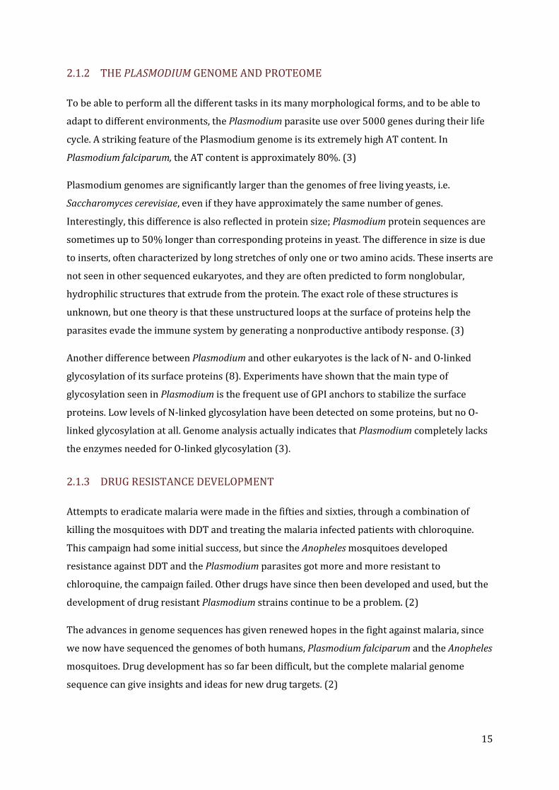
with the water molecule, but with a histidine residue acting as a “proton shuttle”, transferring
the proton from the active site to the surface of the enzyme. (9)
Fig. 3- The catalytic mechanism of the hydration of carbon dioxide by carbonic anhydrase
2.2.2 THE ROLE OF CARBONIC ANHYDRASE IN PLASMODIUM
Plasmodium falciparum cells inside of human erythrocytes contain high levels of carbonic
anhydrase, even higher than the erythrocytes themselves. It has two critical roles in the malaria
parasites; regulation of the CO2 – HCO3 buffer system and production of HCO3- for pyrimidine
synthesis. (4) During the rapid growth of the parasites in their intraerythrocytic stage, high
levels of purines and pyrimidines are required for DNA and RNA synthesis as well as other
metabolical pathways. The parasites are able to acquire the purines from their host, but they
synthesize their own pyrimidines, with HCO3- as one of the substrates. (5)
The enzymatic activity of carbonic anhydrase is destroyed if the zinc ion is removed by the
addition of chelating agents, or if the active site is blocked by inhibitors. It is known that many
types of chelators have antimalarial activity, and it has also been shown that inhibition of CA
activity with acetazolamide can kill intraerythrocytic malaria parasites. (4) Further
characterization of this protein and its inhibitors could lead to the development of new
antimalarial drugs with a new mechanism of action.

18
2.3 CODON BIAS
In the genetic code each amino acid is encoded by more than one codon, and different organisms
have different bias in the usage of the 61 amino acid codons. This bias is reflected in both the
codons of their genes and in the population of tRNA available in the cells. This often poses a
problem in heterologous expression of proteins, since the difference in codon bias can result in
depletion of tRNA molecules that represents codons that are rare in the host organism but
common in the foreign gene. These rare codons can disturb translation of the gene, since the
ribosome stops at these codons waiting for a suitable tRNA. How severe these problems will be
is dependent of the number of rare codons and if they are close to each other. (10) In most cases,
the result is slower translation, but it can also lead to premature termination, frameshifting or
incorporation of incorrect amino acids. (11)
Genes from Plasmodium falciparum are often problematic or seemingly impossible to express in
Escherichia coli (12) or Pichia pastoris (13) due to the high AT content of the malarial genome
and the resulting usage of unusual codons. To counter the problem with rare codons, there are
two different approaches. The tRNA pool of the host can be changed to better reflect the codons
of the expressed heterologous gene, or the gene can be modified to better reflect the tRNA pool
of the expression system. In E. coli, the easiest and in many cases cheapest way to handle it is to
co-transform the E. coli strain used with a plasmid that overexpress the rare codon tRNAs (12),
or to choose an expression strain that already has such a plasmid. The other, more drastic
approach is to redesign the gene so it is optimized for the expression system used. This can be
done either by point mutation of the rare codons or a complete resynthesis of the gene with an
optimal codon usage profile. Earlier, this was an expensive and time consuming method (12) but
now there are several commercial companies specializing in gene optimization and synthesis,
making this an increasingly interesting option. Codon optimization can give a dramatic increase
in expression levels of heterologous proteins, both in E. coli and in P. pastoris. (13)
2.4 ESCHERICHIA COLI AS EXPRESSION SYSTEM
In many cases of heterologous protein expression, the best choice of host system are bacteria,
because of their rapid growth rate, low demands on growth medium and ease of genetic
modification. (14)
There are some limitations, however, that always need to be considered. One of the biggest
problems with protein expression in E. coli is the formation of inclusion bodies; instead of
folding properly, the proteins interact with each other and form large insoluble aggregates. In

19
some cases, the proteins can easily be separated from each other, while some inclusion bodies
are so tightly woven together that it takes strong denaturants to dissociate them. It is not always
a problem, though; if the proteins can be renaturated later in the process, the formation of
inclusion bodies can be seen as a first purification step, since they consist of relatively pure
protein that can be separated from other proteins. (14)
It has been shown that the heterologous expression of codon optimized genes in E. coli is more
likely to form inclusion bodies than the expression of genes with native codon usage. A possible
explanation for this phenomenon is that the codon optimized genes translate much faster, and
this increase in speed can affect the folding mechanism. (13) In some cases the formation of
inclusion bodies can be avoided by using vectors that promote secretion of the protein to the
periplasmic space or to the growth medium. (14)
Escherichia coli and other prokaryotic systems have limitations when it comes to
posttranslational modifications, such as correct folding, formation of disulfide bonds and
glycosylation. (7)
2.5 PICHIA PASTORIS AS EXPRESSION SYSTEM
Pichia pastoris is a methylotrophic yeast often used as an expression system for heterologous
protein production. In this work, Pichia pastoris was chosen since it gives many of the benefits
of a higher eukaryotic system such as folding and processing of proteins, while being almost as
simple and inexpensive to work with as bacteria.
Pichia pastoris was first developed as a source of protein for animal feed, but the interest in
single-cell protein declined during the oil crisis in the seventies. The cheap methanol used as
substrate for the yeast was produced using methane, a petroleum product that suddenly got
significantly more expensive due to the crisis. Later, it was realized that the easy fermentations
methods developed for Pichia and the strong inducible alcohol oxidase promoter AOX1 made it a
system suitable for heterologous protein expression. The ability to grow on methanol also gives
an advantage compared to many other expression systems when it comes to risk of
contamination, since it is an unsuitable environment for many other organisms such as most
bacteria. (7)
One property of Pichia that can be seen as both positive and negative, depending on the
situation, is the ability to glycosylate proteins. Both O-linked and N-linked glycosylation can be
performed, but the glycosylation is not identical to that of mammals and other higher
eukaryotes. (7) Glycosylation is generally not desirable when expressing malaria proteins, since

20
Plasmodium has an extremely low level of glycosylation. To avoid this, the potential sites of N-
linked glycosylation of the expressed protein can be disrupted by amino acid substitutions. (6)
2.5.1 CONTROLLING GENE EXPRESSION IN PICHIA PASTORIS
Many methods for molecular genetic manipulation of Pichia are similar to those of
Saccharomyces cerevisiae, one of the most studied eukaryotic model organisms. Several ways of
transformation are possible, including electroporation. Transformation with a linearized
expression plasmid stimulates homologous recombination between the plasmid and the
chromosomal sequence, leading to integration of the vector into the genome at a specific locus.
Several Pichia strains and vectors are commercially available, simplifying the procedure. (7)
One of the most widely used promoters for heterologous protein expression is the strong,
methanol inducible AOX1 promoter. In Pichia, this promoter normally regulates the expression
of alcohol oxidase (AOX), an enzyme that catalyzes the oxidation of methanol to formaldehyde
and hydrogen peroxide, the first step in the methanol metabolism. The reaction takes place in
the peroxisomes, where the produced hydrogen peroxide is degraded to water by the enzyme
catalase, while the formaldehyde is uses as both an carbon source and an energy source in the
cell. The AOX protein is only expressed during methanol metabolism, but then at very high
levels. In cells grown on methanol, approximately 5% of the poly(A)+ RNA is from AOX1. (7)
An alternative to the AOX1 promoter is the glyceraldehydes 3-phosphate dehydrogenase gene
promoter (GAP). Unlike AOX this promoter gives constitutive expression, even when glucose or
glycerol is used as carbon source. This is not possible for all proteins, since some can affect
growth or be toxic to the yeast cells. The advantage here is easier handling, since there is no
need to change medium to induce expression. (7)
2.5.2 THE pPICZαA VECTOR
In this work the vector chosen for expression in Pichia pastoris is the pPICZαA vector from
Invitrogen. It is a vector that uses the AOX1 promoter for methanol inducible expression. As a
selection marker, it uses the ShBle gene from Streptoalloteichus hindustanus which gives it
resistance to the antibiotic zeocin. It also allows for secretion of the protein to the medium using
the α-factor sequence from Saccharomyces cerevisiae. This makes the subsequent purification
simpler, since there is no need to disrupt the yeast cells to access the recombinant protein. Since
Pichia pastoris normally secretes very low levels of native proteins to the medium, recombinant
proteins expressed with an export signal will usually constitute the majority of the proteins in

21
the medium. (7) The vector also makes it possible to express the proteins with a C-terminal
hexahistidine tag, which can be used for purification of the recombinant protein using IMAC.
2.6 IMMOBILIZED METAL ION AFFINITY CHROMATOGRAPHY (IMAC)
Immobilized metal ion affinity chromatography is a method to separate proteins that use the
affinity of specific amino acids, especially histidine, to metal ions. The technique uses a column
matrix containing metal ions, usually Ni(II), Cu(II), Zn(II), Co(II) or Fe(III), immobilized by
chelating groups bound to the matrix. These metal ions will bind to amino acids on the protein,
the metal ion being an electron pair acceptor and the amino acid residue the electron pair donor.
Histidine binds stronger than other amino acids, and the strength of the affinity is significantly
increased if several histidines close to each other can bind to the matrix. The pH of the solution
needs to be neutral or basic for this binding to occur, since the histidines must be in a non
protonated state. To avoid non specific binding to the matrix, the ion strength of the solution is
often high, with concentrations of NaCl between 0.1 and 1.0 M. When washed, the proteins
bound to the matrix stay on the column, while the others leave the column together with the
wash buffer. To elute the bound proteins, they can either be protonated by lowering the pH, or
by ligand exchange with imidazole (fig. 4). (15)
Even if the technique was developed for native proteins, it is mostly used today as a method to
purify recombinant proteins equipped with a hexahistidine tag. Most naturally occurring
proteins have a rather low affinity for the metal ions, while recombinant proteins with
hexahistidine tags have very high affinity. Some of the advantages of the method compared to
other purification methods are the ability to bind relatively large amounts of protein and the
possibility to use mild conditions for protein elution. (15)
Fig. 4 - A: The interaction between the metal ions on a bead in an IMAC column and the histidine residues
on a his-tagged protein. B: A comparison of the chemical structure of histidine (left) and imidazole (right).

22
23
3 MATERIALS AND METHODS
3.1 EXPRESSION IN PICHIA PASTORIS
3.1.1 DESIGN OF THE SYNTHETIC PfCA GENE
The sequence of the PfCA gene was retrieved from the NCBI online database (GenBank accession
no.: AE014842 region: 82145..83401, GenPept accession no.: AAN35993 “hypothetical protein
PF11_0410”). In previous articles, only the C-terminal part of this hypothetical protein has been
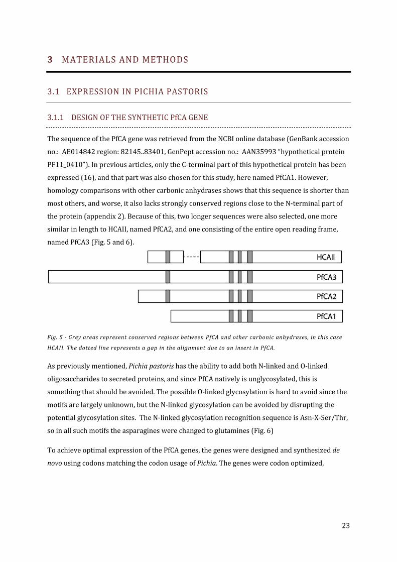
expressed (16), and that part was also chosen for this study, here named PfCA1. However,
homology comparisons with other carbonic anhydrases shows that this sequence is shorter than
most others, and worse, it also lacks strongly conserved regions close to the N-terminal part of
the protein (appendix 2). Because of this, two longer sequences were also selected, one more
similar in length to HCAII, named PfCA2, and one consisting of the entire open reading frame,
named PfCA3 (Fig. 5 and 6).
Fig. 5 - Grey areas represent conserved regions between PfCA and other carbonic anhydrases, in this case
HCAII. The dotted line represents a gap in the alignment due to an insert in PfCA.
As previously mentioned, Pichia pastoris has the ability to add both N-linked and O-linked
oligosaccharides to secreted proteins, and since PfCA natively is unglycosylated, this is
something that should be avoided. The possible O-linked glycosylation is hard to avoid since the
motifs are largely unknown, but the N-linked glycosylation can be avoided by disrupting the
potential glycosylation sites. The N-linked glycosylation recognition sequence is Asn-X-Ser/Thr,
so in all such motifs the asparagines were changed to glutamines (Fig. 6)
To achieve optimal expression of the PfCA genes, the genes were designed and synthesized de
novo using codons matching the codon usage of Pichia. The genes were codon optimized,

24
Fig. 6 - Adjustments of the native PfCA amino acid sequence, with the substitutions marked with boxes. The
start of the different length PfCA constructs are marked with arrows.
synthesized, sequence verified and cloned into the pPICZαA vector as a custom service by
GenScript Corp. (NJ, USA). The synthetic gene was designed to not contain internal restriction
sites for EcoRI, NotI and SacI, since EcoRI and NotI would be used later for cloning of the gene
into the vector, and SacI would be used to linearize the vector prior to transformation. See
appendix 1 for complete sequences.
3.1.2 CONSTRUCTION OF EXPRESSION PLASMID
As expression vector, the pPICZαA from Invitrogen was chosen. It is an E. coli – Pichia shuttle
vector, which uses Zeocin resistance as selection marker in both E. coli and Pichia. It uses the
alcohol oxidase (AOX1) promoter for methanol induced expression and an α-factor sequence for
export of the protein to the growth medium. It also allows proteins to be expressed as fusion
proteins with a C-terminal c-myc epitope for simple antibody detection and C-terminal
hexahistidine tag for efficient affinity purification. A short spacer sequence (AAASFL) separates
the native C-terminal and the fusion tags.
For the construction of the expression plasmids, the synthetic genes were cloned into the
pPICZαA plasmid in frame with the α-factor and the C-terminal fusion tags using the restriction
sites EcoRI and NotI. The schematic of the resulting plasmid is shown in fig. 7. After removal of
the α-factor sequence by the Golgi protease KEX2, the expressed proteins should have the
sequences
EFMKDLKERELKQISDVY…TIIQVSSAVHVGSGNKAAASFLEQKLISEEDLNSAVDHHHHHH (PfCA1)
EFMKNINSVQNNIQKTF…TIIQVSSAVHVGSGNKAAASFLEQKLISEEDLNSAVDHHHHHH (PfCA2)
EFMLEMIDKYNTHFVQT…TIIQVSSAVHVGSGNKAAASFLEQKLISEEDLNSAVDHHHHHH (PfCA3)

25
The underlined sequences represent the actual protein, while the rest of the sequences are
derived from the vector. The N-terminal sequence bears two extra amino acids, the amino acids
in bold form the c-myc epitope, and the C-terminal histidines form the hexahistidine tag used for
immobilized metal ion affinity chromatography (IMAC).
Fig. 7- The pPICZαA-PfCA expression plasmid. The zeocin-r is the ShBle gene that gives resistance to zeocin.
The pUC origin allows replication of the plasmid in E. coli, and the PEM7 promotes the expression of the
zeocin resistance gene in E. coli. PTEF drives expression of ShBle in Pichia, and the CYC1TT is a Pichia
transcription terminator. The 5'AOX1 is the AOX1 promoter, while the AOX1TT is the native termination
signal from the AOX1 gene. The transcription terminators are important for correct mRNA processing, like
polyadenylation in Pichia, and stabilize the mRNA. SP is the spacer sequence AAASFL. SacI, EcoRI and NotI
are unique restriction sites for linearization and cloning.
3.1.3 TRANSFORMATION
The expression plasmids were amplified in the E. coli strain Top10 (Invitrogen), which were
grown over night in low salt Luria-Bertani (LB) medium (yeast extract (5 g/l), peptone (10 g/l),
and NaCl (5 g/l), pH 7.5) containing 25 µg/ml zeocin. Five ml of each bacterial suspension were
used to prepare plasmids using the QIAprep Spin Miniprep Kit (Qiagen). A total of four plasmid
preparations of each construct were made, giving a total volume of 200 µl of each plasmid
solution with an approximate DNA concentration of 80 ng/µl. Before transformation, the vectors
were linearized with SacI (New England Biolabs), and purified with the QIAquick PCR
Purification Kit (Qiagen). The solutions with the linearized plasmids were then concentrated

26
using vacuum centrifugation at room temperature until they had reached a volume of
approximately 10 µl (c≈1 µg/µl).
Pichia pastoris GS115 (Invitrogen) was grown in 10 ml YPD medium (yeast extract (10 g/l),
peptone (20 g/l), and glucose (20 g/l)) overnight in 30º C. Of this culture, 5 µl were used to
inoculate 500 ml of fresh YPD medium in a 4 liter baffled bottle. Competent GS115 cells were
prepared according to the recommendations from Invitrogen (appendix 4). The competent cells
were then transformed using electroporation according to protocol (appendix 4). Aliquots of
200 µl of the cell suspension were plated on YPDSZ plates (appendix 3) and grown at 30º for 5
days. Isolated colonies of successful transformants (12 strains of each construct) were randomly
chosen and replica plated on YPDZ plates in three steps to get pure strains and to minimize the
risk of wild type GS115 contamination. The strains were designated X-Y, where X represents the
PFCA construct number (1, 2 or 3) while Y is a number between 1 and 12.
3.1.4 VERIFICATION OF INSERT
To verify the correct chromosomal integration of the vector, six of the strains, (1-2, 1-3, 1-4, 2-1,
2-11, 2-12) were grown overnight at 30ºC in 10 ml YPD medium with 100 µg/ml zeocin in
sterile 50 ml polypropylene tubes. From these cultures, 1.5 ml were centrifuged at 8500 g and
chromosomal DNA was extracted from the pellets using the FastDNA SPIN Kit for Soil (MP
Biomedicals) (appendix 5). To control the DNA quality, the DNA solutions were analyzed on a
10% agarose gel. The inserts were then amplified using GeneJet Fast PCR (Fermentas) with the
AOX primers recommended by Invitrogen:
5' AOX1, 5'-GACTGGTTCCAATTGACAAGC-3'
3' AOX1, 5'-GCAAATGGCATTCTGACATCC-3'
The PCR conditions were: 95 ºC for 60 s, 35 cycles of 3 s at 95 ºC, 8 s at 52 ºC, and 60s 72 ºC,
with a final extension step at 72 ºC for 22 s. The PCR products were analyzed on a 10% agarose
gel and purified using the QIAquick PCR Purification Kit (Qiagen), before being sent to GATC
(Germany) for sequencing.
3.1.5 EXPRESSION
To screen the Pichia transformant strains for PfCA expression, colonies from the YPDZ plates
were inoculated into 10 ml of BMGY culture medium (appendix 3) containing 1 mM ZnSO4 in
sterile 50 ml polypropylene tubes, and were grown over night at 30ºC with shaking at 230 rpm.
At OD600 = 2.16, the cells were harvested by centrifugation (750 g for 5 min) and the cell pellets
were resuspended in 15 ml BMMY culture medium containing 1 mM ZnSO4 to start induction.

27
The cultures were grown at 30ºC while shaking at 230 rpm for 72 hours, with an addition of
0.5% methanol every 24 hours to maintain induction. To ensure good aeration, the caps of the
tubes were removed, and the tube rack was covered with aluminum foil to avoid contamination.
After 72 hours, the cultures were centrifuged at 4500 g for 3 min, and the supernatants and cell
pellets were separated and stored at -20ºC.
3.1.6 DETECTION OF PROTEIN
To analyze the protein expression of the transformants, the supernatants were analyzed with
SDS-PAGE. To 10 µl of each sample, 5 µl reducing SDS-PAGE buffer was added and heated at
95ºC for 5 minutes to denature all proteins. On a 12% SDS-PAGE gel, 12 µl of each sample were
loaded, and run in a vertical gel apparatus (BioRad) at 90 V for 30 minutes followed by 200 V for
45 minutes. The protein molecular weight marker used was PageRuler Prestained Protein
Ladder (Fermentas). After electrophoresis, the protein bands on the gel were visualized with the
Silver Stain Plus kit according to the manual from BioRad.
To detect expression of PfCA, dot blots of the supernatants were performed using nitrocellulose
membrane and horse radish peroxidase conjugated anti-c-myc antibodies (Invitrogen). The
Positope protein from Invitrogen was used as positive control. Of each sample, 150 µl were
applied to a nitrocellulose membrane using a dot-blot apparatus. The membrane was air dried,
and incubated with gentle agitation for 3 h in blocking buffer (137 mM NaCl, 2.7 mM KCl, 10 mM
Na2HPO4, 1.8 mM KH2PO4, 0.05% Tween-20, v/v, 5% nonfat, dry milk). It was then washed 2x5
minutes with PBST (137 mM NaCl, 2.7 mM KCl, 10 mM Na2HPO4, 1.8 mM KH2PO4, 0.05%
Tween-20, v/v), and then incubated overnight at room temperature with a horse radish
peroxidase conjugated anti-myc antibody diluted 1:5000 in blocking buffer. Blots were then
washed 2x5 minutes with PBST and incubated with HRP chromogen substrate (0.1 M citrate-
phosphate buffer pH 5.0, containing o.o3% hydrogen peroxide and 1 mg/l para-
phenylenediamine) for about 5 minutes.
To prepare the samples for western blotting, the supernatant samples were concentrated using
acetone precipitation. To 1 ml sample, 4 ml acetone was added, and after mixing the samples
were frozen at -20 ºC over night. The samples were then centrifuged, and the supernatants
discarded. The pellets were resuspended in remaining fluid, and 10 µl were dissolved in 10 µl
SDS-PAGE loading buffer and heated to 95ºC for 5 minutes before 12 µl of each sample were
loaded onto the SDS-PAGE gel. The electrophoresis ran at 90 V for 30 minutes and 200 V for 20
minutes. Since the lanes were running in a slightly distorted way, the voltage was lowered to
120 V, and the gel was run for 40 additional minutes. The proteins were then electrophoretically
transferred to a PVDF membrane at 10 V over night, using 20 mM Tris, 150 mM glycine and 20%

28
v/v methanol as a transfer buffer. After transfer, they were blotted with anti-myc HRP-
antibodies according to the same protocol as the dot blot (appendix 6). A dot blot was also made
on the concentrated samples, using the same protocol as before.
3.1.7 PURIFICATION OF PROTEIN
To purify the PfCA protein, the supernatants of each construct were pooled and prepared for
immobilized metal ion affinity chromatography (IMAC). The freezing and thawing of the samples
had caused some precipitation, so they were centrifuged at 3700 g at 4 ºC for 10 minutes to
remove this precipitate. Before the IMAC, 10% of a 10x IMAC buffer (pH 8, 500 mM NaH2PO4, 1.5
M NaCl, and 100 mM imidazole) was added to the samples, and then the NaCl concentration was
increased to 300 mM with 3 M NaCl. Since the supernatants were already buffered to pH 6, after
these additions they had an approximate pH of 6.5, and they were adjusted to pH 8.0 by addition
of solid tris(hydroxymethyl)aminomethane. The increase in salt levels made the samples
precipitate again, so another centrifugation at 3700 g at 4 ºC for 10 minutes was performed. The
samples were then batch purified under native conditions using Ni-NTA Superflow from Qiagen
(appendix 7). The pooled samples, the flow-throughs, the washing buffers and the elution
fractions 1 to 5 were analyzed with SDS-PAGE. Of each sample, 20 µl were mixed with 10 µl SDS-
PAGE loading buffer and heated at 95ºC for 5 minutes before 25 µl of each sample were loaded
onto the SDS-PAGE gel. The electrophoresis ran at 90 V for 35 minutes followed by 180 V for 45
minutes, and the proteins were then visualized by silver staining.

29
4 RESULTS
4.1 PFCA GENE LENGTH AND VECTOR DESIGN
In previous articles, expression of the C-terminal part of the open reading frame of this
hypothetical protein has been reported (16). Homology alignment with other carbonic
anhydrases shows that this sequence is shorter than most others, and worse, it also lacks one
strongly conserved region. Because of this, three different lengths of the gene were selected for
expression, one 235 amino acids long representing the previously studied protein (PfCA1), one
284 amino acids long that is more similar in length to HCAII (PfCA2), and one consisting of 418
amino acids, the entire open reading frame (PfCA3). These three genes were codon optimized,
synthesized and cloned into the pPICZαA expression vector by GenScript Corp.
4.2 TRANSFORMATION
The pPICZαA-PfCAX plasmid was amplified in E. coli strain Top10, prepared and purified using
the QIAprep Spin Miniprep Kit (Qiagen) and then digested with the restriction endonuclease
SacI. The linearized vectors were integrated into the genome of the Pichia pastoris GS115 strain
through electroporation. The transformation with the pPICZαA-PfCA1 and pPICZαA-PfCA2
vectors were successful, with over hundred transformants appearing on each YPDSZ plate, but
transformation with the pPICZα-PfCA3 vector failed, despite repeated attempts. Successful
transformants were replica plated on selective plates in three steps to ensure pure strains.
4.3 VERIFICATION OF INSERT
After extraction of yeast chromosomal DNA from the transformed strains using the FastDNA
SPIN Kit for Soil (MP Biomedicals), agarose gel analysis showed that the extracted DNA was of
good quality with long fragment lengths (fig 8). The inserts were amplified with the AOX primers
recommended by Invitrogen (5'AOX1, 5'-GACTGGTTCCAATTGACAAGC-3'; and 3' AOX1, 5'-
GCAAATGGCATTCTGACATCC-3') using PCR (GeneJet Fast PCR, Fermentas), and the gel analysis
showed strong bands of the correct lengths, 1307 bp (PfCA1) and 1454 bp (PfCA2), without any
visible contamination of non specific amplification (fig 8). The sequence verification performed
by GATC (Germany) showed that the inserts were correct, without mutations.

30
Fig. 8- A: Agarosis gel analysis of the DNA extracted from the transformed Pichia strains. Lane 1 is a
molecular weight marker, lanes 2 to 7 are the extracted DNA samples from strains 1-2, 1-3, 1-4, 2-1, 2-11
and 2-12. B: Agarosis gel analysis of the PCR products. Lane 1 is a molecular weight marker, lanes 2 to 4
are the PCR products from strains 1-2, 1-3 and 1-4, lane 5 is the PCR product of a positive control, pure
pPICZαA-PfCA1 plasmid DNA, lanes 6 to 8 are the PCR products from strains 2-1, 2-11 and 2-12, lane 9 is
the PCR product of a positive control, pure pPICZαA-PfCA2 plasmid DNA. The expected lengths of the
fragments are 1307 bp (PfCA1) and 1454 bp (PfCA2).
4.4 EXPRESSION
Single colonies of the pPICZαA-PfCA1 and pPICZαA-PfCA2 strains were inoculated into BMGY
culture medium and grown over night while shaking at 30 ºC. The cells were then harvested by
centrifugation and resuspended in BMMY culture medium to start induction. The cultures were
grown while shaking for 72 hours at 30ºC, with an addition of 0.5% methanol every 24 hours.
The cultures were then centrifuged, and the supernatants and cell pellets were separated and
stored at -20ºC.
4.4.1 SDS-PAGE ANALYSIS
The expression supernatant samples were analyzed on 12% SDS-PAGE gels, and stained with
silver staining. The supernatants showed no overexpression bands, so the SDS-PAGE did not give
any clear indication whether the expression was successful or not (fig. 9). It did show, however,
that if there is expression of PfCA, the levels of that protein in the medium are low. In fact, the
total protein levels in the supernatant were so low that they could not be detected at all by
Coomassie staining; only with silver staining did the proteins become visible. The expected sizes
of the proteins are 31.3 kDa (PfCA1) and 37,1 kDa (PfCA2), and while there are bands
31
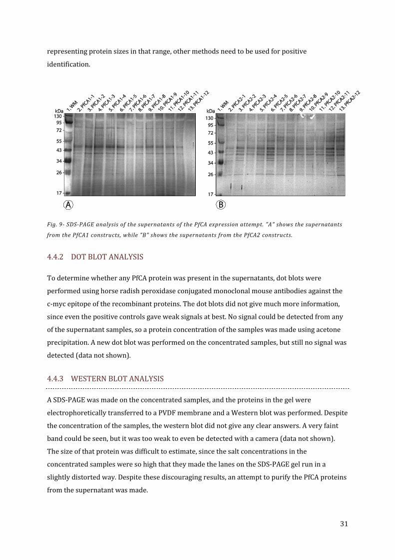
representing protein sizes in that range, other methods need to be used for positive
identification.
Fig. 9- SDS-PAGE analysis of the supernatants of the PfCA expression attempt. "A" shows the supernatants
from the PfCA1 constructs, while "B" shows the supernatants from the PfCA2 constructs.
4.4.2 DOT BLOT ANALYSIS
To determine whether any PfCA protein was present in the supernatants, dot blots were
performed using horse radish peroxidase conjugated monoclonal mouse antibodies against the
c-myc epitope of the recombinant proteins. The dot blots did not give much more information,
since even the positive controls gave weak signals at best. No signal could be detected from any
of the supernatant samples, so a protein concentration of the samples was made using acetone
precipitation. A new dot blot was performed on the concentrated samples, but still no signal was
detected (data not shown).
4.4.3 WESTERN BLOT ANALYSIS
A SDS-PAGE was made on the concentrated samples, and the proteins in the gel were
electrophoretically transferred to a PVDF membrane and a Western blot was performed. Despite
the concentration of the samples, the western blot did not give any clear answers. A very faint
band could be seen, but it was too weak to even be detected with a camera (data not shown).
The size of that protein was difficult to estimate, since the salt concentrations in the
concentrated samples were so high that they made the lanes on the SDS-PAGE gel run in a
slightly distorted way. Despite these discouraging results, an attempt to purify the PfCA proteins
from the supernatant was made.

32
4.5 PURIFICATION
The supernatants of each construct were pooled and prepared for immobilized metal ion affinity
chromatography (IMAC). The precipitation caused by freezing and thawing of the samples was
removed by centrifugation. The samples were prepared for IMAC by adding 10% of a 10x IMAC
buffer (pH 8, 500 mM NaH2PO4, 1.5 M NaCl, and 100 mM imidazole) to the samples, increasing
the NaCl concentration to 300 mM with 3 M NaCl, and adjusting the pH to 8.0 by adding solid
tris(hydroxymethyl)aminomethane. The increase in salt levels caused new precipitation, so
another centrifugation was performed. The samples were then batch purified using the Ni-NTA
Superflow from Qiagen. After purification, a reducing SDS-PAGE analysis was made on the
pooled samples, the flow-throughs, the washing buffers and the elution fractions 1 to 5. After
visualization with silver staining, the gel did show weak bands that indicated some proteins
binding to the column, but their sizes did not match the ones of the PfCA proteins (fig. 10).
Fig. 10-SDS-PAGE analysis of the IMAC purification of the samples. "A" shows the result from the PfCA1
constructs, while "B" shows the results from the PfCA2 constructs. WM: Molecular weight markers; PS:
Pooled sample; FT: Flow-through; W1: 10 mM imidazole wash ; W2: 20 mM imidazole wash; E: Eluates.

33
5 DISCUSSION
The methylotrophic yeast Pichia pastoris has been increasingly popular in recent years for the
recombinant expression of proteins. It has many properties similar to prokaryotes when it
comes to genetic modification and rapid growth, still allowing eukaryotic post translational
protein modifications and folding. By using the α-factor export signal from Saccharomyces
cerevisiae, the expressed protein can be exported to the medium. Simple growth media can be
used, and since Pichia pastoris exports very few native proteins, purification of the expressed
proteins is simplified. (7)
The natively expressed carbonic anhydrase from Plasmodium falciparum has been studied in
previous articles by Krungkrai et. al. and they also report having expressed the protein in E. coli
(16). According to their sequence data, however, only the 235 amino acids long C-terminal part
of this hypothetical protein was expressed. Interestingly, the reported sequencing of the N-
terminal of the CA extracted from Plasmodium falciparum gives a sequence (MAPKAKIVLVG)
(17) that is not present in the PfCA protein sequence derived from the genetic database. Still, the
article from 2004 claims that this native protein is identical to the recombinant PfCA entirely
based on size and enzymatic activity data (16). In a later article from 2007 they instead say that
the PfCA protein is 418 amino acids long, but they still claim to have expressed the full-length
PfCA protein, referring to their earlier articles (5).
Since homology comparisons with other carbonic anhydrases (see appendix 2) indicate that the
shorter 235 amino acids long sequence lacks strongly conserved regions, it is unlikely that this
shorter sequence represents the native form of the PfCA protein. In this work, three different
lengths of the gene were selected for recombinant expression, the shorter sequence previously
studied (PfCA1, 235 amino acids), one sequence similar to the human carbonic anhydrase HCAII
in length (PfCA2, 284 amino acids), and the full length open reading frame (PfCA3, 418 amino
acids).
Since Plasmodium falciparum has very low native glycosylation, while Pichia pastoris has the
ability to add both N-linked and O-linked oligosaccharides to secreted proteins, the native PfCA
sequence was modified to avoid glycosylation. The recognition motifs for N-linked glycosylation
in Pichia are Asn-X-Ser/Thr (7), so all such sequences were changed by replacing the
asparagines with glutamines. Since malarial genes are known to be difficult to express in other
organisms due to the unusual codon bias of Plasmodium (12), the genes were synthesized de
novo using the codon usage preferred by Pichia.

34
The Pichia expression vector chosen was the pPICZαA from Invitrogen. It has many features that
make the protein expression, detection and purification easier. The efficient methanol inducible
alcohol oxidase (AOX1) promoter is used to control the expression, and the expressed protein is
exported to the medium using the α-factor signal sequence. Since the proteins can be expressed
with a C-terminal c-myc epitope and a hexahistidine tag, antibody detection and affinity
purification with IMAC can be made.
The PfCA1 and PfCA2 vectors were successfully integrated into the Pichia genome, as the PCR
and the sequencing made by GATC showed. The transformation of the PfCA3 however, failed
despite several attempts. The PfCA3 vector could be expected to be more problematic because of
its large size, and with increased length comes increased difficulties in entering the yeast cells.
The relative difference in size if the entire vectors are compared is not that big, however, the
extra 402 base pairs of PfCA3 compared to PfCA2 represent an increase in size of 9%. Even if
this is likely to affect the transformation frequency negatively, at least a few successful
transformants could be expected.
The SDS-PAGE analysis of the samples did not give any clear indication of whether or not the
protein was expressed. If the protein would have been overexpressed, it should have been easy
to detect since the normal expression of native Pichia proteins to the medium is very low. Given
the results of the analysis, it would have been wise to grow a Pichia strain transformed with the
pPICZαA vector with another protein sequence inserted instead of PfCA as a control. A
comparison of the protein expression of the control and the PfCA transformed strains might
show protein bands not present in the control.
When this project was planned, the myc tag was intended to be used as the primary indicator of
protein expression levels. The plan was to select several successful transformants, 12 of each
strain, and then use dot blotting to screen them for protein expression. This step could rather
early in the screening process allow a decrease in the number of strains to one or two per
construct, and then use only these for further analysis. This would save time, materials and
effort later in the process. Unfortunately, the blotting turned out to be very problematic. Despite
efforts to optimize the protocols, even the positive controls gave very weak signals, and in many
cases they gave no signals at all. One possible explanation is that the antibodies somehow were
defective, either in specificity or in enzymatic activity. The antibodies used for blotting were
anti-myc mouse monoclonal antibodies conjugated with HRP enzyme, instead of a more
conventional two step system using primary and secondary antibodies. This made the blotting
quicker and easier, but at the cost of slightly lower detection levels. It also made it more difficult
to detect where the problem with the weak signal was, in the binding specificity or in the
35
enzyme. Since the antibodies used in this experiment are mouse antibodies, this could have been
investigated by using them as a primary antibody, and using an anti-mouse antibody with a
different enzyme, i.e. alkaline phosphatase, as a secondary antibody.
Since the positive controls in the dot blotting gave at least a weak signal, an attempt to do a
concentration of the samples and do a Western Blot was made. It did give a signal, but since it
was so weak that it could not be detected with a camera, it cannot alone confirm expression of
the proteins.
The last attempt to detect possible PfCA expression was to pool the supernatants and purify
them using immobilized metal ion affinity chromatography. A successful purification, with a
SDS-PAGE gel showing a sharp band at the expected size of the proteins would be a very strong
indication of successful expression. This was not the case, however; the gels only showed weak
bands at a size that did not match the PfCA proteins. It is likely that these bands represented
some native Pichia proteins that bound weakly to the column even without the presence of a
hexahistidine tag. It could be argued that the size of the expressed PfCA proteins could be bigger
than expected due to O-linked glycosylation, but it seems that the observed size differences are
too large for that explanation to be likely.
This could be interpreted in two ways, either the IMAC failed, or there is no protein expression.
There are several possible explanations that could explain failed affinity purification. During the
preparations of the samples before the IMAC, they were centrifuged twice to remove precipitate.
Those precipitations could be mostly salts, but there is a possibility that they contain proteins.
An analysis of these pellets could give interesting results, since if they contain the proteins, the
precipitations will represent a concentration of the samples. There are indications that this
could be the case; a comparison of the SDS-PAGE gel of the supernatants, and the SDS-PAGE lane
showing the pooled samples after the IMAC preparation shows that many of the bands from the
first gel are not visible on the later one. This is a strong indication that some of the proteins
indeed were lost in the precipitation. Another explanation of the failed purification could be that
the proteins were degraded by proteases. This problem could be prevented in future
experiments by adding protease inhibitors to the samples immediately after expression.
Even if these problems with the expression analysis could be solved, the SDS-PAGE analysis of
the supernatant showed that if the PfCA protein is expressed, it is expressed at low levels.
However, this does not necessarily indicate that the strains are unable of high expression levels
of PfCA proteins. The expression attempt made in this work was intended as a first screening,
and not for optimizing the expression conditions. The cultures were grown in 50 ml
polypropylene tubes with a small surface area. Since it is known that the supply of oxygen to the
36
yeast during the expression is critical (7), these are far from optimal conditions. Further
attempts to express the proteins could be done in larger, baffled culture flasks to ensure optimal
aeration.
5.1 FUTURE RESEARCH
If the levels of expression would still be low, despite better growth conditions, more drastic
changes could be made. One explanation of the low expression levels could be the use of the α-
factor, causing the proteins to be secreted. While this has many obvious advantages, such as easy
purification and possible high expression levels, it is known that this can be problematic with
proteins that are normally expressed intracellularly (7). Another possible risk with the export is
the N-linked glycosylation. While there are examples of proteins being expressed and
successfully exported without any N-glycosylation motifs (13), other report that the removal of
all N-glycosylation sites in a protein can make export impossible due to the degradation of
unglycosylated proteins by the ER quality control system (18). To avoid these possible
problems, the synthetic PfCA genes could instead be cloned into an expression vector that
promotes intracellular expression.
Yet another factor that has been shown to be potentially problematic is the addition of the
hexahistidine tag. While it simplifies purification, some report that the addition of a his-tag to a
protein can greatly reduce expression levels (18) or lower protein stability and enzymatic
activity, possibly due to the his-tag interfering with protein folding (19). The his-tag could be
completely removed, or an attempt could be made to use an N-terminal his-tag instead of a C-
terminal one, since the location of the tag can affect how it interacts with the rest of the protein.
The AOX1 promoter system is widely used, since it often gives very strong expression, and since
an inducible system allows the expression of proteins that limit cell growth or are toxic to Pichia.
Since no expression was detected in this work, it could be interesting to try using a promoter
with constitutive expression like the GAP promoter. This would also simplify the expression,
since the yeast can be grown in standard YPD medium instead of methanol medium.
Once stable expression of the PfCA protein has been accomplished, the protein could be further
characterized, using enzymatic essays to measure its catalytic properties, followed by inhibition
studies.
37
6 CONCLUSIONS
The carbonic anhydrase from Plasmodium falciparum (PfCA) plays a critical role in the
metabolism of the parasite, making it an interesting potential drug target. In this work, the PfCA
gene was codon optimized and cloned into the expression vector pPICZαA. The vector was
successfully transformed into the genome of Pichia, and expression attempts were conducted.
Since no expression of the PfCA protein could be detected, further research should focus on
optimization of expression conditions, or redesigning of the expression vector. Changes that
could be made are removal of the α-factor, since the export of the protein is a known risk factor
in protein expression, or removal of the his-tag, since it may interfere with correct protein
folding.

38

39
7 ACKNOWLEDGEMENTS
First of all, I want to thank Nalle Jonsson at IFM for giving me the opportunity to work with this
interesting project, and for all his enthusiasm and good advice. I would also like to thank Gunnar
Höst, my supervisor, for his help and support during this work and for his great feedback on the
report. Special thanks to Daniel Kanmert, who taught me a lot about practical lab work, and gave
me valuable advice on working with Pichia pastoris. Many thanks to Cecilia Andrésen for the
advice regarding IMAC, and Per-Eric Lindgren, who let me use his lab and equipment at the
Faculty of Health Sciences for the DNA extraction from Pichia.
I would like to thank my opponent, Birgitta Sundén, for the feedback on my report and
presentation; it was very nice working with you! Finally, I want to thank Mirthe Hoekzema for
always listening to my thoughts about this project, and for all her support along the way.

40

41
8 REFERENCES
(1) Snow, R W, C A Guerra, A M Noor, H Y Myint, and S I Hay. "The global distribution of
clinical episodes of Plasmodium falciparum malaria." Nature 434 (2005): 214-217.
(2) Greenwood, B M, et al. "Malaria: progress, perils, and prospect for eradication." The
Journal of Clinical Investigation 118 (2008): 1266-76.
(3) Aravind, L, M I Lakishminarayan, T E Wellems, and LH Miller. "Plasmodium Biology:
Genomic Gleanings." Cell 115 (2003): 771-785.
(4) Sein, Kyaw Kyaw, and M Aikawa. "The pivotal role of carbonic anhydrase in malaria
infection." Medical Hypotheses 50 (1998): 19-23.
(5) Krungkrai, J, S R Krungkrai, and C T Supuran. "Malarial parasite carbonic anhydrase and
its inhibitors." Current Topics in Medicinal Chemistry 7 (2007): 909-917.
(6) Tsai, C W, P F Duggan, R L Jr Shimp, L H Miller, and D L Narum. "Overproduction of
Pichia pastoris or Plasmodium falciparum protein disulfide isomerase affects
expression folding and O-linked glycosylation of a malaria vaccine candidate expressed
in P pastoris." Journal of Biotechnology 121 (2006): 458-470.
(7) Cereghino, Joan Lin, and James M. Cregg. "Heterologous protein expression in the
methylotrophic yeast Pichia pastoris." FEMS Microbiol. Rev. 24, 2000: 45-66.
(8) Davidsson, E A, and D C Gowda. "Glycobiology of Plasmodium falciparum." Biochimie 83
(2001): 601-604.
(9) Lindskog, Sven. "Structure and mechanism of carbonic anhydrase." Pharmacology &
Therapeutics 74, no. 1 (1997): 1-20.
(10) Kurland, C G. "Codon bias and gene expression." FEBS Letters 2 (1991): 165-169.
(11) Kurland, C, and J Gallant. "Errors of heterologous protein expression." Current Opinion
in Biotechnology 7 (1996): 489-493.
(12) Baca, A M, and W G J Hol. "Overcoming codon bias: A method for high-level
overexpression of Plasmodium and other AT-rich parasite genes in Escherichia coli."
Internationa Journal for Parasitology 30 (2000): 113-118.

42
(13) Yadava, A, and C F Ockenhouse. "Effect of Codon Optimization on Expression Levels of a
Functionally Folded Malaria Vaccine Candidate in Prokaryotic and Eukaryotic
Expression Systems." Infection and Immunity 71, no. 9 (2003): 4961-4969.
(14) Harwood, C R, and A Wipat. "Genome management and analysis: Prokaryotes." Chap. 4
in Basic Biotechnology, by C Ratledge and B Kristiansen, 89-93. New York: Cambridge
University Press, 2001.
(15) Gaberc-Porecar, V, and V Menart. "Perspectives of immobilized-metal affinity
chromatography." Journal of Biochemical and Biophysical Methods 49 (2001): 335-360.
(16) Reungprapavut, S, S R Krungkrai, and J Krungkrai. "Plasmodium falciparum carbonic
anhydrase is a possible target for malaria chemotherapy." Journal of Enzyme inhibition
and medical chemistry 19:3 (2004): 249-256.
(17) Krungkrai, S R, N Suraveratum, S Rochanakij, and J Krungkrai. "Characterisation of
carbonic anhydrase in Plasmodium falciparum." International Journal for Parasitology
31 (2001): 661-668.
(18) Muraki, M. "Secretory expression of synthetic human Fas ligand extracellulardomain
gene in Pichia pastoris Influences of tag addition and N-glycosylation site deletion, and
development of a purification method." Protein Expression and Purification 50 (2006):
137-146.
(19) Yu, M, S Lange, S Richter, T Tan, and R D Schmid. "High-level expression of extracellular
lipase Lip2 from Yarrowia lipolytica in Pichia pastoris and its purification and
characterization." Protein Expression and Purification 53 (2007): 255-263.
(20) Images from the US governmental agency “Centers for Disease Control and Prevention”,
http://www.dpd.cdc.gov/, images #5861 and #3405