department of biochemistry school of medicine
TRANSCRIPT

UNIVERSITY OF NAIROBI
DEPARTMENT OF BIOCHEMISTRY
SCHOOL OF MEDICINE
ALKALINE ACTIVE α-AMYLASES FROM ALKALIPHILIC BACILLUS SP.:
SCREENING AND ENZYME PROUCTION
HBC 305: RESEARCH PROJECT
BY
MUDIDI DERRICK LUGONZO
H12/1581/2010
SUPERVISOR: MR. KEVIN RAYMOND OLUOCH
A research dissertation submitted to the Department of Biochemistry in partial fulfillment
for the requirement of Bachelor of Science Degree in Biochemistry, School of Medicine,
University of Nairobi
OCTOBER 2014

D E C L A R A T I O N
Declaration by the student
I hereby declare that this research dissertation is my original work and has not been presented to any other university for examination or any other award.
Submitted by
Name: Mudidi Derrick Lugonzo
RegistrationNo: H12/1581/2010
Declaration by the supervisor
This dissertation has been submitted for examination with my approval as a University of Nairobi supervisor.
Supen isor:
Mr. Ke^ in Raymond Oluoch
Department of Biochemistry
Universit}' of Nairobi
Date
Signature Date

Page | 1
ACKNOWLEDGEMENT
Praise be to GOD for seeing me through it all. All Glory, Honour and Highest praises be to His
Son Jesus Christ, Lord and Saviour.
I wish to express my heartfelt thanks to my very hardworking and dedicated supervisor, Mr.
Kevin R. Oluoch, for his guidance, motivation and optimistic outlook in the course of my
research and dissertation writing.
I wish also to share this achievement with my fellow colleague Janemary Obiny. I would
especially like to thank Mr. C. Wycliff, Nelson Khan, Irene Kiio, Mr. Osawa and Isemeki J. for
assisting me with my research work. My sincere gratitude for the use of the facilities,
cooperation and assistance rendered in one way or another.
My appreciation and thanks to Mama C for assistance with access to Dr. C. A. Omwandho’s
laboratory, to Dr. Muge for his concern and guidance, to the staff and my fellow colleagues
undertaking MSc. for their kind generosity and hospitality in the course of my research in the
Department of Biochemistry, University of Nairobi.
Last but not least, I thank God for my family who are my companion on this academic journey.
In my many moments of disappointment and dismay, the thoughts of your prayers and moral
support have helped me to keep my chin up and journey on in faith and hope. Thank you for
being my sheltering trees.

Page | 2
TABLE OF CONTENTS
ACKNOWLEDGEMENT ............................................................................................................................ 1
LIST OF FIGURES ...................................................................................................................................... 4
LIST OF TABLES ........................................................................................................................................ 5
ABBREVIATIONS ...................................................................................................................................... 6
ABSTRACT .................................................................................................................................................. 7
CHAPTER 1 ................................................................................................................................................. 9
1.0. INTRODUCTION ............................................................................................................................. 9
1.1. PROBLEM STATEMENT .............................................................................................................. 11
1.2. JUSTIFICATION OF THE RESEARCH ........................................................................................ 12
1.3. HYPOTHESIS ................................................................................................................................. 13
1.4. RESEARCH OBJECTIVES ............................................................................................................ 13
CHAPTER 2 ............................................................................................................................................... 14
2.0 LITERATURE REVIEW ................................................................................................................. 14
2.1 EXTREMOPHILES .......................................................................................................................... 14
2.2 ALKALIPHILES .............................................................................................................................. 14
2.2.1 Distribution ................................................................................................................................ 15
2.2.2: The East African Rift valley and the soda lakes found therein ................................................. 15
2.2.3 Microbial diversity of soda lakes ............................................................................................... 17
2.2.4 Isolation ...................................................................................................................................... 17
2.2.5 Special physiological features of alkaliphiles ............................................................................ 18
2.2.6 Alkaliphilic Bacillus sp. and their alkaline active enzymes ....................................................... 21
2.3 STARCH ........................................................................................................................................... 24
2.3.1 Physico-chemical properties ...................................................................................................... 25
Page | 3
CHAPTER 3 ............................................................................................................................................... 30
3.0 MATERIALS AND METHODS ...................................................................................................... 30
3.1 MATERIALS .................................................................................................................................... 30
3.2 RESEARCH SITE ............................................................................................................................ 30
3.3 METHODOLOGY ........................................................................................................................... 30
3.3.1 Screening for amylase producers ............................................................................................... 30
3.3.2 Cultivation of amylase producing bacteria ................................................................................ 31
3.3.3 Analytical methods .................................................................................................................... 31
CHAPTER 4 ............................................................................................................................................... 34
4.0 RESULTS ......................................................................................................................................... 34
4.1 Plate test assay for screening for amylase producing bacteria ...................................................... 34
4.2 Extracellular starch hydrolysing activities of Lake Bogoria isolates ............................................ 36
CHAPTER 5 ............................................................................................................................................... 40
5.0 DISCUSSION ................................................................................................................................... 40
CHAPTER 6 ............................................................................................................................................... 42
6.0 CONCLUSION ................................................................................................................................. 42
REFERENCES ....................................................................................................................................... 43

Page | 4
LIST OF FIGURES
FIGURE 1:………………………………………………………………………………………………..16
FIGURE 2:………………………………………………………………………………………………..25
FIGURE 3:………………………………………………………………………………………………..27
FIGURE4:…………………………………………………………………………………………………32
FIGURE 5:………………………………………………………………………………………………..35
FIGURE 6:………………………………………………………………………………………………..37
Page | 5
LIST OF TABLES
TABLE 1: ………………………………………………………………………………………………..15
TABLE 2: ………………………………………………………………………………………………..17
TABLE 3: ………………………………………………………………………………………………..18
TABLE 4: ………………………………………………………………………………………………..23
TABLE 5: ………………………………………………………………………………………………..35
TABLE 6: ………………………………………………………………………………………………..36

Page | 6
ABBREVIATIONS
α: alpha
β: Beta
CO2: Carbon(ІV) oxide
DNA: Deoxyribonucleic acid
U: Enzyme Unit
Fig: figure
H+:
Hydrogen ion
H20: Water
K2HPO4: Dipotasium hydrogen phosphate
LBW: Lake Bogoria Water
mg: miligram
Mg2SO4: Magnesium sulphate
MPa: Mega Pascal
Na2CO3: Sodium carbonate
NaCl: Sodium chloride
NaOH: Sodium hydroxide
pH: Hydrogen potential
sp: species
w/v: weight per volume
rpm: revolutions per min

Page | 7
ABSTRACT
The term alkaliphiles defines microorganisms that grow optimally at pH values above 9, often
between 10 and 12, but are unable to grow or grow slowly at the near neutral pH value of 6.5.
Consequently, their extremozymes such as amylases also have high catalytic efficiency and
stability at an alkaline pH range of pH9- 11. These alkaline active amylases have potential
applications for hydolysing starch under high pH conditions in the starch-based industries and
also as ingredients in enzyme-based detergents. Potential application of alkaliphilic amylases has
led to their bioprospecting from the microorganisms found thriving in naturally occurring
alkaline environments such as soda lakes found along the East African Rift valley.
In this study, five alkaliphilic bacteria designated Bacilus sp. LBW 213, Bacilus sp LBW 2.719,
Bacilus sp LBW 33, Bacilus sp LBW 35 and Bacilus sp LBW 313 previously isolated from Lake
Bogoria, a soda lake found in the east African Rift Valley in Kenya were screened for the
production of alkaline-tolerant amylases. The five Bacilli sp were grown on solid Horikoshi
medium pH (10.5) for 72 h, after which the plates were flooded with Gram’s iodine solution in
order to identify amylase-producers. All 5 bacteria exhibited extracellular starch hydrolysing
activities, as depicted by the presence of clearance zones or ‘halos’ around their colonies after
staining with Gram’s iodine solution. The ratios of the diameter of halos to that of colonies were
then used as a semi-quantitative method for classifying the bacteria. Bacillus sp. LBW 33 and
LBW 35 were considered as excellent producers while the rest were very good producers. Based
on this analysis, all the bacteria were considered for further experiments.
The five Bacilli sp. from Lake Bogoria in the presence of soluble starch were cultivated in liquid
broth at 37oC, 100rpm in a shaker incubator for up to 48h, during which samples were withdrawn
after every 12h to determine the cell growth by measuring their optical density at 600nm in order
to generate characteristic bacterial growth curves. During each withdrawal, cells were harvested
by centrifugation and the cell free culture supernatants used as crude enzyme source for
extracellular amylase assay using a glucose standard. All the bacteria exhibited both growth and
α-amylase enzyme production.
During the cultivation, all five bacteria exhibited a general growth profile, reaching optimal
growth at 24 h with maximum OD at 600nm of 4.370(Bacillus sp.LBW 213), 4.670 (Bacillus sp.
27 19), 5.206(Bacillus sp.LBW 33), 6.10(Bacillus sp.LBW 35) and 6.310(Bacillus sp.LBW 313).

Page | 8
There was a general increase in α-amylase production by most of the bacteria, reaching optimum
levels during late exponential phases of bacterial growth (24 h) for most of the bacteria except
for Bacillus sp.LBW313, during cell death phase (36 h). Therefore, in this study the range of
enzymatic activity for all the five bacteria was 5.5 x 10⁻³
– 38 x 10-3
U/ml. Although production
levels by the bacteria were low compared to those of other alkaliphilic bacteria studied, Bacillus
sp. LBW 33 was the most promising candidate for enzyme production (highest producer), thus
making it ideal for further studies.

Page | 9
CHAPTER 1
1.0. INTRODUCTION
Amylases are enzymes that digest glycosidic linkages in starch molecules. They are derived from
several sources e.g. fungi, yeast, bacteria and actinomycetes. Fungal and bacterial sources,
however dominated industrial applications. This is because they offer several advantages e.g.
they meet industrial needs, have short growth periods, are biochemical diverse and the ease with
which enzyme concentrations might be increased by environmental and genetic manipulation.
Starch-converting enzymes can be classified into four groups: endoamylases, exoamylases,
debranching enzymes and Transferases: a) Endoamylases cleave the α-(1-4) glycosidic bonds
present in the inner part of amylose and amylopectin polymers in a random fashion, by-passing
the α-(1-6) branching points. The obtained products of hydrolysis are thus linear
oligosaccharides of varying length, with an α-conformation, and branched oligosaccharides (α-
limit dextrins). The most well know endoamylase is α-amylase (EC 3.2.1.1) b) Exoamylases act
on the external glucose residues of amylose or amylopectin and may either exclusively cleave α
(1-4) glycosidic bonds and, thus produce maltose and β-limit dextrin (β-amylase EC 3.2.1.2), or
cleave both α,1-4 and α,1-6 glycosidic bonds and produce only glucose (glucoamylase EC
3.2.1.3) and α-glucosidase (EC 3.2.1.20) c). Debranching enzymes exclusively hydrolyze α (1-6)
glycosidic bonds in amylopectin, glycogen and branched limit dextrins, thus leaving long linear
polysaccharides as end products. Examples include isoamylase (EC 3.2.1.68) and pullulanase
type I (EC 3.2.1.41) which also has the ability to hydrolyse Pullulan (a polysaccharide composed
of repeating maltotriose units linked by α-(1-6) bonds to panose. In addition, pullulanase type II
enzymes (also referred to as α-amylase-pullulanase or amylopullulanase) are also available. They
hydrolyze both α-(1-4) and α-(1-6) glycosidic bonds. Their action on amylopectin results in the
release of small sugars e.g. glucose, maltose and maltotriose. A remarkable example of this
group of enzyme is neopullulanase, which can catalyse the formation of new α-(1-4) and α-(1-6)
glycosidic bonds by transglycosylation d) Transferases cleave α-( 1-4) glycosidic bonds of a
sugar molecule (donor) and transfer part of it to another (acceptor), forming a new glycosidic
bond -transglycosylation. Examples include amylomaltase (EC 2.4.1.25) and cyclodextrin

Page | 10
glycosyltransferase (CGTases) (EC 2.4.1.19) which form new α (1-4) glycosidic bonds and
branching enzymes (EC 2.4.1.18) which form new α-(1-6) glycosidic bonds.
The first alkaline amylase was produced in Horikoshi ІІ medium by Horikoshi after he cultivated
an alkaliphilic B. pseudofirmus No. A-40-2 (Horikoshi, 1971).Several types of alkaline starch-
degrading enzymes were subsequently discovered by cultivating alkaliphilic microorganisms.
The alkaline amylases have also been classified into four types according to their pH activity
curves: The type-І curve has only one peak at pH 10.5; type-І curve has two peaks at pH 4-4.5
and 9- 10; type-ІІІ curve has three peaks at pH 4.5, 7, and 9.5- 10; type-ІV curve has one peak at
pH 4 with a shoulder at pH 10 (Boyer and Ingle, 1972; Yamamoto, 1972).
In the starch conversion industry, alkaline α-amylases are used for starch hydrolysis in the starch
liquefaction process that converts starch into fructose and glucose syrups. α-amylase of Bacillus
stearothermophilus or Bacillus licheniformis are used.
Alkaline amylases are used in textile industry for desizing process. Sizing agents like starch are
applied to yarn before fabric production to prevent breaking of the warp thread during the
weaving process. Desizing involves the removal of starch from the fabric using α-amylases
remove selectively the size and do not attack the fibres. Amylase from Bacillus stain has been
employed for long in this industry.
The use of alkaline α-amylases in the pulp and paper industry is for the modification of starch of
coated paper, i.e. for the production of low-viscosity, high molecular weight starch. The coating
treatment serves to make the surface of paper sufficiently smooth and strong, to improve the
writing quality of the paper.
The addition of α-amylase with an activity around alkline pH to the dough results in enhancing
the rate of fermentation and the reduction of the viscosity of dough, resulting in improvements in
the volume and texture of the product. Moreover, it generates additional sugar in the dough,
which improves the taste, crust colour and toasting qualities of the bread. Besides generating
fermentable compounds, α-amylases also have an anti-staling effect in bread baking, and they
improve the softness retention of baked goods, increasing the shelf life of these products

Page | 11
thermostable maltogenic amylase of Bacillus stearothermophilus is used commercially in the
bakery industry.
Another important application is the industrial production of cyclodextrin by alkaline
cyclomaltodextrin glucanotransferase. This enzyme has reduced the production cost and paved
the way for cyclodextrin use in large quantities in foodstuffs, chemicals, and pharmaceuticals.
The use of enzymes in detergents formulations enhances the detergents ability to remove tough
stains and making the detergent environmentally safe. These enzymes are used in detergents for
laundry and automatic dishwashing to degrade the residues of starchy foods .Amylases have
activity at lower temperatures and alkaline pH, maintaining the necessary stability under
detergent conditions and the oxidative stability of amylases is one of the most important criteria
for their use in detergents where the washing environment is very oxidizing (Kirk, 2002).
1.1. PROBLEM STATEMENT
Conventional detergents are synthetic. They often contain ingredients such as builders,
surfactants, optical brighteners, preservatives, active oxygen bleaches and fabric softeners.
However, they also contain enzymes such as proteases, amylases, cellulases and also lipases.
Currently, the amylases incorporated in detergents are inefficient and costly. Owing to the fact
that they are obtained from mesophilic microorganisms and so the secreted enzymes are not
stable and efficient over a wide range of alkaline pH in the detergent formulation and industrial
processes requiring high pH. There has also been serious public concern about the ecological
problems arising from the use of such synthetic detergents on a large scale. Owing to the fact that
these synthetic detergents are corrosive, toxic and exhibit a slow rate of biodegradation, their
extensive usage leads to the formation of slumps, creating unhygienic conditions in the
surroundings. On individuals these have negative health effects ranging from skin and throat
irritation to carcinogenicity.
Enzyme-based technology has enabled ingredient replacement or a reduction of surfactants,
builders or other chemicals, without compromising performance. Enzyme-based detergents have
found a wide range of applications in laundry, dishwashing, textile and other such industries. The
Page | 12
alkaline enzyme preparations like proteases, amylases, lipases and cellulases are considered as
indispensable ingredients in these detergents mainly due to the synergistic action exhibited by
the different detergent ingredients and the enzyme-preparations. Enzyme detergents are proving
extremely useful in keeping a check on environmental pollution. They offer a suitable option to
the synthetic detergents with regard to their biodegradability, low toxicity, non-corrosiveness,
environment-friendliness, enhanced cleaning properties as well as increased efficiency and
stability in different formulations. The optimized formulations make it possible to:
• Replace costly and toxic surfactants such as linear alkyl sulfonates used in traditional cleaners.
• Eliminate builders, such as phosphates, e.g. sodium tripolyphosphate (STPP).
• Replace other toxic or harmful substances with all-natural ingredients.
. Low volumes of enzymes are required to replace high-volume ingredients such as surfactants
and builders.
• Improve performance. The synergistic effect of combining several enzymes results in improved
stain removal and overall cleaning beyond what single enzymes and traditional surfactants can
achieve.
• Energy-efficient product. Enzymes work well even at low temperatures, allowing wash
temperature reductions and thereby energy savings.
• Enzyme cleaners are naturally occurring elements so they won’t negatively affect the
environment.
1.2. JUSTIFICATION OF THE RESEARCH
The alkaliphiles are unique microorganisms, with great potential for microbiology and
biotechnological exploitation. Alkaliphilic extremozymes are stable and active at high pH hence
their application in processes requiring high pH would increase efficiency and reduce production
cost. Therefore, bioprospecting for new microorganisms that can be used for alkaline stable
amylase production is a necessary continuous process for industrial development and
environmental conservation.

Page | 13
1.3. HYPOTHESIS
Alkaliphilic bacillus species isolated from Lake Bogoria, a soda lake found in the east African
Rift valley, Kenya, produce alkaline active- and stable- amylases that can be incorporated in
detergents for more efficient cleaning of starch-stained garments and dishes.
1.4. RESEARCH OBJECTIVES
1. Screening alkaliphilic bacillus species for alkaline-active amylase producers
2. cultivating the positive amylase producers in broth medium while monitoring both cell
growth and enzyme production levels

Page | 14
CHAPTER 2
2.0 LITERATURE REVIEW
2.1 EXTREMOPHILES
Extremophiles are microorganisms that grow and thrive in extreme environments which were
formerly considered too hostile to support life (Gomes, 2004). The term “extreme environment”
is however, a relative term since environments that are extreme for one organism may be
essential for the survival of another organism. These environments include those with high (55
oC to 121
oC) or low (-2
oC to 20
oC) temperatures, high salinity (at least 1M salt concentration),
high or low pH (pH > 8 and pH < 4, respectively), high pressure (38-110 MPa), high radiation
levels, toxic waste, organic waste and harmful heavy metals. Others include conditions that man
considers unusual such as living in rocks in the interior of the earth, cold deserts, or extremely
dry areas with very low water activity and nutrient concentration, or from the absence of oxygen
(Madigan, 1997).
Extremophiles are classified according to the conditions in which they thrive: thermophiles
(organisms that grow optimally at high temperatures (55-85 oC), hyperthermophiles (organisms
that grow optimally at very high temperatures (85-110 oC), psychrophiles (organisms that grow
best at low temperatures (-2 oC-20
oC), acidophiles (organisms that grow optimally at acidic pH
(0-3), alkalophiles (organisms optimally adapted to alkaline pH (10-12), piezophiles (organisms
that grow optimally under high pressure (38-110 MPa), and halophiles (organisms that require 2-
5M NaCl for optimal growth) (Pabulo, 2013). In addition, there is another special group of
organisms called polyextremophiles. These are organisms that have adapted to live in
environments that have a combination of extreme physico-chemical parameters, such as acidic-
or alkaline- hot springs, deep ocean where it is generally cold, oligotrophic and pressure is high,
and hyper saline lakes which are not only saline, but also very alkaline.
2.2 ALKALIPHILES
Several microorganisms exhibit more than one optimum pH for growth depending on the growth
conditions, in particular, metal ions, nutrients and temperature. This eliminates precise
definitions of what characterizes an alkaliphilic or alkali-tolerant organism. Therefore, the term

Page | 15
alkaliphiles defines microorganisms that grow optimally at pH values above 9, often between 10
and 12, but are unable to grow or grow slowly at the near neutral pH value of 6.5.
2.2.1 Distribution
Alkaliphiles require an alkaline pH of 9 or more for their growth, but generally have an optimal
growth pH of around 10. Human industry activity by processes such as cement manufacture,
mining operations, paper and pulp production, and food processing effluents all generate
examples of highly alkaline environments often in excess of pH 11. Natural geothermal
processes such as weathering of silicate minerals can lead to alkaline water about pH 11 because
of calcium hydroxide (Grant, 1992; Grant et.al, 1996; Bath et.al., 1987). In volcanic areas,
alkaline hot springs have been reported with pH 9 (Hansel, 1997) where alkalinity is also
generated by decomposition of silicates.
Therefore, alkaline environments are placed into several categories depending on the nature of
the process generating alkalinity. They depend on a continuous process, either microbial or
chemical, to maintain an alkaline pH and also counter the buffering capacity of CO2 which tends
to maintain a more neutral or acidic pH.
2.2.2: The East African Rift valley and the soda lakes found therein
Soda lakes and soda deserts are the most stable highly alkaline environments (Table 1). They
represent the most stable high pH environments on earth, where large amounts of carbonate
minerals can generate pH values greater than 11.5.
Table 1: Examples of some alkaline environments (Grant, 2004)
Continent Country Location
Africa
Libya Lake Fezzan
Egypt Wadi Natrun
Ethiopia Lakes Aranguadi, Kilotes, Abiata, Shala, Chilu, Hertale, Metahara
Sudan Dariba Lake
Kenya Lakes Bogoria, Nakuru, Elmenteita, Magadi, Simbi, Crater Lake
Tanzania Lakes Natron, Eyasi, Magad, Manyara, Balangida
North America Canada Manito
USA Ragtown Soda Lakes, Alkali Valley, Albert Lake Lenore, SoapLake
Weathering processes known to occur on earth suggests that alkalinity is likely to arise as result
of excess sodium and calcium ions in basaltic minerals, resulting in carbonate-rich and therefore
alkaline aqueous environments (Mills and Sims, 1995). The East African Rift Valley is part of a

Page | 16
huge volcanic rift that stretches from north of Africa with an eastern branch through Kenya and
Tanzania. The climate of the rift is semi-arid or arid with a geology dominated by Na+
trachyte
lavas resulting from vulcanism. The floor of the rift has a considerable number of highly alkaline
soda lakes, ranging in salinity from around 5% (w/v) total salts, e.g. L. Bogoria and Elementaita
to saturation with respect to NaCl and Na2CO3, e.g. Lakes Magadi and Natron (Fig 1). They also
range in pH values from 10.5-11 for the more dilute lakes e.g. L. Bogoria to values in excess of
11.5 for the hypersaline types e.g. L. Magadi. This alkalinity is as a consequence of the high Na+
and, low Mg2+
and Ca2+
ions. Following evaporative concentration in the absence of significant
amounts of Mg2+
and Ca2+
ions, alkaline sodium carbonate brine develops, typically presented by
the East African soda lakes. The Mg2+
and Na+ ions usually buffer the aquatic environment by
the precipitation of insoluble carbonates (Grant, 1990).
(Fig 1): Map showing the some of the highly alkaline lakes found in the east African Rift valley
Alkaliphiles thrive in such soda lakes, although they have been isolated mainly from alkaline
soils laden with carbonates and, sometimes even from acidic soils pH 4 and neutral soils
(Horikoshi, 1991).

Page | 17
2.2.3 Microbial diversity of soda lakes
Soda lakes have been clearly defined as lakes that contain higher amounts of Na2CO3 and NaCl
salts compared to other salts in the water. Microbiological analyses done on these lakes have
recorded a vast number of microorganisms that are of biotechnological importance to the society.
There is a diversity of prokaryotes with alkaliphiles represented in most of the major taxonomic
groups. Many of the microorganisms so far characterized from soda lakes have relatives in salt
lakes except that they are all alkaliphilic or at least highly alkali-tolerant (Grant, 1990). Table 2
shows some of the microorganisms that have been isolated from soda lakes.
Table 2: Microorganisms from Soda Lake (Grant et al., 2003)
Bacteria Strain
Cyanobacteria Spirulina spp., Cyanospirarippkae, Synechocystis sp.
Corynebacteria Bogoriellacasielyticus, Dietzianatronolimnaea
Microccocci Lake Bogoria isolate 69B4
Streptomyces Lake Nakuru isolate 11AG8
Bacillus/Clostridium Clostridiumparadoxum, Anaerobrancagottshalkii, Tindalliamagadiensis,and
Natronincola histidinovarans
Holoanaerobes Natroniellaacetigena,Halonatronumsaccarohilum,Thermosyntrophlypolyticum,
and Anaerobrancahorikoshiia
Heliobacteria Heliorestisdurensis, Heliorestisbaculata, and Heliospira dauricaa
Proteobacteria Pseudomonassp,,Halomonas sp.
Sulfur oxidizers Thioalkalimicrobiumsibericum, Thioalkalimicrobiumaerophilum,
Thioalcalivibrioversutus, Thioalcalivibrioniitratus, and
Thioalkalivibriojannaschiia
Nitrifiers Nitrobacteralakalicus
Sulfate-reducing
bacteria
Desulfonatronovibro
2.2.4 Isolation
Isolation of alkaliphiles must be carried out in alkaline medium containing Na2CO3, K2CO3, or
Na2B4O7 at recommended concentrations of 0.5- 2.0% (Horikoshi, 1991). The pH of the medium
is maintained at about 8.5- 11.0. Na2CO3, normally autoclaved separately, is often the salt of
choice, as it occurs in most naturally occurring alkaline environments, and provides the much
required Na+ ions for growth, germination and sporulation of most alkaliphilic Bacillus strains
(Horikoshi, 1996). Nitrogen sources other than ammonium should be used because at high pH

Page | 18
the equilibrium between NH4+
and NH3 results in the volatalisation of ammonia from the media
containing ammonium salts. A typical isolation media for alkaliphiles is shown in table 3:
Table 3: A typical basal isolation media for alakliphilic microorganisms
Ingredient(g/L) Horikoshi-Ι Horikoshi-ΙΙ
Glucose 10 0
Soluble starch 0 10
Yeast extract 5 5
Polypeptone 5 5
K2HPO4 1 1
MgSO4. 7H2O 0.2 0.2
Na2CO3 10 10
Agar 20 20
2.2.5 Special physiological features of alkaliphiles
2.2.5.1 Internal pH
The cell surface is a key feature in maintaining the intracellular neutral environment separate
from the extracellular alkaline environment. Alkaliphiles use proton pumps to maintain a neutral
pH internally, and so the intracellular enzymes from these microorganisms need not to be
adapted to extreme growth conditions. The Na+/H
+ antiporter protein present on the plasma
membrane enables cells to adapt to a sudden upward shift in pH and to maintain a cytoplasmic
pH that is 2 to 2.3 units below the external pH in the most alkaline range of pH for growth.
2.2.5.2 Cell Walls
Alkalophiles have a peptidoglycan cell wall, although in addition to this they have negatively
charged cell wall polymers. Alkaliphilic Bacillus species contain certain acidic polymers, such as
galacturonic acid, gluconic acid, glutamic acid, aspartic acid and phosphoric acid (Aono, 1983).
These function as a negatively charged matrix and reduce the pH values at the cell surface
because the cell membrane is very unstable at alkaline pH values below the optimum pH for
growth and therefore must be kept below 9. Therefore, it helps to stabilize the cell membrane
(Gomes, 2003 and Horikoshi, 1999). They may give the cell its ability to adsorb sodium and
hydronium ions and repulse hydroxide ions and, therefore, may assist cells to grow in alkaline
environments. Their cellular fatty acids contain predominantly, saturated and monosaturated

Page | 19
straight chain fatty acids (Gomes, 2003; Horikoshi, 1999). These also enable the cell to adsorb
Na+ and H3O
+ ions, and repulse OH
- ions, thus maintaining the cells internal pH.
The composition of peptidoglycan in alkaliphilic Bacillus species have an excess of hexosamines
and amino acids in the cell walls compared to that of neutrophilic Bacillus sp. Glucosamine,
muramic acid, D- and L- Alanine, D-Glutamic acid, meso-diaminopimelic acid, and acetic acid
were found in hydrolysate contents of amide is also observed in their peptidoglycan. These
components also provide the negative charge density on the cell surface, thus giving the cell its
ability to adsorb Na+ and H3O
+ ions, and repulse OH
- ions and, therefore, may assist cells to
grow in alkaline environments.
2.2.5.3 Na+
ions and membrane transport
Alkaliphiles require Na+
ions for growth and they have exhibited vigorous growth at pH range of
pH 9 to 11. According to the Chemiosmotic theory, the proton motive force in the cells is
generated by the Electron Transport Chain or by excreted H+
derived from ATP metabolism by
ATPase. H+ is then reincorporated into the cells with co-transport of various substrates.
In Na+
ion–dependent transport systems, the H+ ion is exchanged with Na
+ by Na
+/H
+ antiporter
systems, thus generating a sodium motive force, which drives substrates accompanied by Na+
into the cells. Incorporation of substrates at pH 9 is observed to greatly increase and the presence
of Na+
significantly enhances the incorporation. However, other cations including Li+, K
+, NH4
+,
Cs+ and Rb
-, showed no effect, nor did their concentrations.
Plasma membranes play a role in maintaining pH homeostasis by using Na+/H
+ antiporter
systems, K+/H
+ antiporter and ATPase drives H
+ expulsion.
2.2.5.4 Mechanisms of cytoplasmic pH regulation
The Na+/H
+ antiporter protein present on the plasma membrane enables cells to adapt to a sudden
upward shift in pH and to maintain a cytoplasmic pH that is 2 to 2.3 units below the external pH
in the most alkaline range of pH for growth. These mechanisms of these membrane proteins play
a key role in keeping the intracellular pH value in the range between 7 and 8.5 (Horikoshi, 1999).

Page | 20
2.2.5.5 Biotechnological application of alkaliphiles
Alkaliphilic microorganisms, in particular Bacillus sp., have attracted a lot of interest in the last
few decades, due to their ability to produce extracellular enzymes that are active and stable at
high pH values (alkaline active enzymes). The unusual properties of the enzyme offer a potential
opportunity for their utilization in processes demanding such extreme conditions. One such
application of great impact has been the inclusion of alkaline active enzymes in laundry and dish
washing detergents, comprising about 30 % of the total world enzyme production. (Horikoshi,
1996). Other applications of alkaline active enzymes are leather tanning, paper-pulp bleaching,
production of CDs, and treatment of agricultural and food processing wastes (Aguilar et al.,
1998; Horikoshi, 1996; Horikoshi, 1991; Horikoshi, 1999; Horikoshi and Akiba, 1982; Sharp
and Munster, 1986; Grant, 1993; Grant and Horikoshi, 1992; Krulwich and Guffanti 1989 and
Grant et al., 1990). Several enzymes such as proteases, amyalses, CGTases, cellulases, lipases,
mannases, pectinases and xylanases have been purified, purified characterized and evaluated for
industrial use. Alkaline active β-lactamase, catalase, chitinase, D-xylose isomerase, and DNase
and RNase have also been purified and investigated (Horikoshi, 1996; Horikoshi, 1999; Sharp
and Munster, 1986 and Krulwich and Gruuanti, 1989). The use of microbial enzymes from
alkaliphiles and haloalkaliphiles (both from Bacteria and Archaea) in biotransformation at high
pH, synthesis of pure enantiomers, and region specific conversion has been receiving increased
attention (Grant et al., 1990).
Although alkaline active enzymes monopolize the biotechnological interest of alkaliphiles, the
production of metabolites by alkaliphiles is also worth exploring. Carotenoids, 2-phenylamine,
siderophores, cholic acid derivatives, organics acids antibiotics, and enzyme inhibitors are
known to be produced by alkaliphilic Bacillus sp., and other alkaliphilic microorganisms and
have great potential use (Aguilar et al., 1998; Horikoshi, 1999; Sharp and Munster, 1986 and
Grant, 1993). For example, the alkaliphilic cyanobacteria of spirulina sp. is an extraordinary
source of carotenoids, vitamins, essential fatty acids, exopolymers, an dprobably antibiotics
(Grant, 1993)
Another potential application of alkaliphiles is in the construction of secretion vectors. The gene
encoding penicillinase from an alkaliphilic Bacillus sp. has been shown to drive the excretion of
cell products by E. coli (Horikoshi, 1990).

Page | 21
2.2.6 Alkaliphilic Bacillus sp. and their alkaline active enzymes
2.2.6.1 Alkaliphilic Bacillus sp.
In Microbiology, the term bacillus means any rod-shaped microbe. However Bacillus, written
with a capital letter and italicized, refers to a specific genus of bacteria. In the laboratory,
Bacillus sp. can be isolated and readily grown, since they are flexible chemo-heterotrophs and
therefore, capable of utilizing a variety of simple organic compounds e.g., simple sugars
(glucose, amino acids and organic acids), and also complex sugars (starch and cellulose).
Primary isolation can be performed on nutrient agar- or J agar (Tryptone 5g/L, Yeast extract
15g/L, K2HPO4 3g/L, glucose 2g/L, agar 20g/L, pH 7.4) – medium, although media components
may be manipulated to best suit the growth of the Bacillus sp. in question (Kenneth, 2008).
Under optimal conditions of growth they exhibit a generation time of 25 min following 24 h
duration to several days of cultivation. Typical bacillus colonies have been observed to have
various pigmentations, mucoid textures, opaque with smooth surfaces and grow in an elevated
manner. The cells are rod shaped, motile and endospore-forming. Endospores may be central,
terminal or sub terminal, and they can be round or oval. Most Bacillus sp. can be grown in
defined or relatively complex media. Primary isolation can be performed on either nutrient agar
or J- agar (Tryptone 5g/L, Yeast extract 15g/L, K2HPO4 3g/L, glucose 2g/L, agar 20g/L, pH 7.4),
although the media components can be manipulated to best suit the growth of the Bacillus sp.
microorganism grown (Kenneth, 2008). Furthermore, they are also able to degradation almost all
substrates obtained from plant and animal sources.
In Bergy’s manual of systematic Bacteriology (2nd
ed.2004) phylogenetic classification schemes
outlined the two most prominent types of endospore forming bacteria as belonging to the classes
clostridia and bacilli (Kenneth, 2008). The family Bacilaceae has more than 37 genera, including
genus Bacillus. Great diversity has been observed within this genus, and analysis of their 16S
rRNA genes by oligonucleotide sequencing, has led to the establishment of its phylogenetic tree.
The taxonomy hierarchy therefore is:
1. Kingdom: Bacteria
2. Phylum: Firmicutes
3. Class: Bacilli
4. Order: Bacillales

Page | 22
5. Family; Bacilaceae
6. Genus Bacillus
Bacillus sp. may also be classified based on the extreme conditions they thrive in. Majority are
mesophiles with growth temperature optima between 30-50 oC, and some are thermophiles with
optima as high as 65 o
C. Others are halophiles with ability to thrive in high salt concentrations of
2-5 M NaCl, and acidophiles and alkaliphiles being able to thrive in low and high pH,
respectively.
In the past decade a full revision of the alkaliphilic Bacillus sp. classification was done with
respect to the phylogenetic analysis and phenotypic diversity with proposal of 13 validated
alkaliphilc Bacillus species (Spanka and Fritze, 1993; Agnew et al., 1995; Nielsen et al., 1995;
Yumoto et al., 1998; Fritze et al., 1990 and Nielsen et al., 1994).
2.2.6.2 Alkaline active enzymes from alkaliphilic Bacillus sp.
Bacillus strains are well known to produce extracellular enzymes of industrial importance such
as amylases, xylanases, cellulases, and pullulanases. The ability to secrete these enzymes into the
medium, together with high growth rate, and the possibility of manipulation of their environment
have given Bacillus sp. an extraordinary advantage in the production of enzymes of industrial
interest (Ingle and Boyer (1976). Approximately half of the present commercial production of
bulk enzymes is derived from Bacillus sp. strains.
Alkaliphilic and alkalitolerant Bacillus sp. have been shown to produce several active
extracellular enzymes, the majority being relatively thermostable, with potential biotechnological
applications (Table 4), and many of them have already been commercialized. The very first
alkaline enzyme was a protease from an alkaliphilic Bacillus sp. strain reported by Horikoshi in
1971 (Horikoshi, 1971). Since then, several alkaline active enzymes from alkaliphilic Bacillus
sp. strains have been purified and studied (Horikoshi, 1991; Horikoshi, 1999; Horikoshi and
Akiba, 1982).
Table 4: Potential applications of alkaline active enzymes from alkaliphilic Bacillus sp.
(Horikoshi, 1996; Takami and Horikoshi, 2000; Ito et al., 1998; Horikoshi, 1999; Horikoshi and
Akiba, 1982; Grant, 1993; Grant et al., 1990; Jensen, 1972; Kelly and Fogartty, 1976; Ito, 1997
and Hoondall et al., 2002)

Page | 23
Alkaline active
enzymes
Applications
Protease - Detergent industry (removal stains of blood, milk, egg, body secretions etc
- Leather tanning/dehairing
- Decomposition of gelatinous coating of x-ray films, with silver recovery
Amylase - Detergent industry (removal of starch based stains)
- Desizing of denims and paper
Cellulase - Detergent industry (removal of grass stains from cotton garments
- Fabric softening and color brightening
- Waste treatment containing cellulose and hemicellulose
Lipase - Low energy detergents (removal of fats)
CGTase - Cyclodextrins production
Mannase - Hydrolysis of mannans used as food additives or products
- Manufacture of Japanese mannan-based foods (cognac, guar gum)
Pectinase - Japanese paper manufacture
Xylanase - Hydrolysis of xylan into useful products
- Rayon modification
- Waste treatment of industrial containing xylan
- Pulp and paper processing
- Production of beverage water soluble dietery fiber
2.2.6.3 Amylases from alkaliphiles
Alkaliphiles can be isolated from normal environments such as garden soil, although viable
counts of alkaliphiles are higher in samples from alkaline environments.
The first alkaline amylase was produced in Horikoshi ІІ medium by Horikoshi after he cultivated
an alkaliphilic B. pseudofirmus No. A-40-2 (Horikoshi, 1971). From its characterizationit was
determined that it was most active at pH 10.0 to 10.5 and retains 50% of its activity between pH
9.0 and 11.5. The enzyme is not inhibited by 10 mM EDTA at 30°C but is completely
inactivated by 8 M urea. The enzyme can hydrolyze 70% of starch to yield glucose, maltose, and
maltotriose, and it is a type of saccharifying α-amylase.
Boyer and Ingle reported an alkaline amylase in strain NRRL B-3881, which was the second
report of an alkaline amylase(Boyer and Ingle, 1972). The B-3881 amylase has its optimum pH
for enzyme action at 9.2. It yields maltose, maltotriose, and a small amount of glucose and
maltotetraose, all of which have a β-configuration.
McTigue et al. studied the alkaline amylases of three alkaliphilic Bacillus strains. Bacillus
halodurans A-59 (ATCC 21591), Bacillus sp. strain NCIB 11203, and Bacillus sp. strain
IMD370 produced alkaline α-amylases with maxima for activity at pH 10.0 (1994).

Page | 24
Nakamura and Horikoshi discovered many alkaliphilic Bacillus strains producing CGTases. The
crude enzyme of Bacillus sp. strain 38-2 was a mixture of three enzymes: acidic CGTase, having
an optimum pH for enzyme action at 4.6, neutral CGTase, having an optimal pH at 7.0, and
alkaline CGTase, having an optimal pH at 8.5 (1976)).
Kelly et al. found that alkaliphilic B. halodurans A-59 (ATCC 21591) produced three enzymes,
α-amylase, pullulanase, and α-glucosidase, in culture broth (Kelly, 1985). These three enzymes
were separately produced, and the levels of α-glucosidase and pullulanase reached their maxima
after 24 h of cultivation at the initial pH 9.7. Although this pullulanase was not purified, the
indicated pH optimum was at 7.0.
Many alkaline enzymes are very unstable under alkaline condition, compared to their mesophilic
counterparts which are destroyed when exposed to such harsh conditions.
When industrial production of β-CD (cyclodxtrins) began in USA and Japan, they used B.
macerans CGTase. However, there were two serious problems in both production processes: (i)
the yield of CD from starch was not high, and (ii) Toxic organic solvents such as
trichloroethylene, bromobenzene, and toluene were used to precipitate CD owing to the low
conversion rate. The use of CGTase of alkaliphilic Bacillus sp. strain 38-2 overcame all these
weak points and led to the mass production of crystalline α-, β-, γ-CD at low cost without using
any organic solvents (1975).
2.3 STARCH
Starch is a polysaccharide produced by green plants as carbon and energy storage material.
Leaves trap sunlight energy and through a cascade of physico-chemical processes involving CO2
and water, translate this into glucose. In itself, glucose is too mobile to act as a long-term energy
storage molecule. Therefore the plant’s solution is to immobilize the glucose by forming a
condensation polymer in which glucose chains are linked together by a condensation
polymerization reaction, resulting in the formation of starch, an anhydroglucose polymer that is
laid down as insoluble, compact and microscopic semi-crystalline granules in the repository sites
of the plant- seeds, tubers and roots.

Page | 25
2.4.1 Structure
A starch molecule contains two types of polymers; Amylose, a linear polymer made up of about
500 – 1000 α-D-glucopyranosyl units linked up by α-(1-4) glycosidic linkages and amylopectin,
a branched polysaccharide composed of hundreds of short α-(1-4) -glucan chains that are
interlinked by up to 30 glucose units with α-(1-6) linkages (French, 1984, Galliard; Bowler,
1987; Murphy, 2000) (Fig 2) (Murphy, 2000).The fraction of amylose and amylopectin varies
depending on the plant sp. but generally consists of 15-30% amylose.
Fig 2: Linear and branched starch polymers
2.3.1 Physico-chemical properties
2.3.2.1 Iodine-staining properties
Iodine has the ability to detect small amounts of starch (as little as 1μg/ml) and reveal any
changes in the starch’s degree of polymerization caused by enzymes and chemical treatment
(Peat, 1953). The blue color of the stain is due to the amylose component of starch. Amylopectin
gives a red-purple colour which is much less intense than the amylose stain. When hydrolysed by
acid or amylase, both polymers gradually lose the capacity to stain with iodine. The blue colour
becomes purple then red, brown, and finally disappears. Screening microorganisms for amylase

Page | 26
producers is a key step for production of the enzyme. Starch hydrolysis is usually detected
directly on agar medium containing starch as clear zones around bacterial colonies. The area of
the clear zone is in turn related to the potency of the amylase (Dhawale et al., 1982).
2.3.2.2 Solubility
Starch granules are insoluble in water at room temperature due to hydrogen bonds between the
hydroxyl groups in the glucose monomers within starch molecules. These bonds are weak and
many, which makes it difficult for water molecules to enter and dissolve the starch in water.
However, these inter- and intra-hydrogen bonds can become weaker as the temperature of the
suspension is raised to that of boiling water or by autoclaving the suspension under pressure at
121 oC. Therefore, water is absorbed and the granules swell. This process is commonly called
gelatinization because the solution formed has a gelatinous, highly viscous consistency.
Although they still have some structural integrity, a further increase in temperature causes them
to swell and eventually burst, releasing the amylase and amylopectin into the aqueous solution.
2.3.2.3 Hydrolysis
Starch may be hydrolysed chemically or enzymatically:
a) Chemical hydrolysis
Hydrolysis of starch to glucose by the usual acids such as H2SO4 must be followed by
neutralization and the subsequent production of large amounts of salts. Acidic hydrolysis of
glycosidic bonds in starch takes place in two phases (Fig 3):
1. Phase one - Formation of glucoside components that contains protons (activation
complex)
2. Phase two - Splitting of the (1-4) glycosidic linkages in the glycoside residues to form
glucose and water.
NOTE: The splitting of the glycoside bonds (phase 1) undergoes decomposition simultaneously
with the splitting of the glycoside bonds (phase 1) (Bunton and Lewis, 1955).

Page | 27
Fig 3: Mechanism of action for chemical hydrolysis starch
This approach of hydrolysis of starch in industries has however, greatly decreased in paper and
pulp bleaching, textile and detergent industries, and in the developed countries its application
ceased. This is due to the highly concentrated waste resulting from its application which exposes
the workers to risk as well as pollutants in the environment. The method is also accompanied by
high energy consumption and expensive waste treatment which increases the production costs.
b) Enzymatic hydrolysis
Enzymatic hydrolysis of starch can be compared to the acidic hydrolysis in respect to the
probability of splitting of the glycosidic bonds. Bacterial α-amylase, being an endo-enzyme,
catalyzes the hydrolysis of α-(1, 4) glycosidic linkages located in the inner regions of amylose
and amylopectin in starch molecules. These enzymes don't hydrolyze the (1, 6) - α branch points
in amylopectin, but can bypass them (Outtrup, 1984). Acid hydrolysis appears to be a totally
random process which is not influenced by the presence of α-(1, 6) glycosidic linkages.
Amylases are a class of enzymes that are capable of digesting these glycosidic linkages found in
starches. They can be derived from a variety of sources. They are present in all living organisms,
but they vary in activity, specificity and requirements from species to species and even from
tissue to tissue in the same organism.

Page | 28
The effective hydrolysis of starch demands the action of many enzymes due to its complexity,
although a prolonged incubation with one particular enzyme can lead to (almost) complete
hydrolysis.
Starch-converting enzymes can be classified into four groups: endoamylases, exoamylases,
debranching enzymes and Transferases:
a) Endoamylases cleave the α-(1-4) glycosidic bonds present in the inner part of amylose and
amylopectin polymers in a random fashion, by-passing the α-(1-6) branching points. The
obtained products of hydrolysis are thus linear oligosaccharides of varying length, with an α-
conformation, and branched oligosaccharides (α- limit dextrins). The most well know
endoamylase is α-amylase (EC 3.2.1.1)
b) Exoamylases act on the external glucose residues of amylose or amylopectin and may either
exclusively cleave α (1-4) glycosidic bonds and, thus produce maltose and β-limit dextrin (β-
amylase EC 3.2.1.2), or cleave both α,1-4 and α,1-6 glycosidic bonds and produce only glucose
(glucoamylase EC 3.2.1.3) and α-glucosidase (EC 3.2.1.20)
c) Debranching enzymes exclusively hydrolyze α (1-6) glycosidic bonds in amylopectin,
glycogen and branched limit dextrins, thus leaving long linear polysaccharides as end products.
Examples include isoamylase (EC 3.2.1.68) and pullulanase type I (EC 3.2.1.41) which also has
the ability to hydrolyse Pullulan (a polysaccharide composed of repeating maltotriose units
linked by α-(1-6) bonds to panose. In addition, pullulanase type II enzymes (also referred to as α-
amylase-pullulanase or amylopullulanase) are also available. They hydrolyze both α-(1-4) and α-
(1-6) glycosidic bonds. Their action on amylopectin results in the release of small sugars e.g.
glucose, maltose and maltotriose. A remarkable example of this group of enzyme is
neopullulanase, which can catalyse the formation of new α-(1-4) and α-(1-6) glycosidic bonds by
transglycosylation
d) Transferases cleave α-(1-4) glycosidic bonds of a sugar molecule (donor) and transfer part of
it to another (acceptor), forming a new glycosidic bond -transglycosylation. Examples include
amylomaltase (EC 2.4.1.25) and cyclodextrin glycosyltransferase (CGTases) (EC 2.4.1.19)
which form new α (1-4) glycosidic bonds and branching enzymes (EC 2.4.1.18) which form new
α-(1-6) glycosidic bonds. Cyclodextrin glycosyltransferases (CGTase) have a very low
hydrolytic activity and make cyclic oligosaccharides with 6, 7, or 8 glucose residues and highly
branched high molecular weight dextrins, the cyclodextrin glycosyltransferase limit dextrins.

Page | 29
Amylomaltases are very similar to cyclodextrin glycosyltransferases with respect to the type of
enzymatic reaction. The major difference is that amylomaltase performs a transglycosylation
reaction resulting in a linear product while cyclodextrin glycosyltransferase gives a cyclic
product.
Depending on the relative location of the bond under attack as counted from the end of the chain,
the products of this digestive process are dextrin, maltotriose, maltose, and glucose, etc. Dextrins
are shorter, broken starch segments that form as the result of the random hydrolysis of internal
glucosidic bonds. A molecule of maltotriose is formed if the third bond from the end of a starch
molecule is cleaved; a molecule of maltose is formed if the point of attack is the second bond; a
molecule of glucose results if the bond being cleaved is the terminal one; and so on( Suhaila,
2004).

Page | 30
CHAPTER 3
3.0 MATERIALS AND METHODS
3.1 MATERIALS
Five alkaliphilic bacteria designated Bacillus sp.LBW 213, Bacillussp.LBW 2.719,
Bacillussp.LBW 33, Bacillussp.LBW 35 and Bacillussp.LBW 313 were provided by Mr. Kevin
R. Oluoch. The microorganisms were previously isolated from Lake Bogoria, a soda lake found
in the east African Rift Valley in Kenya, and stored as glycerol stocks at -80 oC in our laboratory
at the Department of Biochemistry, University of Nairobi.
The reagents and chemicals used in the research included: tryptone, yeast extract, MgSO4,
iodine, potassium sodium tartarate (Rochelle salt), DNS, Na2CO3, Glucose, glycine, ethanol and
NaOH, which were purchased from Sigma-Aldrich(Missouri, USA). Others included K2HPO4,
and agar from Kobian (Kenya), and glucose and soluble starch from BDH chemicals (Poole,
England) and MercK (Dermsdat, Germany), respectively.
3.2 RESEARCH SITE
The research work described here-in was carried out in Prof. Francis J. Mulaa’s laboratory at the
Department of Biochemistry, University of Nairobi.
3.3 METHODOLOGY
3.3.1 Screening for amylase producers
Screening for amylase producers among the 5 alkaliphilic bacillus species was carried out as
described by Castro, 1983. Briefly, the bacteria were inoculated in Horikoshi II agar medium
(Horikoshi, 1971), pH 10.5. The medium consisted of (g/l): starch 10g, yeast extract 5g, peptone
5g, K2HPO4 1g, MgSO4 0.2g and agar, 20g dissolved in 950 ml distilled water and 50 ml 20%
(w/v) Na2CO3. Both were autoclaved separately at 121oC, 20Psi for 15minafter which the former
was added asceptically to the latter to raise the pH to 10.5. 20 ml of the sterile alkaline medium
was then poured asceptically in a sterile petri dish and allowed to solidify at room temperature.
The five bacillus species were each inoculated in separate spots on the plate, allowed to dry and

Page | 31
the plate incubated at 37oC in a incubator for 72h. Amylase production by the bacteria was
determined by flooding the plate with Gram’s iodine solution (1.27g iodine in 10ml distilled
water containing 2g potassium iodide, and dilute to 300ml with distilled water). The presence of
clear zones around the colonies is an indication of starch hydrolysis, which signifies extracellular
amylase production (Dhawale et al., 1982).
3.3.2 Cultivation of amylase producing bacteria
Positive amylase-producing bacterial colonies were selected and inoculated in separate 100 ml
conical flasks containing 20 ml Horikoshi ΙΙ broth mediumpH10.5, prepared as described above,
but without agar. Cultures were grown for 48h at 37oC and 100 rpm in a shaker incubator, during
which samples were withdrawn after 12, 24, 36 and 48 h of cultivation. I ml of the withdrawn
sample was used to determine bacterial cell growth while the remaining 19 ml was viewed as
crude enzyme source and was kept frozen at – 80 oC when not in use.
3.3.3 Analytical methods
3.3.3.1 Screening for amylase producers
The size of the clearance zone around each colony was determined semi-quantitatively as the
ratio of the halo to that of its colony, which was in turn determined by measuring the diameter of
the clearance zone and that of the colony. The measurements were taken and recorded in
duplicates and their averages obtained.
3.3.3.2 Cell growth determination
Samples of the bacterial cultures were withdrawn after 12, 24, 36 and 48h of cultivation and the
absorbance of the cells measured at 600 nm (in triplicates) using a spectrophotometer in order to
generate characteristic bacterial growth curves.

Page | 32
3.3.3.3 Enzyme assay
Cell cultures were sampled after 12, 24 , 36 and 48 h of growth, centrifuged (5000rpm, 4oC for
30min) (Hanil science industries, Korea) and cell-free supernatants assayed (in triplicates) for
amylase activity using a glucose standard, prepared as described below:
Glucose standard curve
A stock glucose solution (2.5 mg/ml) was prepared by dissolving 250mg D-glucose (w/v) in a
minimum volume of 50 mM NaOH-Glycine buffer pH 10.4 and then topping up to 100ml using
the same buffer. A working glucose solution was prepared from the stock solution by aliquoting
10ml of the stock solution into a clean conical flask and then topping up to 100ml using the same
buffer. Standard glucose solutions were prepared by pipetting the working glucose solution into
test tubes in the range of 0-1ml at intervals of 0.2ml in triplicates and the final volumes in the test
tubes made up to 1ml with the buffer. The standard curve was first generated by determining the
absorbances, at 540 nm, of glucose concentrations in the range 0-0.25mg/ml using the DNS
method.
Fig 4: Glucose standard curve
α- Amylase activity
α-Amylase activityin the crude culture supernatants was determined from the amount of
reducing sugars formed using a modification of the dinitrosalicylic acid (DNS) method (Miller,
1959) with D-glucose as the calibration standard.50µl cell-free culture supernatant was incubated

Page | 33
with 450µl 0.3% (w/v) soluble starch in 50mM glycine/NaOH pH 10.5 at 40oCin a water bath
(Memmert Company, Shwadach, Germany) for 10 min. The reaction was stopped by the addition
of 1ml DNS reagent (2g phenol, 10g dinitrosalicylic acid, 0.5g sodium sulphite, 10g NaOH and,
top up to 1000ml with ditilled water), after which the samples were placed in a boiling water
bath at 90oC for 10 min. 0.3ml 40% (w/v) Rochelle salt(Potassium sodium tartarate) was then
added to the samples, after which they were left to cool at room temperature for 30 min to allow
for colour development, and then absorbance read at 540nm using a spectrophotometer. These
readings were used to calculate enzyme activity after quantifying the amounts of sugars formed
using the D-glucose calibration curve. One unit of enzyme activity was defined as the amount of
enzyme releasing 1µmol reducing sugars per min under the standard assay conditions. The
samples were assayed in triplicates. The control samples used in the assay comprised of 450µl
0.3% (w/v) soluble starch in 50mM glycine/NaOH pH 10.5 and 50µl heat deactivated (90oC for
10 min) crude enzyme.

Page | 34
CHAPTER 4
4.0 RESULTS
4.1 Plate test assay for screening for amylase producing bacteria
The five alkaliphilic bacillus species were grown on Horikoshi agar medium pH 10.5) for 72 h,
after which the plate was flooded with Gram’s iodine solution in order to identify amylase-
producers. All five colonies were circular, flat, and smooth with entire margins. Pigmentation of
the colonies of isolate LBW 2.13 and LBW 2.719 were white, opaque and dull. LBW 33 and
LBW 313 had cream, translucent and shiny pigmentation, and LBW 3.5 had white, translucent
and shiny. Similar results were obtained for these bacteria(personal communication from Mr.
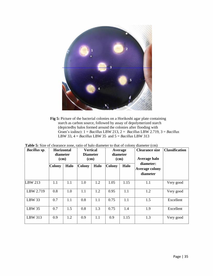
Oluoch, K. R).All 5 bacteria exhibited extracellular starch hydrolysing activities, as depicted by
the presence of clearance zones or ‘halos’ around their colonies after staining with Gram’s
iodine solution( Fig 5). The ratios of the diameter of halos to that of their respective colonies was
then used as a semi-quantitative method for classifying the bacteria as; excellent producers of
starch hydrolysing enzymes (halos > 1.5), very good producers (halo size > 1 but < 1.5), good
producers (halo size > 0.5 but < 1) and poor producers when no clear zones were observed
(Table 1). Based on this criterion, Bacillus sp. LBW 33 and LBW 35 were considered as
excellent producers while the rest were very good producers. All the bacteria were considered for
further experiments.
Page | 35
Fig 5: Picture of the bacterial colonies on a Horikoshi agar plate containing
starch as carbon source, followed by assay of depolymerized starch
(depictedby halos formed around the colonies after flooding with
Gram’s iodine): 1 = Bacillus LBW 213, 2 = Bacillus LBW 2.719, 3 = Bacillus
LBW 33, 4 = Bacillus LBW 35 and 5 = Bacillus LBW 313
Table 5: Size of clearance zone, ratio of halo diameter to that of colony diameter (cm)
Bacillus sp.
Horizontal
diameter
(cm)
Vertical
Diameter
(cm)
Average
diameter
(cm)
Clearance size
Average halo
diameter:
Average colony
diameter
Classification
Colony Halo Colony Halo Colony Halo
LBW 213 1.1 1.1 1.0 1.2 1.05 1.15 1.1 Very good
LBW 2.719 0.8 1.0 1.1 1.2 0.95 1.1 1.2 Very good
LBW 33 0.7 1.1 0.8 1.1 0.75 1.1 1.5 Excellent
LBW 35 0.7 1.5 0.8 1.3 0.75 1.4 1.9 Excellent
LBW 313 0.9 1.2 0.9 1.1 0.9 1.15 1.3 Very good
1
5
4
3
2
Page | 36
4.2 Extracellular starch hydrolysing activities of Lake Bogoria isolates
The five alkaliphilic bacillus species were cultivated separately in Horikoshi broth medium
containing starch as the sole carbon source for a period of 48h. During this period, samples were
withdrawn every 12 h and used to determine both cell growth and amylase activity. Table 6
shows the average OD 600 nm readings for the bacteria and enzyme activities they exhibited
over the entire cultivation period. These readings were used to generate both characteristic
bacterial growth curves (OD 600 nm) and enzyme production profiles for all the bacteria (Fig 6).
The standard curve was first generated by determining the absorbances, at 540 nm, of glucose
concentrations in the range 0-0.25mg/ml using the DNS method and then using the standard
curve to calculate the enzyme activities by determining the quantities of reducing sugars
produced following enzymatic hydrolysis of soluble starch during the assays. The glucose
standard curve was then used to calculate the specific enzyme activities of the five Bacillus sp. as
shown in the Table.
Table 6: O.D(600 nm) readings taken after 0, 12, 24, 36 and 48 h of bacterial cultivation and the
corresponding amounts of reducing sugars and enzyme obtained after the assay
Bacillus sp. Cultivation
time(h)
Average
O.D 600 nm
Glucose
conc.(mg/ml)
Enzyme
activity(U/ml)
LBW 213
0 0.1 0 0
12 3.300 0.0000 0.0000
24 4.370 0.0370 0.0103
36 3.565 0.0000 0.0000
48 4.520 0.0044 0.0012
LBW 2.719
0 0.15 0 0
12 2.805 0.0232 0.0064
24 4.670 0.0830 0.0230
36 3.840 0.0000 0.0000
48 4.430 0.0081 0.0022
LBW 33
0 0.1 0 0
12 3.660 0.0375 0.0104
24 5.206 0.0140 0.0389
36 4.650 0.0500 0.0139
48 4.190 0.0000 0.0000

Page | 37
LBW 35
0 0.2 0 0
12 3.142 0.0221 0.0061
24 6.100 0.0420 0.0117
36 4.073 0.0350 0.0097
48 3.665 0.0000 0.0000
LBW 313
0 0.25 0 0
12 1.940 0.0000 0.0000
24 6.310 0.0078 0.0022
36 3.218 0.0199 0.0055
48 4.650 0.0000 0.0000

Page | 38
Fig 6: characteristic bacterial growth curves (OD 600 nm) and enzyme production profiles for all
the alkaliphilic Baillus sp bacteria.
During the cultivation, all five bacteria exhibited a general growth profile, reaching optimal
growth at 24 h with maximum OD at 600nm of 4.370(Bacillus sp.LBW 213), 4.670 (Bacillus sp.
27 19), 5.206(Bacillus sp.LBW 33), 6.10(Bacillus sp.LBW 35) and 6.310(Bacillus sp.LBW 313)
(Fig 6curve profiles).Thereafter, growth declined gradually for Bacillus sp. 33 and Bacillus sp.
34, reaching OD 600 nm of 4.2 and 3.7, respectively, at the end of the cultivation period (48 h)
(Fig 6). The remaining bacteria showed a similar pattern only until the 36th
h, after which growth

Page | 39
started to increase unexpectedly. This unexpected result may be attributed to pipetting errors
encountered during the research.
Production of α-amylase by the 5 bacterial species was also followed during the cultivation
period. There was a general increase in α-amylase production by most of the bacteria, reaching
optimum levels of 10.3 x 10⁻³
(Bacillus sp. LBW 213), 23 x 10-3
(Bacillus sp. LBW 27 19), 38 x
10-3
(LBWBacillus sp. 33), and 11 x 10-3
(Bacillus sp. LBW35) during late exponential phases of
bacterial growth (24 h), and 5.5 x 10⁻³
(Bacillus sp.LBW313) during cell death phase (36 h) (Fig
6). Therefore, in this study the range of enzymatic activity for all the five bacteria was 5.5 x 10⁻³
– 38 x 10-3
IU/ml. Other alkaliphiles have been reported to produce various amounts of α-
amylases: The alkaliphilic Bacillus halodurans LBK34 was shown to exhibit a much higher α-
amylase activity in the range 1.2- 1.8 U/ml when cultured under optimum temperature conditions
(55- 65o C)(Hashim, 2004).Indira, in his study on α-amylase production from Bacillus sp. SI- 136
isolated from sodic alkaline soil reported that log phase was reached by the 20th h when
cultivation in Horikoshi-ІІ brothmedium at 60oC(Indira, 2012),but maximum amylase production
(2000U/ml) wastowards late log-stationery phase as reported in an earlier study (Annamalai et
al., 2011 (Indira, 2012). Pardeep (2011) reported that during determination of optical production
of alkaline α-amylase from newly isolated Bacillus sp. DLB 9, maximum enzyme synthesis was
recorded at 24 h in pH 10 at 37 oC (10 +/-0.283Uml/min). Bacillus sp. DLB 9 had maximal
enzyme activity at 24 h, pH 9, at 37 oC (12.2+/-0.283U/ml/min) although the production and
enzyme activity was considerably high at 24 h, pH 10, 37 oC (10+/-0.0.848U/ml/min). Bacillus
sp. DLB 9 also had enzyme activity at 24 h, pH 9 at 37 oC of (12.2+/-0.424 U/ml/min) although
its optimum temp was 60 oC (16.2+/-0.141) and at 50
oC it had (16.0+/-0.283U/ml/min)
indicating an increase in enzyme activity with increase in temperature up to 60oC.
Page | 40
CHAPTER 5
5.0 DISCUSSION
Alkaline active amylases have potential applications in the detergent (removal of starch based
stains) and textile industries (de-sizing of denims and paper). Currently, these industries utilize
environment unfriendly chemicals or less active- and/or unstable- amylases to manufacture their
respective products, thus leading to environmental pollution and high cost of production.
Alkaliphilic microorganisms are known to possess alkaline active and stable enzymes. One such
enzyme is α-amylase which can find applications in both the detergent and textile industries.The
study therefore focused on bioprospecting for alkaline-active amylases from alkaliphilic bacillus
species, previously isolated from a Kenyan soda lake.
The research started with the screening of five alkaliphilic bacteria for amylase production. All
the bacteria exhibited extracellular starch hydrolysing activities, as depicted by the presence of
clearance zones around their colonies after staining with Gram’s iodine solution.Bacterial α-
amylase, being an endo-enzyme, catalyzes the hydrolysis of alpha-(1, 4) glycosidic linkages
located in the inner regions of amylose and amylopectin in starch molecules resulting in the
production of α-limit dextrins. Iodine has the ability to detect small amounts of starch and reveal
any changes in the starch’s degree of polymerization caused by enzymes. Hence, in the absence
of starch around the 5 bacterial colonies, iodine does not stain.
The 5 bacterial species were subjected to growth in separate Horikoshi broth media and both cell
growth and α-amylase production levels followed. The general increase in growth of the
bacterial cells followed a typical sigmoid curve, with very short lag phases (not determined)
followed by exponential-, stationary- and death- phases in that order. Enzyme production by the
bacteria correlated to cell growth - the higher the cell growth the higher the enzyme production.
Thus, with no limiting factor, enzyme production was enhanced when most of the bacteria
entered their exponential phases of growth, reaching optimum levels at the log-stationary phases.
The only exception was Bacillus sp. 313 whose unexpected increase in enzyme activity was
enhanced in the death phase of that bacterium when limiting factors such as lack of nutrients and
oxygen rain in. As expected, enzyme activity generally declined in the death phases of all the
bacteria, a fact that is attributed to the depletion of nutrients, oxygen and change in pH during
growth (decreased growth translates to decreased enzyme production).

Page | 41
The general low level of α-amylase production by all the bacteria under this study was probably
due to un-optimized culture conditions (pH, temperature, concentration of carbon and nitrogen,
among others). Assay conditions (e.g. incubation temperature) are other factors to take into
consideration.

Page | 42
CHAPTER 6
6.0 CONCLUSION
The five alkaliphilic bacteria were able to grow under alkaline conditions and produce
extracellular alkaline active α-amylases (Due to limited resources, I was not able to identify other
types of amylases e.g. β-amylase, amyloglucosidase or glucoamylase, CGTases, pullulanase,
isoamylase, and amylolactose). Although production levels by the bacteria were low compared to
those of other alkaliphilic bacteria studied, Bacillus sp. LBW 33 was the most promising
candidate for enzyme production (highest producer), thus making it ideal for further studies.
6.1 RECOMMENDATIONS
1. Assay for other amylases in the crude culture supernatants frozen at – 80 oC.
2. Optimize culture conditions to maximize enzyme yields for best amylase producer.
3. Characterize crude secreted by best amylase producer with respect to temperature, pH,
surfactants, chelators and other additives in order to determine factors required to
maintain enzyme activity and stability during its application in the target industry.
4. Carry out a pilot study to evaluate the efficiency of the enzyme with respect to an
application in the detergent or textile industry.
5. Establish strong collaborations with potential partners in the relevant industry.
6. Identify the alkaliphilic amylase-producing Bacilli sp. used in this study.

Page | 43
REFERENCES
Aguilar, A., Ingemansson, T., Magnien, E., Extremophile microorganism as cell factories:
support from the the European Union. Extremophiles 2 (1998) 367-373.
Akamura N, Horikoshi K. Characterization of acid-cyclodextrin glycosyl-transferase of an
alkalophilic Bacillus sp. Agric Biol Chem. 1976;40: 1647–1648.
Aono R. and Horikoshi K. (1983).Chemical composition of cell walls of alkalophilic strains of
Bacillus. J.Gen. Microbiol. 129,1083-1087.
Bath AH, Christofi N., Neal C, Philip JC, Cave MR.McGnley IG, Berner U (1987). Trace
element and microbiological studies of alkaline ground waters in Oman, Arabian gulf: a natural
analogue for cement pore waters. Report fluid processes Res. Group. Brit. Geol. Surv. FLPU-87-
92.
Boyer E W, Ingle M B, Mercer G D. Bacillus alcalophilus subsp. halodurans subsp. nov.: an
alkaline-amylase-producing alkalophilic organisms. Int. J Syst Bacteriol. 1973;23:238–242.
Boyer E W, Ingle M B. Extracellular alkaline amylase from a Bacillus species. J Bacteriol.
1972;110:992–1000.
Bunton , C.A. Lewis, T.A. Llewellyn, D.R. Vernon, C.A., J. Chem.Soc. 1955,pg 4419.
Dhawale, M., Wilson, J., Kachatourians, G. and Mike W. (1982). Improved method for
detection of starch starch hydrolysis. Appl. Environ. Microbiol., Vol. 34. No.4, (April 1982),747-
750.
Djekrif-Dakhmouche S., Gheribi-Aoulmi Z., Meraihi Z., Bennamoun L. Application of a
statistical design to the optimization of culture medium for α-amylase production by Aspergillus
niger ATCC16404 grown on orange waste powder. J Food Process Eng. 2006; 73:190–197.
Grant W. D.Alkaline Environments In: Lederberg 3(Ed.) Encyclopedia of Microbiology, vol.
Academy press(1992),73-80.

Page | 44
Grant W.D, Jones B.E, Mwetha W.E., (1990) Alkaliphiles: ecology, diversity and applications
.FEMS. Microbial Rev 75:255-270.
Grant W.D. Halophilic Microorganisms, Introductory Chapter: Half a Lifetime in Soda
Lakes.2004,17-31.
Horikoshi K. Production of alkaline enzymes by alkalophilic microorganisms. II. Alkaline
amylase produced by Bacillus No. A-40-2. Agric Biol Chem. 1971; 35:1783–1791.
Horikoshi K., Alkaliphiles: some applications of their products for biotechnology. Microbiol.
Mol. Rev. 63 (1999) 735-750.
Horikoshi, K., Alkaliphiles- from an industrial point of view. FEMS Microbiol. Rev. 18 (1996)
259-270.
Horikoshi, K., Enzymes of alkalophiles. In: Microbial enzymes and biotechnology (Fogarty,
W.M. and Kelly, C.T., eds.) Kluwer Aademic Publishers Dodrecht (1990) pp.275-293.
Horikoshi, K., Microorganisms in alkaline environments, ed. 1991, VCH Publishers Inc. New
York.
J. Gomes., and W. Steinner : Extremophiles and Extremozymes, Food Technol. Biotechnol. 42
(4) 223-235 (2004).
Kelly C T, O’Reilly F, Fogarty W M. Extracellular α-glucosidase of an alkalophilic
microorganism, Bacillus sp. ATCC 21591. FEMS Microbiol Lett. 1983;20:55–59.
Kenneth T.,PhD. Todar,s Online Textbook of Bacteriology.2008.
Kirk O., Borchert T.V., Fuglsang C.C. Industrial enzyme applications. Curr. Opin. Biotechnol.
2002; 13:345–351 .
L. J. Rothschild., Mnicinelli, Life in Extreme Environments. Nature 409(2001)1092-1102.
Matsuzawa M, Kawano M, Nakamura N, Horikoshi K. An improved method for the production
of Schardinger β-dextrin on an industrial scale by cyclodextrin glycosyltransferase of an
alkalophilic Bacillus sp. Starch. 1975;27:410–413.

Page | 45
Michael T. Madigan and Barry L. Marrs., Extremophiles. Scientific American. 276 (1997), 66-
71.
Nielsen J.E., Borchert T.V. Protein engineering of bacterial alpha-amylases. Biochim Biophys
Acta. 2000; 1543:253–274.
Outtrup, H., and B.E. Norman: starc W starkc, 38. (1984), pg 405-411.
P. Murphy, in Handbook of Hydrocolloids (Eds.) G.O. Phillips and P.A. Williams,Woodhead
Publishing, Cambridge , 2000, pg. 41
Pabulo H. Rampelotto, Extremophiles and Extreme Environments.Life 2013, 3, 482-485.
Sharp R.J., Munster, M.J., Biotechnological implications for microorganisms from extreme
environments. In: Microbes in Extreme environments (Herbert R.A. and Codd, G.A., eds.)
Academic Press London (1986) pp.215-295.
Suhaila O. Hashim, An alkaline active maltooligosaccharide forming α-amylase from Bacillus
halodurans. Doctoral thesis, Lund, 2004: pg22-24
Horikoshi K., Garo A., Allan T. Bull, Frank T. Robb, Karl O. Stetter, Springer science and
Business Media. Dec 8,2010.pg166.

Page | 46