dentin remineralization around ceramir restoration
TRANSCRIPT
University of Pennsylvania University of Pennsylvania
ScholarlyCommons ScholarlyCommons
Dental Theses Penn Dental Medicine
Summer 8-15-2016
Dentin Remineralization Around Ceramir Restoration Dentin Remineralization Around Ceramir Restoration
Lujain S. Alhuwayrini University of Pennsylvania, [email protected]
Follow this and additional works at: https://repository.upenn.edu/dental_theses
Part of the Dental Materials Commons
Recommended Citation Recommended Citation Alhuwayrini, Lujain S., "Dentin Remineralization Around Ceramir Restoration" (2016). Dental Theses. 17. https://repository.upenn.edu/dental_theses/17
This paper is posted at ScholarlyCommons. https://repository.upenn.edu/dental_theses/17 For more information, please contact [email protected].
Dentin Remineralization Around Ceramir Restoration Dentin Remineralization Around Ceramir Restoration
Abstract Abstract AIM: AIM:
To determine the efficacy of Ceramir, a modified glass ionomer cement and a surfactant mono-n-dodecyl phosphate in remineralization of dentin around root caries restorations.
MATERIALS & METHODS: MATERIALS & METHODS:
45 permanent intact teeth were embedded in self-cured dental acrylic resin to expose buccal or lingual surfaces. The buccal/lingual surfaces were wet ground with carbide paper, final polishing were accomplished with aluminum to obtain highly polished dentin surface. Baseline Knoop micro hardness values were recorded. All specimens were then etched using 37% phosphoric acid for 5 seconds to demineralize dentin and to expose dentin collagen. The Knoop indenter micro hardness measurements were again performed for each sample four indentations in dentin surface within an area of 75 μm. The mean of Knoop microhardness was calculated. Cavities, 6.35 mm width and 3 mm depth were prepared within the etched area of each tooth with round carbide bur. Samples were divided into 4 groups: 2 samples used as control with no restoration, 13 samples were restored with plain Ceramir, 13 sample were restored with Ceramir containing 2% mono-n-dodecyl phosphate restoration and 13 samples were restored with Ceramir containing 5% mono-n-dodecyl phosphate. Samples were stored in SBF a 37° C incubator. Knoop micro hardness values were recorded at a distance of 75 um from the margins of the restoration at 10,20 and 38 day intervals.
RESULTS: RESULTS:
Knoop hardness of dentin (KHN) was reduced by 33.7% after etching. Knoop hardness of dentin around Ceramir restorations returned to pre-etching levels after 10 days of restoration. There was no statistically significant difference in Knoop micro-hardness (KHN) between the plain Ceramir compared to the Ceramir with surfactant after 10 days. Also, there was no statistically significant difference between the plain Ceramir and Ceramir with 2% surfactant after 20 days. Knoop hardness around cavities restored with Plain Ceramir and Ceramir with 2% Surfactant were significantly higher than around cavities restored with Ceramir with 5% surfactant after 20 and 38 days.
CONCLUSIONS: CONCLUSIONS:
The result of this study shows that Ceramir restorations of dentin lesions lead to remineralization of dentin around the restoration margins in the area of 75 μm where the micro-indentations performed. It shows that addition of 2% surfactant to Ceramir tend to increase the remineralization over time. Toward the end of the observation period samples restored with Ceramir containing 2% surfactant appeared to remineralize at a faster rate than plain Ceramir. On the other hand addition of 5% surfactant was not beneficial as it led to a decrease in the remineralizing effect of Ceramir.
Degree Type Degree Type Thesis
Degree Name Degree Name MSOB (Master of Science in Oral Biology)
Primary Advisor Primary Advisor Francis Mante, BDS, MS, PhD, DMD, MA (Hon)
Keywords Keywords Biomimetic Remineralization, Ceramir, CPP-ACP, Knoop Micro-Hardness, Surfactant
Subject Categories Subject Categories Dental Materials | Dentistry
This thesis is available at ScholarlyCommons: https://repository.upenn.edu/dental_theses/17
1
MASTERTHESISPennDentalMedicineSummer 8-15-2016
DR.LUJAINS.ALHUWAYRINI
Dentin Remineralization Around Ceramir Restoration
2
Table of content Introduction
• Overview
• Caries-Preventive nanofillers
• Recurrent Caries
• Enamel Anatomy and Remineralization
• Dentin Anatomy and Remineralization
• Concepts of Calcium Phosphate Biomineralization
• Biomimetic Remineralization of Dentin
• Bottom-up Remineralization Strategy
• Amorphous calcium phosphate ( ACP ) Development
• Amorphous calcium phosphate ( ACP ) and its role in forming Hydroxy appatite
• Amorphous calcium phosphate ( ACP ) and its application in dentistry
• CPP-ACP
• CPP-ACP Mechanism of Action
• Clinical Safety of CPP-ACP Usage
• ACP-filled polymeric composites
• Enhanced Glass Ionomer - Ceramir
• Surfactant and HA formation
• Aim of the study
3
Materials and methods
• Sample Selection
• Sample Preparation and Baseline Measurement
• Demineralization and Cavity Preparation
Results
Discussion
Conclusion
limitation
Conflict of interest
References
4
AKNOWLEDGEMENTS
I am grateful to my mentor, Dr. Francis Mante for accepting me into his lab as a master student. I am thankful for the kindness, guidance, motivation and thoughtful insight he graciously provided.
I am grateful forever to my parents and brothers for their support and encouragement throughout my life. I wouldn’t be where I am today if it wasn’t for them.
And I will like to express my appreciation to the members of my thesis committee, Dr. Fusun Ozer, Dr. Thomas Sollecito and Dr. Scott Odell for their encouragement and for providing valuable expertise during this project.
5
Introduction
Overview:
Teeth are the most heavily mineralized tissues in the human body. Demineralization
and remineralization processes coexist in teeth during the entire life of an individual. In
pathological conditions, demineralization outweighs remineralization [1]. Fermentation
of dietary carbohydrates by acidogenic bacteria results in the production of acids such as
lactic acid, acetic acid and propionic acid that demineralize enamel and dentin. As the
carious lesion progresses into dentin, activation of endogenous, bound matrix
metalloroteinases and cysteine cathepsins will lead to the degradation of collagen fibrils
and decrease in the mechanical properties of dentin [2,3]
In the last decade, the focus of caries research has shifted from only restoring missing
tooth structure to the development of methodologies for the detection of early caries
lesions and the non-invasive management of caries lesions through remineralization to
preserve tooth structure.[4] Fluoride is generally known to promote remineralization, but
its remineralization process relies on calcium and phosphate ions from saliva. Therefore,
several new remineralizing agents have been introduced. These agents supplement and
enhance the ability of fluoride to restore tooth minerals by increasing availability of
these ions.[5,6] .
Fluoride is the cornerstone of the non-invasive management of non-cavitated caries
lesions, but its ability to promote net remineralization is limited by the availability of
calcium and phosphate ions (Reynolds et al., 2008)[7]. Fluoride ions can drive the
remineralization of extant non-cavitated caries lesions if adequate salivary or plaque
calcium and phosphate ions are available when the fluoride is applied. For fluorapatite or
fluorhydroxyapatite to form, calcium and phosphate ions are required, as well as fluoride
ions. Several authors have now shown that enamel remineralization in situ and the
retention of fluoride in plaque are dependent on the availability of calcium ions (Chow et
al., 2000; Whitford et al., 2005; Reynolds et al., 2008; Vogel et al., 2008)[8,9,10].
6
Hence, on topical application of fluoride ions, the availability of calcium and
phosphate ions can be the limiting factor for fluoride retention and net enamel
remineralization. Under hyposalivation conditions the lack of available calcium and
phosphate ions is highly exacerbated (Reynolds et al., 2008)[7]. When adequate levels
of calcium and phosphate ions are present together with the fluoride ions, it has been
shown in vitro that this combination can produce substantial remineralization of lesions
of enamel and even those penetrating the underlying dentin in pH-cycling experiments
(ten Cate, 2001; ten Cate et al., 2008)[11,12]. Therefore, the challenge now is to achieve
this clinically, since salivary remineralization of enamel promoted by topical fluoride
(particularly high concentrations) has been shown to give rise to predominantly surface
remineralization (Arends and Ten Cate, 1981; ten Cate et al., 1981; Ögaard et al., 1988;
Willmot, 2004).[13,14,15,16] Surface-only remineralization does little to improve the
aesthetics and structural properties of the deeper lesion. Ideally, a remineralization
system should supply stabilized bioavailable calcium, phosphate, and fluoride ions that
favor subsurface mineral gain rather than deposition only in the surface layer. [17]
In recent years, biomimetic treatment of early caries lesions by the application of
various types of nano-sized hydroxyapatite or calcium carbonate has received
considerable attention (Huang S et al., 2009, 2010, 2011; Nakashima et al., 2009).
[18,19,20,21] An experimental dentifrice containing 1% nano-sized amorphous calcium
carbonate particles (several tens to hundreds of nm), applied twice a day over 20 days,
yielded statistically significant mineral gain and remineralization of artificial caries
lesions in an in vitro system that used collagen-coated wells as a model for oral mucosal
surfaces (Nakashima et al., 2009) [21]. The authors concluded that the experimental
dentifrice has the potential to remineralize incipient enamel lesions due to the unique
properties of the nano-sized calcium carbonate, which had been retained on the collagen-
coated surfaces in the in vitro model system and thus might also be retained on oral
surfaces, thereafter releasing Ca ions into oral fluids for remineralization (Nakashima et
al., 2009).[21]

7
Caries-Preventive Nano fillers
Several studies have indicated that nano- apatite, in principle, has the potential to
remineralize, at least in part, initial enamel caries lesions under dynamic pH-cycling
conditions in vitro (Huang S et al., 2009, 2010, 2011).[18,19,20,20] A 10% suspension
of nano-hydroxyapatite particles (10-20 nm diameter, 60-80 nm length) promotes
preferential remineralization of the superficial layer of artificial caries lesions, and thus
might be effective in reversing lesion progression in the outer surface layer of initial
caries lesions measuring 20 to 40 µm (Huang S et al., 2010).[19] However, little
remineralization could be obtained by nano-hydroxyapatite in the body of the lesion
(Huang S et al., 2010, 2011).[19.20] Interestingly, hydroxyapatite nanoparticles promote
remineralizing effects under in vitro conditions, in contrast to a control solution
containing an equivalent concentration of free ions as provided by the nano-HA solution
at equilibrium (Huang et al., 2011).[20] These observations suggest that intrinsic
characteristics of the nano-HA, such as size and structure or chemical composition, may
be of considerable relevance for the remineralization process (Huang et al.,
2011).[20]Apparently, not only the size of the apatite nanoparticles used for
remineralization purpose but also the pH of the remineralizing agent will affect the
process of mineralization (Huang et al., 2011). More mineral was deposited in the body
part of the lesion if the pH-value was reduced from 7.0 to 4.0 (Huang et al., 2011).[20]
Recurrent Caries
Secondary caries and restoration fracture are still the main reasons for dental
restoration failure, thus limiting the longevity of (resin composite) restorations.
Recurrent caries around composites is strongly linked to leakage through marginal gaps
caused by polymerization shrinkage.[22] To control caries-induced demineralization at
the resin composite-tooth interface, calcium and phosphate ion-releasing nanofillers have
been developed, such as nanoparticles of dicalcium phosphate anhydrous (112 nm in
8
size) or of amorphous calcium phosphate (116 nm in size) (Xu et al., 2007a,b, 2010b,
2011; Moreau et al., 2011).[23,24,25,26] These additives enable the resin composite to
release calcium and phosphate when the pH is dropped down under in vitro conditions,
providing caries-inhibiting properties (Xu et al., 2007a,b, 2010b, 2011; Chen,
2010).[23,24,25,27]Nanocomposites containing 40% nanoparticles of amorphous
calcium carbonate have been shown to rapidly neutralize a lactic acid solution of pH 4.0
by increasing the pH to 5.69 within 10 min (Moreau et al., 2011).[26] The mechanical
properties of the calcium- and phosphate-releasing experimental composites match those
of commercial hybrid composites (Chen, 2010; Moreau et al., 2011; Xu et al., 2011).
[25,26]
Most recent developments are novel nanocomposites which contain antibacterial
agents, such as chlorhexidine (10%) and quaternary ammonium dimethacrylate (7%)
alone or in combination with silver nanoparticles (0.028%), in addition to calcium and
phosphate ion-releasing nanofillers (Cheng et al., 2012a,b,c).[28,29,30] Incorporation of
these antibacterial components into nanocomposites has been shown to yield
antibacterial capabilities, thereby reducing the biofilm colony-forming unit counts, the
metabolic activity, and lactic acid production of Streptococcus mutans biofilms under in
vitro conditions (Cheng et al., 2012a,b,c).[28,29,30] In the presence of marginal gaps
caused by polymerization shrinkage.However, the effectiveness of all these strategies for
the control of demineralization processes still needs validation, on the one hand, by in
vitro studies focusing on the caries-inhibiting potential of ion-releasing and antibacterial
resin composites, as well as by subsequent clinical studies, on the other. [31]
Enamel Anatomy and Remineralization
Dental Enamel is the outermost covering of teeth. It is the hardest mineralized tissue
present in the human body. Enamel faces the challenge of maintaining its integrity
through periods of demineralization and remineralization within the oral environment
and it is vulnerable to wear, damage, and caries. Enamel is composed of crystalline
calcium phosphate of 96% mineral with the remaining 4% consisting of organic

9
components and water. The organic content consists of breakdown products of major
enamel protein amelogenin [32]. The main component of enamel includes rods, which
are bundles of aligned crystallites that are woven into intricate architecture that are 3-5
µm in diameter [32]. The second component of the enamel matrix is inter-rod enamel
which surrounds and packs between the rods [33]. The third structure, aprismatic
enamel, refers to the structures containing HA crystals that show no mesoscale or
macroscale alignment.
The mature enamel is acellular and does not regenerate itself unlike other
biomineralized tissues such as bone and dentin [34]. To replace enamel that is damaged
by dental caries, dentistry has formulated artificial replacement materials that mimic the
hardness of enamel [35]. But none of these materials could mimic all the physical,
mechanical, and aesthetic properties of enamel [36]. Recently scientists have shown
much interest in the direction of synthesizing artificial enamel [34]. Thorough
understanding of structure and pattern of ameloblast gene products, control of protein
self-assembly and simultaneous hydroxyapatite crystallization allows one to design
biomimetic approaches to create synthetic enamel [34]. There is now a transition of
emphasis from traditional synthetic biomaterials toward biological materials [37].
Advances in tissue engineering methods paves a way for enamel regeneration.
Based on the understanding of biological process involved in amelogenesis and
advances in nanotechnology, Chen et al., fabricated fluoapatite nanorods, which
resembles enamel prism like structures from a supersaturated chemical solution under
physiological condition. These nanorods have similar characteristics to those of natural
enamel crystals isolated from rat incisor enamel [38]. Yin et al., regenerated enamel like
microstructures using a simple chemical approach, which may have a potential clinical
application to repair enamel damage in dental clinics [39]. Zhang et al., have achieved an
ordered dental enamel-like structure of hydroxyapatite (HAP) through a solution
mediated solid-state conversion process with organic phosphate surfactant and gelatin as
the mediating agent [40].
Stephen Mann and colleagues prepared electrospun hydrogel mats of amorphous
calcium phosphate and polymer nano and micro fibres. Mats generated HAP crystals as

10
an immediate layer, which covers the enamel surface. Hence, it could be used for re-
growing enamel surfaces that have been lost due to erosion/or wear [41]. Ying et al.,
used an agarose hydrogel method, which mimics the natural enamel at secretory or
matrix formation stage. This biomimetic mineralization model regenerates enamel like
prismatic structure with hardness similar to natural enamel [42].
Hontsu et al., successfully fabricated a freestanding flexible HAP sheet, which was
directly attached to enamel surface of extracted teeth using a calcium phosphate solution.
The interface between sheet and surface was not completely adhered. [43] To improve
the adhesiveness HAP sheet coated with a tricalcium phosphate layer. The adhesive
strength of the HAP/TCP sheet was markedly higher than that of the HAP sheet that
indicates sheet may be used for restoration [44,45].
Dentin Anatomy and Remineralization
Dentin is a complex mineralized tissue arranged in an elaborate 3-dimensional
framework composed of tubules extending from the pulp to the dentin–enamel junction.
The mineral portion is composed of carbonate apatites. Fibrillar type I collagen accounts
for 90% of the organic matrix, while the remaining 10% consists of non-collagenous
proteins, such as phosphoproteins and proteoglycans. The peri-tubular dentin, i.e., dentin
surrounding the tubules, is highly mineralized (95 vol% of mineral), while most organic
content is localized at the inter-tubular dentin (30vol% of mineral) [46]. Dentin
undergoes modifications by physiological aging and disease processes to produce
different forms of dentin [47]. This process affects the biomechanics and biochemistry of
the tissue.
Although similar in composition to bone, dentin does not share the same ability to
remodel. This limits site regenerative therapies. An advantage of dentin over enamel is
the presence of a collagen based scaffold that provides an appropriate cell- free backbone
for tissue repair and regeneration. The presence of such a scaffold is a key to advance
new concepts in tissue engineering approaches to the treatment of missing hard tissue.
11
Recently, bio- modification of dentin has been investigated as a biomimetic strategy
therapy to mechanically strengthen the existing collagen network and also control
biodegradation rates of extracellular matrix (ECM) components. [48]
Different strategies have been employed for remineralizing demineralized dentin. For
instance, fluoride, amorphous calcium phosphate (ACP)-releasing resins or resin-based
adhesives containing bioactive glass have been used to improve the resistance of bonded
restorations to secondary caries [49,50] However, most of these studies focused on
remineralizing partially demineralized carious dentin, which was based on the epitaxial
deposition of calcium and phosphate ions over existing apatite seed crystallites [51].
With these traditional ion-based strategies, remineralization does not occur in
locations where seed crystallites are absent [52]. Thus, the classical ion-based
crystallization concept may not be applicable for remineralizing completely
demineralized dentin within hybrid layers created by etch-and-rinse adhesive systems or
the superficial part of a caries-affected dentin lesion left behind after minimally invasive
caries removal, due to the unavailability of seed crystallites in those regions for
accomplishing homogeneous nucleation of apatite crystallites [53,54].
Concepts of Calcium Phosphate Biomineralization
Biomineralization is the process by which living organisms secrete inorganic
minerals in the form of biominerals (e.g. magnetite, silica, oxalates, various crystalline
forms of calcium carbonate and carbonated apatite) within cell cytoplasm, shells, teeth
and bony skeletons [55,56]. This process exhibits a high level of spatial and hierarchical
control as mineralization usually takes place in a confined reaction environment under
ambient temperature and pressure conditions. Calcified human tissues consist of the
collagen matrix and the hierarchically arranged carbonated apatite inorganic phase;
deposition of the latter is regulated by non-collagenous proteins [57,58]. It is generally
believed that non-collagenous proteins, along with specific matrix metalloproteinases
and other important enzymes secreted by odontoblasts, play critical roles to orchestrate
12
dentin mineralization. They possess carboxylic acid and phosphate functional groups that
act as preferential sites for Ca/P nucleation and subsequent apatite crystallization.
[59,60]. As the therapeutic use of native or recombinant non-collagenous proteins for in
situ biomineralization is not yet economically viable, research scientists have resorted to
the use of polyelectrolyte and poly(acid) macromolecules to mimic the functional
domains of these naturally occurring proteins, in biomimetic mineralization [61,62]. In
the past few years, this field of research has attracted a lot of attention, resulting in
changing concepts of calcium phosphate biomineralization.[63]
Biomimetic Remineralization of Dentin
Biomimetic remineralization represents a different approach to this problem by
attempting to backfill the demineralized dentin collagen with liquid-like amorphous
calcium phosphate nano-precursor particles that are stabilized by biomimetic analogs of
non-collagenous proteins [63].
Several nano-technological approaches have been reported for remineralization of
early caries lesions. Casein phosphopeptide-amorphous calcium phosphate nano-
complexes (CPP-ACP) have been shown to promote remineralization and provide anti-
cariogenic activity in laboratory, animal, and human experiments. The casein
phosphopeptides stabilize calcium and phosphate ions by formation of amorphous nano-
complexes. The calcium phosphate from these complexes is biologically available for
remineralization of initial lesions. [31]
This is achieved by adopting the recently discovered, non-classical particle-based
crystallization concept utilized by Nature in various biomineralization schemes, ranging
from the mineralization of sea-shells (calcium carbonate), siliceous shells of diatoms and
sponges (amorphous silica) to the deposition of calcium phosphate salts in fish scales
and bone [64,65].
Intra-fibrillar mineralization of fibrillar collagen not only significantly increases its
mechanical properties [66,67], but also protects the collagen molecules from external
13
challenges, such as temperature, endogenous enzymes, bacterial acids and other
chemical factors. Using this biomimetic remineralization strategy, both hybrid layers
created by etch-and-rinse adhesives and moderately aggressive self-etch adhesives
[53,68,69], as well as 250–300 nm thick completely demineralized dentin lesions can be
remineralized [70,71]. This bottom-up remineralization strategy does not rely on seed
crystallites, and may be considered as a potentially useful mechanism in extending the
longevity of resin–dentin bonds [72] via restoring the dynamic mechanical properties of
the denuded collagen within the hybrid layer to approximate those of mineralized dentin
[73].
Bottom-up Remineralization Strategy
Nanotechnologies involved in the fabrication of biomaterials may be classified as top-
down or bottom-up approaches [74]. The top-down approach starts from a bulk material
that incorporates critical nanoscale details. In this approach, a biomaterial is engineered
by scaling down a complex entity into its component parts, such as creating small
crystals from a bulk mineralized hard tissue via acid-etching. By contrast, the bottom-up
approach assembles materials from the nanoscopic scale, such as molecules and atoms,
to form larger structures [75]. Examples of the bottom-up approach include self-
assembly and molecular patterning [74]. Biomineralization, which involves the spatial
regulation of amorphous mineral phases via matrix proteins and organization of
nanoscopic crystal-line mineral units into hierarchical structures, represents the perfect
example of a bottom-up approach [76,77]
Biominerals found in bone and teeth are carbonated apatites with dimensions that are
small enough to fit within the gap zones of type I collagen molecules. This hierarchical
arrangement of over-lapping platelets can be identified as cross-banded patterns in
unstained, non-demineralized ultrastructural sections [78,79]. Partial demineralization of
a mineralized collagen matrix by acids derived from bacteria or clastic cells represents
an example of a top-down approach in generating seed crystallites [74] as nidi of
heterogeneous nucleation [80]. The orientation of those mineral lattices is determined by

14
the lattice of the seed crystallites [81]. Conventional mineralization strategies often
involve the use of metastable calcium and phosphate ion-containing solutions or gels
[82,83]. This example of a top-down mineralization approach [75] does not occur by
spontaneous nucleation of minerals on the organic matrix but rather by epitaxial growth
over existing seed crystallites [84]. Throughout the evolution of biomineralization,
matrix proteins play a pivotal role in the regulation of mineral nucleation and growth
[85,86]. In the absence of seed crystallites, matrix protein–mineral interactions are
responsible for overcoming the thermodynamic barriers in homogeneous nucleation [87].
The precise control of crystal growth at the nanoscale and the creation of natural
structures based on bottom-up self-assembly mechanisms have inspired scientists to
mimic these non-classical strategies in the fabrication of novel biomaterials [87]. The
literature abounds with examples of the use of bottom-up, self-assembly approaches in
the fabrication of hybrid nanostructures [88]. A biomimetic mineralization strategy has
been developed [89] based on the use of polyanionic molecules to mimic the functions of
matrix proteins in biomineralization [90,91]. In this strategy, a polycarboxylic acid-based
biomimetic analog is employed as a sequestration agent [92] to stabilize amorphous
calcium phosphate (ACP) derived from set Portland cement and simulated body fluid
(SBF) in the form of nanoparticles that are moldable enough to infiltrate the water
compartments of a collagen fibril. Another phosphorus-based analog mimicking the
collagen-binding function of matrix phosphoproteins [93] is used as a template to
promote nanoprecursor recruitment to the gap zones of the collagen fibrils, where they
nucleate and self-assemble into hierarchically arranged apatite nanocrystals within the
fibril. This example of a particle-mediated [94], bottom-up [77] crystallization strategy
differs from the classical top-down crystallization approach in two aspects. Firstly, it
recapitulates the progressive dehydration mechanism in natural biomineralization [95] by
replacing free and loosely bound water within the internal compartments of a collagen
fibril by apatite crystallites [96] Secondly, this self-assembly approach proceeds in the
absence of apatite seed crystallites and natural matrix phosphoproteins in a collagen
matrix and can be duplicated using reconstituted collagen [97].

15
The mineral phase in collagenous hard tissues such as bone and dentin is classified
as intrafibrillar apatites, which are deposited within or immediately adjacent to gap zones
of the collagen molecules and extend along the microfibrillar spaces within the fibril;
and extrafibrillar apatites, which are located within the interstitial spaces separating the
collagen fibrils [78,98]. Previous studies have shown that intrafibrillar apatites play a
significant role in the mechanical properties of mineralized tissues [99,100]. Thus,
biomimetic mineralization must recapitulate both the dimension and hierarchical
arrangement of apatites present in natural mineralized tissues [101,102].
Non-collagenous matrix proteins which serve as promoters or inhibitors of crystal
nucleation or growth play an important role in intrafibrillar mineralization [103]. In the
absence of biomimetic analogs of those matrix proteins, there should be no intrafibrillar
apatite deposition using the top-down mineralization approach.[104]
Amorphous calcium phosphate ( ACP ) Development
Generally, it is believed that ACP was firstly described by Aaron S. Posner [105] in
the mid 1960s. It was obtained as an amorphous precipitate by accident when mixing
high concentrations (30 mM) of calcium chloride and sodium acid phosphate (20 mM) in
buffer [106]. In X-ray diffraction, it was shown to have only two broad and diffuse
peaks, with maximum at 25° 2θ. No other features were obvious and it was clearly not
apatite. This pattern is typical for substances that lack long range periodic regularity. It
was found that immediately after being mixed, the spontaneously formed precipitate was
a non-crystalline, or amorphous, calcium phosphate with calcium to phosphorus molar
ratio (Ca/P) of 1.50. After several hours, it could convert to poorly crystalline apatite on
ageing. Afterwards, this solid converts slowly to crystalline apatite (Ca/P = 1.67) by an
autocatalytic mechanism [107].
In 1965, Eanes et al. identified ACP as a bone component [106]. ACP in bone, along
with the apatite, might account for the broad diffraction pattern and variable composition
of bone minerals. An age-dependent change in the ACP content of bone was also
16
described, with the proportion of ACP decreasing with age [107]. In 1975, ACP was
found in the mineralized cytoplasmic structure isolated from the blue crab
hepatopancreas, with a very similar short-range atomic structure to synthetic amorphous
calcium phosphate [108].
Amorphous calcium phosphate (ACP) is the initial solid phase that precipitates from a
highly supersaturated calcium phosphate solution, and can convert readily to stable
crystalline phases such as octacalcium phosphate or apatite products. Its morphological
form, structural model and X-ray diffraction patterns are typical for non-crystalline
substances with short-range periodic regularity. ACP has been demonstrated to have
better in vivo osteoconductivity than hydroxyapatite (HAP), better bio-degradability than
tricalcium phosphate, good bioactivity but no cytotoxicity [105]. These excellent
biological properties make ACP widely used in dentistry, orthopedics and medicine.
Amorphous calcium phosphate ( ACP ) and its role in forming
Hydroxy appatite
After the discovery of amorphous calcium phosphate, the early studies were focused
on the structure of ACP. It was suggested that synthetic ACP particles, which appear as
300- 1000 Å spheres in the electron microscope, consist of a random assembly of ion
clusters 9.5 Å in diameter, dimensions consistent with the chemical composition of
Ca9(PO4)6 [108]. And the 15-20% of water found in synthetic amorphous calcium
phosphate was shown to be mostly in the interstices between, and not within, the
individual Ca9(PO4)6 clusters [109]. Aggregated ACP particles readily dissolve and
crystallize to form apatite, a thermodynamically stable phase. The typical radial
distribution of noncrystalline ACP cluster structures, calculated from the x-ray
diffraction patterns, is only two broad and diffuse peaks showing the rapid drop-off of
atomic periodicity. Short-range order exists in these amorphous structures but no long-
range order such as that in crystalline hydroxyapatite [109]. Infrared analysis showed a
similar lack of crystalline order about the PO4 anions in the ACP structure

17
[110]However, Wuthier et al reported that ACP, with Ca/PO4 molar ratio as low as 1.15
precipitated at more acidic preparative pHs, i.e.6.9 [111].
More importantly, it has been shown that ACP particles are nanometer particles.
Primary particle sizes of ACP is about 40-100 nm. The morphology of ACP solids
appears to be a curvilinear shape when viewed by TEM, rather than the faceted, angular
shape of crystalline calcium phosphates. However, this curvilinear appearance has only
been clearly established with dried ACP [112]. The initial flocculates collected
immediately after precipitation of highly hydrated ACP have a low-contrast disk-shaped
appearance. High-contrast spherical particles begin to appear as ACP suspensions age,
and become the dominant shape with time [113].
The ACP precipitate, with little long-range order, is a highly unstable phase and
hydrolyzes almost instantaneously to more stable phases. In the presence of other ions or
under in vivo conditions, ACP may persist for appreciable periods due to kinetic
stabilization [114]. Although the exact mechanism of stabilization of ACP is not
understood, the presence of Mg2+
, F-
, carbonate, pyrophosphate, diphosphonates, or
polyphosphorylated metabolites or nucleotides, in sufficient quantity will prevent the
transformation of synthetic ACP to hydroxyapatite [115,116].
Amorphous calcium phosphate (ACP) and its application in
dentistry
ACP has been widely applied in biomedical field due to its excellent bioactivity, high
cell adhesion, adjustable biodegradation rate and good osteoconduction [117,118]. As
discussed above, the first quantitative studies on synthetic ACP were done in the mid
1960s [105]. From then on, more and more attention has been attracted in the
development and the application of ACP-containing products, especially in orthopedic
and dental fields. It is also used as filler in ionomer cements to fill carious lesions or as a
colloidal suspension in toothpastes, chewing gums or mouthwashes to promote
demineralization of carious lesions and/or to prevent tooth demineralization .[119]
18
CPP-ACP
CPP-ACP is an acronym for a complex of casein phosphopeptides (CPP) and
amorphous calcium phosphate (ACP). Caesins are a heterogenous family of proteins
predominated by alpha 1 and 2 and beta caesins. CPPs are phosphorylated casein-derived
peptides produced by trypsin digestion of caesin. This protein nanotechnology combines
specific phosphoproteins from bovine milk with nanoparticles of ACP.
The precise ratio is 144 calcium ions; 96 phosphate ions; and six peptides of CPP. [119]
The possible cariostatic potential of dairy products is the subject of many reports in
the literature.[120,121] .In 1991, the complex CPP-ACP, derived from a major protein
found in milk called casein, was patented in the United States.[122]The complex is
presented as an alternative remineralizing agent that is remarkably capable of stabilizing
calcium phosphate, maintaining a state of supersaturation of these ions in the oral
environment. As a consequence, the tooth structure would benefit from the high levels of
calcium phosphate in the biofilm,and remineralization would occur.[123,124]CPP-ACP
nanocomplexes have been shown to prevent demineralization and promote
remineralization of enamel subsurface lesions in animal and in-situ caries models.By
stabilizing calcium phosphate in solution, the CPP maintains high-concentration
gradients of calcium and phosphate ions and ion pairs in the subsurface lesion and, thus,
causes high rates of enamel remineralization. The calcium phosphate in these complexes
is biologically available for intestinal absorption and remineralization of subsurface
lesions in tooth enamel.[125]
CPP-ACP Mechanism of Action
CPP has been shown to stabilize calcium and phosphate, preserving them in an
amorphous or soluble form called amorphous calcium phosphate (ACP). ACP (Ca3H.,0)
is postulated as a precursor in the formation of hydroxyapatite (HA). The ACPs exhibit a
very high solubility and are readily converted to HA, which makes them suitable
mineralizing agents. The main advantage of ACP is its facile, single solid phase
19
phosphate formulation and its biocompatibility with both hard and soft tissues, which is
equal to that of HA and various di-, tri-, and tetracalcium phosphates.[125]
The following mechanism is responsible for a consistent level of remineralization
through CPP-ACP.Casein phosphopeptide-amorphous calcium phosphate is a
technology based on amorphous calcium and phosphate (ACP) stabilized by casein
phosphopeptides (CPP). CPP containing the cluster sequence -Ser(P)- Ser(P)-Ser(P)-
Glu-Glu- stabilizes ACP in metastable solution. Through the cluster sequence, the CPP
binds to forming clusters of ACP, preventing their growth to the critical size required for
nucleation and phase transformation.[125]
Rose and Hogg investigated the affinity and capacity of Streptococcus mutans for
CPP-ACP. Using the equilibrium dialysis system they described, their results
demonstrated that CPP-ACP binds with about twice the affinity of the bacterial cells for
calcium up to value of 0.16g/g wet weight cells. Application of CPP-ACP to plaque may
cause a transient rise in plaque fluid-free calcium, which may assist remineralization.
Subsequently, CPP-ACP will form a source of readily available calcium to inhibit
demineralization. Hence, CPP-ACP binds well to plaque, providing a large calcium
reservoir, which is likely to restrict mineral loss during a cariogenic episode and provide
a potential source of calcium for subsequent remineralization. In short, once in place,
CPP-ACP will restrict the caries process. [125]
In the United States, up to now, this product is primarily used for abrasive prophylaxis
pastes and secondarily used for the treatment of tooth sensitivity especially after in-
office bleaching procedures, ultrasonic scaling, hand scaling or root planing. However,
its use for remineralizing dentin and enamel and preventing dental caries is an off-label
application. Outside the United States, this product is marketed as GC Tooth Mousse
[126,127].
20
Clinical Safety of CPP-ACP Usage
No serious side effects were reported in studies assessing the clinical safety of CPP-
ACP usage. [128,129,130,131] In Morgan’s two-year follow-up study, no significant
differences were found in the incidence of side effects (i.e. nausea, headache, and
diarrhoea) between the intervention and control groups.[128]
No allergies or serious side effects were recorded in Rao’s and Bailey’s trials as
well.[128,130] Sitthisettapong also confirmed by email corre-spondence that no extra
calculus formation had occurred on the primary teeth in their experimental group.[131]
ACP-filled polymeric composites
ACP has been evaluated as a filler phase in bioactive polymeric composites [132].
Skrtic has developed unique biologically active restorative materials containing ACP as
filler encapsulated in a polymer binder, which may stimulate the repair of tooth structure
because of releasing significant amounts of calcium and phosphate ions in a sustained
manner [133,134]. In addition to excellent biocompatibility, the ACP-containing
composites release calcium and phosphate ions into saliva milieus, especially in the oral
environment caused by bacterial plaque or acidic foods. Then these ions can be
deposited into tooth structures as apatitic mineral, which is similar to the hydroxyapatite
found naturally in teeth and bone [135,136].
Compared with more commonly used silanated glass or ceramic filler, more
hydrophilic and biodegradable ACP-filled composites exhibited inferior mechanical
properties, durability and water sorption characteristics [137]. The uncontrolled
aggregation of ACP particulates along with poor interfacial interaction plays a key role
in adversely affecting their mechanical properties [138]. Their clinical applicability may
be compromised by relatively poor filler/matrix interfacial adhesion and also by
excessive water sorption that occurs in both resin and filler phases of these composites
[139,140].

21
Enhanced Glass Ionomer - Ceramir
Recently Doxa Dental AB,(Uppsala, Sweden) introduced a bioceramic, Ceramir with
a modified GI composition. The cement has been shown to form apatite on storage in
simulated body fluids (SBF)and saliva. [141]. The powder contains approximately 50%
Calcium aluminate in place of Calcium aluminosilicate glass. The high Calcium
aluminate content is responsible for apatite formation in SBF and saliva which has been
reported to contain enough phosphate ions to promote this effect. The setting reaction is
a combination of a glass-ionomer reaction and an acid-base reaction of the type
occurring in hydraulic cements. The set cement has an acidic pH of 5 and gradually
increases to 8.5 after 3-2 hours of setting. The set cement is reported to form
hydroxyapatite on the surface when in contact with phosphate containing solutions.
Initial setting time of 3mins and final setting time of 6 mins have been reported. [142].
Although Vickers hardness increased gradually from 30-110 MPa over a 28 days period
after mixing. The set cement is alkaline and releases calcium and fluoride ions. The
incorporation of Calcium Aluminate is believed to fix the GIC structure and hinders the
ionomer glass from continuous leaching over time. (Parmeijer 2007) When in contact
with phosphate solutions, such as saliva or body fluids it first forms precursors of HA,
which transform to apatite which can integrate with bone and dentin. The high pH of the
set cement inhibits bacterial growth. A comparative study showed that GI cement
modified by addition of calcium aluminate (Ceramir) induced hydroxyapatite formation
when immersed in SBF solution. This represents a significant development because use
of this material could potentially remineralize adjacent hard tissues. Other study
investigated HA formation of Ceramir in saliva and concluded that saliva contained
adequate amounts of calcium and phosphate for HA formation and demonstrated HA
formation on samples stored in saliva. [141]
22
Surfactant and HA formation
Hydroxyapatite (HA) is the principal inorganic constituent of bones and teeth [143].
HA is used mostly as powders and its usefulness depends on the powder properties such
as particle size, surface area, and morphology. Nano-structured HA particles with a
higher surface area would be more desirable for their use in many fields including
separation processes. [144].
HA can be synthesized by a variety of methods including conventional routes such as
solid-state reactions [145] and wet chemical routes [146] based on precipitation at low
temperature.
These conventional methods, however, mostly prepare irregular forms of powders.
Hydrothermal method, which has been proved to be a convenient way to prepare
materials, including salts, metal oxides, etc., has also been applied, but the control on
morphology is poor [147]. Nevertheless, the size and morphology would largely
determine the behavior of a certain material, that’s why a biomineralization process
usually involves complicated mediation and the final products generally have a delicate
microstructure [148]. Bone itself is a composite consisting of HAP nanorods embedded
in the collagen matrix [149].
Synthetic HAP crystals are usually prepared as rods or needles, which are similar in
structure and composition to HAP found in human bone.
Hence, HAP nano-rods are desirable when biocompatibility is considered [150].
Although control over microstructure seems too big a challenge to traditional methods,
the biological process itself has given some clues to achieve this: the controlled
nucleation and crystal growth process mediated by macromolecule control and cell
organization would finally result in uniform products. A method called biomimetics is
then aroused [151,152]. Macromolecules, such as stearic acid, monosaccharides and
related molecules were explored and the molecule addition has exerted significant
control on the morphology. [152]. Bose and Saha synthesized HAP nano-powders with a
controlled surface area and particle size by using nonionic surfactant emulsion. [153].
23
Another surfactant-templating approach has been extensively used in the preparation of
various nanoporous or nano tubular frame-work materials including mesoporous
silica[154] and many other metal oxides.[155]
Y. Wang et al. (2006) shows that the size
and morphology of precipitated hydroxyapatites can be affected by adding adequate a
surfactant, Cetyltrimethylammonium bromide (CTAB) and control of the precipitation
temperature and pH.
One current hypothesis is that the use of remineralizing materials in dentistry would
prevent secondary caries due to a natural formation of apatite between material and
tooth, leading to a stable interface. [141]
Aim of This Study
This study aimed to evaluate the effect of Ceramir and the addition of mono-n-
Dodecy phosphate to Ceramir as a surfactant in dentin remineralization.
Dentin remineralization is expected to occur in two mechanisms : 1. the classical ion-
based crystallization concept. 2. Locations where seed crystallites are absent. The
surfactant can act a biomemitic analogue OF non-collagenous proteins. To modulate
remineralization, the latter can lead to increase the calcium precipitation and modify the
HAP particle size to improve dentin remineralization ability of the Ceramir.
This, might be applicable for remineralizing completely demineralized dentin within
hybrid layers created by etch-and-rinse adhesive systems or the superficial part of a
caries-affected dentin lesion left behind after minimally invasive caries removal . This
method would also prevent secondary caries due to a natural formation of apatite
between material and tooth structure, leading to a stable interface.
Dentin remineralization will be determined by the alteration in micro-hardness of
dentin surface around Ceramir. The null hypothesis is that none of the filling using
including: plain Ceramir or Ceramir with mono-n-Dodecyl phosphate would
significantly affect the Knoop micro-hardness of demineralized dentin surface. [156]
24
Materials and methods: Sample Selection:
In this study, 45 permanent, intact, non-carious, non-hypoplastic, unfractured and non-
malformed, upper and lower, anterior and posterior human teeth, freshly extracted for
orthodontic or periodontic reasons were selected and stored in 0.5% chlorothymol
solution. Teeth with any defect were excluded.
All selected teeth were used within three months of extraction as recommended by
Occupational Safety & Health Administration (OSHA).
Sample Preparation and Baseline Measurement: The teeth were cleaned using an ultrasonic scaler (cavitron). The teeth were then
embedded in self-cured dental acrylic resin to expose buccal or lingual surfaces. The
buccal/lingual surfaces were wet ground starting with 800 grit silicon carbide paper
through 1200 and1600 under water cooling to obtain a smooth dentin surface, final
polishing was accomplished with alumina powder using 9 , 3 and 1 micron grit to obtain
highly polished surface.
The Knoop hardness tester (Leco M-400-G1 Hardness Tester). was calibrated with
standard test blocks provided by the manufacturer. Baseline surface micro-hardness of
sound dentin was measured using Knoop indenter. A loading force of 50 g and dwell
time 10 seconds were used. For each sample four indentations were performed in the
dentin surface and the mean Knoop microhardness value (KHN) was calculated.
Demineralization and Cavity Preparation: The dentin surfaces of all specimens were then etched using 37% phosphoric acid for
5 seconds to demineralize dentin and expose dentin collagen. The dimensions of the acid
etched area ranged from 7.5 to 8 mm. After the demineralization process was
25
completed, all the specimens were rinsed in deionized water and blotted dry. The Knoop
indenter microhardness of the etched dentin was measured. The mean Knoop
microhardness of etched dentin was calculated.
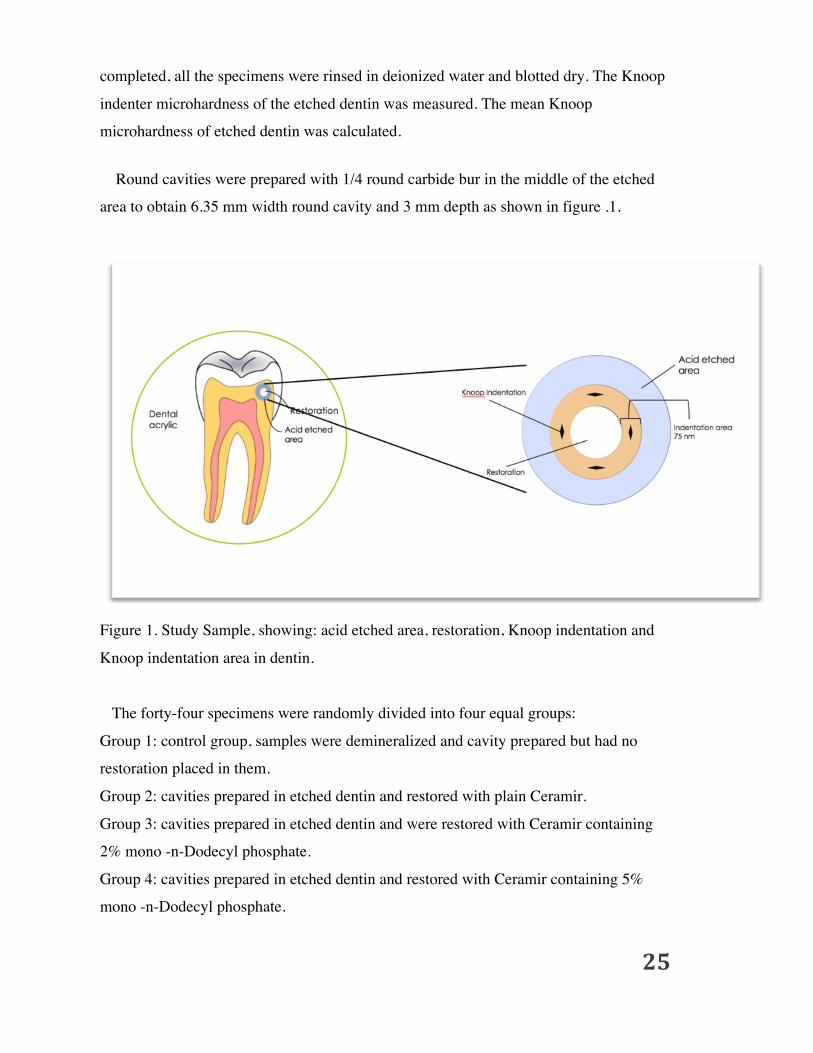
Round cavities were prepared with 1/4 round carbide bur in the middle of the etched
area to obtain 6.35 mm width round cavity and 3 mm depth as shown in figure .1.
Figure 1. Study Sample, showing: acid etched area, restoration, Knoop indentation and
Knoop indentation area in dentin.
The forty-four specimens were randomly divided into four equal groups:
Group 1: control group, samples were demineralized and cavity prepared but had no
restoration placed in them.
Group 2: cavities prepared in etched dentin and restored with plain Ceramir.
Group 3: cavities prepared in etched dentin and were restored with Ceramir containing
2% mono -n-Dodecyl phosphate.
Group 4: cavities prepared in etched dentin and restored with Ceramir containing 5%
mono -n-Dodecyl phosphate.
26
Ceramir capsules were mixed using an amalgamator for 5 seconds following the
manufacturer’s instructions. To fabricate experimental samples containing 2% and 5%
surfactant one Ceramir capsule was mixed and emptied and the weight of the paste
measured in grams to calculate the 2% and 5% surfactant. Then, the powder of mono -n-
Dodecyl phosphate was measured and added to the Ceramir paste to obtain Ceramir with
2% and 5% concentration of surfactant.
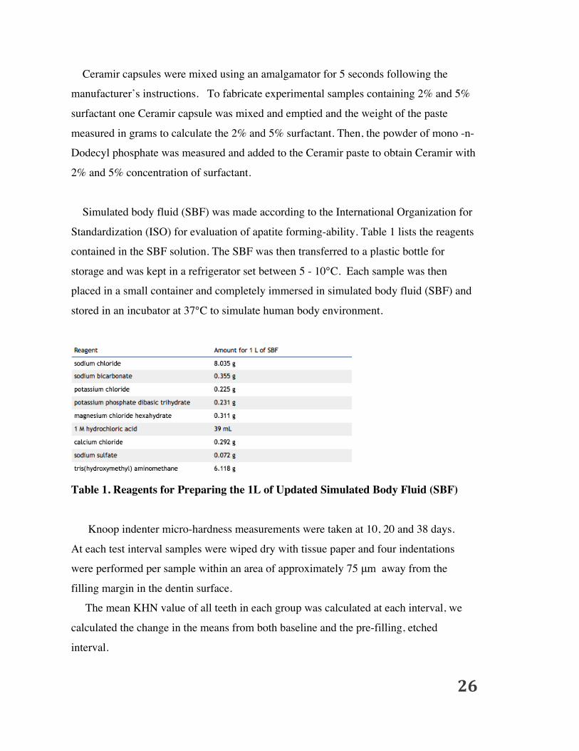
Simulated body fluid (SBF) was made according to the International Organization for
Standardization (ISO) for evaluation of apatite forming-ability. Table 1 lists the reagents
contained in the SBF solution. The SBF was then transferred to a plastic bottle for
storage and was kept in a refrigerator set between 5 - 10°C. Each sample was then
placed in a small container and completely immersed in simulated body fluid (SBF) and
stored in an incubator at 37°C to simulate human body environment.
Table 1. Reagents for Preparing the 1L of Updated Simulated Body Fluid (SBF)
Knoop indenter micro-hardness measurements were taken at 10, 20 and 38 days.
At each test interval samples were wiped dry with tissue paper and four indentations
were performed per sample within an area of approximately 75 μm away from the
filling margin in the dentin surface.
The mean KHN value of all teeth in each group was calculated at each interval, we
calculated the change in the means from both baseline and the pre-filling, etched
interval.

27
Dentin surface was evaluated by FEI Quanta 600 environmental scanning electron
microscope after each testing period. Environmental electronic microscope permits wet
and insulating samples to be imaged without prior specimen preparation. The use of the
environmental microscope eliminated the need for specimen preparation by drying and
sputters coating with gold or carbon that could alter the dentine surfaces.
One sample in each of the groups restored with 2% and 5% surfactant were excluded
because the fillings were dislodged after 10 days.
Data were expressed in Knoop Hardness Number (KHN) and statistically analysed
running a Repeated Measures ANOVA Analysis and pair-wise multiple comparison
procedures on the difference of means for each data point using Sigma Stat version 3.5
(Systat software , Point Richmond, California ,USA). Statistical significance was
determined at p value of 0.05.
Results:
SEM pictures were taken throughout the study period using (FEI Quanta 600
Environmental Scanning Electron Microscope). Samples were imaged in low vacuum
mode without any additional surface treatment. Figure 2 shows SEM of the polished
dentin surface with dentinal tubules partially blocked by smear layer. Figure 3 shows
micrographs of dentin after etching with phosphoric acid with widened dentin tubules
that appear interconnected. Figures 4, 5 and 6 show progressive formation of crystal
precipitates that are blocking the dentin tubules that were opend by acid etching.
28
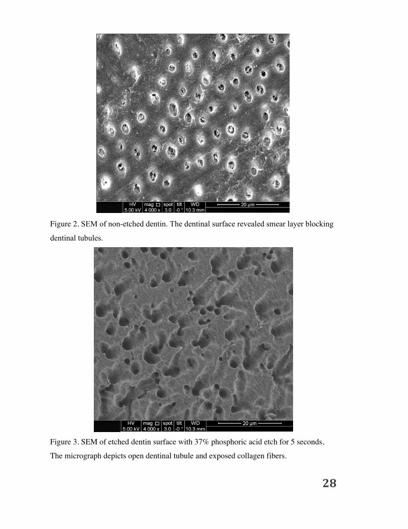
Figure 2. SEM of non-etched dentin. The dentinal surface revealed smear layer blocking
dentinal tubules.
Figure 3. SEM of etched dentin surface with 37% phosphoric acid etch for 5 seconds,
The micrograph depicts open dentinal tubule and exposed collagen fibers.
29
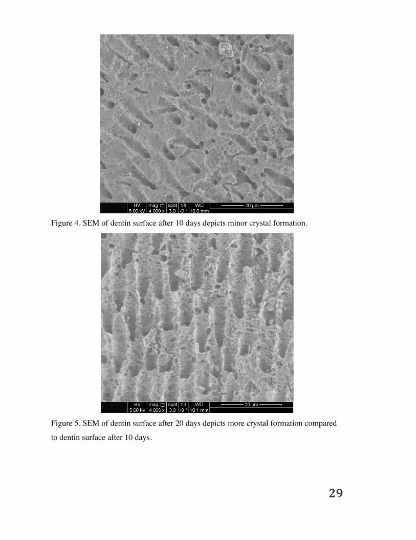
Figure 4. SEM of dentin surface after 10 days depicts minor crystal formation.
Figure 5. SEM of dentin surface after 20 days depicts more crystal formation compared
to dentin surface after 10 days.
30
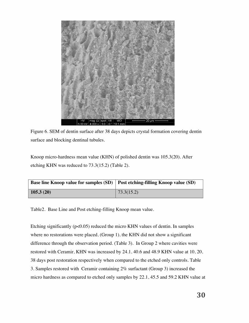
Figure 6. SEM of dentin surface after 38 days depicts crystal formation covering dentin
surface and blocking dentinal tubules.
Knoop micro-hardness mean value (KHN) of polished dentin was 105.3(20). After
etching KHN was reduced to 73.3(15.2) (Table 2).
Base line Knoop value for samples (SD) Post etching-filling Knoop value (SD)
105.3 (20) 73.3(15.2)
Table2. Base Line and Post etching-filling Knoop mean value.
Etching significantly (p<0.05) reduced the micro KHN values of dentin. In samples
where no restorations were placed, (Group 1), the KHN did not show a significant
difference through the observation period. (Table 3). In Group 2 where cavities were
restored with Ceramir, KHN was increased by 24.1, 40.6 and 48.9 KHN value at 10, 20,
38 days post restoration respectively when compared to the etched only controls. Table
3. Samples restored with Ceramir containing 2% surfactant (Group 3) increased the
micro hardness as compared to etched only samples by 22.1, 45.5 and 59.2 KHN value at

31
10,20,38 days respectively
Samples restored with Ceramir containing 5% surfactant (Group 4) increased the micro
hardness as compared to etched only samples by 24.2, 33.9 and 43.5 KHN units at 10,
20, and 38 days respectively.
Filling Type Baseline Knoop value (SD)
Post etching-filling Knoop value (SD)
Post 20 days KHN (SD)
Post 38 days KHN (SD)
Control ( no restoration )
105.3 (20)
73.3 (15.2)
73.3(15.0) 74.3(14.4) 75.2(16.2)
Plain Ceramir 101.2 (22.3) 117.7 (21.4) 126.0 (17.1) Ceramir with 2%
surfactant 90.1 (21.4) 113.5 (18.1) 127.2 (23.4)
Ceramir with 5% surfactant
93.3 (17.5) 102 (20.0) 112.5 (19.3)
Table 3. Change in Knoop values with time.
According to the Repeated Measures Anova Analysis, the increase in KHN values
for the plain Ceramir group were not significantly after 10 days post filling. After 20 and
38 days post filling the KHN readings show significant increase in values vs etched
values. Also, significant change found to be between 10 vs 38 days as shown in table 4.
Time period Significance of KHN value
Etched vs 10 days not significant Etched vs 20 days significant Etched vs 38 days significant 10 days vs 20 days not significant 10 days vs 38 days significant 20 days vs 38 days not significant
Table 4. Significance of KHN value over time period for group 2 (plain Ceramir).
For group 3 in which samples were restored with Ceramir containing 2% surfactant,
the increase in KHN values were statistically not significant (p<0.05) after 10 days post
filling. After 20 and 38 days post filling the KHN readings show statistically significant
32
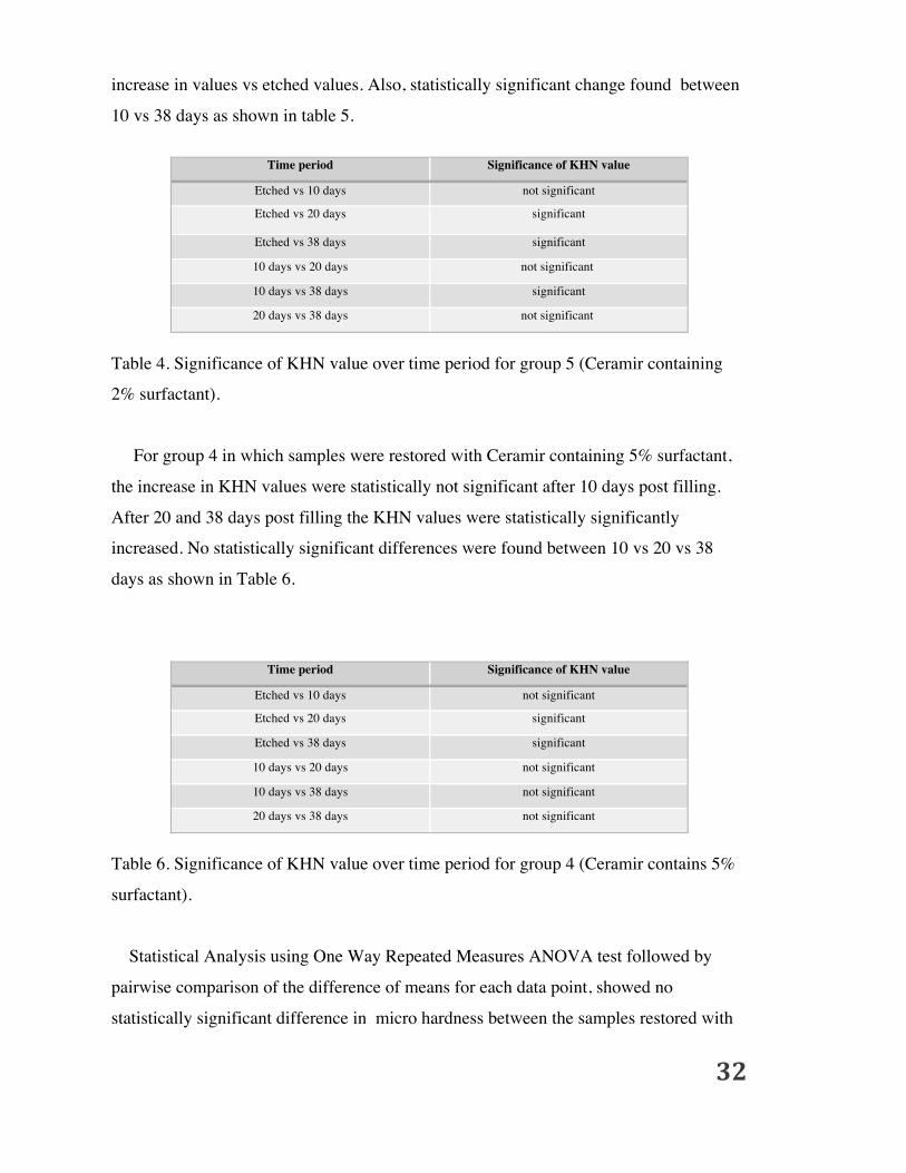
increase in values vs etched values. Also, statistically significant change found between
10 vs 38 days as shown in table 5.
Time period Significance of KHN value
Etched vs 10 days not significant Etched vs 20 days significant Etched vs 38 days significant 10 days vs 20 days not significant 10 days vs 38 days significant 20 days vs 38 days not significant
Table 4. Significance of KHN value over time period for group 5 (Ceramir containing
2% surfactant).
For group 4 in which samples were restored with Ceramir containing 5% surfactant,
the increase in KHN values were statistically not significant after 10 days post filling.
After 20 and 38 days post filling the KHN values were statistically significantly
increased. No statistically significant differences were found between 10 vs 20 vs 38
days as shown in Table 6.
Time period Significance of KHN value
Etched vs 10 days not significant Etched vs 20 days significant Etched vs 38 days significant 10 days vs 20 days not significant 10 days vs 38 days not significant 20 days vs 38 days not significant
Table 6. Significance of KHN value over time period for group 4 (Ceramir contains 5%
surfactant).
Statistical Analysis using One Way Repeated Measures ANOVA test followed by
pairwise comparison of the difference of means for each data point, showed no
statistically significant difference in micro hardness between the samples restored with

33
plain Ceramir compared to the Ceramir with surfactant group after 10 days.
There is no statistically significant difference between the Ceramir (Group 2) and
samples restored with Ceramir with 2% surfactant (Group 3) after 20 days while there is
statistically significant difference between them and the Ceramir with 5% (Group 4).
There is statistically significant difference in the micro hardness between Ceramir
with 2% group and Ceramir with 5% group after 38 days and no statistical difference
between plain Ceramir and Ceramir with 2% groups.
Change in micro-hardness values compared to baseline and etched values with time in
percentage summarized in table 7 and 8.
Filling Type 10 days 20 days 38 days
Control ( no restoration ) -33.69% -32.6% -31.6%
Plain Ceramir -7.57% 10.2% 19.2%
Ceramir with 2% surfactant
-14.66% 9.6% 23.96%
Ceramir with 5% surfactant
-14.77% -4.49% 5.7%
Table 7. Change in micro-hardness values from baseline with time in percentage.
Filling Type 10 days 20 days 38 days
Control ( no restoration ) 0.03% 0.7% 1.39%
Plain Ceramir 18.48% 31.30% 37.7%
Ceramir with 2% surfactant
14.96% 30.6% 40.2%
Ceramir with 5% surfactant
16.56% 22.77% 29.67%
Table 8. Change in micro-hardness values from etched dentin with time in percentage.
34
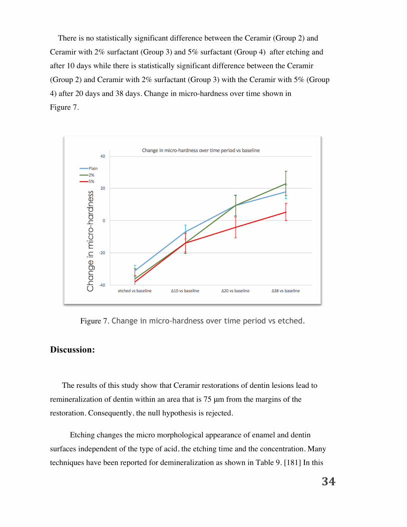
There is no statistically significant difference between the Ceramir (Group 2) and
Ceramir with 2% surfactant (Group 3) and 5% surfactant (Group 4) after etching and
after 10 days while there is statistically significant difference between the Ceramir
(Group 2) and Ceramir with 2% surfactant (Group 3) with the Ceramir with 5% (Group
4) after 20 days and 38 days. Change in micro-hardness over time shown in
Figure 7.
Figure 7. Change in micro-hardness over time period vs etched.
Discussion:
The results of this study show that Ceramir restorations of dentin lesions lead to
remineralization of dentin within an area that is 75 μm from the margins of the
restoration. Consequently, the null hypothesis is rejected.
Etching changes the micro morphological appearance of enamel and dentin
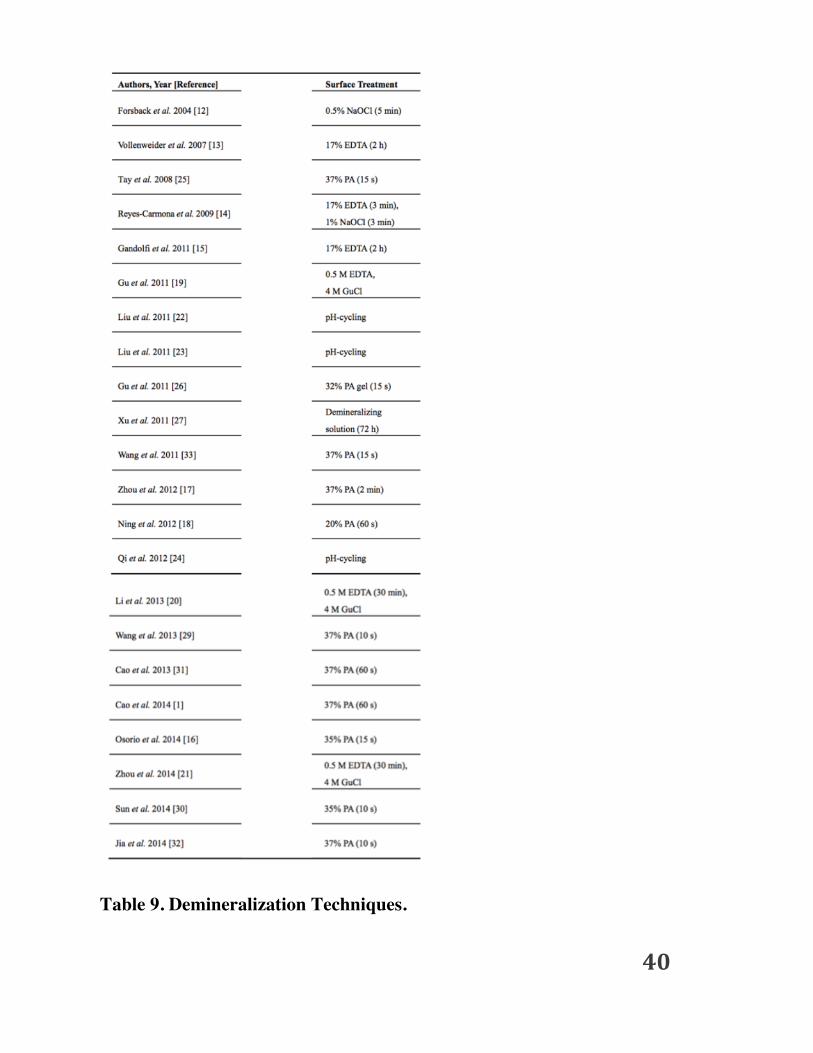
surfaces independent of the type of acid, the etching time and the concentration. Many
techniques have been reported for demineralization as shown in Table 9. [181] In this
35
study 37% phosphoric acid gel was been used for etching because it can be applied for a
small area as needed also it is quick and effective in providing adequate
demineralization.
The scanning electron micrograph study shows that polished dentin surface was
covered by a smear layer covering which partially blocked the dentin tubules. Figure 2.
Etching with phosphoric acid removed the smear layer and widened the openings of the
dentin tubules. The tubules appeared interconnected due to exposure of collagen fibers in
inter- tubular regions of the dentin.
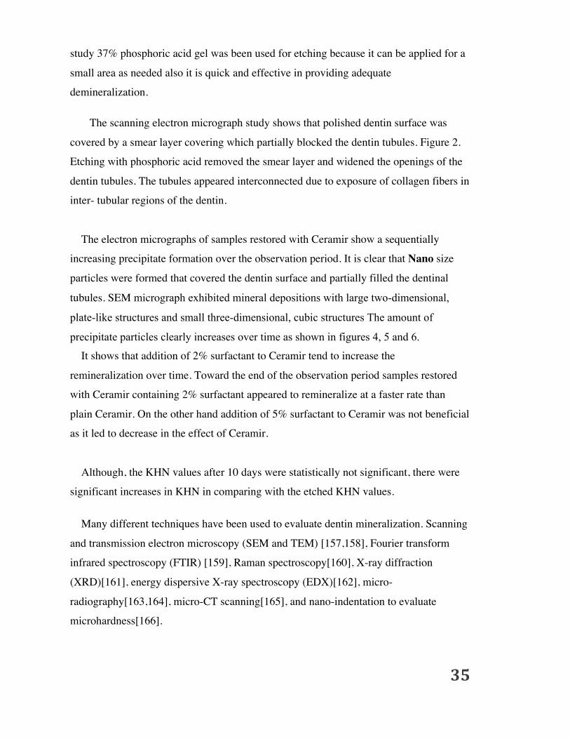
The electron micrographs of samples restored with Ceramir show a sequentially
increasing precipitate formation over the observation period. It is clear that Nano size
particles were formed that covered the dentin surface and partially filled the dentinal
tubules. SEM micrograph exhibited mineral depositions with large two-dimensional,
plate-like structures and small three-dimensional, cubic structures The amount of
precipitate particles clearly increases over time as shown in figures 4, 5 and 6. It shows that addition of 2% surfactant to Ceramir tend to increase the
remineralization over time. Toward the end of the observation period samples restored
with Ceramir containing 2% surfactant appeared to remineralize at a faster rate than
plain Ceramir. On the other hand addition of 5% surfactant to Ceramir was not beneficial
as it led to decrease in the effect of Ceramir.
Although, the KHN values after 10 days were statistically not significant, there were
significant increases in KHN in comparing with the etched KHN values.
Many different techniques have been used to evaluate dentin mineralization. Scanning
and transmission electron microscopy (SEM and TEM) [157,158], Fourier transform
infrared spectroscopy (FTIR) [159], Raman spectroscopy[160], X-ray diffraction
(XRD)[161], energy dispersive X-ray spectroscopy (EDX)[162], micro-
radiography[163,164], micro-CT scanning[165], and nano-indentation to evaluate
microhardness[166].
36
Microhardness is defined as the resistance to local deformation. [167] Microhardness
tests are commonly used to study the physical properties of materials and they are widely
used to measure the hardness of teeth. [168.169] These tests are based on the induced
permanent surface deformation that remains after removal of a load. [167] Two
microhardness tests, Knoop and Vickers hardness are commonly used for evaluation
dental materials. Both measurements can be correlated with other mechanical properties
such as fracture resistance, [170] modulus of elasticity, and yield strength. [171,172]
For our study we used a Knoop indenter (Leco M-400-G1 Hardness Tester) for the
micro hardness -indentation assessment and evaluation. The dental literature shows that
Knoop microhardness has been employed extensively in testing the hardness of both
enamel and dentin and is an effect measure of demineralization. [174]
The Knoop micro-indentation method requires only a tiny area of specimen surface
for testing. Using this technique, the specimen surfaces are impressed with a diamond
indenter. The geometry of this indenter is an extended pyramid with the length to width
ratio being 7:1 and respective face angles are 172 degrees for the long edge and 130
degrees for the short edge. The depth of the indentation can be approximated as 1/30 of
the long dimension at a certain load for a certain period of time. After load removal,
diagonals of the indentation are measured with an optical microscope. The hardness
number is defined by the ratio between the indentation load and the area of the residual
impression, which depends on the indenter shape. Then the hardness of materials was
calculated using these equations:
KHN = 14230 (F/d2) for Knoop microhardness or HV = 1854 (F/d2) for Vickers
microhardness. [173]
The Knoop indentation is longer and shallower than Vickers indentation and the load
impression can be applied to brittle materials without cracking. Also, the longer diagonal
is easier to read than the short diagonal of the Vickers. However, the advantage of the
Knoop’s longer diagonal is offset by the difficulty in deciding where the tapered tip ends
on the surface of the dentin. [173]
The chief characteristic of the Knoop microhardness test is its sensitivity to surface

37
effects and textures. [175,176] For a given load, the Vickers indenter penetrates about
twice as far into the specimen as the more shallow Knoop indenter, and the diagonal is
about one-third the length of the longest diagonal of the Knoop indentation. Thus, the
Vickers test is less sensitive to surface conditions and, due to its shorter diagonals, more
sensitive to measurement errors when equal loads are applied. [175,176,177,178]
Figure 8: difference between Vickers (A) and Knoop indenter (B).
The indentation load for the micro hardness test can range from 1 to 1,000 g, and with
various loading dwell times.
Dentin Knoop micro-hardness KHN values for baseline measurements ranges from
70 up to 90 depending on tooth location and area of indentation. [180][187][188]
Anterior teeth tend to show lower hardness value than posterior. Victoria Fuentes et al.
(2003) evaluated microhardness of superficial and deep sound human dentin using
Knoop indenter. [173]
Indentations closer to the dentin-enamel junction give higher KHN and decrease as we
move toward pulp direction. Because the tubules in dentin are not randomly oriented,
properties may be directionally dependent. This is because the mineral content in dentin
is higher at the dentin-enamel junction and as we move toward the pulp the mineral
content decrease as the organic content increase. [187]
In this study KHN was measured at sites close to the DEJ (Figure 1) and found to be
slightly higher than literature values reported by Huang et al. (20) Knoop micro-hardness
values increase as the mineral content increase and decrease as mineral content decrease.
38
This can explain the results in this study. After etching which is the process in which
mineral content of material removed the KHN was found to be decreased. And gradually
as particles forms and mineral precipitate increase, the KHN would increase as well.
[187]
Similar to our study, several studies have used microhardness measurements to
evaluate both enamel and dentin remineralization process.
Manuel Toledano et al. (2004) used Knoop indenter to asess microhardness of acid-
treated and resin infiltrated human dentine. The study concluded that treating dentine
with either H3PO4 alone or H3PO4 followed by NaOCl caused marked reduction of its
surface hardness. The removal of the mineral phase of dentine surfaces by acidic
treatments modifies their surface morphology and properties, and undoubtedly their
hardness. [180]
E. Bresciani et al. (2010) used Knoop indenter to evaluate dentin Microhardness
beneath a calcium-phosphate cement [188]. The study reported that according to the
structure of dentin, microhardness may be related to 3 different forms of mineralization,
represented as: (a) plate-shaped crystals within tubule lumina, (b) uniform mineral
distribution in peritubular and intertubular dentin; and (c) intra- and interfibrillar mineral
in collagen. Also the study suggested that acid-etching opens dentin tubules and may
assist in the mineralization of intertubular and peritubular dentin close to the interface.
Hussam Milly et al. (2014) also used Knoop indenter in their study of enamel white
spot lesion remineralization using bio-active glass and polyacrylic acid-modified bio-
active glass powders , and showed that increasing KHN represents increasing in the
mineral content of enamel.
In our study, the mean Knoop microhardness found to show marked reduction of its
surface hardness after treating dentine with 37% phosphoric acid. We assume that the
removal of the mineral phase of dentine surfaces by acidic treatments modifies their
surface morphology and properties, and undoubtedly their hardness. In agreement with
our study Panighi and G’Sell also observed a positive correlation between hardness and
39
the mineral content of the tooth. They indicate that a comparable decrease in mechanical
properties of dentine can be observed after acid etching treatment. [180] In this study we
used the SBF to mimic saliva rule in providing phosphateions.
40
Table 9. Demineralization Techniques.
41
In this study SBF was used as storage medium for samples to mimic saliva role for
providing phosphate. Using human saliva in this study is not applicable for two
reasons; first, it difficult to obtain large amount of saliva needed for all samples,
second, the relatively long study period affects sterility of samples.
The control etched sample (group 1) showed slight increase in micro-hardness in
storage in SBF.
The simulated body fluid (SBF) is widely used for the study of biomineralization.
[182][183].When a material is incubated in SBF solution, the formation of apatite layer
on the surface of pellet goes through a sequence of chemical reactions like spontaneous
precipitation, nucleation and growth of calcium phosphate [184]. It has been suggested
that surface chemistry plays an important role in this process [185] and even the
functional groups of materials have a large effect on the bone-bonding property. It is
well known that HAp structure consists of Ca, PO4 and OH groups closely packed
together. The OH and PO4 3−groups are responsible for negative charge of HAp surface
and Ca2+ ions form the positive group. The process of apatite formation mainly depends
on negative group, which in turn depends on the large number of negative ions (i.e. OH
and PO43−) on the surface. During incubation period, the positive Ca2+ ions from SBF
are attracted by the OH and PO43−ions present on HAp surface. Therefore, the surface
gains positive charge with respective to the surrounding SBF and further attracts the
negatively charged OH- and PO43− ions from the SBF. This promotes formation of the
apatite layer [186].
Spanos et al. (2006) conducted a study about the the precipitation of calcium
phosphates in simulated body fluid (SBF) with pH 7.40 and 37°C.The crystal growth
experiments in which SBF solutions of variable supersaturations were seeded with
hydroxyapatite crystals showed that the precipitation of calcium phosphates took place
on specific active sites provided on the surface of the synthetic seed crystals.[182]
42
Chavan et al (2009) showed that the Simulated Body Fluid (SBF) can support Hap
formation. The ion exchange process is carried out to exchange calcium cation by
sodium and potassium. The pure HAp and ion exchanged HAp pellets are used as source
of nucleating agent for apatite layer formation, in SBF maintained at 37◦C using
incubator for different periods of time to study the bioactivity. [183]
Conclusion:
Within the limitations of this study, Ceramir was found to have a remineralization
effect on demineralized dentin. Adding 2% surfactant to plain Ceramir increased the rate
of remineralization. More than 2% surfactant is still questionable while adding 5%
surfactant to Ceramir cement decreased the material’s effect in remineralization. Our
findings confirm that Ceramir can be used clinically for restorations of root lesions and
remineralization of the margin of the cavity to reduce the secondary caries. Based on the
remineralization effect observed, Ceramir could also have a beneficial effect in reducing
root sensitivity.
Further studies of the biomimetic molecules involved in calcium fluoride phosphate
stabilization and nucleation may provide improvements in the development of novel
remineralization treatments. Of the remineralization technologies currently commercially
available, the CPP-ACP technology has the most evidence to support its use. The clinical
benefits of using Ceramir are still being investigated. Well-designed random clinical
trials are needed to improve the level of evidence in this area.
Conflict of interest: The authors received no financial support and declare no potential conflicts of interest with respect to the authorship and/or publication of this article.
References:
43
[1] White DJ. The application of in vitro models to research on demineralization and remineralization of the teeth. Advances in Dental Research 1995;9:175–93 [194–7].
[2] Tersariol IL, Geraldeli S, Minciotti CL, Nascimento FD, Paakkonen V, Martins MT, et al. Cysteine cathepsins in dental materials 30 (2014) 77–96 human dentin–pulp complex. Journal of Endodontics 2010;36:475–81.
[3] Tjäderhane L, Nascimento FD, Breschi L, Mazzoni A, Tersariol IL, Geraldeli S, et al. Optimizing dentin bond durability: control of collagen degradation by matrix metalloproteinases and cysteine cathepsins. Dental Materials 2013;29:116–35.
[4] .Ismail A, Tellze M, Pitts NB, Ekstrand KR, Ricketts D, Longbottom C, et al. Caries management pathways.preserve dental tissue and promote oral health. Community Dentistry and Oral Epidemiology 2013;41:12–40.
[5] Fejerskov O. Changing paradigms in concepts on dental caries: consequences for oral health care. Caries Research 2004;38:182–91.
[6] Cummins D. The development and validation of a new technology, based upon 1.5% arginine, an insoluble calcium compound and fluoride, for everyday use in the prevention and treatment of dental caries,. Journal of Dentistry 2013;41S::S1–1.
[7] Reynolds EC, Cai F, Cochrane NJ, Shen P, Walker G, Morgan MV, et al.. Fluoride and casein phosphopeptide-amorphous calcium phosphate. J Dent Res 2008 87:344-348.
[8] Chow LC, Takagi S, Carey CM, Sieck BA. Remineralization effects of a two-solution fluoride mouthrinse: an in situ study. J Dent Res 2000, 79:991-995.
[9] Whitford GM, Buzalaf MA, Bijella MF, Waller JL. Plaque fluoride concentrations in a community without water fluoridation: effects of calcium and use of a fluoride or placebo dentifrice. Caries Res2005, 39:100-107.
[10] Vogel GL, Schumacher GE, Chow LC, Takagi S, Carey CM. Ca pre-rinse greatly increases plaque and plaque fluid F. J Dent Res 2008,87:466-469.
[11] Ten Cate JM. Remineralization of caries lesions extending into dentin. J Dent Res 2001, 80:1407-1411.
[12] Ten Cate JM, Buijs MJ, Miller CC, Exterkate RA Elevated fluoride products enhance remineralization of advanced enamel lesions. J Dent Res 2008, 87:943-947.
[13] Arends J, Ten Cate JM. Tooth enamel remineralization. J Crystal Growth1981,53:135-147.
[14] Ten Cate JM, Jongebloed WL, Arends J. Remineralization of artificial enamel lesions in vitro. IV. Influence of fluorides and diphosphonates on short- and long-term remineralization. Caries Res1981,15:60-69.
[15] Ögaard B, Rölla G, Arends J, ten Cate JM. Orthodontic appliances and enamel demineralization. Part 2. Prevention and treatment of lesions. Am J Orthod Dentofacial Orthop1988, 94:123-128.
[16] Willmot DR. White lesions after orthodontic treatment: does low fluoride make a difference? J Orthod 2004, 31:235-242.
[17] N.J. Cochrane, F. Cai, N.L. Huq, M.F. Burrow, and E.C. Reynolds .New Approaches to Enhanced Remineralization of Tooth Enamel.J Dent Res 2010, 89(11):1187-1197
[18] Huang SB, Gao SS, Yu HY. Effect of nano-hydroxyapatite concentration on remineralization of initial enamel lesion in vitro.. Biomed Mater 2009,4:034104.
[19] Huang S, Gao S, Cheng L, Yu H. Combined effects of nano-hydroxy-apatite and Galla chinensis on remineralization of initial enamel lesion in vitro. J Dent 2010,38:811-819.
[20] Huang S, Gao S, Cheng L, Yu H. Remineralization potential of nano-hydroxyapatite on initial enamel lesions: an in vitro study. Caries Res 2011,45:460-468.
[21] Nakashima S, Yoshie M, Sano H, Bahar A. Effect of a test dentifrice containing nano-sized calcium carbonate on remineralization of enamel lesions in vitro. J Oral Sci 2009,51:69-77.
44
[22] T. Alptekin, F. Ozer, N. Unlu, N. Cobanoglu, and M. B. Blatz (2010) In Vivo and In Vitro Evaluations of Microleakage Around Class I Amalgam and Composite Restorations. Operative Dentistry: November 2010, Vol. 35, No. 6, pp. 641-648.
[23] Xu HH, Weir MD, Sun L. Nanocomposites with Ca and PO4 release: effects of reinforcement, dicalcium phosphate particle size and silanization. Dent Mater2007,23:1482-1491.
[24] Xu HH, Weir MD, Sun L, Takagi S, Chow LC. Effects of calcium phosphate nanoparticles on Ca-PO4 composite. J Dent Res 2007,84:378-383.
[25] Xu HH, Moreau JL, Sun L, Chow LC. Novel CaF2 nanocompos-ite with high strength and fluoride ion release. J Dent Res 2010,89:739- 745.
[26] Xu HH, Moreau JL, Sun L, Chow LC. Nanocomposite containing amorphous calcium phosphate nanoparticles for caries inhibition. Dent Mater 2011,27:762-769.
[27] Moreau JL, Sun L, Chow LC, Xu HH. Mechanical and acid neutralizing properties and bacteria inhibition of amorphous calcium phosphate dental nanocomposite. J Biomed Mater Res B Appl Biomater2011, 98:80-88.
[28] Chen MH. Update on dental nanocomposites. J Dent Res 2010, 89:549-560.
[29] Cheng L, Weir MD, Xu HH, Kraigsley AM, Lin NJ, Zhou X, et al. Antibacterial and physical properties of calcium-phosphate and calcium-fluoride nanocomposites with chlorhexidine. Dent Mater2012, 28:573-583.
[30] Cheng L, Weir MD, Zhang K, Xu SM, Chen Q, Zhou X, et al. Antibacterial nanocomposites with calcium phosphate and quaternary ammonium. J Dent Res2012, 91:460-466.
[31 m. Hannig, C. Hannig. Nanotechnology and its Role in Caries therapy. Adv Dent Res2012,24(2):53-57.
[32] M Baldassarri, HC Margolis, E Beniash. Compositional Determinants of Mechanical Properties of Enamel. J Dent Res. 2008;87(7):645–49.
[33] Norberto Roveri and Michele Ia sco. Evolving application of biomimetic nanostructured hydroxyapatite. Nanotechnology Science and Applications. 2010; 3:107–25.
[34] Janet Moradian-Oldak. Protein- mediated enamel mineralization. Front Biosci. 2013;17:1996–2023.
[35] Masaki J. Honda and Ken-ichiroHata. Enamel Tissue Engineering. In Daniel Eberli Editor. Tissue Engineering, publisher. In tech 2010.
[36] Chatzistavrou X, Papagerakis S, Ma PX, Papagerakis P. Innovative approaches to regenerate enamel and dentin. International Journal of Dentistry. 2012;2012:856470.
[37] Stephen C. Bayne Dental Biomaterials: Where Are We and Where Are We Going? Journal of Dental Education. 2005;69(5):571-85.
[38] H Chen, K Sun, Z Tang, et al. Synthesis of uorapatite nanorods and nanowires by direct precipitation from solution. Crystal Growth and Design. 2006;6(6): 1504–08.
[39] Yujing Yin, Song Yun, Jieshi Fang, Haifeng Chen. Chemical regeneration of human tooth enamel under near-physiological conditions. Chem Commun. 2009; 5892-94.
[40] Junling Zhang, Dongliang Jiang, Jingxian Zhang, Qingling Lin, Zhengren Huang. Synthesis of Dental Enamel-like Hydroxyapaptite through Solution Mediated Solid-State Conversion. Langmuir. 2010;26(5):2989–94.
[41] Jane Fletcher, Dominic Walsh, Christabel Emma Fowlerband, Stephen Mann. Electrospun mats of PVP/ACP nano bres for remineralization of enamel tooth surfaces. Cryst Eng Comm. 2011;13(11):3692-97.
[42] Ying Cao, May Lei Mei, Quan-Li Li, Edward Chin Man Lo, Chun Hung Chu. Agarose Hydrogel Biomimetic Mineralization Model for the Regeneration of Enamel Prismlike Tissue. ACS Appl. Mater. Interfaces. 2014;6:410−20.
[43] S Hontsu, K Yoshikawa, N Kato, Y Kawakami, T Hayami, H Nishikawa, et al. Restoration and Conservation of Dental Enamel using a Flexible Apatite Sheet. Journal of the Australian Ceramic Society. 2011; 47(1):11-

45
13.
[44] Ei Yamamoto, Nobuhiro Kato, Arata Isai, Hiroaki Nishikawa, Masanobu Kusunoki, Kazushi Yoshikawa,et al. Restoration of Tooth Enamel Using a Flexible Hydroxyapatite Sheet Coated with Tricalcium Phosphate. Bioceram Dev Appl. 2013;3(1):S1:006.
[45] JayaSuDha, BaSwaraJ, NaviN h.K, PraSaNNa K.B.Enamel Regeneration - Current Progress and Challenges Journal of Clinical and Diagnostic Research. 2014 Sep, Vol-8(9): ZE06-ZE09
[46] Marshall Jr GW. Dentin: microstructure and characterization. Quintessence Int 1993;24:606–17. [47] Marshall Jr GW, Marshall SJ, Kinney JH, Balooch M. The dentin substrate: structure and properties related to
bonding. J Dent 1997;25:441–58.
[48] Ana K. Bedran-Russo, Guido F. Pauli, Shao-Nong Chen, James McAlpine, Carina S. Castellan, Rasika S. Phansalkar, Thaiane R. Aguiar, Cristina M.P. Vidal, José G. Napotilano, Joo-Won Nam, Ariene A. Leme. Dentin biomodification: strategies, renewable resources and clinical applications. dental materials 30 (2014) 62–76 .
[49] Shen C, Zhang NZ, Anusavice KJ. Fluoride and chlorhexidine release from filled resins. Journal of Dental Research 2010;89:1002–6.
[50] Sauro S, Osorio R, Osorio E, Watson TF, Toledano M. Novel light-curable materials containing experimental bioactive micro-fillers remineralise mineral-depleted bonded-dentine interfaces. Journal of Biomaterials Science, Polymer Edition 2013;24:940–56.
[51] Klont B, ten Cate JM. Remineralization of bovine incisor root lesions in vitro: the role of the collagenous matrix. Caries Research 1991;25:39–45.
[52] Kim YK, Yiu CK, Kim JR, Gu L, Kim SK, Weller RN, et al. Failure of a glass ionomer to remineralize apatite-depleted dentin. Journal of Dental Research 2010;89:230–5.
[53] Liu Y, Mai S, Li N, Yiu CK, Mao J, Pashley DH, et al. Differences between top-down and bottom-up approaches in mineralizing thick, partially demineralized collagen scaffolds. Acta Biomaterialia 2011;7:1742–51.
[54] Nancollas G, Wu W. Biomineralization mechanisms: a kinetics and interfacial energy approach. Journal of Crystal Growth 2000;211:137–42.
[55] Xu A, Ma Y, Cölfen H. Biomimetic mineralization. Journal of Materials Chemistry 2007;17:415–49.
[56] Baeuerlein E. Handbook of biomineralization. Volume I: Biological aspects and structure formation. Weinheim: Wiley-VCH; 2007.
[57] Veis A. Mineral–matrix interactions in bone and dentin. Journal of Bone and Mineral Research 1993;8(Suppl. 2):493–7.
[58] Boskey AL. Biomineralization: conflicts, challenges, and opportunities. Journal of Cell Biochemistry (Supplement) 1998;30–31:83–91.
[59] Hunter GK, Hauschka PV, Poole AR, Rosenberg LC, Goldberg HA. Nucleation and inhibition of hydroxyapatite formation by mineralized tissue proteins. Biochemical Journal 1996;317(Pt):59–64.
[60] Qin C, D’Souza R, Feng JQ. Dentin matrix protein 1 (DMP1): new and important roles for biomineralization and phosphate homeostasis. Journal of Dental Research 2007;86:1134–41.
[61] Goldberg HA, Warner KJ, Li MC, Hunter GK. Binding of bone sialoprotein, osteopontin and synthetic polypeptides to hydroxyapatite. Connective Tissue Research 2001;42: 25–37.
[62] Gower LB. Biomimetic model systems for investigating the amorphous precursor pathway and its role in biomineralization. Chemistry Review 2008;108:4551–627.
[63]Li-na Niu, Wei Zhang, David H. Pashley, Lorenzo Breschi, Jing Mao, Ji-hua Chen, Franklin R. Tay.Biomimetic remineralization of dentin. dental materials 30 (2014) 77–96 .
46
[64] Niederberger M, Cölfen H. Oriented attachment and mesocrystals: non-classical crystallization mechanisms based on nanoparticle assembly. Physical Chemistry Chemical Physics 2006;8:3271–97.
[65] Cölfen H, Antonietti M. Mesocrystals and nonclassical crystallization: new self-assembled structures. Hoboken, NJ: Wiley-Blackwell; 2008.
[66]Lees S, Page EA. A study of some properties of mineralized turkey leg tendon. Connective Tissue Research 1992;28:263–87.
[67] Bertassoni LE, Habelitz S, Marshall SJ, Marshall GW. Mechanical recovery of dentin following remineralization in vitro – an indentation study. Journal of Biomechanics 2011;44:176–81.
[68] Kim J, Vaughn RM, Gu L, Rockman RA, Arola DD, Schafer TE, et al. Imperfect hybrid layers created by an aggressive one-step self-etch adhesive in primary dentin are amendable to biomimetic remineralization in vitro. Journal of Biomedical Materials Research A 2010;93: 1225–34.
[69] Kim J, Mai S, Carrilho MR, Yiu CK, Pashley DH, Tay FR. An all-in-one adhesive does not etch beyond hybrid layers. Journal of Dental Research 2010;89:482–7.
[70] LiuY,LiN,QiY,NiuLN,ElshafiyS,MaoJ,etal.Theuseof sodium trimetaphosphate as a biomimetic analog of matrix phosphoproteins for remineralization of artificial caries-like dentin. Dental Materials 2011;27:465–77.
[71] Qi YP, Li N, Niu LN, Primus CM, Ling JQ, Pashley DH, et al. Remineralization of artificial dentinal caries lesions by biomimetically modified mineral trioxide aggregate. Acta Biomaterialia 2012;8:836–42.
[72] Kim YK, Mai S, Mazzoni A, Liu Y, Tezvergil-Mutluay A, Takahashi K, et al. Biomimetic remineralization as a progressive dehydration mechanism of collagen matrices – implications in the aging of resin–dentin bonds. Acta Biomaterialia 2010;6:3729–39.
[73] Ryou H, Niu LN, Dai L, Pucci CR, Arola DD, Pashley DH, et al. Effect of biomimetic remineralization on the dynamic nanomechanical properties of dentin hybrid layers. Journal of Dental Research 2011;90:1122–8.
[74] Silva GA. Neuroscience nanotechnology: progress, opportunities and challenges. Nat Rev Neurosci 2006;7:65–74.
[75] Zhang S. Fabrication of novel biomaterials through molecular self-assembly. Nat Biotechnol 2003;21:1171–8.
[76] Niederberger M, Cölfen H. Oriented attachment and mesocrystals: non- classical crystallization mechanisms based on nanoparticle assembly. Phys Chem Chem Phys 2006;8:3271–87.
[77] Wong TS, Brough B, Ho CM. Creation of functional micro/nano systems through top-down and bottom-up approaches. Mol Cell Biomech 2009;6:1–55.
[78] Traub W, Arad T, Weiner S. Three-dimensional ordered distribution of crystals in turkey tendon collagen fibers. Proc Natl Acad Sci USA 1989;86: 9822–6.
[79] Weiner S, Wagner HD. The material bone: structure–mechanical function relations. Ann Rev Mater Sci 1998;28:271–98.
[80] Jiang H, Liu XY. Principles of mimicking and engineering the self-organized structure of hard tissues. J Biol Chem 2004;279:41286–93.
[81] Nelson DG, Barry JC. High resolution electron microscopy of nonstoichiometric apatite crystals. Anat Rec 1989;224:265–76.
[82] Klont B, ten Cate JM. Remineralization of bovine incisor root lesions in vitro: the role of the collagenous matrix. Caries Res 1991;25:39–45.
[83] Silverman L, Boskey AL. Diffusion systems for evaluation of biomineralization. Calcif Tissue Int 2004;75:494–501.
[84] Koutsoukos PG, Nancollas GH. Crystal growth of calcium phosphates – epitaxial considerations. J Crystal Growth 1981;53:10–9.
[85] Mann S. The chemistry of form. Angew Chem Int Ed Engl 2000;39:3393–406.
47
[86] Boskey AL. Biomineralization: an overview. Connect Tissue Res 2003;44(Suppl.1):5–9.
[87] Xu A-W, Ma Y, Cölfen H. Biomimetic mineralization. J Mater Chem 2007;17:415–49.
[88] Mann S. Self-assembly and transformation of hybrid nano-objects and nanostructures under equilibrium and non-equilibrium conditions. Nat Mater 2009;8:781–92.
[89] Tay FR, Pashley DH. Guided tissue remineralisation of partially demineralised human dentine. Biomaterials 2008;29:1127–37.
[90] Gajjeraman S, Narayanan K, Hao J, Qin C, George A. Matrix macromolecules in hard tissues control the nucleation and hierarchical assembly of hydroxyapatite. J Biol Chem 2007;282:1193–204.
[91] Gericke A, Qin C, Sun Y, Redfern R, Redfern D, Fujimoto Y, et al. Different forms of DMP1 play distinct roles in mineralization. J Dent Res 2010;89:355–9.
[92] Tsortos A, Nancollas GH. The role of polycarboxylic acids in calcium phosphate mineralization. J Colloid Interface Sci 2002;250:159–67.
[93] George A, Veis A. Phosphorylated proteins and control over apatite nucleation, crystal growth, and inhibition. Chem Rev 2008;108:4670–93.
[94] Gower LB. Biomimetic model systems for investigating the amorphous precursor pathway and its role in biomineralization. Chem Rev 2008;108:4551–627.
[95] Wehrli FW, Fernández-Seara MA. Nuclear magnetic resonance studies of bone water. Ann Biomed Eng 2005;33:79–86.
[96] Kim YK, Mai S, Mazzoni A, Liu Y, Tezvergil-Mutluay A, Takahashi K, et al. Biomimetic remineralization as a progressive dehydration mechanism of collagen matrices – implications in the aging of resin–dentin bonds. Acta Biomater 2010;6:3729–39.
[97] Kim YK, Gu L-S, Bryan TE, Kim JR, Chen L, Liu Y, et al. Mineralisation of reconstituted collagen using polyvinylphosphonic acid/polyacrylic acid templating matrix protein analogues in the presence of calcium, phosphate and hydroxyl ions. Biomaterials 2010;31:6618–27.
[98] Landis WJ, Hodgens KJ, Arena J, Song MJ, McEwen BF. Structural relations between collagen and mineral in bone as determined by high voltage electron microscopic tomography. Microsc Res Technol 1996;33:192–202.
[99] Jäger I, Fratzl P. Mineralized collagen fibrils: a mechanical model with a staggered arrangement of mineral particles. Biophys J 2000;79:1737–46.
[100] Balooch M, Habelitz S, Kinney JH, Marshall SJ, Marshall GW. Mechanical properties of mineralized collagen fibrils as influenced by demineralization. J Struct Biol 2008;162:404–10.
[101] Sanchez C, Arribart H, Guille MM. Biomimetism and bioinspiration as tools for the design of innovative materials and systems. Nat Mater 2005;4:277–88.
[102] Weiner S. Biomineralization: a structural perspective. J Struct Biol 2008;163:229–34.
[103] Wiesmann HP, Meyer U, Plate U, Höhling HJ. Aspects of collagen mineralization in hard tissue formation. Int Rev Cytol 2005;242:121–56.
[104] Yan Liu, Sui Mai, Nan Li, Cynthia K.Y. Yiu, Jing Mao, David H. Pashley, Franklin R. Tay.Differences between top-down and bottom-up approaches in mineralizing thick, partially demineralized collagen scaffolds.Acta Biomaterialia 7 (2011) 1742–1751.
[105] Boskey AL: Amorphous calcium phosphate: the contention of bone. J Dent Res 1997, 76:1433-1436.
[106] Eanes ED, Gillessen IH, Posner AS: Intermediate states in the precipitation of hydroxyapatite. Nature 1965, 208:365-367.
[107] Betts F, Blumenthal NC, Posner AS, Becker GL, Lehninger AL: Atomic structure of intracellular amorphous calcium phosphate deposits. Proc Natl Acad Sci 1975, 72:2088-2090.

48
[108] Blumenthal NC, Betts F, Posner AS: Stabilization of amorphous calcium phosphate by Mg and ATP. Calcif Tissue Res 1977, 23:245-250.
[109] Termine JD, Eanes ED: Comparative chemistry of amorphous and apatitic calcium phosphate preparations. Calcif Tissue Res 1972, 10:171-197.
[110] Eanes ED, Termine JD, Nylen MU: An electron microscopic study of the formation of amorphous calcium phosphate and its transformation to crystalline apatite. Calcif Tissue Res 1973, 12:143-158.
[111] Wuthier RE, Rice GS, Wallace JE, Weaver RL, LeGeros RZ, Eanes ED: In vitro precipitation of calcium phosphate under intracellular conditions: formation of brushite from an amorphous precursor in the absence of ATP. Calcif Tissue Int 1985, 37:401-410.
[112] Dorozhkin SV: Amorphous calcium (ortho) phosphates. Acta Biomater 2010, 6:4457-4475.
[113] Harries JE, Hukins DW, Holt C, Hasnain SS: Conversion of Amorphous Calcium Phosphate into Hydroxyapatite. J Cryst Growth 1987, 84:563-570.
[114] Boskey AL, Posner AS: Magnesium Stabilization of Amorphous Calcium Phosphate: A Kinetic Study. Mater Res Bull 1974, 9:907-916.
[115] Posner AS, Betts F, Blumenthal NC: Role of ATP and Mg in the stabilization of biological and synthetic amorphous calcium phosphates. Calcif Tissue Res 1977, 22:208-212.
[116] Li YB, Weng WJ: In vitro synthesis and characterization of amorphous calcium phosphates with various Ca/P atomic ratios. J Mater Sci Mater Med 2007, 18:2303-2308.
[117] Li YB, Li DX, Weng WJ: Amorphous calcium phosphates and its biomedical application. J Inorgan Mater 2007, 22:775-782.
[118] Dorozhkin SV: Calcium Orthophosphates as Bioceramics: State of the Art. J Funct Biomater 2010, 1:22-107.
[119] Jie Zhao, Yu Liu, Wei-bin Sun and Hai Zhang.Amorphous calcium phosphate and its application in dentistry .Zhao et al. Chemistry Central Journal 2011, 5:40
[120] Jensen ME, Wefel JS. Effects of processed cheese on human plaque pH and demineralization and remineralization. Am J Dent. 1990;3(5):217-223.
[121] Shaw JH, Ensfield BJ, Wollman DH. Studies on the relation of dairy products to dental caries in caries-susceptible rats. J Nutr. 1959;67(2):253-273.
[122] Reynolds EC. The prevention of sub-surface demineralization of bovine enamel and change in plaque composition by casein in an intra-oral model. J Dent Res. 1987;66(6):1120-1127.
[123] Reynolds EC, Cain CJ, Webber FL, et al. Anticariogenicity of cal cium phosphate complexes of tryptic casein phosphopeptides in the rat. J Dent Res. 1995;74(6):1272-1279.
[124] Reynolds EC. Remineralization of enamel subsurface lesions by casein phosphopeptide-stabilized calcium phosphate solutions. J Dent Res. 1997;76(9):1587-1595.
[125] Neetu Gupta,Charu Mohan Marya, Ruchi Nagpal,Sukhvinder Singh Oberoi and Chandan Dhingra. A Review of Casein Phosphopeptide- Amorphous Calcium Phosphate (CPP-ACP) and Enamel Remineralization.Compendium of Continuing Education in Dentistry 2016 ,37:1, 36-40.
[126] Iijima Y, Cai F, Shen P, Walker G, Reynolds C, Reynolds EC: Acid resistance of enamel subsurface lesions remineralized by a sugar-free chewing gum containing casein phosphopeptide-amorphous calcium phosphate. Caries Res 2004, 38:551-556.
[127] Shen P, Cai F, Nowicki A, Vincent J, Reynolds EC: Remineralization of enamel subsurface lesions by sugar-free chewing gum containing casein phosphopeptide-amorphous calcium phosphate. J Dent Res 2001, 80:2066-2070.
[128] Beniash E, Metzler RA, Lam RS, Gilbert PU: Transient amorphous calcium phosphate in forming enamel. J Struct Biol 2009, 166:133-143.
49
[129] Lowenstam HA, Weiner S: Transformation of Amorphous Calcium Phosphate to Crystalline Dahillite in the Radular Teeth of Chitons. Science 1985, 227:51-53.
[130] Tung MS, Brown WE: An intermediate state in hydrolysis of amorphous calcium phosphate. Calcif Tissue Int 1983, 3:783-790.
[131] Li YB, Kong FZ, Weng WJ: Preparation and Characterization of Novel Biphasic Calcium Phosphate Powders (alpha-TCP/HA) Derived From Carbonated Amorphous Calcium Phosphates. J Biomed Mater Res B 2009, 89B:508-17.
[132] Dorozhkin SV, Epple M: Biological and medical significance of calcium phosphates. Angew. Chem Int Ed Engl 2002, 41:3130-3146.
[133] Skrtic D, Antonucci JM, Eanes ED, Eichmiller FC, Schumacher GE: Physicochemical evaluation of bioactive polymeric composites based on hybrid amorphous calcium phosphates. J Biomed Mater Res 2000, 53:381-391.
[134] Skrtic D, Antonucci JM, McDonough WG, Liu DW: Effect of chemical structure and composition of the resin phase on mechanical strength and vinyl conversion of amorphous calcium phosphate-based composites. J Biomed Mater ResA 2004, 68:763-772.
[135] Olszta MJ, Cheng X, Jee XX: Bone structure and formation: A new perspective. Mat Sci Eng 2007, 58:77-116.
[136] Llena C, Forner L, Baca P: Anticariogenicity of casein phosphopeptide- amorphous calcium phosphate: A review of the literature. J Contemp Dent Pract 2009, 10:1-9.
[137] O’Donnell JN, Antonucci JM, Skrtic D: Amorphous calcium phosphate composites with improved mechanical properties. J Bioact Compat Polym 2006, 21:169-184.
[138] Schumacher GE, Antonucci JM, O’Donnell JN, Skrtic D: The use of amorphous calcium phosphate composites as bioactive basing materials: their effect on the strength of the composite/adhesive/dentin bond. J Am Dent Assoc 2007, 138:1476-1484.
[139] Reynolds EC: Calcium phosphate-based remineralization systems: scientific evidence? Aus Dent J 2008, 53:268-273.
[140] Skrtic D, Antonucci JM, Eanes ED, Eichmiller FC, Schumacher GE: Physicochemical evaluation of bioactive polymeric composites based on hybrid amorphous calcium phosphates. J Biomed Mater Res 2000, 53:381-391.
[141] Johanna Engstrand, Erik Unosson, and Håkan Engqvist,Hydroxyapatite Formation on a Novel Dental Cement in Human Saliva,Volume 2012 (2012), Article ID 624056, 7 pages
[142] Harmansson & Engqvist, Formation of Apatite Coatings on Chemically Bonded Ceramics,Ceramic Nanomaterials and Nanotechnologies IV, Volume 172.
[143] YingJun Wang, JingDi Chen, Kun Wei , ShuHua Zhang, XiDong Wang,Surfactant-assisted synthesis of hydroxyapatite particles ,Materials Letters 60 (2006) 3227–3231.
[144] Masafumi Uota, Hiroshi Arakawa,Nana Kitamura, Takumi Yoshimura,Junzo Tanaka,and Tsuyoshi Kijima,Synthesis of High Surface Area Hydroxyapatite, Nanoparticles by Mixed Surfactant-Mediated Approach,Langmuir 2005, 21, 4724-4728
[145] Fowler O. Inorg Chem Infrared studies of apatites. II. Preparation of normal and isotopically substituted calcium, strontium, and barium hydroxyapatites and spectra-structure-composition correlations, Inorg. Chem., 1974, 1974;13:207.
[146] Moreno C, Gregory TM, Brown WE. J Res Composition of Human Plaque Fluid Sci, Natl Bur Stand A 1968;72:773.
[147] Yoshimura M, Suda H, Okamoto K, Ioku K. J Mater Hydrothermal synthesis of biocompatible whiskers, 1994;29:3299 – 402.
[148] Mann S, Ozin GA. Synthesis of inorganic materials with complex form,Nature 1996;382:313.
50
[149] Tiselius A, Hjerten S, Levin O. Arch Biochem Biophys Protein chromatography on calcium phosphate
columns. Arch. Biochem. Biophys. 65:132-55, 1956.
[150] Meldrum, F. C. , Heywood, B. R. & Mann, S. Magnetoferritin: In vitro synthesis of a novel magnetic protein. Science 257, 522-523
[151] Lu HB, Ma CL, Cui H, Zhou LF, Wang RZ, Cui FZ. J Cryst Growth 1995;155:120 – 5.
[152] L. Yan et al. Surfactant-assisted hydrothermal synthesis of hydroxyapatite nanorods International Journal of Inorganic Materials 3 (2001) 633–637
[153] H. Tanaka, K. Miyajima, M. Nakagaki, S. Shimabayashi, Chem. Pharm. Bull. 37 (1989) 2897.
[154] T.Y. Lu, S.Y. Chen, D.W. Liu, S.C. Liou, J. Control. Release 101 (2005) 112–121.
[155] K. de Groot, Biomaterials 1 (1980) 47.
[156] YingJun Wang, JingDi Chen, Kun Wei, ShuHua Zhang, XiDong Wang ,Surfactant-assisted hydrothermal synthesis of hydroxyapatite nanorods,Materials Letters 60 (2006) 3227–3231
[157] LiuY,LiN,QiY,NiuLN,ElshafiyS,MaoJ,etal.Theuseof sodium trimetaphosphate as a biomimetic analog of matrix phosphoproteins for remineralization of artificial caries-like dentin. Dent Mater 2011;27(May (5)):465–77.
[158] Tay FR, Pashley DH. Guided tissue remineralisation of partially demineralised human dentine. Biomaterials 2008;29(March (8)):1127–37.
[159] Eisenmann DR, Yaeger JA. In-vitro mineralization of hypomineralized dentine induced by strontium and fluoride in the rat. Arch Oral Biol 1972;17(June (6)):987–99.
[160] Li X, Chang J. Preparation of bone-like apatite-collagen nanocomposites by a biomimetic process with phosphorylated collagen. J Biomed Mater Res A 2008;85(May (2)):293–300.
[161] Saito T, Toyooka H, Ito S, Crenshaw MA. In vitro study of remineralization of dentin: effects of ions on mineral induction by decalcified dentin matrix. Caries Res 2003;37(November–December (6)):445–9.
[162] Zhang X, Neoh KG, Lin CC, Kishen A. Remineralization of partially demineralized dentine substrate based on a biomimetic strategy. J Mater Sci Mater Med 2012;23(March (3)):733–42.
[163] Arends J, Ruben JL, Christoffersen J, Jongebloed WL, Zuidgeest TG. Remineralization of human dentine in vitro. Caries Res 1990;24(6):432–5.
[164] Kawasaki K, Ruben J, Stokroos I, Takagi O, Arends J. The remineralization of EDTA-treated human dentine. Caries Res 1999;33(July–August (4)):275–80.
[165] Sauro S, Toledano M, Aguilera FS, Mannocci F, Pashley DH, Tay FR, et al. Resin-dentin bonds to EDTA-treated vs. acid-etched dentin using ethanol wet-bonding. Dent Mater 2010;26(4):368–79.
[166] Bertassoni LE, Habelitz S, Marshall SJ, Marshall GW. Mechanical recovery of dentin following remineralization in vitro—an indentation study. J Biomech 2011;44(January (1)):176–81.
[167] Van Meerbeck B, Willems G, Celis JP, Roos JR, Braem M, Lambrechts P, Vanherle G. Assessment by nanoindentation of the hardness and elasticity of the resin-dentin bonding area. J Dent Res 1993;72:1434–1442.
[168] Marshall GW, Marshall SJ, Kinney JH, Balooch M. The dentin substrate: Structure and properties related to bonding. J Dent Res 1997;25:441–458.
[169] Pashley D. Dentin: A dynamic substrate—a review. Scanning Microscopy 1989;3:161–176.
[170] Perinka L, Sano H, Hosoda H. Dentin thickness, hardness, and Ca-concentration vs bond strength of dentin adhesives. Dent Mater 1992;8:229–233.
[171] Currey JD, Brear K. Young’s modulus and yield strength in mammalian mineralized tissues. J Mater Sci Mater
51
Med 1990; 1:14 –20.
[172] Mahoney E, Holt A, Swain M, Kilpatrick N. The hardness and modulus of elasticity of primary molar teeth: An ultra-micro- indentation study. J Dent 2000;28:589–594.
[173]Chanya Chuenarrom, Pojjanut Benjakul, Paitoon Daosodsai ,Effect of Indentation Load and Time on Knoop and Vickers Microhardness Tests for Enamel and Dentin ,Materials Research, Vol. 12, No. 4, 473-476, 2009
[174] Akihiko SHIMIZU, Syozi NAKASHIMA, Toru NIKAIDO, Toyotaro SUGAWARA, Takatsugu YAMAMOTO and Yasuko MOMOI.,Newly developed hardness testing system, “Cariotester”: Measurement principles and development of a program for measuring Knoop hardness of carious dentin ,Dental Materials Journal 2013; 32(4): 643–647.
[175] Lysaght VE, DeBellis A. Microhardness testing. In: Wilkes B, editor. Hardness testing handbook. New York: American Chain and Cable Co.; 1969. p 76–105.
[176] Knoop F, Peters CG, Emersoon WB. A sensitive pyramidal- diamond tool for indentation measurements. J Res Nat Bur Stand 1939;23:39.
[177] Lasheras Esteban JM. Materiales industriales. Zaragoza: Cedel; 1981. p 901.
[178] Victoria Fuentes, Manuel Toledano, Raquel Osorio,Ricardo M. Carvalho.Microhardness of superficial and deep sound human dentin ,2003 Wiley Periodicals ,p.850-855
[179] Van Meerbeek B, Willems G, Cellis JP, Roos JR, Braem M, Lambrechts P, Vanherle G. Assessment by nano-indentation of the hardness and elasticity of the resin–dentin bonding area. Journal of Dental Research 1993;72:1434–42.
[180]Manuel Toledano, Raquel Osorio, Estrella Osorio, Carlo Prati, Ricardo M. Carvalho,Microhardness of acid-treated and resin infiltrated human dentine ,Journal of Dentistry (2005) 33, 349–354 .
[181]David H. Pashley, Franklin R. Tay,Van B. Haywood, Marie A. Collins, Connie L. Drisko, Dentin Hypersensitivity Consensus-Based Recommendations for the Diagnosis & Management of Dentin Hypersensitivity,AEGIS Publications, LLC October 2008 Volume 4, Number 9
[182] N. Spanos,D. Y. Misirlis,D. G. Kanellopoulou,P. G. Koutsoukos, Seeded growth of hydroxyapatite in simulated body fluid. Journal of Materials Science , Mater Sci (2006) 41: 1805. [183] Pradnya N. Chavan, Manjushri M. Bahir, Ravindra U. Mene, Megha P. Mahabole, Rajendra S. Khairnar Study of nanobiomaterial hydroxyapatite in simulated body fluid: Formation and growth of apatite,,Materials Science and Engineering B 168 (2010) 224–230 [184] F.H. Jones, Surf. Sci. Rep. 42 (3–5) (2001) 75–205. [185] W. Wu, H. Zhuang, G.H. Nancollas, J. Biomed. Mater. Res. 35 (1997) 93–99. [186] S. Prakash Parthiban, R.V. Sugandhi, E.K. Girija, K. Elayaraja, P.K. Kulriya, Y.S. Katharria, F. Singh, I. Sulania, A. Tripathi, K. Asokan, D. Kanjilal, S. Yadav, T.P.Singh, Y. Yokogawa, S. Narayana Kalkura, Nucl. Instrum. Method Phys. Res. B 266 (2008) 911–917. [187] Cavalcanti AN1, Mitsui FH, Lima AF, Mathias P, Marchi GM. Evaluation of Dentin Hardness and Bond Strength at Different Walls of Class II Preparations. J Adhes Dent 2010; 12: 183-188.