dental caries experience and oral health in down …
TRANSCRIPT

DENTAL CARIES EXPERIENCE AND ORAL HEALTH IN DOWN SYNDROME
CHILDREN IN DUBAI, UNITED ARAB EMIRATES: A CASE CONTROL STUDY
By
Dr. Batool Ghaith
M Paed Dent, Royal College of Surgeons Edinburgh, 2016
MFD, Royal College of Surgeons Ireland, 2011
BDS, Trinity College Dublin, 2009
BA, Trinity College Dublin, 2009
Presented to the Hamdan Bin Mohammed College of Dental Medicine of
Mohammed Bin Rashid University of Medicine and Health Sciences
in Partial Fulfilment of the Requirements for the Degree of
Master of Science in Paediatric Dentistry
2016

II
ABSTRACT
DENTAL CARIES EXPERIENCE AND ORAL HEALTH IN DOWN SYNDROME
CHILDREN IN DUBAI, UNITED ARAB EMIRATES: A CASE CONTROL STUDY
Dr. Batool Ghaith, BDS, BA, MFD RCSI, M PAED DENT RCSEd
Supervisors: Associate Professor Mawlood Kowash and Associate Professor Manal Al Halabi
Aims:
The purpose of this study was to assess the oral health status in Down syndrome (DS) children in
Dubai, United Arab Emirates (UAE).
Materials and Methods:
A total of 106 DS children (mean age = 9.3 ± 2.8) and 125 healthy children (mean age = 11.7 ±
4.4) were recruited from both special needs centres and private/public schools in Dubai. A dental
examination including caries assessment using dmft/DMFT indices, oral hygiene assessment using
the Simplified Oral Hygiene Index, an assessment of occlusal anomalies, dentofacial
abnormalities, soft tissue abnormalities and erosion were conducted.
Results:
The mean number of DMFT in DS children was significantly higher than that in healthy children
(3.32 ± 4.62 vs. 2.16 ± 2.86). The dmft scores were highest among the youngest age groups in DS
with primary dentition compared to their controls. The Met Need Index (MNI) and Restorative
Index (RI) were calculated from the mean dmft/DMFT of the studied DS sample. DS children in
the primary dentition group had higher RI and MNI scores than the control group (RI= 27% and
MNI= 40% vs RI= 2.52% and MNI= 2.54%). On the other hand, Calculus Index (CI) was found

III
to be significantly higher among children with DS (0.25±0.52) compared with healthy controls
(0.07± 0.27) (p-value < 0.004). DS subjects had a significantly higher proportion of open bite
compared to the control group (40% vs 11.2%), crossbite (42% vs. 28%), scissor bite (9.5% vs
2.4%), anterior spacing (45.3% vs 32%) and posterior spacing (20.8% vs. 8%). Class III molar
Angle malocclusion was significantly higher in DS (66%) compared to controls (11.2%). DS
individuals had remarkably increased frequencies of dentofacial anomalies such as shovel shaped
incisors, high arched palate and microdontia compared to controls. In addition, erosion was
significantly higher among DS children compared to healthy control (34% vs. 15.3%).
Conclusions:
Individuals with DS feature unique medical and orofacial characteristics that might interfere with
their oral health. This current study had concluded that DS children in Dubai had higher caries rate
compared to healthy children. Despite the high caries rate among DS subjects, they received more
restorations and dental treatment compared to the control group. DS subjects in Dubai
demonstrated most of the dentofacial anomalies usually seen in DS individuals.

IV
DEDICATION
This thesis is dedicated to my parents
For their endless love, support and encouragement

V
DECLERATION
I declare that all the content of the thesis is my own work. There is no conflict of interest with
any other entity or organization.
Name: Batool Ghaith
Signature:

VI
ACKNOWLEDGMENTS
There have been many people who have walked alongside me during the last three years, without
whom this thesis might not have been written and to whom I am greatly indebted.
First and foremost, I have to thank my parents, for their love and support throughout my life.
Thank you for giving me strength to reach for the stars and chase my dreams. My sisters and
brothers deserve my wholehearted thanks as well.
I would like to sincerely thank my supervisors, Dr Mawlood Kowash, Dr Manal Halabi and Dr
Iyad Hussien, in a special way, I express my heartfelt gratefulness for their guide and support
that I believed I learned from the best. Thank you for your confidence in me. To Dr Ammar
Hassan, I was grateful for your help in statistical data handling and analysis in this thesis. I
learned from his insights a lot.
A very big thanks to my colleagues, indeed friends, Dr Haifa Al-Hashimi, Dr Ghada Hussain, Dr
Eman Al-Nuaimi, Dr Shaikha Al-Raeesi and Dr Dina Mansour. I would like to thank each and
every one of them for their contributions and support. I am truly blessed by their friendship.
Particular thanks must also be recorded to the paediatric nursing staff in Hamdan Bin Mohamed
College of Dental Medicine of The Mohammed Bin Rashid University of Health and Medical
Sciences. They have been exceptionally helpful, charming and patient throughout the data
collection visits in the schools.
I would like to thank Dubai Health Authority for the sponsorship they awarded that enabled my
financial support for the Master degree.
I would like to extend my sincerest gratitude to the following ministries, organizations, schools
and centres for their great contributions and participation in this study:
Ministry of Health – Dubai
Ministry of Social Affairs – Dubai
Al Noor Training Centre for Children with Special Needs
UAE Down Syndrome Association
Rashid Centre for Disabled
Dubai Rehabilitation Centre
Jumeirah Model Girls School
Zayed Bin Sultan School
Al Maaref Private School
I am indebted to the teachers and nurses in all the special needs centres and schools. They helped
in organizing my visits to the schools and made the data collection process easy. Finally and
without hesitation, I would like to thank the parents and children who participated in this study.

TABLE OF CONTENTS
Contents ABSTRACT ......................................................................................................................................................II
DEDICATION ................................................................................................................................................ IV
DECLERATION ............................................................................................................................................... V
ACKNOWLEDGMENTS ................................................................................................................................. VI
List of Tables ..................................................................................................................................................1
List of figures .................................................................................................................................................2
1. Introduction ...........................................................................................................................................3
1.1 Literature Review ....................................................................................................................................6
1.1.1 Definition ..........................................................................................................................................6
1.1.2 History ..............................................................................................................................................6
1.1.3 Epidemiology ....................................................................................................................................7
1.1.4 Aetiology ...........................................................................................................................................8
1.1.5 Diagnosis ........................................................................................................................................ 10
1.1.6 General Characteristics ................................................................................................................. 11
1.1.7 Medical Problems .......................................................................................................................... 11
1.1.8 Craniofacial Features ..................................................................................................................... 18
1.1.9 Access to Oral Care ........................................................................................................................ 28
1.2 Aims ...................................................................................................................................................... 30
2. Materials and Methods ...................................................................................................................... 31
2.1 Study Design, Location and Population ............................................................................................ 31
2.1.1 Sample Size ................................................................................................................................ 32
2.1.2 Sampling Technique .................................................................................................................. 32
2.1.3 Participating Schools ................................................................................................................. 33
2.2 Inclusion and Exclusion Criteria ........................................................................................................ 34
2.2.1 Inclusion Criteria ........................................................................................................................ 34
2.2.2 Exclusion Criteria ....................................................................................................................... 34
2.3 Data Collection ................................................................................................................................. 35
2.3.1 Examiners Calibration ................................................................................................................ 35
2.3.2 Dental Examination ................................................................................................................... 35
2.3.3 Cross Infection Control .............................................................................................................. 36

2.3.4 Indices ........................................................................................................................................ 37
2.4 Statistical Analysis ............................................................................................................................ 41
2.5 Ethical Considerations ...................................................................................................................... 41
3. Results ................................................................................................................................................ 42
3.1 Study Sample Characteristics ........................................................................................................... 42
3.2 Dental Caries .................................................................................................................................... 43
3.3 Oral Hygiene Status .......................................................................................................................... 47
3.4 Occlusal Anomalies ........................................................................................................................... 49
3.5 Dentofacial Anomalies ...................................................................................................................... 51
3.6 Oral Soft Tissues ............................................................................................................................... 54
3.7 Medical Conditions ........................................................................................................................... 54
3.8 Erosion .............................................................................................................................................. 54
4. Discussion ........................................................................................................................................... 55
4.1 Dental Caries .................................................................................................................................... 56
4.2 Oral Hygiene Status .......................................................................................................................... 59
4.3 Occlusal Anomalies ........................................................................................................................... 60
4.4 Dentofacial Anomalies ...................................................................................................................... 61
4.5 Oral Soft Tissues ............................................................................................................................... 62
4.6 Medical Conditions ........................................................................................................................... 62
4.7 Erosion .............................................................................................................................................. 63
4.8 Study limitations ............................................................................................................................... 64
5. Conclusions and Rrecommendations ................................................................................................. 65
6. Bibliography ........................................................................................................................................ 67
7. Appendix ............................................................................................................................................. 76

1
List of Tables
Table 1: Erosion Index
Table 2: Demographic characteristics and type of dentition among children with DS and healthy
subjects
Table 3: Caries status, Restorative Index and Met Treatment Index in DS and control children
(Mean values in both permanent and mixed dentition)
Table 4: Proportion of patients with decayed, missing, filled teeth along with mean dmft and
DMFT averages (± standard deviation) among children and adolescents in both healthy
and DS groups by type of dentition
Table 5: Oral hygiene status in DS children and control
Table 6: Occlusal anomalies and traumatic dental injuries in DS children and control
Table 7: Dentofacial anomalies in DS children and control
Table 8: Oral soft tissues findings in DS children and control

2
List of figures
Figure 1: Portable dental chair used to examine the children
Figure 2: Study methodology summary flowchart
Figure 3: Prevalence of caries in DS and control
Figure 4: Distribution of debris, calculus and gingivitis in children with DS and control
Figure 5: Type of Angle malocclusion classification in DS and control
Figure 6: Transposition of upper canine
Figure 7: Microdontia of upper second premolar
Figure 8: Nipple appearance of the lower canine
Figure 9: (Left) Fissured tongue, (Right) Gorlin Sign
Figure 10: Peg shaped upper lateral incisors and clinically absent lower central incisors
Figure 11: Erosion severity in DS children and control group.

3
1. Introduction
Down Syndrome (DS) is a syndrome named after John Langdon Down, a British doctor who first
described it in 1866 1. DS or Trisomy 21 is a genetic disorder caused by a trisomy of chromosome
21 2,3, which is an extra chromosome No.21. An abnormal segregation of chromosomes during cell
division gives the affected individuals three instead of the normal two chromosomes 4,5,6. These
individuals have a total chromosome count of 47 4,6. There are at least three forms of DS, the
typical trisomy 21 with 47 chromosomes accounts for 95% of the cases 1. The other cases are
associated with other chromosomal abnormalities, including translocation (3%), mosaicism (2%)
and partial trisomy 1,3,4,7.
Patients with DS have multiple general defects in their bodies. In general, these patients have a
high incidence of congenital heart disease, gastrointestinal tract anomalies, immunodeficiency,
visual impairment, skeletal defects, audiological dysfunction, nutritional difficulties during
infancy, dermatological disorders, seizure disorders, sleep apnoea, increased weight in
adolescence, mental deficiency, high risk of acute leukaemia and thyroid disorders 1,3,4,8.
DS patients have distinct features in the head and neck region. The most prominent features consist
of brachycephy, thin cranium with late closure of fontanelle, fine and sparse hair, frontal bossing,
blocked tear ducts, small and broad nasal bridge, hypotonia of muscles with tendency to keep the
mouth open and protrude the tongue, mid face deficiency, short neck, small or absent ear lobes,
hearing loss, fluid accumulation in the middle ear and a risk of atlanto-axial dislocation 1,4.
DS is the most common chromosomal condition diagnosed in the US 7. Approximately one out of
every 700 infants born in the United States is diagnosed with DS 9. Regionally, DS prevalence is
around 1 in 554 in Saudi Arabia and 1 in 853 in Hyderabad in India 10,11. Surprisingly, Dubai has

4
scored the highest incidence in the region, One in every 319 live birth among UAE nationals and
1 in every 602 live births among non-nationals are diagnosed with DS 12.
As the life expectancy among this population is increasing 1, schools, work and community settings
are becoming the norm for DS persons 1,7. The demand for dental care for this group with special
needs is also increasing with this incidence trend and thus every practitioner should have a clear
understanding of DS’s unique characteristics that would undoubtedly influence their dental care
and treatment.
Intra orally, DS patients suffer from a higher prevalence of periodontal disease compared to the
normal population, hypoplasia of teeth, supernumerary teeth, atypical patterns of eruption,
bruxism, ectopic eruption, macroglossia, high arched palates, prognathism, open bite, fissured
tongues, angular cheilitis, smaller permanent dentition and larger deciduous dentitions 1,4,13. A
very common finding is congenitally missing teeth 1. Also, during tooth development a number of
dental morphologic abnormalities occur such as shovel-shaped incisors, missing or reduced
marginal ridges, nipple appearance of the canine tips and wrinkled occlusal surfaces of molars1.
Individuals with DS have different degrees of intellectual impairment. Most of them have a mild
to moderate range of IQ and are able to cooperate in the normal dental setting. However, there are
some with severe delay in language development 7. The reduced intellectual capacity of DS
persons has made it more challenging for them to access health care services 14. Several studies
have concluded that persons with intellectual disabilities experience higher health disparities
compared to the healthy individuals 14.
In general, children with disabilities tend to have poorer oral hygiene, and an increased prevalence
and severity of periodontal disease compared to their normal counterparts 15. The compromised
immune system with a decrease in the number of T cells in DS individuals makes them more prone

5
to infections and periodontal disease 7. Moreover, several papers have noted that disabled subjects
have higher levels of caries, untreated lesions and lower levels of care 16,17. These children usually
receive less treatment than the normal population 15. A study conducted to observe differences in
dental care behaviours and experiences within the families with DS and non-DS siblings
demonstrated that DS subjects were less likely to consult a dentist yearly and less likely to receive
oral preventive and restorative care 18.
The US Surgeon General’s report 19 stated that dental caries is the most common infectious disease
of childhood. Moreover, it is the major cause of tooth loss in individuals with physical and mental
disabilities 15. The prevalence of dental caries in DS persons in different areas of the world is
inconsistent 13. For instance, in Jordan and Portugal, the prevalence of dental caries in children
with DS is reported to be low compared to other normal children 8,13. However, in Saudi Arabia,
DS patients have higher caries prevalence compared to healthy children 20. On the other hand,
some studies have shown that there is no difference in caries prevalence between children with DS
and children without DS 13. The precise cause of this conflict in caries prevalence is not well
understood and depends on a lot of factors across the world.
In the United Arab Emirates (UAE), there is only one study conducted on oral health condition of
DS patients in the city of Sharjah. This study has shown that DS children have poorer oral health
and increased prevalence of periodontal disease and dental caries compared to healthy children 16.
Given that Dubai has scored the highest DS incidence in the region 12, there is a pressing need to
study the oral condition of the DS group in Dubai as well as the rest of the UAE. Investigating this
group with special needs will help highlight their dental health issues and re-direct the national
dental programs to provide the best oral health treatment to different groups according to their
needs.

6
1.1 Literature Review
1.1.1 Definition
The normal human cell is a diploid cell that contains two sets of chromosomes. One set of
chromosomes is donated from each parent. The total number of chromosomes in each cell is 46
chromosomes arranged in 23 pairs. Any abnormality in the number or constitute of these
chromosomes will result in a chromosomal disorder 21,22.
Down syndrome (DS) is a chromosomal anomaly that occurs during the embryonic development
and is caused by an extra chromosome (no.21) resulting in a total of 47 chromosomes instead of
the normal 46 chromosomes in each cell. The presence of three chromosomes instead of two is
termed a trisomy 23.
1.1.2 History
In the 19th century, John Langdon Down, an English physician, was the first to describe a person
with DS 24,25. Dr Down was named as the “father” of the syndrome after his publication in 1866.
Later on, a French physician Jerome Lejeune proved that DS is a chromosomal condition. He
identified that the cell of DS individuals contains 47 chromosomes instead of the normal 46
chromosomes 26.
During the first half of the 20th century, the majority of children with DS in the United States (US)
were isolated from their parents and the society. They were locked in institutions and people were
convinced that DS children were less than human and it would be difficult to raise them. Parents,
who did not follow this standardized advice, were given no support or services for their DS
children. Kate and Marty McGee were the first to stand up for the rights of DS children as their
child was born with the condition. They were the founder of the first organization that recognized

7
the great values of DS individuals and provided great support to their families. This organization
was first called Mongoloid Development Council and now is the National Association for Down
syndrome 27.
1.1.3 Epidemiology
DS is the most common chromosomal condition diagnosed in the United States (US) 7.
Approximately one out of every 700 infants born in the US is diagnosed with DS 9. In the United
Kingdom (UK), the number of diagnosed DS has increased by 71% , whereas the number of live
births decreased by 1%, owing to antenatal screening and pregnancy termination 28. Looking at the
Middle East region, DS incidence is around 1 in 554 in Saudi Arabia and 1 in 853 in Hyderabad
in India 10,11.
As mentioned previously, Dubai has scored the highest incidence in the region, 1 in every 319
live birth among UAE nationals and 1 in 602 live births among non-nationals are born with Down
Syndrome 12. This relatively high incidence may be explained by advanced maternal age, with
mothers bearing children as late as their 50s 12.
However, the data in Dubai was generated through a single study that was conducted in a
specialized maternity hospital where there are higher chances of finding DS new-borns. This might
not be solid data since it might not be a true reflection of the incidence in the UAE, but it is the
only incidence reported.
The life expectancy of DS individuals has improved and the prevalence is increasing 1. The death
rate is highest between the ages of 28 days and 1 year. The death rate is stable until 40 years of
age and it falls to one in 3250 by 45-49 years of age 24.

8
1.1.4 Aetiology
The chromosomal anomaly that results in DS occurs mostly before or around the time of
conception. Therefore, it is unlikely that effects happening during pregnancy can increase the
incidence of this anomaly. No clear behavioural or environmental factors that might cause DS have
been identified, however a strong dependency on the maternal age has been advocated in the
literature 23.
In order for the fertilization to occur, both paternal (sperm) and maternal (egg) gametes must fuse
to form a zygote that contains a mixture of chromosomes from both parents. Before this process
takes place the gametes undergo a specialized type of cell division which reduces the chromosome
number by half. This latter process is called meiosis 23. There are three types of DS that occur
during the meiosis process as follow:
1. Typical trisomy 21 (non-disjunction), which accounts for 95% of the cases. The child has
three copies of chromosome 21 instead of the usual two copies. This extra chromosome
addition occurs during the development of the egg cell and the sperm cell specifically in
the meiosis stage that has been explained earlier on 4,23.
2. Mosaic type, which accounts for 2% of the cases. In this rare type the child has normal
(cells that contain 46 chromosomes) along with abnormal cells that contain an extra copy
of chromosome 21(a total of 47 chromosomes). This mosaic pattern is caused by abnormal
cell division after fertilization 4,23.
3. Translocation type, which accounts for 3% of the cases. The child will have two copies of
normal chromosome 21, but also an additional material from chromosome 21 gets attached
to another chromosome which is known as translocation. Individuals with this type exhibit
fewer characteristics of DS than those with other types of DS 4,23

9
DS is not commonly an inherited disorder; however the translocated type is the only type that
can be passed on to the child. This happens when one of the parents has no extra genetic
material but only a translocated genetic material. This means that the father or the mother is a
balanced carrier. They usually exhibit no signs or symptoms of DS, but can pass on the
translocation to the child, causing an extra genetic material from chromosome 21 4,23. The risk
of recurrence is 20-25% if the mother is a balanced carrier and 2.5% if the father is the carrier.
If neither of the parents is a carrier the risk is less than 1%. If one of the parents carries the rare
21:21 translocation, all of their children will have the syndrome 24,29.
DS occurs in all races and economic levels; however the maternal age is strongly associated
with increasing the chance of having a child with DS. This chance is 1 in 350 in women of 35
years of age and it increases gradually to 1 in 100 by age 40. By age 45 the incidence becomes
around 1 in 30 23,30–33.
Other risk factors had been investigated in the literature such as smoking, maternal obesity and
drugs during early pregnancy. Maternal smoking had no significant effect on conceiving a
child with DS 23,34, however maternal obesity increased the risk 23,35. Researches had shown
that the use of drugs during early pregnancy increased the risk slightly 23. An example would
be the use of thyroid drugs which may suggest an association between thyroid disease and an
increased risk of DS 23,36.

10
1.1.5 Diagnosis
Diagnosing children with multiple malformations is essential. Accurate diagnosis of a specific
syndrome will help in determining the appropriate medical management plan and future needs
and prognosis 4.
The UK National Screening Committee advised that all pregnant mothers should be screened
for foetal chromosomal anomalies including DS in the first trimester, and the tests used should
have a detection rate of more than 75% and a false positive rate of less than 3% 28.
The main screening test used for DS is the “combined test”. This test includes a blood test and
an ultrasound scan which is also known as a nuchal translucency scan 27,37–39. The screening
tests are only designed to tell whether a foetus might be at increased risk of having a
chromosomal disorder 27,40. The screening tests are different from the prenatal diagnostic tests
because they do not give a diagnosis of DS nor rule it out 27,40.
DS can be diagnosed during pregnancy or after birth. There are several techniques to screen
for DS prenatally such as the combined test, quadruple blood test, amniocentesis, chorionic
villus sampling, chromosomal analysis and fluorescent in situ hybridization 27,41. After birth
the diagnosis is based on the child’s appearance looking for the typical physical characteristic
of DS. In addition, a chromosomal analysis can be performed after birth to look for the extra
copy of chromosome 21 that will confirm the diagnosis 27,41. Early diagnosis can help arrange
genetic counselling for the parents where they will be given different options including
pregnancy termination 27,42,43.

11
1.1.6 General Characteristics
DS is a multisystem disorder that has well-recognized features. Each DS individual can have
different system involvement with varying severity. In general DS children have physical, medical
problems, intellectual impairment along with delayed cognitive and motor development 44,45. Also,
they have respiratory hematologic, neurological, cardiac, dermatological, immunological,
neuropsychiatric, infectious diseases, endocrinal, ophthalmologic, hearing loss and gastrointestinal
problems 4,45.
Some DS Children have short stature, infertility, epicanthus, obesity, palpebral fissure, hypotonic
tongue, hyper flexibility of the joints, short extremities and a single palmar crease 4,45.
The different abnormalities that characterize DS can be divided into medical and craniofacial
features.
1.1.7 Medical Problems
Cardiac Anomalies
Congenital heart defects are very frequent in DS individuals, they occur in 40-60% of all children
born with the condition 44. Different types of defects are found and the most common ones are
atrio-ventricular septal defect (AVSD) (45%), ventricular septal defect (35%), atrial septal defect
(26%), coarctation of the aorta (1%), tetralogy of Fallot (5%) and other defects (9%) 46.
When a child with DS is born, an assessment is done to establish his/her cardiac status in the first
few weeks of life. Even those who have no congenital cardiac defects at birth can still develop
heart disease later on in life. The latter is usually due to upper airway obstruction. During adulthood

12
they are at risk of developing primary valvular dysfunction such as mitral valve prolapse and aortic
regurgitation 44.
It is well known that some cardiac conditions put the patient at an increased risk of developing
Infective Endocarditis (IE) especially following bacteraemia 47. IE is a rare and fatal condition that
is caused by bacteria in the blood stream that infects the heart’s inner lining or valves. IE is a
serious condition with a significant morbidity and mortality 48. Therefore, when dealing with a DS
individual the healthcare professional including the dentist should have an updated cardiac status
and follow the up to date subacute bacterial endocarditis antimicrobial prophylaxis protocol prior
to any interventional procedure 47.
Cervical Spine Stability
Persons with DS have a number of musculoskeletal abnormalities due to defective collagen
structure. This abnormal collagen leads to an increased ligamentous laxity and decreased muscle
tone 32,49,50. The major area that is to the dentist’s concern is the cervical spine.
The atlanto-axial joint is the joint responsible for movement between the first 51 and the second
vertebrae 45,52,53. Laxity of the transverse ligament between the atlas and the odontoid processes of
the cervical vertebrae and between the atlas and occipital condyles at the base of the skull or
dysplasia of C1/C2 can predispose DS individuals to dislocations or subluxation of the atlanto-
axial joint 44. The atlanto-axial joint instability (AAI) is found in 20% of the DS individuals 44.
Most of these cases are asymptomatic and difficult to detect by clinical examination alone 32,54.
Those who have asymptomatic AAI are advised to avoid contact sports, trampoline and diving
24,55. Sudden movements of flexion and extension of the neck in individuals with AAI can cause
spinal cord compression 45,56. Unfortunately, the first presentation of AAI can be catastrophic with
quadriplegia and death after severe hyper flexion and hyperextension injuries 32,57. Consequently,

13
DS children who are scheduled to undergo treatment under general anaesthesia which is very
common in dentistry are at increased risk for spinal cord injury 32,58. A pre anaesthesia assessment
is really important, where a thorough assessment by the anaesthetist should be carried out looking
for signs and symptoms of cervical spine compression 32. The dentist must also be very careful
while manipulating the head and neck of an unconscious DS child 45,56. Head and neck
manipulation during dental procedures might result in increased morbidity in patients with AAI.
The cervical spine must be carefully stabilized during intubation and restoration placement
especially stainless steel crowns. The anaesthesiologist might consider fibre optic laryngoscopy
for intubation in patients with limited neck movement 59.
The operator should be aware of warning signs such as abnormal head posture, restricted neck
movement, neck pain, altered gait, deteriorating manipulative skills, incoordination, weakness,
spasticity and bladder control problems 32,60–63. If the child presents with any of the previous signs,
an urgent referral should be done to a paediatric orthopaedic surgeon or paediatric neurosurgeon
for management 44.
Compromised Immune System
Many differences have been found between the immune system of DS and non-DS individuals.
DS individuals have partial reduction in the number and function of T lymphocytes besides
antibodies deficiency. The combination of immunodeficiency and immune dysregulation makes
them more prone to haematological malignancies (leukaemia), autoimmune diseases (acquired
hypothyroidism, coeliac disease and diabetes mellitus), infections and hepatitis B surface antigen
carriers 64.
In addition, DS children frequently present with recurrent ear-nose-throat (ENT) and airway
infections in their early years, followed by increasing frequency of autoimmune diseases and

14
lympho-proliferation at older ages. The altered anatomy, hypotonia and macroglossia play an
important role in their ENT infections 64–66.
DS individuals have up to 20 times higher risk of developing leukaemia 33,45, thus the dentist must
address any persistent lesions and spontaneous gingival bleeding seriously as it might be the only
sign of leukaemia at that point 45,67.
Thyroid Dysfunction
Both hypothyroidism and hyperthyroidism are seen frequently in DS individuals, however
hypothyroidism is more common. The prevalence increases with age and all DS children should
be screened for thyroid dysfunction 44,68. Hypothyroidism is found to be associated with
underdevelopment of the bones and teeth as well as delayed tooth eruption 45,69.
Epilepsy
Although epilepsy was not included in the original description of DS, it has been shown that the
prevalence of seizures in DS is higher than in the general population 24,25,70. Epilepsy rates in DS
range from 1 to 13% 71. Seizure develops in 40% of DS children before the age of 1 and the other
60% have an onset in their thirties or later 72.
Caution must be taken when treating DS individuals in the dental clinic. The dentist must avoid
the known triggers for seizures such as flickering lights, anxiety, etc. In addition, the dental team
must be trained and ready to handle seizure attacks. Some antiepileptic drugs such as Phenytoin
can cause gingival fibrous overgrowth (hypertrophy and hyperplasia) 24,73.

15
ENT Problems
Patients with DS have multiple ENT problems including chronic nasal obstruction, short
Eustachian tube and recurrent sinusitis as a result of craniofacial malformations and hypotonia.
All the previous problems together with their immunodeficiency, predispose them to chronic otitis
media and conductive hearing loss 45,74,75.
Over 50% of children with DS have hearing loss. This could be either congenitally due to
sensorineural deafness or due to glue ear. The former becomes more prevalent with age 44. The
hearing disability can greatly affect their speech and language acquisition 45,75,76.
Obstructive Sleep Apnoea (OSA)
OSA was mentioned in the literature under seven different names; the latest is upper airway
resistance syndrome (UARS). It is defined by the British Snoring and Sleep Apnoea Association
as a disorder that is characterised specifically by “the occurrence of repetitive episodes of partial
or complete collapse of the upper airway which prevents breathing” 77. This is usually
accompanied by a series of signs and symptoms such as loud snoring, reduction of blood oxygen
saturation, excessive daytime sleepiness, night sweats, gasping or choking during sleep 31,77.
The prevalence of OSA in DS children ranges between 31% - 100% 31,78. DS individuals have
physical characteristics that predispose them to OSA such as short neck, relative macroglossia,
maxillary hypoplasia and hypotonia. Studies showed that DS children have similar tongue sizes
compared to normal children, but significantly smaller tonsils and adenoids. The smaller skeletal
structures leaves less room to accommodate the soft tissue in the upper airway which results in
crowding 31,79. They also have physical characteristics that predispose them to OSA such as short
neck, , maxillary hypoplasia and facial muscle hypotonia 77.
16
OSA can have a lot of negative consequences on DS children and can affect their quality of life.
OSA can adversely affect their behaviour, growth and neurodevelopment 45,56. It is associated with
cognitive deficits, attention deficit, hyperactivity disorder and poor school performance 31.
Recently, dentists have become part of the team players in the field of sleep medicine.DS children
should be examined for the risk of OSA. Every DS patient should be asked and examined for
snoring, sleepiness, and morning headaches in the presence of obesity, change in behaviour, large
tonsils, xerostomia and other oral malformation. An appropriate referral should be done if OSA is
suspected 80. Different treatment modalities are available to treat OSA such as adeno-
tonsillectomy, continuous positive airway pressure (CPAP) and oral appliances 31,80. There are two
types of oral appliances used for the treatment of OSA, those that bring the tongue forward and
those that reposition the mandible forward during sleep. The treatment should be in a
multidisciplinary approach involving both the dental and medical team 80.
Mental Health and Behavioural Problems
Individuals with DS have variable severity of cognitive impairment, learning disability and
behavioural problems which include hyperactivity, attention deficit disorder ,obsessive-
compulsive disorder and depression 45.
Providing dental treatment for DS children can be challenging and depends on the level of learning
disability, visual or hearing disability and any speech impairment. Recent studies on behavioural
phenotype in DS revealed that DS individuals have strengths in nonverbal social functioning,
visual processing and receptive language. On the other hand, they have weakness in expressive
language skills and motor skills 81. Therefore, it is advised to use simple language and nonverbal
communication such as smiling and reassuring touch 24,82. Also, a better result can be achieved by
using visual teaching. For example, pictures and models can be used when giving oral hygiene

17
instructions instead of verbal advice. Other successful behavioural techniques that had been
advocated in the literature are short and focused appointments, give one advice at a time, speak
clearly and slowly, positive verbal reinforcement, use tell show and do technique and give rewards
24,83.
DS individuals’ cooperation can range from being compliant and cooperative to mild to moderate
phobia or complete lack of cooperation. For many patients, simple restorative and preventive
treatment is possible to carry out. However, for less cooperative or anxious patients sedation can
be used to provide dental treatment, but there might be complications of airway management due
to the relatively large tongue, short neck and obstructed nasal passages that are often associated
with DS 24. On the other end of the spectrum, general anaesthesia might be the only option for
some DS patients to allow a proper examination and treatment 24,84.
Grip Strength and Manual Dexterity
Part of the characteristics of DS is delay in the neuro-motor development, which includes delays
in motor skills acquirement such as grip strength and manual dexterity. The reasons for this delay
has been attributed to the syndrome characteristics itself such as hypotonia, obesity and hyper-
mobility of the joints 85. For instance, if we examine a DS patient’s hand we will find it small and
thick having short and sometimes arched fingers.
The hand characteristics of DS individuals make it very difficult to perform actions quickly or
skilfully. Priosti and his colleagues studied the manual dexterity in different ages in DS and found
that they have lower performance in both grip strength and manual dexterity compared to normal
individuals 85. Another study also found that neither DS manual dexterity nor their grip strength
improved by age. Both DS age groups studied, the first group which was 7 to 9 years old and the
second group between 14 to 15 years old, had similar weakness in their grip strength 85.

18
A person who lacks grip strength or have weak manual dexterity cannot perform proper oral
hygiene, thus DS individuals have higher plaque, debris accumulation and sometimes poor oral
hygiene depending on their skills levels 45,56,86. The dentist should assess the child’s manual
dexterity and should advise parental supervision during brushing as needed 87.
1.1.8 Craniofacial Features
DS individuals exhibit distinguished head and neck features including brachycephalic cranium
(short head) and flatter cranial base with late closure of fontanels, reduced or absent frontal sinuses,
small nose, small or absent ear, accumulation of fluid in middle ear and short neck 4,45,51,88,89.
In regards to the eyes of DS children, both the high external angles and the slanting fissure with
the presence of an epicanthic fold give an almond shaped appearance to the eyes 90. The risk of
developing refractive eyesight errors is around 50% in DS children especially between the age of
3 to 5 years old 32. In addition to that, they have Brushfield spots, which is speckling in the iris
with peripheral hypoplasia 4. In adulthood, around 30-60% of DS individuals develop cataracts
and blocked tear ducts. If eyesight impairment remains uncorrected, it results in poor performance
and weakness in different activities 32.
Oral and Occlusal Anomalies
The orofacial characteristics are greatly influenced by facial muscle hypotonia. The tongue in
particular looks abnormally large as a result of muscle weakness which makes it sit anteriorly and
in a low position in the mouth (relative macroglossia) 91. This unfavourable muscular weakness of
the tongue will unfavourably influence the shape of the maxilla leading to malocclusion 45. The
maxilla will be underdeveloped and the maxillary teeth will erupt in an edge to edge relationship
or with a reverse overjet. Both the lingual tongue posture and facial muscle hypotonia result in

19
imbalance in the muscular forces between the lip and the tongue, which develops an anterior open
bite and an incomplete lip closure. Lack of lip seal is also caused by the hypotonic lip muscles 91.
As a result of this open bite, the tongue is forced to form an oral seal which will affect the
swallowing action. The swallowing action can be compromised further if the tongue is used to
stabilise the mandible against the maxilla 91. Muscle hypotonicity can also cause joint hyper-
flexibility and saliva drooling at the labial commissure. The latter will lead to angular cheilitis,
aphthous ulcers, cracking, and candidiasis 45.
A. Fissured Tongue
Another tongue abnormality that has been reported in DS children is fissured tongue 4,16,45.
Fissured tongue is a non-pathological variation of the normal tongue, where the dorsal surface of
the tongue is altered by the presence of a central groove and several clefts assembling veins of a
leaf 92,93. Microscopically, the main feature is the presence of various sizes of papillae and more
inflammatory cells than in normal tongue 92. This condition is usually asymptomatic and can
sometimes be associated with geographic tongue. The exact aetiology is unknown and a polygenic
mode of inheritance is suspected. Fissured tongue is noted on routine dental examination. These
deep fissures can act as bacterial reservoir and cause glossitis 94
B. Mouth Breathing
Another significant finding in this population is mouth breathing. This is usually as a result of
several factors such as narrow nasal airways, recurrent upper respiratory tract infections due to
immune deficiency, inflamed adenoids and tonsils which results in congestion of the upper airway
91. When a person breathes through his mouth, the tongue takes an anterior position covering the
mandibular anterior teeth, the lips will be apart and the mandible will assume a lowered position

20
to allow air passage 91,95. Persistent Mouth breathing will lead to persistence of a primary swallow
function, where the tongue moves back and forth against the palate. This latter action will
encourage tongue thrust and a high narrow palate will develop as a result 91,96. Tongue thrust and
posture will also contribute to the anterior open bite that is commonly found in DS children 76. DS
children have greater tendency to acquire finger sucking habit, which contributes to the anterior
open bite problem 92.
One fairly common finding is hypoplasia of the mid-facial region which consequently makes DS
individuals present with flat nasal bridge and hypertelorism 51. As mentioned earlier, the maxilla
is narrow and underdeveloped, while the mandible follows normal development. Although the
mandible measurements are not different from normal subjects, the tongue pressure lingually may
produce a transverse expansion. The discrepancy between the upper and the lower arches as well
shortened palate in the anteroposterior dimension will give various occlusal abnormalities such as
anterior open bite, posterior crossbite, crowding of teeth in the upper arch, widely spaced teeth and
Class III occlusal relationship 4,45.
In general, the combination of the oral motor dysfunction, orofacial hypotonicity and the restricted
craniofacial growth can cause significant disturbances in occlusion, lead to weak lip closure,
temporomandibular joint laxity, drooling, respiratory and orthodontic, speech, sucking, chewing,
and swallowing problems 45,91.
The malocclusion in DS children must be prevented and treated if possible. Early orthodontic
assessment is advisable including airway examination and consideration of palate expansion,
tonsillectomy and tongue crib appliances depending on the child’s cooperation 45,97,98. Their
orofacial muscular dysfunction must be noted and orofacial physiotherapy is important to
strengthen it 97.

21
Prevalence of Malocclusions and Craniofacial Features
The prevalence of different malocclusion types has been investigated in different studies. Soares
et al found that Class III malocclusion is more common in DS children 45,99. This Class III
malocclusion is due to underdevelopment of the midface and not to prognathism of the mandible.
The presence of Class III malocclusion has been confirmed as well by Jaber, where his DS study
group showed higher prevalence of Class III malocclusion than the normal group 16.
In regards to posterior and anterior crossbite, Soares et al found a 39% prevalence of posterior
crossbite and 26% anterior crossbite 45. Another study reported a prevalence of 31% posterior
crossbite and 33% anterior crossbite 45,76. Almost similar results were found in a sample of DS in
Sharjah city in the United Arab Emirates, where the prevalence of crossbite was 26% and 10% of
open bite. In addition, high arched palate, fissured tongue and macroglossia have been reported to
be more frequent in DS children than normal children 16,45,51,88,100.
Dental Anomalies
Anomalies of number, shape, structure and position are frequently observed in DS patients. Both
primary and permanent dentition are affected and the incidence is approximately five times greater
in DS children than in general population 45,101. The most observed anomalies are hypodontia,
delayed eruption, hypoplasia, supernumerary teeth, ectopic eruption, atypical patterns of eruption
and abnormal dental morphology.
The prevalence of hypodontia is diverse in different ethnic groups 89. Third molar agenesis was
found to be around 4 times greater in DS than normal individuals. The latter is followed in
decreasing order by agenesis of mandibular central incisors, maxillary lateral incisors, maxillary
second premolars and mandibular second premolars 89,102. The pattern of agenesis in DS is thought

22
to be associated with peripheral nervous system abnormalities and abnormal cartilaginous tissue
89,102. Hypodontia prevalence is reported to be around 60% in DS children 89,103,104. Other
anomalies described in the literature are macrodontia, microdontia, talon cusp, dens evaginatus,
double teeth, amelogenesis imperfecta, dentinogenesis imperfecta, taurodontia, peg shaped teeth
and impacted teeth 45,89. The roots tend to be conical in DS individuals and this finding is
significant when considering orthodontic tooth movement and periodontal disease 4.Tooth
anatomy can affect the degree of root resorption as teeth with pipette shaped and blunt roots are
significantly at greater risk of root resorption 105.
Non Carious Tooth Wear
A. Bruxism
Bruxism is defined as parafunctional behaviour of the mandible, characterized by clenching or/and
grinding of the teeth” 106,107. It has been reported in the literature that bruxism prevalence is higher
in children with cognitive impairment compared to normal children 106. DS children have bruxism
at a young age and usually it persists throughout life 45. The factors that are thought to contribute
to this phenomenon are that DS children have underdeveloped nervous system, malocclusion,
chronic anxiety, temporomandibular joint dysfunction, hypotonicity and laxity of the supporting
ligaments 8,45.
The discomfort of the malocclusion in DS children might unconsciously make them protrude their
mandible to get a more comfortable position. This latter position traps the maxilla behind and
retards its growth furthermore. This mandibular protrusion is also facilitated by the TMJ laxity
91,96. The child might also clench or grind his teeth in an attempt to eliminate interferences in
his/her occlusion and find a comfortable position 91,96. Bruxism on the long term creates tooth wear
facets, teeth fractures and overloading of the supporting tissues 106. On the other hand, other studies

23
found similar or less bruxism habits in DS compared to normal children and that could be a result
of the variability in the diagnostic criteria of bruxism between studies 106.
B. Erosion
Tooth wear due to acidic and chemical assault to the teeth are commonly noticed in DS children
53. This issue is related to the fact that 13.8% to 59% of DS children suffer from gastric
dysfunctions like vomiting and gastroesophageal reflux 53,108,109. A study by Bell 107 showed that
dental erosion was significantly higher in DS individuals than the normal population which
accounts to 67% compared to 34% in normal people.
The dentist should take a careful note of tooth wear in DS children and try to identify the aetiology
in order to avoid the problems of dentinal hypersensitivity and dental destruction 53.
Oral Diseases
A. Caries
History
Caries is a word derived from the Latin word for rotten. Historically many theories have been
brought up to explain this condition. The ancient Chinese in 2005 BC came up with the first theory
“tooth worm theory”. They proposed that a tooth warm is the main cause of rotten teeth. Later on,
Aristotle noticed that teeth developed cavities caused by eating sweets and figs 110. Dental caries
treatment in the 12th century was either extraction or plugging the cavities with home remedies
such as tobacco ash and other materials 110. Many theories followed until Miles and Underwood
suggested that dental caries is caused by microorganisms that enter the dentinal tubules destroying
the mineral content and leaving the organic components to be washed away by the saliva 110,111.
Aetiology

24
In 1988, Dr Willoughby and Dr. Miller proposed the chemoparasitic theory which is still accepted
today. Their theory suggests that dental caries is caused by microorganisms that metabolize
fermentable carbohydrates and then produce acids. The acid will cause demineralization of enamel
and breakdown of the tooth 110. However this theory failed to recognize the plaque biofilm as the
main source of bacteria that produces acid. Researchers later on concluded that the presence of
cariogenic bacteria is the main cause of dental caries, not the fermentable carbohydrates alone 110.
The current proven theory is that dental caries is a multifactorial disease that needs three primary
factors namely a susceptible host, cariogenic microflora and fermentable carbohydrates 112,113.
The bacteria found to play a major role in the carious process are Mutans Streptococci and
Lactobacilli. Under certain conditions, streptococcus mutans, which is part of the normal oral flora
becomes cariogenic. The latter is essential in the formation of the carious lesion and have the
ability to survive in low PH conditions generated from sugar metabolism and production of
intracellular and extracellular polysaccharide 110,114–116. Lactobacilli are acidogenic (acid-
producing) and aciduric (acid-tolerating) type of bacteria found in the deep parts of the carious
lesion and plays a major role in the progression of an established lesion 110
Sucrose has been highlighted as the “Arch Criminal” of dental caries 117. Sucrose is a disaccharide
and is considered to be the most important sugar in the production of extracellular polysaccharides.
Sucrose is consumed directly by the bacteria and broken down into glucose and fructose to form
the extracellular polysaccharides. This extracellular polysaccharides act as a reservoir of substrate
for plaque microorganism and a structural matrix of dental plaque. The matrix enables the bacterial
adherence to the enamel surface 117. Studies have shown no association between the quantity of
sugar and dental caries, a strong association has been demonstrated between the frequency of sugar
use and dental caries 117.

25
Protective Role of the Saliva
Saliva plays an important role in maintaining oral health and provides protection against
demineralization. Saliva prevents dental caries by several means such as mechanical cleansing of
debris and plaque, the presence of calcium, phosphate and fluoride which reduces enamel
solubility, capability of neutralizing the acids produced by the cariogenic bacteria and the
antibacterial activity 118. The major components that form buffering system are bicarbonate,
phosphate and proteins 119.
Caries in DS Children
As far as caries is concerned, the majority of the literature and researches describe a low prevalence
of dental caries in DS children both in the primary and permanent dentition 8,45,90,120–123. However,
some studies reported similar caries rate between DS and normal control children 120,124,125, while
others reported that DS children have more caries than healthy children 16,17. The results are
conflicting and this could be attributed to the inappropriate study designs, the sample number used,
and not controlling covariates 126.
The literature attributes the reduced caries risk in DS individuals to several factors such as higher
salivary pH 24, higher salivary bicarbonate levels which improves its buffering capacity 119,
eruptive pattern ( delayed eruption of teeth so they have less time to be exposed to cariogenic
factors), bruxism ( teeth are flatter and have reduced fissure depths so debris do not accumulate
easily and the surfaces are self-cleansing), hypodontia ( makes the dentition spaced ) and
microdontia (spaces are present between teeth and visual detection of caries is easier and earlier).
Also, due to the nature of their complex medical condition, their parents tend to be more concerned
about their dental health and seek dental advice earlier 8,45,56,86,120.

26
Regardless of the favourable factors mentioned above, the dentist should not underestimate the
occurrence of dental caries in this group of children. DS children might have some dietary and oral
hygiene habits that put them at a higher risk of developing gross caries 24. When compared to
normal children , DS children are more likely to bottle feed during sleep ( 50% compared to 12%),
are on medications that contains sugar, have less help with their brushing and are weaned off bottle
at an older age 127.
B. Periodontal Disease
Gingivitis is an “inflammation of the gingiva in the absence of clinical attachment loss”. Clinically,
it is noted as redness and oedema of the gingiva with bleeding upon probing. Gingivitis has no
radiographic evidence of bone loss 128. On the other hand, periodontitis is inflammation that
involves the gingiva and the adjacent apparatus. Periodontitis is characterized by clinical
attachment loss and loss of the adjacent supporting bone 128.
Periodontal disease in DS individuals has been first described by Nash, where she reported that
90% of DS patients exhibit some evidence of periodontal disease 129. The sample she examined
included children below 7 years and she suggested that the gonads hypofunction is the main reason,
which is not accepted nowadays 129. The occurrence of periodontal disease in DS patients is mostly
due to defective immune system rather than poor dental hygiene on its own 90,130. All of the
longitudinal studies along with the cross sectional studies reported that the prevalence of
periodontal disease in DS individuals is very high and can rapidly progress especially in the young
age groups 1,45,129. The prevalence has been reported between 90% and 96% in adults with DS 7,90.
The periodontal disease is also noted in the deciduous dentition 129.
The limited manual dexterity in DS children, lowered self-home care and limited access to care all
lead to poor oral hygiene and increased level of gingivitis 1. Gingivitis can differ in DS children

27
than healthy children. In an experimental gingivitis study, it was found that DS children developed
rapid and more extensive gingivitis around deciduous teeth than normal control children. The
amount of plaque between the two groups was similar 131.Other studies tried to explain this and
reported that there are no differences between the plaque composition in DS children and healthy
children; however abnormalities in host defence particularly in leucocyte response may be the
reason 132.This pattern of gingivitis in DS has been also explained by the presence of defective
connective tissue and altered vascularisation 132.
The greater concern about the periodontal disease in DS individuals is the progressive pattern of
the disease. Children with the syndrome can present with marginal gingivitis, gingival recession,
advanced periodontitis and pocket formation. Brown and Cunningham found that 36% of DS
children had pocket formation below the age of 6 years 129. They can also experience acute
necrotizing ulcerative periodontal disease more frequently before the age of 12 129. Clinical and
radiographic presentation of periodontitis in DS individuals resembles the pattern of bone loss in
aggressive periodontitis and it might be seen as early as 11 years of age. The most affected teeth
are mandibular incisors followed by maxillary and mandibular first molars and canines 1. DS
children might have congenital structural abnormalities of capillaries; they tend to be thin and
narrowed, which leads to anoxia of the tissues especially in the anterior mandibular region 129. The
latter might be the reason for severe periodontal breakdown in the mandibular incisors.
There are many researches that highlighted the abnormalities in the DS immune system including
the non-specific defence mechanism, the cellular and the humoral immune systems 129. Several
defects have been reported in advanced periodontal destructions in DS such as diminished
chemotaxis of neutrophils, decreased phagocytic ability and shortened half-life of the neutrophils
1. The polymorphonuclear leucocytes (PMN) activity towards aggregatibacter
actinomycetemcomitans (AA) is reduced in DS individuals compared to age matched controls 1.

28
The PMN defect in DS is a qualitative type, where there bactericidal function fails and the
neutrophil adhesiveness to bacteria is reduced 129. An integral feature of DS immune system is
defective T-cell maturation, low level of immunoglobulins IgM and altered function of B-cell
lymphocyte 129.
The amount of periodontal pathogens in DS individuals has been found to be higher than patients
with other mental challenges. Higher amounts of P. Gingivalis, motile organism, Tannerella
forsythia and spirochetes have been reported in different studies. Viruses have also been reported
to co-exist with the periodontal pathogens in some DS cases such as Epstien-Barr virus, human
cytomegalovirus and herpes virus 1.
Treatment of periodontal disease in DS children can be very challenging and the family plays an
important role in the treatment. DS children have compromised capacity in performing oral
hygiene and parents should get involved and supervise them 1. Despite treatment, some cases can
show severe destructive pattern. A longitudinal study was done by Barr-Agholme to measure the
progression of periodontal disease 1. He found that most of the patients showed increased bone
loss from 35% to 74% particularly in the mandibular incisors.
1.1.9 Access to Oral Care
Today, no statement of reasons is needed to say that optimal oral health is an essential prerequisite
for good general health. Oral health optimises self-esteem, nutrition communication and quality of
life 133. Although, the oral health has been improved over the decades, some groups might still
experience suboptimal oral care.

29
Children with special needs have specific intellectual, physical and psychological problems and
should get special oral care in the dental office 134. Special need children may have jeopardized
oral health because of their medical issues, use of medications, craniofacial defects, teeth
anomalies, enamel abnormalities and difficulty in practicing the routine oral hygiene measures
133.
The insufficiency in these children’s oral care is not only because of their physical and intellectual,
but could also be due to barriers to proper oral healthcare 133. These barriers are either
environmental or non-environmental. The environmental barriers are focused on the oral care
delivery system such as insurance, financial aspects, finding a dentist that will accept to treat the
disabled child. On the other hand, the non-environmental barriers are those that originate in the
special need individual himself such as anxiety, dental phobia, medical conditions that complicated
his/her dental treatment 133.
A study was conducted to compare oral health care utilization between special needs children and
healthy ones during a 7 year period in Belgium. This study found that 50% of the special needs
children had only one dental visit in four or more of the seven observation years. Most of the visits
were emergency visits. On the other hand, the healthy group had dental visits for radiographs,
restorations, orthodontic assessments and treatments. The same study stated that there were very
low rate of attendance among the special needs children group and preventive oral health care was
not frequently received 133. Another study which was conducted in Canada found that children
with DS received less restorative work and more extractions compared to their healthy siblings 126.
Looking at another study in India, where they compared dental care between DS children and
their siblings through a questionnaire filled by their parents, the study found that DS children
received different oral care than their siblings. DS children were less likely to visit a dentist yearly,

30
less likely to receive restorative treatment and caries prevention and less likely to have dental
extractions 18. The author concluded that it is a cumulative neglect and mostly parental neglect of
their children’s basic health measures. It also reflects lack in the overall scheme of health
management to this disadvantaged group of children 18.
Children with DS vary and often they lack cooperation or have neuromuscular problems,
craniofacial deformities and joint laxity which make the routine oral hygiene measures difficult
134. Several studies have found that DS children exhibit poor oral hygiene compared to normal
children 134,135. In the UAE, a study of DS children showed that they have poor oral hygiene, higher
occurrence of periodontal disease and dental caries compared to normal matched children 16.
Parents and caregivers should be encouraged to assist their children to accomplish acceptable oral
hygiene measures. Therefore, the dentist should educate the parents as part of the prevention plan
for DS children 134.
1.2 Aims
The aim of this study was to assess to assess the oral health status among children with DS and in
a control subjects in Dubai, UAE. There is little information on the status of oral health and the

31
dental treatment needs among DS children in Dubai, UAE. This data is very important in order to
develop interventions to improve the oral health of this group of special needs children.
2. Materials and Methods
2.1 Study Design, Location and Population
A quantitative case control study design was used to compare oral health characteristics of DS
children and healthy control in Dubai. The study group consisted of DS individuals from the
special needs centres located in Dubai. The controls were healthy children from both government

32
and private schools that were located in the same demographic region as with the special needs
centres in Dubai. The controls were matched to the DS group in age and sex. The calculation of
the sample size is explained in the below section.
2.1.1 Sample Size
The sample size calculation based on Cochran Equation of sample size using the formula:
Our calculation depended on the prevalence of caries among DS in a comparable community in
the region. Using the data reported in previous study in the UAE 16. The following data was
available:
The estimation was 13.2 ± 0.84, where the s was 0.84
B = 1.96 * 0.84/Square Root(60) = 0.2
N = (1.96*1.96)*(0.84*(0.84)/(0.2*0.2) = 68
A 20% of the nonresponse was added to the sample size calculated to yield the working
sample size, which was 82. The total sample size projected was 82 Down syndrome and 82 healthy
children.
2.1.2 Sampling Technique
B = width of 95% confidence interval and S is standard error of the mean estimation

33
1. DS group:
For this group, census sampling technique was used. All DS children that were registered in the
special needs centres in Dubai were invited to participate in the study (2013-2015).
2. Control group:
For this group, stratified random sampling technique was used. This was done by random
selection of every 3rd student from schools that were living nearby to the special needs centres. In
regards to the age, for each DS age group a matching grade in the school were chosen that had
similar age ranges. Also, females and males number were matched between DS group and the
control within the age groups.
2.1.3 Participating Schools
1. Special needs centres in Dubai :
An approval letter was obtained from the Ministry of Social Affairs in Dubai to access DS
children in the special need centres that are located in Dubai (Appendix I). All the special needs
centres agreed to participate in the study except Dubai Centre for Special Needs, where the
principal decline to take part. The following centres agreed to participate in the study:
Al Noor Training Centre for Children with Special Needs
UAE Down Syndrome Association
Rashid Centre for Disabled
Dubai Rehabilitation Centre
2. Public/private schools in Dubai:

34
An approval letter was obtained from the Ministry of Health in Dubai to access the control group
in the public and private schools in Dubai (Appendix II). The following schools agreed to
participate in the study:
Jumeirah Model Girls School
Zayed Bin Sultan School
Al Maaref Private School
2.2 Inclusion and Exclusion Criteria
2.2.1 Inclusion Criteria
1. DS Group :
DS patients age 4 to18 were invited to participate
Both UAE and non-UAE nationals were eligible to participate
All children previously diagnosed with DS according to the centre’s medical
records.
Consent was obtained from parents or legal guardians for both DS and control
groups (Appendices III, IV, V and VI).
Approval to access the centres and schools was obtained from the headmasters.
2. Control Group :
Healthy with no known disease
Children were matched to the DS group in age and sex
2.2.2 Exclusion Criteria
Uncooperative DS children and the healthy non DS who were difficult to examine were excluded

35
2.3 Data Collection
Data was collected using standard form (Appendix VII) through dental examination. The
examination was conducted by two principal investigators and an assistant recorded the findings
in the data sheet. Initially the data sheet was identifiable by the child’s name. Once the data sheet
was checked for completeness the sheets were coded.
2.3.1 Examiners Calibration
A pilot study was conducted before starting the data collection. The two examiners were trained
and calibrated to use the basic WHO Oral Health survey methods by Dr Mawlood Kowash
Associate Professor in Hamdan Bin Mohamed College of Dental Medicine , who was also
calibrated for all the indices used in this research 136. Intra and inter examiner reliability was
calculated using Kappa statistics prior to starting the data collection. The results were as follow:
- Intra Kappa: There was matching between before and after reading (X (1, P=0.317)).
- Inter Kappa (Mc Nemar’s test): There was matching between the two examiners’ reading
(Kappa= 0.029, P=0.606).
2.3.2 Dental Examination
The dental exam was performed on a portable dental chair (Figure 1) at the centre/school nursing
room. One student at a time was examined using sterile gloves, artificial light, disposable mouth
mirror and a WHO ball ended dental probe 137. The probe was only used to remove debris and not
to probe the fissures 136.

36
Figure 1: Portable dental chair used to examine the children.
2.3.3 Cross Infection Control
National Institute for Clinical Excellence (NICE) guidelines were followed for cross infection
measures during the examinations 138:
Hands were decontaminated immediately before and after examining each patient. This
was done by using alcohol hand rubs or hand washing.

37
Alcohol hand rubs were used preferably, while liquid soap and water were used if the hands
were visibly soiled with body fluids.
Gloves were used as a single use items for each candidate since there was contact with oral
mucosal surfaces and saliva. Gloves were worn immediately before patient contact and
removed after completing the examination.
Alternative to natural rubber latex gloves were available for patients with history of latex
allergy
Gloves were discarded immediately by the examiner into waste disposable bags
All instruments were disposable. The gloves were discarded immediately by the examiner
into waste disposable bags. However, the sharp instruments were placed in the sharp
container.
2.3.4 Indices
The following indices were used:
1. Angle malocclusion classification and primary molar terminal plane relationship
This was used to record the various molar relationships in each individual. For the
permanent dentition, the classification is based on where the buccal groove of the mandibular first

38
molar contacts the mesiobuccal cusp of the maxillary first molar: on the cusp (Class I, normal
occlusion); distal to the cusp (Class II); or mesial to the cusp (Class III) 139. However, the primary
molars are classified as flush terminal plane, distal step or mesial step according to the relationship
of the distal surfaces of the primary second molars to each other 140.
2. Caries Index: dmft/DMFT
This index was used to examine the dentition status of the child. Both primary and permanent teeth
were examined and given a specific code as in D (decayed), M (missing) and F (filled). The WHO
criteria was followed to correctly record the findings 136. Met Need Index (MNI), an indication of
treatment received by an individual is determined using the ratio of the mean missing (M) plus
filled (F) teeth to mean decayed, missing and filled teeth (DMF) that is M+F/DMF. While
Restorative Index (RI) which reflects the restorative care of those who have suffered the disease
is measured by the ratio of filled (F) to filled plus decayed teeth (F+D) percent that is F/F+D
percent as described by Jackson 141.
3. Simplified Oral Hygiene Index of Greene and Vermillion
This index is a combination of debris index (i.e Plaque and calculus). Six Index teeth were
examined (buccal and lingual surfaces of each) and scored according to specific criteria from 0 to
3. The scores ranges from zero, which is absence of debris to 3, which is debris covering more
than two thirds of the examined tooth surface 142. This index was used for permanent dentition,
however for primary dentition a separate record was done, where the presence of gingivitis,
calculus and debris was marked as present or absent.
4. Erosion Index
Erosion was measured using the erosion index that was modified by Walker et al 143. In regards to
the permanent dentition, both labial and palatal surfaces of the upper incisors and occlusal surfaces

39
of the first permanent molars were assessed. However, in the primary dentition, only the primary
incisors were assessed. The highest score from the below list is marked (Table1):
Table 1: Erosion Index
Code Depth Area of surface affected
0 Normal normal
1 Enamel only Less than 1/3 of surface involved
2 Enamel and dentine 1/3 up to 2/3 of surface involved
3 Enamel dentine and pulp 2/3 or more of surface involved
9 Assessment cannot be made Assessment cannot be made

40
Figure 2: Study methodology summary flowchart.
Approval to conduct the study was obtained from the following Authorities
in Dubai :
- Ethical committee in Dubai Health Care City
- Ministry of Social Affairs
- Ministry of Health
Study population
- Power sample: 82
- 4 special need centres agreed to participate
- 125 Consent sheets were sent to the parents/guardians of all DS children in these centres
- A total of 118 consent sheet were signed by parents and agreed to participate
106 DS children were examined
12 DS children were uncoopeartive and
were excluded
Control
- Power sample: 82
- 3 schools participated
- Every 3rd student was given a consent to be signed by his parents/guardians
- A total of 150 consent sheets were sent to parents
- A total of 125 consent sheets were signed by parents and agreed to participate
125 healthy children were examined and were all cooperative
Study Aim: to study the oral health in Down syndrome
children and a control group in Dubai – UAE
Study Design: case-control study
41
2.4 Statistical Analysis
The collected data from the data sheet were transferred to computer spreadsheets and
analysed using computerized Statistical Package for Social Sciences (SPSS, version 20, Chicago,
SPSS Inc). Descriptive statistics were performed for general description of the data. Chi-square
and Exact Fisher test were performed to examine differences between categorical data and t-test
was performed to compare continuous variable. The level of statistical significance was set at 5%.
2.5 Ethical Considerations
This study was conducted in full conformance with principles of the “Declaration of
Helsinki”, Good Clinical Practice (GCP), and within the laws and regulations of the UAE/DHCC.
The ethical approval was obtained from the Research Ethics Review Committee in Dubai
Healthcare City (Appendix VIII).
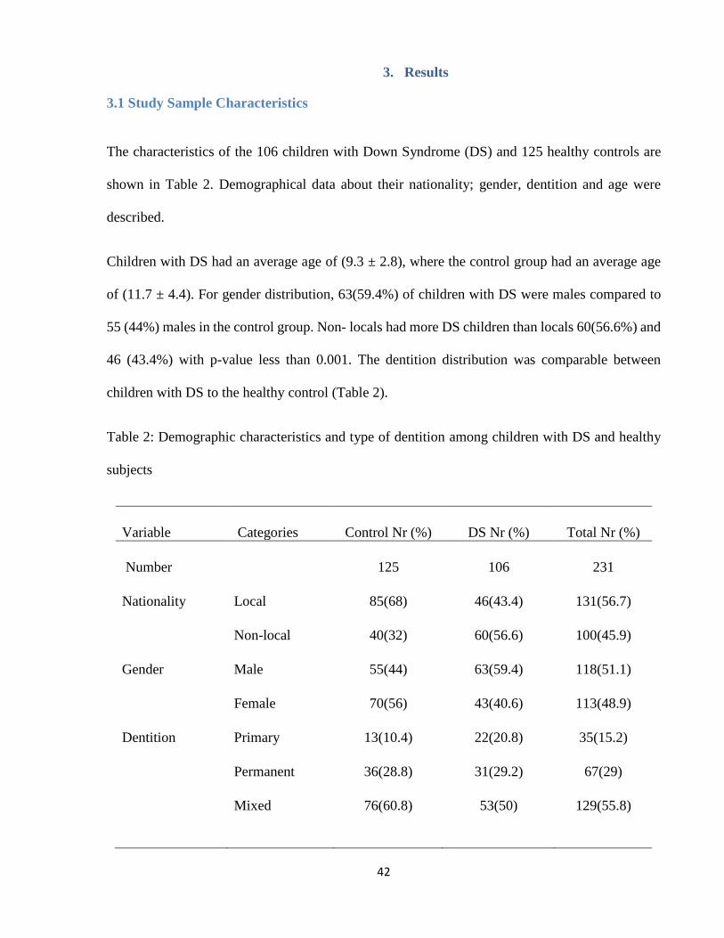
42
3. Results
3.1 Study Sample Characteristics
The characteristics of the 106 children with Down Syndrome (DS) and 125 healthy controls are
shown in Table 2. Demographical data about their nationality; gender, dentition and age were
described.
Children with DS had an average age of (9.3 ± 2.8), where the control group had an average age
of (11.7 ± 4.4). For gender distribution, 63(59.4%) of children with DS were males compared to
55 (44%) males in the control group. Non- locals had more DS children than locals 60(56.6%) and
46 (43.4%) with p-value less than 0.001. The dentition distribution was comparable between
children with DS to the healthy control (Table 2).
Table 2: Demographic characteristics and type of dentition among children with DS and healthy
subjects
Variable Categories Control Nr (%) DS Nr (%) Total Nr (%)
Number 125 106 231
Nationality Local 85(68) 46(43.4) 131(56.7)
Non-local 40(32) 60(56.6) 100(45.9)
Gender Male 55(44) 63(59.4) 118(51.1)
Female 70(56) 43(40.6)
113(48.9)
Dentition Primary 13(10.4) 22(20.8) 35(15.2)
Permanent 36(28.8) 31(29.2) 67(29)
Mixed 76(60.8) 53(50) 129(55.8)

43
3.2 Dental Caries
Prevalence
The overall occurrence of dental caries among children with DS and healthy controls were equal
as it was 57.6% (57/106) in the DS group where as for the healthy controls was 57.6% (72/125).
The prevalence of caries in the primary dentition among children with DS was 62.8% (49/78)
while for the healthy control it was 70.5% (62/88). On the same context the prevalence of caries
in the permanent dentition among children with DS was 28.6% (24/84) while for the healthy
controls was 17.9 (20/112) as shown in Figure 3.
Figure 3: Prevalence of caries in DS and control.
No Yes No Yes
Control DS
29.5
70.5
37.2
62.8
82.1
17.9
71.4
28.6
42.4
57.6
46.253.8
dmft DMFT Dmft/dmft

44
DMFT/dmft Caries Index
Tables 3 and 4 demonstrate the caries status of the sample population. The mean numbers of the
decayed component of DMFT in DS children was significantly higher than that in healthy children
(2.73 ± 0.22 vs. 1.65 ± 2.46, p value = 0.01). There was a clear statistical significant difference in
caries experience amongst DS children compared to the healthy controls as measured by
DMFT/dmft in all age groups which is shown in Tables 3 and 4. The mean number of DMFT in
DS children was significantly higher than that in healthy children (3.32 ± 4.62 vs. 2.16 ± 2.86, p
value= 0.02). The dmft scores were highest among the youngest age groups in DS with primary
dentition compared to their controls.
In regards to the treatment of the decayed teeth (fillings), Children in the DS group received more
treatment than their controls in all age groups; however, this was not shown to be statistically
significant.
The restorative care and the treatment received in both study groups had been represented by the
Restorative Index (RI) and the Met Need index (MNI) in Table 3 and 4. Looking at different age
and dentition groups, DS children in the mixed dentition had the least RI and MNI scores compared
to the primary dentition as shown in Table 4: A, B. Surprisingly, DS children in the primary
dentition group had the highest RI and MNI scores (RI= 27% and 40% respectively) as shown in
Table 4: A.

45
Table 3: Caries status, Restorative Index and Met Treatment Index in DS and control children
(Mean values in both permanent and mixed dentition)
Control DS
n = 112 n = 84 p-value
DMFT index 2.16 ± 2.89 3.32 ± 4.62 0.021
Decayed 1.65 ± 2.46 2.73 ± 0.22 0.018
Missing 0.29 ± 0.83 0.48 ± 1.62 0.269
Filled 0.22 ± 0.61 1.0 ± 0.41 0.076
Restorative Index (RI) 11.76% 26.81% -
Met Need Index (MNI) 23.6% 35.6% -
Table 4: Proportion of patients with decayed, missing, filled teeth along with mean dmft and
DMFT averages (± standard deviation) among children and adolescents in both healthy and DS
groups by type of dentition
(A) Primary Dentition
Control DS
n = 13 n = 22
dmft index 2.69 ± 3.04 4 ± 4.74
decayed 2.69 ± 3.33 3.09 ± 4.62
missing 0 0.91 ± 3.11
filled 0.07 ± 0.27 1.14 ± 0.64
Restorative Index (RI) 2.52% 27%
Met Need Index (MNI) 2.54% 40%

46
Table 4: Continued
(B) Mixed Dentition
Control DS
n = 76 n = 53
dmft index 2.78 ± 2.93 3.32 ± 3.97
decayed 2.05 ± 2.58 2.96 ± 3.96
missing 0.6 ± 1.36 0.58 ± 1.68
filled 0.31 ± 0.73 0.06 ± 0.23
Restorative Index (RI) 13.1% 2%
Met Need Index (MNI) 34% 18%
DMFT 0.22 ± 0.58 0.55 ± 1.07
Decayed 0.15 ± 0.56 0.28 ± 1
Missing 0.13 ± 0.14 0.08 ± 0.55
Filled 0.03 ± 0.16 0.04 ± 0.19
Restorative Index (RI) 16.7% 12.5%
Met Need Index (MNI) 51.6% 30%
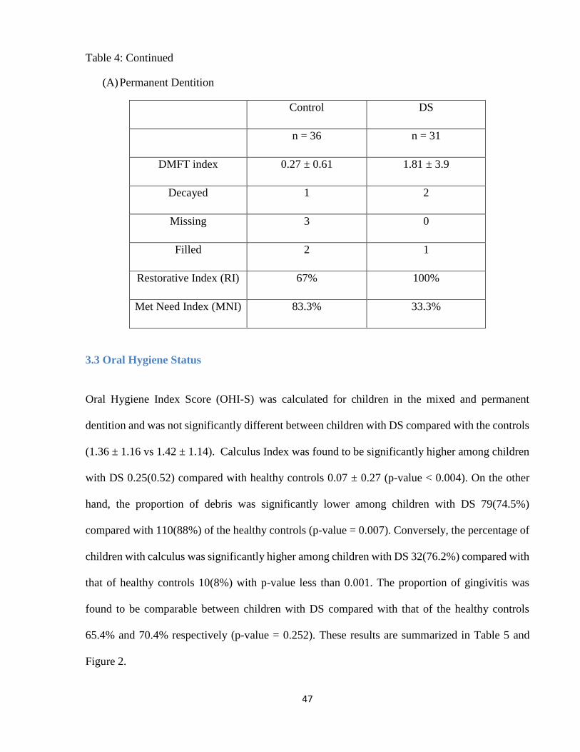
47
Table 4: Continued
(A) Permanent Dentition
Control DS
n = 36 n = 31
DMFT index 0.27 ± 0.61 1.81 ± 3.9
Decayed 1 2
Missing 3 0
Filled 2 1
Restorative Index (RI) 67% 100%
Met Need Index (MNI) 83.3% 33.3%
3.3 Oral Hygiene Status
Oral Hygiene Index Score (OHI-S) was calculated for children in the mixed and permanent
dentition and was not significantly different between children with DS compared with the controls
(1.36 ± 1.16 vs 1.42 ± 1.14). Calculus Index was found to be significantly higher among children
with DS 0.25(0.52) compared with healthy controls 0.07 ± 0.27 (p-value < 0.004). On the other
hand, the proportion of debris was significantly lower among children with DS 79(74.5%)
compared with 110(88%) of the healthy controls (p-value = 0.007). Conversely, the percentage of
children with calculus was significantly higher among children with DS 32(76.2%) compared with
that of healthy controls 10(8%) with p-value less than 0.001. The proportion of gingivitis was
found to be comparable between children with DS compared with that of the healthy controls
65.4% and 70.4% respectively (p-value = 0.252). These results are summarized in Table 5 and
Figure 2.
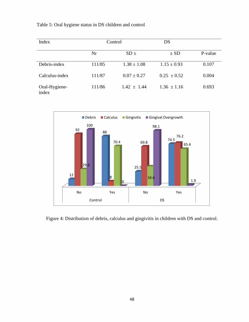
48
Table 5: Oral hygiene status in DS children and control
Index Control
DS
Nr SD ± ± SD P-value
Debris-index 111/85 1.38 ± 1.08 1.15 ± 0.93 0.107
Calculus-index 111/87 0.07 ± 0.27 0.25 ± 0.52 0.004
Oral-Hygiene-
index
111/86 1.42 ± 1.44 1.36 ± 1.16 0.693
Figure 4: Distribution of debris, calculus and gingivitis in children with DS and control.
No Yes No Yes
Control DS
12
88
25.5
74.5
92
8
69.8
76.2
29.6
70.4
34.6
65.4
100
0
98.1
1.9
Debris Calculus Gingivitis Gingival Overgrowth

49
3.4 Occlusal Anomalies
Open bite was more frequent among children with DS 42 (40%) compared with healthy controls,
14 (11.2%) and p-value was 0.001. There was a significant difference between the proportion of
deep bite among children with DS compared to the proportion among healthy controls 10 (9.4%)
and 23 (18.4%) respectively, p-value was 0.039. The proportions of posterior crossbite and scissor
bite were significantly higher among children with DS compared with healthy controls 52 (49.1%)
and 10 (9.5%) versus 35 (28%) and 3(2.4%) respectively (p-value < 0.02). In relation to spacing
between teeth, it was found that the proportion of anterior spacing was significantly higher among
children with DS 48(45.3%) compared with that of the healthy controls 40(32%) (P-value 0.026).
Consistently, the proportion of posterior spacing was significantly higher among children with DS
compared with that among healthy controls, 22(20.8%) and 10(8%) respectively (p-value = 0.004)
(Table 6).
Regarding molar Angle classification, the proportion of Class I was more likely among healthy
controls 68(54.4%) compared with children with DS 4(3.8%) as Class I. The same outcome applied
to Class II, as the proportion of Class II was lower among children with DS 7(6.6%) compared
with proportion of healthy controls with Class II, 28(22.4%). Conversely, proportion of Class III
was found higher among children with DS compared to the healthy controls 70(66%) and
14(11.2%) respectively (p-value < 0.001) (Figure 5). On the other side, primary molar relationship
in DS individuals were found to have higher occurrence of mesial step relationship compared to
the control group (13.3% vs. 4%).
The proportion of trauma was found to be significantly higher among children with DS 12(11.3%)
compared with that of healthy controls 4(3.2%) (P-value < 0.015). The mean value of positive

50
overjet between children with DS and controls was significantly lower 1.51 ± 1.59 mm and 3.39
± 2.2 mm respectively (p-value < 0.001) (Table 6).
Table 6: Occlusal anomalies and traumatic dental injuries in DS children and control
Anomalies Control DS
Nr (%) Nr (%) P-value
Vertical
open bite 14(11.2) 63(60) 0.001
deep bite 23(18.4) 96(90.6) 0.039
Transverse
crossbite 35(28) 54(50.9) 0.001
scissor bite 3(2.4) 95(90.5) 0.02
Spacing
anterior spacing 40(32) 58(54.7) 0.026
posterior spacing 10(8) 84(79.2) 0.004
Trauma 4(3.2) 94(88.7) 0.015
Figure 5: Distribution of molar Angle malocclusion classification in DS and control.
Class I Class II Class III
54.4
22
113.8 7
66
Control DS

51
3.5 Dentofacial Anomalies
Children with DS had shown higher proportion of high shovel shape incisors, high arched plate,
microdontia, nipple appearance of canine tip, Tongue thrust and lymphadenopathy compared with
that among healthy controls (Table 7). A single case of transposition of canine was found among
DS group. An example of the anomalies found in the study population are shown in Figure
6,7,8,9,10.
Table 7: Dentofacial anomalies in DS children and control
Anomalies Control DS
Nr (%) Nr (%) P-value
Shovel shape incisors 0 19(18.1) 0.001
High arched palate 42(33.6) 99(93.4) 0.001
Microdontia 1(0.8) 48(45.3) 0.001
Nipple appearance of canine 1(0.8) 40(37.7) 0.001
Tongue thrust 2(1.6) 37(34.9) 0.001
Lymphadenopathy 5(4) 17(16) 0.002

52
Figure 6: Transposition of upper canine.
Figure 7: Microdontia of upper second premolar.
Figure 8: Nipple appearance of the lower canine

53
Figure 9: (Left) Fissured Tongue, (Right) Gorlin Sign.
Figure 10: Peg shaped upper lateral incisors and clinically absent lower central incisors.

54
3.6 Oral Soft Tissues
In Table 8, children with DS had significantly higher proportion of all the conditions listed. The
controls scored zero for all the listed conditions.
Table 8: Oral soft tissues findings in DS children and control
Condition Control DS
Nr (%) Nr (%) P-value
Atrophy of tongue 0 8(7.5) 0.002
Median rhomboid
glossitis
0 0 -
Geographic tongue 0 9(8.5) 0.001
Fissure tongue 0 72(67.9) 0.001
Irritation fibroma 0 1(1) 0.454
Angular cheilitis 0 23(21.7) 0.001
Macroglossia 1(0.8) 49(46.2) 0.001
Ulcer 0 3(2.8) 0.096
Trauma to soft tissue/lip 0 3(2.8) 0.096
Drooling 0 25(23.6) 0.001
3.7 Medical Conditions
According to the medical history that was obtained from the children’s medical file in the schools,
DS children had variable conditions. The cardiac conditions were more common, where 24 % of
DS sample had a history of cardiac anomalies. The cardiac conditions varied from atrioventricular
septal defect, mitral valve cleft and patent ductus arteriosus. Other medical conditions reported in
the medical history were hypothyroidism (10%), epilepsy (5%) and recurrent ear infection (4%).
Also, there was a single case of thoracolumbar scoliosis among DS children.
3.8 Erosion

55
According to the pyramid graph (Figure 11), the severity of erosion was significantly higher among
DS children compared to healthy control (p-value=0.006). The proportion of DS children with
erosion was 34% vs. 15.3% in the control group. The percentage of DS children with erosion into
enamel only was 19.8 %( 21/106) compared to 11.3 %( 14/125) in the control group. The
percentage of erosion into enamel and dentine was 12.3% (13/106) in DS children vs. 4% (5/125)
in healthy children. In addition, the percentage of severe erosion, which is into enamel, dentine
and pulp, was 0% in the control group compared to 1.9% (2/106) in DS group.
Figure 11: Erosion severity in DS children and control group.
4. Discussion

56
As the life expectancy of DS individuals is improving and the prevalence is increasing 1, it is of a
great benefit to understand the oral health problems experienced by this group. Since there is no
central DS registry data in the UAE particularly in Dubai, an accurate estimate of the percentage
of DS is difficult. The only generated data suggests that DS incidence in Dubai was 1 in every 319
live birth among UAE nationals and 1 in 602 live births among non-nationals 12, which is the
highest in the Middle East region due to increased maternal age and consanguineous marriages.
This study provided an opportunity to assess the oral health problems among DS children in Dubai
who are enrolled in special centres.
The sample used in this study presented a fair distribution with respect to age and sex. The males
(63 out of 106, 59%) in the DS group were more than females (43 out of 106, 41%), which might
reflect the higher occurrence of DS in males as reported by a study conducted in Sharjah city and
other parts of the world 3,16,144. In regards to the geographic distribution, the special needs centres
were from different areas in Dubai and the control group were matched accordingly. This wide
distribution allowed us to cover Dubai as a whole city instead of a particular area. A sample size
calculation was done prior to data collection to make sure sufficient number of children was
included. This might have strengthened the validity of the results and made them applicable to the
neighbouring cities in the UAE. A total of 106 DS individuals along with 125 controls were
examined which exceeded the calculated sample size.
4.1 Dental Caries

57
This current study used the dmft/DMFT index to detect dental caries, according to WHO standards
136. Several studies stated that this method is efficient to detect dental cavities, but not the non-
cavitated lesions 96,145,146. Of course, the inclusion of non-cavitated lesions would give a better idea
of disease prevalence which means a better understanding of treatment needs 147. However, the
dmft/DMFT index was used in this study due to the large sample size and its objectivity.
Additionally, the WHO criteria of caries diagnosis 136 are still the standard in epidemiological
studies and is use allowed comparison of this study results with national and international studies.
Also, using radiographs to detect non-cavitated lesions for screening purposes would not be ethical
nor practical.
In this study, the mean number of the decayed component of DMFT in DS children was higher
than the value in healthy children (2.73 ± 0.22 vs. 1.65 ± 2.46, p value = 0.01). The mean number
of DMFT in DS children was significantly higher than that in healthy children (3.32 ± 4.62 vs.
2.16 ± 2.86, p value= 0.02). Additionally, the mean DMFT/dmft was found to be higher in DS
compared to healthy children in all age groups and dentition types especially in the primary
dentition (4 ± 4.74) and has a clear statistical significance. This study findings are consistent with
a previous study conducted in Sharjah 16. In Sharjah the mean number of the decayed component
of DMFT in DS patients was almost twice as high as that in healthy controls (13.2 ± 0.84 vs 7.4 ±
3.94), which is even higher than the finding in this current study.
The prevalence of caries in DS children varied in the literature and the findings are conflicting.
Some studies reported lower caries rate 120,121, while others reported either similar 124,125or higher
caries rate 16,17. An older study reported dramatically lower caries rate in DS individuals compared
to healthy children. This old study used institutionalized population whose diet was controlled and
probably low in cariogenic foods 121. Today, most DS children are raised at home and only attend
special needs schools and might have higher exposure to cariogenic foods. Also, lower caries rate

58
in DS children is explained to be as a result of several factors such as increased spacing between
teeth, delayed eruption of the teeth , possible different salivary chemical content, hypodontia,
microdontia and flatter teeth due to bruxism 8,24,45,56.
A high caries rate among DS children in Dubai is not surprizing because they seem to follow the
normal caries pattern reported by a recent dental survey among healthy children. The latter survey
in the U.A.E found that the prevalence of dental caries among healthy school children was 76.1%
and the average dmfs score was 10.2 148. This high prevalence of dental disease in U.A.E might be
attributed to cultural factors 149 such as strong family cohesion and the involvement of extended
family members in taking care of the child, high sugary diet and lack of dental visits 150. Likewise,
Studies conducted in neighbouring countries also reported high decay rate among DS children and
young adults 17,151,152.
The Met Need Index (MNI) and Restorative Index (RI) were calculated from the mean dmft/DMFT
of the studied DS sample and were high compared with healthy control subjects (Table 3). This
means that the restorative treatment needs of the studied DS children in the present study were
more addressed compared to the control group. These values were inconsistent with the results
found in Sharjah and other studies 16–18,126,151, as the MNI and RI were lower for the DS sample.
This difference could be because DS children in Dubai might have better access to dental clinics
than those in Sharjah. Also, The Ministry of Health in Dubai provides free dental treatment for
special needs children who are holders of a special needs medical card. Moreover, the children
recruited for this study are all in special need centres which might imply a high socio-economic
status of their parents making them more able to afford dental treatment. In addition, some
literature suggest that due to the nature of their complex medical condition, DS parents tend to be
more concerned about their children’s dental health and seek dental advice earlier 45. However,

59
the fact that we were unable to recruit DS children who are not in the special need centres might
bias this finding because we are not aware of their dental health status and treatment needs.
This study was able to demonstrate that despite the higher percentage of treatment received among
DS children; they still had a higher caries rate compared to healthy children. The author suggests
that a lot of factors might contribute to this including cultural habits, lack of parental awareness
on the importance of prevention and regular dental follow up, parental neglect, long waiting lists
for dental treatment especially in the government hospitals and lack of sufficient general
anaesthesia facilities and paediatric dental specialists often needed for the comprehensive
treatment of DS patients. Further studies into the aforementioned factors and their possible
contribution, is necessary in order to help shed some light on this dilemma.
4.2 Oral Hygiene Status
Since periodontal disease is a significant oral health problem in people with DS, it is very important
to understand the status of oral hygiene in this population. The proportion of debris was
significantly lower among children with DS 79(74.5%) compared with 110(88%) of the healthy
controls (p-value = 0.007). DS children have flatter occlusal surfaces, which facilitate oral hygiene
and self-cleaning, eliminating debris that serve as a substrate for oral bacteria 45. The DS group
attended special needs centres. As part of these centres’ usual routine, the caregivers either
supervise or carry out tooth brushing for the DS children. This might have contributed to the lower
debris index measured in the study group. On the other hand, Calculus Index was found to be
significantly higher among children with DS (0.25 ± 0.52) compared with healthy controls (0.07
± 0.27) (p-value < 0.004). The higher calculus in DS might be attributed to the high calcium content
in their saliva 2.

60
The proportion of gingivitis was found to be 65.4%, although less than the control group, gingivitis
was present in more than half of the DS sample. It has been reported that gingivitis in DS develops
more rapidly and is more extensive around deciduous teeth compared to healthy children 131. In
addition, DS subjects have other factors that aggravate the periodontal disease such as
abnormalities in host defence particularly leucocyte response, defective connective tissue and
altered vascularisation 132. Periodontal disease is of a great concern in DS subjects because it has
a progressive pattern and children can present with marginal gingivitis, gingival recession,
advanced periodontitis and pocket formation. Pocket formation has been reported in 36% of DS
children below the age of 6 years 129.
The hallmark of managing periodontal disease in DS subjects is prevention. A comprehensive
preventive dental program is needed to promote better oral hygiene, prevent the development of
periodontal disease and halt its progression. DS children must be always screened for periodontal
disease and early, aggressive treatment is needed. They may be needed to be seen more often for
scaling and root planning.
4.3 Occlusal Anomalies
DS children have unique occlusal and dentofacial anomalies that are present frequently and might
lead to improper functioning and ads up to the complexity of their condition. In the present study,
DS subjects had significantly higher proportion of open bite (40% vs. 11.2%), crossbite (42% vs.
28%), scissor bite (9.5% vs. 2.4%), anterior spacing (45.3% vs 32%) and posterior spacing (20.8%
vs. 8%). These findings are confirmed by another study, where Soares et al found an incidence of
39% posterior crossbite and 26% anterior crossbite in DS patients 45. More to add, the DS sample
studied in Sharjah had similar findings 16.

61
It is well nestablished that Class III malocclusion is more common in DS children, due to
underdevelopment of the midface and mandibular prognathism 45,99. This typical malocclusion was
confirmed in our study, where DS subjects had significantly higher Class III malocclusion (66%)
compared to controls (11.2%). Additionally, Primary molar relationship in DS individuals was
found to have higher occurrence of mesial step relationship compared to the control group (13.3%
vs. 4%). Due to the small number of subjects with primary dentition, no statistical significance
was found in the latter finding. This mesial step relationship explains the Class III malocclusion
later on in DS children, as mesial step relationship may progress to a Class III during the molar
transition with continued mandibular growth 153.
4.4 Dentofacial Anomalies
In this study, DS individuals had remarkably increased frequencies of shovel shape incisors, high
arched palate, microdontia, nipple appearance of canine tip, tongue thrust and lymphadenopathy
compared to controls. These findings were also reported in the literature along with other findings
such as dentinogenesis imperfecta, taurodontia, peg shaped teeth, impacted teeth, dens evaginatus
and talon cusp 16,45,89.
Other dental anomalies reported in DS individuals are hypodontia, delayed eruption,
supernumerary teeth and ectopic eruption. Unfortunately, these anomalies were not investigated
in this study as radiographs are needed to confirm their presence or absence. Hypodontia
prevalence is reported to be around 60% in DS children 103,104. Third molar agenesis was found to
be around 4 times greater in DS than controls 154. The latter is followed by mandibular central
incisors, maxillary lateral incisors, maxillary second premolars and mandibular second molars 102.
The pattern of hypodontia in DS is thought to be associated with peripheral nervous system
abnormalities and abnormal cartilaginous tissues 102,154.

62
4.5 Oral Soft Tissues
DS sample in this study had significantly higher proportion of geographic tongue, atrophy of the
tongue, fissure tongue, irritation fibroma, angular cheilitis, macroglossia, ulcers, trauma to soft
tissues and drooling compared to healthy controls. Fissured tongue was found in 67% of DS
children in this study. Fissured tongue is a non-pathological variation of the normal tongue and
often seen in DS individual. Fissured tongue is asymptomatic , often associated with geographic
tongue and the only clinical relevance it plays is that it acts as bacterial reservoir and causes
glossitis 94. However, Desai reported that expressive language of children with DS is commonly
more delayed than receptive language which might be due to several factors including the
relatively large tongue in a small oral cavity 3.
Other findings related to the tongue in DS individuals is relative macroglossia, where the tongue
gives the impression of being large due to muscle weakness and low position in the mouth 45,76.
Macroglossia within DS subjects in this study is found in 46.2%, which is similar to the incidence
in other studies (11-60%) 16,45,76.
4.6 Medical Conditions
It is very important to note the medical conditions in DS individuals especially those that may
impinge on oral care such as congenital heart defects, epilepsy and cervical spine abnormalities.
Congenital heart defects are very frequent in DS individuals, they occur in 40-60% of all DS
children 44.The percentage of DS children with cardiac conditions in our sample was 24%. The
dentist must always have an up to date cardiac status and follow the most current subacute bacterial
endocarditis antimicrobial prophylaxis protocol prior to any procedure 47.

63
Other conditions are also found in the study DS subjects such as hypothyroidism (10%), recurrent
ear infection (4%) and one case of thoracolumbar scoliosis. Hypothyrodism is reported in DS
children and is associated with underdevelopment of the bones and teeth as well as delayed tooth
eruption 45,69. Ear-nose-throat (ENT) infections are frequent in DS children and some factors such
as immune defects, altered anatomy, hypotonia and macroglossia play an important role in these
infections 64,65. Epilepsy rates in DS were reported to be from 1 to 13% 71 . In this study 5% of DS
children were epileptic. The dental team must be trained and ready to handle seizure attacks and
be aware of some antiepileptic drugs side effects on the gingiva such as Phenytoin 24,73. Not to
forget, DS individuals have a number of musculoskeletal abnormalities due to defective collagen
structure that leads to increased ligamentous laxity. The major concern to the dentist is the effect
on cervical spine as the incidence of atlanto-axial joint subluxation was reported to be 20% in DS
individuals. However, other spinal deformities were reported in the thoracic area and were known
to occur after an early surgery for heart disease 155.
4.7 Erosion
Dental erosion is a multifactorial condition that occurs due to the interaction of chemical,
biological and behavioural factors 156. Tooth wear as a result of acidic and chemical insult to the
teeth are commonly noticed in DS children 53. Erosion in DS children is related to the fact that
13.8% to 59% of DS children suffer from gastric dysfunction such as gastroesophageal reflux and
vomiting 53,108,109. In This study, the severity of erosion was significantly higher among DS
children compared to healthy control (p-value=0.006).The proportion of DS children with erosion
was 34% vs 15.3% in the control group. Another study also found that erosion was significantly
higher in DS individuals than the normal population 107.
Erosion is often associated with other forms of tooth wear such as attrition and abrasion, hence,

64
the term ‘tooth wear’ is used to reflect all three conditions (erosion, attrition and abrasion) 157.
The combination of this triad (erosion, attrition and abrasion) makes the diagnosis of dental
erosion more difficult, therefore careful history taking is important 158
4.8 Study limitations
As with every study, perfection is desired, however, there are always obstacles and challenges at
the time of conducting a study. The author believes that pointing out the study limitations is a good
practice in order to understand the overall outcome and come up with better ways to overcome
these challenges in future researches. The limitations in this current study are as follow:
As mentioned previously, there is no central DS registry data in the United Arab Emirates,
hence only DS children in special need schools and centres were invited to participate in
the study. DS children who are raised at home are out of reach and cannot be tracked. This
might had affected the results.
The study population was all from Dubai city. It would have been beneficial if DS children
from all around UAE participated but this was unachievable due to time limitation, number
of researchers examining the children and facilities to accommodate the large number of
participants.
Since periodontal disease is a major concern in DS individuals, it would have been
important to include other periodontal examination alongside the Oral Hygiene Index.
Basic Periodontal Examination BPE 159 which includes probing index teeth would have
been a good method however, due to the difficulties in examining some children this was
not practical. Also, a high proportion of these children might have cardiac conditions and
might be at risk of infective endocarditis; hence, a cardiologist consultation and

65
antimicrobial prophylaxis (American Heart Association guidelines are followed in the
UAE) might be needed prior to probing.
5. Conclusions and Rrecommendations
This current study had concluded that DS children in Dubai had higher caries rate
compared to healthy children.

66
The youngest age group among DS children with primary dentition had the highest dmft
score compared to the rest.
Despite the high caries rate among DS subjects, they received more restorations and dental
treatment compared to the control, which suggests that DS children had better access to
dental care than their control.
Additionally, DS children had significantly more calculus than healthy children.
DS population had similar occlusal anomalies to DS subjects worldwide. They had
significantly higher proportion of open bite, crossbite, scissor bite, anterior spacing, and
posterior spacing. In addition, they had more Class III molar relationship compared to the
control.
Other dentofacial anomalies that were more frequent in DS children were shovel shaped
incisors, high arched palate, microdontia, hypodontia, nipple appearance of canine tip,
tongue thrust, macroglossia and lymphadenopathy.
Soft tissue findings in the DS group revealed higher frequencies of atrophy of the tongue,
geographic tongue, fissured tongue, irritation fibroma, angular cheilitis, ulcers, trauma to
soft tissue/lip and frequent drooling.
The severity of erosion was significantly higher among DS children compared to healthy
control.
As with any DS subject, our group had several medical conditions that are of a great
concern to the dentist such as cardiac conditions, hypothyroidism, epilepsy, recurrent ear
infections and spinal deformities.
Looking at the outcome of this study, the following recommendations are suggested for future
research:
67
To present the findings of this study to the newly restructured Federal Ministry of Health
and Community Prevention.
To investigate the health care system provided for DS children including general
anaesthesia facilities, dental appointments, dental follow ups and waiting lists.
To focus on parental awareness programs that stress the importance of oral health of special
needs children.
To establish proper prevention and community oral health care programs that target special
needs children in Dubai.
To suggest establishing data registry for DS children in the United Arab Emirates. This
will help in epidemiological studies and the provision of comprehensive oral healthcare for
these children.
To conduct a similar study to include all DS children in The United Arab Emirates to have
a better understanding of their oral health and treatment needs.
6. Bibliography
1. Frydman A, Nowzari H. Down syndrome-associated periodontitis: a critical review of the

68
literature. Compend Contin Educ Dent. 2012;33(5):356–61.
2. Davidovich E, Aframian DJ, Shapira J, Peretz B. A comparison of the sialochemistry, oral pH, and
oral health status of Down syndrome children to healthy children. Int J Paediatr Dent.
2010;20(4):235–41.
3. Desai SS. Down syndrome: a review of the literature. Oral Surg Oral Med Oral Pathol Oral Radiol
Endod. 1997;84(3):279–85.
4. Prakash A, Raghuwanshi B, Hameed A. Down syndrome- diagnosis and guidelines of dental and
orthodontic management. orthodontics. 2013;40–3.
5. Cohen I. Health care guidelines for individuals with Down syndrome. Down Syndr Q. 1996;1(2).
6. Khocht A, Janal M, Turner B. Periodontal health in down syndrome: Contributions of mental
disability, personal, and professional dental care. Spec Care Dent. 2010;30(3):118–23.
7. Pilcher E. Dental Care for the Patient with Down Syndrome. Down Syndr Res Pract [Internet].
1998;5(3):111–6. Available from: http://www.down-syndrome.org/reviews/84/
8. Areias CM, Sampaio-Maia B, Guimaraes H, Melo P, Andrade D. Caries in Portuguese children
with Down syndrome. Clinics. 2011;66(7):1183–6.
9. Parker SE, Mai CT, Canfield M a., Rickard R, Wang Y, Meyer RE, et al. Updated national birth
prevalence estimates for selected birth defects in the United States, 2004-2006. Birth Defects Res
Part A - Clin Mol Teratol. 2010;88(12):1008–16.
10. Niazi MA, Al-Mazyad AS, Al-Husain MA, Al-Mofada SM, Al-Zamil FA KT. Down’s syndrome
in Saudi Arabia: incidence and cytogenetics. Hum Hered. 1995;45(2):65–9.
11. Isaac GS, Krishnamurty PS, Reddy YR AY. Down’s syndrome in Hyderabad, India.
ActaAnthropogenet. 1985;9(4):256–60.
12. Murthy SK, Malhotra AK, Mani S, Shara ME a, Al-Rowaished EEM, Naveed S, et al. Incidence
of Down syndrome in Dubai, UAE. Med Princ Pract. 2006;16(1):25–8.
13. Alsakarna BK OE. Dentofacial Changes and Oral Health Status in Individuals with Down
Syndrome in Jordan - Cross Sectional Study. Pakistan Oral Dent J. 2010;30(1):159–61.
14. Booth KVP. Health disparities and intellectual disabilities: Lessons from individuals with down
syndrome. Dev Disabil Res Rev. 2011;17:32–5.
15. Hashemi Z, Hajizamani A BE. Oral Health Status of a Sample of Disabled Population in Iran. Oral
Heal Oral Epidemiol. 2012;1(1):23–8.
16. Jaber MA. Oral Health Condition and Treatment Needs of a Group of UAE Children with Down
Syndrome. Ibnosina J Med Biomed Sci. 2010;2(2):62–71.
17. Al-maweri S, Al-sufyani G. Dental caries and treatment needs of Yemeni children with down
syndrome. Dent Res J (Isfahan). 2013;11(6):631–6.
18. Jain M. A Paired Comparison among Siblings of Common Parents About Dental Care with and
without Down Syndrome. Pesqui Bras Odontopediatria Clin Integr. 2010;10(1):9–14.
19. U.S. Department of Health and Human Services. Oral Health in America: A Report of the Surgeon
General. US Dep Heal Hum Serv Natl Inst Dent Craniofacial Res Natl Institutes Heal. 2000;

69
20. Alkhadra TA. Prevalence of Dental Caries and Oral Hygiene Status among Down’s Syndrome
Patients in Riyadh. Pakistan Oral Dent J. 2011;31(1):113–5.
21. Zacharias M, Np C, Gp H, Sivalingam P, Rj W, Carson N. Anesthetic Management in a Down
Syndrome Patient. Int Student Iournal Nurse Anesth. 2010;18(1):19–23.
22. Lee C, Luginbuehl I, Bissonnette B ML. Pediatric disorders. Anesthesia and Co-Existing Disease.
5th ed. Stoelting R DS, editor. Philadelphia: Churchill Livingstone; 2008. 611-612 p.
23. Källén B. Epidemiology of Human Congenital Malformations. Vol. 189, Journal of anatomy.
2014. 45-60 p.
24. Fiske J, Shafik HH. Down’s syndrome and oral care. Dent Update. 2001;28(3):148–56.
25. Down JL. Observations on an ethnic classification of idiots. 1866. Ment Retard. 1995;33(1):54–6.
26. What is Down Syndrome? Natl Down Syndr Soc [Internet]. 2015; Available from:
http://www.ndss.org/Down-Syndrome/What-Is-Down-Syndrome/
27. NHS. Down’s syndrome - Diagnosis. UK Natl Heal Serv [Internet]. 2015; Available from:
http://www.nhs.uk/Conditions/downs-syndrome/Pages/diagnosis.aspx
28. Morris JK, Alberman E. Trends in Down’s syndrome live births and antenatal diagnoses in
England and Wales from 1989 to 2008: analysis of data from the National Down Syndrome
Cytogenetic Register. Bmj. 2009;339(3):3794–3794.
29. Lissauer T, Clayden G. Illustrated Textbook of Paediatrics. London: Mosby; 1998.
30. Cocchi G, Gualdi S, Bower C, Halliday J, Jonsson B, Myrelid A, et al. International trends of
Down syndrome 1993-2004: Births in relation to maternal age and terminations of pregnancies.
Birth Defects Res A Clin Mol Teratol. 2010;88(6):474–9.
31. Soediono B. Sleep Disordered Breathing in Children. Vol. 53, Journal of Chemical Information
and Modeling. 1989. 160 p.
32. Saperstein A, Nicholas S. Pediatric and adolescent sports medicine. Vol. 43, Pediatric clinics of
North America. 1996. 1013-1033 p.
33. Weijerman ME, de Winter JP. Clinical practice. The care of children with Down syndrome. Eur J
Pediatr. 2010;169(12):1445–52.
34. Kallen K. Down’s syndrome and maternal smoking in early pregnancy. Genet Epidemiol.
1997;14(1):77–84.
35. Blomberg MI, Källén B. Maternal obesity and morbid obesity: The risk for birth defects in the
offspring. Birth Defects Res Part A - Clin Mol Teratol. 2010;88(August 2009):35–40.
36. Gustafsson J, Anneren G, Ericsson UB, Svanberg L IS. Thyroid antibodies are not a risk factor for
pregnancies with Down syndrome. Prenat Diagn. 1995;15:451–4.
37. Vassy C. From a genetic innovation to mass health programmes: the diffusion of Down’s
Syndrome prenatal screening and diagnostic techniques in France. Soc Sci Med. 2006;63(8):2041–
51.
38. Reddy UM, Mennuti MT. Incorporating first-trimester Down syndrome studies into prenatal
screening: executive summary of the National Institute of Child Health and Human Development
workshop. Obstet Gynecol. 2006;107(1):167–73.

70
39. Malone FD, Canick JA, Ball RH, Nyberg DA, Comstock CH, Bukowski R, et al. First-Trimester
or Second-Trimester Screening, or Both, for Down’s Syndrome. N Engl J Med.
2005;353(19):2001–11.
40. American College of Obstetricians and Gynecologists. ACOG Practice Bulletin No. 27: Clinical
Management Guidelines for Obstetrician-Gynecologists. Prenatal diagnosis of fetal chromosomal
abnormalities. Obs Gynecol. 2001;97(5-1):1–12.
41. Skotko BG. Prenatally diagnosed Down syndrome: Mothers who continued their pregnancies
evaluate their health care providers. Am J Obstet Gynecol. 2005;192(3):670–7.
42. pueschel S. Ethical issues pretaining to prenatal diagnosis of Down syndrome, in Down syndrome:
A review of current knowledge. Rondal J NL, editor. London: Whorr Publishers Ltd.; 1999. 170-
177 p.
43. Siffel C, Correa A, Cragan J, Alverson CJ. Prenatal diagnosis, pregnancy terminations and
prevalence of down syndrome in Atlanta. Birth Defects Res Part A - Clin Mol Teratol.
2004;70:565–71.
44. Charleton PM, Dennis J, Marder E. Medical management of children with Down syndrome.
Paediatr Child Health (Oxford). 2014;24(8):362–9.
45. Macho V, Coelho A, Areias C, Macedo P, Andrade D. Craniofacial features and specific oral
characteristics of Down syndrome children. Oral Health Dent Manag. 2014;13(2):408–11.
46. Freeman SB, Taft LF, Dooley KJ, Allran K, Sherman SL, Hassold TJ, et al. Population-based
study of congenital heart defects in Down syndrome. Am J Med Genet. 1998;80(3):213–7.
47. Nice. Prophylaxis against infective endocarditis NG64. Evid Based Dent. 2008;9(2):37–8.
48. American Heart Association. What Is Infective Endocarditis ? Am Hear Assoc. 2015;1.
49. Bull M. American Academy of Pediatrics: Health supervision for children with Down syndrome.
Pediatrics. 2001;107(2):442–9.
50. Brockmeyer D. Down syndrome and craniovertebral instability. Topic review and treatment
recommendations. Pediatr Neurosurg. 1999;31(2):71–7.
51. Article O. Dentofacial Anomalies and Oral Hygiene Status in Mentally Challenged Children : A
Survey. J indian Acad Oral Med Radiol. 2011;23(March):51–3.
52. Odeh M, Hershkovits M, Bornstein J, Loberant N, Blumenthal M, Ophir E. Congenital absence of
salivary glands in Down syndrome. Arch Dis Child. 2013;98(10):781–3.
53. Abanto J, Ciamponi AL, Francischini E, Murakami C, de Rezende NPM, Gallottini M. Medical
problems and oral care of patients with Down syndrome: a literature review. Spec Care Dentist.
2011;31(6):197–203.
54. Cremers MJG, Beijer HJM. No relation between general laxity and atlantoaxial instability in
children with Down syndrome. J Pediatr Orthop. 1993;(3):318–21.
55. Rogers PT CM. Medical Care in Down Syndrome: A Preventative Medicine Approach. New
York: Marcel Dekker Inc; 1993.
56. Määttä T, Tervo-Määttä T, Taanila A, Kaski M, Iivanainen M. Mental health, behaviour and
intellectual abilities of people with Down syndrome. Down Syndr Res Pract. 2006;11(1):37–43.

71
57. Burke SW, French HG, Roberts JM, Johnston CE, Whitecloud III TS, Edmunds Jr. JO. Chronic
atlanto-axial instability in Down syndrome. J Bone Jt Surg - Am Vol. 1985;67(9):1356–60.
58. Mitchell V, Howard R, Facer E. Down’s syndrome and anaesthesia. Paediatr Anaesth.
1995;5(6):379–84.
59. Unkel JH, Edwards JS, Piscitelli WP, Tye GW. Dental surgery and anesthetic precautions of a
patient with Down syndrome and juvenile rheumatoid arthritis: a case report. Pediatr Dent.
2012;34(7):517–20.
60. Caird MS, Wills BPD, Dormans JP. Down syndrome in children: the role of the orthopaedic
surgeon. J Am Acad Orthop Surg. 2006;14:610–9.
61. Mik G, Gholve P a, Scher DM, Widmann RF, Green DW. Down syndrome: orthopedic issues.
Curr Opin Pediatr. 2008;20(1):30–6.
62. Birrer RB. The Special Olympics athlete: evaluation and clearance for participation. Clin Pediatr.
2004;43(9):777–82.
63. Davidson M a. Primary Care for Children and Adolescents with Down Syndrome. Pediatr Clin
North Am. 2008;55(5):1099–111.
64. Kusters M, Verstegen RHJ, Gemen EFA, De Vries E. Intrinsic defect of the immune system in
children with Down syndrome: A review. Clin Exp Immunol. 2009;156(2):189–93.
65. Liston A, Enders A, Siggs OM. Unravelling the association of partial T-cell immunodeficiency
and immune dysregulation. Nat Rev Immunol. 2008;8(7):545–58.
66. Cunningham-Rundles C. Autoimmune manifestations in common variable immunodeficiency. J
Clin Immunol. 2008;28 Suppl 1:S42–5.
67. Contestabile A, Benfenati F, Gasparini L. Communication breaks-Down: From neurodevelopment
defects to cognitive disabilities in Down syndrome. Prog Neurobiol. 2010;91(1):1–22.
68. Noble SE, Leyland K, Findlay CA, Clark CE, Redfern J, Mackenzie JM, et al. School based
screening for hypothyroidism in Down’s syndrome by dried blood spot TSH measurement. Arch
Dis Child. 2000;82(1):27–31.
69. Thiel R, Fowkes SW. Down syndrome and thyroid dysfunction: should nutritional support be the
first-line treatment? Med Hypotheses. 2007;69(4):809–15.
70. Corbett J a. Retardation and developmental disabilities. Behav Res Ther. 1974;12(2):362.
71. Hayashi M, Iwamoto H, Suzuki H KY. Epilepsy in childhood Down syndrome. Brain Dev.
1984;6(1):37–44.
72. Pueschel SM, Louis S, McKnight P. Seizure disorders in Down syndrome. Arch Neurol.
1991;48(1825777):318–20.
73. Scully C, Cawson R. Medical problems in Dentistry. 4th ed. Oxford: Wright; 1998.
74. Alió J, Lorenzo J, Iglesias MC, Manso FJ, Ramíreze EM. Longitudinal maxillary growth in Down
syndrome patients. Angle Orthod. 2011;81:253–9.
75. van Gameren-Oosterom HBM, Fekkes M, Buitendijk SE, Mohangoo AD, Bruil J, Van Wouwe JP.
Development, problem behavior, and quality of life in a population based sample of eight-year-old
children with Down syndrome. PLoS One. 2011;6(7):218.

72
76. Oliveira ACB, Paiva SM, Campos MR, Czeresnia D. Factors associated with malocclusions in
children and adolescents with Down syndrome. Am J Orthod Dentofac Orthop. 2008;133(4):1–8.
77. Tierney NM, Pollard BJ, Doran BR. Obstructive sleep apnoea. Anaesthesia. 1989;44(3):235–7.
78. Fitzgerald D a, Paul A, Richmond C. Severity of obstructive apnoea in children with Down
syndrome who snore. Arch Dis Child. 2007;92(5):423–5.
79. Uong EC, McDonough JM T-KC. Magnetic resonance imaging of the upper airway in children
with Down syndrome. Am J Respir Crit Care Med. 2001;163:731–6.
80. Prabhat K, Verma S, Maheshwari S, Sharma N. Role of oral health professional in pediatric
obstructive sleep apnea. Natl J Maxillofac Surg. 2010;1(1):35.
81. Fidler DJ. The Emerging Down Syndrome Behavioral Phenotype in Early Childhood. Infants
Young Child. 2005;18(2):86–103.
82. Newton T. Dentist/patient communication: A review. Dent Updat. 1995;22:118–22.
83. Down’s Syndrome: Your Questions Answered [Internet]. Down’s Syndrome Association. 2000.
Available from: https://www.downs-syndrome.org.uk
84. Wilson MD. Special considerations for patients with Down syndrome. Okla Dent Assoc J.
1994;184:24–5.
85. Priosti PA, Blascovi-assis SM, Cymrot R, Vianna DL, Caromano FA. Grip strength and manual
dexterity in Down Syndrome children. 2013;278–85.
86. Oredugba F a. Oral health condition and treatment needs of a group of Nigerian individuals with
Down syndrome. Downs Syndr Res Pract. 2007;12(1):72–6.
87. Sdcep. Prevention and Management of Dental Caries in Children. Scottish Dent Clin Eff Program
[Internet]. 2010;(April). Available from: www.sdcep.org.uk/index.aspx?o=2866?
88. Jesuino FAS, Valladares-Neto J. Craniofacial morphological differences between down syndrome
and maxillary defi ciency children. Eur J Orthod. 2013;35(1):124–30.
89. Sekerci AE, Cantekin K, Aydinbelge M. Prevalence of Dental Anomalies in the Permanent
Dentition of Children with Down Syndrome. J Dent Child. 2014;81(2):78–83.
90. Alan M, Bittencourt V. Dental and skeletal characteristics of patients with Down Syndrome. Rev
Gauch Odontol. 2013;61(1):121–6.
91. Faulks D, Colladc V, Mazille M, Veyrune J, Hennequin M. Masticatory dysfunction in persons
with Down’s syndrome. Part 1: aetiology and incidence. J Oral Rehabil. 2008;35(11):854–62.
92. Weiss L, Chen LT. A scanning electron microscopic study of fissured tongue. Blood.
1974;43(5):665–91.
93. Pindborg JJ. Atlas of Diseases of the Oral Mucosa. 2nd ed. Copenhagen: Munksgaard; 1973. 176
p.
94. Goswami M, Verma A, Verma M. Benign migratory glossitis with fissured tongue. J Indian Soc
Pedod Prev Dent. 2012;30(2):173–5.
95. Gise E, Lange L NC. Tongue movements in 4 and 5 year old Down’s syndrome children during
eating: a comparison ith normal children. Am J Occup Ther. 1984;38:660–5.

73
96. Hennequin M, Allison PJ, Veyrune JL. Prevalence of oral health problems in a group of
individuals with Down syndrome in France. Dev Med Child Neurol. 2000;42:691–8.
97. Carlstedt K, Henningsson G, McAllister A, Dahllöf G. Long-term effects of palatal plate therapy
on oral motor function in children with Down syndrome evaluated by video registration. Acta
Odontol Scand. 2001;59(2):63–8.
98. Carlstedt K, Henningsson G, Dahllöf G. A four-year longitudinal study of palatal plate therapy in
children with Down syndrome: effects on oral motor function, articulation and communication
preferences. Acta Odontol Scand. 2003;61(1):39–46.
99. Musich DR. Orthodontic intervention and patients with Down syndrome. Angle Orthod.
2006;76:734–5.
100. Westerman GH, Johnson R, Cohen MM. Variations of Palatal Dimensions in Patients with
Down’s Syndrome. J Dent Res. 1974;54(4):767–72.
101. de Moraes MEL, de Moraes LC, Dotto GN, Dotto PP, dos Santos LRDA. Dental anomalies in
patients with down syndrome. Braz Dent J. 2007;18(4):346–50.
102. Bjorn GR IK. Tooth agenesis in Down syndrome. Am J Med Cener. 1995;55:466–71.
103. Suri S, Tompson BD, Atenafu E. Prevalence and patterns of permanent tooth agenesis in Down
syndrome and their association with craniofacial morphology. Angle Orthod. 2011;81(2):260–9.
104. Acerbi A. Prevalence of numeric anomalies in the permanent dentition of patients with Down
syndrome. Spec Care Dent. 2001;75–8.
105. Levander E, Malmgren O. Evaluation of the risk of root resorption during orthodontic treatment:
A study of upper incisors. Eur J Orthod. 1988;10(1):30–8.
106. Miamoto CB, Pereira LJ, Ramos-Jorge ML, Marques LS. Prevalence and predictive factors of
sleep bruxism in children with and without cognitive impairment. Braz Oral Res. 2011;25(5):439–
45.
107. Bell EJ, Kaidonis J, Townsend GC. Tooth wear in children with Down syndrome. Aust Dent J.
2002;47(1):30–5.
108. Mitchell RB, Call E, Kelly J. Ear, nose and throat disorders in children with Down syndrome.
Laryngoscope. 2003;113(2):259–63.
109. Zárate N, Mearin F, Hidalgo A, Malagelada JR. Prospective evaluation of esophageal motor
dysfunction in Down’s syndrome. Am J Gastroenterol. 2001;96(6):1718–24.
110. Hurlbutt M. Dental Caries: A ph-mediated disease. 2010;9–15.
111. barrett W. The septic theory of dental caries. Am Dent Assoc Trans. 1885;24:204–12.
112. Holst D. Causes and prevention of dental caries: a perspective on cases and incidence. Oral Health
Prev Dent. 2005;3(1):9–14.
113. Keyes PH. Recent advances in dental caries research. Bacteriology. 1962;12:43–64.
114. Marsh PD. Are dental diseases examples of ecological catastrophes? Microbiology.
2003;149(2):279–94.
115. Bradshaw DJ, Lynch RJM. Diet and the microbial aetiology of dental caries: new paradigms. Int

74
Dent J. 2013;63 Suppl 2:64–72.
116. Bradshaw DJ, McKee a S, Marsh PD. Effects of carbohydrate pulses and pH on population shifts
within oral microbial communities in vitro. J Dent Res. 1989;68(9):1298–302.
117. Anderson C a., Curzon MEJ, Van Loveren C, Tatsi C, Duggal MS. Sucrose and dental caries: A
review of the evidence. Obes Rev. 2009;10(SUPPL. 1):41–54.
118. Animireddy D, Reddy Bekkem VT, Vallala P, Kotha SB, Ankireddy S, Mohammad N. Evaluation
of pH, buffering capacity, viscosity and flow rate levels of saliva in caries-free, minimal caries and
nursing caries children: An in vivo study. Contemp Clin Dent. 2014;5(3):324–8.
119. Siqueira WL, Bermejo PR, Mustacchi Z, Nicolau J. Buffer capacity, pH, and flow rate in saliva of
children aged 2-60 months with Down syndrome. Clin Oral Investig. 2005;9(1):26–9.
120. Macho V, Palha M, Macedo AP, Ribeiro O, Andrade C. Comparative study between dental caries
prevalence of Down syndrome children and their siblings. Spec Care Dentist. 2013;33(1):2–7.
121. Creighton WE, Wells HB, Carie EDE, In D, Juveniles I, In MEN, et al. Dental Caries Experience
in Institutionalized Mongoloid and Nonmongoloid Children in North Carolina and Oregon. J Dent
Res. 1964;45(1):66–75.
122. Shapira J SA. Caries levels, strep mutans counts, salivary ph and periodontal treatment needs of
adult Down syndrome patients. Spec Care Dent. 1991;11(6):248–51.
123. Areias C, Sampaio-Maia B, Macho V, Leal I, Melo P, de Andrade C. Does the chemistry in the
saliva of Down syndrome children explain their low caries prevalence? Eur J Paediatr Dent.
2013;14(1):23–6.
124. Fung K, Allison PJ. A comparison of caries rates in non-institutionalized individuals with and
without Down syndrome. Spec Care Dentist. 2005;25(6):302–10.
125. Guaré RDO, Ciamponi AL, Romano MM. Behavioral and physiological changes in children with
Down syndrome using mechanical and chemomechanical (Carisolv) caries removal methods. Spec
Care Dentist. 2008;28(5):195–200.
126. Fung K, Lawrence H, Allison P. A paired analysis of correlates of dental restorative care in
siblings with and without Down syndrome. Spec Care Dentist. 2008;28(3):85–91.
127. Randell DM, Harth S, Seow WK. Preventive dental health practices of non-institutionalized Down
syndrome children: a controlled study. J Clin Pediatr Dent. 1992;16(3):225–9.
128. Adams DA, Barrington EP, Caton J, Genco RJ, Goodman SF, Hildebrand CN, et al. Parameters of
Care Supplement. J Periodontol. 2000;71(5):847–83.
129. Reuland-Bosma W, Dijk J. Periodontal disease in Down’s syndrome: a review. J Clin Periodontol.
1986;13(1):64–73.
130. Barnett ML, Press KP, Friedman D, Sonnenberg EM. The prevalence of periodontitis and dental
caries in a Down’s syndrome population. J Periodontol. 1986;57(5):288–93.
131. Reuland-Bosma W, van Dijk J, van der Weele L. Experimental gingivitis around deciduous teeth
in children with Down’s syndrome. J Clin Periodontol. 1986;13(4):294–300.
132. Reuland-Bosma W, Liem RS, Jansen HW, van Dijk LJ, van der Weele LT. Morphological aspects
of the gingiva in children with Down’s syndrome during experimental gingivitis. J Clin
75
Periodontol. 1988;15(5):293–302.
133. Leroy R, Declerck D. Oral health care utilization in children with disabilities. Clin Oral Investig.
2013;17(8):1855–61.
134. Jokic N, Majstorovic M, Bakarcic D, Katalinic A, Szirovicza L. Dental caries in disabled children.
Coll Antropol. 2007;31(1):321–4.
135. Al-Sufyani GA, Al-Maweri SA, Al-Ghashm AA, Al-Soneidar WA. Oral hygiene and gingival
health status of children with Down syndrome in Yemen: A cross-sectional study. J Int Soc Prev
Community Dent. 2014;4(2):82–6.
136. WHO Oral Health Surveys - Basic methods. Dentition status and criteria for diagnosis and coding
(caries). 4th ed. Geneva: WHO; 1997.
137. Emslie RD. The 621 periodontal probe. Int Dent J. 1980;30:287–8.
138. Prevention and control of healthcare-associated infections in primary. NICE Clin Guidel 139.
2012;
139. Angle EH. Classification of malocclusion. Dent Cosm. 1899;41:248–64.
140. Burdi Ar MR. Developmenet of the dentition and the occlusion. In: Moyers RE, editor. Handbook
of Orthodontics. 4ed ed. Chicago, London: Year Book Medical Publishers; 1988. 99-146 p.
141. D. J. measuring restorataive dental care in communities. Br Dent J. 1973;134:358–8.
142. Greene JC VJ. The Simplified Oral Hygiene Index. J Am Dent Assoc. 1964;68:7–13.
143. Walker A, Gregory J, Bradnock G, Nunn J, White D. National Diet and Nutrition Survey: Young
People Age 4-18 Years. Volume 2: Report of the oral health survey. London: The Stationary
Office; 2000.
144. Hennequin M, Faulks D, Veyrune IJ BP. Significance of oral health in persons with Down
Syndrome: a literatre review. Dev MEd Child Neurol. 1999;41:275–83.
145. GM. D. “Department of health, British Association for the study of community Dentistry,
Delivering better oral health: an evidence-based toolkit for prevention” (second edition). Dep Heal
London Dent Updat. 2008;35:460–2.
146. Allison P, Hennequin M, Faulks D. Dental care access among individuals with Down syndrome in
France. Vol. 20, Special care in dentistry : official publication of the American Association of
Hospital Dentists, the Academy of Dentistry for the Handicapped, and the American Society for
Geriatric Dentistry. 2000. p. 28–34.
147. Shore S, Lightfoot T, Ansell P. Oral disease in children with Down syndrome: causes and
prevention. Community Pract [Internet]. 2010;83(2):18–21. Available from:
http://www.ncbi.nlm.nih.gov/pubmed/20222361
148. Hashim R, Thomson WM, Ayers KMS, Lewsey JD, Awad M. Dental caries experience and use of
dental services among preschool children in Ajman, UAE. Int J Paediatr Dent. 2006;16(4):257–62.
149. Pine CM, Adair PM, Burnside G, Nicoll AD, Gillett A, Aida Borges-Yáñez S, et al. Barriers to the
treatment of childhood caries perceived by dentists working in different countries. Community
Dent Health. 2004;21(1 SUPPL.):112–20.
150. Kowash MB. Severity of early childhood caries in preschool children attending Al-Ain Dental

76
Centre, United Arab Emirates. Eur Arch Paediatr Dent. 2015;16(4):319–24.
151. Asokan S, Muthu MS, Sivakumar N. Dental caries prevalence and treatment needs of Down
syndrome children in Chennai, India. Indian J Dent Res. 2008;19(3):224–9.
152. Shyama M, Al-Mutawa SA, Morris RE, Sugathan T, Honkala E, Shyama M; Al-Mutawa SA;
Morris RE; Sugathan T; Honkala E. Dental caries experience of disabled children and young
adults in Kuwait. Community Dent Health. 2001;18(3):181–6.
153. Moyers R. Handbook of Orthodontics. 3rd editio. Mosley, Year Book.Chicago; 1973.
154. Lomholt JF, Russell BG, Stoltze K, Kjaer I. Third molar agenesis in Down syndrome. Acta
Odontol Scand. 2002;60(3):151–4.
155. Dimar JR, Carreon LY. Spinal Deformity in Down Syndrome. Spine Deform. 2012;
156. Lussi A, Jaeggi T. Erosion:diagnosis and risk factors. Clin Oral Investig. 2008;12 Suppl 1:5–13.
157. Huew R, Waterhouse PJ, Moynihan PJ, Maguire A. Dental erosion among 12 year-old Libyan
schoolchildren. Community Dent Health. 2012;29(4):279–83.
158. O’Sullivan E, Barry S, Milosevic A, Brock G. Diagnosis, Prevention and Management of Dental
Erosion. RCS Guidel. 2013;
159. British Society of Periodontology. Basic Periodontal Examination (BPE). 2011;1–2.
7. Appendix
Appendix I: Letter of approval from the Ministry of Social Affairs – Dubai
Appendix II: Letter of approval from the Ministry of Health – Dubai

77
Appendix III, IV, V & VI: Study Consent sheets to be signed by parents/legal guardians
Appendix VII: Data sheet
Appendix VIII: Ethical approval from the Research Ethics Review Committee in Dubai
Healthcare City, Dubai, UAE

78
Appendix I

79

80
Appendix II

81
Appendix III
(Down syndrome children)
Consent for research:
Research Title: Dental Caries Experience and Oral Health in Down syndrome Children in
Dubai-UAE
Dear parents\guardian:
We are a group of paediatric dentists from Dubai School of Dental Medicine currently studying
the oral health condition in healthy children and special needs children with Down syndrome in Dubai-the
United Arab Emirates. Recognizing the oral health problems in this group of children will help us structure
a proper dental program according to their treatment needs. We will be visiting the schools and special
needs centres to conduct a clinical dental check-up that will take an average of ten minutes depending on
the child’s cooperation and it is free of charge. You will be provided with a copy of your child’s dental
examination results. The collected data will be used for research purposes only and your child’s
information will be kept confidential (The data will be scanned and saved in the electronic file dedicated
for the research data on Dubai School of Dental Medicine server). Your child’s voluntary participation will
serve a great value to the community. This research will provide base line data to be used by the health
care providers for the planning of the preventive and curative needs of Down syndrome children and help
the families of the children with Down syndrome to understand the importance of preventing oral disease.
If you are interested to participate, please sign below.
If the child’s brother or sister (healthy) would like to participate as well for comparison, Please
fill a separate consent form and questionnaire.
Kindly note that only signed forms with complete answers will be eligible to participate.
Only children who meet the study’s inclusion criteria will be included.
The child’s medical records maybe reviewed to obtain necessary information which will be
kept confidential.
The parent can withdraw their child from the study at any time without any liability on their
part and withdrawal will not affect any treatment they receive.
The data of the child will be destroyed upon withdrawal from the study.
The child’s information will be written in the data sheet for the purpose of collecting data.
After confirmation of completeness of data the name will be coded.

82
I have read and understood the above information and:
☐ Agree
For my child (Name: --------------------------------------------------) to participate.
The data might be used for future research projects provided that your child’s identity will not be
revealed. Approval by Research Ethics Review Committee in Dubai HealthCare City will be
obtained for any future use of your child’s information.
☐ Agree
☐ Disagree
Name ____________________________________________
Relationship to child ___________________________________
Contact number – mobile: _________________________________
Signature _______________________________________
For inquires please call:
Dr Batool Ghaith: 052-7776684
Rim Turki : 04-4248624
Appendix IV This research study has been approved by the Research Ethics Review Committee in Dubai Healthcare City,
Dubai, and UAE

83
)أطفال متلازمة داون(
عنوان البحث :صحة الفم والاسنان متلازمة داون في مدينة دبي.
:عزيزي ولي الأمر
طب أسنان الأطفال من كليه دبي لطب الأسنان ، نجري دراسه حول صحة الفم و الأسنان لدى الأطفال (نحن مجموعة أخصائيين
الأصحاء و الأطفال الذين يعانون من متلازمة داون على مستوى دوله الإمارات العربية المتحدة .
تواجهها هذه الفئة في مجتمعنا، وكيفية وضع الحلول الهدف الأساسي من هذه الدراسة: التعرف على مشاكل صحة الفم و الأسنان التي
المناسبة عن طريق هيكلة برنامج طبي متكامل لصحة الفم والأسنان يلبي احتياجاتهم العلاجية والوقائية و مساعدة الاهالي على تفهم
اهمية الجانب الوقائي و تشجيعهم على العنايه بصحة الفم و الاسنان لاطفالهم.
فإن أي دراسة بحث تستلزم وجود أشخاص ينضمون ويشتركون في الدراسة ،لذا سنقوم بزيارة المدارس و مراكز ذوي كما هو معلوم
وإجراء فحص أسنان مجاني للأطفال وتسليم نتائج الفحص لكل طفل ، علماً بأن هذا الفحص سيستغرق بمعدل (الإحتياجات الخاصة
انات التي سيتم رصدها ستسخدم لهذا البحث ،مع الإحتقاظ بسريه تامه للمعلومات العشرة دقائق اعتمادا على تعاون طفلكم، البي
. علماً بأن (الاسنان لطب دبي كلية في الجاري بالبحث الخاص الالكتروني الملف في تحفظ سوف المعلومات ان حيث)الشخصية لطفلك
لجنة البحوث الطبية وافقت على هذه الدراسة مسبقاً.
في هذه الدراسةهو عمل تطوعي ويعتبر خطوة فعالة نحو مجتمع واعي و صحي.مشاركة طفلك
اذا كنت ترغب في المشاركة الرجاء التوقيع أدناه
لكل طفل. ملء إستمارة أخرىمن دواعي سرورنا مشاركة أبناؤكم الأخرون في الاستبيان ،ويفضل
هي التي ستكون مؤهلة للمشاركة في الدراسة. يرجى العلم بان الاستمارات الموقعة و الاستبيانات الكاملة
.الأطفال المستوفون لشروط البحث سوف يتم ادراجهم في هذه الدراسة
.قد نحتاج لمراجعه الملف الطبي لطفلك للحصول على المعلومات اللازمه ،حيث سيتم ذلك بخصوصيه تامه
اية تبعات.ولي الامر له الحق باخراج طفله من الدراسة في اي وقت من غير
نهائي بشكل المعلومات من التخلص يتم سوف الدراسة من الطفل انسحاب حالة في.
معلومات الطفل ستكون مكتوبة على ورقة البيانات و الاستبيان لغرض جمع البيانات ، بعد التأكد من اكتمال البينات اسم
الطفل و بياناته ستكون مشفرة

84
أوافق أعلاه , و : لقد قرات وفهمت المعلومات
بمشاركة طفلي )...............................................................( في هذه الدراسة .
التوقيع : اسم ولي الأمر:
رقم الهاتف المتحرك :
التاريخ :
تعاونك معنا .............................. شاكرين لكم
للاستفسار ارجو الاتصال على الارقام التالية:
7776684-052بتول الغيث : .د
04- 4248624: تركي ريم,
خدم رصدها ستستيمكن أن تستخدم البيانات للمشاريع البحثية في المستقبل شريطة أن هوية طفلك لن يتم التعرف عليها. البيانات التي سيتم
للبحوث المستقبلية مع الاحتفاظ بسرية تامة للمعلومات الشخصية لطفلك، مع العلم بأنه ستؤخذ موافقة أخرى من قبل لجنة مراجعة أخلاقيات
البحوث العلمية في مدينة دبي الطبية للدراسة المستقبلية
لا أوافق أوافق
متحدةتمت الموافقة على هذه الدراسة البحثية من قبل لجنة مراجعة أخلاقيات البحث العلمي في مدينة دبي الطبية ، دبي ، الامارات العربية ال

85
Appendix V
(Healthy children)
Consent for research:
Research Title: Dental Caries Experience and Oral Health in Down syndrome Children in
Dubai-UAE
Dear parents\guardian:
We are a group of paediatric dentists from Dubai School of Dental Medicine currently studying the oral
health of healthy children and special needs children in the United Arab Emirates. Recognizing the oral
health problems in this group of children will help us structure a proper dental program according to
their treatment needs. We will be visiting the schools and special needs centres to conduct a clinical
dental check-up that will take an average of ten minutes depending on the child’s and it is free of
charge. You will be provided with a copy of your child’s dental examination results. The collected data
will be used for this research only and your child’s information will be kept confidential (The data will be
scanned and save in the electronic file dedicated for the research data on Dubai School of Dental
Medicine server). This study will provide base line data to be used by the health care providers for the
planning of the preventive and curative needs of CP children and help the families of the children with
CP to understand the importance of preventing oral disease for CP children. Your child’s voluntary
participation will serve a great value to the community.
If you are interested to participate, please sign below
Kindly note that only signed forms with complete answers will be eligible to participate.
Only children who meet the study’s inclusion criteria will be included.
The child’s medical records maybe reviewed to obtain necessary information which will be kept confidential.
The parent can withdraw their child from the study at any time without any liability on their part and withdrawal will not affect any treatment they receive.
The data of the child will be destroyed upon withdrawal from the study. The child’s information will be written in the data sheet and questionnaire for the purpose of
collecting data. After confirmation of completeness of data the name will be coded.

86
I have read and understood the above information and:
☐ Agree
For my child (Name: --------------------------------------------------) to participate.
The data might be used for future research projects provided that your child’s identity will not be
revealed. Approval by Research Ethics Review Committee in Dubai HealthCare City will be
obtained for any future use of your child’s information.
☐ Agree
☐ Disagree
Name ________________________________________________
Relationship to child ___________________________________
Contact number – mobile: ________________________________ Signature ______________________________________________
For inquires please call:
Dr Batool Ghaith: 052-7776684
Rim Turki : 04-4248624
Appendix VI
This research study has been approved by the Research Ethics Review Committee in Dubai Healthcare City,
Dubai, and UAE

87
( الاصحاء الاطفال )
.مدينة دبي في لاطفال متلازمة داون والاسنان الفم صحةعنوان البحث :
عزيزي ولي الأمر :
(نحن مجموعة أخصائيين طب أسنان الأطفال من كليه دبي لطب الأسنان ، نجري دراسه حول صحة الفم و الأسنان لدى الأطفال )
الأصحاء و الأطفال من ذوي الإحتياجات الخاصة على مستوى دوله الإمارات العربية المتحدة .
تمعنا، وكيفية وضع الحلول الهدف الأساسي من هذه الدراسة: التعرف على مشاكل صحة الفم و الأسنان التي تواجهها هذه الفئة في مج
المناسبة عن طريق هيكلة برنامج طبي متكامل لصحة الفم والأسنان يلبي احتياجاتهم العلاجية والوقائية.
كما هو معلوم فإن أي دراسة بحث تستلزم وجود أشخاص ينضمون ويشتركون في الدراسة ،لذا سنقوم بزيارة المدارس و مراكز ذوي
راء فحص أسنان مجاني للأطفال وتسليم نتائج الفحص لكل طفل ، علماً بأن هذا الفحص سيستغرق بمعدل وإج (الإحتياجات الخاصة
العشرة دقائق اعتمادا على تعاون طفلكم، البيانات التي سيتم رصدها ستسخدم لهذا البحث ،مع الإحتقاظ بسريه تامه للمعلومات
. (الاسنان لطب دبي كلية في الجاري بالبحث الخاص الالكتروني لفالم في تحفظ سوف المعلومات ان حيث)الشخصية لطفلك
مشاركة طفلك في هذه الدراسةهو عمل تطوعي ويعتبر خطوة فعالة نحو مجتمع واعي و صحي.
المشاركة الرجاء التوقيع أدناه اذا كنت ترغب في
.يرجى العلم بان الاستمارات الموقعة و الاستبيانات الكاملة هي التي ستكون مؤهلة للمشاركة في الدراسة
.الأطفال المستوفون لشروط البحث سوف يتم ادراجهم في هذه الدراسة
.قد نحتاج لمراجعه الملف الطبي لطفلك للحصول على المعلومات اللازمه ،حيث سيتم ذلك بخصوصيه تامه
الامر له الحق باخراج طفله من الدراسة في اي وقت من غير اية تبعات.ولي
.في حالة انسحاب الطفل من الدراسة سوف يتم التخلص من المعلومات بشكل نهائي
معلومات الطفل ستكون مكتوبة على ورقة البيانات و الاستبيان لغرض جمع البيانات ، بعد التأكد من اكتمال البينات اسم
ياناته ستكون مشفرةالطفل و ب

88
لقد قرات وفهمت المعلومات أعلاه , و :
أوافق
بمشاركة طفلي )...............................................................( في هذه الدراسة .
اسم ولي الأمر: التوقيع :
اريخ : رقم الهاتف المتحرك : الت
شاكرين لكم تعاونك معنا.............
الارقام التالية: على الاتصال الرجاء ستفسار للا
7776684-052بتول الغيث : .د
04- 4248624: تركي ريم,
البيانات للمشاريع البحثية في المستقبل شريطة أن هوية طفلك لن يتم التعرف عليها. البيانات التي سيتم رصدها ستستخدم للبحوث يمكن أن تستخدم
العلمية في المستقبلية مع الاحتفاظ بسرية تامة للمعلومات الشخصية لطفلك، مع العلم بأنه ستؤخذ موافقة أخرى من قبل لجنة مراجعة أخلاقيات البحوث
دينة دبي الطبية للدراسة المستقبليةم
لا أوافق أوافق
مدينة دبي الطبية ، دبي ، الامارات العربية المتحدة تمت الموافقة على هذه الدراسة البحثية من قبل لجنة مراجعة أخلاقيات البحث العلمي في

89
Appendix VII
D=
M=
F=
D+M+FT=
d=
m=
f=
d+m+ft=
Child’s Name: Age: Sex: Date:
Child’s condition : School/Area:

90
Oral Hygiene StatusOral Hygiene Status
Debris index (0,1,2,3) Calculus Index (0,1,2,3)
Oral Hygiene Index – Simplified = PI + CI
Debris -67
Right molar Anterior Left molar Total
Buccal Lingual Labial Labial Buccal Lingual Buccal Lingual
Upper - - - -
Lower - - -
Debris Index = (The buccal-scores) + (The lingual-scores) / (Total number of examined buccal and
lingual surfaces).
Calculus
Right molar Anterior Left molar Total
Buccal Lingual Labial Labial Buccal Lingual Buccal Lingual
Upper - - - -
Lower - - -
Calculus Index = (The buccal-scores) + (The lingual-scores) / (Total number of examined buccal and
lingual surfaces).
Gingivitis Yes No
Gingival Hyperplasia Yes No
Debris Index =
Calculus Index
=
Oral Hygiene Index = Debris Index + Calculus Index
6 6
6 6 1
1 6 6
6 6 1
1

91
Debris Yes No
Calculus Yes No
Occlusion anomalies
Vertical :
1. Open bite
2. Deep bite
Transverse
1. Crossbite (63)
2. Scissor bite
Spacing
1. Anterior
2. Posterior
Trauma – Anterior teeth
Yes No
Dentofacial abnormalities
Yes No
Yes No
Yes No
Yes No
Angle malocclusion
classification:
Class I
Class II
Class III
Primary Molar classification:
Flush Terminal Plane
Mesial step
Distal step
Overjet = ……………. mm
If reverse overjet = ………………. mm

92
Soft tissue abnormalities
Tooth wear Index - walker et al
Permanent Dentition
Circle the highest score you observe ( only upper incisors –Labial + palatal and 1st molars _
Occlusaly) or 2nd molar
Primary Dentition
Circle the highest score you observe ( only primary incisors )
Code Depth Area of surface affected
0 Normal normal
1 Enamel only Less than 1/3 of surface involved
2 Enamel and dentine 1/3 up to 2/3 of surface involved
3 Enamel dentine and pulp 2/3 or more of surface involved
9 Assessment cannot be made Assessment cannot be made
Appendix VIII
1. Shovel shaped incisors
2. High arched palate
3. Microdontia
4. Nippled appearance of canine tip
5. Tongue thrust
6. lymphadenopathy
Oral lesion ( yes / No) Yes No
Atrophy of tongue papilla
Median rhomboid glossitis
Geographic tongue
Fissure tongue
Irritation fibroma
Angular chilitis
Gingival hyperplasia
Macroglossia
Ulcer
Trauma to soft tissue/lip

93