dengue update: who 2009 guideline - · pdf filedengue update: who 2009 guideline ......
TRANSCRIPT

Dengue Update:
WHO 2009 Guideline
Ma. Rosario Z. Capeding, MDResearch Institute for Tropical Medicine
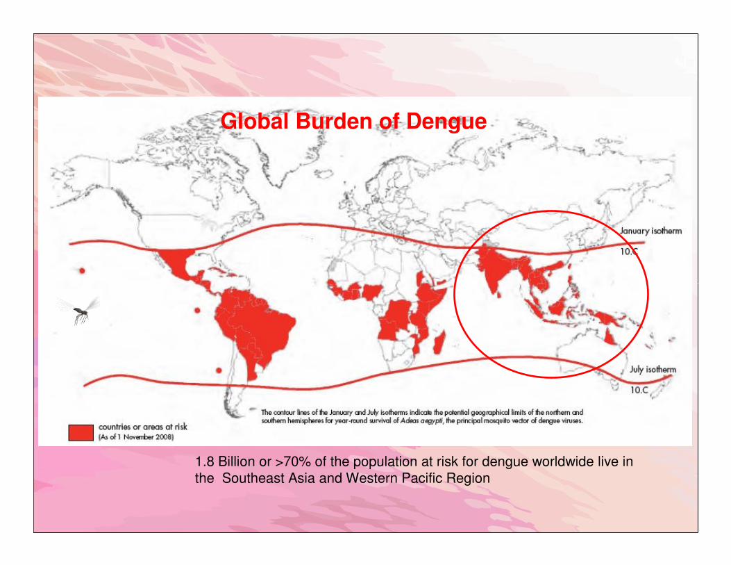
1.8 Billion or >70% of the population at risk for dengue worldwide live in
the Southeast Asia and Western Pacific Region
Global Burden of Dengue

• Chapter 1 Epidemiology, burden of disease and
transmission
• Chapter 2 Clinical management and delivery of
clinical services
• Chapter 3 Vector management and delivery of
vector control services
• Chapter 4 Laboratory diagnosis and diagnostic
tests
• Chapter 5 Surveillance, emergency
preparedness and response
• Chapter 6 New avenues

Dengue Case Classification and Levels of Severity

The Course of Dengue Illness

Clinical Problems During the Different Phases of Dengue
1 Febrile phase Dehydration; high fever may cause neurological disturbances and febrileseizures in young children
2 Critical phase Shock from plasma leakage; severe haemorrhage; organ impairment
3 Recovery phase Hypervolaemia (excessive IVF therapy)

Differential Diagnosis of Dengue Fever
Conditions that mimic the febrile phase of dengue infection
Flu-like syndromes
Illnesses with a rash
Diarrhoeal diseases
Illnesses with neurological
manifestations
Influenza, measles,
Chikungunya, infectious
mononucleosis , HIV
seroconversion illness
Rubella, measles, scarlet fever,
meningococcal infection,
Chikungunya, drug reactions
Rotavirus, other enteric infections
Meningo/encephalitis febrile
seizures

Differential Diagnosis of Dengue Fever
Conditions that mimic the critical phase of dengue infection
Infectious
Malignancies
Other clinical pictures
Acute gastroenteritis, malaria, leptospirosis,
typhoid, viral hepatitis, acute HIV seroconversion
illness, bacterial sepsis, septic shock
Acute leukemia and other malignancies
Acute abdomen
– acute appendicitis, cholecystitis, perforated
viscus
Diabetic ketoacidosis
Lactic acidosis
Leukopenia and thrombocytopaenia ± bleeding
Platelet disorders
Renal failure Respiratory distress (Kussmaul’s
breathing)
Systemic Lupus Erythematosus

A Stepwise Approach to the Management of Dengue
Step I. Overall assessment
I.1 History, including information on symptoms, past medical and family history
I.2 Physical examination, including full physical and mental assessment
I.3 Investigation, including routine laboratory and dengue-specific laboratory
Step II. Diagnosis, assessment of disease phase and severity
Step III. Management
III.1 Disease notification
III.2 Management decisions. Depending on the clinical manifestations and other
circumstances,
patients may:
– be sent home (Group A);
– be referred for in-hospital management (Group B);
– require emergency treatment and urgent referral (Group C).

A Stepwise Approach to the Management of Dengue
Step I—Overall assessment
History
The history should include:
– date of onset of fever/illness;
– quantity of oral intake;
– assessment for warning signs
– diarrhoea;
– change in mental state/seizure/dizziness;
– urine output (frequency, volume and time of last voiding);
– other important relevant histories, such as family or neighbourhood dengue, travel
to dengue endemic areas, co-existing conditions, jungle trekking and swimming in
waterfall, recent unprotected sex or drug abuse (consider acute HIV seroconversion
illness).
Physical Examination
– assessment of mental state;
– assessment of hydration status;
– assessment of haemodynamic status
– checking for tachypnoea/acidotic breathing/pleural effusion;
– checking for abdominal tenderness/hepatomegaly/ascites;
– examination for rash and bleeding manifestations;
– tourniquet test (repeat if previously negative or if there is no bleeding manifestation).
Investigation
A full blood count should be done at the first visit. A hct test in the early febrile
phase establishes the patient’s own baseline hct. A decreasing white blood cell count
makes dengue very likely. A rapid decrease in platelet count in parallel with a rising hct is
suggestive of progress to the plasma leakage/critical phase. In the absence of the
patient’s baseline, age-specific population hct levels could be used as a surrogate during
the critical phase.

Step II—Diagnosis, assessment of disease phase and severity
Step III—Management
Disease notification
In dengue-endemic countries, cases of suspected, probable and confirmed dengue should be
notified for appropriate public health measures. Suggested criteria for early notification of
suspected cases are that the patient lives in or has travelled to a dengue-endemic area, has
fever for three days or more, has low or decreasing white cell counts, and/or has
thrombocytopaenia ± positive tourniquet test. In dengue-endemic countries, the later the
notification, the more difficult it is to prevent dengue transmission.
Management decisions
Patient may be sent home (Group A), be referred for in-hospital management (Group B), or
require emergency treatment and urgent referral (Group C).


Normal maintenance fluid per hour can be calculated on the basis of the following formula*
(equivalent to Holliday-Segar formula):
4 mL/kg/h for first 10 kg body weight
+ 2 mL/kg/h for next 10 kg body weight
+ 1 mL/kg/h for subsequent kg body weight
*For overweight/obese patients calculate normal maintenance fluid based on ideal body weight (IBW)
(Adapted from reference 16)
IBW for overweight/obese adults can be estimated on the basis of the following formula
Female: 45.5 kg + 0.91(height -152.4) cm
Male: 50.0 kg + 0.91(height -152.4) cm
(17)
Calculations for normal maintenance of intravenous fluid infusion


Compensated shock (systolic pressuremaintained but has signs of reduced perfusion)
Fluid resuscitation with isotonic crystalloid5–10 ml/kg/hr over 1 hour
Algorithm for fluid management in compensated shock


Hypotensive shockFluid resuscitation with 20 ml/kg isotonic crystalloid or
colloid over 15 minutesTry to obtain a HCT level before fluid resuscitation
Algorithm for fluid management in Hypotensive shock

Treatment of complications and other areas of treatment
Fluid overload
Large pleural effusions and ascites is a common cause of acute respiratory distress and
failure in severe dengue.
Causes of fluid overload are:
– excessive and/or too rapid IVFs;
– incorrect use of hypotonic rather than isotonic crystalloid solutions;
– inappropriate use of large volumes of IVFs in patients with unrecognized severe
bleeding;
– inappropriate transfusion of fresh-frozen plasma, platelet concentrates and
cryoprecipitates;
– continuation of IVFs after plasma leakage has resolved (24–48 hours from
defervescence);
– co-morbid conditions such as congenital or ischaemic heart disease, chronic
lung and renal diseases.

Early clinical features of fluid overload
– respiratory distress, difficulty in breathing;
– rapid breathing;
– chest wall in-drawing;
– wheezing (rather than crepitations);
– large pleural effusions;
– tense ascites;
– increased jugular venous pressure (JVP).
Late Clinical Features
– pulmonary oedema (cough with pink or frothy sputum ± crepitations,
cyanosis);
– irreversible shock (heart failure, often in combination with ongoing
hypovolaemia).

Additional Investigations
– Chest x-ray shows cardiomegaly, pleural effusion, upward displacement of
the diaphragm by the ascites and varying degrees of “bat’s wings” appearance
± Kerley B lines suggestive of fluid overload and pulmonary oedema;
– ECG to exclude ischaemic changes and arrhythmia;
– ABG;
– Echocardiogram for assessment of left ventricular function,
– Cardiac enzymes.

The management of fluid overload varies according to the phase of
the diseaseand the patient’s haemodynamic status.
•Stable haemodynamic status and is out of the critical phase (more than 24–48 hours of defervescence), stop IVF but continue close monitoring. If necessary, give oral or IV
furosemide 0.1–0.5 mg/kg/dose once or twice daily, or a continuous infusion of
furosemide 0.1 mg/kg/hour.
- Monitor serum potassium and correct the ensuing hypokalaemia.
• Stable haemodynamic status but is still within the critical phase, reduce the
intravenous fluid accordingly.
-Avoid diuretics during the plasma leakage phase because they may lead to
intravascular volume depletion.
• In shock with low or normal haematocrit levels but show signs of fluid overload,
consider occult haemorrhage.
- Fresh whole blood transfusion should be initiated as soon as possible.
- If the patient remains in shock and the haematocrit is elevated, repeated small
boluses of a colloid solution may help.

Treatment of Fluid Overload
• Oxygen therapy should be given immediately.
• Stop IVF therapy during the recovery phase will allow fluid in the pleural and
peritoneal cavities to return to the intravascular compartment.
• IVF should be discontinued or reduced to the minimum rate when the
following signs are present:
– signs of cessation of plasma leakage;
– stable blood pressure, pulse and peripheral perfusion;
– HCT decreases in the presence of a good pulse volume;
– afebrile for more than 24–48 days (without the use of antipyretics);
– resolving bowel/abdominal symptoms;
– improving urine output.
Recognizing when to decrease or stop IVF is key to preventing fluid overload.
Other complications of dengue
•Hyperglycaemia and hypoglycaemia, even in the absence of diabetes mellitus and/or hypoglycaemic agents.
• Electrolyte and acid-base imbalances are probably related to gastrointestinal losses. Hyponatraemia, hypokalaemia,
hyperkalaemia, serum calcium imbalances and metabolic
acidosis (sodium bicarbonate for metabolic acidosis is not
recommended for pH ≥ 7.15) can occur.
•Co-infections and nosocomial infections.

Clinical No fever for 48 hours.Improvement in clinical status (general well-being, appetite, haemodynamic status, urine output, no respiratory distress).
LaboratoryIncreasing trend of platelet count.
Stable haematocrit without intravenous fluids.
Discharge criteria(all of the following conditions must be present)
