2007 focused update of the acc/aha/scai 2005 guideline ... · acc/aha/scai 2005 guideline update...
TRANSCRIPT
Journal of the American College of Cardiology Vol. 51, No. 2, 2008© 2008 by the American College of Cardiology Foundation and the American Heart Association, Inc. ISSN 0735-1097/08/$34.00P
PCI FOCUSED UPDATE
2007 Focused Update of theACC/AHA/SCAI 2005 Guideline Updatefor Percutaneous Coronary InterventionA Report of the American College of Cardiology/AmericanHeart Association Task Force on Practice Guidelines
2007 Writing Group to Review New Evidence and Update theACC/AHA/SCAI 2005 Guideline Update for Percutaneous Coronary Intervention,
Writing on Behalf of the 2005 Writing Committee
Spencer B. King III, MD, MACC, FAHA,FSCAI, Co-Chair*†
Sidney C. Smith, JR, MD, FACC, FAHA,Co-Chair*†
John W. Hirshfeld, JR, MD, FACC, FAHA,FSCAI‡
Douglass A. Morrison, MD, PHD, FACC,FSCAI‡
David O. Williams, MD, FACC, FAHA,FSCAI§
*Chair of 2005 Writing Committee; †Recused from voting on Section 7:Antiplatelet Therapy; ‡Society for Cardiovascular Angiography and Interven-tions Representative; §Recused from voting on Section 8: Bare-Metal and
ublished by Elsevier Inc. doi:10.1016/j.jacc.2007.10.002
Alice K. Jacobs, MD, FACC, FAHA, FSCAI Drug-Eluting Stents
S
TJ
AM
DWHPD
2005WritingCommitteeMembers
S
SA
CJCMSJS
idney C. Smith, JR, MD, FACC, FAHA, Chair
ed E. Feldman, MD, FACC, FSCAI‡ohn W. Hirshfeld, JR, MD, FACC, FAHA,
FSCAI‡lice K. Jacobs, MD, FACC, FAHA, FSCAIorton J. Kern, MD, FACC, FAHA, FSCAI‡
pencer B. King III, MD, MACC, FSCAI ‡
haron A. Hunt, MD, FACC, FAHA�
HFBRRBLC
�
ouglass A. Morrison, MD, PHD, FACC, FSCAI‡illiam W. O’Neill, MD, FACC, FSCAIartzell V. Schaff, MD, FACC, FAHAatrick L. Whitlow, MD, FACC, FAHAavid O. Williams, MD, FACC, FAHA,FSCAI
Society for Cardiovascular Angiography and Interventions Representative
TaskForceMembers
idney C. Smith, JR, MD, FACC, FAHA, Chairlice K. Jacobs, MD, FACC, FAHA, Vice-Chair
ynthia D. Adams, MSN, PHD, FAHA�
effrey L. Anderson, MD, FACC, FAHA�
hristopher E. Buller, MD, FACCark A. Creager, MD, FACC, FAHA
teven M. Ettinger, MD, FACConathan L. Halperin, MD, FACC, FAHA�
arlan M. Krumholz, MD, FACC, FAHArederick G. Kushner, MD, FACC, FAHAruce W. Lytle, MD, FACC, FAHAick Nishimura, MD, FACC, FAHAichard L. Page, MD, FACC, FAHAarbara Riegel, DNSC, RN, FAHA�
ynn G. Tarkington, RNlyde W. Yancy, MD, FACC
Former Task Force member during this writing effort
P
1
2
3
4
5
6
7
8
9
R
A
A
P
AgwmCFutircospanro
spbpitfogi
•••
•••
•
••
Tr
TCA
adMGClAC
e
Chsm
doHwR
173JACC Vol. 51, No. 2, 2008 King III et al.January 15, 2008:172–209 PCI Focused Update
TABLE OF CONTENTS
reamble . . . . . . . . . . . . . . . . . . . . . . . . . . . . . . . . . . . . . . . . . . . . . . . . . . . . .173
. Introduction . . . . . . . . . . . . . . . . . . . . . . . . . . . . . . . . . . . . . . . . . . . . .175
1.1. Evidence Review . . . . . . . . . . . . . . . . . . . . . . . . . . . . . . . . . . .175
1.2. Organization of Committee andRelationships With Industry . . . . . . . . . . . . . . . . . . . . . . .175
1.3. Review and Approval . . . . . . . . . . . . . . . . . . . . . . . . . . . . . . .175
. Patients With Unstable Angina/Non–ST-Elevation Myocardial Infarction . . . . . . . . . . . .176
2.1. Electrocardiogram . . . . . . . . . . . . . . . . . . . . . . . . . . . . . . . . . .1792.1.1. Comparison of Early Invasive and Initial
Conservative Strategies for UA/NSTEMI . . . . .1812.1.2. Selection for Coronary Angiography . . . . . . . . . . .1842.1.3. Chronic Kidney Disease . . . . . . . . . . . . . . . . . . . . . . .185
. Facilitated PCI . . . . . . . . . . . . . . . . . . . . . . . . . . . . . . . . . . . . . . . . . .186
. Rescue PCI . . . . . . . . . . . . . . . . . . . . . . . . . . . . . . . . . . . . . . . . . . . . . .187
. PCI After Fibrinolysis or for Patients NotUndergoing Primary Reperfusion . . . . . . . . . . . . . . . . . . . .190
. Ancillary Therapy for Patients Undergoing PCIfor STEMI . . . . . . . . . . . . . . . . . . . . . . . . . . . . . . . . . . . . . . . . . . . . . . . . .191
. Antiplatelet Therapy . . . . . . . . . . . . . . . . . . . . . . . . . . . . . . . . . . .192
. Bare-Metal and Drug-Eluting Stents . . . . . . . . . . . . . . . . .195
8.1. Selection of a Bare-Metal or Drug-ElutingStent . . . . . . . . . . . . . . . . . . . . . . . . . . . . . . . . . . . . . . . . . . . . . . . . .195
his document is a limited update to the 2005 guideline update and is based on aeview of certain evidence, not a full literature review.
This document was approved by the American College of Cardiology Board ofrustees in October 2007, by the American Heart Association Science Advisory andoordinating Committee in October 2007, and by the Society for Cardiovascularngiography and Interventions Board of Trustees in November 2007.The American College of Cardiology Foundation, American Heart Association,
nd Society for Cardiovascular Angiography and Interventions request that thisocument be cited as follows: King SB III, Smith SC Jr., Hirshfeld JW Jr., Jacobs AK,orrison DA, Williams DO. 2007 focused update of the ACC/AHA/SCAI 2005uideline Update for Percutaneous Coronary Intervention: a report of the Americanollege of Cardiology/American Heart Association Task Force on Practice Guide-
ines: (2007 Writing Group to Review New Evidence and Update the 2005CC/AHA/SCAI Guideline Update for Percutaneous Coronary Intervention). J Amoll Cardiol 2008;51:172–209.This article has been copublished in the January 15, 2008, issue of Circulation and
-published in Catheterization and Cardiovascular Interventions.Copies: This document is available on the World Wide Web sites of the Americanollege of Cardiology (www.acc.org), American Heart Association (www.american-eart.org), and Society for Cardiovascular Angiography and Interventions (www.cai.org). For copies of this document, please contact Elsevier Inc. Reprint Depart-ent, fax 212-633-3820, e-mail [email protected]: Modification, alteration, enhancement, and/or distribution of this
ocument are not permitted without the express permission of the American Collegef Cardiology and the American Heart Association. Please contact the Americaneart Association: Instructions for obtaining permission are located at http://
ww.americanheart.org/presenter.jhtml?identifier�4431. A link to the “Permissionequest Form” appears on the right side of the page.. Secondary Prevention . . . . . . . . . . . . . . . . . . . . . . . . . . . . . . . . .197
eferences . . . . . . . . . . . . . . . . . . . . . . . . . . . . . . . . . . . . . . . . . . . . . . . . . . .202
ppendix 1 . . . . . . . . . . . . . . . . . . . . . . . . . . . . . . . . . . . . . . . . . . . . . . . . . . .206
ppendix 2 . . . . . . . . . . . . . . . . . . . . . . . . . . . . . . . . . . . . . . . . . . . . . . . . . . .207
reamble
primary challenge in the development of clinical practiceuidelines is keeping pace with the stream of new data uponhich recommendations are based. In an effort to respondore quickly to new evidence, the American College ofardiology/American Heart Association (ACC/AHA) Taskorce on Practice Guidelines has created a new “focusedpdate” process to revise the existing guideline recommenda-ions that are affected by evolving data or opinion. Before thenitiation of this focused approach, periodic updates andevisions of existing guidelines required up to 3 years toomplete. Now, however, new evidence will be reviewed in anngoing fashion to more efficiently respond to importantcience and treatment trends that could have a major impact onatient outcomes and quality of care. Evidence will be reviewedt least twice a year, and updates will be initiated on an aseeded basis as quickly as possible while maintaining theigorous methodology that the ACC and AHA have devel-ped during their more than 20 years of partnership.
These updated guideline recommendations reflect a con-ensus of expert opinion following a thorough reviewrimarily of late-breaking clinical trials identified through aroad-based vetting process as important to the relevantatient population and of other new data deemed to have anmpact on patient care (see Section 1.1 for details regardinghis focused update). It is important to note that thisocused update is not intended to represent an update basedn a full literature review from the date of the previousuideline publication. Specific criteria/considerations fornclusion of new data include:
Publication in a peer-reviewed journalLarge, randomized, placebo-controlled trial(s)Nonrandomized data deemed important on the basis ofresults that impact current safety and efficacy assumptionsStrengths/weakness of research methodology and findingsLikelihood of additional studies influencing current findingsImpact on current performance measure(s) and/or likeli-hood of the need to develop new performance measure(s)Requests and requirements for review and update fromthe practice community, key stakeholders, regulatoryagencies, and other sources free of relationships withindustry or other potential biasNumber of previous trials showing consistent resultsNeed for consistency with other new guidelines or
guideline revisions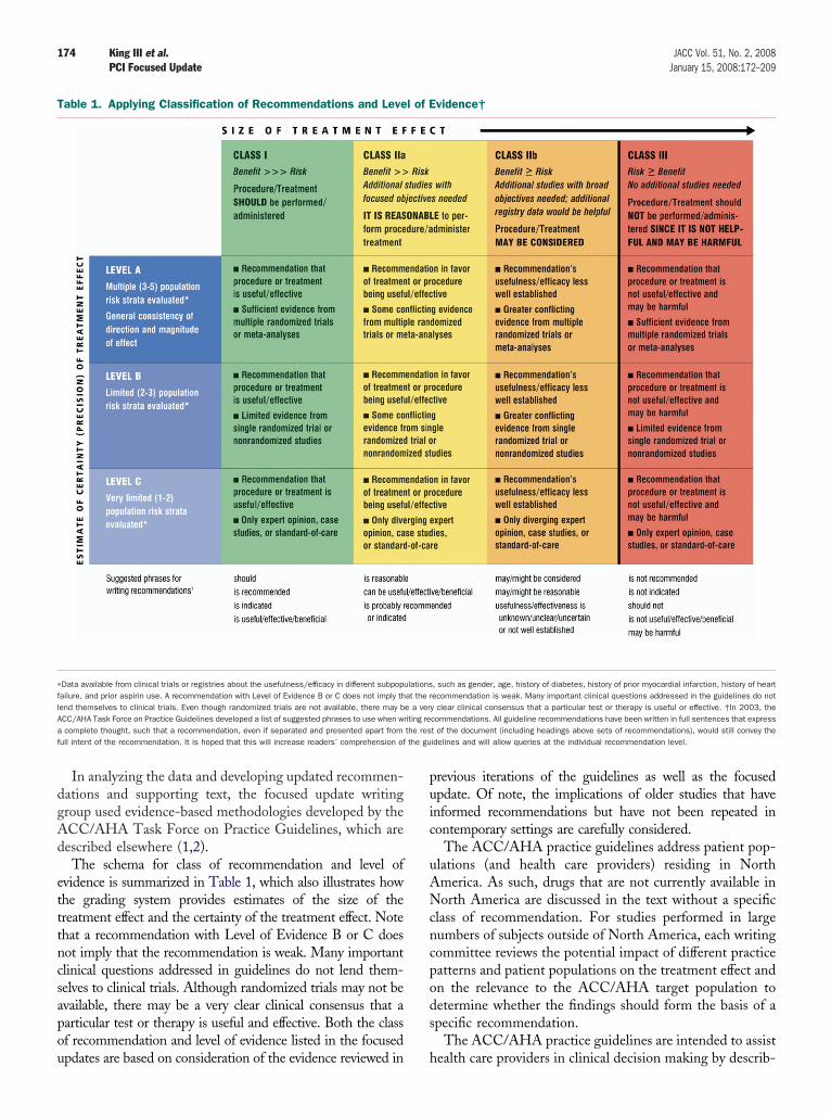
dgAd
etttncsapou
puic
uANcncpods
T
�
flAa the resf the gu
174 King III et al. JACC Vol. 51, No. 2, 2008PCI Focused Update January 15, 2008:172–209
In analyzing the data and developing updated recommen-ations and supporting text, the focused update writingroup used evidence-based methodologies developed by theCC/AHA Task Force on Practice Guidelines, which areescribed elsewhere (1,2).The schema for class of recommendation and level of
vidence is summarized in Table 1, which also illustrates howhe grading system provides estimates of the size of thereatment effect and the certainty of the treatment effect. Notehat a recommendation with Level of Evidence B or C doesot imply that the recommendation is weak. Many importantlinical questions addressed in guidelines do not lend them-elves to clinical trials. Although randomized trials may not bevailable, there may be a very clear clinical consensus that aarticular test or therapy is useful and effective. Both the classf recommendation and level of evidence listed in the focused
able 1. Applying Classification of Recommendations and Leve
Data available from clinical trials or registries about the usefulness/efficacy in different subpopuailure, and prior aspirin use. A recommendation with Level of Evidence B or C does not imply thend themselves to clinical trials. Even though randomized trials are not available, there may bCC/AHA Task Force on Practice Guidelines developed a list of suggested phrases to use when wrcomplete thought, such that a recommendation, even if separated and presented apart from
ull intent of the recommendation. It is hoped that this will increase readers’ comprehension of
pdates are based on consideration of the evidence reviewed in h
revious iterations of the guidelines as well as the focusedpdate. Of note, the implications of older studies that havenformed recommendations but have not been repeated inontemporary settings are carefully considered.
The ACC/AHA practice guidelines address patient pop-lations (and health care providers) residing in Northmerica. As such, drugs that are not currently available inorth America are discussed in the text without a specific
lass of recommendation. For studies performed in largeumbers of subjects outside of North America, each writingommittee reviews the potential impact of different practiceatterns and patient populations on the treatment effect andn the relevance to the ACC/AHA target population toetermine whether the findings should form the basis of apecific recommendation.
The ACC/AHA practice guidelines are intended to assist
vidence†
, such as gender, age, history of diabetes, history of prior myocardial infarction, history of heartecommendation is weak. Many important clinical questions addressed in the guidelines do noty clear clinical consensus that a particular test or therapy is useful or effective. †In 2003, thecommendations. All guideline recommendations have been written in full sentences that expresst of the document (including headings above sets of recommendations), would still convey theidelines and will allow queries at the individual recommendation level.
l of E
lationsat the re a veriting re
ealth care providers in clinical decision making by describ-
idotTmlTtiiuui
rtamt
mcowqscdW$rmp
trgrltmmc
cfftJCdASsa
1
1
SaEdceicoeSI
wattahp
Aocdil
1R
FwagumfiarbmRv
1
Tn
175JACC Vol. 51, No. 2, 2008 King III et al.January 15, 2008:172–209 PCI Focused Update
ng a range of generally acceptable approaches for theiagnosis, management, and prevention of specific diseasesr conditions. The guidelines attempt to define practiceshat meet the needs of most patients in most circumstances.he ultimate judgment regarding care of a particular patientust be made by the health care provider and patient in
ight of all the circumstances presented by that patient.hus, there are circumstances in which deviations from
hese guidelines may be appropriate. Clinical decision mak-ng should consider the quality and availability of expertisen the area where care is provided. These guidelines may besed as the basis for regulatory or payer decisions, but theltimate goal is quality of care and serving the patient’s bestnterests.
Prescribed courses of treatment in accordance with theseecommendations are only effective if they are followed byhe patient. Because lack of patient adherence may adverselyffect treatment outcomes, health care providers shouldake every effort to engage the patient in active participa-
ion with prescribed treatment.The ACC/AHA Task Force on Practice Guidelinesakes every effort to avoid any actual, potential, or per-
eived conflict of interest arising from industry relationshipsr personal interests of a writing committee member. Allriting committee members and peer reviewers were re-uired to provide disclosure statements of all such relation-hips pertaining to the trials and other evidence underonsideration (see Appendixes 1 and 2). Final recommen-ations were balloted to all writing committee members.
riting committee members with significant (greater than10 000) relevant relationships with industry (RWI) wereequired to recuse themselves from voting on that recom-endation. Writing committee members who did not
articipate are not listed as authors of this focused update.With the exception of the recommendations presented in
his statement, the full guidelines remain current. Only theecommendations from the affected section(s) of the fulluidelines are included in this focused update. For easyeference, all recommendations from any section of guide-ines impacted by a change are presented with a notation aso whether they remain current, are new, or have beenodified. When evidence impacts recommendations inore than 1 set of guidelines, those guidelines are updated
oncurrently.The recommendations in this focused update will be
onsidered current until they are superseded by anotherocused update or the full-text guidelines are revised. Thisocused update is published in the January 15, 2008, issue ofhe Journal of the American College of Cardiology and theanuary 15, 2008, issue of Circulation and e-published inatheterization and Cardiovascular Interventions as an up-ate to the full-text guidelines and is also posted on theCC (www.acc.org), AHA (www.americanheart.org), andociety for Angiography and Interventions (SCAI) (www.cai.org) Web sites. Copies of the focused update are
vailable from all organizations. SSidney C. Smith, Jr., MD, FACC, FAHAChair, ACC/AHA Task Force on Practice Guidelines
Alice K. Jacobs, MD, FACC, FAHAVice-Chair, ACC/AHA Task Force on Practice Guidelines
. Introduction
.1. Evidence Review
elected late-breaking clinical trials presented at the 2005nd 2006 annual scientific meetings of the ACC, AHA, anduropean Society of Cardiology, as well as selected otherata, were reviewed by the standing guideline writingommittee along with the parent Task Force and otherxperts to identify those trials and other key data that mightmpact guideline recommendations. On the basis of theriteria/considerations noted above, recent trial data andther clinical information were considered importantnough to prompt a focused update of the ACC/AHA/CAI 2005 Guideline Update for Percutaneous Coronaryntervention (3–13).
To provide clinicians with a comprehensive set of data,henever possible, the exact event rates in various treatment
rms of clinical trials are presented to permit calculation ofhe absolute risk difference (ARD) and number needed toreat (NNT) or harm (NNH); the relative treatment effectsre described either as odds ratio (OR), relative risk (RR), orazard ratio (HR), depending on the format in the originalublication.Consult the full-text version or executive summary of the
CC/AHA/SCAI 2005 Guideline Update for Percutane-us Coronary Intervention for policy on clinical areas notovered by the focused update (13a). Individual recommen-ations updated in this focused update will be incorporatednto future revisions and/or updates of the full-text guide-ines.
.2. Organization of Committee andelationships With Industry
or this focused update, all members of the 2005 PCIriting committee were invited to participate; those who
greed (referred to as the 2007 focused update writingroup) were required to disclose all RWI relevant to the datander consideration (2). Focused update writing groupembers who had no significant relevant RWI wrote the
rst draft of the focused update; the draft was then reviewednd revised by the full writing group. Each recommendationequired a confidential vote by the writing group membersefore external review of the document. Any writing com-ittee member with a significant (greater than $10 000)WI relevant to the recommendation was recused from
oting on that recommendation.
.3. Review and Approval
his document was reviewed by 2 outside reviewers nomi-ated by each cosponsoring organization (ACC, AHA, and
CAI) and 24 individual content reviewers. All reviewer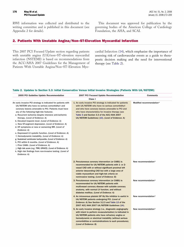
RwA
g
2
TwitP
T
A
176 King III et al. JACC Vol. 51, No. 2, 2008PCI Focused Update January 15, 2008:172–209
WI information was collected and distributed to theriting committee and is published in this document (see
. Patients With Unstable Angina/Non–ST-Elevat
atients With Unstable Angina/Non–ST-Elevation Myo-
capt
This document was approved for publication by theoverning bodies of the American College of Cardiology
ppendix 2 for details). Foundation, the AHA, and SCAI.
ion Myocardial Infarction
his 2007 PCI Focused Update section regarding patientsith unstable angina (UA)/non–ST-elevation myocardial
nfarction (NSTEMI) is based on recommendations fromhe ACC/AHA 2007 Guidelines for the Management of
ardial Infarction (14), which emphasize the importance ofssessing risk of cardiovascular events as a guide to thera-eutic decision making and the need for interventionalherapy (see Table 2).
able 2. Updates to Section 5.3: Initial Conservative Versus In
2005 PCI Guideline Update Recommendation 2007 PCI F
C
n early invasive PCI strategy is indicated for patients withUA/NSTEMI who have no serious comorbidity† andcoronary lesions amenable to PCI. Patients must haveany of the following high-risk features:
a. Recurrent ischemia despite intensive anti-ischemictherapy. (Level of Evidence: A)
b. Elevated troponin level. (Level of Evidence: A)c. New ST-segment depression. (Level of Evidence: A)d. HF symptoms or new or worsening MR. (Level of
Evidence: A)e. Depressed LV systolic function. (Level of Evidence: A)f. Hemodynamic instability. (Level of Evidence: A)g. Sustained ventricular tachycardia. (Level of Evidence: A)h. PCI within 6 months. (Level of Evidence: A)i. Prior CABG. (Level of Evidence: A)j. High risk score (e.g., TIMI, GRACE). (Level of Evidence: A)k. High risk findings from non-invasive testing. (Level of
Evidence: A)
1. An early invasiwith UA/NSTEMand who havewho have charTable 3 and SeUA/NSTEMI Gu
2. Percutaneousrecommendedvessel CAD witanterior desceviable myocardnoninvasive te
3. Percutaneousrecommendedmultivessel coanatomy, withdiabetes melli
4. An intravenousUA/NSTEMI paEvidence: A) S2007 ACC/AH
5. An early invasiwith intent to pUA/NSTEMI pahemodynamiccomorbidities(Level of Evide
itial Invasive Strategies (Patients With UA/NSTEMI)
ocused Update Recommendation Comments
lass I
ve PCI strategy is indicated for patientsI who have no serious comorbidity†
coronary lesions amenable to PCI andacteristics for invasive therapy (seection 3.3 of the ACC/AHA 2007idelines) (14). (Level of Evidence: A)
Modified recommendation*
coronary intervention (or CABG) isfor UA/NSTEMI patients with 1- or 2-h or without significant proximal leftnding CAD but with a large area ofium and high-risk criteria on
sting. (Level of Evidence: B)
New recommendation*
coronary intervention (or CABG) isfor UA/NSTEMI patients with
ronary disease with suitable coronarynormal LV function, and without
tus. (Level of Evidence: A)
New recommendation*
platelet GP IIb/IIIa inhibitor is useful intients undergoing PCI. (Level ofee Section 3.2.3 and Table 13 of theA 2007 UA/NSTEMI Guidelines (14).
New recommendation*
ve strategy (i.e., diagnostic angiographyerform revascularization) is indicated intients who have refractory angina oror electrical instability (without seriousor contraindications to such procedures).nce: B)
New recommendation*
T
I
I
U
I
P
177JACC Vol. 51, No. 2, 2008 King III et al.January 15, 2008:172–209 PCI Focused Update
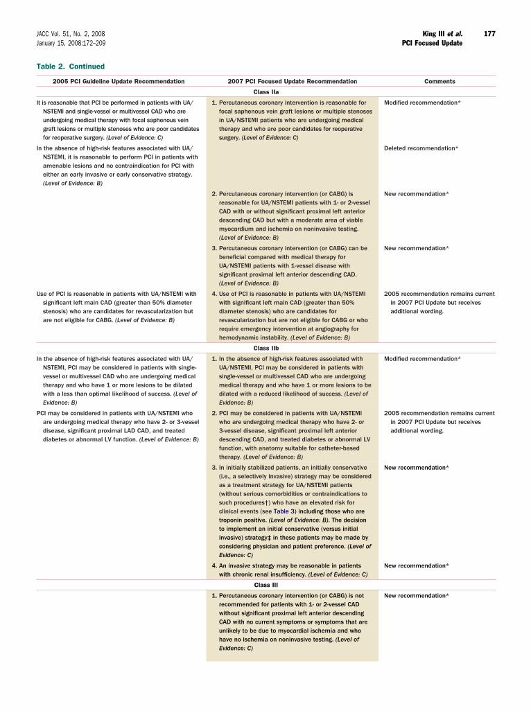
able 2. Continued
2005 PCI Guideline Update Recommendation 2007 PCI Focused Update Recommendation Comments
Class IIa
t is reasonable that PCI be performed in patients with UA/NSTEMI and single-vessel or multivessel CAD who areundergoing medical therapy with focal saphenous veingraft lesions or multiple stenoses who are poor candidatesfor reoperative surgery. (Level of Evidence: C)
1. Percutaneous coronary intervention is reasonable forfocal saphenous vein graft lesions or multiple stenosesin UA/NSTEMI patients who are undergoing medicaltherapy and who are poor candidates for reoperativesurgery. (Level of Evidence: C)
Modified recommendation*
n the absence of high-risk features associated with UA/NSTEMI, it is reasonable to perform PCI in patients withamenable lesions and no contraindication for PCI witheither an early invasive or early conservative strategy.(Level of Evidence: B)
Deleted recommendation*
2. Percutaneous coronary intervention (or CABG) isreasonable for UA/NSTEMI patients with 1- or 2-vesselCAD with or without significant proximal left anteriordescending CAD but with a moderate area of viablemyocardium and ischemia on noninvasive testing.(Level of Evidence: B)
New recommendation*
3. Percutaneous coronary intervention (or CABG) can bebeneficial compared with medical therapy forUA/NSTEMI patients with 1-vessel disease withsignificant proximal left anterior descending CAD.(Level of Evidence: B)
New recommendation*
se of PCI is reasonable in patients with UA/NSTEMI withsignificant left main CAD (greater than 50% diameterstenosis) who are candidates for revascularization butare not eligible for CABG. (Level of Evidence: B)
4. Use of PCI is reasonable in patients with UA/NSTEMIwith significant left main CAD (greater than 50%diameter stenosis) who are candidates forrevascularization but are not eligible for CABG or whorequire emergency intervention at angiography forhemodynamic instability. (Level of Evidence: B)
2005 recommendation remains currentin 2007 PCI Update but receivesadditional wording.
Class IIb
n the absence of high-risk features associated with UA/NSTEMI, PCI may be considered in patients with single-vessel or multivessel CAD who are undergoing medicaltherapy and who have 1 or more lesions to be dilatedwith a less than optimal likelihood of success. (Level ofEvidence: B)
1. In the absence of high-risk features associated withUA/NSTEMI, PCI may be considered in patients withsingle-vessel or multivessel CAD who are undergoingmedical therapy and who have 1 or more lesions to bedilated with a reduced likelihood of success. (Level ofEvidence: B)
Modified recommendation*
CI may be considered in patients with UA/NSTEMI whoare undergoing medical therapy who have 2- or 3-vesseldisease, significant proximal LAD CAD, and treateddiabetes or abnormal LV function. (Level of Evidence: B)
2. PCI may be considered in patients with UA/NSTEMIwho are undergoing medical therapy who have 2- or3-vessel disease, significant proximal left anteriordescending CAD, and treated diabetes or abnormal LVfunction, with anatomy suitable for catheter-basedtherapy. (Level of Evidence: B)
2005 recommendation remains currentin 2007 PCI Update but receivesadditional wording.
3. In initially stabilized patients, an initially conservative(i.e., a selectively invasive) strategy may be consideredas a treatment strategy for UA/NSTEMI patients(without serious comorbidities or contraindications tosuch procedures†) who have an elevated risk forclinical events (see Table 3) including those who aretroponin positive. (Level of Evidence: B). The decisionto implement an initial conservative (versus initialinvasive) strategy‡ in these patients may be made byconsidering physician and patient preference. (Level ofEvidence: C)
New recommendation*
4. An invasive strategy may be reasonable in patientswith chronic renal insufficiency. (Level of Evidence: C)
New recommendation*
Class III
1. Percutaneous coronary intervention (or CABG) is notrecommended for patients with 1- or 2-vessel CADwithout significant proximal left anterior descendingCAD with no current symptoms or symptoms that areunlikely to be due to myocardial ischemia and whohave no ischemia on noninvasive testing. (Level ofEvidence: C)
New recommendation*
Afa
tptt
m(
sieiuTvcS
TI
I
C
R
Epa
T
I
a
b
d
*‡
d n; SVGa
TN
TipabiAM(
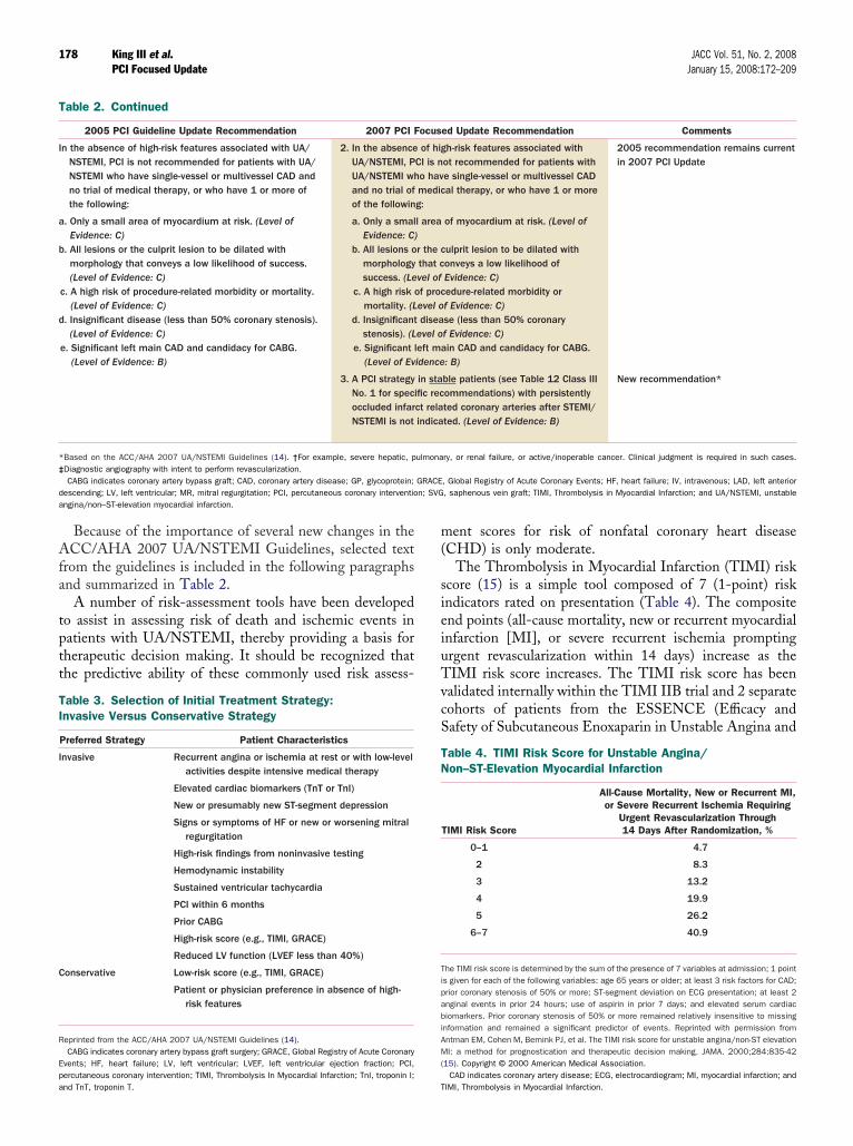
178 King III et al. JACC Vol. 51, No. 2, 2008PCI Focused Update January 15, 2008:172–209
Because of the importance of several new changes in theCC/AHA 2007 UA/NSTEMI Guidelines, selected text
rom the guidelines is included in the following paragraphsnd summarized in Table 2.
A number of risk-assessment tools have been developedo assist in assessing risk of death and ischemic events inatients with UA/NSTEMI, thereby providing a basis forherapeutic decision making. It should be recognized thathe predictive ability of these commonly used risk assess-
able 3. Selection of Initial Treatment Strategy:nvasive Versus Conservative Strategy
Preferred Strategy Patient Characteristics
nvasive Recurrent angina or ischemia at rest or with low-levelactivities despite intensive medical therapy
Elevated cardiac biomarkers (TnT or TnI)
New or presumably new ST-segment depression
Signs or symptoms of HF or new or worsening mitralregurgitation
High-risk findings from noninvasive testing
Hemodynamic instability
Sustained ventricular tachycardia
PCI within 6 months
Prior CABG
High-risk score (e.g., TIMI, GRACE)
Reduced LV function (LVEF less than 40%)
onservative Low-risk score (e.g., TIMI, GRACE)
Patient or physician preference in absence of high-risk features
eprinted from the ACC/AHA 2007 UA/NSTEMI Guidelines (14).CABG indicates coronary artery bypass graft surgery; GRACE, Global Registry of Acute Coronary
vents; HF, heart failure; LV, left ventricular; LVEF, left ventricular ejection fraction; PCI,
able 2. Continued
2005 PCI Guideline Update Recommendation 2007 PCI F
n the absence of high-risk features associated with UA/NSTEMI, PCI is not recommended for patients with UA/NSTEMI who have single-vessel or multivessel CAD andno trial of medical therapy, or who have 1 or more ofthe following:
2. In the absenceUA/NSTEMI, PUA/NSTEMI whand no trial ofof the followin
. Only a small area of myocardium at risk. (Level ofEvidence: C)
. All lesions or the culprit lesion to be dilated withmorphology that conveys a low likelihood of success.(Level of Evidence: C)
c. A high risk of procedure-related morbidity or mortality.(Level of Evidence: C)
. Insignificant disease (less than 50% coronary stenosis).(Level of Evidence: C)
e. Significant left main CAD and candidacy for CABG.(Level of Evidence: B)
a. Only a smalEvidence: C)
b. All lesions omorphologysuccess. (Le
c. A high risk omortality. (L
d. Insignificantstenosis). (L
e. Significant l(Level of Ev
3. A PCI strategyNo. 1 for specioccluded infarNSTEMI is not
Based on the ACC/AHA 2007 UA/NSTEMI Guidelines (14). †For example, severe hepatic, pDiagnostic angiography with intent to perform revascularization.CABG indicates coronary artery bypass graft; CAD, coronary artery disease; GP, glycoprotein;
escending; LV, left ventricular; MR, mitral regurgitation; PCI, percutaneous coronary interventiongina/non–ST-elevation myocardial infarction.
ercutaneous coronary intervention; TIMI, Thrombolysis In Myocardial Infarction; TnI, troponin I;nd TnT, troponin T. T
ent scores for risk of nonfatal coronary heart diseaseCHD) is only moderate.
The Thrombolysis in Myocardial Infarction (TIMI) riskcore (15) is a simple tool composed of 7 (1-point) riskndicators rated on presentation (Table 4). The compositend points (all-cause mortality, new or recurrent myocardialnfarction [MI], or severe recurrent ischemia promptingrgent revascularization within 14 days) increase as theIMI risk score increases. The TIMI risk score has been
alidated internally within the TIMI IIB trial and 2 separateohorts of patients from the ESSENCE (Efficacy andafety of Subcutaneous Enoxaparin in Unstable Angina and
ed Update Recommendation Comments
h-risk features associated withot recommended for patients withe single-vessel or multivessel CADal therapy, or who have 1 or more
2005 recommendation remains currentin 2007 PCI Update
of myocardium at risk. (Level of
ulprit lesion to be dilated withonveys a low likelihood ofEvidence: C)edure-related morbidity orf Evidence: C)se (less than 50% coronaryf Evidence: C)in CAD and candidacy for CABG.: B)
ble patients (see Table 12 Class IIIommendations) with persistentlyted coronary arteries after STEMI/ted. (Level of Evidence: B)
New recommendation*
ry, or renal failure, or active/inoperable cancer. Clinical judgment is required in such cases.
, Global Registry of Acute Coronary Events; HF, heart failure; IV, intravenous; LAD, left anterior, saphenous vein graft; TIMI, Thrombolysis in Myocardial Infarction; and UA/NSTEMI, unstable
able 4. TIMI Risk Score for Unstable Angina/on–ST-Elevation Myocardial Infarction
TIMI Risk Score
All-Cause Mortality, New or Recurrent MI,or Severe Recurrent Ischemia Requiring
Urgent Revascularization Through14 Days After Randomization, %
0–1 4.7
2 8.3
3 13.2
4 19.9
5 26.2
6–7 40.9
he TIMI risk score is determined by the sum of the presence of 7 variables at admission; 1 points given for each of the following variables: age 65 years or older; at least 3 risk factors for CAD;rior coronary stenosis of 50% or more; ST-segment deviation on ECG presentation; at least 2nginal events in prior 24 hours; use of aspirin in prior 7 days; and elevated serum cardiaciomarkers. Prior coronary stenosis of 50% or more remained relatively insensitive to missing
nformation and remained a significant predictor of events. Reprinted with permission fromntman EM, Cohen M, Bernink PJ, et al. The TIMI risk score for unstable angina/non-ST elevationI: a method for prognostication and therapeutic decision making. JAMA. 2000;284:835-42
15). Copyright © 2000 American Medical Association.
ocus
of higCI is no havmedicg:
l area
r the cthat cvel off procevel odisea
evel oeft maidence
in stafic recct relaindica
ulmona
GRACE
CAD indicates coronary artery disease; ECG, electrocardiogram; MI, myocardial infarction; andIMI, Thrombolysis in Myocardial Infarction.
NrregaswtwcivoeuaN
Artmtwoas
EttwaUnptacdd1mintcPa((d1br
pSipSdod
NATAath
pt(s
2
TedwwesrdNun
CsAtesa(ImrioPttbN
179JACC Vol. 51, No. 2, 2008 King III et al.January 15, 2008:172–209 PCI Focused Update
on–Q-Wave Myocardial Infarction) trial (16). The modelemained a significant predictor of events and appearedelatively insensitive to missing information, such as knowl-dge of previously documented coronary stenosis of 50% orreater. The model’s predictive ability remained intact, withcutoff of 65 years of age. The TIMI risk score was recently
tudied in an unselected emergency department populationith chest pain syndrome; its performance was similar to
hat in the acute coronary syndrome (ACS) population fromhich it was derived and validated (17). The TIMI risk
alculator is available at www.timi.org. The TIMI riskndex, a modification of the TIMI risk score that uses theariables age, systolic blood pressure, and heart rate, has notnly been shown to predict short-term mortality in ST-levation myocardial infarction (STEMI) but also has beenseful in prediction of 30-day and 1-year mortality ratescross the spectrum of patients with ACS, including UA/STEMI (18).The PURSUIT (Platelet Glycoprotein IIb-IIIa in Unstable
ngina: Receptor Suppression Using Integrilin Therapy) trialisk model (19), based on patients enrolled in the PURSUITrial, is another useful tool to guide the clinical decision-aking process when the patient is admitted to the hospital. In
he PURSUIT risk model, critical clinical features associatedith an increased 30-day incidence of death and the compositef death or myocardial (re)infarction were (in order of strength)ge, heart rate, systolic blood pressure, ST-segment depression,igns of heart failure (HF), and cardiac enzymes (19).
The GRACE (Global Registry of Acute Coronaryvents) study risk model, which predicts in-hospital mor-
ality (and death or MI), can be useful to clinicians to guidereatment type and intensity (20,21). The GRACE risk toolas developed on the basis of 11 389 patients in GRACE
nd validated in subsequent GRACE and GUSTO (Globaltilization of Streptokinase and TPA for Occluded Coro-ary Arteries) IIb cohorts and predicts in-hospital death inatients with STEMI, NSTEMI, or UA (C statis-ic�0.83). The 8 variables used in the GRACE risk modelre older age (OR 1.7 per 10 years), Killip class (OR 2.0 perlass), systolic blood pressure (OR 1.4 per 20 mm Hgecrease), ST-segment deviation (OR 2.4), cardiac arresturing presentation (OR 4.3), serum creatinine level (OR.2 per 1 mg per dL increase), positive initial cardiacarkers (OR 1.6), and heart rate (OR 1.3 per 30-bpm
ncrease). The sum of scores is applied to a referenceonogram to determine the corresponding all-cause mor-ality from hospital discharge to 6 months. The GRACElinical application tool can be downloaded to a handheldDA (personal digital assistant) to be used at the bedsidend is available at www.outcomes-umassmed.org/graceFigure 1) (21). An analysis comparing the 3 risk scoresTIMI, GRACE, and PURSUIT) concluded that all 3emonstrated good predictive accuracy for death and MI atyear, thus identifying patients who might be likely to
enefit from aggressive therapy, including early myocardial
evascularization (22). fiThe electrocardiogram (ECG) provides unique and im-ortant diagnostic and prognostic information (see alsoection 2.1 below). Accordingly, ECG changes have been
ncorporated into quantitative decision aids for the triage ofatients who present with chest discomfort (23). AlthoughT elevation carries the highest early risk of death, STepression on the presenting ECG portends the highest riskf death at 6 months, with the degree of ST-segmentepression showing a strong relationship to outcome (24).The recommendations in the ACC/AHA 2007 UA/STEMI Guidelines (14) recognize recent data from theCUITY (Acute Catheterization and Urgent Interventionriage strategY) trial, which showed that in patients withCS who were undergoing invasive treatment, bivalirudin
lone was associated with rates of ischemia similar to thosereated with glycoprotein (GP) IIb/IIIa inhibitors pluseparin and significantly less bleeding (25).The ACC/AHA 2007 UA/NSTEMI Guidelines cite a
rogressively greater benefit from newer, more aggressiveherapies such as low-molecular-weight heparin (LMWH)16,26), platelet GP IIb/IIIa inhibition (27), and an invasivetrategy (28) with increasing risk score.
.1. Electrocardiogram
he ECG lies at the center of the decision pathway for thevaluation and management of patients with acute ischemiciscomfort (Table 5). The diagnosis of MI is confirmedith serial cardiac biomarkers in more than 90% of patientsho present with ST-segment elevation greater than or
qual to 1 mm (0.1 mV) in at least 2 contiguous leads, anduch patients should be considered candidates for acuteeperfusion therapy. Patients who present with ST-segmentepression are initially considered to have either UA orSTEMI; the distinction between the 2 diagnoses is
ltimately based on the detection of markers of myocardialecrosis in the blood (29–31).Up to 25% of patients with NSTEMI and elevated
K-MB go on to develop Q-wave MI during their hospitaltay, whereas the remaining 75% have non–Q-wave MI.cute fibrinolytic therapy is contraindicated for ACS pa-
ients without ST-segment elevation, except for those withlectrocardiographic true posterior MI manifested as ST-egment depression in 2 contiguous anterior precordial leadsnd/or isolated ST-segment elevation in posterior chest lead32–34). Inverted T waves may also indicate UA/NSTEMI.n patients suspected of having ACS on clinical grounds,arked (greater than or equal to 2 mm [0.2 mV]) symmet-
ical precordial T-wave inversion strongly suggests acuteschemia, particularly that associated with a critical stenosisf the left anterior descending coronary artery (LAD) (35).atients with this ECG finding often exhibit hypokinesis of
he anterior wall and are at high risk if given medicalreatment alone (36). Revascularization will often reverseoth the T-wave inversion and wall-motion disorder (37).onspecific ST-segment and T-wave changes, usually de-
ned as ST-segment deviation less than 0.5 mm (0.05 mV)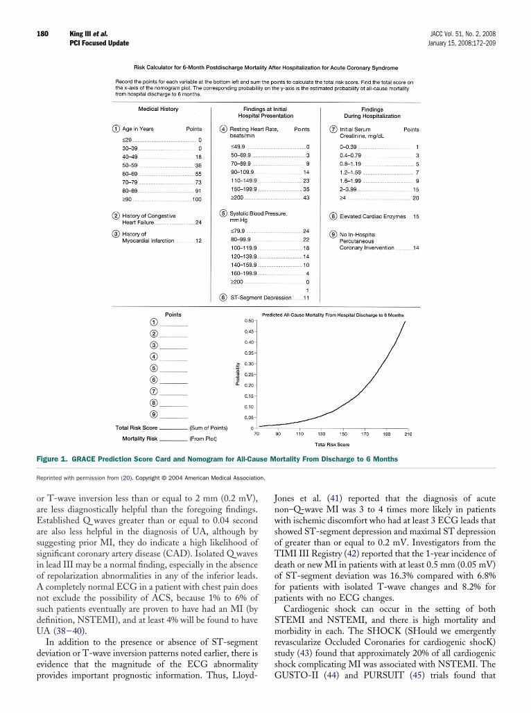
oaEassioAnsdU
dep
JnwsoTdofp
Smrss
F
R n.
180 King III et al. JACC Vol. 51, No. 2, 2008PCI Focused Update January 15, 2008:172–209
r T-wave inversion less than or equal to 2 mm (0.2 mV),re less diagnostically helpful than the foregoing findings.stablished Q waves greater than or equal to 0.04 second
re also less helpful in the diagnosis of UA, although byuggesting prior MI, they do indicate a high likelihood ofignificant coronary artery disease (CAD). Isolated Q wavesn lead III may be a normal finding, especially in the absencef repolarization abnormalities in any of the inferior leads.completely normal ECG in a patient with chest pain does
ot exclude the possibility of ACS, because 1% to 6% ofuch patients eventually are proven to have had an MI (byefinition, NSTEMI), and at least 4% will be found to haveA (38–40).In addition to the presence or absence of ST-segment
eviation or T-wave inversion patterns noted earlier, there isvidence that the magnitude of the ECG abnormality
igure 1. GRACE Prediction Score Card and Nomogram for All-Cau
eprinted with permission from (20). Copyright © 2004 American Medical Associatio
rovides important prognostic information. Thus, Lloyd- G
ones et al. (41) reported that the diagnosis of acuteon–Q-wave MI was 3 to 4 times more likely in patientsith ischemic discomfort who had at least 3 ECG leads that
howed ST-segment depression and maximal ST depressionf greater than or equal to 0.2 mV. Investigators from theIMI III Registry (42) reported that the 1-year incidence ofeath or new MI in patients with at least 0.5 mm (0.05 mV)f ST-segment deviation was 16.3% compared with 6.8%or patients with isolated T-wave changes and 8.2% foratients with no ECG changes.Cardiogenic shock can occur in the setting of both
TEMI and NSTEMI, and there is high mortality andorbidity in each. The SHOCK (SHould we emergently
evascularize Occluded Coronaries for cardiogenic shocK)tudy (43) found that approximately 20% of all cardiogenichock complicating MI was associated with NSTEMI. The
ortality From Discharge to 6 Months
se MUSTO-II (44) and PURSUIT (45) trials found that
cNTo
2v
P(an[nbaawmmwcaiswvsdtapa
Ibsh
ririoLttpipatp(mpdTasacstcm
riIrbd
T
H
E
E
C
Mrdiogra
181JACC Vol. 51, No. 2, 2008 King III et al.January 15, 2008:172–209 PCI Focused Update
ardiogenic shock occurs in up to 5% of patients withSTEMI and that mortality rates are greater than 60%.hus, hypotension and evidence of organ hypoperfusion canccur and constitute a medical emergency in NSTEMI.
.1.1. Comparison of Early Invasive and Initial Conser-ative Strategies for UA/NSTEMI
rior meta-analyses concluded that routine invasive therapythe “invasive” or “early” strategy triages patients to undergon invasive diagnostic evaluation without first getting aoninvasive stress test or without failing medical treatmenti.e., an initial conservative diagnostic strategy or sometimesow known as the “selective invasive strategy”] (14)) isetter than an initial conservative or selectively invasivepproach (the “initial conservative strategy” [also referred tos “selective invasive management”] calls for proceedingith an invasive evaluation only for those patients who failedical therapy [refractory angina or angina at rest or withinimal activity despite rigorous medical therapy] or inhom objective evidence of ischemia [dynamic ECG
hanges, high-risk stress test] is identified (14)). Mehta etl. (47) concluded that the routine invasive strategy resultedn an 18% relative reduction in death or MI, including aignificant reduction in MI alone. The routine invasive armas associated with higher in-hospital mortality (1.8%ersus 1.1%), but this disadvantage was more than compen-ated for by a significant reduction in mortality betweenischarge and the end of follow-up (3.8% versus 4.9%). Inhose analyses, the invasive strategy was associated with lessngina and fewer rehospitalizations than the conservativeathway. Patients undergoing routine invasive treatmentlso had improved quality of life.
In contrast to these findings, other studies, most recentlyCTUS (Invasive versus Conservative Treatment in Unsta-le coronary Syndromes), have favorably highlighted atrategy of selective invasive therapy (48). In ICTUS, 1200
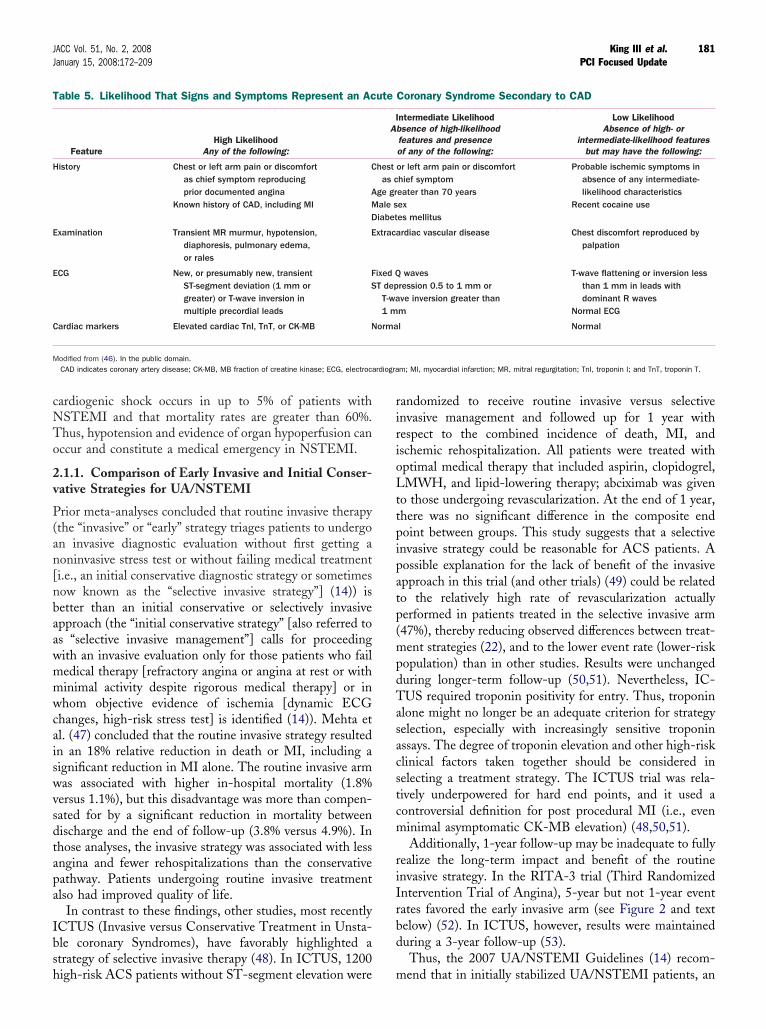
able 5. Likelihood That Signs and Symptoms Represent an Ac
FeatureHigh Likelihood
Any of the following:
istory Chest or left arm pain or discomfortas chief symptom reproducingprior documented angina
Known history of CAD, including MI
C
AMD
xamination Transient MR murmur, hypotension,diaphoresis, pulmonary edema,or rales
E
CG New, or presumably new, transientST-segment deviation (1 mm orgreater) or T-wave inversion inmultiple precordial leads
FS
ardiac markers Elevated cardiac TnI, TnT, or CK-MB N
odified from (46). In the public domain.CAD indicates coronary artery disease; CK-MB, MB fraction of creatine kinase; ECG, electroca
igh-risk ACS patients without ST-segment elevation were m
andomized to receive routine invasive versus selectivenvasive management and followed up for 1 year withespect to the combined incidence of death, MI, andschemic rehospitalization. All patients were treated withptimal medical therapy that included aspirin, clopidogrel,MWH, and lipid-lowering therapy; abciximab was given
o those undergoing revascularization. At the end of 1 year,here was no significant difference in the composite endoint between groups. This study suggests that a selectivenvasive strategy could be reasonable for ACS patients. Aossible explanation for the lack of benefit of the invasivepproach in this trial (and other trials) (49) could be relatedo the relatively high rate of revascularization actuallyerformed in patients treated in the selective invasive arm47%), thereby reducing observed differences between treat-ent strategies (22), and to the lower event rate (lower-risk
opulation) than in other studies. Results were unchangeduring longer-term follow-up (50,51). Nevertheless, IC-US required troponin positivity for entry. Thus, troponin
lone might no longer be an adequate criterion for strategyelection, especially with increasingly sensitive troponinssays. The degree of troponin elevation and other high-risklinical factors taken together should be considered inelecting a treatment strategy. The ICTUS trial was rela-ively underpowered for hard end points, and it used aontroversial definition for post procedural MI (i.e., eveninimal asymptomatic CK-MB elevation) (48,50,51).Additionally, 1-year follow-up may be inadequate to fully
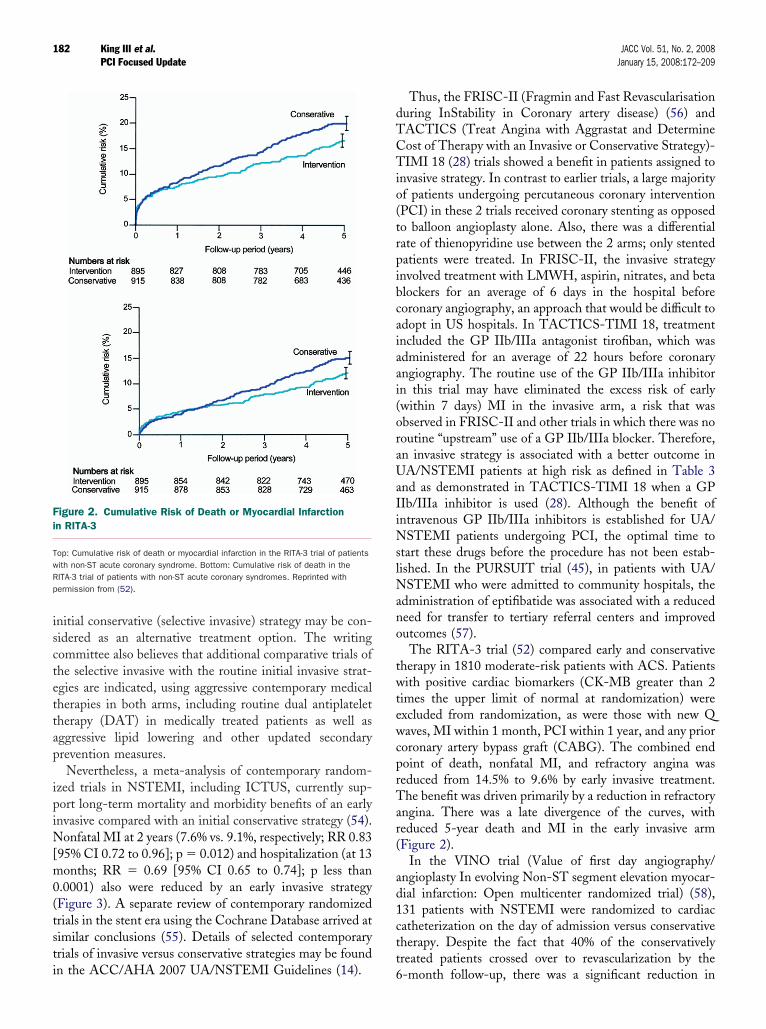
ealize the long-term impact and benefit of the routinenvasive strategy. In the RITA-3 trial (Third Randomizedntervention Trial of Angina), 5-year but not 1-year eventates favored the early invasive arm (see Figure 2 and textelow) (52). In ICTUS, however, results were maintaineduring a 3-year follow-up (53).Thus, the 2007 UA/NSTEMI Guidelines (14) recom-
Coronary Syndrome Secondary to CAD
ntermediate Likelihoodsence of high-likelihood
features and presenceof any of the following:
Low LikelihoodAbsence of high- or
intermediate-likelihood featuresbut may have the following:
or left arm pain or discomforthief symptomeater than 70 yearsexes mellitus
Probable ischemic symptoms inabsence of any intermediate-likelihood characteristics
Recent cocaine use
rdiac vascular disease Chest discomfort reproduced bypalpation
wavesression 0.5 to 1 mm orve inversion greater thanm
T-wave flattening or inversion lessthan 1 mm in leads withdominant R waves
Normal ECG
l Normal
m; MI, myocardial infarction; MR, mitral regurgitation; TnI, troponin I; and TnT, troponin T.
ute
IAb
hestas c
ge grale siabet
xtraca
ixed QT depT-wa1 m
orma
end that in initially stabilized UA/NSTEMI patients, an
isctettap
ipiN[m0(tsti
dTCTio(trpibcaiaai(oraUaIiNslNano
twtewcprTar(
ad1ctt
Fi
TwRp
182 King III et al. JACC Vol. 51, No. 2, 2008PCI Focused Update January 15, 2008:172–209
nitial conservative (selective invasive) strategy may be con-idered as an alternative treatment option. The writingommittee also believes that additional comparative trials ofhe selective invasive with the routine initial invasive strat-gies are indicated, using aggressive contemporary medicalherapies in both arms, including routine dual antiplateletherapy (DAT) in medically treated patients as well asggressive lipid lowering and other updated secondaryrevention measures.Nevertheless, a meta-analysis of contemporary random-
zed trials in NSTEMI, including ICTUS, currently sup-ort long-term mortality and morbidity benefits of an earlynvasive compared with an initial conservative strategy (54).
onfatal MI at 2 years (7.6% vs. 9.1%, respectively; RR 0.8395% CI 0.72 to 0.96]; p � 0.012) and hospitalization (at 13onths; RR � 0.69 [95% CI 0.65 to 0.74]; p less than
.0001) also were reduced by an early invasive strategyFigure 3). A separate review of contemporary randomizedrials in the stent era using the Cochrane Database arrived atimilar conclusions (55). Details of selected contemporaryrials of invasive versus conservative strategies may be found
igure 2. Cumulative Risk of Death or Myocardial Infarctionn RITA-3
op: Cumulative risk of death or myocardial infarction in the RITA-3 trial of patientsith non-ST acute coronary syndrome. Bottom: Cumulative risk of death in theITA-3 trial of patients with non-ST acute coronary syndromes. Reprinted withermission from (52).
n the ACC/AHA 2007 UA/NSTEMI Guidelines (14). 6
Thus, the FRISC-II (Fragmin and Fast Revascularisationuring InStability in Coronary artery disease) (56) andACTICS (Treat Angina with Aggrastat and Determineost of Therapy with an Invasive or Conservative Strategy)-IMI 18 (28) trials showed a benefit in patients assigned to
nvasive strategy. In contrast to earlier trials, a large majorityf patients undergoing percutaneous coronary interventionPCI) in these 2 trials received coronary stenting as opposedo balloon angioplasty alone. Also, there was a differentialate of thienopyridine use between the 2 arms; only stentedatients were treated. In FRISC-II, the invasive strategynvolved treatment with LMWH, aspirin, nitrates, and betalockers for an average of 6 days in the hospital beforeoronary angiography, an approach that would be difficult todopt in US hospitals. In TACTICS-TIMI 18, treatmentncluded the GP IIb/IIIa antagonist tirofiban, which wasdministered for an average of 22 hours before coronaryngiography. The routine use of the GP IIb/IIIa inhibitorn this trial may have eliminated the excess risk of earlywithin 7 days) MI in the invasive arm, a risk that wasbserved in FRISC-II and other trials in which there was nooutine “upstream” use of a GP IIb/IIIa blocker. Therefore,n invasive strategy is associated with a better outcome inA/NSTEMI patients at high risk as defined in Table 3
nd as demonstrated in TACTICS-TIMI 18 when a GPIb/IIIa inhibitor is used (28). Although the benefit ofntravenous GP IIb/IIIa inhibitors is established for UA/
STEMI patients undergoing PCI, the optimal time totart these drugs before the procedure has not been estab-ished. In the PURSUIT trial (45), in patients with UA/
STEMI who were admitted to community hospitals, thedministration of eptifibatide was associated with a reducedeed for transfer to tertiary referral centers and improvedutcomes (57).The RITA-3 trial (52) compared early and conservative
herapy in 1810 moderate-risk patients with ACS. Patientsith positive cardiac biomarkers (CK-MB greater than 2
imes the upper limit of normal at randomization) werexcluded from randomization, as were those with new Qaves, MI within 1 month, PCI within 1 year, and any prior
oronary artery bypass graft (CABG). The combined endoint of death, nonfatal MI, and refractory angina waseduced from 14.5% to 9.6% by early invasive treatment.he benefit was driven primarily by a reduction in refractory
ngina. There was a late divergence of the curves, witheduced 5-year death and MI in the early invasive armFigure 2).
In the VINO trial (Value of first day angiography/ngioplasty In evolving Non-ST segment elevation myocar-ial infarction: Open multicenter randomized trial) (58),31 patients with NSTEMI were randomized to cardiacatheterization on the day of admission versus conservativeherapy. Despite the fact that 40% of the conservativelyreated patients crossed over to revascularization by the
-month follow-up, there was a significant reduction in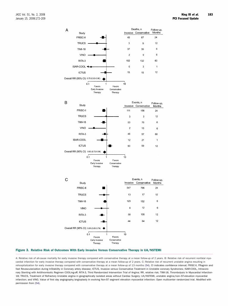
F
Acrfn1ip
183JACC Vol. 51, No. 2, 2008 King III et al.January 15, 2008:172–209 PCI Focused Update
igure 3. Relative Risk of Outcomes With Early Invasive Versus Conservative Therapy in UA/NSTEMI
: Relative risk of all-cause mortality for early invasive therapy compared with conservative therapy at a mean follow-up of 2 years. B: Relative risk of recurrent nonfatal myo-ardial infarction for early invasive therapy compared with conservative therapy at a mean follow-up of 2 years. C: Relative risk of recurrent unstable angina resulting inehospitalization for early invasive therapy compared with conservative therapy at a mean follow-up of 13 months (54). CI indicates confidence interval; FRISC-II, FRagmin andast Revascularization during InStability in Coronary artery disease; ICTUS, Invasive versus Conservative Treatment in Unstable coronary Syndromes; ISAR-COOL, Intracoro-ary Stenting with Antithrombotic Regimen COOLing-off; RITA-3, Third Randomized Intervention Trial of Angina; RR, relative risk; TIMI-18, Thrombolysis In Myocardial Infarction-8; TRUCS, Treatment of Refractory Unstable angina in geographically isolated areas without Cardiac Surgery; UA/NSTEMI, unstable angina/non–ST-elevation myocardial
nfarction; and VINO, Value of first day angiography/angioplasty In evolving Non-ST segment elevation myocardial infarction: Open multicenter randomized trial. Modified withermission from (54).
dr
t4retdtciwavata
2
Ip
omrfvP
wiruLaPCnip(tsc
TA
E
R
F
M
TAP
A
A
C
T
H
I
L
*0
184 King III et al. JACC Vol. 51, No. 2, 2008PCI Focused Update January 15, 2008:172–209
eath or reinfarction for patients assigned to early angiog-aphy and revascularization (6% versus 22%).
The ISAR-COOL (Intracoronary Stenting with Anti-hrombotic Regimen Cooling-off) trial (59) randomized10 intermediate- to high-risk patients to very early angiog-aphy and revascularization versus a delayed invasive strat-gy. All patients were treated with intensive medical therapyhat included aspirin, heparin, clopidogrel (600-mg loadingose), and the intravenous GP IIb/IIIa receptor inhibitorirofiban. In the very early arm, patients underwent cardiacatheterization at a mean time of 2.4 hours versus 86 hoursn the delayed invasive arm. The very early invasive strategyas associated with significantly better outcome at 30 days,
s measured by reduction in death and large MI (5.9%ersus 11.6%). More importantly, the benefit seen wasttributable to a reduction in events before cardiac cathe-erization, which raises the possibility that there is a hazardssociated with a “cooling-down” period.
.1.2. Selection for Coronary Angiography
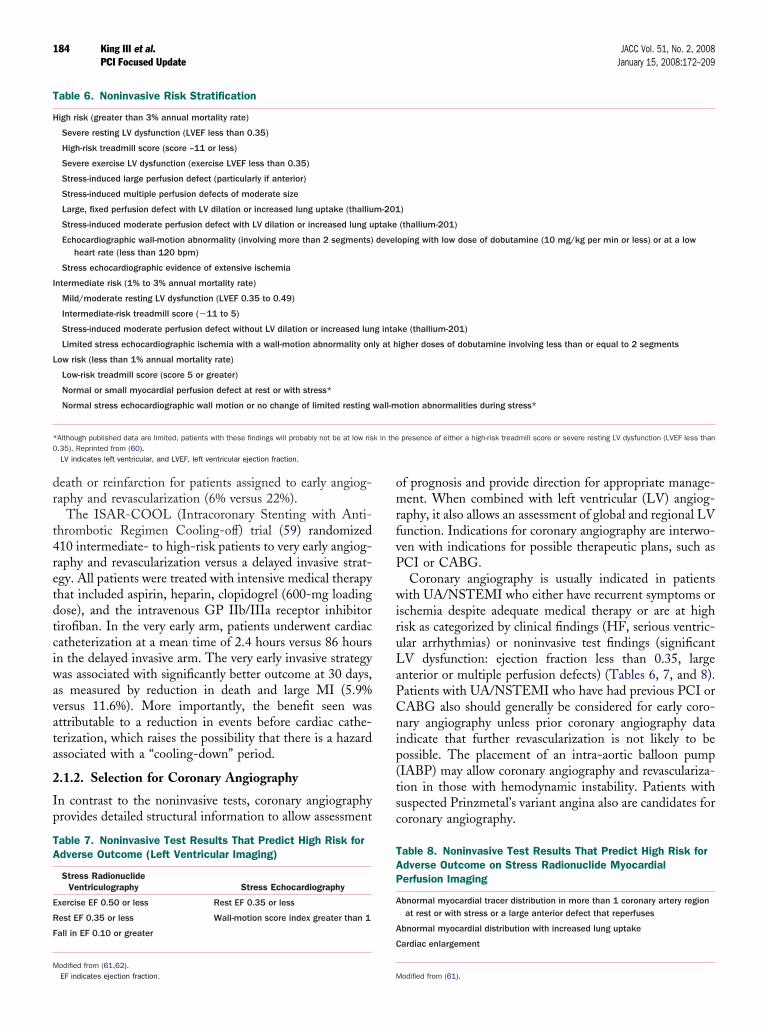
n contrast to the noninvasive tests, coronary angiographyrovides detailed structural information to allow assessment
able 7. Noninvasive Test Results That Predict High Risk fordverse Outcome (Left Ventricular Imaging)
Stress RadionuclideVentriculography Stress Echocardiography
xercise EF 0.50 or less Rest EF 0.35 or less
est EF 0.35 or less Wall-motion score index greater than 1
all in EF 0.10 or greater
able 6. Noninvasive Risk Stratification
igh risk (greater than 3% annual mortality rate)
Severe resting LV dysfunction (LVEF less than 0.35)
High-risk treadmill score (score –11 or less)
Severe exercise LV dysfunction (exercise LVEF less than 0.35)
Stress-induced large perfusion defect (particularly if anterior)
Stress-induced multiple perfusion defects of moderate size
Large, fixed perfusion defect with LV dilation or increased lung uptake (thalliu
Stress-induced moderate perfusion defect with LV dilation or increased lung u
Echocardiographic wall-motion abnormality (involving more than 2 segments)heart rate (less than 120 bpm)
Stress echocardiographic evidence of extensive ischemia
ntermediate risk (1% to 3% annual mortality rate)
Mild/moderate resting LV dysfunction (LVEF 0.35 to 0.49)
Intermediate-risk treadmill score (�11 to 5)
Stress-induced moderate perfusion defect without LV dilation or increased lun
Limited stress echocardiographic ischemia with a wall-motion abnormality on
ow risk (less than 1% annual mortality rate)
Low-risk treadmill score (score 5 or greater)
Normal or small myocardial perfusion defect at rest or with stress*
Normal stress echocardiographic wall motion or no change of limited resting
Although published data are limited, patients with these findings will probably not be at low ris.35). Reprinted from (60).LV indicates left ventricular, and LVEF, left ventricular ejection fraction.
odified from (61,62).EF indicates ejection fraction. M
f prognosis and provide direction for appropriate manage-ent. When combined with left ventricular (LV) angiog-
aphy, it also allows an assessment of global and regional LVunction. Indications for coronary angiography are interwo-en with indications for possible therapeutic plans, such asCI or CABG.Coronary angiography is usually indicated in patients
ith UA/NSTEMI who either have recurrent symptoms orschemia despite adequate medical therapy or are at highisk as categorized by clinical findings (HF, serious ventric-lar arrhythmias) or noninvasive test findings (significantV dysfunction: ejection fraction less than 0.35, largenterior or multiple perfusion defects) (Tables 6, 7, and 8).atients with UA/NSTEMI who have had previous PCI orABG also should generally be considered for early coro-ary angiography unless prior coronary angiography data
ndicate that further revascularization is not likely to beossible. The placement of an intra-aortic balloon pumpIABP) may allow coronary angiography and revasculariza-ion in those with hemodynamic instability. Patients withuspected Prinzmetal’s variant angina also are candidates fororonary angiography.
able 8. Noninvasive Test Results That Predict High Risk fordverse Outcome on Stress Radionuclide Myocardialerfusion Imaging
bnormal myocardial tracer distribution in more than 1 coronary artery regionat rest or with stress or a large anterior defect that reperfuses
bnormal myocardial distribution with increased lung uptake
ardiac enlargement
)
(thallium-201)
oping with low dose of dobutamine (10 mg/kg per min or less) or at a low
ke (thallium-201)
igher doses of dobutamine involving less than or equal to 2 segments
otion abnormalities during stress*
presence of either a high-risk treadmill score or severe resting LV dysfunction (LVEF less than
m-201
ptake
devel
g inta
ly at h
wall-m
k in the
odified from (61).
rpcictp
2
TPd(m
efdaoGpCer1pd(adatccnpuP
adaoabictP
T
*
185JACC Vol. 51, No. 2, 2008 King III et al.January 15, 2008:172–209 PCI Focused Update
In all cases, the general indications for coronary angiog-aphy and revascularization are tempered by individualatient characteristics and preferences. Patient and physi-ian judgments regarding risks and benefits are particularlymportant for patients who might not be candidates fororonary revascularization, such as very frail older adults andhose with serious comorbid conditions (i.e., severe hepatic,
CI in patients with CKD is associated with a higher rate
odipb
caiaciT(BPAic
tpuwFsgmfimFupa
m
MI, un
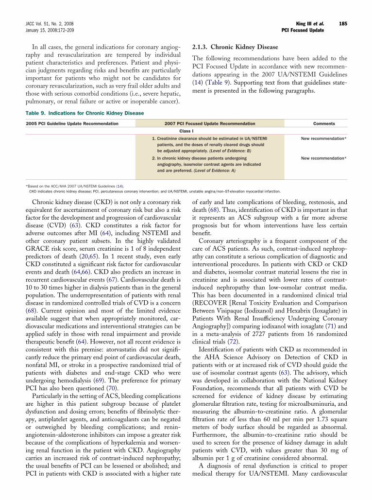
.1.3. Chronic Kidney Disease
he following recommendations have been added to theCI Focused Update in accordance with new recommen-ations appearing in the 2007 UA/NSTEMI Guidelines14) (Table 9). Supporting text from that guidelines state-ent is presented in the following paragraphs.
ulmonary, or renal failure or active or inoperable cancer).
Chronic kidney disease (CKD) is not only a coronary riskquivalent for ascertainment of coronary risk but also a riskactor for the development and progression of cardiovascularisease (CVD) (63). CKD constitutes a risk factor fordverse outcomes after MI (64), including NSTEMI andther coronary patient subsets. In the highly validatedRACE risk score, serum creatinine is 1 of 8 independent
redictors of death (20,65). In 1 recent study, even earlyKD constituted a significant risk factor for cardiovascular
vents and death (64,66). CKD also predicts an increase inecurrent cardiovascular events (67). Cardiovascular death is0 to 30 times higher in dialysis patients than in the generalopulation. The underrepresentation of patients with renalisease in randomized controlled trials of CVD is a concern68). Current opinion and most of the limited evidencevailable suggest that when appropriately monitored, car-iovascular medications and interventional strategies can bepplied safely in those with renal impairment and provideherapeutic benefit (64). However, not all recent evidence isonsistent with this premise: atorvastatin did not signifi-antly reduce the primary end point of cardiovascular death,onfatal MI, or stroke in a prospective randomized trial ofatients with diabetes and end-stage CKD who werendergoing hemodialysis (69). The preference for primaryCI has also been questioned (70).Particularly in the setting of ACS, bleeding complications
re higher in this patient subgroup because of plateletysfunction and dosing errors; benefits of fibrinolytic ther-py, antiplatelet agents, and anticoagulants can be negatedr outweighed by bleeding complications; and renin-ngiotensin-aldosterone inhibitors can impose a greater riskecause of the complications of hyperkalemia and worsen-ng renal function in the patient with CKD. Angiographyarries an increased risk of contrast-induced nephropathy;he usual benefits of PCI can be lessened or abolished; and
2005 PCI Guideline Update Recommendation 2007 PC
C
1. Creatinine clpatients, andbe adjusted
2. In chronic kiangiographyand are pref
Based on the ACC/AHA 2007 UA/NSTEMI Guidelines (14).CKD indicates chronic kidney disease; PCI, percutaneous coronary intervention; and UA/NSTE
f early and late complications of bleeding, restenosis, andeath (68). Thus, identification of CKD is important in thatt represents an ACS subgroup with a far more adverserognosis but for whom interventions have less certainenefit.Coronary arteriography is a frequent component of the
are of ACS patients. As such, contrast-induced nephrop-thy can constitute a serious complication of diagnostic andnterventional procedures. In patients with CKD or CKDnd diabetes, isosmolar contrast material lessens the rise inreatinine and is associated with lower rates of contrast-nduced nephropathy than low-osmolar contrast media.his has been documented in a randomized clinical trial
RECOVER [Renal Toxicity Evaluation and Comparisonetween Visipaque (Iodixanol) and Hexabrix (Ioxaglate) inatients With Renal Insufficiency Undergoing Coronaryngiography]) comparing iodixanol with ioxaglate (71) and
n a meta-analysis of 2727 patients from 16 randomizedlinical trials (72).
Identification of patients with CKD as recommended inhe AHA Science Advisory on Detection of CKD inatients with or at increased risk of CVD should guide these of isosmolar contrast agents (63). The advisory, whichas developed in collaboration with the National Kidneyoundation, recommends that all patients with CVD becreened for evidence of kidney disease by estimatinglomerular filtration rate, testing for microalbuminuria, andeasuring the albumin-to-creatinine ratio. A glomerular
ltration rate of less than 60 ml per min per 1.73 squareeters of body surface should be regarded as abnormal.urthermore, the albumin-to-creatinine ratio should besed to screen for the presence of kidney damage in adultatients with CVD, with values greater than 30 mg oflbumin per 1 g of creatinine considered abnormal.
A diagnosis of renal dysfunction is critical to proper
used Update Recommendation Comments
ce should be estimated in UA/NSTEMIoses of renally cleared drugs should
priately. (Level of Evidence: B)
New recommendation*
isease patients undergoingolar contrast agents are indicated(Level of Evidence: A)
New recommendation*
stable angina/non–ST-elevation myocardial infarction.
able 9. Indications for Chronic Kidney Disease
I Foc
lass I
earanthe d
appro
dney d, isosmerred.
edical therapy for UA/NSTEMI. Many cardiovascular
dciNretoipst
mi(griG
coa
3
FPrprdI(PfiiPPtsahtcc
ti
T
F
L
186 King III et al. JACC Vol. 51, No. 2, 2008PCI Focused Update January 15, 2008:172–209
rugs used in patients with UA/NSTEMI are renallyleared; their doses should be adjusted for estimated creat-nine clearance [see also Section 3 of the 2007 UA/
STEMI Guidelines (14)]. In a large community-basedegistry study, 42% of patients with UA/NSTEMI receivedxcessive initial dosing of at least 1 antiplatelet or anti-hrombin agent (unfractionated heparin [UFH], LMWH,r GP IIb/IIIa inhibitor) (73). Renal insufficiency was anndependent predictor of excessive dosing. Dosing errorsredicted an increased risk of major bleeding. Clinicaltudies and labeling that defines adjustments for several of
. Facilitated PCI
nfarct size or improving outcomes. The largest of these was
tNtrmagcwtt4pbpsttmhmtr
ion myo
ula for estimating creatinine clearance, which is notdentical to the Modification of Diet and Renal DiseaseMDRD) formula. Use of the Cockcroft-Gault formula toenerate dose adjustments is recommended. The impact ofenal dysfunction on biomarkers of necrosis (i.e., troponin)s discussed in Section 2.2.8.2.1 of the 2007 UA/NSTEMI
uidelines (14).To increase the meager evidence base and to optimize
are for this growing high-risk population, the recognitionf CKD patients with or at risk of CVD and the inclusionnd reporting of renal disease in large CVD trials must be
hese drugs have been based on the Cockcroft-Gault for- increased in the future.
acilitated PCI refers to a strategy of planned immediateCI after administration of an initial pharmacological
egimen intended to improve coronary patency before therocedure. These regimens have included high-dose hepa-in, platelet GP IIb/IIIa inhibitors, full-dose or reduced-ose fibrinolytic therapy, and the combination of a GPIb/IIIa inhibitor with a reduced-dose fibrinolytic agente.g., fibrinolytic dose typically reduced 50%). FacilitatedCI should be differentiated from primary PCI withoutbrinolytic therapy, from primary PCI with a GP IIb/IIIa
nhibitor started at the time of PCI, from early or delayedCI after successful fibrinolytic therapy, and from rescueCI after unsuccessful fibrinolytic therapy. Potential advan-
ages of facilitated PCI include earlier time to reperfusion,maller infarct size, improved patient stability, lower infarctrtery thrombus burden, greater procedural success rates,igher TIMI flow rates, and improved survival rates. Po-ential risks include increased bleeding complications, espe-ially in older patients; potential limitations include addedost.
Despite the potential advantages, clinical trials of facili-ated PCI have not demonstrated any benefit in reducing
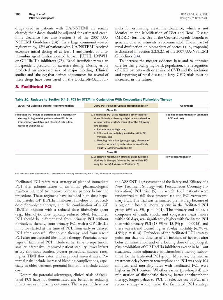
able 10. Updates to Section 5.4.3: PCI for STEMI in Conjunct
2005 PCI Guideline Update Recommendation 2007 PCI Fo
Cl
acilitated PCI might be performed as a reperfusionstrategy in higher-risk patients when PCI is notimmediately available and bleeding risk is low.(Level of Evidence: B)
1. Facilitated Pdose fibrinola reperfusionare present:a. Patients ab. PCI is not
minutes, ac. Bleeding r
poorly conweight). (L
C
1. A planned refibrinolytic thmay be harm
OE indicates level of evidence; PCI, percutaneous coronary intervention; and STEMI, ST-elevat
he ASSENT-4 (Assessment of the Safety and Efficacy of aew Treatment Strategy with Percutaneous Coronary In-
ervention) PCI trial (5), in which 1667 patients wereandomized to full-dose tenecteplase and PCI versus pri-ary PCI. The trial was terminated prematurely because ofhigher in-hospital mortality rate in the facilitated PCI
roup (6% vs. 3%, p � 0.01). The primary end point, aomposite of death, shock, and congestive heart failureithin 90 days, was significantly higher with facilitated PCI
han with primary PCI (18.6% vs. 13.4%; p � 0.0045), andhere was a trend toward higher 90-day mortality (6.7% vs..9%; p � 0.14). Defenders of the facilitated PCI strategyoint out that the absence of an infusion of heparin afterolus administration and of a loading dose of clopidogrel,lus prohibition of GP IIb/IIIa inhibitors except in bail-outituations, made adjunctive antithrombotic therapy subop-imal for the facilitated PCI group. Moreover, the medianreatment delay between tenecteplase and PCI was only 104inutes, and mortality rates with facilitated PCI were
igher in PCI centers. Whether earlier (pre-hospital) ad-inistration of fibrinolytic therapy, better antithrombotic
herapy, longer delays to PCI, or selective use of PCI as a
ith Concomitant Fibrinolytic Therapy
Update Recommendation Comments
b
g regimens other than full-erapy might be considered asegy when all of the following
igh risk,diately available within 90
ow (younger age, absence ofhypertension, normal body
f Evidence: C)
Modified recommendation (changedLOE and text)
I
ion strategy using full-dosefollowed by immediate PCIevel of Evidence: B)
New recommendation
cardial infarction.
ion W
cused
ass II
CI usinytic th
strat
re at himmendisk is ltrolledevel o
lass II
perfuserapyful. (L
escue strategy would make the facilitated PCI strategy
bob
1(i(aFhvmdG
t1whotwApdiwtpr6MI
nicqwoh(itigsSt
4
InsrihtusoEbn
F
T tios. L
187JACC Vol. 51, No. 2, 2008 King III et al.January 15, 2008:172–209 PCI Focused Update
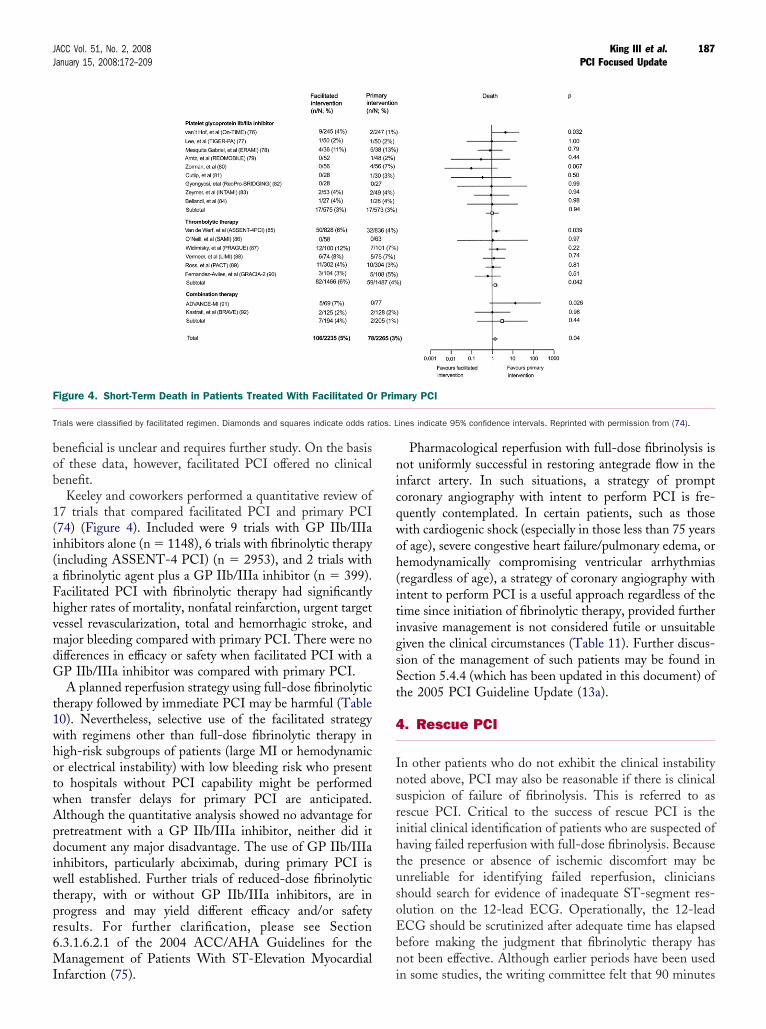
eneficial is unclear and requires further study. On the basisf these data, however, facilitated PCI offered no clinicalenefit.Keeley and coworkers performed a quantitative review of
7 trials that compared facilitated PCI and primary PCI74) (Figure 4). Included were 9 trials with GP IIb/IIIanhibitors alone (n � 1148), 6 trials with fibrinolytic therapyincluding ASSENT-4 PCI) (n � 2953), and 2 trials withfibrinolytic agent plus a GP IIb/IIIa inhibitor (n � 399).acilitated PCI with fibrinolytic therapy had significantlyigher rates of mortality, nonfatal reinfarction, urgent targetessel revascularization, total and hemorrhagic stroke, andajor bleeding compared with primary PCI. There were no
ifferences in efficacy or safety when facilitated PCI with aP IIb/IIIa inhibitor was compared with primary PCI.A planned reperfusion strategy using full-dose fibrinolytic
herapy followed by immediate PCI may be harmful (Table0). Nevertheless, selective use of the facilitated strategyith regimens other than full-dose fibrinolytic therapy inigh-risk subgroups of patients (large MI or hemodynamicr electrical instability) with low bleeding risk who presento hospitals without PCI capability might be performedhen transfer delays for primary PCI are anticipated.lthough the quantitative analysis showed no advantage forretreatment with a GP IIb/IIIa inhibitor, neither did itocument any major disadvantage. The use of GP IIb/IIIa
nhibitors, particularly abciximab, during primary PCI isell established. Further trials of reduced-dose fibrinolytic
herapy, with or without GP IIb/IIIa inhibitors, are inrogress and may yield different efficacy and/or safetyesults. For further clarification, please see Section.3.1.6.2.1 of the 2004 ACC/AHA Guidelines for theanagement of Patients With ST-Elevation Myocardial
igure 4. Short-Term Death in Patients Treated With Facilitated O
rials were classified by facilitated regimen. Diamonds and squares indicate odds ra
nfarction (75). i
Pharmacological reperfusion with full-dose fibrinolysis isot uniformly successful in restoring antegrade flow in the
nfarct artery. In such situations, a strategy of promptoronary angiography with intent to perform PCI is fre-uently contemplated. In certain patients, such as thoseith cardiogenic shock (especially in those less than 75 yearsf age), severe congestive heart failure/pulmonary edema, oremodynamically compromising ventricular arrhythmiasregardless of age), a strategy of coronary angiography withntent to perform PCI is a useful approach regardless of theime since initiation of fibrinolytic therapy, provided furthernvasive management is not considered futile or unsuitableiven the clinical circumstances (Table 11). Further discus-ion of the management of such patients may be found inection 5.4.4 (which has been updated in this document) ofhe 2005 PCI Guideline Update (13a).
. Rescue PCI
n other patients who do not exhibit the clinical instabilityoted above, PCI may also be reasonable if there is clinicaluspicion of failure of fibrinolysis. This is referred to asescue PCI. Critical to the success of rescue PCI is thenitial clinical identification of patients who are suspected ofaving failed reperfusion with full-dose fibrinolysis. Becausehe presence or absence of ischemic discomfort may benreliable for identifying failed reperfusion, clinicianshould search for evidence of inadequate ST-segment res-lution on the 12-lead ECG. Operationally, the 12-leadCG should be scrutinized after adequate time has elapsedefore making the judgment that fibrinolytic therapy hasot been effective. Although earlier periods have been used
ary PCI
ines indicate 95% confidence intervals. Reprinted with permission from (74).
r Prim
n some studies, the writing committee felt that 90 minutes
T
R
R
R
I
R
CS
188 King III et al. JACC Vol. 51, No. 2, 2008PCI Focused Update January 15, 2008:172–209
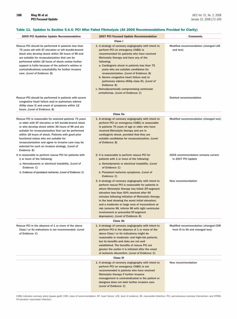
able 11. Updates to Section 5.4.4: PCI After Failed Fibrinolysis (All 2005 Recommendations Provided for Clarity)
2005 PCI Guideline Update Recommendation 2007 PCI Focused Update Recommendation Comments
Class I
escue PCI should be performed in patients less than75 years old with ST elevation or left bundle-branchblock who develop shock within 36 hours of MI andare suitable for revascularization that can beperformed within 18 hours of shock unless furthersupport is futile because of the patient’s wishes orcontraindications/unsuitability for further invasivecare. (Level of Evidence: B)
1. A strategy of coronary angiography with intent toperform PCI (or emergency CABG) isrecommended for patients who have receivedfibrinolytic therapy and have any of thefollowing:a. Cardiogenic shock in patients less than 75
years who are suitable candidates forrevascularization. (Level of Evidence: B)
b. Severe congestive heart failure and/orpulmonary edema (Killip class III). (Level ofEvidence: B)
c. Hemodynamically compromising ventriculararrhythmias. (Level of Evidence: C)
Modified recommendation (changed LOEand text)
escue PCI should be performed in patients with severecongestive heart failure and/or pulmonary edema(Killip class 3) and onset of symptoms within 12hours. (Level of Evidence: B)
Deleted recommendation
Class IIa
escue PCI is reasonable for selected patients 75 yearsor older with ST elevation or left bundle-branch blockor who develop shock within 36 hours of MI and aresuitable for revascularization that can be performedwithin 18 hours of shock. Patients with good priorfunctional status who are suitable forrevascularization and agree to invasive care may beselected for such an invasive strategy. (Level ofEvidence: B)
1. A strategy of coronary angiography with intent toperform PCI (or emergency CABG) is reasonablein patients 75 years of age or older who havereceived fibrinolytic therapy and are incardiogenic shock, provided that they aresuitable candidates for revascularization. (Levelof Evidence: B)
Modified recommendation (changed text)
t is reasonable to perform rescue PCI for patients with1 or more of the following:
2. It is reasonable to perform rescue PCI forpatients with 1 or more of the following:
2005 recommendation remains currentin 2007 PCI Update
a. Hemodynamic or electrical instability. (Level ofEvidence: C)
a. Hemodynamic or electrical instability. (Levelof Evidence: C)
b. Evidence of persistent ischemia. (Level of Evidence: C) b. Persistent ischemic symptoms. (Level ofEvidence: C)
3. A strategy of coronary angiography with intent toperform rescue PCI is reasonable for patients inwhom fibrinolytic therapy has failed (ST-segmentelevation less than 50% resolved after 90minutes following initiation of fibrinolytic therapyin the lead showing the worst initial elevation)and a moderate or large area of myocardium atrisk (anterior MI, inferior MI with right ventricularinvolvement or precordial ST-segmentdepression). (Level of Evidence: B)
New recommendation
Class IIb
escue PCI in the absence of 1 or more of the aboveClass I or IIa indications is not recommended. (Levelof Evidence: C)
1. A strategy of coronary angiography with intent toperform PCI in the absence of 1 or more of theabove Class I or IIa indications might bereasonable in moderate- and high-risk patients,but its benefits and risks are not wellestablished. The benefits of rescue PCI aregreater the earlier it is initiated after the onsetof ischemic discomfort. (Level of Evidence: C)
Modified recommendation (changed CORfrom III to IIb and changed text)
Class III
1. A strategy of coronary angiography with intent toperform PCI (or emergency CABG) is notrecommended in patients who have receivedfibrinolytic therapy if further invasivemanagement is contraindicated or the patient ordesignee does not wish further invasive care.(Level of Evidence: C)
New recommendation
ABG indicates coronary artery bypass graft; COR, class of recommendation; HF, heart failure; LOE, level of evidence; MI, myocardial infarction; PCI, percutaneous coronary intervention; and STEMI,T-elevation myocardial infarction.
aetdt
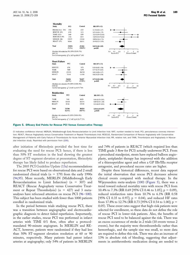
fr(RRmaTe
wgiamAtma
aTapoa
tcWt1r[f0soraeha1
F
CtMd
189JACC Vol. 51, No. 2, 2008 King III et al.January 15, 2008:172–209 PCI Focused Update
fter initiation of fibrinolysis provided the best time forvaluating the need for rescue PCI: hence, if there is lesshan 50% ST resolution in the lead showing the greatestegree of ST-segment elevation at presentation, fibrinolyticherapy has likely failed to produce reperfusion.
The 2005 PCI Guideline Update (13a) recommendationsor rescue PCI were based on observational data and 2 smallandomized clinical trials (n � 179) from the early 1990s94,95). More recently, MERLIN (Middlesbrough Earlyevascularization to Limit Infarction) (n � 307) andEACT (Rescue Angioplasty versus Conservative Treat-ent or Repeat Thrombolysis) (n � 427) and 3 meta-
nalyses have refocused attention on rescue PCI (96–100).his subject has been studied with fewer than 1000 patients
nrolled in randomized trials.In the period between trials studying rescue PCI, there
as a transition between angiographic and electrocardio-raphic diagnosis to detect failed reperfusion. Importantly,n the earlier studies, rescue PCI was performed in infarctrteries with TIMI 0/1 flow, often after a protocol-andated 90-minute angiogram. In MERLIN and RE-CT, however, patients were randomized if they had less
han 50% ST-segment elevation resolution at 60 or 90inutes, respectively. Many patients had patent infarct
igure 5. Efficacy End Points for Rescue PCI Versus Conservative
I indicates confidence interval; MERLIN, Middlesbrough Early Revascularization to Lion; REACT, Rescue Angioplasty versus Conservative Treatment or Repeat Thrombolanagement of Patients with Early Failure of Thrombolysis for Acute Anterior Myocarial Infarction study. Reprinted with permission from (100).
rteries at angiography; only 54% of patients in MERLIN m
nd 74% of patients in REACT (which required less thanIMI grade 3 flow for PCI) actually underwent PCI. Fromprocedural standpoint, stents have replaced balloon angio-lasty, antiplatelet therapy has improved with the additionf a thienopyridine agent and often a GP IIb/IIIa receptorntagonist, and procedural success rates are higher.
Despite these historical differences, recent data supporthe initial observation that rescue PCI decreases adverselinical events compared with medical therapy. In the
ijeysundera meta-analysis (100) (Figure 5), there was arend toward reduced mortality rates with rescue PCI from0.4% to 7.3% (RR 0.69 [95% CI 0.46 to 1.05]; p � 0.09),educed reinfarction rates from 10.7% to 6.1% (RR 0.5895% CI 0.35 to 0.97]; p � 0.04), and reduced HF ratesrom 17.8% to 12.7% (RR 0.73 [95% CI 0.54 to 1.00]; p �.05). These event rates suggest that high-risk patients wereelected for enrollment, so these data do not define the rolef rescue PCI in lower-risk patients. Also, the benefits ofescue PCI need to be balanced against the risk. There wasn excess occurrence of stroke in 2 trials (10 events versus 2vents), but the majority were thromboembolic rather thanemorrhagic, and the sample size was small, so more datare required to define this risk. There was also an increase of3% in absolute risk of bleeding, suggesting that adjust-
apy
farction trial; NNT, number needed to treat; PCI, percutaneous coronary interven-ial; RESCUE, Randomized Comparison of Rescue Angioplasty with Conservativefarction trial; RR, relative risk; and TAMI, Thrombolysis and Angioplasty in Myocar-
Ther
imit Inysis trdial In
ents in antithrombotic medication dosing are needed to
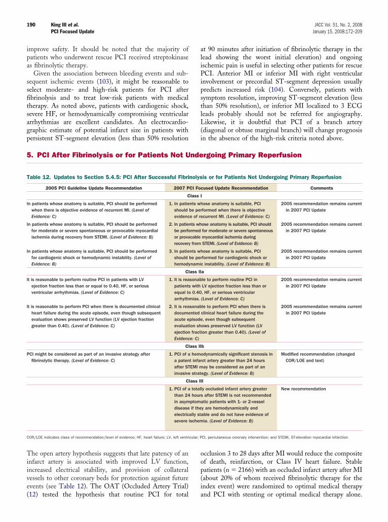
ipa
ssfitsagp
aliPipstlL(
5
Tiive(
T
I
I
I
I
I
P
C
190 King III et al. JACC Vol. 51, No. 2, 2008PCI Focused Update January 15, 2008:172–209
mprove safety. It should be noted that the majority ofatients who underwent rescue PCI received streptokinases fibrinolytic therapy.
Given the association between bleeding events and sub-equent ischemic events (103), it might be reasonable toelect moderate- and high-risk patients for PCI afterbrinolysis and to treat low-risk patients with medicalherapy. As noted above, patients with cardiogenic shock,evere HF, or hemodynamically compromising ventricularrrhythmias are excellent candidates. An electrocardio-raphic estimate of potential infarct size in patients with
. PCI After Fibrinolysis or for Patients Not Unde
12) tested the hypothesis that routine PCI for total
oop(ia
cular; P
t 90 minutes after initiation of fibrinolytic therapy in theead showing the worst initial elevation) and ongoingschemic pain is useful in selecting other patients for rescueCI. Anterior MI or inferior MI with right ventricular
nvolvement or precordial ST-segment depression usuallyredicts increased risk (104). Conversely, patients withymptom resolution, improving ST-segment elevation (lesshan 50% resolution), or inferior MI localized to 3 ECGeads probably should not be referred for angiography.ikewise, it is doubtful that PCI of a branch artery
diagonal or obtuse marginal branch) will change prognosis
ersistent ST-segment elevation (less than 50% resolution in the absence of the high-risk criteria noted above.rgoing Primary Reperfusion
he open artery hypothesis suggests that late patency of annfarct artery is associated with improved LV function,ncreased electrical stability, and provision of collateralessels to other coronary beds for protection against futurevents (see Table 12). The OAT (Occluded Artery Trial)
able 12. Updates to Section 5.4.5: PCI After Successful Fibri
2005 PCI Guideline Update Recommendation 2007 P
C
n patients whose anatomy is suitable, PCI should be performedwhen there is objective evidence of recurrent MI. (Level ofEvidence: C)
1. In patieshouldevidenc
n patients whose anatomy is suitable, PCI should be performedfor moderate or severe spontaneous or provocable myocardialischemia during recovery from STEMI. (Level of Evidence: B)
2. In patiebe perfoor provorecovery
n patients whose anatomy is suitable, PCI should be performedfor cardiogenic shock or hemodynamic instability. (Level ofEvidence: B)
3. In patieshouldhemody
Cl
t is reasonable to perform routine PCI in patients with LVejection fraction less than or equal to 0.40, HF, or seriousventricular arrhythmias. (Level of Evidence: C)
1. It is reapatientsequal toarrhyth
t is reasonable to perform PCI when there is documented clinicalheart failure during the acute episode, even though subsequentevaluation shows preserved LV function (LV ejection fractiongreater than 0.40). (Level of Evidence: C)
2. It is readocumeacute eevaluatejectionEvidenc
Cl
CI might be considered as part of an invasive strategy afterfibrinolytic therapy. (Level of Evidence: C)
1. PCI of aa patenafter STinvasive
C
1. PCI of athan 24in asymdiseaseelectricsevere
OR/LOE indicates class of recommendation/level of evidence; HF, heart failure; LV, left ventri
cclusion 3 to 28 days after MI would reduce the compositef death, reinfarction, or Class IV heart failure. Stableatients (n � 2166) with an occluded infarct artery after MIabout 20% of whom received fibrinolytic therapy for thendex event) were randomized to optimal medical therapy
is or for Patients Not Undergoing Primary Reperfusion
used Update Recommendation Comments
ose anatomy is suitable, PCIformed when there is objectivecurrent MI. (Level of Evidence: C)
2005 recommendation remains currentin 2007 PCI Update
ose anatomy is suitable, PCI shouldfor moderate or severe spontaneousmyocardial ischemia duringSTEMI. (Level of Evidence: B)
2005 recommendation remains currentin 2007 PCI Update
ose anatomy is suitable, PCIformed for cardiogenic shock orc instability. (Level of Evidence: B)
2005 recommendation remains currentin 2007 PCI Update
a
le to perform routine PCI inLV ejection fraction less than or, HF, or serious ventricular(Level of Evidence: C)
2005 recommendation remains currentin 2007 PCI Update
le to perform PCI when there isclinical heart failure during the, even though subsequentows preserved LV function (LVion greater than 0.40). (Level of
2005 recommendation remains currentin 2007 PCI Update
b
odynamically significant stenosis inct artery greater than 24 hoursay be considered as part of an
egy. (Level of Evidence: B)
Modified recommendation (changedCOR/LOE and text)
I
ly occluded infarct artery greaters after STEMI is not recommendedatic patients with 1- or 2-vessely are hemodynamically andable and do not have evidence ofia. (Level of Evidence: B)
New recommendation
CI, percutaneous coronary intervention; and STEMI, ST-elevation myocardial infarction.
nolys
CI Foc
lass I
nts whbe pere of re
nts whrmedcablefrom
nts whbe pernami
ass II
sonabwith0.40
mias.
sonabntedpisodeion shfract
e: C)
ass II
hemt infarEMI mstrat
lass II
totalhour
ptomif the
ally stischem
nd PCI with stenting or optimal medical therapy alone.
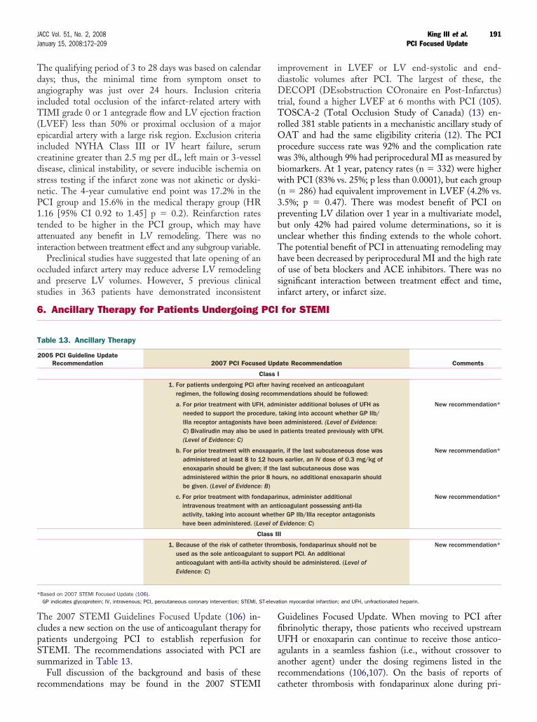
TdaiT(eicdsnP1tai
oas
idDtTrOpwbw(3pbuThos
6
TcpSs
r
T
*
191JACC Vol. 51, No. 2, 2008 King III et al.January 15, 2008:172–209 PCI Focused Update
he qualifying period of 3 to 28 days was based on calendarays; thus, the minimal time from symptom onset tongiography was just over 24 hours. Inclusion criteriancluded total occlusion of the infarct-related artery withIMI grade 0 or 1 antegrade flow and LV ejection fraction
LVEF) less than 50% or proximal occlusion of a majorpicardial artery with a large risk region. Exclusion criteriancluded NYHA Class III or IV heart failure, serumreatinine greater than 2.5 mg per dL, left main or 3-vesselisease, clinical instability, or severe inducible ischemia ontress testing if the infarct zone was not akinetic or dyski-etic. The 4-year cumulative end point was 17.2% in theCI group and 15.6% in the medical therapy group (HR.16 [95% CI 0.92 to 1.45] p � 0.2). Reinfarction ratesended to be higher in the PCI group, which may havettenuated any benefit in LV remodeling. There was nonteraction between treatment effect and any subgroup variable.
Preclinical studies have suggested that late opening of anccluded infarct artery may reduce adverse LV remodelingnd preserve LV volumes. However, 5 previous clinical
. Ancillary Therapy for Patients Undergoing PCI
ecommendations may be found in the 2007 STEMI
GfiUaarc
T-eleva
mprovement in LVEF or LV end-systolic and end-iastolic volumes after PCI. The largest of these, theECOPI (DEsobstruction COronaire en Post-Infarctus)
rial, found a higher LVEF at 6 months with PCI (105).OSCA-2 (Total Occlusion Study of Canada) (13) en-
olled 381 stable patients in a mechanistic ancillary study ofAT and had the same eligibility criteria (12). The PCI
rocedure success rate was 92% and the complication rateas 3%, although 9% had periprocedural MI as measured byiomarkers. At 1 year, patency rates (n � 332) were higherith PCI (83% vs. 25%; p less than 0.0001), but each group
n � 286) had equivalent improvement in LVEF (4.2% vs..5%; p � 0.47). There was modest benefit of PCI onreventing LV dilation over 1 year in a multivariate model,ut only 42% had paired volume determinations, so it isnclear whether this finding extends to the whole cohort.he potential benefit of PCI in attenuating remodeling mayave been decreased by periprocedural MI and the high ratef use of beta blockers and ACE inhibitors. There was noignificant interaction between treatment effect and time,
tudies in 363 patients have demonstrated inconsistent infarct artery, or infarct size.
for STEMI
he 2007 STEMI Guidelines Focused Update (106) in-ludes a new section on the use of anticoagulant therapy foratients undergoing PCI to establish reperfusion forTEMI. The recommendations associated with PCI areummarized in Table 13.
Full discussion of the background and basis of these
able 13. Ancillary Therapy
2005 PCI Guideline UpdateRecommendation 2007 PCI Focuse
C
1. For patients undergoing PCI afregimen, the following dosing r
a. For prior treatment with UFHneeded to support the proceIIIa receptor antagonists havC) Bivalirudin may also be u(Level of Evidence: C)
b. For prior treatment with enoadministered at least 8 to 1enoxaparin should be given;administered within the priobe given. (Level of Evidence:
c. For prior treatment with fondintravenous treatment with aactivity, taking into accounthave been administered. (Le
C
1. Because of the risk of catheterused as the sole anticoagulantanticoagulant with anti-IIa actiEvidence: C)
Based on 2007 STEMI Focused Update (106).GP indicates glycoprotein; IV, intravenous; PCI, percutaneous coronary intervention; STEMI, S
uidelines Focused Update. When moving to PCI afterbrinolytic therapy, those patients who received upstreamFH or enoxaparin can continue to receive those antico-
gulants in a seamless fashion (i.e., without crossover tonother agent) under the dosing regimens listed in theecommendations (106,107). On the basis of reports of
ate Recommendation Comments
ving received an anticoagulantmendations should be followed:
inister additional boluses of UFH astaking into account whether GP IIb/n administered. (Level of Evidence:patients treated previously with UFH.
New recommendation*
in, if the last subcutaneous dose wasrs earlier, an IV dose of 0.3 mg/kg oflast subcutaneous dose wasurs, no additional enoxaparin should
New recommendation*
nux, administer additionalicoagulant possessing anti-IIaer GP IIb/IIIa receptor antagonistsEvidence: C)
New recommendation*
I
bosis, fondaparinux should not bepport PCI. An additionalould be administered. (Level of
New recommendation*
tion myocardial infarction; and UFH, unfractionated heparin.
d Upd
lass I
ter haecom
, admdure,e beesed in
xapar2 houif ther 8 hoB)
aparin ant
whethvel of
lass II
thromto su
vity sh
atheter thrombosis with fondaparinux alone during pri-
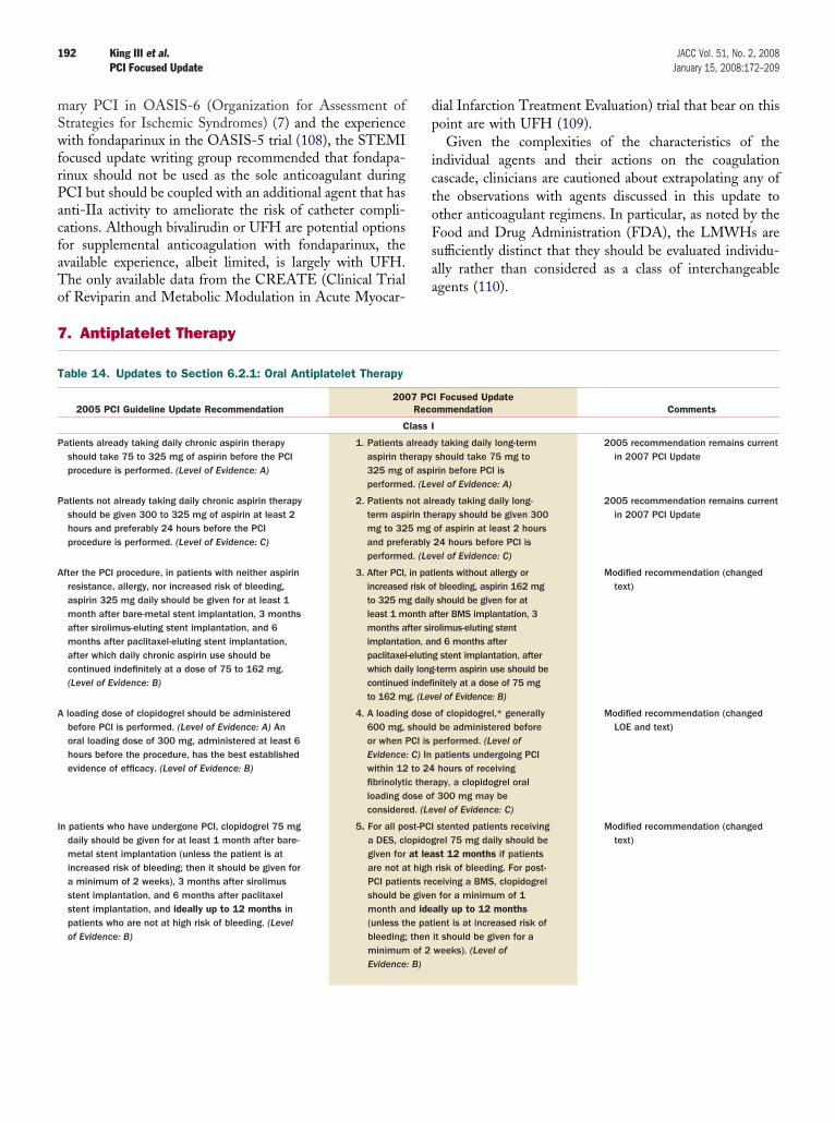
mSwfrPacfaTo
dp
ictoFsa
7
T
P
P
A
A
I
192 King III et al. JACC Vol. 51, No. 2, 2008PCI Focused Update January 15, 2008:172–209
ary PCI in OASIS-6 (Organization for Assessment oftrategies for Ischemic Syndromes) (7) and the experienceith fondaparinux in the OASIS-5 trial (108), the STEMI
ocused update writing group recommended that fondapa-inux should not be used as the sole anticoagulant duringCI but should be coupled with an additional agent that hasnti-IIa activity to ameliorate the risk of catheter compli-ations. Although bivalirudin or UFH are potential optionsor supplemental anticoagulation with fondaparinux, thevailable experience, albeit limited, is largely with UFH.he only available data from the CREATE (Clinical Trial
ial Infarction Treatment Evaluation) trial that bear on thisoint are with UFH (109).Given the complexities of the characteristics of the
ndividual agents and their actions on the coagulationascade, clinicians are cautioned about extrapolating any ofhe observations with agents discussed in this update tother anticoagulant regimens. In particular, as noted by theood and Drug Administration (FDA), the LMWHs areufficiently distinct that they should be evaluated individu-lly rather than considered as a class of interchangeable
f Reviparin and Metabolic Modulation in Acute Myocar-agents (110).
. Antiplatelet Therapy
able 14. Updates to Section 6.2.1: Oral Antiplatelet Therapy
2005 PCI Guideline Update Recommendation2007 PCI Focused Update
Recommendation Comments
Class I
atients already taking daily chronic aspirin therapyshould take 75 to 325 mg of aspirin before the PCIprocedure is performed. (Level of Evidence: A)
1. Patients already taking daily long-termaspirin therapy should take 75 mg to325 mg of aspirin before PCI isperformed. (Level of Evidence: A)
2005 recommendation remains currentin 2007 PCI Update
atients not already taking daily chronic aspirin therapyshould be given 300 to 325 mg of aspirin at least 2hours and preferably 24 hours before the PCIprocedure is performed. (Level of Evidence: C)
2. Patients not already taking daily long-term aspirin therapy should be given 300mg to 325 mg of aspirin at least 2 hoursand preferably 24 hours before PCI isperformed. (Level of Evidence: C)
2005 recommendation remains currentin 2007 PCI Update
fter the PCI procedure, in patients with neither aspirinresistance, allergy, nor increased risk of bleeding,aspirin 325 mg daily should be given for at least 1month after bare-metal stent implantation, 3 monthsafter sirolimus-eluting stent implantation, and 6months after paclitaxel-eluting stent implantation,after which daily chronic aspirin use should becontinued indefinitely at a dose of 75 to 162 mg.(Level of Evidence: B)
3. After PCI, in patients without allergy orincreased risk of bleeding, aspirin 162 mgto 325 mg daily should be given for atleast 1 month after BMS implantation, 3months after sirolimus-eluting stentimplantation, and 6 months afterpaclitaxel-eluting stent implantation, afterwhich daily long-term aspirin use should becontinued indefinitely at a dose of 75 mgto 162 mg. (Level of Evidence: B)
Modified recommendation (changedtext)
loading dose of clopidogrel should be administeredbefore PCI is performed. (Level of Evidence: A) Anoral loading dose of 300 mg, administered at least 6hours before the procedure, has the best establishedevidence of efficacy. (Level of Evidence: B)
4. A loading dose of clopidogrel,* generally600 mg, should be administered beforeor when PCI is performed. (Level ofEvidence: C) In patients undergoing PCIwithin 12 to 24 hours of receivingfibrinolytic therapy, a clopidogrel oralloading dose of 300 mg may beconsidered. (Level of Evidence: C)
Modified recommendation (changedLOE and text)
n patients who have undergone PCI, clopidogrel 75 mgdaily should be given for at least 1 month after bare-metal stent implantation (unless the patient is atincreased risk of bleeding; then it should be given fora minimum of 2 weeks), 3 months after sirolimusstent implantation, and 6 months after paclitaxelstent implantation, and ideally up to 12 months inpatients who are not at high risk of bleeding. (Levelof Evidence: B)
5. For all post-PCI stented patients receivinga DES, clopidogrel 75 mg daily should begiven for at least 12 months if patientsare not at high risk of bleeding. For post-PCI patients receiving a BMS, clopidogrelshould be given for a minimum of 1month and ideally up to 12 months(unless the patient is at increased risk ofbleeding; then it should be given for aminimum of 2 weeks). (Level ofEvidence: B)
Modified recommendation (changedtext)
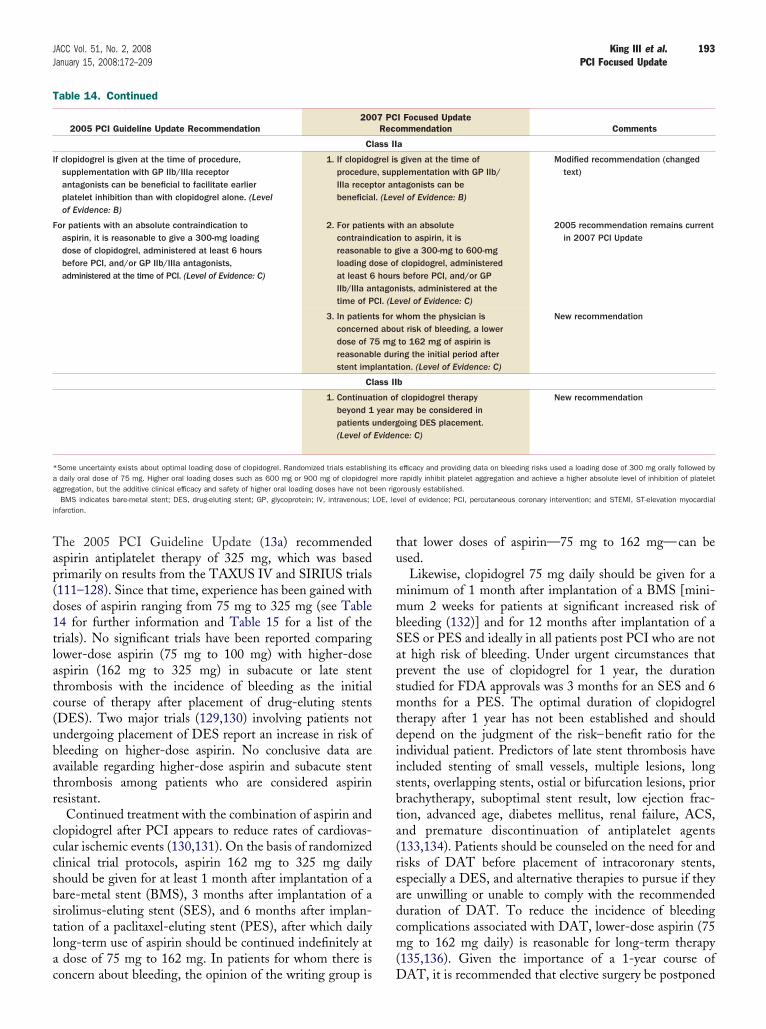
Tap(d1tlatc(ubatr
cccsbstlac
tu
mmbSapsmtdiisbta(readcm(
T
I
F
*aa
OE, lei
193JACC Vol. 51, No. 2, 2008 King III et al.January 15, 2008:172–209 PCI Focused Update
he 2005 PCI Guideline Update (13a) recommendedspirin antiplatelet therapy of 325 mg, which was basedrimarily on results from the TAXUS IV and SIRIUS trials111–128). Since that time, experience has been gained withoses of aspirin ranging from 75 mg to 325 mg (see Table4 for further information and Table 15 for a list of therials). No significant trials have been reported comparingower-dose aspirin (75 mg to 100 mg) with higher-dosespirin (162 mg to 325 mg) in subacute or late stenthrombosis with the incidence of bleeding as the initialourse of therapy after placement of drug-eluting stentsDES). Two major trials (129,130) involving patients notndergoing placement of DES report an increase in risk ofleeding on higher-dose aspirin. No conclusive data arevailable regarding higher-dose aspirin and subacute stenthrombosis among patients who are considered aspirinesistant.
Continued treatment with the combination of aspirin andlopidogrel after PCI appears to reduce rates of cardiovas-ular ischemic events (130,131). On the basis of randomizedlinical trial protocols, aspirin 162 mg to 325 mg dailyhould be given for at least 1 month after implantation of aare-metal stent (BMS), 3 months after implantation of airolimus-eluting stent (SES), and 6 months after implan-ation of a paclitaxel-eluting stent (PES), after which dailyong-term use of aspirin should be continued indefinitely at
dose of 75 mg to 162 mg. In patients for whom there is
able 14. Continued
2005 PCI Guideline Update Recommendation200
Cl
f clopidogrel is given at the time of procedure,supplementation with GP IIb/IIIa receptorantagonists can be beneficial to facilitate earlierplatelet inhibition than with clopidogrel alone. (Levelof Evidence: B)
1. If clopidoprocedureIIIa recepbeneficia
or patients with an absolute contraindication toaspirin, it is reasonable to give a 300-mg loadingdose of clopidogrel, administered at least 6 hoursbefore PCI, and/or GP IIb/IIIa antagonists,administered at the time of PCI. (Level of Evidence: C)
2. For patiecontraindreasonabloading dat least 6IIb/IIIa antime of P
3. In patientconcernedose of 7reasonabstent imp
Cl
1. Continuabeyond 1patients u(Level of
Some uncertainty exists about optimal loading dose of clopidogrel. Randomized trials establishdaily oral dose of 75 mg. Higher oral loading doses such as 600 mg or 900 mg of clopidogre
ggregation, but the additive clinical efficacy and safety of higher oral loading doses have not bBMS indicates bare-metal stent; DES, drug-eluting stent; GP, glycoprotein; IV, intravenous; L
nfarction.
oncern about bleeding, the opinion of the writing group is D
hat lower doses of aspirin—75 mg to 162 mg—can besed.Likewise, clopidogrel 75 mg daily should be given for ainimum of 1 month after implantation of a BMS [mini-um 2 weeks for patients at significant increased risk of
leeding (132)] and for 12 months after implantation of aES or PES and ideally in all patients post PCI who are nott high risk of bleeding. Under urgent circumstances thatrevent the use of clopidogrel for 1 year, the durationtudied for FDA approvals was 3 months for an SES and 6onths for a PES. The optimal duration of clopidogrel
herapy after 1 year has not been established and shouldepend on the judgment of the risk–benefit ratio for thendividual patient. Predictors of late stent thrombosis havencluded stenting of small vessels, multiple lesions, longtents, overlapping stents, ostial or bifurcation lesions, priorrachytherapy, suboptimal stent result, low ejection frac-ion, advanced age, diabetes mellitus, renal failure, ACS,nd premature discontinuation of antiplatelet agents133,134). Patients should be counseled on the need for andisks of DAT before placement of intracoronary stents,specially a DES, and alternative therapies to pursue if theyre unwilling or unable to comply with the recommendeduration of DAT. To reduce the incidence of bleedingomplications associated with DAT, lower-dose aspirin (75g to 162 mg daily) is reasonable for long-term therapy
135,136). Given the importance of a 1-year course of
I Focused Updatemmendation Comments
a
given at the time oflementation with GP IIb/
tagonists can beel of Evidence: B)
Modified recommendation (changedtext)
h an absoluten to aspirin, it isive a 300-mg to 600-mgclopidogrel, administeredbefore PCI, and/or GP
ists, administered at thevel of Evidence: C)
2005 recommendation remains currentin 2007 PCI Update
hom the physician ist risk of bleeding, a lowerto 162 mg of aspirin ising the initial period afterion. (Level of Evidence: C)
New recommendation
b
clopidogrel therapymay be considered inoing DES placement.ce: C)
New recommendation
efficacy and providing data on bleeding risks used a loading dose of 300 mg orally followed byrapidly inhibit platelet aggregation and achieve a higher absolute level of inhibition of platelet
orously established.vel of evidence; PCI, percutaneous coronary intervention; and STEMI, ST-elevation myocardial
7 PCReco
ass II
grel is, supp
tor anl. (Lev
nts witicatiole to gose ofhourstagonCI. (Le
s for wd abou5 mgle durlantat
ass II
tion ofyearnderg
Eviden
ing itsl more
een rig
AT, it is recommended that elective surgery be postponed
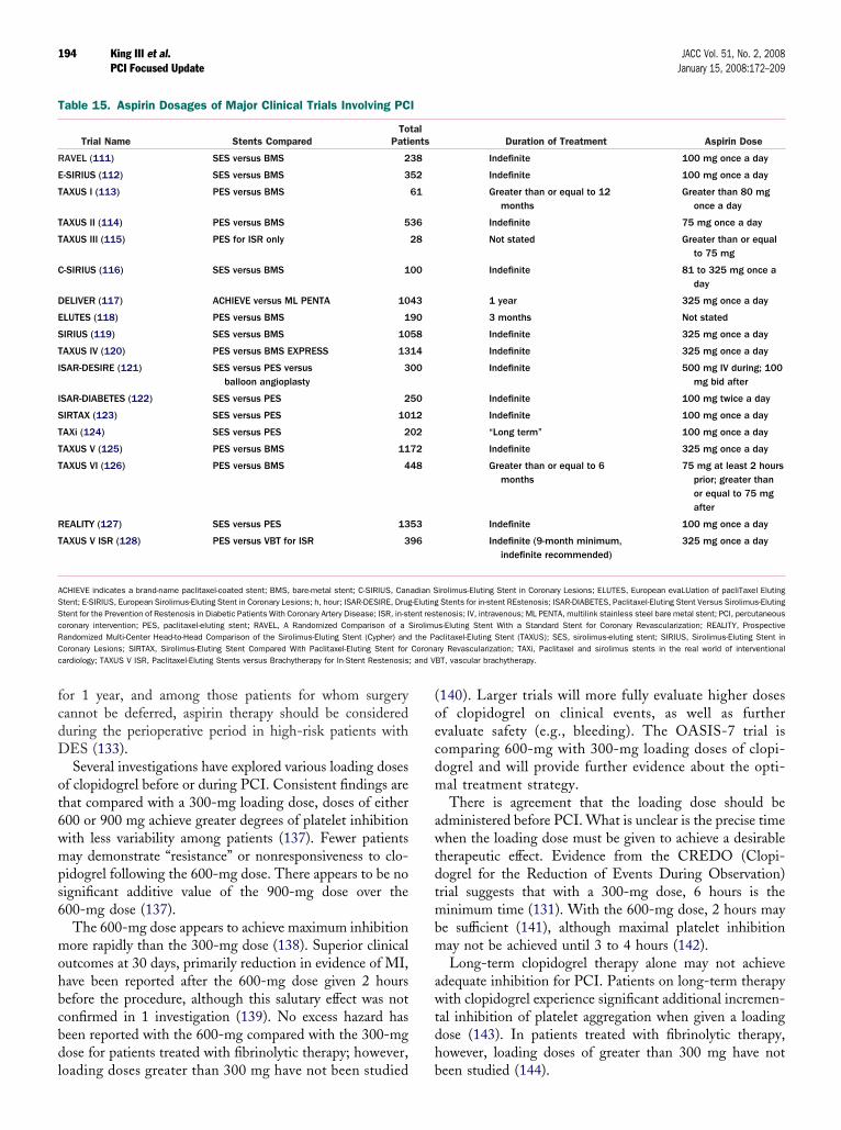
fcdD
ot6wmps6
mohbcbdl
(oecdm
awtdtmbm
awtdh
T
R
E
T
T
T
C
D
E
S
T
I
I
S
T
T
T
R
T
ASScRC Coronac ; and V
194 King III et al. JACC Vol. 51, No. 2, 2008PCI Focused Update January 15, 2008:172–209
or 1 year, and among those patients for whom surgeryannot be deferred, aspirin therapy should be considereduring the perioperative period in high-risk patients withES (133).Several investigations have explored various loading doses
f clopidogrel before or during PCI. Consistent findings arehat compared with a 300-mg loading dose, doses of either00 or 900 mg achieve greater degrees of platelet inhibitionith less variability among patients (137). Fewer patientsay demonstrate “resistance” or nonresponsiveness to clo-
idogrel following the 600-mg dose. There appears to be noignificant additive value of the 900-mg dose over the00-mg dose (137).The 600-mg dose appears to achieve maximum inhibitionore rapidly than the 300-mg dose (138). Superior clinical
utcomes at 30 days, primarily reduction in evidence of MI,ave been reported after the 600-mg dose given 2 hoursefore the procedure, although this salutary effect was notonfirmed in 1 investigation (139). No excess hazard haseen reported with the 600-mg compared with the 300-mgose for patients treated with fibrinolytic therapy; however,
able 15. Aspirin Dosages of Major Clinical Trials Involving PC
Trial Name Stents ComparedTo
Pati
AVEL (111) SES versus BMS 2
-SIRIUS (112) SES versus BMS 3
AXUS I (113) PES versus BMS
AXUS II (114) PES versus BMS 5
AXUS III (115) PES for ISR only
-SIRIUS (116) SES versus BMS 1
ELIVER (117) ACHIEVE versus ML PENTA 10
LUTES (118) PES versus BMS 1
IRIUS (119) SES versus BMS 10
AXUS IV (120) PES versus BMS EXPRESS 13
SAR-DESIRE (121) SES versus PES versusballoon angioplasty
3
SAR-DIABETES (122) SES versus PES 2
IRTAX (123) SES versus PES 10
AXi (124) SES versus PES 2
AXUS V (125) PES versus BMS 11
AXUS VI (126) PES versus BMS 4
EALITY (127) SES versus PES 13
AXUS V ISR (128) PES versus VBT for ISR 3
CHIEVE indicates a brand-name paclitaxel-coated stent; BMS, bare-metal stent; C-SIRIUS, Cantent; E-SIRIUS, European Sirolimus-Eluting Stent in Coronary Lesions; h, hour; ISAR-DESIRE, Drugtent for the Prevention of Restenosis in Diabetic Patients With Coronary Artery Disease; ISR, in-storonary intervention; PES, paclitaxel-eluting stent; RAVEL, A Randomized Comparison of a Sandomized Multi-Center Head-to-Head Comparison of the Sirolimus-Eluting Stent (Cypher) andoronary Lesions; SIRTAX, Sirolimus-Eluting Stent Compared With Paclitaxel-Eluting Stent forardiology; TAXUS V ISR, Paclitaxel-Eluting Stents versus Brachytherapy for In-Stent Restenosis
oading doses greater than 300 mg have not been studied b
140). Larger trials will more fully evaluate higher dosesf clopidogrel on clinical events, as well as furthervaluate safety (e.g., bleeding). The OASIS-7 trial isomparing 600-mg with 300-mg loading doses of clopi-ogrel and will provide further evidence about the opti-al treatment strategy.There is agreement that the loading dose should be
dministered before PCI. What is unclear is the precise timehen the loading dose must be given to achieve a desirable
herapeutic effect. Evidence from the CREDO (Clopi-ogrel for the Reduction of Events During Observation)rial suggests that with a 300-mg dose, 6 hours is theinimum time (131). With the 600-mg dose, 2 hours may
e sufficient (141), although maximal platelet inhibitionay not be achieved until 3 to 4 hours (142).Long-term clopidogrel therapy alone may not achieve
dequate inhibition for PCI. Patients on long-term therapyith clopidogrel experience significant additional incremen-
al inhibition of platelet aggregation when given a loadingose (143). In patients treated with fibrinolytic therapy,owever, loading doses of greater than 300 mg have not
Duration of Treatment Aspirin Dose
Indefinite 100 mg once a day
Indefinite 100 mg once a day
Greater than or equal to 12months
Greater than 80 mgonce a day
Indefinite 75 mg once a day
Not stated Greater than or equalto 75 mg
Indefinite 81 to 325 mg once aday
1 year 325 mg once a day
3 months Not stated
Indefinite 325 mg once a day
Indefinite 325 mg once a day
Indefinite 500 mg IV during; 100mg bid after
Indefinite 100 mg twice a day
Indefinite 100 mg once a day
“Long term” 100 mg once a day
Indefinite 325 mg once a day
Greater than or equal to 6months
75 mg at least 2 hoursprior; greater thanor equal to 75 mgafter
Indefinite 100 mg once a day
Indefinite (9-month minimum,indefinite recommended)
325 mg once a day
irolimus-Eluting Stent in Coronary Lesions; ELUTES, European evaLUation of pacliTaxel ElutingStents for in-stent REstenosis; ISAR-DIABETES, Paclitaxel-Eluting Stent Versus Sirolimus-Eluting
tenosis; IV, intravenous; ML PENTA, multilink stainless steel bare metal stent; PCI, percutaneouss-Eluting Stent With a Standard Stent for Coronary Revascularization; REALITY, Prospectiveclitaxel-Eluting Stent (TAXUS); SES, sirolimus-eluting stent; SIRIUS, Sirolimus-Eluting Stent inry Revascularization; TAXi, Paclitaxel and sirolimus stents in the real world of interventionalBT, vascular brachytherapy.
I
talents
38
52
61
36
28
00
43
90
58
14
00
50
12
02
72
48
53
96
adian S-Elutingent resirolimuthe Pa
een studied (144).

8
8
OiTettDatD
oEhddtrdaD
T
A
A
B
195JACC Vol. 51, No. 2, 2008 King III et al.January 15, 2008:172–209 PCI Focused Update
. Bare-Metal and Drug-Eluting Stents
ifods
tpdcAussut
ifBs
I, percu
.1. Selection of a Bare-Metal or Drug-Eluting Stent
bservational studies indicate that physicians routinelymplant stents when performing coronary interventions.wo types of stents are available: BMS and DES. Drug-
luting stents have become increasingly popular as standardherapy. In 2005, a sampling of 140 US hospitals indicatedhat 94% of patients treated with a stent received at least 1ES (145). More recently, however, because of concerns
bout stent thrombosis and the mandate that each DES-reated patient take prolonged DAT, the proportion ofES use has declined to 60% to 70%.The results of the clinical trials that led to FDA approval
f the DES provide support for its use in suitable patients.xtended follow-up of the initial investigated patient co-orts to 4 years confirms the sustained benefit of DES inecreasing the need for repeat revascularization but withoutifferences in death or MI (146–148). Randomized clinicalrials in selected clinical subsets such as BMS in-stentestenosis, total occlusions, diabetes mellitus, and small-iameter arteries have also demonstrated the value of DESnd have prompted physicians to extend the application of
able 16. Updates to Section 7.3.5: Drug-Eluting and Bare-Me
2005 PCI Guideline Update Recommendation 2007 PCI
C
drug-eluting stent (DES) should be consideredas an alternative to the bare-metal stent insubsets of patients in whom trial datasuggest efficacy. (Level of Evidence: A)
1. A DES shouBMS in thoindicate a(Level of E
2. Before impcardiologisneed for anpatient’s atherapy for
3. In patientsPCI and arproceduresduring thebe given toof balloonimplantatio(Level of E
Cl
1. In patientsabout risk162 mg ofEvidence: C
Cl
DES may be considered for use in anatomicsettings in which the usefulness,effectiveness, and safety have not been fullydocumented in published trials. (Level ofEvidence: C)
1. A DES mayanatomic seffectivenehas not be(Level of E
MS indicates bare-metal stent; DAT, dual antiplatelet therapy; DES, drug-eluting stent; and PC
ES beyond the narrow patient populations included in the M
nitial approval trials (122,126,149–154). The duration ofollow-up of these “off-label” studies and the small numberf patients enrolled, however, limit the detection of subtleifferences in important end points such as stent thrombo-is, death, or MI.
It is important to recognize certain differences betweenhe BMS and DES when selecting a stent for an individualatient or lesion. First, in general, a DES may be moreifficult to implant than a BMS. The DES has a polymeroating that stiffens the stent and makes it less conformable.ccordingly, one reason for using a BMS is that it can besed in patients in whom a DES cannot be implanteduccessfully. Second, the DES is substantially more expen-ive than the BMS. When financial resources are limited,se of the DES may be rationed, with implantation only inhose patients at greatest risk for restenosis.
A third but very important difference relates to thenhibition of endothelial coverage of the DES and the needor extended DAT (Table 16). After introduction of theMS, it was associated with a disturbingly high incidence of
tent thrombosis (141). Stent thrombosis often presented as
tents
sed Update Recommendation Comments
considered as an alternative to atients for whom clinical trialsble effectiveness/safety profile.e: A)
Modified recommendation(changed text)
g a DES, the interventionalld discuss with the patient theation of DAT and confirm theo comply with the recommended(Level of Evidence: B)
New recommendation
re undergoing preparation fory to require invasive or surgicalhich DAT must be interrupted2 months, consideration shouldntation of a BMS or performancelasty with a provisional stentead of the routine use of a DES.e: C)
New recommendation
a
om the physician is concernededing, a lower dose of 75 mg ton is reasonable. (Level of
New recommendation
b
nsidered for clinical ands in which thefety profile appears favorable buty confirmed by clinical trials.e: C)
Modified recommendation(changed text)
taneous coronary intervention.
tal S
Focu
lass I
ld bese pafavoravidenc
lantint shoud dur
bility tDES.
who ae likelfor w
next 1impla
angiopn inst
videnc
ass II
for whof bleaspiri)
ass II
be coettingss/saen fullvidenc
I or even death and usually occurred in the first 30 days
aigri(4oswpebthanw
wTdaatAad“t
bhtafiDDiptnt
DcjwbnbrSBD
tyDbvd
toaDpmpersesos
wpevst“rpratap
mSbyLpbcbrbfvht
196 King III et al. JACC Vol. 51, No. 2, 2008PCI Focused Update January 15, 2008:172–209
fter implantation. Changes in technique such as highnflation pressure and intravascular ultrasound (IVUS)-uided deployment and use of concomitant combined aspi-in and thienopyridine therapy substantially reduced thencidence of stent thrombosis to a clinically acceptable level155). Importantly, the requisite duration of DAT was onlyweeks, and some advocated only 2 weeks. The importancef DAT in preventing stent thrombosis was furthertrengthened by the outcome of patients for whom DATas discontinued prematurely because of the need for thoseatients to undergo surgical procedures. These patientsxperienced a disturbingly high incidence of stent throm-osis (156). The critical role of DAT in preventing stenthrombosis was also noted among patients with BMS whoad received brachytherapy for in-stent restenosis. Presum-bly these patients were less likely to develop subsequenteointimal coverage of the endoluminal stent surface andere accordingly then more susceptible to stent thrombosis.In the initial randomized trials that compared the DES
ith BMS, DAT was administered for 30 days to 6 months.he most recent guidelines update describes a minimumuration of 3 months of DAT for an SES and 6 months forPES. On the basis of results from other trials that suggestsustained benefit of DAT, these guidelines further state
hat ideally DAT should be extended to 12 months.lthough these recommendations were to some extent
rbitrary, subsequent studies have confirmed that prematureiscontinuation of DAT, that is, at a time less thanminimal duration” (3 months for the SES and 6 months forhe PES) was highly associated with stent thrombosis (157).
The tight relationship between DAT and stent throm-osis for patients treated with DES warrants emphasis andas implications for selecting the type of stent deployed athe time of PCI. For example, the clinician should not selectDES for a patient who does not have access to DAT fornancial reasons or who is unlikely to be compliant in takingAT. One study revealed that 14% of patients had stoppedAT 1 month after implantation of the DES (158). Also,
mplantation of a BMS may be more appropriate in aatient with a known increased risk of bleeding. In situa-ions such as these, the consequences of developing reste-osis are considered less untoward than those of stenthrombosis or significant bleeding.
Furthermore, prescribed premature discontinuation ofAT in patients treated with a DES should not be done
asually. For example, routine dental procedures should notustify cessation of DAT even though it is anticipated DATill be subsequently resumed (133). Consideration shoulde given to delay scheduling of elective procedures thatormally warrant discontinuation of antiplatelet agents. Theenefit of DES in reducing the need for target vesselevascularization (TVR) also should be taken into account.ome registries have shown 1-digit TVR rates with theMS, and the absolute reduction in these events using the
ES depends on patient and lesion characteristics. pThere are also concerns related to the appropriate dura-ion of DAT. More recently, the occurrence of late (up to 1ear) or very late (beyond 1 year) stent thrombosis amongES-treated patients has been described (159). One data-
ase analysis suggests that extended use of DAT may havealue in preventing late stent thrombosis, whereas othersisagree (160).Outcomes of patients in the initial FDA-approval trials
o 4 years provides reassurance that, at least for those typesf patients, despite a small excess of stent thrombosis, thereppears to be no increase in death or MI when comparingES-treated groups with BMS-treated groups. As noted,
rotocol-recommended DAT in these patients was notore than 6 months, although extended DAT was not
rohibited. (These results are observed despite a significantxcess occurrence of stent thrombosis among patients whoeceived a paclitaxel stent.) Some have postulated that theubstantial additional revascularization procedures experi-nced by BMS patients were associated with a small butignificant excess rate of death and MI that offset any deathsr MIs that may have occurred in the DES group related totent thrombosis.
Less data are available regarding the outcomes of patientsho receive a DES for an “off-label” indication. Suchatients have characteristics of their coronary disease, forxample, a lesion in an artery less than 2.5 mm in diameter,ery long lesions, bifurcation lesions, or a clinical syndromeuch as acute MI, that were excluded in the FDA-approvalrials. Reports from large observational studies indicate thatoff-label” patients may experience higher rates of repeatevascularization and death and MI at 1 year than DESatients with “on-label” features. Importantly, a similarelationship is observed for patients treated with a BMS. Inddition, there appears to be a significant association be-ween “off-label” use and stent thrombosis. Accordingly, theppropriate selection for DAT among “off-label” DESatients may be different than for “on-label” patients.At this point in time, 12 months of DAT is recom-ended for all patients who receive a DES (120) (see
ection 6.2.1) unless there is a high risk of bleeding. Theenefits and indications for treatment with DAT beyond 1ear in patients with DES are the subject of ongoing studies.ow-dose aspirin should be continued indefinitely. Foratients with clinical features associated with stent throm-osis, such as renal insufficiency, diabetes, or proceduralharacteristics such as multiple stents or treatment of aifurcation lesion, extended DAT beyond 1 year may beeasonable. The risk of stent thrombosis needs to bealanced with other medical conditions and nonmedicalactors that might affect the risk-benefit ratio of DATersus other therapies. Finally, certain DES-treated patientsave already discontinued DAT 1 year after stent implan-ation. No information yet supports restarting DAT in these
atients.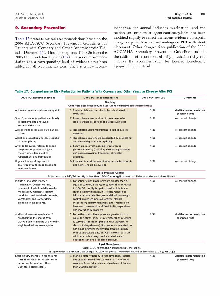
9
T2Pc2da
msmdpAta
T
A
S
A
A
A
U
I
A
S
197JACC Vol. 51, No. 2, 2008 King III et al.January 15, 2008:172–209 PCI Focused Update
. Secondary Prevention
able 17 presents revised recommendations based on the006 AHA/ACC Secondary Prevention Guidelines foratients with Coronary and Other Atherosclerotic Vas-ular Diseases (11). This table replaces Table 26 from the005 PCI Guideline Update (13a). Classes of recommen-ation and a corresponding level of evidence have been
dded for all recommendations. There is a new recom- lendation for annual influenza vaccination, and theection on antiplatelet agents/anticoagulants has beenodified slightly to reflect the recent evidence on aspirin
osage in patients who have undergone PCI with stentlacement. Other changes since publication of the 2006CC/AHA Secondary Prevention Guidelines include
he addition of recommended daily physical activity andClass IIa recommendation for lowered low-density
ipoprotein cholesterol.
able 17. Comprehensive Risk Reduction for Patients With Coronary and Other Vascular Disease After PCI
2005 PCI Recommendations 2007 PCI Recommendations 2007 COR and LOE Comments
SmokingGoal: Complete cessation, no exposure to environmental tobacco smoke
sk about tobacco status at every visit. 1. Status of tobacco use should be asked about atevery visit.
I (B) Modified recommendation(changed text)
trongly encourage patient and familyto stop smoking and avoidsecondhand smoke.
2. Every tobacco user and family members whosmoke should be advised to quit at every visit.
I (B) No content change
ssess the tobacco user’s willingnessto quit.
3. The tobacco user’s willingness to quit should beassessed.
I (B) No content change
ssist by counseling and developing aplan for quitting.
4. The tobacco user should be assisted by counselingand developing a plan for quitting.
I (B) No content change
rrange follow-up, referral to specialprograms, or pharmacologicaltherapy (including nicotinereplacement and bupropion).
5. Follow-up, referral to special programs, orpharmacotherapy (including nicotine replacementand pharmacological treatment) should bearranged.
I (B) No content change
rge avoidance of exposure toenvironmental tobacco smoke atwork and home.
6. Exposure to environmental tobacco smoke at workand home should be avoided.
I (B) No content change
Blood Pressure ControlGoal: Less than 140/90 mm Hg or less than 130/80 mm Hg if patient has diabetes or chronic kidney disease
nitiate or maintain lifestylemodification (weight control,increased physical activity, alcoholmoderation, moderate sodiumrestriction, and emphasis on fruits,vegetables, and low-fat dairyproducts) in all patients.
1. For patients with blood pressure greater than orequal to 140/90 mm Hg (or greater than or equalto 130/80 mm Hg for patients with diabetes orchronic kidney disease), it is recommended toinitiate or maintain lifestyle modification—weightcontrol; increased physical activity; alcoholmoderation; sodium reduction; and emphasis onincreased consumption of fresh fruits, vegetables,and low-fat dairy products.
I (B) No content change
dd blood pressure medication,*emphasizing the use of betablockers and inhibitors of the renin-angiotensin-aldosterone system.
2. For patients with blood pressure greater than orequal to 140/90 mm Hg (or greater than or equalto 130/80 mm Hg for patients with diabetes orchronic kidney disease), it is useful as tolerated, toadd blood pressure medication, treating initiallywith beta blockers and/or ACE inhibitors, with theaddition of other drugs such as thiazides asneeded to achieve goal blood pressure.
I (A) Modified recommendation(changed text)
Lipid ManagementGoal: LDL-C substantially less than 100 mg per dL
(If triglycerides are greater than or equal to 200 mg per dL, non–HDL-C should be less than 130 mg per dL†.)
tart dietary therapy in all patients(less than 7% of total calories assaturated fat and less than200 mg/d cholesterol).
1. Starting dietary therapy is recommended. Reduceintake of saturated fats (to less than 7% of totalcalories), trans fatty acids, and cholesterol (to lessthan 200 mg per day).
I (B) Modified recommendation(changed text)
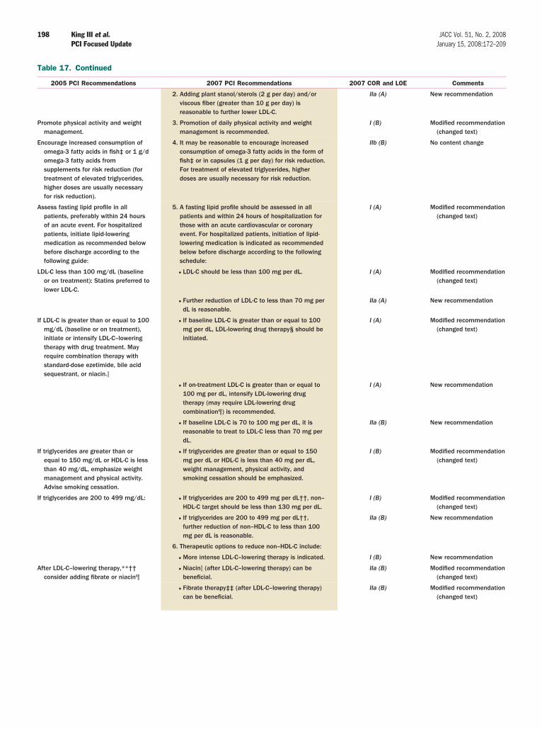
T
P
E
A
L
I
I
I
A
198 King III et al. JACC Vol. 51, No. 2, 2008PCI Focused Update January 15, 2008:172–209
able 17. Continued
2005 PCI Recommendations 2007 PCI Recommendations 2007 COR and LOE Comments
2. Adding plant stanol/sterols (2 g per day) and/orviscous fiber (greater than 10 g per day) isreasonable to further lower LDL-C.
IIa (A) New recommendation
romote physical activity and weightmanagement.
3. Promotion of daily physical activity and weightmanagement is recommended.
I (B) Modified recommendation(changed text)
ncourage increased consumption ofomega-3 fatty acids in fish‡ or 1 g/domega-3 fatty acids fromsupplements for risk reduction (fortreatment of elevated triglycerides,higher doses are usually necessaryfor risk reduction).
4. It may be reasonable to encourage increasedconsumption of omega-3 fatty acids in the form offish‡ or in capsules (1 g per day) for risk reduction.For treatment of elevated triglycerides, higherdoses are usually necessary for risk reduction.
IIb (B) No content change
ssess fasting lipid profile in allpatients, preferably within 24 hoursof an acute event. For hospitalizedpatients, initiate lipid-loweringmedication as recommended belowbefore discharge according to thefollowing guide:
5. A fasting lipid profile should be assessed in allpatients and within 24 hours of hospitalization forthose with an acute cardiovascular or coronaryevent. For hospitalized patients, initiation of lipid-lowering medication is indicated as recommendedbelow before discharge according to the followingschedule:
I (A) Modified recommendation(changed text)
DL-C less than 100 mg/dL (baselineor on treatment): Statins preferred tolower LDL-C.
● LDL-C should be less than 100 mg per dL. I (A) Modified recommendation(changed text)
● Further reduction of LDL-C to less than 70 mg perdL is reasonable.
IIa (A) New recommendation
f LDL-C is greater than or equal to 100mg/dL (baseline or on treatment),initiate or intensify LDL-C–loweringtherapy with drug treatment. Mayrequire combination therapy withstandard-dose ezetimide, bile acidsequestrant, or niacin.�
● If baseline LDL-C is greater than or equal to 100mg per dL, LDL-lowering drug therapy§ should beinitiated.
I (A) Modified recommendation(changed text)
● If on-treatment LDL-C is greater than or equal to100 mg per dL, intensify LDL-lowering drugtherapy (may require LDL-lowering drugcombination¶) is recommended.
I (A) New recommendation
● If baseline LDL-C is 70 to 100 mg per dL, it isreasonable to treat to LDL-C less than 70 mg perdL.
IIa (B) New recommendation
f triglycerides are greater than orequal to 150 mg/dL or HDL-C is lessthan 40 mg/dL, emphasize weightmanagement and physical activity.Advise smoking cessation.
● If triglycerides are greater than or equal to 150mg per dL or HDL-C is less than 40 mg per dL,weight management, physical activity, andsmoking cessation should be emphasized.
I (B) Modified recommendation(changed text)
f triglycerides are 200 to 499 mg/dL: ● If triglycerides are 200 to 499 mg per dL††, non–HDL-C target should be less than 130 mg per dL.
I (B) Modified recommendation(changed text)
● If triglycerides are 200 to 499 mg per dL††,further reduction of non–HDL-C to less than 100mg per dL is reasonable.
IIa (B) New recommendation
6. Therapeutic options to reduce non–HDL-C include:
● More intense LDL-C–lowering therapy is indicated. I (B) New recommendation
fter LDL-C–lowering therapy,**††consider adding fibrate or niacin¶
● Niacin� (after LDL-C–lowering therapy) can bebeneficial.
IIa (B) Modified recommendation(changed text)
● Fibrate therapy‡‡ (after LDL-C–lowering therapy)can be beneficial.
IIa (B) Modified recommendation(changed text)
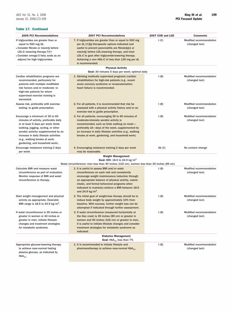
T
I
●
●
C
A
E
E
C
S
I
A
199JACC Vol. 51, No. 2, 2008 King III et al.January 15, 2008:172–209 PCI Focused Update
able 17. Continued
2005 PCI Recommendations 2007 PCI Recommendations 2007 COR and LOE Comments
f triglycerides are greater than orequal to 500 mg/dL:
Consider fibrate or niacin§ beforeLDL-C–lowering therapy.¶††Consider omega-3 fatty acids as anadjunct for high triglycerides.
7. If triglycerides are greater than or equal to 500 mgper dL,††§§ therapeutic options indicated anduseful to prevent pancreatitis are fibrate§‡‡ orniacin§� before LDL-lowering therapy, and treatLDL-C to goal after triglyceride-lowering therapy.Achieving a non–HDL-C of less than 130 mg per dLis recommended.
I (C) Modified recommendation(changed text)
Physical ActivityGoal: 30 minutes 5 days per week; optimal daily
ardiac rehabilitation programs arerecommended, particularly forpatients with multiple modifiablerisk factors and/or moderate- tohigh-risk patients for whomsupervised exercise training iswarranted.
1. Advising medically supervised programs (cardiacrehabilitation) for high-risk patients (e.g., recentacute coronary syndrome or revascularization,heart failure) is recommended.
I (B) Modified recommendation(changed text)
ssess risk, preferably with exercisetesting, to guide prescription.
2. For all patients, it is recommended that risk beassessed with a physical activity history and/or anexercise test to guide prescription.
I (B) Modified recommendation(changed text)
ncourage a minimum of 30 to 60minutes of activity, preferably dailyor at least 5 days per week (briskwalking, jogging, cycling, or otheraerobic activity) supplemented by anincrease in daily lifestyle activities(e.g., walking breaks at work,gardening, and household work).
3. For all patients, encouraging 30 to 60 minutes ofmoderate-intensity aerobic activity isrecommended, such as brisk walking on most—preferably all—days of the week, supplemented byan increase in daily lifestyle activities (e.g., walkingbreaks at work, gardening, and household work).
I (B) Modified recommendation(changed text)
ncourage resistance training 2 daysper week.
4. Encouraging resistance training 2 days per weekmay be reasonable.
IIb (C) No content change
Weight ManagementGoal: BMI: 18.5 to 24.9 kg/m2
Waist circumference: men less than 40 inches (102 cm), women less than 35 inches (89 cm)
alculate BMI and measure waistcircumference as part of evaluation.Monitor response of BMI and waistcircumference to therapy.
1. It is useful to assess BMI and/or waistcircumference on each visit and consistentlyencourage weight maintenance/reduction throughan appropriate balance of physical activity, caloricintake, and formal behavioral programs whenindicated to maintain/achieve a BMI between 18.5and 24.9 kg/m2.
I (B) Modified recommendation(changed text)
tart weight management and physicalactivity as appropriate. DesirableBMI range is 18.5 to 24.9 kg/m2.
2. The initial goal of weight-loss therapy should be toreduce body weight by approximately 10% frombaseline. With success, further weight loss can beattempted if indicated through further assessment.
I (B) Modified recommendation(changed text)
f waist circumference is 35 inches orgreater in women or 40 inches orgreater in men, initiate lifestylechanges and treatment strategiesfor metabolic syndrome.
3. If waist circumference (measured horizontally atthe iliac crest) is 35 inches (89 cm) or greater inwomen and 40 inches (102 cm) or greater in men,it is useful to initiate lifestyle changes and considertreatment strategies for metabolic syndrome asindicated.
I (B) Modified recommendation(changed text)
Diabetes ManagementGoal: HbA1c less than 7%
ppropriate glucose-lowering therapyto achieve near-normal fastingplasma glucose, as indicated byHbA1c.
1. It is recommended to initiate lifestyle andpharmacotherapy to achieve near-normal HbA1c.
I (B) Modified recommendation(changed text)
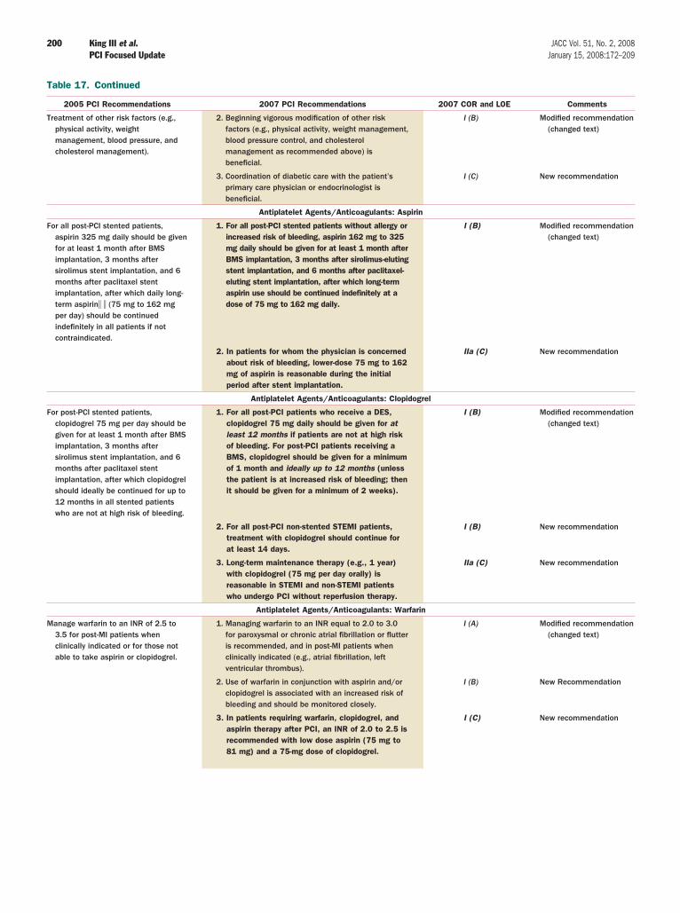
T
T
F
F
M
200 King III et al. JACC Vol. 51, No. 2, 2008PCI Focused Update January 15, 2008:172–209
able 17. Continued
2005 PCI Recommendations 2007 PCI Recommendations 2007 COR and LOE Comments
reatment of other risk factors (e.g.,physical activity, weightmanagement, blood pressure, andcholesterol management).
2. Beginning vigorous modification of other riskfactors (e.g., physical activity, weight management,blood pressure control, and cholesterolmanagement as recommended above) isbeneficial.
I (B) Modified recommendation(changed text)
3. Coordination of diabetic care with the patient’sprimary care physician or endocrinologist isbeneficial.
I (C) New recommendation
Antiplatelet Agents/Anticoagulants: Aspirin
or all post-PCI stented patients,aspirin 325 mg daily should be givenfor at least 1 month after BMSimplantation, 3 months aftersirolimus stent implantation, and 6months after paclitaxel stentimplantation, after which daily long-term aspirin� � (75 mg to 162 mgper day) should be continuedindefinitely in all patients if notcontraindicated.
1. For all post-PCI stented patients without allergy orincreased risk of bleeding, aspirin 162 mg to 325mg daily should be given for at least 1 month afterBMS implantation, 3 months after sirolimus-elutingstent implantation, and 6 months after paclitaxel-eluting stent implantation, after which long-termaspirin use should be continued indefinitely at adose of 75 mg to 162 mg daily.
I (B) Modified recommendation(changed text)
2. In patients for whom the physician is concernedabout risk of bleeding, lower-dose 75 mg to 162mg of aspirin is reasonable during the initialperiod after stent implantation.
IIa (C) New recommendation
Antiplatelet Agents/Anticoagulants: Clopidogrel
or post-PCI stented patients,clopidogrel 75 mg per day should begiven for at least 1 month after BMSimplantation, 3 months aftersirolimus stent implantation, and 6months after paclitaxel stentimplantation, after which clopidogrelshould ideally be continued for up to12 months in all stented patientswho are not at high risk of bleeding.
1. For all post-PCI patients who receive a DES,clopidogrel 75 mg daily should be given for atleast 12 months if patients are not at high riskof bleeding. For post-PCI patients receiving aBMS, clopidogrel should be given for a minimumof 1 month and ideally up to 12 months (unlessthe patient is at increased risk of bleeding; thenit should be given for a minimum of 2 weeks).
I (B) Modified recommendation(changed text)
2. For all post-PCI non-stented STEMI patients,treatment with clopidogrel should continue forat least 14 days.
I (B) New recommendation
3. Long-term maintenance therapy (e.g., 1 year)with clopidogrel (75 mg per day orally) isreasonable in STEMI and non-STEMI patientswho undergo PCI without reperfusion therapy.
IIa (C) New recommendation
Antiplatelet Agents/Anticoagulants: Warfarin
anage warfarin to an INR of 2.5 to3.5 for post-MI patients whenclinically indicated or for those notable to take aspirin or clopidogrel.
1. Managing warfarin to an INR equal to 2.0 to 3.0for paroxysmal or chronic atrial fibrillation or flutteris recommended, and in post-MI patients whenclinically indicated (e.g., atrial fibrillation, leftventricular thrombus).
I (A) Modified recommendation(changed text)
2. Use of warfarin in conjunction with aspirin and/orclopidogrel is associated with an increased risk ofbleeding and should be monitored closely.
I (B) New Recommendation
3. In patients requiring warfarin, clopidogrel, andaspirin therapy after PCI, an INR of 2.0 to 2.5 isrecommended with low dose aspirin (75 mg to81 mg) and a 75-mg dose of clopidogrel.
I (C) New recommendation
T
C
U
C
U
A
S
U
Rm1oLsbooTe2
ia
201JACC Vol. 51, No. 2, 2008 King III et al.January 15, 2008:172–209 PCI Focused Update
able 17. Continued
2005 PCI Recommendations 2007 PCI Recommendations 2007 COR and LOE Comments
Renin-Angiotensin-Aldosterone System Blockers: ACE Inhibitors
onsider use of ACE inhibitors for allCHD patients indefinitely; start earlyafter MI in stable high-risk patients(anterior MI, previous MI, Killip classgreater than or equal to II [S3 gallop,rales, radiographic HF]).
1. ACE inhibitors should be started and continuedindefinitely in all patients with LVEF less than orequal to 40% and for those with hypertension,diabetes, or chronic kidney disease, unlesscontraindicated.
I (A) Modified recommendation(changed text)
se as needed to manage bloodpressure or consider for long-termtherapy in all other patients.
2. ACE inhibitors should be started and continuedindefinitely in patients who are not lower risk(lower risk defined as those with normal LVEF inwhom cardiovascular risk factors are wellcontrolled and revascularization has beenperformed), unless contraindicated.
I (B) Modified recommendation(changed text)
ontinue indefinitely for all patientswith LV dysfunction (ejection fractionless than or equal to 0.40) orsymptoms of heart failure.
3. Among lower risk patients (i.e., those with normalLVEF in whom cardiovascular risk factors are wellcontrolled and revascularization has been performed)use of ACE inhibitors is reasonable.
IIa (B) Modified recommendation(changed text)
Renin-Angiotensin-Aldosterone System Blockers: Angiotensin Receptor Blockers
se angiotensin receptor blockers inpost-STEMI patients who are intolerantof ACE inhibitors and who have eitherclinical or radiological signs of heartfailure or LVEF less than 0.40.
1. Use of angiotensin receptor blockers isrecommended in patients who are intolerant ofACE inhibitors and have HF or have had an MI withLVEF less than or equal to 40%.
I (A) Modified recommendation(changed text)
2. Angiotensin receptor blockers are useful inother patients who are ACE-inhibitor intolerantand have hypertension.
I (B) New recommendation
3. Considering use in combination with ACE inhibitorsin systolic dysfunction HF may be reasonable.
IIb (B) New recommendation
Renin-Angiotensin-Aldosterone System Blockers: Aldosterone Blockade
ldosterone blockade in post-STEMIpatients without significant renaldysfunction¶¶ or hyperkalemia***who are already receivingtherapeutic doses of an ACEinhibitor, have an LVEF less than orequal to 0.40, and have eitherdiabetes or heart failure.
1. Use of aldosterone blockade in post-MI patientswithout significant renal dysfunction¶¶ orhyperkalemia*** is recommended in patients whoare already receiving therapeutic doses of an ACEinhibitor and beta blocker, have an LVEF of lessthan or equal to 40%, and have either diabetes orHF.
I (A) Modified recommendation(changed text)
Beta Blockers
tart in all post-MI and acute patients(arrhythmia, LV dysfunction,inducible ischemia). Continue for aminimum of 6 months; continueindefinitely in patients with STEMI.Observe usual contraindications.
1. It is beneficial to start and continue beta-blockertherapy indefinitely in all patients who have hadMI, acute coronary syndrome, or LV dysfunctionwith or without HF symptoms, unlesscontraindicated.
I (A) Modified recommendation(changed text)
se as needed to manage angina,rhythm, or blood pressure in all otherpatients.
2. It is reasonable to consider long-term therapy forall other patients with coronary or other vasculardisease or diabetes unless contraindicated.
IIa (C) Modified recommendation(changed text)
Influenza Vaccination
1. Patients with cardiovascular disease should havean annual influenza vaccination.
I (B) New recommendation
ecommendations in bold type are those the writing committee felt deserved extra emphasis. The 2007 PCI recommendations are written in complete sentences, in accordance with ACC/AHA Guidelinesethodology. “No content change” indicates the updated recommendation now includes a LOE and COR and a verb consistent with that LOE and COR as outlined in the ACC/AHA LOE/COR table (Table). *For compelling indications for individual drug classes in specific vascular diseases, see the Seventh Report of the Joint National Committee on Prevention, Detection, Evaluation, and Treatmentf High Blood Pressure (JNC 7) (161). †Non-HDL-C indicates total cholesterol minus HDL-C. ‡Pregnant and lactating women should limit their intake of fish to minimize exposure to methylmercury. §WhenDL-lowering medications are used, obtain at least a 30% to 40% reduction in LDL-C levels. If LDL-C less than 70 mg per dL is the chosen target, consider drug titration to achieve this level to minimizeide effects and cost. When LDL-C less than 70 mg per dL is not achievable because of high baseline LDL-C levels, it generally is possible to achieve reductions of greater than 50% in LDL-C levelsy either statins or LDL-C–lowering drug combinations. �Dietary supplement niacin must not be used as a substitute for prescription niacin. ¶Standard dose of statin with ezetimibe, bile acid sequestrant,r niacin. **Treat to a goal of non–HDL-C substantially less than 130 mg/dL. ††The use of resin is relatively contraindicated when triglycerides are greater than 200 mg per dL. ‡‡The combinationf high-dose statin plus fibrate can increase risk for severe myopathy. Statin doses should be kept relatively low with this combination. §§Patients with very high triglycerides should not consume alcohol.he use of bile acid sequestrant is relatively contraindicated when triglycerides are greater than 200 mg/dL. ��Some recommend avoiding regular use of ibuprofen, which may limit the cardioprotectiveffects of aspirin. Use of cyclo-oxygenase-2 inhibitors may be associated with an increased incidence of cardiovascular events. ¶¶Creatinine should be less than 2.5 mg per dL in men and less than.0 mg per dL in women. ***Potassium should be less than 5.0 mEq per L.ACE indicates angiotensin-converting enzyme; BMI, body mass index; COR, class of recommendation; CHF, congestive heart failure; HDL-C, high-density lipoprotein cholesterol; HF, heart failure; INR,
nternational normalized ratio; LDL-C, low-density lipoprotein cholesterol; LOE, level of evidence; LVEF, left ventricular ejection fraction; MI, myocardial infarction; PCI, percutaneous coronary intervention;nd STEMI, ST-elevation myocardial infarction.
S
AJCLKMBSMED
AMReJA
R
202 King III et al. JACC Vol. 51, No. 2, 2008PCI Focused Update January 15, 2008:172–209
taff
merican College of Cardiology Foundationohn C. Lewin, MD, Chief Executive Officerharlene May, Director, Clinical Policy and Documentsisa Bradfield, Associate Director, Practice Guidelinesristen N. Fobbs, MS, Senior Specialist, Practice Guidelinesark D. Stewart, MPH, Associate Director, Evidence-
ased Medicineue Keller, BSN, MPH, Senior Specialist, Evidence-Basededicine
rin A. Barrett, Senior Specialist, Clinical Policy andocuments
merican Heart Association. Cass Wheeler, Chief Executive Officer
ose Marie Robertson, MD, FACC, FAHA, Chief Sci-nce Officerudy Bezanson, DSN, CNS, RN, Science and Medicinedvisor
EFERENCES
1. Gibbons RJ, Smith S, Antman E. American College of Cardiology/American Heart Association clinical practice guidelines: Part I: wheredo they come from? Circulation. 2003;107:2979–86.
2. Antman, EM. Manual for ACC/AHA Guideline Writing Commit-tees: Methodologies and Policies from the ACC/AHA Task Forceon Practice Guidelines. 2006. Available at: http://www.acc.org/qualityandscience/clinical/manual/pdfs/Methodology.pdf. AccessedSeptember 24, 2007.
3. Chen ZM, Jiang LX, Chen YP, et al. Addition of clopidogrel toaspirin in 45,852 patients with acute myocardial infarction: random-ised placebo-controlled trial. Lancet 2005;366:1607–21.
4. Chen ZM, Pan HC, Chen YP, et al. Early intravenous then oralmetoprolol in 45,852 patients with acute myocardial infarction:randomised placebo-controlled trial. Lancet 2005;366:1622–32.
5. Assessment of the Safety and Efficacy of a New Treatment Strategywith Percutaneous Coronary Intervention (ASSENT-4 PCI) inves-tigators. Primary versus tenecteplase-facilitated percutaneous coro-nary intervention in patients with ST-segment elevation acute myo-cardial infarction (ASSENT–PCI): randomised trial. Lancet 2006;367:569–78.
6. Antman EM, Morrow DA, McCabe CH, et al. Enoxaparin versusunfractionated heparin with fibrinolysis for ST-elevation myocardialinfarction. N Engl J Med 2006;354:1477–88.
7. Yusuf S, Mehta SR, Chrolavicius S, et al. Effects of fondaparinux onmortality and reinfarction in patients with acute ST-segment eleva-tion myocardial infarction: the OASIS–randomized trial. JAMA2006;295:1519–30.
8. Bhatt DL, Fox KA, Hacke W, et al. Clopidogrel and aspirin versusaspirin alone for the prevention of atherothrombotic events. N EnglJ Med 2006;354:1706–17.
9. Sabatine MS, Cannon CP, Gibson CM, et al. Addition of clopi-dogrel to aspirin and fibrinolytic therapy for myocardial infarctionwith ST-segment elevation. N Engl J Med 2005;352:1179–89.
10. Bennett JS, Daugherty A, Herrington D, Greenland P, Roberts H,Taubert KA. The use of nonsteroidal anti-inflammatory drugs(NSAIDs): a science advisory from the American Heart Association.Circulation 2005;111:1713–6.
11. Smith SC Jr., Allen J, Blair SN, et al. AHA/ACC guidelines forsecondary prevention for patients with coronary and other athero-sclerotic vascular disease: 2006 update. J Am Coll Cardiol 2006;47:2130–9.
12. Hochman JS, Lamas GA, Buller CE, et al. Coronary intervention for
persistent occlusion after myocardial infarction. N Engl J Med2006;355:2395–407.13. Dzavik V, Buller CE, Lamas GA, et al. Randomized trial ofpercutaneous coronary intervention for subacute infarct-relatedcoronary artery occlusion to achieve long-term patency and im-prove ventricular function: the Total Occlusion Study of Canada(TOSCA)–trial. Circulation 2006;114:2449–57.
13a.Smith SC Jr., Feldman TE, Hirshfeld JW Jr., et al. ACC/AHA/SCAI 2005 guideline update for percutaneous coronary intervention:a report of the American College of Cardiology/American HeartAssociation Task Force on Practice Guidelines (ACC/AHA/SCAIWriting Committee to Update the 2001 Guidelines for PercutaneousCoronary Intervention). J Am Coll Cardiol 2006;47:e1–121.
14. Anderson JL, Adams CD, Antman EM, et al. ACC/AHA 2007guidelines for the management of patients with unstable angina/non-ST-elevation myocardial infarction: a report of the American Collegeof Cardiology/American Heart Association Task Force on PracticeGuidelines (Writing Committee to Revise the 2002 Guidelines forthe Management of Patients With Unstable Angina/Non–ST-Elevation Myocardial Infarction). J Am Coll Cardiol 2007;50:e1–e157.
15. Antman EM, Cohen M, Bernink PJ, et al. The TIMI risk score forunstable angina/non–ST elevation MI: a method for prognosticationand therapeutic decision making. JAMA. 2000;284:835–42.
16. Cohen M, Demers C, Gurfinkel EP, et al. A comparison oflow-molecular-weight heparin with unfractionated heparin for un-stable coronary artery disease. Efficacy and Safety of SubcutaneousEnoxaparin in Non–Q-Wave Coronary Events Study Group. N EnglJ Med. 1997;337:447–52.
17. Pollack CV Jr., Sites FD, Shofer FS, Sease KL, Hollander JE.Application of the TIMI risk score for unstable angina and non-STelevation acute coronary syndrome to an unselected emergencydepartment chest pain population. Acad Emerg Med 2006;13:13–8.
18. Morrow DA, Antman EM, Giugliano RP, et al. A simple risk indexfor rapid initial triage of patients with ST-elevation myocardialinfarction: an InTIME II substudy. Lancet 2001;358:1571–5.
19. Boersma E, Pieper KS, Steyerberg EW, et al. Predictors of outcomein patients with acute coronary syndromes without persistent ST-segment elevation. Results from an international trial of 9461patients. The PURSUIT Investigators. Circulation 2000;101:2557–67.
20. Eagle KA, Lim MJ, Dabbous OH, et al. A validated predictionmodel for all forms of acute coronary syndrome: estimating the risk of6-month postdischarge death in an international registry. JAMA2004;291:2727–33.
21. Granger CB, Goldberg RJ, Dabbous O, et al. Predictors of hospitalmortality in the global registry of acute coronary events. Arch InternMed 2003;163:2345–53.
22. Giugliano RP, Braunwald E. The year in non–ST-segment elevationacute coronary syndromes. J Am Coll Cardiol 2005;46:906–19.
23. Selker HP, Zalenski RJ, Antman EM, et al. An evaluation oftechnologies for identifying acute cardiac ischemia in the emergencydepartment: a report from a National Heart Attack Alert ProgramWorking Group. Ann Emerg Med 1997;29:13–87.
24. Savonitto S, Cohen MG, Politi A, et al. Extent of ST-segmentdepression and cardiac events in non–ST-segment elevation acutecoronary syndromes. Eur Heart J 2005;26:2106–13.
25. Stone GW, McLaurin BT, Cox DA, et al. Bivalirudin for patientswith acute coronary syndromes. N Engl J Med 2006;355:2203–16.
26. Antman EM, McCabe CH, Gurfinkel EP, et al. Enoxaparin pre-vents death and cardiac ischemic events in unstable angina/non–Q-wave myocardial infarction. Results of the thrombolysis in myocardialinfarction (TIMI) IIB trial. Circulation 1999;100:1593–601.
27. Morrow DA, Antman EM, Snapinn SM, McCabe CH, Theroux P,Braunwald E. An integrated clinical approach to predicting thebenefit of tirofiban in non-ST elevation acute coronary syndromes.Application of the TIMI Risk Score for UA/NSTEMI in PRISM-PLUS. Eur Heart J 2002;23:223–9.
28. Cannon CP, Weintraub WS, Demopoulos LA, et al. Comparison ofearly invasive and conservative strategies in patients with unstablecoronary syndromes treated with the glycoprotein IIb/IIIa inhibitortirofiban. N Engl J Med 2001;344:1879–87.
29. Wu AH, Apple FS, Gibler WB, Jesse RL, Warshaw MM, Valdes RJr. National Academy of Clinical Biochemistry Standards of Labo-ratory Practice: recommendations for the use of cardiac markers in
coronary artery diseases. Clin Chem 1999;45:1104–21.
203JACC Vol. 51, No. 2, 2008 King III et al.January 15, 2008:172–209 PCI Focused Update
30. Theroux P, Fuster V. Acute coronary syndromes: unstable angina andnon-Q-wave myocardial infarction. Circulation 1998;97:1195–206.
31. Adams JE III, Abendschein DR, Jaffe AS. Biochemical markers ofmyocardial injury. Is MB creatine kinase the choice for the 1990s?Circulation 1993;88:750–63.
32. Matetzky S, Freimark D, Feinberg MS, et al. Acute myocardialinfarction with isolated ST-segment elevation in posterior chest leadsV7–9: “hidden” ST-segment elevations revealing acute posteriorinfarction. J Am Coll Cardiol 1999;34:748–53.
33. Boden WE, Kleiger RE, Gibson RS, et al. Electrocardiographicevolution of posterior acute myocardial infarction: importance of earlyprecordial ST-segment depression. Am J Cardiol 1987;59:782–7.
34. Zalenski RJ, Rydman RJ, Sloan EP, et al. Value of posterior and rightventricular leads in comparison to the standard 12-lead electrocar-diogram in evaluation of ST-segment elevation in suspected acutemyocardial infarction. Am J Cardiol 1997;79:1579–85.
35. de Zwaan C, Bar FW, Janssen JH, et al. Angiographic and clinicalcharacteristics of patients with unstable angina showing an ECGpattern indicating critical narrowing of the proximal LAD coronaryartery. Am Heart J 1989;117:657–65.
36. Haines DE, Raabe DS, Gundel WD, Wackers FJ. Anatomic andprognostic significance of new T-wave inversion in unstable angina.Am J Cardiol 1983;52:14–8.
37. Renkin J, Wijns W, Ladha Z, Col J. Reversal of segmental hypoki-nesis by coronary angioplasty in patients with unstable angina,persistent T wave inversion, and left anterior descending coronaryartery stenosis. Additional evidence for myocardial stunning inhumans. Circulation 1990;82:913–21.
38. Rouan GW, Lee TH, Cook EF, Brand DA, Weisberg MC,Goldman L. Clinical characteristics and outcome of acute myocardialinfarction in patients with initially normal or nonspecific electrocar-diograms (a report from the Multicenter Chest Pain Study). Am JCardiol 1989;64:1087–92.
39. McCarthy BD, Wong JB, Selker HP. Detecting acute cardiacischemia in the emergency department: a review of the literature.J Gen Intern Med 1990;5:365–73.
40. Slater DK, Hlatky MA, Mark DB, Harrell FE Jr., Pryor DB, CaliffRM. Outcome in suspected acute myocardial infarction with normalor minimally abnormal admission electrocardiographic findings. Am JCardiol 1987;60:766–70.
41. Lloyd-Jones DM, Camargo CA Jr., Lapuerta P, Giugliano RP,O’Donnell CJ. Electrocardiographic and clinical predictors of acutemyocardial infarction in patients with unstable angina pectoris. Am JCardiol 1998;81:1182–6.
42. Cannon CP, McCabe CH, Stone PH, et al. The electrocardiogrampredicts one-year outcome of patients with unstable angina andnon-Q wave myocardial infarction: results of the TIMI III RegistryECG Ancillary Study. Thrombolysis in Myocardial Ischemia. J AmColl Cardiol 1997;30:133–40.
43. Hochman JS, Sleeper LA, White HD, et al. One-year survivalfollowing early revascularization for cardiogenic shock. JAMA 2001;285:190–2.
44. Holmes DR Jr., Berger PB, Hochman JS, et al. Cardiogenic shock inpatients with acute ischemic syndromes with and without ST-segment elevation. Circulation 1999;100:2067–73.
45. The PURSUIT Trial Investigators. Inhibition of platelet glyco-protein IIb/IIIa with eptifibatide in patients with acute coronarysyndromes. N Engl J Med 1998;339:436–43.
46. Braunwald E, Mark DB, Jones RH, et al. Unstable Angina: Diag-nosis and Management. 3-1-1994;AHCPR Publication No 94-0602:1-154.
47. Mehta SR, Cannon CP, Fox KA, et al. Routine vs selective invasivestrategies in patients with acute coronary syndromes: a collaborativemeta-analysis of randomized trials. JAMA 2005;293:2908–17.
48. de Winter RJ, Windhausen F, Cornel JH, et al. Early invasive versusselectively invasive management for acute coronary syndromes.N Engl J Med 2005;353:1095–104.
49. Cannon CP. Revascularisation for everyone? Eur Heart J 2004;25:1471–2.
50. Hirsch A, Windhausen F, Tijssen JG, Verheugt FW, Cornel JH, deWinter RJ. Long-term outcome after an early invasive versus selectiveinvasive treatment strategy in patients with non-ST-elevation acute
coronary syndrome and elevated cardiac troponin T (the ICTUStrial): a follow-up study. Lancet 2007;369:827–35.51. Stone GW. Non-ST-elevation acute coronary syndromes. Lancet2007;369:801–3.
52. Fox KA, Poole-Wilson P, Clayton TC, et al. 5-year outcome of aninterventional strategy in non-ST-elevation acute coronary syndrome:the British Heart Foundation RITA 3 randomised trial. Lancet2005;366:914–20.
53. Boden WE, O’Rourke RA, Teo KK, et al. Optimal medical therapywith or without PCI for stable coronary disease. N Engl J Med2007;356:1503–16.
54. Bavry AA, Kumbhani DJ, Rassi AN, Bhatt DL, Askari AT. Benefitof early invasive therapy in acute coronary syndromes: a meta-analysisof contemporary randomized clinical trials. J Am Coll Cardiol2006;48:1319–25.
55. Hoenig MR, Doust JA, Aroney CN, Scott IA. Early invasive versusconservative strategies for unstable angina & non-ST-elevation myo-cardial infarction in the stent era. Cochrane Database Syst Rev2006;3:CD004815.
56. FRagmin and Fast Revascularisation during InStability in Coronaryartery disease Investigators. Invasive compared with non-invasivetreatment in unstable coronary-artery disease: FRISC II prospectiverandomised multicentre study. Lancet 1999;354:708–15.
57. Greenbaum AB, Harrington RA, Hudson MP, et al. Therapeuticvalue of eptifibatide at community hospitals transferring patients totertiary referral centers early after admission for acute coronarysyndromes. PURSUIT Investigators. J Am Coll Cardiol 2001;37:492–8.
58. Spacek R, Widimsky P, Straka Z, et al. Value of first day angiogra-phy/angioplasty in evolving Non-ST segment elevation myocardialinfarction: an open multicenter randomized trial. The VINO Study.Eur Heart J 2002;23:230–8.
59. Neumann FJ, Kastrati A, Pogatsa-Murray G, et al. Evaluation ofprolonged antithrombotic pretreatment (“cooling-off” strategy) be-fore intervention in patients with unstable coronary syndromes: arandomized controlled trial. JAMA 2003;290:1593–9.
60. Gibbons RJ, Abrams J, Chatterjee K, et al. ACC/AHA 2002guideline update for the management of patients with chronic stableangina—summary article: a report of the American College ofCardiology/American Heart Association Task Force on PracticeGuidelines (Committee on the Management of Patients WithChronic Stable Angina). J Am Coll Cardiol 2003;41:159–68.
61. Guidelines for clinical use of cardiac radionuclide imaging, December1986. A report of the American College of Cardiology/AmericanHeart Association Task Force on Assessment of CardiovascularProcedures (Subcommittee on Nuclear Imaging). J Am Coll Cardiol1986;8:1471–83.
62. Cheitlin MD, Alpert JS, Armstrong WF, et al. ACC/AHA guide-lines for the clinical application of echocardiography: a report of theAmerican College of Cardiology/American Heart Association TaskForce on Practice Guidelines (Committee on Clinical Application ofEchocardiography). Developed in collaboration with the AmericanSociety of Echocardiography. Circulation 1997;95:1686–744.
63. Brosius FC III, Hostetter TH, Kelepouris E, et al. Detection ofchronic kidney disease in patients with or at increased risk ofcardiovascular disease: a science advisory from the American HeartAssociation Kidney and Cardiovascular Disease Council;the Councilson High Blood Pressure Research, Cardiovascular Disease in theYoung, and Epidemiology and Prevention;and the Quality of Careand Outcomes Research Interdisciplinary Working Group: devel-oped in collaboration with the National Kidney Foundation. Circu-lation 2006;114:1083–7.
64. Anavekar NS, McMurray JJ, Velazquez EJ, et al. Relation betweenrenal dysfunction and cardiovascular outcomes after myocardialinfarction. N Engl J Med 2004;351:1285–95.
65. Avezum A, Makdisse M, Spencer F, et al. Impact of age onmanagement and outcome of acute coronary syndrome: observationsfrom the Global Registry of Acute Coronary Events (GRACE). AmHeart J 2005;149:67–73.
66. Go AS, Chertow GM, Fan D, McCulloch CE, Hsu CY. Chronickidney disease and the risks of death, cardiovascular events, andhospitalization. N Engl J Med 2004;351:1296–305.
67. Weiner DE, Tighiouart H, Stark PC, et al. Kidney disease as a riskfactor for recurrent cardiovascular disease and mortality. Am J Kidney
Dis 2004;44:198–206.
1
1
11
1
204 King III et al. JACC Vol. 51, No. 2, 2008PCI Focused Update January 15, 2008:172–209
68. Coca SG, Krumholz HM, Garg AX, Parikh CR. Underrepresenta-tion of renal disease in randomized controlled trials of cardiovasculardisease. JAMA 2006;296:1377–84.
69. Wanner C, Krane V, Marz W, et al. Atorvastatin in patients withtype 2 diabetes mellitus undergoing hemodialysis. N Engl J Med2005;353:238–48.
70. Dragu R, Behar S, Sandach A, et al. Should primary percutaneouscoronary intervention be the preferred method of reperfusion therapyfor patients with renal failure and ST-elevation acute myocardialinfarction? Am J Cardiol 2006;97:1142–5.
71. Jo SH, Youn TJ, Koo BK, et al. Renal toxicity evaluation andcomparison between visipaque (iodixanol) and hexabrix (ioxaglate) inpatients with renal insufficiency undergoing coronary angiography:the RECOVER study: a randomized controlled trial. J Am CollCardiol 2006;48:924–30.
72. McCullough PA, Bertrand ME, Brinker JA, Stacul F. A meta-analysis of the renal safety of isosmolar iodixanol compared withlow-osmolar contrast media. J Am Coll Cardiol 2006;48:692–9.
73. Alexander KP, Chen AY, Roe MT, et al. Excess dosing of antiplate-let and antithrombin agents in the treatment of non-ST-segmentelevation acute coronary syndromes. JAMA 2005;294:3108–16.
74. Keeley EC, Boura JA, Grines CL. Comparison of primary andfacilitated percutaneous coronary interventions for ST-elevationmyocardial infarction: quantitative review of randomised trials. Lan-cet 2006;367:579–88.
75. Antman EM, Anbe DT, Armstrong PW, et al. ACC/AHA guide-lines for the management of patients with ST-elevation myocardialinfarction: a report of the American College of Cardiology/AmericanHeart Association Task Force on Practice Guidelines (Committee toRevise the 1999 Guidelines for the Management of patients withacute myocardial infarction). J Am Coll Cardiol 2004;44:E1–E211.
76. van’t Hof AW, Ernst N, de Boer MJ, et al. Facilitation of primarycoronary angioplasty by early start of a glycoprotein 2b/3a inhibitor:results of the ongoing tirofiban in myocardial infarction evaluation(On-TIME) trial. Eur Heart J 2004;25:837–46.
77. Lee DP, Herity NA, Hiatt BL, et al. Adjunctive platelet glycoproteinIIb/IIIa receptor inhibition with tirofiban before primary angioplastyimproves angiographic outcomes: results of the TIrofiban Given inthe Emergency Room before Primary Angioplasty (TIGER-PA)pilot trial. Circulation 2003;107:1497–501.
78. Mesquita Gabriel H, Oliveira J, Canas da Silva P, et al. Earlyadministration of abciximab bolus in the emergency room improvesmicroperfusion after primary percutaneous coronary intervention, asassessed by TIMI frame count: results of the ERAMI trial (abstr).Eur Heart J 2003;24:543.
79. Arntz HR, Schroeder J, Pels K, Schwimmbeck P, Witzenbichler B,Schultheiss H. Prehospital versus periprocedural administration ofabciximab in STEMI: early and late results from the randomisedREOMOBILE study (abstr). Eur Heart J 2003;24:268.
80. Zorman S, Zorman D, Noc M. Effects of abciximab pretreatment inpatients with acute myocardial infarction undergoing primary angio-plasty. Am J Cardiol 2002;90:533–6.
81. Cutlip DE, Ricciardi MJ, Ling FS, et al. Effect of tirofiban beforeprimary angioplasty on initial coronary flow and early ST-segmentresolution in patients with acute myocardial infarction. Am J Cardiol2003;92:977–80.
82. Gyongyosi M, Domanovits H, Benzer W, et al. Use of abciximabprior to primary angioplasty in STEMI results in early recanalizationof the infarct-related artery and improved myocardial tissue reperfu-sion—results of the Austrian multi-centre randomized ReoPro-BRIDGING Study. Eur Heart J 2004;25:2125–33.
83. Zeymer U, Zahn R, Schiele R, et al. Early eptifibatide improvesTIMI 3 patency before primary percutaneous coronary interventionfor acute ST elevation myocardial infarction: results of the random-ized integrilin in acute myocardial infarction (INTAMI) pilot trial.Eur Heart J 2005;26:1971–7.
84. Bellandi F, Maioli M, Leoncini M, Toso A, Dabizzi RP. Earlyabciximab administration in acute myocardial infarction treated withprimary coronary intervention. Int J Cardiol 2006;108:36–42.
85. van de Werf F, Janssens L, Brzostek T, et al. Short-term effects ofearly intravenous treatment with a beta-adrenergic blocking agent ora specific bradycardiac agent in patients with acute myocardial
infarction receiving thrombolytic therapy. J Am Coll Cardiol 1993;22:407–16.1
86. O’Neill WW, Weintraub R, Grines CL, et al. A prospective,placebo-controlled, randomized trial of intravenous streptokinase andangioplasty versus lone angioplasty therapy of acute myocardialinfarction. Circulation 1992;86:1710–7.
87. Widimsky P, Groch L, Zelizko M, Aschermann M, Bednar F,Suryapranata H. Multicentre randomized trial comparing transportto primary angioplasty vs immediate thrombolysis vs combinedstrategy for patients with acute myocardial infarction presenting to acommunity hospital without a catheterization laboratory. The PRA-GUE study. Eur Heart J 2000;21:823–31.
88. Vermeer F, Oude Ophuis AJM, vd Berg EJ, et al. Prospectiverandomised comparison between thrombolysis, rescue PTCA, andprimary PTCA in patients with extensive myocardial infarctionadmitted to a hospital without PTCA facilities: a safety and feasibilitystudy. Heart 1999;82:426–31.
89. Ross AM, Coyne KS, Reiner JS, et al., for the PACT investigators.A randomized trial comparing primary angioplasty with a strategy ofshort-acting thrombolysis and immediate planned rescue angioplastyin acute myocardial infarction: the PACT trial. J Am Coll Cardiol1999;34:1954–62.
90. Fernandez-Aviles F, Alonso J, Castor-Beiras A, et al. Primary versusfacilitated percutaneous coronary intervention (tenecteplase plusstenting) in patients with ST-elevated myocardial infarction: the finalresults of the GRACIA–2 randomized trial (abstr). Eur Heart J2004;25:33.
91. Facilitated percutaneous coronary intervention for acute ST-segmentelevation myocardial infarction: results from the prematurely termi-nated ADdressing the Value of facilitated ANgioplasty after Com-bination therapy or Eptifibatide monotherapy in acute MyocardialInfarction (ADVANCE MI) trial. Am Heart J 2005;150:116–22.
92. Kastrati A, Mehilli J, Schlotterbeck K, et al. Early administration ofreteplase plus abciximab vs abciximab alone in patients with acutemyocardial infarction referred for percutaneous coronary interven-tion: a randomized controlled trial. JAMA 2004;291:947–54.
93. Deleted in proof.94. Belenkie I, Traboulsi M, Hall CA, et al. Rescue angioplasty during
myocardial infarction has a beneficial effect on mortality: a tenablehypothesis. Can J Cardiol 1992;8:357–62.
95. Ellis SG, da Silva ER, Heyndrickx G, et al. Randomized comparisonof rescue angioplasty with conservative management of patients withearly failure of thrombolysis for acute anterior myocardial infarction.Circulation 1994;90:2280–4.
96. Sutton AG, Campbell PG, Graham R, et al. A randomized trial ofrescue angioplasty versus a conservative approach for failed fibrino-lysis in ST-segment elevation myocardial infarction: the Middles-brough Early Revascularization to Limit INfarction (MERLIN) trial.J Am Coll Cardiol 2004;44:287–96.
97. Gershlick AH, Stephens-Lloyd A, Hughes S, et al. Rescue angio-plasty after failed thrombolytic therapy for acute myocardial infarc-tion. N Engl J Med 2005;353:2758–68.
98. Patel TN, Bavry AA, Kumbhani DJ, Ellis SG. A meta-analysis ofrandomized trials of rescue percutaneous coronary intervention afterfailed fibrinolysis. Am J Cardiol 2006;97:1685–90.
99. Collet JP, Montalescot G, Le MM, Borentain M, Gershlick A.Percutaneous coronary intervention after fibrinolysis: a multiplemeta-analyses approach according to the type of strategy. J Am CollCardiol 2006;48:1326–35.
00. Wijeysundera HC, Vijayaraghavan R, Nallamothu BK, et al. Rescueangioplasty or repeat fibrinolysis after failed fibrinolytic therapy forST-segment myocardial infarction: a meta-analysis of randomizedtrials. J Am Coll Cardiol 2007;49:422–30.
01. Ellis SG, Lincoff AM, George BS, et al. Randomized evaluation ofcoronary angioplasty for early TIMI 2 flow after thrombolytic therapyfor the treatment of acute myocardial infarction: a new look at an oldstudy. he Thrombolysis and Angioplasty in Myocardial Infarction(TAMI) Study Group. Coron Artery Dis 1994;5:611–5.
02. Deleted in proof.03. Rao SV, O’Grady K, Pieper KS, et al. Impact of bleeding severity on
clinical outcomes among patients with acute coronary syndromes.Am J Cardiol 2005;96:1200–6.
04. Bates ER. Revisiting reperfusion therapy in inferior myocardialinfarction. J Am Coll Cardiol 1997;30:334–42.
05. Steg PG, Thuaire C, Himbert D, et al. DECOPI (DEsobstructionCOronaire en Post-Infarctus): a randomized multi-centre trial of
1
1
1
1
1
1
1
1
1
1
1
1
1
1
1
1
1
1
1
1
1
1
1
1
1
1
1
1
1
1
1
1
1
1
1
1
1
205JACC Vol. 51, No. 2, 2008 King III et al.January 15, 2008:172–209 PCI Focused Update
occluded artery angioplasty after acute myocardial infarction. EurHeart J 2004;25:2187–94.
06. Antman E, Hand M, Armstrong PW, et al. ACC/AHA 2007focused update of the ACC/AHA 2004 guidelines for the manage-ment of patients with ST-elevation myocardial infarction. J Am CollCardiol 2008;51:XXX–XXX.
07. Gibson CM, Murphy SA, Montalescot G, et al. Percutaneouscoronary intervention in patients receiving enoxaparin or unfraction-ated heparin after fibrinolytic therapy for ST-segment elevationmyocardial infarction in the ExTRACT-TIMI 25 trial. J Am CollCardiol 2007;49:2238–46.
08. Yusuf S, Mehta SR, Chrolavicius S, et al. Comparison of fondapa-rinux and enoxaparin in acute coronary syndromes. N Engl J Med2006;354:1464–76.
09. Yusuf S, Mehta SR, Xie C, et al. Effects of reviparin, a low-molecular-weight heparin, on mortality, reinfarction, and strokes inpatients with acute myocardial infarction presenting with ST-segment elevation. JAMA 2005;293:427–35.
10. Nightingale SL. From the Food and Drug Administration. JAMA1993;270:1672.
11. Morice MC, Serruys PW, Sousa JE, et al. A randomized comparisonof a sirolimus-eluting stent with a standard stent for coronaryrevascularization. N Engl J Med 2002;346:1773–80.
12. Schofer J, Schluter M, Gershlick AH, et al. Sirolimus-eluting stentsfor treatment of patients with long atherosclerotic lesions in smallcoronary arteries: double-blind, randomised controlled trial (E-SIRIUS). Lancet 2003;362:1093–9.
13. Grube E, Silber S, Hauptmann KE, et al. TAXUS I: six- andtwelve-month results from a randomized, double-blind trial on aslow-release paclitaxel-eluting stent for de novo coronary lesions.Circulation 2003;107:38–42.
14. Colombo A, Drzewiecki J, Banning A, et al. Randomized study toassess the effectiveness of slow- and moderate-release polymer-basedpaclitaxel-eluting stents for coronary artery lesions. Circulation 2003;108:788–94.
15. Tanabe K, Serruys PW, Grube E, et al. TAXUS III trial: in-stentrestenosis treated with stent-based delivery of paclitaxel incorporated ina slow-release polymer formulation. Circulation 2003;107:559–64.
16. Schampaert E, Cohen EA, Schluter M, et al. The Canadian study ofthe sirolimus-eluting stent in the treatment of patients with long denovo lesions in small native coronary arteries (C-SIRIUS). J Am CollCardiol 2004;43:1110–5.
17. Lansky AJ, Costa RA, Mintz GS, et al. Non-polymer-basedpaclitaxel-coated coronary stents for the treatment of patients with denovo coronary lesions: angiographic follow-up of the DELIVERclinical trial. Circulation 2004;109:1948–54.
18. Gershlick A, De Scheerder, I, Chevalier B, et al. Inhibition ofrestenosis with a paclitaxel-eluting, polymer-free coronary stent: theEuropean evaLUation of pacliTaxel Eluting Stent (ELUTES) trial.Circulation 2004;109:487–93.
19. Holmes DR Jr., Leon MB, Moses JW, et al. Analysis of 1-yearclinical outcomes in the SIRIUS trial: a randomized trial of asirolimus-eluting stent versus a standard stent in patients at high riskfor coronary restenosis. Circulation 2004;109:634–40.
20. Stone GW, Ellis SG, Cox DA, et al. One-year clinical results withthe slow-release, polymer-based, paclitaxel-eluting TAXUS stent: theTAXUS-IV trial. Circulation 2004;109:1942–7.
21. Kastrati A, Dibra A, Eberle S, et al. Sirolimus-eluting stents vspaclitaxel-eluting stents in patients with coronary artery disease:meta-analysis of randomized trials. JAMA 2005;294:819–25.
22. Dibra A, Kastrati A, Mehilli J, et al. Paclitaxel-eluting or sirolimus-eluting stents to prevent restenosis in diabetic patients. N Engl J Med2005;353:663–70.
23. Windecker S, Remondino A, Eberli FR, et al. Sirolimus-eluting andpaclitaxel-eluting stents for coronary revascularization. N Engl J Med2005;353:653–62.
24. Goy JJ, Stauffer JC, Siegenthaler M, Benoit A, Seydoux C. Aprospective randomized comparison between paclitaxel and sirolimusstents in the real world of interventional cardiology: the TAXi trial.J Am Coll Cardiol 2005;45:308–11.
25. Stone GW, Ellis SG, Cannon L, et al. Comparison of a polymer-based paclitaxel-eluting stent with a bare metal stent in patients with
complex coronary artery disease: a randomized controlled trial.JAMA 2005;294:1215–23.26. Dawkins KD, Grube E, Guagliumi G, et al. Clinical efficacy ofpolymer-based paclitaxel-eluting stents in the treatment of complex,long coronary artery lesions from a multicenter, randomized trial:support for the use of drug-eluting stents in contemporary clinicalpractice. Circulation 2005;112:3306–13.
27. Morice MC, Colombo A, Meier B, et al. Sirolimus- vs paclitaxel-eluting stents in de novo coronary artery lesions: the REALITY trial:a randomized controlled trial. JAMA 2006;295:895–904.
28. Stone GW, Ellis SG, O’Shaughnessy CD, et al. Paclitaxel-elutingstents vs vascular brachytherapy for in-stent restenosis within bare-metal stents: the TAXUS V ISR randomized trial. JAMA 2006;295:1253–63.
29. Topol EJ, Easton D, Harrington RA, et al. Randomized, double-blind, placebo-controlled, international trial of the oral IIb/IIIaantagonist lotrafiban in coronary and cerebrovascular disease. Circu-lation 2003;108:399–406.
30. Mehta SR, Yusuf S, Peters RJ, et al. Effects of pretreatment withclopidogrel and aspirin followed by long-term therapy in patientsundergoing percutaneous coronary intervention: the PCI-CUREstudy. Lancet 2001;358:527–33.
31. Steinhubl SR, Berger PB, Mann JT III, et al. Early and sustaineddual oral antiplatelet therapy following percutaneous coronary inter-vention: a randomized controlled trial. JAMA 2002;288:2411–20.
32. Berger PB, Mahaffey KW, Meier SJ, et al. Safety and efficacy of only2 weeks of ticlopidine therapy in patients at increased risk of coronarystent thrombosis: results from the Antiplatelet Therapy alone versusLovenox plus Antiplatelet therapy in patients at increased risk ofStent Thrombosis (ATLAST) trial. Am Heart J 2002;143:841–6.
33. Grines CL, Bonow RO, Casey DE Jr., et al. Prevention of prematurediscontinuation of dual antiplatelet therapy in patients with coronaryartery stents: a science advisory from the American Heart Associa-tion, American College of Cardiology, Society for CardiovascularAngiography and Interventions, American College of Surgeons, andAmerican Dental Association, with representation from the Ameri-can College of Physicians. J Am Coll Cardiol 2007;49:734–9.
34. Luscher TF, Steffel J, Eberli FR, et al. Drug-eluting stent andcoronary thrombosis: biological mechanisms and clinical implica-tions. Circulation 2007;115:1051–8.
35. Yusuf S, Zhao F, Mehta SR, Chrolavicius S, Tognoni G, Fox KK.Effects of clopidogrel in addition to aspirin in patients with acutecoronary syndromes without ST-segment elevation. N Engl J Med2001;345:494–502.
36. Collaborative meta-analysis of randomised trials of antiplatelet ther-apy for prevention of death, myocardial infarction, and stroke in highrisk patients. BMJ 2002;324:71–86.
37. von Beckerath N, Taubert D, Pogatsa-Murray G, Schomig E,Kastrati A, Schomig A. Absorption, metabolization, and antiplateleteffects of 300-, 600-, and 900-mg loading doses of clopidogrel: resultsof the ISAR-CHOICE (Intracoronary Stenting and AntithromboticRegimen: Choose Between 3 High Oral Doses for ImmediateClopidogrel Effect) trial. Circulation 2005;112:2946–50.
38. Gurbel PA, Bliden KP, Zaman KA, Yoho JA, Hayes KM, TantryUS. Clopidogrel loading with eptifibatide to arrest the reactivity ofplatelets: results of the Clopidogrel Loading With Eptifibatide toArrest the Reactivity of Platelets (CLEAR PLATELETS) study.Circulation 2005;111:1153–9.
39. van der Heijden DJ, Westendorp IC, Riezebos RK, et al. Lack ofefficacy of clopidogrel pre-treatment in the prevention of myocardialdamage after elective stent implantation. J Am Coll Cardiol 2004;44:20–4.
40. Patti G, Colonna G, Pasceri V, Pepe LL, Montinaro A, Di SciascioG. Randomized trial of high loading dose of clopidogrel for reductionof periprocedural myocardial infarction in patients undergoing coro-nary intervention: results from the ARMYDA-2 (Antiplatelet ther-apy for Reduction of MYocardial Damage during Angioplasty) study.Circulation 2005;111:2099–106.
41. Hochholzer W, Trenk D, Frundi D, et al. Time dependence ofplatelet inhibition after a 600-mg loading dose of clopidogrel in alarge, unselected cohort of candidates for percutaneous coronaryintervention. Circulation 2005;111:2560–4.
42. Bates ER, Lau WC, Bleske BE. Loading, pretreatment, and inter-individual variability issues with clopidogrel dosing. Circulation
2005;111:2557–9.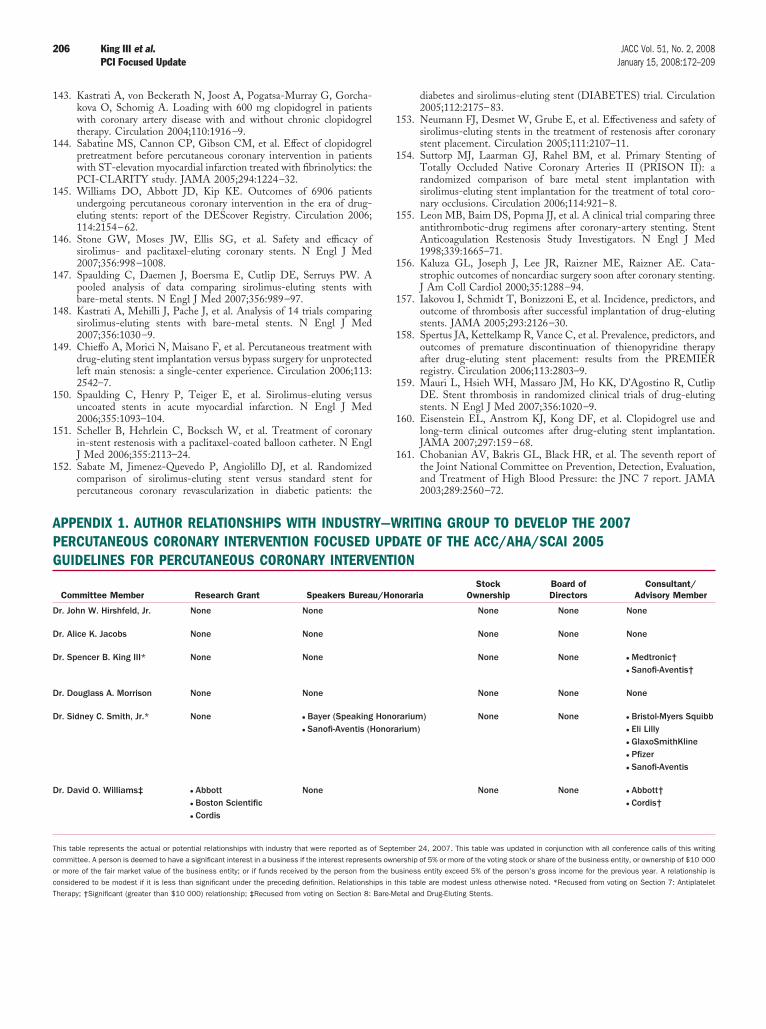
1
1
1
1
1
1
1
1
1
1
1
1
1
1
1
1
1
1
1
APG
D
D
D
D
D
D
T
c
o
c
T
206 King III et al. JACC Vol. 51, No. 2, 2008PCI Focused Update January 15, 2008:172–209
43. Kastrati A, von Beckerath N, Joost A, Pogatsa-Murray G, Gorcha-kova O, Schomig A. Loading with 600 mg clopidogrel in patientswith coronary artery disease with and without chronic clopidogreltherapy. Circulation 2004;110:1916–9.
44. Sabatine MS, Cannon CP, Gibson CM, et al. Effect of clopidogrelpretreatment before percutaneous coronary intervention in patientswith ST-elevation myocardial infarction treated with fibrinolytics: thePCI-CLARITY study. JAMA 2005;294:1224–32.
45. Williams DO, Abbott JD, Kip KE. Outcomes of 6906 patientsundergoing percutaneous coronary intervention in the era of drug-eluting stents: report of the DEScover Registry. Circulation 2006;114:2154–62.
46. Stone GW, Moses JW, Ellis SG, et al. Safety and efficacy ofsirolimus- and paclitaxel-eluting coronary stents. N Engl J Med2007;356:998–1008.
47. Spaulding C, Daemen J, Boersma E, Cutlip DE, Serruys PW. Apooled analysis of data comparing sirolimus-eluting stents withbare-metal stents. N Engl J Med 2007;356:989–97.
48. Kastrati A, Mehilli J, Pache J, et al. Analysis of 14 trials comparingsirolimus-eluting stents with bare-metal stents. N Engl J Med2007;356:1030–9.
49. Chieffo A, Morici N, Maisano F, et al. Percutaneous treatment withdrug-eluting stent implantation versus bypass surgery for unprotectedleft main stenosis: a single-center experience. Circulation 2006;113:2542–7.
50. Spaulding C, Henry P, Teiger E, et al. Sirolimus-eluting versusuncoated stents in acute myocardial infarction. N Engl J Med2006;355:1093–104.
51. Scheller B, Hehrlein C, Bocksch W, et al. Treatment of coronaryin-stent restenosis with a paclitaxel-coated balloon catheter. N EnglJ Med 2006;355:2113–24.
52. Sabate M, Jimenez-Quevedo P, Angiolillo DJ, et al. Randomizedcomparison of sirolimus-eluting stent versus standard stent for
percutaneous coronary revascularization in diabetic patients: thediabetes and sirolimus-eluting stent (DIABETES) trial. Circulation2005;112:2175–83.
53. Neumann FJ, Desmet W, Grube E, et al. Effectiveness and safety ofsirolimus-eluting stents in the treatment of restenosis after coronarystent placement. Circulation 2005;111:2107–11.
54. Suttorp MJ, Laarman GJ, Rahel BM, et al. Primary Stenting ofTotally Occluded Native Coronary Arteries II (PRISON II): arandomized comparison of bare metal stent implantation withsirolimus-eluting stent implantation for the treatment of total coro-nary occlusions. Circulation 2006;114:921–8.
55. Leon MB, Baim DS, Popma JJ, et al. A clinical trial comparing threeantithrombotic-drug regimens after coronary-artery stenting. StentAnticoagulation Restenosis Study Investigators. N Engl J Med1998;339:1665–71.
56. Kaluza GL, Joseph J, Lee JR, Raizner ME, Raizner AE. Cata-strophic outcomes of noncardiac surgery soon after coronary stenting.J Am Coll Cardiol 2000;35:1288–94.
57. Iakovou I, Schmidt T, Bonizzoni E, et al. Incidence, predictors, andoutcome of thrombosis after successful implantation of drug-elutingstents. JAMA 2005;293:2126–30.
58. Spertus JA, Kettelkamp R, Vance C, et al. Prevalence, predictors, andoutcomes of premature discontinuation of thienopyridine therapyafter drug-eluting stent placement: results from the PREMIERregistry. Circulation 2006;113:2803–9.
59. Mauri L, Hsieh WH, Massaro JM, Ho KK, D’Agostino R, CutlipDE. Stent thrombosis in randomized clinical trials of drug-elutingstents. N Engl J Med 2007;356:1020–9.
60. Eisenstein EL, Anstrom KJ, Kong DF, et al. Clopidogrel use andlong-term clinical outcomes after drug-eluting stent implantation.JAMA 2007;297:159–68.
61. Chobanian AV, Bakris GL, Black HR, et al. The seventh report ofthe Joint National Committee on Prevention, Detection, Evaluation,and Treatment of High Blood Pressure: the JNC 7 report. JAMA
2003;289:2560–72.PPENDIX 1. AUTHOR RELATIONSHIPS WITH INDUSTRY—WRITING GROUP TO DEVELOP THE 2007ERCUTANEOUS CORONARY INTERVENTION FOCUSED UPDATE OF THE ACC/AHA/SCAI 2005UIDELINES FOR PERCUTANEOUS CORONARY INTERVENTION
Committee Member Research Grant Speakers Bureau/HonorariaStock
OwnershipBoard ofDirectors
Consultant/Advisory Member
r. John W. Hirshfeld, Jr. None None None None None
r. Alice K. Jacobs None None None None None
r. Spencer B. King III* None None None None ● Medtronic†● Sanofi-Aventis†
r. Douglass A. Morrison None None None None None
r. Sidney C. Smith, Jr.* None ● Bayer (Speaking Honorarium)● Sanofi-Aventis (Honorarium)
None None ● Bristol-Myers Squibb● Eli Lilly● GlaxoSmithKline● Pfizer● Sanofi-Aventis
r. David O. Williams‡ ● Abbott● Boston Scientific● Cordis
None None None ● Abbott†● Cordis†
his table represents the actual or potential relationships with industry that were reported as of September 24, 2007. This table was updated in conjunction with all conference calls of this writing
ommittee. A person is deemed to have a significant interest in a business if the interest represents ownership of 5% or more of the voting stock or share of the business entity, or ownership of $10 000
r more of the fair market value of the business entity; or if funds received by the person from the business entity exceed 5% of the person’s gross income for the previous year. A relationship is
onsidered to be modest if it is less than significant under the preceding definition. Relationships in this table are modest unless otherwise noted. *Recused from voting on Section 7: Antiplatelet
herapy; †Significant (greater than $10 000) relationship; ‡Recused from voting on Section 8: Bare-Metal and Drug-Eluting Stents.
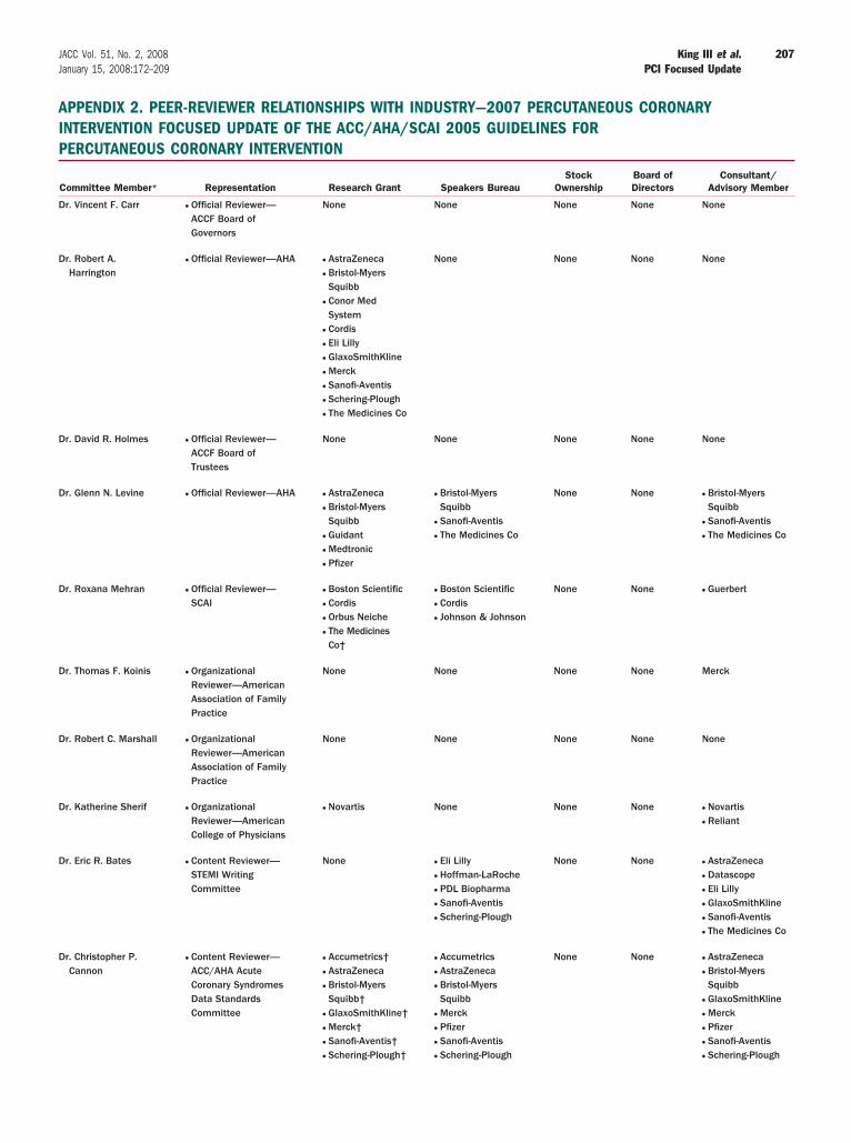
AIP
D
D
D
D
D
D
D
D
D
D
207JACC Vol. 51, No. 2, 2008 King III et al.January 15, 2008:172–209 PCI Focused Update
PPENDIX 2. PEER-REVIEWER RELATIONSHIPS WITH INDUSTRY—2007 PERCUTANEOUS CORONARYNTERVENTION FOCUSED UPDATE OF THE ACC/AHA/SCAI 2005 GUIDELINES FORERCUTANEOUS CORONARY INTERVENTION
Committee Member* Representation Research Grant Speakers BureauStock
OwnershipBoard ofDirectors
Consultant/Advisory Member
r. Vincent F. Carr ● Official Reviewer—ACCF Board ofGovernors
None None None None None
r. Robert A.Harrington
● Official Reviewer—AHA ● AstraZeneca● Bristol-MyersSquibb
● Conor MedSystem
● Cordis● Eli Lilly● GlaxoSmithKline● Merck● Sanofi-Aventis● Schering-Plough● The Medicines Co
None None None None
r. David R. Holmes ● Official Reviewer—ACCF Board ofTrustees
None None None None None
r. Glenn N. Levine ● Official Reviewer—AHA ● AstraZeneca● Bristol-MyersSquibb
● Guidant● Medtronic● Pfizer
● Bristol-MyersSquibb
● Sanofi-Aventis● The Medicines Co
None None ● Bristol-MyersSquibb
● Sanofi-Aventis● The Medicines Co
r. Roxana Mehran ● Official Reviewer—SCAI
● Boston Scientific● Cordis● Orbus Neiche● The MedicinesCo†
● Boston Scientific● Cordis● Johnson & Johnson
None None ● Guerbert
r. Thomas F. Koinis ● OrganizationalReviewer—AmericanAssociation of FamilyPractice
None None None None Merck
r. Robert C. Marshall ● OrganizationalReviewer—AmericanAssociation of FamilyPractice
None None None None None
r. Katherine Sherif ● OrganizationalReviewer—AmericanCollege of Physicians
● Novartis None None None ● Novartis● Reliant
r. Eric R. Bates ● Content Reviewer—STEMI WritingCommittee
None ● Eli Lilly● Hoffman-LaRoche● PDL Biopharma● Sanofi-Aventis● Schering-Plough
None None ● AstraZeneca● Datascope● Eli Lilly● GlaxoSmithKline● Sanofi-Aventis● The Medicines Co
r. Christopher P.Cannon
● Content Reviewer—ACC/AHA AcuteCoronary SyndromesData StandardsCommittee
● Accumetrics†● AstraZeneca● Bristol-MyersSquibb†
● GlaxoSmithKline†● Merck†● Sanofi-Aventis†● Schering-Plough†
● Accumetrics● AstraZeneca● Bristol-MyersSquibb
● Merck● Pfizer● Sanofi-Aventis● Schering-Plough
None None ● AstraZeneca● Bristol-MyersSquibb
● GlaxoSmithKline● Merck● Pfizer● Sanofi-Aventis● Schering-Plough
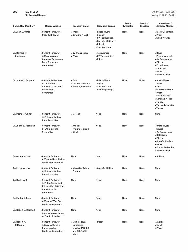
D
D
D
D
D
D
D
D
D
D
D
208 King III et al. JACC Vol. 51, No. 2, 2008PCI Focused Update January 15, 2008:172–209
Committee Member* Representation Research Grant Speakers BureauStock
OwnershipBoard ofDirectors
Consultant/Advisory Member
r. John G. Canto ● Content Reviewer—Individual Review
● Pfizer● Schering-Plough†
● Bristol-MyersSquibb†
● CV Therapeutics● GlaxoSmithKline†● Pfizer†● Sanofi-Aventis†
None None ● NRMI/Genentech● Pfizer● Sanofi-Aventis
r. Bernard R.Chaitman
● Content Reviewer—ACC/AHA AcuteCoronary SyndromesData StandardsCommittee
● CV Therapeutics● Pfizer
● AstraZeneca● CV Therapeutics● Pfizer
None None ● BayerPharmaceuticals
● CV Therapeutics● Eli Lilly● F. Hoffman-La Roche
● Merck● Sanofi-Aventis
r. James J. Ferguson ● Content Reviewer—ACCF CardiacCatheterization andInterventionCommittee
● Eisai● The Medicines Co● Vitatron/Medtronic
● Bristol-MyersSquibb
● Sanofi-Aventis● Schering-Plough
None None ● Bristol-MyersSquibb
● Eisai● GlaxoSmithKline● Prism● Sanofi-Aventis● Schering-Plough● Takeda● The Medicines Co● Therox
r. Michael A. Fifer ● Content Reviewer—AHA Acute CardiacCare Committee
● Merck† None None None None
r. Judith S. Hochman ● Content Reviewer—STEMI GuidelineCommittee
● ArginoxPharmaceuticals
● Eli Lilly
None None None ● Bristol-MyersSquibb
● CV Therapeutics● Datascope● Eli Lilly● GlaxoSmithKline● Merck● Procter & Gamble● Sanofi-Aventis
r. Sharon A. Hunt ● Content Reviewer—ACC/AHA Heart FailureGuideline Committee
None None None None ● Guidant
r. Ik-Kyung Jang ● Content Reviewer—AHA Acute CardiacCare Committee
● Mitsubishi-TokyoPharma
● GlaxoSmithKline None None None
r. Hani Jneid ● Content Reviewer—AHA Diagnostic andInterventional CardiacCatheterizationCommittee
None None None None None
r. Morton J. Kern ● Content Reviewer—ACC/AHA/SCAI PCIGuideline Committee
None None None None None
r. Robert C. Marshall ● Content Reviewer—American Associationof Family Practice
None None None None None
r. Robert A.O’Rourke
● Content Reviewer—ACC/AHA ChronicStable AnginaGuideline Committee
● Multiple drugcompaniesfunding BARI 2Dand COURAGEtrials
● Pfizer None None ● Aventis● Merck● Pfizer
D
D
D
D
D
D
D
Ttoti
C
209JACC Vol. 51, No. 2, 2008 King III et al.January 15, 2008:172–209 PCI Focused Update
Committee Member* Representation Research Grant Speakers BureauStock
OwnershipBoard ofDirectors
Consultant/Advisory Member
r. Martha J. Radford ● Content Reviewer—ACS Data Standards
None None None None None
r. Rita F. Redberg ● Content Reviewer—ACC PreventionCommittee
None None None None None
r. Charanjit S. Rihal ● Content Reviewer—AHA Diagnostic andInterventional CardiacCatheterizationCommittee
None None None None None
r. Allan M. Ross ● Content Reviewer—Individual Review
● BoehringerIngelheim
● Genentech● Roche
None None None ● BoehringerIngelheim
● Roche
r. Samuel J.Shubrooks
● Content Reviewer—ACCF Board ofGovernors
None None None None None
r. Chittur A. Sivaram ● Content Reviewer—ACCF Board ofGovernors
None None None None None
r. YeremYeghiazarians
● Content Reviewer—AHA Diagnostic andInterventional CardiacCatheterizationCommittee
None ● Pfizer● Sanofi-Aventis
None None None
his table represents the relationships of peer reviewers with industry that were disclosed at the time of peer review of this guideline. It does not necessarily reflect relationships with industry at theime of publication. Participation in the peer review process does not imply endorsement of this document. A person is deemed to have a significant interest in a business if the interest representswnership of 5% or more of the voting stock or share of the business entity, or ownership of $10 000 or more of the fair market value of the business entity; or if funds received by the person fromhe business entity exceed 5% of the person’s gross income for the previous year. A relationship is considered to be modest if it is less than significant under the preceding definition. Relationshipsn this table are modest unless otherwise noted. *Names are listed in alphabetical order with each category of review. †Significant (greater than $10,000) relationship.
ACC indicates American College of Cardiology; ACCF, American College of Cardiology Foundation; ACS, American College of Surgeons; AHA, American Heart Association; and SCAI, Society forardiovascular Angiography and Interventions.