delayed treatment with chondroitinase abc promotes - brain
TRANSCRIPT

BRAINA JOURNAL OF NEUROLOGY
Delayed treatment with chondroitinase ABCpromotes sensorimotor recovery and plasticityafter stroke in aged ratsSara Soleman,1 Ping K. Yip,1,2 Denise A. Duricki1,3 and Lawrence D.F. Moon1,3
1 Neurorestoration Group, Wolfson Centre for Age-Related Diseases, King’s College London, Guy’s Campus, London, UK
2 Centre for Neuroscience and Trauma, Blizard Institute, Barts and The London School of Medicine and Dentistry, Queen Mary University of London,
London, UK
3 Centre for Integrative Biology, King’s College London, Franklin-Wilkins Building, 150 Stamford Street, London, UK
Correspondence to: Dr Lawrence Moon,
Neurorestoration Group,
Wolfson Centre for Age-Related Diseases,
King’s College London, Guy’s Campus,
16-18 Newcomen Street, London,
SE1 1UL, UK
E-mail: [email protected]
Stroke is the dominant cause of sensorimotor disability that primarily affects the elderly. We now show that neuroplasticity and
functional recovery after stroke is constrained by inhibitory chondroitin sulphates. In two blinded, randomized preclinical trials,
degradation of chondroitin sulphate using chondroitinase ABC reactivated neuroplasticity and promoted sensorimotor recovery
after stroke in elderly rats. Three days after stroke, chondroitinase ABC was microinjected into the cervical spinal cord to induce
localized plasticity of forelimb sensorimotor spinal circuitry. Chondroitinase ABC effectively removed chondroitin sulphate from
the extracellular matrix and perineuronal nets. Three different tests of sensorimotor function showed that chondroitinase ABC
promoted recovery of forelimb function. Anterograde and retrograde tracing showed that chondroitinase ABC also induced
sprouting of the contralesional corticospinal tract in the aged treated hemicord. Chondroitinase ABC did not neuroprotect the
peri-infarct region. We show for the first time delayed chondroitinase ABC treatment promotes neuroanatomical and functional
recovery after focal ischaemic stroke in an elderly nervous system.
Keywords: stroke; chondroitin sulphate proteoglycans; plasticity; ageing; perineuronal nets
Abbreviations: BDA = biotinylated dextran amine
IntroductionStroke is the largest cause of long-term sensorimotor disability
(Di Carlo, 2009), with nearly 90% of incidents occurring in individ-
uals aged 465 years (Truelsen et al., 2006). The elderly population
are more likely to become disabled by stroke and they have a
reduced capacity to recover from these disabilities compared to
younger stroke survivors (Kelly-Hayes et al., 2003). However, the
majority of experimental research is still conducted on young
animals, despite recommendations by the Stroke Therapy
Academic Industry Round Table and other stroke committees
urging for the use of aged animals in preclinical studies when mod-
elling the clinical disorder (Macleod et al., 2009; Saver et al., 2009).
Many human stroke survivors have small infarcts (Brott et al.,
1989) and the location of these focal strokes predicts the type of
functional deficit, thus it is important to model both these aspects
in an elderly system. The most common functional deficit follow-
ing stroke are motor impairments of the contralateral upper limb
doi:10.1093/brain/aws027 Brain 2012: 135; 1210–1223 | 1210
Received October 15, 2011. Revised November 29, 2011. Accepted December 17, 2011. Advance Access publication March 6, 2012
� The Author (2012). Published by Oxford University Press on behalf of the Guarantors of Brain. All rights reserved.
For Permissions, please email: [email protected]
Dow
nloaded from https://academ
ic.oup.com/brain/article/135/4/1210/359507 by guest on 24 N
ovember 2021

with 480% of subjects experiencing this acutely (Cramer et al.,
1997) and 60% chronically (Dobkin, 2004). Additionally, reports
also indicate up to 94% of human stroke survivors experience
sensory deficits and disturbed performance of motor tasks requir-
ing somatosensory information (Carey et al., 1993). Structural
imaging of human stroke subjects demonstrates highly significant
correlations between the extent of these sensorimotor impairments
and focal ischaemic damage to the corticospinal tract (Lindenberg
et al., 2010; Zhu et al., 2010). This indicates that corticospinal
tract integrity plays a critical role in functional outcome and that
localized ischaemic damage to this pathway is an important goal in
animal models of focal stroke. Focal ischaemic lesions can be pro-
duced using endothelin-1, which occludes arteries and restricts
regional blood flow to produce localized and dose dependent is-
chaemic injury in various brain regions in young animals (Adkins
et al., 2004; Windle et al., 2006). Importantly, we have previously
shown that focal ischaemic damage to the corticospinal tract using
endothelin-1 produces sustained sensorimotor deficits similar to
those observed in human stroke survivors, great reproducibility
and low mortality rates in aged animals (Soleman et al., 2010).
We now show that this model provides a powerful method for
evaluating therapies that promote plasticity, which is vital as there
is currently no drug treatment available beyond the acute stage of
stroke.
Following injury to the adult CNS, undamaged neurons can
undergo plastic responses and neuroanatomical reorganization
to replace damaged synaptic connections (Carmichael, 2003;
Dancause et al., 2005; Hsu and Jones, 2006). However, the
extent of reorganization remains constrained by many intrinsic
and extrinsic factors that limit neuronal growth. Age-related
changes are also known to alter neuroplasticity and the physio-
logical responses after ischaemic injury (Badan et al., 2003; Ward,
2005; Li et al., 2010) which can influence the effects of some
drugs (Fisher and Ratan, 2003). Thus, this reveals age as an im-
portant factor when assessing potential therapeutics for neural
repair. Various drug interventions that enhance events of struc-
tural plasticity have been shown to cause functional recovery after
CNS injury (Chen et al., 2002; Wiessner et al., 2003; Lee et al.,
2004; Clarkson et al., 2010). Of particular interest is chondroiti-
nase ABC, which degrades inhibitory chondroitin sulphate proteo-
glycans present in the extracellular matrix. We and others have
shown that degradation of chondroitin sulphates with chondroiti-
nase ABC reduces the neurite-inhibitory environment, reactivates
plasticity and promotes functional recovery after different central
and peripheral nervous system injuries (Moon et al., 2001;
Bradbury et al., 2002; Barritt et al., 2006; Massey et al., 2006;
Pizzorusso et al., 2006; Galtrey et al., 2007; Cafferty et al., 2008;
Garcia-Alias et al., 2009; Alilain et al., 2011). However, to date
the effects of chondroitinase ABC following stroke have yet to be
explored.
Unlike most conventional stroke therapies that typically target
the brain or the entire neuraxis, we administered chondroitinase
ABC into the denervated cervical spinal cord to induce localized
plasticity of spinal circuitry. Here, we tested the efficacy of this
intraspinal chondroitinase ABC delivered 3 days after focal ischae-
mic stroke on neuroplasticity and functional recovery from sensori-
motor deficits in aged animals. Our results demonstrate that
delayed intraspinal chondroitinase ABC is able to alter the com-
position of diffuse chondroitin sulphate proteoglycans and con-
densed chondroitin sulphate proteoglycan-rich perineuronal nets
present in the aged spinal cord to promote axonal reorganization
and behavioural recovery after stroke. These findings constitute
evidence that chondroitinase ABC promotes plasticity of the unin-
jured CNS after injury and propose chondroitinase ABC as a novel
therapeutic candidate in ischaemic stroke for aged individuals.
Materials and methods
SubjectsForty aged male and female Long Evans rats (304–595 g; 16–19
months of age) were housed 2–3 to a cage in standard laboratory
conditions. Experiments were performed in accordance with guidelines
from the Stroke Therapy Academic Industry Round Table and others
(Fisher et al., 2005; Macleod et al., 2009; Saver et al., 2009) and our
findings were reported in accordance with the Animals in Research:
Reporting In Vivo Experiments guidelines (Kilkenny et al., 2010).
Surgeries, behavioural testing and analysis were performed with inves-
tigators blinded to treatment groups.
Experimental designWe investigated whether delayed intraspinal administration of chon-
droitinase ABC would promote functional recovery after unilateral
stroke in aged (416 month old) rats. Anterograde tracing was used
to assess sprouting of uninjured corticospinal tract collaterals into areas
of partial corticospinal tract denervation in the spinal cord.
In our second study, early behavioural improvements observed in
the first study following delayed intraspinal administration of chondroi-
tinase ABC were further investigated. Retrograde tracing was used to
further assess sprouting of uninjured corticospinal tract collaterals and
any neuroprotective effect on surviving corticospinal neurons.
Experimental designs and groups are outlined in Supplementary Fig. 1.
All surgeries were performed using a randomized block design and the
experimenters were blinded to treatment groups during behavioural and
histological assessment. Penicillinase served as a control as it is an
enzyme shown not to degrade chondroitin sulphate proteoglycans
(Moon et al., 2001; Pizzorusso et al., 2002).
SurgeryAll procedures were in accordance with guidelines from the UK Home
Office and Animals (Scientific Procedures) Act of 1986. Animals were
anaesthetized with isoflurane (4% in O2 for induction) and maintained
at 1.5–2% in O2 delivered via a facemask. Body temperature was
monitored via a rectal thermometer and maintained at �36�C with
a heating pad.
Stroke lesionsPrior to surgery, rats were allocated to an experimental group in a
counterbalanced fashion to ensure no difference in group mean pre-
operative performance of the dominant forepaw on the staircase test.
Unilateral lesions were performed in the hemisphere contralateral to
the dominant forelimb (Supplementary Fig. 3), as determined by the
staircase behavioural test. Stroke lesions (n = 32) or sham surgeries
were performed as previously described (Soleman et al., 2010) using
Chondroitinase ABC as a stroke therapy Brain 2012: 135; 1210–1223 | 1211
Dow
nloaded from https://academ
ic.oup.com/brain/article/135/4/1210/359507 by guest on 24 N
ovember 2021
a randomized block design. Briefly, animals were transferred to a
stereotaxic frame (David Kopf Instruments) where a midline incision
was made, the sensorimotor cortex was then exposed via craniotomy
and the dura mater was incised. Since skull thickness varied amongst
aged rats, a craniotomy was performed to enable accurate depth
placement of endothelin-1 intracortical injections. Four 2 ml injections
of endothelin-1 (200 pmol/ml; 0.5 mg/ml dissolved in sterile saline;
CalBioChem) were delivered via a glass micropipette connected to a
syringe (Hamilton). The first 1 ml was administered at a depth of 1 mm
from the brain surface and the subsequent 1 ml applied to the surface
of the cortex at four coordinates. Prior to suturing, the animal was left
undisturbed for 5 min. Modifying previous work (Soleman et al.,
2010), the skull fragment was then replaced and sealed using bone
wax (Aesculap, Tuttlingen). Sham-operated rats (n = 8) received all
procedures up to, but not including, craniotomy. Animals were admin-
istered buprenorphine (0.01 mg/kg, subcutaneously) for postoperative
pain relief after recovering from anaesthesia. Our method of inducing
stroke with endothelin-1 is advantageous for evaluating regenerative
stroke therapies for four reasons: (i) our model produces ischaemic
lesions that model small focal human strokes rather than larger
‘malignant’ strokes, which tend to be fatal in humans (Carmichael,
2005); (ii) our model targets specific neuronal circuits that are typically
affected after stroke including the corticospinal tract pathway that
originates in the sensorimotor and agranular cortex; (iii) our stroke
model has statistically powerful reproducibility (Soleman et al., 2010)
and involves only low mortality rates in aged animals; and (iv) our
model causes sustained sensorimotor deficits that are the most
common neurological symptoms of human stroke.
Spinal delivery of chondroitinase ABCAnimals received two unilateral spinal injections of either chondroiti-
nase ABC (10 U/ml; Seikagaku) or the control enzyme penicillinase
(same mg/ml of protein; Sigma) delivered into the partially denervated
side of the C5 and C8 spinal cord. Chondroitinase ABC was delivered
intraspinally as previously described (Wong et al., 2006; Galtrey et al.,
2007). Briefly, a partial laminectomy was performed to expose the
cervical spinal cord at level C5 and C8. At each site, 1 ml injections
of either chondroitinase ABC or penicillinase were delivered using a
glass micropipette connected to a 10 ml syringe (Hamilton) at a rate of
0.25 ml/min. The micropipette was positioned 1 mm lateral from the
midline and lowered 1.5 mm below the spinal cord surface; this was
left in situ for 2 min before being withdrawn. To assess the extent of
chondroitin sulphate degradation after chondroitinase ABC treatment,
rats were perfused 1 week after injections (n = 2). In our two main
experiments, animals received unilateral intraspinal injections 3 days
post-stroke injury. The number of rats per treatment group was
based on sample size calculations from previous work (Soleman
et al., 2010), which showed that a minimum of eight rats per
treatment group would be required to identify a treatment which
caused a 50% improvement on the staircase test (� = 0.05;
power4 0.80).
Anterograde tracingIn the first experiment, 7 weeks post-stroke, animals were given bio-
tinylated dextran amine (BDA; 10%; 10 000 MW, Invitrogen) unilat-
erally into the uninjured sensorimotor cortex to visualize uninjured
corticospinal tract axons. Animals were placed in a sterotaxic frame
and six burr holes were made into the skull at the following
coordinates defined as anterioposterior (AP), mediolateral (ML):
(i) AP: + 1 mm, ML: 1.5 mm; (ii) AP: + 0.5 mm, ML: 2.5 mm;
(iii) AP: + 1.5 mm, ML: 2.5 mm; (iv) AP: + 0.5 mm, ML: 3.5 mm;
(v) AP: + 2.0 mm, ML: 3.5 mm; and (vi) AP: �0.5 mm, ML: 3.5 mm,
relative to bregma. At each site, 0.5 ml injections of BDA were delivered
using a glass micropipette attached to a Hamilton syringe inserted 2 mm
from the skull surface and delivered at a rate of 0.25ml/min. Animals
were subsequently left for 2 weeks before being perfused.
Retrograde tracingIn the second experiment, 5 weeks post-stroke, animals were given
unilateral intraspinal injections of the retrograde fluorescent tracer Fast
Blue (2% in phosphate-buffered saline; Sigma) into the denervated
side of the spinal cord and at areas of chondroitinase ABC-mediated
digestion. The spinal cord (C6–C8) was exposed as described above.
Each animal had five injections of Fast Blue (200 nl/site) spaced at
�1 mm apart along the spinal cord. A glass micropipette was pos-
itioned 1 mm lateral and 1.8 mm below the spinal cord surface.
Animals were left for 10 days before being perfused. Location of the
injections was verified post-mortem. Animals that failed to have con-
sistent unilateral tracing mostly confined to the spinal grey matter
were excluded from analysis (chondroitinase ABC group, n = 3; peni-
cillinase group, n = 3).
Behavioural testingTests previously found to be effective in assessing sensory and motor
deficits were included (Soleman et al., 2010). Rats were given 4 weeks
of daily training on the staircase test to identify forepaw preference.
All behavioural testing was carried out by an experimenter blinded to
surgery and treatment groups. Baseline values were recorded 3 days
before surgery on all behavioural tasks.
Assessment of fine motor function
The staircase test was used to assess reaching performance; this pro-
vides a sensitive measure of skilled forepaw motor function (Montoya
et al., 1991). The staircase apparatus (Campden Instruments Ltd.)
consists of a chamber with a central platform for the rat to climb
onto and a set of seven steps located on either side. Three sucrose
pellets (45 mg, Research Diets Inc.) were placed in the well on each
step and could be retrieved by the rat reaching down either side of the
platform. The number of pellets retrieved (maximum of 21 pellets per
side) and the maximum step reached using each forepaw was
recorded during each 15 min trial. Scoring the number of pellets
retrieved indicates successful grasp and retrieval (fine motor function),
while the maximum step reached assesses the lowest step a pellet is
displaced regardless of successful grasping (gross limb control). Each
weekly session consisted of two trials; mean scores per rat per week
were calculated. The minimum criterion to be included in the study
was the retrieval of 11 pellets using their dominant paw during pre-
operative testing.
Assessment of forelimb asymmetry
The cylinder test was used to assess asymmetries in forelimb use for
postural support during rearing (Schallert et al., 2000) within a trans-
parent 20 cm diameter and 30 cm high cylinder. An angled mirror was
placed behind the cylinder to allow movements to be recorded when
the animal turned away from the camera. During exploration, rats rear
against the vertical surface of the cylinder. The first forelimb to touch
the wall was scored as an independent placement for that forelimb.
Subsequent placement of the other forelimb against the wall to main-
tain balance was scored as ‘both’. If both forelimbs were simultan-
eously placed against the wall during rearing this was scored as ‘both’.
1212 | Brain 2012: 135; 1210–1223 S. Soleman et al.
Dow
nloaded from https://academ
ic.oup.com/brain/article/135/4/1210/359507 by guest on 24 N
ovember 2021
Lateral movements along the wall using both forelimbs alternately
were also scored as ‘both’. Scores were obtained from a total
number of 10 full rears to control for differences in rearing between
animals. Once scores had been acquired, forelimb asymmetry was
calculated using the formula: 100 � (ipsilateral forelimb use + 1/2 bi-
lateral forelimb use)/total forelimb use observations (Hsu and Jones,
2005).
Assessment of somatosensory function
The magnitude of somatosensory asymmetry and sensorimotor impair-
ments in forepaw function after stroke was assessed using the bilateral
tactile stimulation test (Schallert et al., 1982, 2000). For each trial,
round adhesive patches (13 mm diameter, Ryman) were applied to
the plantar surface of both forepaws and the animal was returned
to its home cage. Two times were recorded for both forepaws: (i)
contact; and (ii) remove; where ‘contact’ represents the time taken
for the animal to notice the adhesive patch on its forepaw and bring
it to its mouth, and ‘remove’ represents the time taken for the animal
to remove the adhesive patch from its forepaw. To determine whether
the rats showed bias for their affected or less-affected forelimbs, the
order and side of label removal was recorded. This was repeated four
times per session until a 475% preference had been found; if this was
not the case a fifth trial was conducted. The magnitude of asymmetry
was established using the seven levels of stimulus pairs on both fore-
paws as previously described (Fig. 2D, E) (Schallert and Whishaw,
1984; Schallert et al., 2000). During this phase, to determine the
extent of ipsilateral response bias, the size of the stimulus was pro-
gressively increased on the affected forepaw and decreased on the
less-affected forepaw by an equal amount (14.1 mm2), until the rat
removed the stimulus on the affected forepaw first (reversal of original
bias). This reversal represents the magnitude of asymmetry, where the
higher the score indicates the greater the degree of somatosensory
impairment.
HistologyAnimals were terminally anaesthetized with sodium pentobarbital
(80 mg/kg; intraperitoneally) and perfused transcardially with hepari-
nized saline followed by 4% paraformaldehyde in 0.1 M phosphate
buffer. Brains and spinal cords were postfixed for 2 h at 4�C then
transferred to 30% sucrose in phosphate-buffered saline for 2 days.
The brain and spinal cord were separately embedded in 10% gelatin in
distilled water and blocks were postfixed in 4% paraformaldehyde for
24 h. Free-floating serial sections were then cut using a freezing stage
microtome (Kryomat). Ten series of rostral to caudal tissue sections
were collected in 24-well plates containing phosphate-buffered saline
(with 0.1% sodium azide) and stored at 4�C.
ImmunohistochemistryAll sections used were transverse C4–C8 spinal cord sections (40 mm).
To confirm chondroitin sulphate proteoglycan digestion, sections were
incubated in mouse anti-chondroitin-4-sulphate (1:1000; MP
Biomedicals) and goat anti-mouse Alexa 488 (1:500; Invitrogen). To
assess degradation of chondroitin sulphate proteoglycan-rich perineur-
onal nets, sections were incubated in biotin-conjugated Wisteria flori-
bunda agglutinin (15 mg/ml; Sigma) and extra-avidin tetramethyl
rhodamine isothiocyanate (1:1000; Sigma). To assess fibre sprouting
from the uninjured corticospinal tract, sections were incubated in
hydrogen peroxide (0.3%), ABC reagent (1:400; Vector
Laboratories), biotinyl tyramide (1:75; PerkinElmer Life Sciences) and
extra-avidin fluorescein isothiocyanate (1:1000; Sigma). All sections
were coverslipped with FluorsaveTM mounting medium.
Histological analysisImages were captured using a Carl Zeiss AxioImager Z1 microscope
with Z-stack intervals. All images were re-coded during analysis so the
investigator was blinded to treatment groups.
Analysis of biotinylated dextran amine labelled axons
Transverse spinal cord sections in the treated area (C6–C8) were se-
lected. To ensure consistent tracing within animals, the number of
BDA-labelled axons present in the dorsal column were quantified
and averaged from 10 sections per animal. In the denervated grey
matter of these sections, sprouting BDA-labelled axons were quanti-
fied. To reduce between animal tracing variations, the axon index was
calculated as previously described (Zheng et al., 2005; Lee et al.,
2010). For this, the number of BDA-labelled axons present in the
denervated grey matter was normalized against the total number of
labelled axons present in the dorsal column. The distance (mm) of each
BDA-labelled axon along the mediolateral axis from the midline was
determined from the central canal.
Analysis of Fast Blue-labelled corticospinal neurons
Coronal brain sections from regions + 3.5 mm to �1 mm relative to
bregma were selected. The total number of retrogradely labelled cor-
ticospinal neurons present in both hemispheres was quantified in 12
sections per animal. In the injured hemisphere, cell area (mm2) was
assessed in all corticospinal neurons from two cortical 500 mm-wide
regions in the ipsilesional hemisphere: (i) the area directly around
the lesion cavity (Supplementary Fig. 2A0); and (ii) the area neighbour-
ing the injury site (Supplementary Fig. 2A0 0). For the latter, as no vis-
ible cavity was present, we used the following approach: in sections
that had lesion cavities, the distance from the lesion centre to the
midline interhemispheric fissure was measured and applied to the
sections with no visible lesion. A region 500 mm from this calculated
lesion midline was applied on either side and cells within this area were
also measured. Cell size distributions were then tabulated.
Quantification of infarct volume
Serial coronal sections from + 3.5 mm to �2.5 mm relative to bregma
were captured using a light microscope with a high-resolution digital
camera (MiniVID Digital Eyepiece Camera, LW Scientific). Infarct size
was calculated as previously described (Soleman et al., 2010). For each
section, the total area of the ipsilesional hemisphere was subtracted
from the area of the contralesional hemisphere. Hemisphere area
measurements excluded necrotic tissue, cysts and cavities, and ven-
tricles. Volume of injury (mm3) was calculated as the sum of the area
from each section, multiplied by the distance between sections
(Buchan et al., 1992). Before the end of the study, one animal died
due to age-related pituitary tumours and is thus absent from
histological analysis (chondroitinase ABC, n = 1).
Statistical analysisBehavioural data were analysed using repeated measures analysis of
covariance (ANCOVA), using preoperative performances as covariates.
Pairwise group comparisons were performed using two-tailed Fisher’s
protected Least Significant Difference post hoc tests. Infarct volumes
were compared using two-tailed t-tests. The number of Fast Blue-
labelled cells and BDA axon counts were compared between groups
using the Kruskal–Wallis and Mann–Whitney tests. Cell size
Chondroitinase ABC as a stroke therapy Brain 2012: 135; 1210–1223 | 1213
Dow
nloaded from https://academ
ic.oup.com/brain/article/135/4/1210/359507 by guest on 24 N
ovember 2021

distributions between groups were analysed using repeated measures
analysis of variance (ANOVA) and the Kolmogorov–Smirnov test. Data
are presented as mean � standard error of the mean (SEM) and
asterisks indicate significance as follows: *P4 0.05; **P4 0.01;
***P4 0.001. SPSS (IBM) version 18 was used for analysis.
Results
Delayed chondroitinase ABC promotesforelimb motor recovery followingstroke in aged ratsTo assess motor function, dexterity of the affected forepaw
was examined using the staircase test (Fig. 1A; Supplementary
Video 1). Here, two parameters were measured: the number of
pellets retrieved (skilled motor function) and maximum step from
which pellets were retrieved or displaced (unskilled limb control).
Concerning the number of pellets retrieved, 1 week after stroke all
animals showed a marked reduction in their ability to retrieve
pellets with their affected forepaw (sham versus either stroke
group, P5 0.001), with no difference between treatment groups
at this time-point (chondroitinase ABC versus penicillinase,
P = 0.19; Fig. 1B). Compared to preoperative levels, penicillinase-
treated animals were able to recover to a maximum of 59%; in
comparison, chondroitinase ABC-treated animals were more suc-
cessful at retrieving pellets (P = 0.007) and were able to recover to
a maximum of 85%. Concerning the maximum step, stroke
groups remained impaired relative to sham animals (chondroitinase
ABC, P = 0.01; penicillinase, P50.001; Fig. 1C), with no differ-
ence between treatment groups (chondroitinase ABC versus peni-
cillinase, P = 0.24). Thus, our data show that chondroitinase ABC
treatment is capable of significantly improving forepaw dexterity
to aid successful pellet retrieval, while unskilled limb movements
were unaffected by treatment demonstrating some spontaneous
recovery.
To further assess forelimb motor function, we employed the
cylinder test. This measures forelimb use for postural support
during rearing (Fig. 1D). Sham animals demonstrated approxi-
mately equal use of forelimbs during postural support (asymmetry
score� 50). One week after stroke, animals exhibited an increase
in asymmetry score indicating preferential use of their less-affected
forelimb for support during rearing (sham versus penicillinase,
P = 0.002; sham versus chondroitinase ABC, P = 0.08; Fig. 1E).
Sustained functional deficits were apparent in the penicillinase-
treated animals, with little recovery occurring beyond the first
week of stroke (Week 1 versus Week 9, P = 0.87; Supplementary
Video 2). In contrast, chondroitinase ABC-treatment significantly
reduced the functional deficits compared to penicillinase animals
(P = 0.002), indicating use of their affected forelimb for postural
support. By Week 9 post-stroke, there was no significant differ-
ence between chondroitinase ABC-treated and sham animals
(P = 0.17; Supplementary Video 3).
Interestingly, our data showed a rapid effect of chondroitinase
ABC as animals had smaller functional deficits by the first week of
behavioural testing (4 days after treatment) compared to the
penicillinase group (P = 0.06). To confirm the immediate effects
of chondroitinase ABC were not due to baseline differences from
the lesion, a second experimental study was repeated with a be-
havioural testing time point at 2 days post-stroke (1 day before
drug intervention). Importantly, both groups prior to treatment
showed an equal extent of functional impairment (chondroitinase
ABC versus penicillinase, P = 0.68; Fig. 1F) indicating both stroke
groups possessed equivalent stroke lesions and behavioural def-
icits. After treatment, penicillinase-treated rats remained persist-
ently impaired (Day 2 versus Week 5; P = 0.36). Interestingly,
chondroitinase ABC-treated rats exhibited immediate behavioural
recovery in the first week (Day 2 versus Week 1; P = 0.06) and
improved until the end of the study (Day 2 versus Week 5;
P = 0.005). By Week 5, chondroitinase ABC-treated animals
were significantly different to penicillinase-treated animals
(P = 0.04) and no different to sham animals (P = 0.23). Together
these two studies show chondroitinase ABC is able to promote use
of the affected forelimb during postural support and reduces fore-
limb deficits following cerebral ischaemia in aged rats.
Delayed chondroitinase ABC promotesforelimb sensory recovery followingstroke in aged ratsTo assess forepaw impairments and asymmetries in somatosensory
function, we used the bilateral tactile stimulation test. A small
adhesive patch was stuck to the plantar surface of each forepaw
and the time taken to contact and remove both stimuli with their
mouth was recorded (Fig. 2A). Sham animals rapidly contacted
and removed both stimuli. Stroke rats showed impairments in
both sensing and removing the adhesive patch from their affected
forepaw (Fig. 2B and C). However, there was no effect of drug
treatment as animals displayed a similar extent of recovery in con-
tact (chondroitinase ABC versus penicillinase, P = 0.19; Fig. 2B)
and removal time (chondroitinase ABC versus penicillinase,
P = 0.71; Fig. 2C).
We also assessed recovery from tactile extinction, a phenom-
enon manifested in many stroke patients who fail to detect a
touch stimulus on their affected hand once it has been applied
simultaneously on both hands (Rose et al., 1994). We obtained
sensory asymmetry scores using pairs of sensory stimuli (Schallert
et al., 2000) (Fig. 2D and E). After stroke, all animals had high
asymmetry scores, indicating that animals neglected the larger
stimulus on their affected forepaw and preferentially removed
the smaller stimulus from their less-affected forepaw first.
Penicillinase-treated animals remained persistently impaired rela-
tive to sham animals (P50.001; Fig. 2F). In comparison, chon-
droitinase ABC induced significant recovery from asymmetrical
sensory impairments with animals able to detect smaller stimuli
on their affected forepaw (chondroitinase ABC versus penicillinase,
P = 0.04). In conclusion, while chondroitinase ABC had no effect
on sensory response times, it overcame somatosensory neglect of
the affected forepaw following stroke. This is important as tactile
extinction was shown to be the single most important predictor of
functional outcome (Rose et al., 1994), thus improving this ability
1214 | Brain 2012: 135; 1210–1223 S. Soleman et al.
Dow
nloaded from https://academ
ic.oup.com/brain/article/135/4/1210/359507 by guest on 24 N
ovember 2021

to detect and process sensory data should enable improvements in
motor function.
Chondroitinase ABC injectionseffectively degrade diffuse chondroitinsulphate proteoglycans present in theaged spinal cordUnilateral intraspinal injections of chondroitinase ABC were carried
out at C5 and C8 to encourage localized plasticity and midline
crossing of the intact corticospinal tract into chondroitinase
ABC-treated areas of the aged cervical spinal cord. We examined
the pattern of chondroitin sulphate proteoglycan digestion using
an antibody that recognizes the chondroitin-4-sulphate stub epi-
tope on chondroitin sulphate proteoglycan core proteins following
chondroitin sulphate removal. As expected, no immunoreactivity
for chondroitin-4-sulphate was detected in sham controls or
penicillinase-treated animals (Fig. 3A and B). In a pilot study,
we assessed the extent and spread of chondroitin sulphate pro-
teoglycan degradation. One week following chondroitinase ABC
injections, extensive and predominantly unilateral chondroitin-4-
sulphate immunoreactivity spanned segments C4 to T1 (Fig. 3C
and D). Even 10 weeks after chondroitinase ABC intraspinal injec-
tions, intense immunoreactivity was still present in the cervical
cord, confirming chondroitinase ABC effectively degraded chon-
droitin sulphate proteoglycans and that its effects are long lasting
(Fig. 3E). Quantification of chondroitin-4-sulphate immunoreactiv-
ity revealed intense digestion in both the spinal cord grey and
white matter, (Fig. 3F) and spanning dorsal, ventral and midline
regions of the spinal cord (Fig. 3G). The greatest degree of chon-
droitin sulphate proteoglycan digestion was observed in the
Figure 1 Delayed chondroitinase ABC promotes motor recovery following stroke in aged rats. The staircase test assesses forepaw
dexterity; (A) a photograph and Supplementary Video 1 show a rat performing this task. (B) After stroke, rats treated with chondroitinase
ABC (ChABC) retrieved more pellets with their affected forelimb than rats treated with penicillinase (P-ase) [overall effect F(2,13) = 34.6,
P = 0.0001; interaction of group by time F(14,91) = 1.88, P = 0.04], confirming that chondroitinase ABC promotes improvements in
dexterity. (C) There was no difference in the maximum step reached by either treatment groups of rats, although they were both impaired
relative to shams [group differences F(2,13) = 9.5, P = 0.003]. (D) The cylinder test assesses forelimb use for postural support during
rearing. (E) Following stroke, rats exhibited an increase in asymmetry score indicating preferential use of the less-affected forelimb for
weight support. Penicillinase-treated animals remained impaired whereas animals treated with chondroitinase ABC significantly recovered
[group differences F(2,17) = 13.2, P = 0.0001; Supplementary Videos 2 and 3]. (F) In a second experiment, testing at 2 days confirmed
both treatment groups possessed similar behavioural deficits before drug intervention. As previously observed, penicillinase-treated ani-
mals remained impaired whereas chondroitinase ABC-treated animals significantly recovered [group differences F(2,11) = 6.1, P = 0.02].
Results are presented as mean � SEM and were analysed using repeated measures ANCOVA with post hoc Least Significant Difference
tests. Significance is denoted as: *P5 0.05, **P50.01, ***P5 0.001 versus sham animals (Experiment 1, n = 5; Experiment 2, n = 3)
and XP50.05, XXP50.01, XXXP50.001 versus treatment groups (Experiment 1, n = 9 per group; Experiment 2, n = 7 per group).
Chondroitinase ABC as a stroke therapy Brain 2012: 135; 1210–1223 | 1215
Dow
nloaded from https://academ
ic.oup.com/brain/article/135/4/1210/359507 by guest on 24 N
ovember 2021

ventral horn and funiculus. Thus, these results indicate that
intraspinal chondroitinase ABC injections were effective in degrad-
ing growth inhibitory chondroitin sulphate proteoglycans.
Chondroitinase ABC injections degradeperineuronal nets present in the agedspinal cordPrevious studies have demonstrated that digestion of chondroitin
sulphate proteoglycans in perineuronal nets with chondroitinase
ABC can reactivate plasticity (Pizzorusso et al., 2002; Massey
et al., 2006), leading to the hypothesis that perineuronal nets
are involved in the control of plasticity in the CNS (Carulli et al.,
2010). We examined W. floribunda agglutinin reactivity, a marker
of chondroitin sulphate proteoglycan-rich perineuronal nets, 1
week following chondroitinase ABC delivery. Here, we observed
that chondroitinase ABC was able to reduce W. floribunda agglu-
tinin reactivity (Fig. 4A), and this effect was only present in areas
of chondroitin sulphate proteoglycan degradation (Fig. 4B and C).
Intact perineuronal nets surrounding motor neurons in the ventral
horn were observed outside areas of chondroitinase ABC-mediated
digestion (Fig. 4D and F) and were completely absent within
degraded areas (Fig. 4E and G). Thus, our results confirm that
intraspinal chondroitinase ABC delivery not only alters the com-
position of the diffuse extracellular matrix but also alters the
Figure 2 Delayed chondroitinase ABC promotes forelimb sensory recovery following stroke in aged rats. The bilateral tactile stimulation
test assesses sensory function using adhesive sticky patches placed on the rats’ forepaws (A). Stroke increased the time animals took to
sense (B) and remove (C) these adhesive patches placed on their affected forepaw, and both treatment groups showed a similar extent
of recovery [sense: F(2,17) = 1.57, P = 0.24; remove: F(2,17) = 0.63, P = 0.55]. (D, E) The magnitude of sensory asymmetry (tactile
extinction) was determined using the seven pairs of sensory stimuli (see text). (D) Stimulus size was progressively increased on the affected
forepaw and decreased on the less-affected forepaw by an equal amount, until the rat removed the stimulus on the affected forepaw first.
This reversal represents the magnitude of asymmetry. (E) Testing begins at Level 3 (black box) and asymmetry scores indicate the severity
of sensory asymmetry (e.g. severe impairment = 7). (F) Stroke increased the magnitude of sensory asymmetry (tactile extinction) in both
groups. Penicillinase-treated (P-ase) animals remained impaired whereas animals treated with chondroitinase ABC (ChABC) showed
significant improvements in detecting the sensory stimulus on their affected forepaw [group differences F(2,17) = 12.2, P = 0.001].
Results are presented as mean � SEM and were analysed using repeated measures ANCOVA with post hoc Least Significant Difference
tests. Significance is denoted as: *P50.05, **P50.01, ***P50.001 versus sham animals (n = 5) and XP5 0.05, XXP50.01,XXXP5 0.001 versus treatment groups (n = 9 per group).
1216 | Brain 2012: 135; 1210–1223 S. Soleman et al.
Dow
nloaded from https://academ
ic.oup.com/brain/article/135/4/1210/359507 by guest on 24 N
ovember 2021
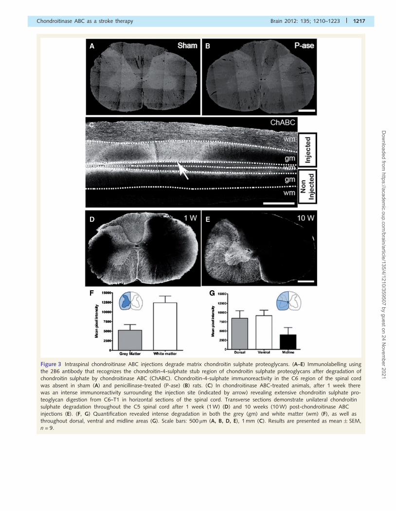
Figure 3 Intraspinal chondroitinase ABC injections degrade matrix chondroitin sulphate proteoglycans. (A–E) Immunolabelling using
the 2B6 antibody that recognizes the chondroitin-4-sulphate stub region of chondroitin sulphate proteoglycans after degradation of
chondroitin sulphate by chondroitinase ABC (ChABC). Chondroitin-4-sulphate immunoreactivity in the C6 region of the spinal cord
was absent in sham (A) and penicillinase-treated (P-ase) (B) rats. (C) In chondroitinase ABC-treated animals, after 1 week there
was an intense immunoreactivity surrounding the injection site (indicated by arrow) revealing extensive chondroitin sulphate pro-
teoglycan digestion from C6–T1 in horizontal sections of the spinal cord. Transverse sections demonstrate unilateral chondroitin
sulphate degradation throughout the C5 spinal cord after 1 week (1 W) (D) and 10 weeks (10 W) post-chondroitinase ABC
injections (E). (F, G) Quantification revealed intense degradation in both the grey (gm) and white matter (wm) (F), as well as
throughout dorsal, ventral and midline areas (G). Scale bars: 500mm (A, B, D, E), 1 mm (C). Results are presented as mean � SEM,
n = 9.
Chondroitinase ABC as a stroke therapy Brain 2012: 135; 1210–1223 | 1217
Dow
nloaded from https://academ
ic.oup.com/brain/article/135/4/1210/359507 by guest on 24 N
ovember 2021

condensed matrix in these chondroitin sulphate proteoglycan-rich
perineuronal nets that are found around neurons present in the
spinal cord, particularly around motor neurons.
Delayed chondroitinase ABC promotescollateral sprouting of the uninjuredcorticospinal tract in the aged spinalcordIn our first experiment, BDA was unilaterally injected into the
contralesional hemisphere to anterogradely label intact corticosp-
inal tract axons and collaterals (Fig. 5A). Immunofluorescent visu-
alization of BDA was used to assess axonal sprouting into
chondroitinase ABC-treated areas of the spinal cord grey matter.
To control for variations in BDA labelling amongst individual ani-
mals, axonal counts in the denervated grey matter were normal-
ized against the total number of BDA labelled corticospinal tract
axons present in the dorsal column (Zheng et al., 2005). No group
differences in BDA labelling were observed (P = 0.77; Fig. 5B–D),
confirming similar tracing efficiency. Penicillinase-treated animals
possessed few BDA-labelled corticospinal tract collaterals in the
grey matter of the treated side (Fig. 5E). In contrast, chondroiti-
nase ABC-treatment significantly increased corticospinal tract col-
lateral sprouting on the treated side (P = 0.02; Fig. 5F and G).
Furthermore, the distance of uninjured corticospinal tract collat-
erals sprouting from the midline revealed a strong trend for chon-
droitinase ABC to encourage more axons to sprout at longer
distances compared to penicillinase [F(1,6) = 4.85, P = 0.07; Fig.
5H]. In chondroitinase ABC-treated animals, it was also observed
that some BDA-positive axons were sprouting within areas of
chondroitin sulphate proteoglycan degradation (Fig. 5I).
In our second experiment, Fast Blue was used to further assess
corticospinal tract collateral sprouting by retrogradely labelling cor-
ticospinal neurons of the uninjured corticospinal tract (Fig. 6A):
Fast Blue was carefully and unilaterally injected into chondroitinase
ABC-treated areas of the cervical grey matter (Fig. 6B). We quan-
tified the number of corticospinal neurons present in the intact
sensorimotor cortex that projected collaterals into these chondroi-
tinase ABC-treated areas. Labelled corticospinal neurons were pri-
marily located rostral to Bregma, particularly from the primary
motor (M1) and somatosensory forelimb cortical regions. Sham
and penicillinase-treated animals displayed similar numbers of
Fast Blue-labelled corticospinal neurons (P = 0.13; Fig. 6C–E).
However, chondroitinase ABC-treated animals possessed
significantly greater numbers of labelled corticospinal neurons
in the sensorimotor cortex (Fig. 6C and F) compared to
penicillinase-treated animals (P = 0.03). Together these results
demonstrate that chondroitinase ABC is able to induce plasticity
and cause axonal collateral sprouting from the intact corticospinal
tract into chondroitinase ABC-treated areas of the spinal cord in
aged rats.
Delayed chondroitinase ABC does notpromote neuroprotection after strokein aged ratsWe and others previously reported that surviving corticospinal
neurons in the ipsilesional hemisphere following stroke display a
degree of cell atrophy (Enright et al., 2007; Soleman et al., 2010).
As chondroitinase ABC has been shown to reverse atrophy of
corticospinal neurons after spinal cord injury (Carter et al.,
2008), we analysed the soma sizes of corticospinal neurons
Figure 4 Intraspinal chondroitinase ABC injections degrade perineuronal nets. Transverse C5 spinal cord sections were stained with W.
floribunda agglutinin (WFA, red) to label chondroitin sulphate proteoglycans present in perineuronal nets. (A) One week following
intraspinal chondroitinase ABC injections, WFA reactivity was reduced. (B) Chondroitin-4-sulphate immunolabelling (green) revealed
perineuronal nets were only reduced in areas of chondroitin sulphate proteoglycan degradation (C). (D, E) Dashed boxes denote the area
of high power magnification highlighting perineuronal nets present in undigested areas (D) and absent in areas of chondroitin sulphate
proteoglycan digestion (E). (F, G) Perineuronal nets were also observed encasing Nissl-stained (green) motor neurons in the ventral horn in
undigested regions (F) and appeared absent around motor neurons in digested regions (G). Scale bar: 250 mm (A–C), 20mm (D–G).
1218 | Brain 2012: 135; 1210–1223 S. Soleman et al.
Dow
nloaded from https://academ
ic.oup.com/brain/article/135/4/1210/359507 by guest on 24 N
ovember 2021

retrogradely labelled with Fast Blue at 5 weeks post-injury
(Supplementary Fig. 2A). At 7 weeks post-stroke, cell atrophy
was evident in penicillinase-treated animals compared to sham
controls, particularly for cell sizes between 150 and 300 mm2
(Supplementary Fig. 2B, C and E). Following chondroitinase
ABC treatment, corticospinal neurons appeared to possess slightly
larger-looking somata (Supplementary Fig. 2D), with cell size
distribution analysis revealing a trend towards significance be-
tween treatment groups for cells between 150–300mm2
(P = 0.07). However, overall drug treatment had no significant
effect on cell size distribution [F(2,6) = 0.73, P = 0.52] and cumu-
lative frequency (P = 0.3; Supplementary Fig. 2E and F). There was
also no difference in the total number of surviving Fast
Blue-labelled corticospinal neurons between treatment groups
Figure 5 Chondroitinase ABC induces cervical collateral sprouting from the intact corticospinal tract following stroke. (A) Schematic
diagram illustrating that BDA injections were administered into the uninjured hemisphere to label projections from the intact corticospinal
tract (CST). (B, C) Photomicrographs of BDA-positive fibres present in the dorsal column of the cervical spinal cord in penicillinase-treated
(P-ase) animals (B) and chondroitinase ABC-treated (ChABC) animals (C). (D) Quantification of these fibres revealed no significant
difference between treatments groups, confirming similar tracing efficiency between animals. (E, F) Photomicrographs of BDA-positive
axons projecting to the denervated side of the spinal cord in penicillinase-treated animals (E) and chondroitinase ABC-treated animals (F).
Dashed lines indicate the spinal cord midline and arrow-heads highlight axons present on the treated side of the spinal cord. (G)
Quantification revealed that animals treated with chondroitinase ABC showed a significant increase in the number of BDA-labelled axons
present on the treated side compared to penicillinase treated animals. (H) Mediolateral spatial distribution of BDA-positive fibres
throughout the treated side of the spinal cord in chondroitinase ABC-treated animals compared to penicillinase-treated animals. (I)
Example of axons (red) in areas of chondroitin sulphate proteoglycan digestion (green). Results are presented as mean � SEM and were
analysed using Kruskal–Wallis and Mann–Whitney tests (D, G) or repeated measures ANOVA (H). Significance is denoted as: *P50.05
between treatment groups (n = 4 per group). Scale bars: 100mm (B, C, E and F).
Chondroitinase ABC as a stroke therapy Brain 2012: 135; 1210–1223 | 1219
Dow
nloaded from https://academ
ic.oup.com/brain/article/135/4/1210/359507 by guest on 24 N
ovember 2021

(P = 0.29; Supplementary Fig. 2G), showing that delayed chon-
droitinase ABC does not prevent corticospinal neuron cell death
in the injured hemisphere.
Additionally, histological analysis of the brain at 10 weeks after
stroke revealed the extent of ischaemic damage in lesioned aged
rats (Supplementary Fig. 3A and B). Affected areas primarily
included the primary motor cortex (M1) with varying amounts
of the secondary motor cortex (M2), agranular cortex and primary
somatosensory cortex (S1, particularly the forelimb and hindlimb
regions). Infarct volume analysis revealed no differences between
drug treatment groups (P = 0.1; Supplementary Fig. 3C), confirm-
ing both groups had similar extents of ischaemic injury. Thus, our
results suggest that intraspinal delivery of chondroitinase ABC does
not reverse corticospinal neuron atrophy associated with ischaemic
stroke or affect infarct size and therefore we attribute recovery to
plasticity of the corticospinal tract and other intraspinal circuits.
DiscussionIschaemic stroke induces sustained sensorimotor impairments and
limited spontaneous behavioural recovery. This was supported in
our study where control-treated animals showed little improve-
ment in behavioural function after stroke, consistent with our pre-
vious work (Soleman et al., 2010). Importantly, we show for the
first time that intraspinal delivery of chondroitinase ABC initiated 3
days post-stroke in an aged CNS is significantly able to reduce
sensorimotor impairments of the affected forelimb, including fine
Figure 6 Chondroitinase ABC increases the number of corticospinal neurons projecting to areas of denervation following stroke. (A)
Schematic diagram illustrating the five unilateral Fast Blue injection sites which retrogradely labelled corticospinal neurons (CSNs) in the
uninjured hemisphere that sprouted uninjured corticospinal tract projections into areas of chondroitinase ABC-mediated digestion. (B)
Transverse C5 spinal cord sections confirmed that the Fast Blue injections were unilateral and within the grey matter of the spinal cord. (C)
Quantification of the number of labelled corticospinal neurons present in the uninjured sensorimotor cortex revealed that chondroitinase
ABC-treatment (ChABC) increased the number of corticospinal neurons projecting to the denervated side of the spinal cord. (D–F)
Photomicrographs of the sensorimotor cortex (Bregma + 0.5 mm) showing the number of Fast Blue-labelled corticospinal neurons in sham
(D), penicillinase-treated (P-ase) (E) and chondroitinase ABC-treated animals (F). Arrow-heads highlight Fast Blue-labelled corticospinal
neurons in sensorimotor cortex. Results were presented as mean � SEM and were analysed using Kruskal–Wallis and Mann–Whitney
tests. Significance is denoted as: *P50.05 between treatment groups (n = 4 per group). Scale bar: 1 mm (B), 500mm (D–F).
1220 | Brain 2012: 135; 1210–1223 S. Soleman et al.
Dow
nloaded from https://academ
ic.oup.com/brain/article/135/4/1210/359507 by guest on 24 N
ovember 2021

motor function, forelimb use during rearing and somatosensory
function (reversal of neglect). Our results extend previous work
showing chondroitinase ABC promotes behavioural recovery after
different neurological injuries (Bradbury et al., 2002; Pizzorusso
et al., 2006; Galtrey et al., 2007; Cafferty et al., 2008;
Garcia-Alias et al., 2009; Alilain et al., 2011). Approximately 16
million people worldwide suffer from stroke every year (Strong
et al., 2007) and there is still no restorative drug treatment
beyond the acute stage of stroke. Our study proposes chondroi-
tinase ABC as a potential treatment to promote neural repair after
stroke that is clinically attractive as it also counteracts existing
problems of treatment time windows and age as chondroitinase
ABC was administered 3 days post-stroke in an elderly CNS
system.
Human and animals studies have shown that the adult CNS is
capable of some functional reorganization that is associated with
the limited spontaneous recovery observed following stroke
(Cramer, 2008). Two main mechanisms considered for reorganiza-
tional and plastic changes after stroke are: (i) formation of new
neuronal circuitry; and (ii) unmasking/strengthening of existing
pathways. In the present study, chondroitinase ABC was adminis-
tered into the denervated side of the spinal cord to encourage
localized plasticity of spinal circuitry and limit the extent of chon-
droitinase ABC-mediated digestion throughout the neuraxis, thus
minimizing effects on other brain regions. As delivery of chondroi-
tinase ABC was spatially restricted this suggests recovery was pos-
sibly a consequence of enhanced plasticity within local spinal
circuitry. It is likely that chondroitinase ABC stimulated anatomical
changes in a number of spinal pathways that may have contrib-
uted to the observed functional improvements. However, here we
anatomically traced and primarily focused on the intact corticosp-
inal tract, a major pathway known to modulate both sensory and
motor function. Our results demonstrate that chondroitinase ABC
is able to enhance plastic changes and axonal sprouting of the
intact corticospinal tract from the contralesional hemisphere. This
is consistent with other stroke studies that have enhanced ana-
tomical sprouting of uninjured spinal pathways from the contrale-
sional hemisphere and consequently improved recovery of
function (Chen et al., 2002; Papadopoulos et al., 2002; Lee
et al., 2004; Markus et al., 2005). Other human and animal stu-
dies have also shown that the contralesional hemisphere is
involved in recovery following stroke, including changes in den-
dritic arborization (Jones and Schallert, 1992; Biernaskie and
Corbett, 2001; Uryu et al., 2001) and enhanced cortical activation
(Dijkhuizen et al., 2003; Schaechter and Perdue, 2008). It has
been reported that stroke induces reorganization in two regions
of the cortex: an area immediately adjacent to the infarct core that
has a substantial increase in chondroitin sulphate proteoglycans,
and a more distant area from the infarct that has reduced levels of
chondroitin sulphate proteoglycan and a loss of perineuronal nets
that contributes to anatomical plasticity (Carmichael et al., 2005).
Whether further digestion of chondroitin sulphate proteoglycans in
these peri-infarct regions enhances local plasticity and functional
recovery has yet to be determined, but may reveal the potential
for local as well as distant chondroitinase ABC treatment in stroke.
Additionally, the rapid recovery of function may be attributed to
the unmasking of physiologically silent synapses (Carmel et al.,
2010) which may be recruited after alterations in neural activity
following treatment. Recently, it has been shown that intraspinal
injections of chondroitin sulphate proteoglycan acutely depresses
axonal conduction in a dose-dependent manner (Hunanyan et al.,
2010) and intraspinal chondroitinase ABC injections prevent the
decline in axonal conduction in intact fibres after spinal cord
injury (Hunanyan et al., 2010). This demonstrates an inhibitory
action of chondroitin sulphate proteoglycans on axonal conduction
and that their removal using chondroitinase ABC can enhance
neural activity. Thus, it may also be possible that chondroitinase
ABC enhanced conduction and synaptic transmission in the pre-
sent study.
Another mechanism for both rapid and progressive recovery
may be the removal of perineuronal nets. In the adult spinal
cord, chondroitin sulphate proteoglycans are distributed diffusely
in both the grey and white matter extracellular matrix (Tang et al.,
2003) and are also highly condensed into structures known as
perineuronal nets that surround specific neuronal cell bodies,
such as motor neurons (Takahashi-Iwanaga et al., 1998). The
time of the appearance of perineuronal nets is closely correlated
with the termination of plasticity and the end of the critical period
in development (Pizzorusso et al., 2002). Developmental attenu-
ation of perineuronal nets extends this critical period (Carulli et al.,
2010) and digestion of chondroitin sulphate proteoglycan-rich
perineuronal nets in the adult visual cortex using chondroitinase
ABC is known to reactivate plasticity (Pizzorusso et al., 2002),
confirming that perineuronal nets control plasticity in the CNS. A
recent study has also shown that removal of perineuronal nets
around phrenic motor neurons using chondroitinase ABC promotes
rapid functional recovery of diaphragmatic function after spinal
cord injury 1 week after treatment (Alilain et al., 2011). We
have shown that microinjections of chondroitinase ABC directly
into the spinal cord produces rapid, localized and long-lasting
chondroitin sulphate proteoglycan digestion throughout both the
grey and white matter, consistent with previous studies (Galtrey
et al., 2007; Cafferty et al., 2008; Garcia-Alias et al., 2009). We
also found chondroitinase ABC effectively removed both diffuse
chondroitin sulphate proteoglycans in the cervical spinal cord and
chondroitin sulphate proteoglycan-rich perineuronal nets present
around motor neurons. Our data indicate that removal of peri-
neuronal nets may be a probable mechanism through which
chondroitinase ABC rapidly reactivates plasticity and recovery of
function.
In summary, our work is the first to demonstrate that chondroi-
tinase ABC can promote recovery of sensorimotor forelimb func-
tion after stroke and plasticity in the uninjured CNS. Excitingly, we
show that recovery and neuroplasticity occurs in an elderly CNS,
even when treatment is delayed by 3 days, and proposes chon-
droitinase ABC as a novel therapeutic candidate in ischaemic
stroke for aged individuals.
AcknowledgementsWe thank Gary Fulcher for assistance during behavioural testing.
Chondroitinase ABC as a stroke therapy Brain 2012: 135; 1210–1223 | 1221
Dow
nloaded from https://academ
ic.oup.com/brain/article/135/4/1210/359507 by guest on 24 N
ovember 2021
FundingThis work was supported by the Medical Research Council (grant
number G0600998), by a Research Councils UK Academic
Fellowship and by the British Pharmacological Society (BPS)’s
Integrative Pharmacology Fund. This study was also supported
by a Capacity Building Award in Integrative Mammalian Biology
funded by the Biotechnology and Biological Sciences Research
Council, BPS, Higher Education Funding Council for England,
Knowledge Transfer Partnerships, MRC and Scottish Funding
Council.
Supplementary materialSupplementary material is available at Brain online.
ReferencesAdkins DL, Voorhies AC, Jones TA. Behavioral and neuroplastic effects of
focal endothelin-1 induced sensorimotor cortex lesions. Neuroscience
2004; 128: 473–86.Alilain WJ, Horn KP, Hu H, Dick TE, Silver J. Functional regeneration of
respiratory pathways after spinal cord injury. Nature 2011; 475:
196–200.Badan I, Buchhold B, Hamm A, Gratz M, Walker LC, Platt D, et al.
Accelerated glial reactivity to stroke in aged rats correlates with
reduced functional recovery. J Cereb Blood Flow Metab 2003; 23:
845–54.
Barritt AW, Davies M, Marchand F, Hartley R, Grist J, Yip P, et al.
Chondroitinase ABC promotes sprouting of intact and injured spinal
systems after spinal cord injury. J Neurosci 2006; 26: 10856–67.
Biernaskie J, Corbett D. Enriched rehabilitative training promotes im-
proved forelimb motor function and enhanced dendritic growth after
focal ischemic injury. J Neurosci 2001; 21: 5272–80.
Bradbury EJ, Moon LD, Popat RJ, King VR, Bennett GS, Patel PN, et al.
Chondroitinase ABC promotes functional recovery after spinal cord
injury. Nature 2002; 416: 636–40.
Brott T, Marler JR, Olinger CP, Adams HP Jr, Tomsick T, Barsan WG,
et al. Measurements of acute cerebral infarction: lesion size by com-
puted tomography. Stroke 1989; 20: 871–5.
Buchan AM, Slivka A, Xue D. The effect of the NMDA receptor antag-
onist MK-801 on cerebral blood flow and infarct volume in experi-
mental focal stroke. Brain Res 1992; 574: 171–7.
Cafferty WB, Bradbury EJ, Lidierth M, Jones M, Duffy PJ, Pezet S, et al.
Chondroitinase ABC-mediated plasticity of spinal sensory function.
J Neurosci 2008; 28: 11998–2009.
Carey LM, Matyas TA, Oke LE. Sensory loss in stroke patients: effective
training of tactile and proprioceptive discrimination. Arch Phys Med
Rehabil 1993; 74: 602–11.Carmel JB, Berrol LJ, Brus-Ramer M, Martin JH. Chronic electrical stimu-
lation of the intact corticospinal system after unilateral injury restores
skilled locomotor control and promotes spinal axon outgrowth. J
Neurosci 2010; 30: 10918–26.
Carmichael ST. Plasticity of cortical projections after stroke.
Neuroscientist 2003; 9: 64–75.Carmichael ST. Rodent models of focal stroke: size, mechanism, and
purpose. NeuroRx 2005; 2: 396–409.
Carmichael ST, Archibeque I, Luke L, Nolan T, Momiy J, Li S.
Growth-associated gene expression after stroke: evidence for a
growth-promoting region in peri-infarct cortex. Exp Neurol 2005;
193: 291–311.
Carter LM, Starkey ML, Akrimi SF, Davies M, McMahon SB,
Bradbury EJ. The yellow fluorescent protein (YFP-H) mouse reveals
neuroprotection as a novel mechanism underlying chondroitinase
ABC-mediated repair after spinal cord injury. J Neurosci 2008; 28:
14107–20.
Carulli D, Pizzorusso T, Kwok JC, Putignano E, Poli A, Forostyak S, et al.
Animals lacking link protein have attenuated perineuronal nets and
persistent plasticity. Brain 2010; 133: 2331–47.Chen P, Goldberg DE, Kolb B, Lanser M, Benowitz LI. Inosine induces
axonal rewiring and improves behavioral outcome after stroke. Proc
Natl Acad Sci U S A 2002; 99: 9031–6.Clarkson AN, Huang BS, Macisaac SE, Mody I, Carmichael ST. Reducing
excessive GABA-mediated tonic inhibition promotes functional recov-
ery after stroke. Nature 2010; 468: 305–9.
Cramer SC. Repairing the human brain after stroke: I. Mechanisms of
spontaneous recovery. Ann Neurol 2008; 63: 272–87.Cramer SC, Nelles G, Schaechter JD, Kaplan JD, Finklestein SP.
Computerized measurement of motor performance after stroke.
Stroke 1997; 28: 2162–8.Dancause N, Barbay S, Frost SB, Plautz EJ, Chen D, Zoubina EV, et al.
Extensive cortical rewiring after brain injury. J Neurosci 2005; 25:
10167–79.
Di Carlo A. Human and economic burden of stroke. Age Ageing 2009;
38: 4–5.Dijkhuizen RM, Singhal AB, Mandeville JB, Wu O, Halpern EF,
Finklestein SP, et al. Correlation between brain reorganization, ische-
mic damage, and neurologic status after transient focal cerebral ische-
mia in rats: a functional magnetic resonance imaging study. J Neurosci
2003; 23: 510–7.Dobkin BH. Strategies for stroke rehabilitation. Lancet Neurol 2004; 3:
528–36.
Enright LE, Zhang S, Murphy TH. Fine mapping of the spatial relationship
between acute ischemia and dendritic structure indicates selective vul-
nerability of layer V neuron dendritic tufts within single neurons
in vivo. J Cereb Blood Flow Metab 2007; 27: 1185–200.
Fisher M, Albers GW, Donnan GA, Furlan AJ, Grotta JC, Kidwell CS,
et al. Enhancing the development and approval of acute stroke thera-
pies: Stroke Therapy Academic Industry roundtable. Stroke 2005; 36:
1808–13.Fisher M, Ratan R. New perspectives on developing acute stroke ther-
apy. Ann Neurol 2003; 53: 10–20.
Galtrey CM, Asher RA, Nothias F, Fawcett JW. Promoting plasticity in
the spinal cord with chondroitinase improves functional recovery after
peripheral nerve repair. Brain 2007; 130: 926–39.Garcia-Alias G, Barkhuysen S, Buckle M, Fawcett JW. Chondroitinase
ABC treatment opens a window of opportunity for task-specific re-
habilitation. Nat Neurosci 2009; 12: 1145–51.Hsu JE, Jones TA. Time-sensitive enhancement of motor learning with
the less-affected forelimb after unilateral sensorimotor cortex lesions in
rats. Eur J Neurosci 2005; 22: 2069–80.
Hsu JE, Jones TA. Contralesional neural plasticity and functional changes
in the less-affected forelimb after large and small cortical infarcts in
rats. Exp Neurol 2006; 201: 479–94.
Hunanyan AS, Garcia-Alias G, Alessi V, Levine JM, Fawcett JW,
Mendell LM, et al. Role of chondroitin sulfate proteoglycans in
axonal conduction in Mammalian spinal cord. J Neurosci 2010; 30:
7761–9.Jones TA, Schallert T. Overgrowth and pruning of dendrites in adult
rats recovering from neocortical damage. Brain Res 1992; 581:
156–60.
Kelly-Hayes M, Beiser A, Kase CS, Scaramucci A, D’Agostino RB,
Wolf PA. The influence of gender and age on disability following is-
chemic stroke: the Framingham study. J Stroke Cerebrovasc Dis 2003;
12: 119–26.
Kilkenny C, Browne WJ, Cuthill IC, Emerson M, Altman DG. Improving
bioscience research reporting: The ARRIVE guidelines for reporting
animal research. J Pharmacol Pharmacother 2010; 1: 94–9.
1222 | Brain 2012: 135; 1210–1223 S. Soleman et al.
Dow
nloaded from https://academ
ic.oup.com/brain/article/135/4/1210/359507 by guest on 24 N
ovember 2021

Lee JK, Geoffroy CG, Chan AF, Tolentino KE, Crawford MJ, Leal MA,et al. Assessing spinal axon regeneration and sprouting in Nogo-,
MAG-, and OMgp-deficient mice. Neuron 2010; 66: 663–70.
Lee JK, Kim JE, Sivula M, Strittmatter SM. Nogo receptor antagonism
promotes stroke recovery by enhancing axonal plasticity. J Neurosci2004; 24: 6209–17.
Li S, Overman JJ, Katsman D, Kozlov SV, Donnelly CJ, Twiss JL, et al.
An age-related sprouting transcriptome provides molecular control of
axonal sprouting after stroke. Nat Neurosci 2010; 13: 1496–504.Lindenberg R, Renga V, Zhu LL, Betzler F, Alsop D, Schlaug G. Structural
integrity of corticospinal motor fibers predicts motor impairment in
chronic stroke. Neurology 2010; 74: 280–7.Macleod MR, Fisher M, O’Collins V, Sena ES, Dirnagl U, Bath PM, et al.
Good laboratory practice: preventing introduction of bias at the bench.
Stroke 2009; 40: e50–e52.
Markus TM, Tsai SY, Bollnow MR, Farrer RG, O’Brien TE, Kindler-Baumann DR, et al. Recovery and brain reorganization after stroke
in adult and aged rats. Ann Neurol 2005; 58: 950–3.
Massey JM, Hubscher CH, Wagoner MR, Decker JA, Amps J, Silver J,
et al. Chondroitinase ABC digestion of the perineuronal net promotesfunctional collateral sprouting in the cuneate nucleus after cervical
spinal cord injury. J Neurosci 2006; 26: 4406–14.
Montoya CP, Campbell-Hope LJ, Pemberton KD, Dunnett SB. The "stair-
case test": a measure of independent forelimb reaching and graspingabilities in rats. J Neurosci Methods 1991; 36: 219–28.
Moon LD, Asher RA, Rhodes KE, Fawcett JW. Regeneration of CNS
axons back to their target following treatment of adult rat brainwith chondroitinase ABC. Nat Neurosci 2001; 4: 465–6.
Papadopoulos CM, Tsai SY, Alsbiei T, O’Brien TE, Schwab ME, Kartje GL.
Functional recovery and neuroanatomical plasticity following middle
cerebral artery occlusion and IN-1 antibody treatment in the adultrat. Ann Neurol 2002; 51: 433–41.
Pizzorusso T, Medini P, Berardi N, Chierzi S, Fawcett JW, Maffei L.
Reactivation of ocular dominance plasticity in the adult visual cortex.
Science 2002; 298: 1248–51.Pizzorusso T, Medini P, Landi S, Baldini S, Berardi N, Maffei L. Structural
and functional recovery from early monocular deprivation in adult rats.
Proc Natl Acad Sci U S A 2006; 103: 8517–22.Rose L, Bakal DA, Fung TS, Farn P, Weaver LE. Tactile extinction and
functional status after stroke. A preliminary investigation. Stroke 1994;
25: 1973–6.
Saver JL, Albers GW, Dunn B, Johnston KC, Fisher M. Stroke TherapyAcademic Industry Roundtable (STAIR) recommendations for extended
window acute stroke therapy trials. Stroke 2009; 40: 2594–600.
Schaechter JD, Perdue KL. Enhanced cortical activation in the contrale-
sional hemisphere of chronic stroke patients in response to motor skillchallenge. Cereb Cortex 2008; 18: 638–47.
Schallert T, Fleming SM, Leasure JL, Tillerson JL, Bland ST. CNS plasticity
and assessment of forelimb sensorimotor outcome in unilateral rat
models of stroke, cortical ablation, parkinsonism and spinal cord
injury. Neuropharmacology 2000; 39: 777–87.
Schallert T, Upchurch M, Lobaugh N, Farrar SB, Spirduso WW, Gilliam P,
et al. Tactile extinction: distinguishing between sensorimotor and
motor asymmetries in rats with unilateral nigrostriatal damage.
Pharmacol Biochem Behav 1982; 16: 455–62.
Schallert T, Whishaw IQ. Bilateral cutaneous stimulation of the somato-
sensory system in hemidecorticate rats. Behav Neurosci 1984; 98:
518–40.
Soleman S, Yip P, Leasure JL, Moon L. Sustained sensorimotor impair-
ments after endothelin-1 induced focal cerebral ischemia (stroke) in
aged rats. Exp Neurol 2010; 222: 13–24.
Strong K, Mathers C, Bonita R. Preventing stroke: saving lives around the
world. Lancet Neurol 2007; 6: 182–7.
Takahashi-Iwanaga H, Murakami T, Abe K. Three-dimensional microa-
natomy of perineuronal proteoglycan nets enveloping motor neurons
in the rat spinal cord. J Neurocytol 1998; 27: 817–27.Tang X, Davies JE, Davies SJ. Changes in distribution, cell associations,
and protein expression levels of NG2, neurocan, phosphacan, brevican,
versican V2, and tenascin-C during acute to chronic maturation of
spinal cord scar tissue. J Neurosci Res 2003; 71: 427–44.
Truelsen T, Piechowski-Jozwiak B, Bonita R, Mathers C, Bogousslavsky J,
Boysen G. Stroke incidence and prevalence in Europe: a review of
available data. Eur J Neurol 2006; 13: 581–98.
Uryu K, MacKenzie L, Chesselet MF. Ultrastructural evidence for differ-
ential axonal sprouting in the striatum after thermocoagulatory and
aspiration lesions of the cerebral cortex in adult rats. Neuroscience
2001; 105: 307–16.
Ward NS. Plasticity and the functional reorganization of the human
brain. Int J Psychophysiol 2005; 58: 158–61.
Wiessner C, Bareyre FM, Allegrini PR, Mir AK, Frentzel S, Zurini M, et al.
Anti-Nogo-A antibody infusion 24 hours after experimental stroke im-
proved behavioral outcome and corticospinal plasticity in normotensive
and spontaneously hypertensive rats. J Cereb Blood Flow Metab 2003;
23: 154–65.
Windle V, Szymanska A, Granter-Button S, White C, Buist R, Peeling J,
et al. An analysis of four different methods of producing focal cerebral
ischemia with endothelin-1 in the rat. Exp Neurol 2006; 201: 324–34.
Wong LF, Yip PK, Battaglia A, Grist J, Corcoran J, Maden M, et al.
Retinoic acid receptor beta2 promotes functional regeneration of sen-
sory axons in the spinal cord. Nat Neurosci 2006; 9: 243–50.
Zheng B, Atwal J, Ho C, Case L, He XL, Garcia KC, et al. Genetic de-
letion of the Nogo receptor does not reduce neurite inhibition in vitro
or promote corticospinal tract regeneration in vivo. Proc Natl Acad Sci
U S A 2005; 102: 1205–10.
Zhu LL, Lindenberg R, Alexander MP, Schlaug G. Lesion load of the
corticospinal tract predicts motor impairment in chronic stroke.
Stroke 2010; 41: 910–5.
Chondroitinase ABC as a stroke therapy Brain 2012: 135; 1210–1223 | 1223
Dow
nloaded from https://academ
ic.oup.com/brain/article/135/4/1210/359507 by guest on 24 N
ovember 2021