defects in heart and lung development in compound heterozygotes
TRANSCRIPT
INTRODUCTION
N-myc is a member of the myc family of proto-oncogenes,which includes N-myc, c-myc, and L-myc. Myc proteins aresite-specific DNA-binding proteins (Blackwell et al., 1990;Prendergast and Ziff, 1991; Alex et al., 1992), belonging tothe basic-helix-loop-helix class of transcription factors,which includes genes that control cell fate determination insuch diverse processes as myogenesis, neurogenesis and sexdetermination (reviewed in Garrell and Campuzano, 1991).
Deregulated expression of myc genes has been implicatedin the genesis or progression of a number of naturallyoccurring tumours, in the transformation of cells in cultureand in the formation of tumours in transgenic mice(reviewed in DePinho et al., 1991). In general, the sites ofexpression of a given myc gene in vivo reflect the types oftumours associated with its elevated expression. Thus N-myc is expressed predominantly in the embryo where it isrestricted to undifferentiated subsets of cells in the centraland peripheral nervous system, lung, kidney and eye
(Mugrauer et al., 1988; Hirning et al., 1991; Zimmerman etal., 1986) and overexpression of N-myc has been associatedwith tumours of embryonic origin such as neuroblastoma(Kohl et al., 1983; Schwab et al., 1983), small-cell lungcancer (Nau et al., 1986; Wong et al., 1986), Wilm’s tumour(Nisen et al., 1986) and retinoblastoma (Lee et al., 1984).N-myc is also expressed in the skin (Mugrauer et al., 1988),in the epithelial layer of the intestine (Hirning et al., 1991)and, earlier in development, in the heart, sclerotome andvisceral arches (Katoh et al., 1991).
The functioning of a Myc protein in vivo should dependnot only on its own level of expression, but also on the levelsof Max, a protein which, like the Myc proteins, possesses abasic-helix loop helix-leucine zipper (bHLH-LZ) domain(Blackwood and Eisenman, 1991), and which associateswith N-Myc, L-Myc and c-Myc proteins in vivo(Blackwood et al., 1992; Wenzel et al., 1991; Mukherjee etal., 1992). Max is required for specific DNA binding by Mycproteins (Blackwood and Eisenman, 1991; Prendergast andZiff, 1991; Kato et al., 1992; Barrett et al., 1992), and has
485Development 119, 485-499 (1993)Printed in Great Britain © The Company of Biologists Limited 1993
Two types of mutant allele, one leaky and one null, havebeen generated by gene targeting at the N-myc locus inembryonic stem cells and the phenotypes of micehomozygous for these mutations have been described.These mutations have shown that N-myc has a numberof functions during development, including a role inbranching morphogenesis in the lung, which manifestsitself at birth in mice homozygous for the leaky allele,and roles in the development of the mesonephric tubules,the neuroepithelium, the sensory ganglia, the gut and theheart, which become evident at midgestation in embryoshomozygous for the null allele. In an attempt to defineroles for N-myc at other stages of development, we havecombined the two types of N-myc mutant allele in acompound heterozygote that as a result contains approx-imately 15% of normal levels of N-Myc protein.Compound heterozygotes died during gestation at a time
intermediate to the times of death of embryos homozy-gous for either mutation individually, and their deathappeared to result from cardiac failure stemming fromhypoplasia of the compact subepicardial layer of themyocardium. Investigation of the expression pattern ofN-myc and various markers of differentiation in wild-type and compound heterozygote mutant hearts hassuggested that N-myc may function in maintaining theproliferation and/or preventing the differentiation ofcompact layer myocytes. This study illustrates theimportance of generating different mutations at a givenlocus to elucidate fully the function of a particular geneduring development.
Key words: N-myc, heart development, targeted mutagenesis,mouse mutant
SUMMARY
Defects in heart and lung development in compound heterozygotes for
two different targeted mutations at the N-myc locus
Cecilia B. Moens1,3, Brian R. Stanton2, Luis F. Parada2 and Janet Rossant1,3,*1Division of Molecular and Developmental Biology, Samuel Lunenfeld Research Institute, 600 University Ave, Toronto,Ontario, M5G 1X5, Canada2Molecular Embryology Section, ABL-Basic Research Program, National Cancer Institute - Frederick Cancer Research andDevelopment Center, Frederick, Maryland 21702-1201, USA3Department of Molecular and Medical Genetics, University of Toronto
*Author for correspondence
486
been shown to be required for transcriptional activation(Amati et al., 1992) and transformation (Amati et al., 1993)by c-myc. Unlike Myc proteins, Max is able to form homo-dimers in vitro and thereby to bind the myc-binding site(Prendergast and Ziff, 1991; Kato et al., 1992). HoweverMax does not transactivate downstream genes on its own(Amati et al., 1992; Kretzner et al., 1992) because it lacks atransactivation domain (Kato et al., 1992) which Mycproteins possess (Kato et al., 1990). Both transformation offibroblasts by c-myc and N-myc (Mukherjee et al., 1992;Makela et al., 1992; Prendergast et al., 1992) and transcrip-tional transactivation by c-myc (Amati et al., 1992; Kretzneret al., 1992) have been shown to be enhanced by low levelsof Max and inhibited by excess Max, suggesting that Mycfunction is indeed influenced by levels of Max. Recently,bHLH-LZ proteins have been isolated which also bind theMyc recognition site and which suppress transcription asheterodimers with Max (Ayer et al., 1993; Zervos et al.,1993). One of these, Mad (Ayer et al., 1993), has beenshown to compete with Myc for Max in vitro and in trans-fected cells. Thus high levels of expression of Max dimer-ization partners may indirectly affect Myc function in vivoby competing for available Max protein and for DNA-recog-nition sites. Finally, Myc function in vivo may be affectedby the levels of other Myc proteins (Mukherjee et al., 1992;Resar et al., 1993).
In an effort to understand the function of myc genes inembryogenesis, leaky and null mutations were made in N-myc in embryonic stem cells by homologous recombination(Charron et al., 1990; Stanton et al., 1990; Sawai et al., 1991;Moens et al., 1992). These mutations have allowed adescription of the function of N-myc at different stages ofdevelopment. Mice homozygous for the null mutations dieat midgestation (Stanton et al., 1992; Charron et al., 1992)while mice homozygous for the leaky mutation survive untilbirth, when they die due to a defect in lung branching mor-phogenesis (Moens et al., 1992). The latter phenotype isconsistent with the normal expression of N-myc in the devel-oping lung epithelium, and has led us to postulate that N-myc plays a role in the response of the lung epithelium tothe signals from the lung mesenchyme that induce epithelialbranching.
There have been two detailed reports of the phenotype ofmice carrying null mutations in N-myc (Stanton et al., 1992;Charron et al., 1992). In the homozygous condition, thesemutations cause embryonic death at around 11.5 days p.c.and cause hypoplasia in a number of components of theembryo, different aspects of which have been emphasizedby the two groups. The developing genitourinary systemshows a reduced number of mesonephric tubules in mutantembryos, which is consistent with N-myc’s normalexpression in the newly induced epithelium of the meso- andmetanephric tubules (Katoh et al., 1991; Mugrauer et al.,1988). The genital ridge is also hypoplastic. Stanton et al.(1992) also describe a defect in the development of thestomach and intestine, consistent with N-myc’s expressionin the epithelium of these tissues (Hirning et al., 1991).Homozygotes also have reduced cranial and spinal gangliaand a thin neuroepithelium in the telencephalon; both ofthese are sites of N-myc expression in the embryo. Finally,homozygotes have a defect in heart development in which
the heart appears to cease development at 9.5 days p.c.(Charron et al., 1992) and as a result is poorly compart-mentalized and appears not to have undergone the normalepithelial-to-mesenchymal transitions that result in theformation of the septa and valves. This is again consistentwith a previous description of N-myc expression in themyocardium of the 9.5 day embryo (Katoh et al., 1991).
By combining leaky and null alleles of N-myc in acompound heterozygote, we hoped to investigate N-mycfunction in the mouse embryo at a stage intermediate tothose identified by the null and leaky alleles individually.We find that compound heterozygotes indeed survive longerthan null homozygotes, but that they die before birth. Themutant phenotype includes a more severe defect inbranching morphogenesis of the lung, as well as a cardiacmyocyte hypoplasia in the compact subepicardial layer ofthe ventricular myocardium. We show that, consistent withthis phenotype, N-myc expression in the developingmyocardium is restricted to the compact layer. A role formyc genes in the control of myocyte differentiation in theheart is discussed.
MATERIALS AND METHODS
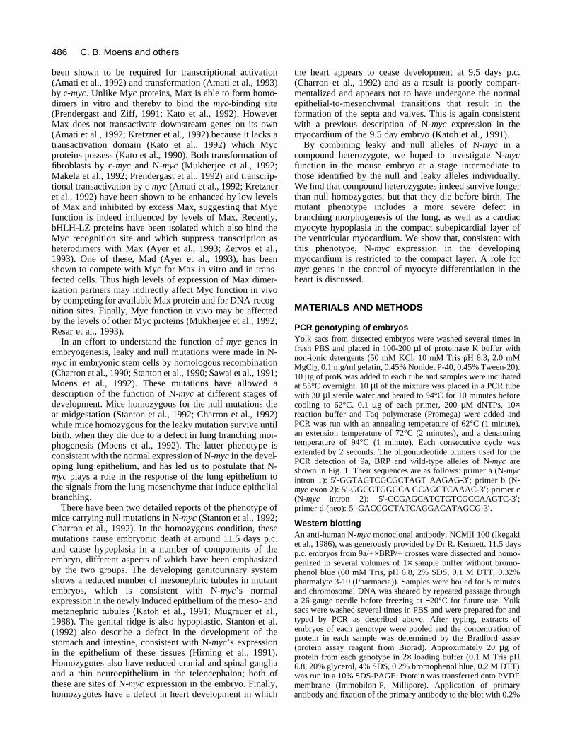
PCR genotyping of embryosYolk sacs from dissected embryos were washed several times infresh PBS and placed in 100-200 µl of proteinase K buffer withnon-ionic detergents (50 mM KCl, 10 mM Tris pH 8.3, 2.0 mMMgCl2, 0.1 mg/ml gelatin, 0.45% Nonidet P-40, 0.45% Tween-20).10 µg of proK was added to each tube and samples were incubatedat 55°C overnight. 10 µl of the mixture was placed in a PCR tubewith 30 µl sterile water and heated to 94°C for 10 minutes beforecooling to 62°C. 0.1 µg of each primer, 200 µM dNTPs, 10×reaction buffer and Taq polymerase (Promega) were added andPCR was run with an annealing temperature of 62°C (1 minute),an extension temperature of 72°C (2 minutes), and a denaturingtemperature of 94°C (1 minute). Each consecutive cycle wasextended by 2 seconds. The oligonucleotide primers used for thePCR detection of 9a, BRP and wild-type alleles of N-myc areshown in Fig. 1. Their sequences are as follows: primer a (N-mycintron 1): 5′-GGTAGTCGCGCTAGT AAGAG-3′; primer b (N-myc exon 2): 5′-GGCGTGGGCA GCAGCTCAAAC-3′; primer c(N-myc intron 2): 5′-CCGAGCATCTGTCGCCAAGTC-3′;primer d (neo): 5′-GACCGCTATCAGGACATAGCG-3′.
Western blottingAn anti-human N-myc monoclonal antibody, NCMII 100 (Ikegakiet al., 1986), was generously provided by Dr R. Kennett. 11.5 daysp.c. embryos from 9a/+×BRP/+ crosses were dissected and homo-genized in several volumes of 1× sample buffer without bromo-phenol blue (60 mM Tris, pH 6.8, 2% SDS, 0.1 M DTT, 0.32%pharmalyte 3-10 (Pharmacia)). Samples were boiled for 5 minutesand chromosomal DNA was sheared by repeated passage througha 26-gauge needle before freezing at −20°C for future use. Yolksacs were washed several times in PBS and were prepared for andtyped by PCR as described above. After typing, extracts ofembryos of each genotype were pooled and the concentration ofprotein in each sample was determined by the Bradford assay(protein assay reagent from Biorad). Approximately 20 µg ofprotein from each genotype in 2× loading buffer (0.1 M Tris pH6.8, 20% glycerol, 4% SDS, 0.2% bromophenol blue, 0.2 M DTT)was run in a 10% SDS-PAGE. Protein was transferred onto PVDFmembrane (Immobilon-P, Millipore). Application of primaryantibody and fixation of the primary antibody to the blot with 0.2%
C. B. Moens and others

487N-myc mutant mice
glutaraldehyde was performed as described (Ikegaki and Kennett,1989). Horseradish peroxidase-conjugated goat anti-mousesecondary antibody and reagents for the chemiluminescentdetection of HRP activity were obtained in the ECL westernblotting kit (Amersham) and were used as suggested by the man-ufacturer.
HistologyDissected embryos were fixed in 10% buffered formalin overnightat room temperature, then were dehydrated and cleared by soakingsequentially in 70%, 80%, 90% and 95% ethanol, 1:1ethanol:xylene, xylene, 1:1 xylene:paraffin wax, (TissuePrep 2,Fisher Scientific) each for 1 hour. Embryos were then oriented andembedded in paraffin wax. Mutant embryos and control littermateswere oriented either for sagittal or frontal sections. 5 µm sectionswere placed on slides, which were dried overnight at 37°C. Slideswere dewaxed, rehydrated, and stained with hematoxylin andeosin. To analyse mutant phenoypes, sections throughout wild-typeand mutant embryos were carefully examined in order to find thosesections that passed through corresponding regions of the differentembryos. Mutant embryos were always compared to non-mutantlittermates rather than to non-mutant embryos from other litters, inorder to rule out differences due to age.
RNA in situ hybridizationsProbes for in situs were as follows. N-myc: a 541 bp Pst1-Sca1fragment of the N-myc genomic clone N7.7 (DePinho et al., 1986),including largely 3′-untranslated sequence; c-myc: a 351 bp HaeIIIfragment including the untranslated first exon (Bossone et al.,1992); flk-1: an 800 bp fragment spanning the transmembranedomain and part of the extracellular ligand-binding domain(Yamaguchi et al., 1993) and α-cardiac actin, a 130 bp BamHIfragment including the first untranslated exon of the mouse α-cardiac actin gene (Sassoon et al., 1988). In situ hybridization wascarried out essentially as described (Frohman et al., 1990) with thefollowing modifications: in vitro-transcribed, α-35S-labeled RNAprobes were used at 5×10⁴ disintegrations/minute per 1 µl ofhybridization solution for the α-cardiac actin probe, and at 1×10⁵
disintegrations/minute per 1 µl of hybridization solution for c-myc,N-myc and flk-1 probes. Slides were hybridized at 53°C overnight,and were washed in the presence of 0.1% β-mercaptoethanol as areducing agent. High stringency washes were in 50% formamide,0.1% β-mercaptoethanol, and 1× SSC at 65°C. Final rinses in 2×and 0.1× SSC were at 65°C for 15 minutes. Slides were exposedfor either 6 days (for α-cardiac actin probe) or for 14 days (for N-myc, flk-1 and c-myc probes). After developing and photographingwith dark-field illumination, slides were stained with hematoxylinand eosin and were rephotographed with bright-field illumination.
RESULTS
Time of death of N-myc compound heterozygotesThe leaky mutation that was generated in N-myc (Moens etal., 1992) was termed N-myc9a and the null allele of N-mycused in this study was named N-mycBRP (Stanton et al.,1992). Both the N-myc9a mutation and the N-mycBRP
mutation are lethal in the homozygous condition. Therefore,N-myc9a/BRP embryos were generated by crossing N-myc9a/+
females to N-mycBRP/+ males, with the expected ratio ofembryos being 1 N-myc+/+: 1 N-myc9a/+: 1 N-mycBRP/+: 1N-myc9a/BRP. The reciprocal cross was also performed: nodifferences in mutant phenotypes were observed.
In order to expedite the genotypic analysis of offspringfrom this cross, a PCR strategy was designed that used four
oligonucleotide primers to distinguish 9a, BRP, and wild-type alleles of N-myc (Fig. 1). 161 live offspring from N-myc9a/+×N-mycBRP/+ crosses were typed by PCR between 1day and 3 weeks after birth. Among these pups, 59 were N-myc+/+, 53 were N-myc9a/+ and 49 were N-mycBRP/+. Nolive animals were N-myc9a/BRP. No pups were observed todie postnatally as occurred in the case of N-myc9a/9a
newborns, suggesting that the N-myc9a/BRP phenotype wasindeed more severe than the N-myc9a/9a phenotype.
We dissected embryos from N-myc9a/+×N-mycBRP/+
crosses at various stages of gestation in order to determinethe time during gestation when N-myc9a/BRP embryos died.Between 8.5 and 11.5 days p.c., N-myc9a/BRP embryos werephenotypically normal and constituted approximately 25%of the total number of embryos, by PCR analysis. Of 268embryos dissected between 8.5 and 11.5 days p.c., 61 (23%)were N-myc9a/BRP embryos. This is clearly different fromthe situation in N-mycBRP/BRP embryos, which were allfound to be dead by 11.5 days p.c. (Stanton et al., 1992).The observed differences in phenotype among the variousmutants were unlikely to be due to different genetic back-ground effects, since all mutant phenotypes were analysedin outbred mice.
Occasionally, N-myc9a/BRP embryos at 11.5 days wereslightly smaller than their littermates but were otherwiseindistinguishable from their wild-type or single heterozy-gote littermates. However, by 12.5 days, most N-myc9a/BRP
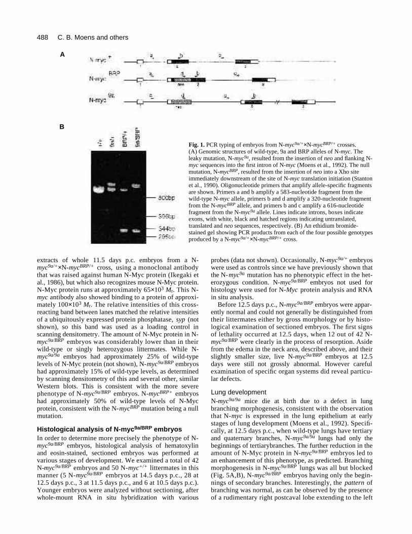
embryos were distinguishable from their littermates eitherbecause they were dead and necrotic, or because they werealive but had a characteristic edema of the body wall in theneck area (Fig. 2). Subsequent histological analysis identi-fied this swelling as likely being the result of extravasion offluid from the vascular compartment into the connectivetissue, since the jugular veins are dilated in these embryos(Fig. 7g,h). Of 230 embryos dissected at 12.5 days p.c., 42(18%) were N-myc9a/BRP, but among these, 12 were deadand necrotic, 23 had the characteristic edema describedabove, two were smaller than normal and five were pheno-typically normal. After 12.5 days p.c., live N-myc9a/BRP
embryos were progressively less frequent and all were phe-notypically abnormal. N-myc9a/BRP embryos that surviveduntil 14.5 days p.c. were always smaller than normal andhad a large edema in the neck area. Live N-myc9a/BRP
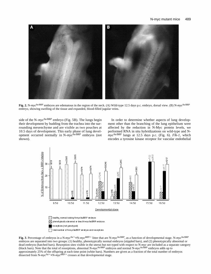
embryos were not observed after 14.5 days p.c. Coincidentwith the loss of N-myc9a/BRP embryos there was an increasein the number of resorptions visible in the uterus. Theseresorptions were not typed, but presumably were derivedfrom N-myc9a/BRP embryos, since the sum of the number ofN-myc9a/BRP embryos and the number of resorptions addedup to approximately 25% of the total number of embryosand resorptions at each stage of gestation. These data areshown graphically in Fig. 3.
N-Myc protein levels in mutant embryosAlthough we have previously shown reduced N-m y cmRNA levels in N-m y c9 a / 9 a embryos (Moens et al., 1992),N-Myc protein levels have not been determined in micebearing either the leaky or the null N-m y c a l l e l e s . In thepresent study, we have used western blot analysis to assessN-Myc protein levels in embryos with the various N-m y cgenotypes. Fig. 4 shows a western blot performed on
488
extracts of whole 11.5 days p.c. embryos from a N-m y c9 a / +×N -m y cB R P / + cross, using a monoclonal antibodythat was raised against human N-Myc protein (Ikegaki etal., 1986), but which also recognizes mouse N-Myc protein.N-Myc protein runs at approximately 65×1 03 Mr. This N-m y c antibody also showed binding to a protein of approxi-mately 100×1 03 Mr. The relative intensities of this cross-reacting band between lanes matched the relative intensitiesof a ubiquitously expressed protein phosphatase, syp ( n o tshown), so this band was used as a loading control inscanning densitometry. The amount of N-Myc protein in N-m y c9 a / B R P embryos was considerably lower than in theirwild-type or singly heterozygous littermates. While N-m y c9 a / 9 a embryos had approximately 25% of wild-typelevels of N-Myc protein (not shown), N-m y c9 a / B R P e m b r y o shad approximately 15% of wild-type levels, as determinedby scanning densitometry of this and several other, similarWestern blots. This is consistent with the more severephenotype of N-m y c9 a / B R P embryos. N-m y cB R P + e m b r y o shad approximately 50% of wild-type levels of N-Mycprotein, consistent with the N-m y cB R P mutation being a nullm u t a t i o n .
Histological analysis of N-myc9a/BRP embryosIn order to determine more precisely the phenotype of N-m y c9 a / B R P embryos, histological analysis of hematoxylinand eosin-stained, sectioned embryos was performed atvarious stages of development. We examined a total of 42N -m y c9 a / B R P embryos and 50 N-m y c+ / + littermates in thismanner (5 N-m y c9 a / B R P embryos at 14.5 days p.c., 28 at12.5 days p.c., 3 at 11.5 days p.c., and 6 at 10.5 days p.c.).Younger embryos were analyzed without sectioning, afterwhole-mount RNA in situ hybridization with various
probes (data not shown). Occasionally, N-m y c9a/+ e m b r y o swere used as controls since we have previously shown thatthe N-m y c9 a mutation has no phenotypic effect in the het-erozygous condition. N-m y c9 a / B R P embryos not used forhistology were used for N-M y c protein analysis and RNAin situ analysis.
Before 12.5 days p.c., N-m y c9 a / B R P embryos were appar-ently normal and could not generally be distinguished fromtheir littermates either by gross morphology or by histo-logical examination of sectioned embryos. The first signsof lethality occurred at 12.5 days, when 12 out of 42 N-m y c9 a / B R P were clearly in the process of resorption. Asidefrom the edema in the neck area, described above, and theirslightly smaller size, live N-m y c9 a / B R P embryos at 12.5days were still not grossly abnormal. However carefulexamination of specific organ systems did reveal particu-lar defects.
Lung developmentN -m y c9 a / 9 a mice die at birth due to a defect in lungbranching morphogenesis, consistent with the observationthat N-m y c is expressed in the lung epithelium at earlystages of lung development (Moens et al., 1992). Specifi-cally, at 12.5 days p.c., when wild-type lungs have tertiaryand quaternary branches, N-m y c9 a / 9 a lungs had only thebeginnings of tertiarybranches. The further reduction in theamount of N-Myc protein in N-m y c9 a / B R P embryos led toan enhancement of this phenotype, as predicted. Branchingmorphogenesis in N-m y c9 a / B R P lungs was all but blocked(Fig. 5A,B), N-m y c9 a / B R P embryos having only the begin-nings of secondary branches. Interestingly, the p a t t e r n o fbranching was normal, as can be observed by the presenceof a rudimentary right postcaval lobe extending to the left
C. B. Moens and others
A
B
Fig. 1. PCR typing of embryos from N-myc9a/+×N-mycBRP/+ crosses.(A) Genomic structures of wild-type, 9a and BRP alleles of N-myc. Theleaky mutation, N-myc9a, resulted from the insertion of neo and flanking N-myc sequences into the first intron of N-myc (Moens et al., 1992). The nullmutation, N-mycBRP, resulted from the insertion of neo into a Xho siteimmediately downstream of the site of N-myc translation initiation (Stantonet al., 1990). Oligonucleotide primers that amplify allele-specific fragmentsare shown. Primers a and b amplify a 583-nucleotide fragment from thewild-type N-myc allele, primers b and d amplify a 320-nucleotide fragmentfrom the N-mycBRP allele, and primers b and c amplify a 616-nucleotidefragment from the N-myc9a allele. Lines indicate introns, boxes indicateexons, with white, black and hatched regions indicating untranslated,translated and neo sequences, respectively. (B) An ethidium bromide-stained gel showing PCR products from each of the four possible genotypesproduced by a N-myc9a/+×N-mycBRP/+ cross.
489N-myc mutant mice
side of the N-m y c9 a / B R P embryo (Fig. 5B). The lungs begintheir development by budding from the trachea into the sur-rounding mesenchyme and are visible as two pouches at10.5 days of development. This early phase of lung devel-opment occurred normally in N-m y c9 a / B R P embryos (nots h o w n ) .
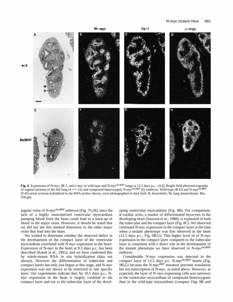
In order to determine whether aspects of lung develop-ment other than the branching of the lung epithelium wereaffected by the reduction in N-Myc protein levels, weperformed RNA in situ hybridizations on wild-type and N-myc9a/BRP lungs at 12.5 days p.c. (Fig. 6). Flk-1, whichencodes a tyrosine kinase receptor for vascular endothelial
Fig. 2. N-myc9a/BRP embryos are edematous in the region of the neck. (A) Wild-type 12.5 days p.c. embryo, dorsal view. (B) N-myc9a/BRP
embryo, showing swelling of the tissue and expanded, blood-filled jugular veins.
Fig. 3. Percentage of embryos in a N-myc9a/+×N-mycBRP/+ litter that are N-myc9a/BRP, as a function of developmental stage. N-myc9a/BRP
embryos are separated into two groups: (1) healthy, phenotypically normal embryos (stippled bars), and (2) phenotypically abnormal ordead embryos (hatched bars). Resorption sites visible in the uterus but not typed with respect to N-myc are included as a separate category(black bars). Note that the total of resorptions, abnormal N-myc9a/BRP embryos and normal N-myc9a/BRP embryos adds up toapproximately 25% of the offspring at each time point (white bars). Numbers are given as a fraction of the total number of embryosdissected from N-myc9a/+×N-mycBRP/+ crosses at that developmental stage.

490
growth factor, marks endothelial cells in the developing cap-illaries and blood vessels in the mouse (Yamaguchi et al.,1993; Millauer et al., 1993), and as such is expressed in apunctate manner throughout the lung mesenchyme wherethe lung vasculature is developing (Fig. 6C). The pattern andintensity of flk-1 expression was not affected in N-myc9a/BRP
lungs (Fig. 6G), indicating that the development of the lungvasculature is not affected by the reduction in N-Mycprotein observed in N-myc9a/BRP embryos. Fig. 6B,F showsthe pattern of expression of N-myc in wild-type and N-myc9a/BRP lungs, respectively, at 12.5 days p.c., reconfirm-ing that expression is restricted to the lung epithelium andshowing that the reduced N-Myc protein levels in N-myc9a/BRP embryos did not lead to a complete loss of N-myc-expressing cells in the lung epithelium. N-myc9a/BRP lungsappeared to express more than 15% of normal levels of N-
myc RNA (Fig. 6F). This is because the N-mycBRP alleleencodes a transcript that includes the entire N-myc openreading frame, but which contains stop codons that preventtranslation of any part of the N-myc protein (Stanton et al.,1990). Fig. 6D,H demonstrate that levels of c-myc mRNAat the cellular level were not affected by the reduction in N-Myc protein in the lungs of N-myc9a/BRP embryos. C-myc isnormally expressed in the lung mesenchyme (Fig. 6D,Hirning et al., 1991) and the pattern and level of c-mycexpression was not altered in the N-myc9a/BRP lung, despiteits reduced size (Fig. 6H).
Heart developmentThe other clearly visible defect in N-myc9a/BRP embryosexamined at 12.5 days p.c. was in the morphology of theheart. While N-myc9a/BRP hearts had the normal four-chamber structure, normal endocardial cushions, valves andsepta, the heart was small and the myocardium was abnor-mally thin (Fig. 7A-D). The latter defect was particuarlyapparent in the ‘compact’, or subepicardial layer of the ven-tricular myocardium while the atrial myocardium and theinner trabecular layer of the ventricles were less severelyaffected. In mammals, trabeculation occurs early duringheart development and, at later stages, growth occurs largelyin the compact layer (Rumyantsev, 1991). In N-myc9a/BRP
embryos, this growth of the compact layer appeared not tooccur, so that the N-myc9a/BRP myocardium at 12.5 days p.c.was no thicker than it had been at 10.5 days p.c.
Occasionally, N-myc9a/BRP embryos survived until 14.5days p.c. These embryos were always grossly abnormal,with large edemas in the neck area, and were smaller thantheir littermates. Fig. 7E and F compares wild-type and N-myc9a/BRP hearts at this stage, showing that the compactlayer of the myocardium of the compound heterozygote wasstill no more developed than it had been at 10.5 days.
It seems likely that this failure in the proliferation of theventricular myocardium leads to a failure of the circulatorysystem and ultimately in fetal death. The inefficient functionof the heart was demonstrated by the hugely expanded
C. B. Moens and others
Fig. 4. Western analysis of N-Myc protein levels in extracts of11.5 days p.c. embryos of the four possible types of offspringfrom an N-myc9a/+×N-mycBRP/+ cross. Each lane containsapproximately 20 µg of protein. The N-Myc protein runs atapproximately 65×103 Mr. A cross-reacting band appears atapproximately 100×103 Mr.
Fig. 5. Comparison of lung development in wild-type (A) and compound heterozygote N-myc9a/BRP (B) embryos at 12.5 days p.c.Comparable frontal sections were taken at the level of the right postcaval lobe. E, esophagus; L, left lung; RP; right lung, postcaval lobe.Bar, 200 µm.
491N-myc mutant mice
jugular veins of N-myc9a/BRP embryos (Fig. 7G,H), since thelack of a highly muscularized ventricular myocardiumpumping blood from the heart could lead to a back-up ofblood in the major veins. However, it should be noted thatwe did not see this marked distension in the other majorveins that lead into the heart.
We wished to determine whether the observed defect inthe development of the compact layer of the ventricularmyocardium correlated with N-myc expression in the heart.Expression of N-myc in the heart at 9.5 days p.c. has beendescribed (Katoh et al., 1991), and we have confirmed thisby whole-mount RNA in situ hybridization (data notshown). However the differentiation of trabecular andcompact layers has only just begun at this stage, and N-mycexpression was not shown to be restricted to one specificlayer. Our experiments indicate that, by 10.5 days p.c., N-myc expression in the heart is largely confined to thecompact layer and not to the trabecular layer of the devel-
oping ventricular myocardium (Fig. 8B). For comparison,α-cardiac actin, a marker of differentiated myocytes in thedeveloping heart (Sassoon et al., 1988), is expressed in boththe trabeculae and the compact layer (Fig. 8C). We observedcontinued N-myc expression in the compact layer at the timewhen a mutant phenotype was first observed in the heart(12.5 days p.c., Fig. 9B,G). This higher level of of N-mycexpression in the compact layer compared to the trabecularlayer is consistent with a direct role in the development ofthe mutant phenotype we have observed in N-myc9a/BRP
embryos.Considerable N-myc expression was detected in the
compact layer of 12.5 days p.c. N-myc9a/BRP hearts (Fig.9B,G) because the N-mycBRP mutation prevents translationbut not transcription of N-myc, as noted above. However, asexpected, the layer of N-myc-expressing cells was narrowerin the ventricular myocardium of compound heterozygotesthan in the wild-type myocardium (compare Figs 9B and
Fig. 6. Expression of N-myc, flk-1, and c-myc in wild-type and N-myc9a/BRP lungs at 12.5 days p.c.. (A,E) Bright-field photomicrographsof sagittal sections of the left lung of +/+ (A) and compound heterozygote N-myc9a/BRP (E) embryos. Wild-type (B-D) and N-myc9a/BRP
(F-H) serial sections hybridized to the RNA probes shown, were photographed in dark field. B, bronchiole; M, lung mesenchyme. Bar,100 µm.

492 C. B. Moens and others
Fig. 7. Comparison of heart development in wild-type and compound heterozygote N-m y c9 a / B R P embryos at 12.5 and 14.5 days p.c.Left-hand photographs show wild-type embryos, and right-hand photographs show N-m y c9 a / B R P embryos. (A,B) Low-powerm a g n i fication photomicrographs of frontal sections of 12.5 days p.c. hearts taken at the level of the left atrioventricular canal.(C,D) High-power magnification of the ventricle of 12.5 day hearts. (E,F) Sagittal sections of 14.5-day hearts. (G,H) Frontal sectionsthrough the jugular veins at 12.5 days. A, atrium; AT, aortic trunk; AV, aortic valve; C, compact layer; E, endocardial cushion; En,endocardium; Ep, epicardium; J, jugular veins; LV, left ventricle; P, pharynx; V, ventricle; T, trabeculae; Tr, trachea. For A,B,E,F,G,H:bar, 250 µm; for C,D: bar, 50 µm .

493N-myc mutant mice
8B). The observation that the mutant alleles continue to betranscribed in the N-myc9a/BRP myocardium suggests thatcells that normally express N-myc are present but in reducednumbers.
Flk-1, the endothelial cell marker described above, isexpressed in the endocardium (Yamaguchi et al., 1993),which is the mesodermally derived inner lining of the heartthat later contributes to the endocardial cushion and heartvalves through an epithelial-to-mesenchymal transition (Fig.9D,I; Markwald et al., 1990). The differentiation of theendocardium was not affected in N-myc9a/BRP embryos, as
demonstrated by the continued expression of flk-1 in theendocardium (compare Figs 10D,I and 8D,I). The N-myc9a/BRP genotype also did not prevent the differentiationof cardiac myocytes in the myocardium, as indicated by thestrong expression of α-cardiac actin in both the compact andtrabecular layers of the mutant myocardium (compare Figs10C,H and 9C,H).
The finding that reduction in N-m y c expression in thecompact layer of the heart caused a myocyte hypoplasiaevident at 12.5 days p.c. was interesting, in that overexpres-sion of the closely related c-myc proto-oncogene in the heartof transgenic mice has been described to cause a hyperplasiaof the myocytes which is also evident during development(Jackson et al., 1990). The normal expression pattern of c-m y cin the heart has not been described, so the relative roles ofthese two genes in myocyte proliferation in vivo was notclear. Fig. 9E,J demonstrates that c-m y c is expressed at lowlevels in the heart, and that its pattern of expression isdifferent, and to some extent complementary to that of N-m y c.While N-m y c is expressed in the compact layer of the ven-tricular myocardium, c-m y c expression is largely restricted tocells adjacent to and on the outside of the compact layer, andto isolated cells or clusters of cells adjacent to and on theinside of the compact layer. The expression on the outersurface of the heart is similar to that of flk - 1 (Fig. 8D,I) andprobably represents expression in the capillaries that runbetween the myocardium and epicardium (Viragh andChallice, 1981). The small foci of grains on the inner surfaceof the myocardium also appears to represent c-myc e x p r e s s i o nin endocardial cells, as they are found specifically in thevicinity of red-blood-cell-containing capillaries. c- m y cmRNA was also observed in endothelial cells lining the endo-cardial cushion. c-m y c is only expressed at low levels in themyocardium itself, in spite of the fact that when it is expressedectopically in the myocardium it causes myocytic hyperpla-sia (Jackson et al., 1990). In N-m y c9 a / B R P hearts, theexpression of c-m y c did not appear to be affected (Fig. 10E,J).
Other tissuesCareful examination of N-myc9a/BRP embryos between 10.5and 14.5 days p.c. gave no evidence of defects in the kidneyor brain, both of which are major sites of N-myc expressionin the embryo, and both of which are affected in N-mycBRP/BRP embryos (Stanton et al., 1992; Charron et al.,1992). In four N-myc9a/BRP embryos that survived until 14.5days p.c., the kidneys were small but were structurallynormal (not shown). N-mycBRP/BRP mice have also beendescribed as having reduced genital ridges (Stanton et al.,1992) and cranial and spinal ganglia. However, these struc-tures were indistinguishable in N-myc9a/BRP embryos fromthose of their wild-type littermates (not shown).
DISCUSSION
We have generated mice that carry two different mutantalleles of the N-myc proto-oncogene in order to identifyfunctions for N-myc that were not revealed in mice homozy-gous for each mutation individually. The N-myc9a allele isa leaky mutation which in the homozygous condition causesperinatal lethality due to a defect early in lung branching
Fig. 8. Expression patterns of N-myc and α-cardiac actin in theheart at 10.5 days p.c. (A) Bright-field photomicrograph of ahematoxylin and eosin-stained parasagittal section of a wild-typeventricle. (B,C) Dark-field photomicrographs of serial sectionsshowing N-myc (B) and α-cardiac actin (C) expression by RNA insitu hybridization. C, compact subepicardial layer; T, trabeculae;Ep, epicardium. Bar, 100 µm.

494 C. B. Moens and others

495N-myc mutant mice

496
morphogenesis. The levels of residual N-Myc protein(approximately 25% of normal levels) are presumably suf-ficient to support the normal development of other tissues inwhich N-myc normally functions. The N-mycBRP allele islikely to be a mutation which, in the homozygous condition,results in embryonic lethality at approximately 11.5 daysp.c., at which point the epithelial component of themesonephros, brain, lung, stomach and intestine are allhypoplastic (Stanton et al., 1992). The phenotype of N-myc9a/BRP compound heterozygotes is intermediate to thetwo homozygous mutant phenotypes, consistent with theobservation that the N-myc9a/BRP heterozygotes containlower levels of N-Myc protein than are present in N-myc9a/9a
embryos. N-myc9a/BRP embryos die between 12 and 15 daysp.c., and a number of tissues that are affected in N-mycBRP/BRP homozygotes, such as the kidney, brain andcranial and spinal sensory ganglia, appear to be normal. Thelungs, which are the main organ affected in N-myc9a/9a
homozygotes, are even more severely affected in N-myc9a/BRP embryos. Compound heterozygotes also showeda defect in the development of the compact subepicardiallayer of the heart, and appeared to die from a failure of heartfunction caused by a hypoplasia of ventricular myocytes inthe compact layer. These results indicate a critical role forN-myc in the development of the heart.
N-myc in heart developmentThe reduced levels of N-Myc protein found in N-myc9a/BRP
embryos result in a considerable thinning of the subepicar-dial compact layer of the myocardium by 12.5 days of devel-opment. Consistent with this phenotype, N-myc expressionis expressed much more strongly in the compact layer of theheart at 10.5 and 12.5 days p.c. than in the trabecular layerof the myocardium or in the endothelium. Trabeculation, orformation of the myocardial projections that form a latticeof contractile cells throughout much of the ventricles of theembryonic heart, occurs early during heart development inthe mouse, beginning around day 9.5 of gestation (Challiceand Viragh, 1973). By a number of ultrastructural and cyto-chemical criteria, the myocytes within the trabeculae aremore highly differentiated than the myocytes in the compactlayer (Rumyantsev, 1991). Thus compact layer myocytesare more basophilic and richer in RNA, while trabecularmyocytes contain more mitochondria, ribosomes andgranular endoplasmic reticulum. Myofibrils in trabecularmyocytes are thicker and more highly organized than incompact layer myocytes, where myofibrils are present butare scattered randomly relative to one another in thecytoplasm. Consistent with this picture is the observationthat the rate of cell proliferation in the compact layer is 2-to 3-fold higher than in the trabeculae (Rumyantsev, 1977;Tokuyasu, 1990). The highly differentiated myocytes in thetrabecular layer have been postulated to be responsible forthe early beating of the heart while, at later stages, thethickened compact layer becomes the major contractileforce. The cardiac hypoplasia that we observe in N-myc9a/BRP embryos is more apparent in the compact layer.This may explain our observation that embryonic lethalitydoes not occur until later during heart development, whenheart function may depend more on the compact layer thanon the trabecular layer.
Our observation of N-myc expression in the compact layerand the absence of development in the compact layer in N-myc9a/BRP mice suggests that N-myc is required either for theproliferation of myocytes in the compact layer, and/or forpreventing the differentiation of these cells, although thereare other possible explanations, such as that they are dyingprematurely. The simplest explanation is the former.However, the second hypothesis, in which N-mycexpression prevents the terminal differentiation of compactlayer myocytes into trabecular-type myocytes, is consistentwith previous descriptions of N-myc expression in theembryo, in which N-myc expression is correlated with cellsin an undifferentiated state in the kidney, brain and skin,regardless of their proliferative state, and the further differ-entiation of these cells is correlated with down-regulation ofN-myc (Mugrauer et al., 1988).
In their description of embryos homozygous for a nullmutation in N-myc, Charron et al. (1992) noted a defect inthe development in the heart, visible as early as 9.5 days p.c.,in which development was apparently slowed as evidencedby the absence of endocardial cushion tissue and of intera-trial and interventricular septa. It was postulated that N-mycis involved in the generation of the inductive signal sent bythe myocardium to the endocardium to induce the epithelial-to-mesenchymal transition of the endocardium, which formsthese anlagen of the cardiac valves and septa. Our resultsconfirm a function for N-myc in the development in theheart, but tend to support a role for N-myc within themyocardium itself, although this could presumably have asecondary impact on endocardial differentiation in moresevere mutants. We have not observed defects in theformation of septal or endocardial cushion tissue in N-myc9a/BRP embryos.
c-myc in heart developmentWhen c-myc is overexpressed in the heart of RSV/c-myctransgenic mice, these mice develop a fetal cardiac myocytehyperplasia and at birth have more than twice the normalnumber of cardiac myocytes (Jackson et al., 1990). This hassuggested that endogenous c-myc may play a role in cardiacmyocyte proliferation in vivo. However, we have demon-strated that c-myc is only expressed at low levels in the heartat 12.5 days p.c. and that this expression is largely in endo-thelial cells and not in the myocardium. Background levelsof c-myc expression in the heart were also observed inmouse embryos at 13.5 days p.c. (Stanton et al., 1992) andin first trimester human embryos (Pfeifer-Ohlsson et al.,1985). N-myc, in contrast, is expressed at high levels in thecompact layer of the ventricular myocardium at 10.5 and12.5 days. Combined with the hypoplasia of themyocardium that we have observed in N-myc9a/BRP
embryos, these data suggest that N-myc rather than c-mycmay play a primary role in the regulation of proliferationand/or differentiation of ventricular myocytes during heartdevelopment, and that the effect of c-myc in these transgenicmice may reflect the possibility that in this instance c-myccan mimic the normal effects of N-myc. It has previouslybeen observed that different myc family genes can cause thesame tumour types in transgenic mice when they are over-expressed using identical promoters, even though thisexpression may be ectopic (Rosenbaum et al., 1989; Dildrop
C. B. Moens and others
497N-myc mutant mice
et al., 1989; Adams et al., 1985). Furthermore, high levelsof expression of one myc gene in transgenic mice can represstranscription of itself and of other myc genes (Rosenbaumet al., 1989; Dildrop et al., 1989; Adams et al., 1985). Theseresults have suggested that at high levels, the various mycgenes may be able to mimic each other’s effects on down-stream targets. This hypothesis has been strengthened by theobservations that N-Myc and c-Myc proteins bind the samecore DNA sequence in vitro, and that N-, L- and c-myc allform heterodimers with Max in vivo (Blackwood et al.,1992; Wenzel et al., 1991; Mukherjee et al., 1992), and alltransform cells in culture through their interaction with Max(Mukherjee et al., 1992). We are presently crossing RSV/c-myc mice with the N-myc mutant mice to generate N-myc9a/BRP embryos that also express this c-myc transgene inthe heart. If c-myc in these transgenics truly mimicks thenormal role of N-myc, the cardiac myocyte hypoplasia ofcompound heterozygous embryos is expected to be rescuedby the transgene.
Recently, Davis et al. (1993) have described thephenotype of mice that bear a null mutation in c-myc. Theseembryos die before 10.5 days p.c. and exhibit, among otherabnormalities, an enlargement of the heart and a dilated,fluid-filled pericardium. This is unexpected in light of theobservation, described above, that mice with ectopicexpression of c-myc in the heart have enlarged hearts.However, it is still unclear whether the heart abnormality inthe c-myc null mutants is a direct result of the mutation oris secondary to other defects that are causing the embryo todie.
In spite of the possibility that c-myc expression may beable to replace N-myc function, we observe no up-regula-tion of c-myc in either the compact layer of the myocardiumor in the lung epithelium of N-myc9a/BRP mice. This may bebecause cross-regulation of the myc family genes does notnormally occur in these tissues as it does when they are over-expressed in transformed cells or in transgenic mice. Stantonet al. (1992) showed that c-myc is expressed in the telen-cephalon of N-mycBRP/BRP embryos, and this observationwas interpreted to indicate cross regulation of N-myc and c-myc in the neuroepithelium.
N-myc in lung developmentMice homozygous for the N-myc9a mutation die at birth dueto a defect in lung morphogenesis which is visible as earlyas 12.5 days p.c. (Moens et al., 1992). We have postulatedthat N-myc is required for the lung epithelium to respond tolocal inductive signals emanating from the lung mes-enchyme, which cause branching to occur. N-myc9a/BRP
embryos have more severely affected lungs, with only arudimentary branching pattern at 12.5 days. However, theearliest events of lung development, in which two buds areinduced to grow from the trachea by surrounding mes-enchyme (Spooner and Wessells, 1970), occur normally inN-myc9a/BRP and indeed in N-mycBRP/BRP embryos (Stantonet al., 1992). We and others (Hirning et al., 1991; Moens etal., 1992) have shown that N-myc is expressed in the lungepithelium and, further, that expression is largely restrictedto bronchioles and is present at very low levels in the tracheaand bronchi. These results suggest that N-myc is involved inbranching morphogenesis in the lung but not in the initial
induction of budding of the tracheal epithelium. Experi-mental manipulations in vitro have suggested that there aredifferent mechanisms for budding versus branching of thelung epithelium. A number of different stimuli, includingsalivary gland mesenchyme and bronchial mesenchyme, caninduce supernumerary buds in tracheal epithelium, but onlybronchial mesenchyme is able to induce those buds tobranch (Wessells, 1970; Spooner and Wessells, 1970). Thephenotypes of mice bearing mutations in N-myc providegenetic evidence for such a mechanism for lung develop-ment and provide a candidate gene that is involved in thecontrol of one process (branching) and not the other(budding).
Tissue-specific effects of N-myc mutant allelesOnly a subset of the tissues that normally express N-myc arevisibly affected in N-myc9a/BRP embryos. N-myc9a/9a
embryos have approximately 25% of wild-type levels of N-Myc protein and, in these embryos, the lungs and spleen arethe only tissues affected (Moens et al., 1992). N-myc9a/BRP
embryos have approximately 15% of wild-type levels of N-Myc protein and, in these embryos, the heart is also affected.However, 15% of normal levels of N-Myc protein appear tobe sufficient for normal genitourinary and nervous systemdevelopment. The molecular basis for this remains to bedetermined. It is possible that different tissues within theembryo make different amounts of normal protein relativeto the amounts in wild-type embryos. We have previouslyattempted to correlate the relative levels of normal N-mycmRNA in different tissues of N-myc9a/9a embryos with thepresence or absence of a mutant phenotype (Moens et al.,1992) and, although there were differences among thetissues examined, no strong correlation could be established.Another explanation for the tissue-specific effects of N-mycmutant alleles is that different tissues are affected differentlyby approximately the same reduction in N-Myc proteinbecause of differences in the ratio of N-Myc protein to Max(Blackwood and Eisenman, 1991), to Max-associatedproteins such as Mad and Mxi1 (Ayer et al., 1993; Zervoset al., 1993), or to other Myc proteins.
Myc proteins require dimerization with Max for DNAbinding (Blackwood and Eisenman, 1991; Prendergast andZiff, 1991; Kato et al., 1992). A number of lines of evidencehave suggested that Max overexpression can inhibit trans-formation (Mukherjee et al., 1992; Makela et al., 1992;Prendergast et al., 1992) and transactivation (Kretzner et al.,1992; Amati et al., 1992) by myc genes. Mad, cloned byvirtue of its ability to dimerize with Max, has been shownto compete with Myc for binding to Max, and to therebyinhibit transactivation by Myc (Ayer et al., 1993). Mxi1(Zervos et al., 1993) and other, as yet unidentified, Maxpartners presumably act in a similar manner and are alsolikely thereby to inhibit N-myc function. Also, L-Myc, apoorly transforming member of the Myc family, has beenshown to prevent transformation by other myc genes, pre-sumably by competing for and forming less active DNA-bound complexes with Max (Mukherjee et al., 1992). In celltypes where a number of Max-associated and Myc proteinscompete with N-Myc protein for dimerization with Max andsites on DNA, an 85% reduction in N-Myc protein isexpected to reduce the response of downstream targets of N-

498
Myc more strongly than in cells where there are no com-petitors for Max binding. Interestingly, L-myc is co-expressed with N-myc in the lung, perhaps in the same celltype (Zimmerman et al., 1986), but the two genes are notco-expressed in the kidney (Mugrauer and Ekblom, 1991),where neither the N-myc9a/9a nor the N-myc9a/BRP embryoshave an abnormal phenotype. It will be interesting tocompare the detailed expression patterns of the variousinteracting factors with the tissues affected by the N-mycmutations.
ConclusionsWe have generated a third N-myc mutant phenotype bycombining leaky and null alleles in a compound heterozy-gote. These mice have allowed us to study the function ofN-myc at a stage in development that is not reached inembryos homozygous for the null allele (Stanton et al.,1992) and that is not affected in embryos homozygous forthe leaky allele (Moens et al., 1992). Classical geneticstudies of development in a number of systems have demon-strated the importance of studying the phenotypic effects ofdifferent mutant alleles and combinations of mutant allelesin a given gene in order to determine its multiple roles inthe course of development. Our results have shown that thetechnique of gene targeting by homologous recombinationin the mouse can be used to the same ends. The clear delin-eation of a function for N-myc in both lung branching mor-phogenesis and myocardial development also providestarget tissues in which to search for the elusive downstreamgenes in the N-myc signaling pathway.
Note added after acceptanceRecently, a third description of embryos homozygous for aputative null allele of N-myc has been published (Sawai etal., 1993). The overall phenotype of these mutant embryosis very similar to those described by Stanton et al. (1992)and Charron et al. (1992), but the defect in heart develop-ment is shown to be primarily in the myocardium.
We wish to thank Dr R. Kennett for the anti-N-myc antibody,Dr M. Buckingham for the α-cardiac actin probe, T. Yamaguchifor the flk-1 probe, Dr C. Asselin for the c-myc probe and Dr R.DePinho for the N-myc genomic clone from which probes for RNAin situ hybridization were subcloned. Our gratitude also to Dr ArchPerkins for his help in the phenotypic analysis of N-myc9a/BRP
mutants, to Alexandra Joyner for her critical reading of the manu-script, and to Valerie Prideaux, Chi-Chong Hui, Benny Motro andEster Ivanyi for their generous assistance in various aspects of thiswork. This work was supported by a Terry Fox program projectgrant from the National Cancer Institute of Canada. C. B. M. wassupported by a Natural Sciences and Engineering Research Councilof Canada ‘Centennial’ Scholarship and a Medical ResearchCouncil of Canada Studentship. J. R. is an International Scholar ofthe Howard Hughes Medical Institute and a Terry Fox CancerResearch Scientist of the National Cancer Institute of Canada. B.R. S. and L. F. P. are sponsored by the NCI-DHHS under contractNO1-CO-74101 with ABL.
REFERENCES
Adams, J. M., Harris, A. W., Pinkert, C. A., Corcoran, L. M.,Alexander, W. S., Cory, S., Palmiter, R. D. and Brinster, R. L. (1985).
The c-myc oncogene driven by immunoglobulin enhancers induceslymphoid malignancy in transgenic mice. Nature 318, 533-538.
Alex, R., Soezeri, O., Meyer, S. and Dildrop, R. (1992). Determination ofthe DNA sequence recognized by the bHLH-zip domain of the N-Mycprotein. Nucleic Acids Res. 20, 2257-2263.
Amati, B., Dalton, S., Brooks, M. W., Littlewood, T. D., Evan, G. I. andLand, H. (1992). Transcriptional activation by the human c-Myconcoprotein in yeast requires interaction with Max. Nature 359, 423-426.
Amati, B., Brooks, M. W., Levy, N., Littlewood, T. D., Evan, G. I. andLand, H. (1993). Oncogenic activity of the c-Myc protein requiresdimerization with Max. Cell 72, 233-245.
Ayer, D. E., Kretzner, L. and Eisenman, R. N. (1993). Mad: aheterodimeric partner for Max that antagonizes Myc transcriptionalactivity. Cell 72, 211-222.
Barrett, J., Birrer, M. J., Kato, G. J., Dosaka-Akita, H. and Dang, C. V.(1992). Activation domains of L-Myc and c-Myc determine theirtransforming potencies in rat embryo cells. Mol. Cell Biol. 12, 3130-3137.
Blackwell, T. K., Kretzner, L., Blackwood, E. M., Eisenman, R. N. andWeintraub, H. (1990). Sequence-specific DNA binding by the c-Mycprotein. Science 250, 1149-1151.
Blackwood, E. M., Luescher, B. and Eisenman, R. N. (1992). Myc andMax associate in vivo. Genes Dev. 6, 71-80.
Blackwood, E. M. and Eisenman, R. N. (1991). Max: a helix-loop-helixzipper protein that forms a sequence-specific DNA-binding complex withMyc. Science 251, 1211-1217.
Bossone, S. A., Asselin, C., Patel, A. J. and Marcu, K. B. (1992). Maz, azinc finger protein, binds to c-MYC and C2 gene sequences regulatingtranscriptional initiation and termination. Proc. Natl. Acad. Sci. USA 89,7452-7456.
Challice, C. E. and Viragh, S. (1973). The architectural development of theearly mammalian heart. Tissue Cell 6, 447-462.
Charron, J., Malynn, B. A., Robertson, E. J., Goff, S. P. and Alt, F. W.(1990). High-frequency disruption of the N-myc gene in embryonic stemand pre-B cell lines by homologous recombination. Mol. Cell Biol. 10,1799-1804.
Charron, J., Malynn, B. A., Fisher, P., Stewart, V., Jeannotte, L., Goff,S. P., Robertson, E. J. and Alt, F. W. (1992). Embryonic lethality inmice homozygous for a targeted disruption of the N-myc gene. GenesDev. 6, 2248-2257.
Davis, A. C., Wims, M., Spotts, G. D., Hann, S. R. and Bradley, A.(1993). A null c-myc mutation causes lethality before 10.5 days ofgestation in homozygotes and reduced fertility in heterozygous femalemice. Genes Dev. 7, 671-682.
DePinho, R. A., Legouy, E., Feldman, L. B., Kohl, N. E., Yancopoulos,G. D. and Alt, F. W. (1986). Structure and expression of the murine N-myc gene. Proc. Natl. Acad. Sci. USA 83, 1827-1831.
DePinho, R. A., Schreiber-Agus, N. and Alt, F. W. (1991). myc familyoncogenes in the development of normal and neoplastic cells. Adv.Cancer Res. 57, 1-46.
Dildrop, R., Ma, A., Zimmerman, K., Hsu, E., Tesfaye, A., DePinho, R.A. and Alt, F. W. (1989). IgH enhancer-mediated deregulation of N-mycgene expression in transgenic mice: generation of lymphoid neoplasiasthat lack c-myc expression. EMBO J. 8, 1121-1128.
Frohman, M. A., Boyle, M. and Martin, G. R. (1990). Isolation of themouse Hox-2.9 gene; Analysis of embryonic expression suggests thatpositional information along the anterior-posterior axis is specified bymesoderm. Development 110, 589-607.
Garrell, J. and Campuzano, S. (1991). The helix-loop-helix domain: Acommon motif for bristles, muscles and sex. BioEssays 13, 493-498.
Hirning, U., Schmid, P., Schulz, W. A., Rettenberger, G. and Hameister,H. (1991). A comparative analysis of N-myc and c-myc expression andcellular proliferation in mouse organogenesis. Mech. Dev. 33, 119-126.
Ikegaki, N., Bukovsky, J. and Kennett, R. H. (1986). Identification andcharacterization of the NMYC gene product in human neuroblastomacells by monoclonal antibodies with defined specificities. Proc. Natl.Acad. Sci. USA 83, 5929-5933.
Ikegaki, N. and Kennett, R. H. (1989). Glutaraldehyde fixation of theprimary antibody-antigen complex on nitrocellulose paper increases theoverall sensitivity of immunoblot assay. J. Immunol. Met.124, 205-210.
Jackson, T., Allard, M. F., Sreenan, C. M., Doss, L. K., Bishop, S. P. andSwain, J. L. (1990). The c-myc proto-oncogene regulates cardiacdevelopment in transgenic mice. Mol. Cell Biol.10, 3709-3716.
Kato, G. J., Barrett, J., Villa-Garcia, M. and Dang, C. V. (1990). An
C. B. Moens and others

499N-myc mutant mice
amino-terminal c-Myc domain required for neoplastic transformationactivates transcription. Mol. Cell Biol.10, 5914-5920.
Kato, G. J., Lee, W. M. F., Chen, L. and Dang, C. V. (1992). Max:Functional domains and interaction with c-Myc. Genes Dev. 6, 81-92.
Katoh, K., Kanamori, A., Wakamatsu, Y., Sawai, S. and Kondoh, H.(1991). Tissue distribution of N-myc expression in the earlyorganogenesis period of the mouse embryo. Dev. Growth Diff.33, 29-36.
Kohl, N. E., Kanda, N., Schrenck, R. R., Bruns, G., Latt, S. A., Gilbert,F. and Alt, F. W. (1983). Transposition and amplification of oncogene-related sequences in human neuroblastomas. Cell 35, 359-367.
Kretzner, L., Blackwood, E. M. and Eisenman, R. N. (1992). Myc andMax proteins possess distinct transcriptional activities. Nature 359, 426-429.
Lee, W. H., Murphree, A. L. and Benedict, W. F. (1984). Expression andamplification of the N-myc gene in primary retinoblastoma. Nature 309,458-460.
Makela, T. P., Koskinen, P. J., Vastrik, I. and Alitalo, K. (1992).Alternative forms of Max as enhancers or suppressors of Myc-Rascotransformation. Science 256, 373-377.
Markwald, R. R., Mjaatvedt, C. H., Krug, E. L. and Sinning, A. R.(1990). Inductive interactions in heart development: role of cardiacadherons in cushion tissue formation. Ann. NY Acad. Sci.588, 13-25.
Millauer, B., Wizigmann-Voos, S., Schnurch, H., Martinez, R., Moller,N. P. H., Risau, W. and Ullrich, A. (1993). High affinity VEGF bindingand developmental expression suggest Flk-1 as a major regulator ofvasculogenesis and angiogenesis. Cell 72, 835-846.
Moens, C. Bernelot, Auerbach, A. B., Conlon, R. A., Joyner, A. L. andRossant, J. (1992). A targeted mutation reveals a role for N-myc inbranching morphogenesis in the embryonic mouse lung. Genes Dev. 6,691-704.
Mugrauer, G., Alt, F. W. and Ekblom, P. (1988). N-myc proto-oncogeneexpression during organogenesis in the developing mouse as revealed byin situ hybridization. J. Cell Biol. 107, 1325-1335.
Mugrauer, G. and Ekblom, P. (1991). Contrasting expression patterns ofthree members of the myc family of protooncogenes in the developingand adult mouse kidney. J. Cell Biol. 112, 13-25.
Mukherjee, B., Morgenbesser, S. D. and DePinho, R. A. (1992). Mycfamily oncoproteins function through a common pathway to transformnormal cells in culture: cross-interference by Max and trans-actingdominant mutants. Genes Dev. 6, 1480-1492.
Nau, M. M., Brooks, B. J., Jr., Carney, D. N., Gazdar, A. F., Battey, J.F., Sausville, E. A. and Minna, J. D. (1986). Human small-cell lungcancers show amplification and expression of the N-myc gene. Proc. Natl.Acad. Sci. USA 83, 1092-1096.
Nisen, P. D., Zimmerman, K., Cotter, S. V., Gilbert, F. and Alt, F. W.(1986). Enhanced expression of the N-myc gene in Wilms’ tumours.Cancer Res. 46, 6217-6222.
Pfeifer-Ohlsson, S., Rydnert, J., Goustin, A. S., Larsson, E., Betsholtz,C. and Ohlsson, R. (1985). Cell-type-specific pattern of mycprotooncogene expression in developing human embryos. Proc. Natl.Acad. Sci. USA82, 5050-5054.
Prendergast, G. C., Lawe, D. and Ziff, E. B. (1991). Association of Myn,the murine homolog of Max, with c-Myc stimulates methylation-sensitiveDNA binding and Ras cotransformation. Cell 65, 395-407.
Prendergast, G. C., Hopewell, R., Gorham, B. J. and Ziff, E. B. (1992).Biphasic effect of Max on Myc cotransformation activity and dependenceon amino- and carboxy-terminal Max functions. Genes Dev. 6, 2429-2439.
Prendergast, G. C. and Ziff, E. B. (1991). Methylation-sensitive sequence-specific DNA binding by the c-myc basic region. Science 251, 186-189.
Resar, L. M. S., Dolde, C., Barrett, J. F. and Dang, C. V. (1993). B-mycinhibits neoplastic transformation and transcriptional activation by c-myc. Mol. Cell Biol. 13, 1130-1136.
Rosenbaum, H., Webb, E., Adams, J. M., Cory, S. and Harris, A. W.
(1989). N-myc transgene promotes B lymphoid proliferation, elicitslymphomas and reveals cross-regulation with c-myc. EMBO J. 8, 749-755.
Rumyantsev, P. P. (1977). Interrelations of the proliferation anddifferentiation processes during cardiac myogenesis and regeneration.Int. Rev. Cytol. 51, 187-273.
Rumyantsev, P. P. (1991). Growth and Hyperplasia of Cardiac MuscleCells. London: Harwood Academic Publishers.
Sassoon, D. A., Garner, I. and Buckingham, M. (1988). Transcripts of α-cardiac and α-skeletal actins are early markers for myogenesis in themouse embryo. Development 104, 155-164.
Sawai, S., Shimono, A., Hanaoka, K. and Kondoh, H. (1991). Embryoniclethality resulting from disruption of both N-myc alleles in mousezygotes. New Biologist 3, 861-869.
Sawai, S., Shimono, A., Wakamatsu, Y., Palmes, C., Hanaoka, K., andKondoh, H. (1993). Defects of embryonic organogenesis resulting fromtargeted disruption of the N-myc gene in the mouse. Development 117,1445-1455.
Schwab, M., Alitalo, K., Klempnauer, K., Varmus, H. E., Bishop, J. M.,Gilbert, F., Brodeur, G. M., Boldstein, M. and Trent, J. (1983).Amplified DNA with limited homology to myc cellular oncogene isshared by human neuroblastoma cell lines and a neuroblastoma tumour.Nature 305, 245-248.
Spooner, B. S. and Wessells, N. K. (1970). Mammalian lung development:interactions in primordium formation and bronchial morphogenesis. J.Exp. Zool. 175, 445-454.
Stanton, B. R., Reid, S. W. and Parada, L. F. (1990). Germ linetransmission of an inactive N-myc allele generated by homologousrecombination in mouse embryonic stem cells. Mol. Cell Biol. 10, 6755-6758.
Stanton, B. R., Perkins, A. S., Tessarollo, L., Sassoon, D. A. and Parada,L. F. (1992). Loss of N-myc function results in embryonic lethality andfailure of the epithelial component of the embryo to develop. Genes Dev.6, 2235-2247.
Tokuyasu, K. T. (1990). Co-development of embryonic myocardium andmyocardial circulation. In Developmental Cardiology: Morphogenesisand Function (ed. E. B. Clark and A. Takao), pp. 205-218. Mount Kisko,NY: Futura Publishing Co., Inc.
Viragh, S. and Challice, C. E. (1981). The origin of the epicardium and theembryonic myocardial circulation in the mouse. Anat. Rec. 201, 157-168.
Wenzel, A., Cziepluch, C., Hamann, U., Schuermann, J. and Schwab,M. (1991). The N-myc oncoprotein is associated in vivo with thephosphoprotein Max(p20/22) in human neuroblastoma cells. EMBO J.10, 3703-3712.
Wessells, N. K. (1970). Mammalian lung development: interactions in theformation and morphogenesis of tracheal buds. J. Exp. Zool. 175, 455-466.
Wong, A. J., Ruppert, J. M., Eggleston, J., Hamilton, S. R., Baylin, S. B.and Vogelstein, B. (1986). Gene amplification of c-myc and N-myc insmall cell carcinoma of the lung. Science 233, 461-464.
Yamaguchi, T. P., Dumont, D. J., Conlon, R. A., Breitman, M. andRossant, J. (1993). Flk-1, a flt-1-related receptor tyrosine kinase is anearly marker for heart and blood for endothelial cell precursors.Development in press,
Zervos, A. S., Gyuris, J. and Brent, R. (1993). Mxi1, a protein thatspecifically interacts with Max to bind Myc-Max recognition sites. Cell72, 223-232.
Zimmerman, K., Yancopoulos, G. D., Collum, R. G., Smith, R. K., Kohl,N. E., Denis, K. A., Nau, M. M., Witte, O. N., Toran-Allerand, D.,Gee, C. E., Minna, J. D. and Alt, F. W. (1986). Differential expressionof myc family genes during murine development. Nature 319, 780-783.
(Accepted 30 June 1992)