deduced structural model for animal rhabdovirus glycoproteinserueda/bioinformatica/clustal3.pdf ·...
TRANSCRIPT

Journal of General Virology (1999), 80, 1211–1220. Printed in Great Britain. . . . . . . . . . . . . . . . . . . . . . . . . . . . . . . . . . . . . . . . . . . . . . . . . . . . . . . . . . . . . . . . . . . . . . . . . . . . . . . . . . . . . . . . . . . . . . . . . . . . . . . . . . . . . . . . . . . . . . . . . . . . . . . . . . . . . . . . . . . . . . . . . . . . . . . . . . . . . . . . . . . . . . . . . . . . . . . . . . . . . . . . . . . . . . . . . . . . . . . . . . . . . . . . . . . . . . . . . . . . . . . . . . . . . . . . . . . . . . . . . . . . . . . . . . . . . . . . . . . . . . . . . . . . . . . . . . .
Deduced structural model for animal rhabdovirusglycoproteins
Peter J. Walker and Kritaya Kongsuwan
CSIRO Tropical Agriculture, PMB 3, Indooroopilly, Qld 4068, Australia
The G protein sequences of fourteen animal rhabdoviruses, representing all four recognizedgenera (Vesiculovirus, Lyssavirus, Ephemerovirus and Novirhabdovirus) and the ungrouped sigmavirus, were aligned using CLUSTAL W and adjusted to account for obvious sequence similarities notdetected by the algorithm. Analysis of the alignment indicated remarkable preservation of Gprotein structural features including cysteine residues, antigenic sites and significant elements ofsecondary structure (α-helices, β-strands and loops). Twelve highly conserved cysteine residueswere assigned numbers (CI to CXII) according to their location in the alignment. Other cysteineresidues were assigned numbers (C0 to CXIIe) according to their position relative to the conservedcysteines. The pattern of conservation of cysteine residues and the structural characteristics ofidentified discontinuous antigenic sites were used to deduce a model for G protein structure. Sixabsolutely conserved cysteines are predicted to associate in three disulphide bridges (CI–CXII ;CVIII–CXI ; CIX–CX) that form the core of the G protein structure and define the common discontinuousantigenic site. The associations of six other highly conserved cysteines (CII–CIV ; CIII–CV ; CVI–CVII) arepredicted by the absence of a specific pair in all viruses within a genus. Of the other cysteines, onepair occurs only in ephemeroviruses and novirhabdoviruses (C0–CXIIa) ; two pairs occur only inephemeroviruses (CIb–CVIIIa ; CXIIb–CXIIe) ; and two pairs occur only in lyssaviruses (CIa–CVIIIb ; CXIIc–CXIId).The structures predicted by the model account for the preservation of conformational antigenicsites, accommodate genus-specific variations, and are generally consistent with previousobservations of G protein structure.
IntroductionIn all rhabdoviruses, the G protein is an N-glycosylated
class I transmembrane protein which forms trimeric peplomerson the virion surface (Tordo et al., 1997). The G proteinmediates attachment of the virus to cellular receptors, endo-cytosis and fusion with vesicular membranes. In animalrhabdoviruses, the G protein can induce antibodies whichneutralize infection in vitro and protect against experimentalinfection. Competitive antibody-binding studies and analysesof neutralization-escape mutants have allowed the identi-fication and location of major antigenic sites in the G proteinsof several rhabdoviruses including rabies virus (Lafon et al.,1983 ; Sief et al., 1985 ; Prehaud et al., 1988), vesicular stomatitisvirus (VSV) Indiana (Vanderpol et al., 1986), VSV New Jersey(Bricker et al., 1987 ; Luo et al., 1988), bovine ephemeral fever
Author for correspondence: Peter Walker.
Fax 61 7 3214 2718. e-mail Peter.Walker!tag.csiro.au
virus (BEFV) (Cybinski et al., 1990, 1992 ; Kongsuwan et al.,1998) and infectious haematopoietic necrosis virus (IHNV)(Huang et al., 1994, 1996). Although G proteins of rhabdo-viruses from different genera share a very low level of aminoacid sequence identity, alignment of the sequences revealsremarkable conservation of cysteine residues, glycosylationsites and themajor antigenic domains. Of particular significanceis the alignment of 12 highly conserved cysteine residues andelements of a common discontinuous antigenic site comprisingwidely separated regions of the polypeptide chain (Walker etal., 1992 ; Huang et al., 1996 ; Kongsuwan et al., 1998). Theextent of these similarities suggests that, despite the widedifferences in host range and tissue tropism, the core elementsof animal rhabdovirus G protein structure are preserved.However, attempts to determine the pattern of disulphidebridges in rabies virus and VSV G proteins by analysis ofcleavage products, expressed fragments or by site-directedmutation of individual cysteine residues have been incon-clusive (Dietzschold et al., 1982 ; Keil & Wagner, 1989 ; Grigera
0001-6041 # 1999 SGM BCBB

P. J. Walker and K. KongsuwanP. J. Walker and K. Kongsuwan
Fig. 1. For legend see facing page.
BCBC
G protein structural modelG protein structural model
et al., 1992), and G protein crystals of sufficient quality for X-ray crystallographic analysis have not yet been obtained. Inthis paper, we propose a general structural model forrhabdovirus glycoproteins based on the pattern of conser-vation of cysteine residues, available experimental data and thestructural characteristics of discontinuous antigenic sites.
Methods+ Alignments and correction of Chandipura sequence. Theectodomain sequences of fourteen available animal rhabdovirus Gproteins were aligned using the CLUSTAL W multiple sequencealignment program (Thompson et al., 1994). The viruses includedmembers of the four recognized genera [Vesiculovirus (VSV Indiana, VSVNew Jersey, Chandipura virus, Piry virus, spring viraemia of carp virus) ;Lyssavirus (rabies virus, Mokola virus, Australian bat lyssavirus) ;Ephemerovirus (bovine ephemeral fever virus, Adelaide River virus) ; andNovirhabdovirus (approved by the Executive Committee of the ICTV andto be included in the 7th Report : infectious haemorrhagic necrosis virus,viral haemorrhagic septicaemia virus, Hirame rhabdovirus)] and theungrouped sigma virus of Drosophila. The published Chandipura Gprotein sequence (GenBank accession no. J04350) contains an unevennumber of cysteine residues in the ectodomain, one of which failed toalign with those of other vesiculoviruses. PCR primers targeted tosequences flanking the unaligned cysteine were used to amplify a 262 bpfragment (four clones) from RNA extracted from the same Chandipuravirus isolate (I653514). Sequence analysis of the fragment identifiedmultiple deletion, addition and substitution errors that eliminated onecysteine residue. The multiple sequence alignment was reconstructedusing the corrected Chandipura G sequence, and adjusted to account forobvious sequence similarities not detected by the algorithm.
+ Secondary structure predictions. Structural predictions wereconducted by using the PHD algorithm of Rost and Sander(http :}}www.embl-heidelberg.de}predictprotein}predictprotein.html).The method determines secondary structures (α-helix, β-strand or loop)and solvent accessibility (exposed or buried) from a multiple sequencealignment by employing a neutral network of non-redundant referencesequences (Rost & Sander, 1993a, b). Predictions were obtained for theectodomains of G protein sequences submitted individually to the serverfor automatic sequence alignment.
Results and DiscussionThe aligned sequences of fourteen animal rhabdovirus G
proteins, representing four recognized genera (Vesiculovirus,Lyssavirus, Ephemerovirus and Novirhabdovirus) and the
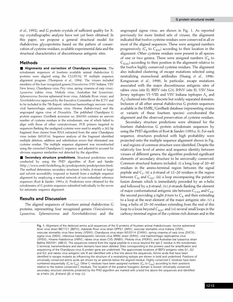
Fig. 1. Alignment of the deduced amino acid sequences of the G proteins of fourteen animal rhabdoviruses : bovine ephemeralfever virus strain BB7721 (BEFV) ; Adelaide River virus strain DPP61 (ARV); vesicular stomatitis virus Indiana (VSIV) ;vesicular stomatitis virus New Jersey (VSNV); Chandipura virus strain I653514 (CHNV); spring viraemia of carp virus (SVCV) ;sigma virus (SIGV) ; infectious haematopoeitic necrosis virus WRAC strain (IHNV); viral haemorrhagic septicaemia virus(VHSV); Hirame rhabdovirus (HIRV); rabies virus strain CVS (RABV) ; Mokola virus (MOKV) ; and Australian bat lyssavirus strainBallina 960591 (ABLV). The sequences extend from the signal peptide to a locus beyond the last C residue in the ectodomain.C-terminal, transmembrane and stem domains have been deleted. Sites corresponding to the primers used for amplification andsequencing of the Chandipura virus G protein gene are underlined. The approximate locations of BEFV antigenic sites G1, G2and G3, and rabies virus antigenic site III are identified with a fine line above the sequences. Amino acids that have beenidentified in escape mutants as influencing the structure of a neutralizing epitope are shown in bold and underlined. Positions ofuniversally conserved amino acids are shown by an asterisk below the aligned residue. Highly conserved C residues have beennumbered sequentially (CI to CXII). Other C residues have been assigned numbers (C0 to CXIIe) according to their positionsrelative to the twelve conserved C residues. The location of the putative fusogenic domain is boxed. Universally conservedsecondary structure elements predicted by the PHD algorithm are marked with a bold line above the sequences and identifiedas α-helix (α), β-strand (β) or loop (λ).
ungrouped sigma virus, are shown in Fig. 1. As reportedpreviously for more limited sets of viruses, the alignmentindicated that twelve cysteine residues were conserved in all ormost of the aligned sequences. These were assigned numbersprogressively (C
Ito C
XII) according to their location in the
alignment. Other cysteine residues were present in all speciesof one or two genera. These were assigned numbers (C
!to
CXIIe
) according to their position in the alignment relative tothe twelve highly conserved cysteine residues. The alignmentalso indicated clustering of escape mutations selected usingneutralizing monoclonal antibodies (Huang et al., 1996 ;Kongsuwan et al., 1998). In particular, escape mutationsassociated with the major discontinuous antigenic sites ofrabies virus (site II), BEFV (site G3), IHNV (site II), VSV NewJersey (epitopes VI–VIII) and VSV Indiana (epitopes A
"and
A#) clustered into three discrete but widely separated domains.
Inclusion of all other animal rhabdovirus G protein sequencesavailable in the EMBL}GenBank database (representing strainsor variants of these fourteen species) corroborated thealignment and the observed preservation of cysteine residues.
Secondary structure predictions were obtained for thefourteen rhabdovirus G protein ectodomain sequences byusing the PHD algorithm of Rost & Sander (1993a, b). For eachsequence, structures predicted with high probability wereprojected onto the multiple sequence alignment shown in Fig.1 and regions of common structure were identified. Despite therelatively low level of amino acid sequence identity betweenviruses of different genera, the algorithm predicted significantelements of secondary structure to be universally conserved.Common structural features included : (i) a long loop of 20–40residues in the amino-terminal region between the signalpeptide and C
I; (ii) a β-strand of 12–20 residues in the region
between CII
and CIII
; (iii) a loop encompassing the putativefusion domain which is immediately preceded by an α-helixand followed by a β-strand ; (iv) β-strands flanking the elementof major conformational antigenic site between C
VIIIand C
IX,
the second providing a tight β-turn to CX
and then extendingto a loop at the next element of the major antigenic site ; (v) along α-helix of 25–30 residues extending from the end of thisloop to a locus beyond C
XII; and (vi) several small loops in the
carboxy-terminal region of the cysteine-rich domain and in the
BCBD

P.J.Walker
andK.K
ongsuwan
P.J.Walker
andK.K
ongsuwan
Table 1. Deduced assignment of disulphide bonds between cysteine residues in the G proteins of animal rhabdoviruses representing four recognizedgenera and the ungrouped sigma virus
Abbreviations for virus names are according to Wunner et al. (1995) except for rabies virus (RV) and the inclusion of strain designations where appropriate.
Disulphide bridges
A B C D E F G H I J KGenus/virus CI–CXII CII–CIV CIII–CV CVI–CVII CVIII–CXI CIX–CX C0–CXIIb CIa–CVIIIa CIb–CVIIIa CXIIa–CXIIe CXIIc–CXIId
EphemerovirusBEFV E E E E E E E E E
ARV E E E E E E E E E
VesiculovirusVSIV E E E E E E
VSIV-OG E E E E E E
VSNV E E E E E E
CHNV E E E E E E
PIRV E E E E E E
SVCV E E E E E E
LyssavirusRV-PV E E E E E E E
RV-ERA E E E E E E E
RV-SAD E E E E E E E
RV-HEP E E E E E E E
RV-STR E E E E E E E
RV-VNU E E E E E E E
MOKV E E E E E E E
ABLV E E E E E E E
NovirhabdovirusIHNV E E E E E E
VHSV E E E E E E
HIRV E E E E E E
UngroupedSigma virus E E E E E
BCBE

G protein structural modelG protein structural model
Fig. 2. Sequences of CNBr-generated peptides (Cr1 to Cr7) of rabies virus (ERA) G protein as described by Dietzschold et al.(1982). C residues are numbered according to the alignment shown in Fig. 1. Disulphide bridges deduced to be consistentwith the linkage of fragments Cr1, Cr2 and Cr6 are illustrated.
vicinity of rabies virus antigenic site III (Fig. 1). The universalconservation of these predicted structural elements, as well asthe conservation of cysteine residues and elements of themajor conformational antigenic site, indicate that the essentialfolded structure is preserved in animal rhabdovirus G proteins.
The multiple sequence alignment illustrated in Fig. 1 wasused to deduce the arrangement of disulphide bridges byapplying the following basic assumptions : (i) all cysteineresidues in the ectodomain of rhabdovirus G proteins areinvolved in intramolecular disulphide bridges ; (ii) six cysteineresidues (C
I, C
VIII, C
IX, C
X, C
XIand C
XII) which are
conserved in all animal rhabdovirus G proteins associate inthree universally preserved disulphide bridges ; (iii) six cysteineresidues (C
II, C
III, C
IV, C
V, C
VIand C
VII) which are variously
conserved in all but one genus also associate in three highlypreserved disulphide bridges ; (iv) additional cysteine residues(C
!to C
XIIe) present in all species of some genera pair to form
similarly preserved disulphide bridges ; and (v) the deducedfolded structure of the G protein will be consistent with thestructure of known discontinuous antigenic sites and otheravailable experimental data. As a logical corollary to theseassumptions, themodel anticipates that the presence or absenceof unique cysteine pairs in a limited set of viruses will indicatetheir association in a disulphide bridge.
The deduced pairing of cysteine residues in disulphidebridges is shown in Table 1. The number and locations ofpaired cysteine residues was unique for each genus but
absolutely conserved among viruses within a genus. ResiduesC
II, C
III, C
IV, C
V, C
VIand C
VIIare variously conserved in all
but one genus. CII
and CIV
occur in all viruses other thannovirhabdoviruses and are predicted to pair. Similarly, C
IIIand
CV
are present in all except lyssaviruses and CVI
and CVII
arepresent in all except sigma virus, and these are also predictedto form respective disulphide bridges. The association of C
VI
and CVII
is also consistent with the identification of escapemutations at residues L
"'*and I
")(in discontinuous antigenic
site G2 of BEFV which would require a tight loop in thepolypeptide chain for alignment (Kongsuwan et al., 1998), andescape mutations at residues K
"''and R
#!$which appear to be
remotely associated with the major discontinuous site II ofrabies virus (Prehaud et al., 1988).
Other cysteine residues are less conserved but certain pairsare preserved within all representatives of the genus or generain which they occur. Cysteines C
!and C
XIIaoccur only in
ephemeroviruses and novirhabdoviruses. These are predictedto bridge, linking the amino terminus of the G protein to a sitedownstream of the highly conserved cysteine-rich domain.Cysteines C
Ia, C
VIIIb, C
XIIcand C
XIIdoccur only in
lyssaviruses. Their respective associations can be deduced byreference to the results of cyanogen bromide (CNBr) cleavageanalysis of rabies virus G protein under reducing and non-reducing conditions (Dietzschold et al., 1982). As shown in Fig.2, residues C
XIIcand C
XIIdare the only cysteine residues
located within CNBr fragment Cr5, which was not observed to
BCBF

P. J. Walker and K. KongsuwanP. J. Walker and K. Kongsuwan
Fig. 3. For legend see facing page.
be linked to any other peptide, indicating they form adisulphide bridge. The association of the remaining tworesidues unique to lyssaviruses (C
Ia–C
VIIIb) is deduced by
elimination, and is also consistent with the CNBr cleavage datawhich linked fragments Cr1 and Cr6 (Table 1). Cysteines C
Ib,
CVIIIa
, CXIIb
and CXIIe
occur only in ephemeroviruses. Adisulphide bridge linking C
Ib–C
VIIIaclosely parallels the
CIa
–CVIIIb
bridge deduced in lyssaviruses. The association ofthe remaining two ephemerovirus cysteines (C
XIIb–C
XIIe) is
deduced by elimination and would generate a loop encom-passing antigenic site III of rabies virus, epitope V in VSVIndiana and site B in VSV New Jersey. No antigenicdeterminants have been detected in this region for BEFV orIHNV.
BCBG
G protein structural modelG protein structural model
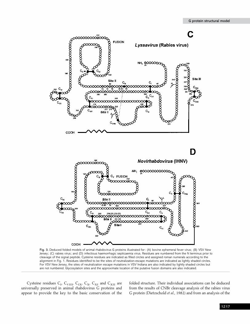
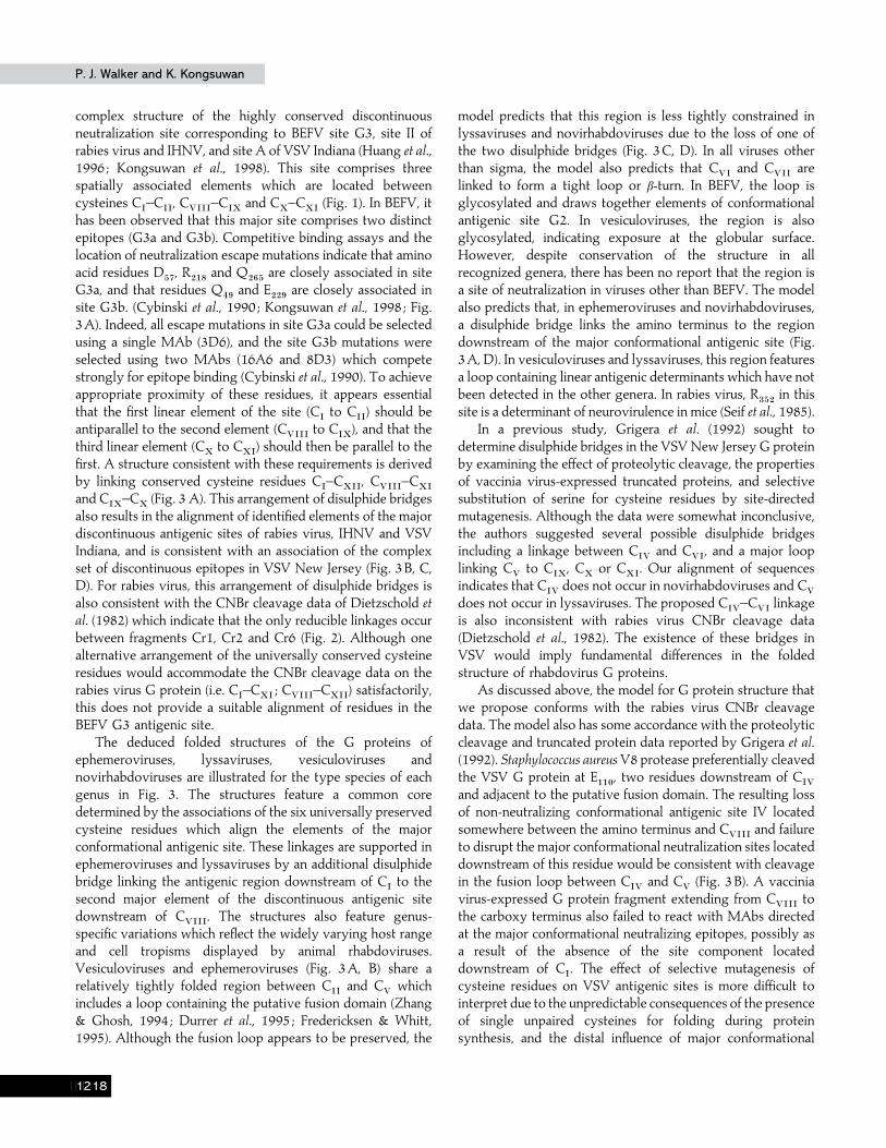
Fig. 3. Deduced folded models of animal rhabdovirus G proteins illustrated for : (A) bovine ephemeral fever virus ; (B) VSV NewJersey ; (C) rabies virus ; and (D) infectious haemorrhagic septicaemia virus. Residues are numbered from the N terminus prior tocleavage of the signal peptide. Cysteine residues are indicated as filled circles and assigned roman numerals according to thealignment in Fig. 1. Residues identified to be the sites of neutralization-escape mutations are indicated as lightly shaded circles.For VSV New Jersey, the sites of neutralization escape mutations in VSV Indiana are also indicated by lightly shaded circles butare not numbered. Glycosylation sites and the approximate location of the putative fusion domains are also indicated.
Cysteine residues CI, C
VIII, C
IX, C
X, C
XIand C
XIIare
universally preserved in animal rhabdovirus G proteins andappear to provide the key to the basic conservation of the
folded structure. Their individual associations can be deducedfrom the results of CNBr cleavage analysis of the rabies virusG protein (Dietzschold et al., 1982) and from an analysis of the
BCBH
P. J. Walker and K. KongsuwanP. J. Walker and K. Kongsuwan
complex structure of the highly conserved discontinuousneutralization site corresponding to BEFV site G3, site II ofrabies virus and IHNV, and site A of VSV Indiana (Huang et al.,1996 ; Kongsuwan et al., 1998). This site comprises threespatially associated elements which are located betweencysteines C
I–C
II, C
VIII–C
IXand C
X–C
XI(Fig. 1). In BEFV, it
has been observed that this major site comprises two distinctepitopes (G3a and G3b). Competitive binding assays and thelocation of neutralization escape mutations indicate that aminoacid residues D
&(, R
#")and Q
#'&are closely associated in site
G3a, and that residues Q%*
and E##*
are closely associated insite G3b. (Cybinski et al., 1990 ; Kongsuwan et al., 1998 ; Fig.3A). Indeed, all escape mutations in site G3a could be selectedusing a single MAb (3D6), and the site G3b mutations wereselected using two MAbs (16A6 and 8D3) which competestrongly for epitope binding (Cybinski et al., 1990). To achieveappropriate proximity of these residues, it appears essentialthat the first linear element of the site (C
Ito C
II) should be
antiparallel to the second element (CVIII
to CIX
), and that thethird linear element (C
Xto C
XI) should then be parallel to the
first. A structure consistent with these requirements is derivedby linking conserved cysteine residues C
I–C
XII, C
VIII–C
XI
and CIX
–CX
(Fig. 3 A). This arrangement of disulphide bridgesalso results in the alignment of identified elements of the majordiscontinuous antigenic sites of rabies virus, IHNV and VSVIndiana, and is consistent with an association of the complexset of discontinuous epitopes in VSV New Jersey (Fig. 3B, C,D). For rabies virus, this arrangement of disulphide bridges isalso consistent with the CNBr cleavage data of Dietzschold etal. (1982) which indicate that the only reducible linkages occurbetween fragments Cr1, Cr2 and Cr6 (Fig. 2). Although onealternative arrangement of the universally conserved cysteineresidues would accommodate the CNBr cleavage data on therabies virus G protein (i.e. C
I–C
XI; C
VIII–C
XII) satisfactorily,
this does not provide a suitable alignment of residues in theBEFV G3 antigenic site.
The deduced folded structures of the G proteins ofephemeroviruses, lyssaviruses, vesiculoviruses andnovirhabdoviruses are illustrated for the type species of eachgenus in Fig. 3. The structures feature a common coredetermined by the associations of the six universally preservedcysteine residues which align the elements of the majorconformational antigenic site. These linkages are supported inephemeroviruses and lyssaviruses by an additional disulphidebridge linking the antigenic region downstream of C
Ito the
second major element of the discontinuous antigenic sitedownstream of C
VIII. The structures also feature genus-
specific variations which reflect the widely varying host rangeand cell tropisms displayed by animal rhabdoviruses.Vesiculoviruses and ephemeroviruses (Fig. 3A, B) share arelatively tightly folded region between C
IIand C
Vwhich
includes a loop containing the putative fusion domain (Zhang& Ghosh, 1994 ; Durrer et al., 1995 ; Fredericksen & Whitt,1995). Although the fusion loop appears to be preserved, the
model predicts that this region is less tightly constrained inlyssaviruses and novirhabdoviruses due to the loss of one ofthe two disulphide bridges (Fig. 3C, D). In all viruses otherthan sigma, the model also predicts that C
VIand C
VIIare
linked to form a tight loop or β-turn. In BEFV, the loop isglycosylated and draws together elements of conformationalantigenic site G2. In vesiculoviruses, the region is alsoglycosylated, indicating exposure at the globular surface.However, despite conservation of the structure in allrecognized genera, there has been no report that the region isa site of neutralization in viruses other than BEFV. The modelalso predicts that, in ephemeroviruses and novirhabdoviruses,a disulphide bridge links the amino terminus to the regiondownstream of the major conformational antigenic site (Fig.3A, D). In vesiculoviruses and lyssaviruses, this region featuresa loop containing linear antigenic determinants which have notbeen detected in the other genera. In rabies virus, R
$&#in this
site is a determinant of neurovirulence in mice (Seif et al., 1985).In a previous study, Grigera et al. (1992) sought to
determine disulphide bridges in the VSV New Jersey G proteinby examining the effect of proteolytic cleavage, the propertiesof vaccinia virus-expressed truncated proteins, and selectivesubstitution of serine for cysteine residues by site-directedmutagenesis. Although the data were somewhat inconclusive,the authors suggested several possible disulphide bridgesincluding a linkage between C
IVand C
VI, and a major loop
linking CV
to CIX
, CX
or CXI
. Our alignment of sequencesindicates that C
IVdoes not occur in novirhabdoviruses and C
V
does not occur in lyssaviruses. The proposed CIV
–CVI
linkageis also inconsistent with rabies virus CNBr cleavage data(Dietzschold et al., 1982). The existence of these bridges inVSV would imply fundamental differences in the foldedstructure of rhabdovirus G proteins.
As discussed above, the model for G protein structure thatwe propose conforms with the rabies virus CNBr cleavagedata. The model also has some accordance with the proteolyticcleavage and truncated protein data reported by Grigera et al.(1992). Staphylococcus aureus V8 protease preferentially cleavedthe VSV G protein at E
""!, two residues downstream of C
IV
and adjacent to the putative fusion domain. The resulting lossof non-neutralizing conformational antigenic site IV locatedsomewhere between the amino terminus and C
VIIIand failure
to disrupt the major conformational neutralization sites locateddownstream of this residue would be consistent with cleavagein the fusion loop between C
IVand C
V(Fig. 3B). A vaccinia
virus-expressed G protein fragment extending from CVIII
tothe carboxy terminus also failed to react with MAbs directedat the major conformational neutralizing epitopes, possibly asa result of the absence of the site component locateddownstream of C
I. The effect of selective mutagenesis of
cysteine residues on VSV antigenic sites is more difficult tointerpret due to the unpredictable consequences of the presenceof single unpaired cysteines for folding during proteinsynthesis, and the distal influence of major conformational
BCBI

G protein structural modelG protein structural model
disruptions. In accordance with our model, the mutagenesisdata indicated that elimination of linked cysteines C
IXor C
X
resulted in a significant reduction in the efficiency of immuno-precipitation by MAbs directed to the major conformationalantigenic site. However, substitutions at other key cysteineresidues either did not significantly reduce the efficiency ofimmunoprecipitation (e.g. C
XII), or caused a significant
reduction in efficiency when only one residue of a putativelykey cysteine bridge was eliminated (e.g. C
XIbut not C
VIII).
This may be due to a partial stabilization of the folded structureby non-covalent interactions. The disulphide bridgespostulated by Grigera et al. (1992) are consistent with themutagenesis data. The C
IV–C
VIbridge is also consistent with
the greatly reduced electrophoretic mobility of the non-reduced G protein following limited V8 proteolytic cleavagewhich occurs close to C
IV. Although this does appear to be
inconsistent with our model, there is a total of 35 potential V8cleavage sites in the VSV New Jersey G protein. A largeproportion of G protein molecules may have been cleaved atvarious other sites, resulting in relaxation of a major loop in theG protein.
The analysis presented in this study has been confined tothe cysteine-rich head region of rhabdovirus G proteins whichcontains most of the functional domains associated with cellattachment and fusion. Immediately downstream of this regionis a highly conserved glycine residue which appears to markthe beginning of the stalk domain. Gaudin et al. (1996) haveidentified that this conserved glycine is followed by twopredicted α-helices separated by a highly conserved HPsequence. It has been proposed that the HP sequence mayfunction as a hinge between these helices which appear to havea key role in controlling conformational transitions duringvirus morphogenesis and membrane fusion (Gaudin et al.,1993 ; Li et al., 1993). These structures are not evidentlyconserved in ephemeroviruses which feature an extended stalkcontaining a major linear neutralization site and multipleputative glycosylation sites in the corresponding domain(Kongsuwan et al., 1998).
The deduced model presented in this paper should providea useful basis for the rational design of further studies to proberhabdovirus G protein structure. We are aware that workpresented recently at the Fourth International Symposium onViruses of Lower Vertebrates has used mass spectroscopicanalysis of proteolytic cleavage products of the VHSV Gprotein to identify a pattern of disulphide bridges which isconsistent with this model (Einer-Jensen et al., 1998).
The authors wish to thank Dr Robert Tesh (Center for TropicalDiseases, University of Texas, Galveston, Texas) for providingChandipura virus isolates and Dr Gael Kurath and Bill Batts (WesternFisheries Research Center, BRD-USGS, Seattle, Washington) for as-sistance with RT–PCR of Chandipura RNA. We also thank Dr JimWinton (Western Fisheries Research Center) and Dr Ross Tellam (CSIROTropical Agriculture) for helpful discussions.
ReferencesBricker, B. J., Snyder, R. M., Fox, J. W., Volk, W. A. & Wagner, R. R.(1987). Monoclonal antibodies to the glycoprotein of vesicularstomatitis virus (New Jersey serotype) : a method for preliminarymappingof epitopes. Virology 161, 533–540.
Cybinski, D. H., Walker, P. J., Byrne, K. A. & Zakrzewski, H. (1990).Mapping of antigenic sites on the bovine ephemeral fever virusglycoprotein using monoclonal antibodies. Journal of General Virology 71,2065–2072.
Dietzschold, B., Wiktor, T. J., Macfarlan, R. & Varrichio, A. (1982).Antigenic structure of rabies virus glycoprotein : ordering and immuno-logical characterization of the large CNBr cleavage fragments. Journal ofVirology 44, 595–602.
Durrer, P., Gaudin, Y., Ruigrok, R. W., Graf, R. & Brunner, J. (1995).Photolabeling identifies a putative fusion domain in the envelopeglycoprotein of rabies and vesicular stomatitis viruses. Journal of BiologicalChemistry 270, 17575–17581.
Einer-Jensen, K., Krogh, T. N., Roepstorff, P. & Lorenzen, N. (1998).Characterization of intramolecular disulfide bonds and secondarymodifications of the glycoprotein from viral hemorrhagic septicemiavirus, a fish rhabdovirus. Journal of Virology 72, 10189–10196.
Fredericksen, B. L. & Whitt, M. A. (1995). Vesicular stomatitis virusglycoprotein mutations that affect membrane fusion activity and abolishvirus infectivity. Journal of Virology 69, 1435–1443.
Gaudin, Y., Ruigrok, R. W. H., Knossow, M. & Flamand, A. (1993).Low-pH conformational changes of rabies virus glycoprotein and theirrole in membrane fusion. Journal of Virology 67, 1365–1372.
Gaudin, Y., Raux, H., Flamand, A. & Ruigrok, R. W. H. (1996).Identification of amino acids controlling low-pH-induced conformationalchanges of rabies virus glycoprotein. Journal of Virology 70, 7371–7378.
Grigera, P. R., Keil, W. & Wagner, R. R. (1992). Disulphide bondeddiscontinuous epitopes on the glycoprotein of vesicular stomatitis virus(New Jersey serotype). Journal of Virology 66, 3749–3757.
Huang, C., Chien, M.-S., Landolt, M. & Winton, J. (1994). Charac-terization of the infectious haematopoietic necrosis virus glycoproteinusing neutralizing monoclonal antibodies. Diseases of Aquatic Organisms18, 29–35.
Huang, C., Chien, M.-S., Landolt, M., Batts, W. & Winton, J. (1996).Mapping the neutralizing epitopes on the glycoprotein of infectioushaematopoietic necrosis virus, a fish rhabdovirus. Journal of GeneralVirology 77, 3033–3040.
Keil, W. & Wagner, R. R. (1989). Epitope mapping by deletion mutantsand chimeras of two vesicular stomatitis virus glycoprotein genesexpressed by a vaccinia virus vector. Virology 170, 392–407.
Kongsuwan, K., Cybinski, D. H., Cooper, J. & Walker, P. J. (1998).Location of neutralizing epitopes on the G protein of bovine ephemeralfever rhabdovirus. Journal of General Virology 79, 2573–2581.
Lafon, M., Wiktor, T. J. & Macfarlan, R. I. (1983). Antigenic sites on theCVS rabies virus glycoprotein : analysis with monoclonal antibodies.Journal of General Virology 64, 843–851.
Li, Y., Drone, C., Sat, E., Ghosh, H. P. & Banerjee, A. K. (1993).Mutational analysis of the vesicular stomatitis virus glycoprotein G formembrane fusion domains. Journal of Virology 67, 4070–4077.
Luo, L., Li, Y., Snyder, R. M. & Wagner, R. R. (1988). Point mutationsin glycoprotein gene of vesicular stomatitis virus (New Jersey serotype)selected by resistance to neutralization by epitope-specific monoclonalantibodies. Virology 163, 341–348.
BCBJ

P. J. Walker and K. KongsuwanP. J. Walker and K. Kongsuwan
Prehaud, C., Coulon, P., Lafay, F., Thiers, C. & Flamand, A. (1988).Antigenic site II of the rabies virus glycoprotein : structure and role inviral virulence. Journal of Virology 62, 1–7.
Rost, B. & Sander, C. (1993a). Improved prediction of proteinsecondary structure by use of sequence profiles and neutral networks.Proceedings of the National Academy of Sciences, USA 90, 7558–7562.
Rost, B. & Sander, C. (1993b). Prediction of protein secondary structureat better than 70% accuracy. Journal of Molecular Biology 232, 584–599.
Seif, I., Coulon, P., Rollin, P. E. & Flamand, A. (1985). Rabies virulence :effect on pathogenicity and sequence characterization of rabies virusmutations affecting antigenic site III of the glycoprotein. Journal ofVirology 53, 926–934.
Thompson, J. D., Higgins, D. G. & Gibson, T. J. (1994). CLUSTAL W:improving the sensitivity of progressive multiple sequence alignmentthrough sequence weighting, position-specific gap penalties and weightmatrix choice. Nucleic Acids Research 22, 4673–4680.
Tordo, N., Charlton, K. & Wandeler, A. (1997). Rhabdoviruses : rabies.In Topley and Wilson’s Principles of Bacteriology, Virology and Immunity, 9thedn, pp. 665–692. Edited by W. W. C. Topley, G. S. Wilson & L. Collier.London : Arnold.
Vanderpol, S. B., Lefrancois, L. & Holland, J. J. (1986). Sequences ofthe major antibody binding epitopes of the Indiana serotype of vesicularstomatitis virus. Virology 148, 312–325.
Walker, P. J., Byrne, K. A., Riding, G. A., Cowley, J. A., Wang, Y. &McWilliam, S. (1992). The genome of bovine ephemeral feverrhabdovirus contains two related glycoprotein genes. Virology 191,49–61.
Wunner, W. H., Calisher, C. H., Dietzgen, R. G., Jackson, A. O.,Kitajima, E. W., Lafon, M. F., Leong, J. C., Nichol, S. T., Peters, D.,Smith, J. S. & Walker, P. J. (1995). Rhabdoviridae. In Virus Taxonomy.Sixth Report of the International Committee on Taxonomy of Viruses, pp.288–293. Edited by F. A. Murphy, C. M. Fauquet, D. H. L. Bishop, S. A.Ghabrial, A. W. Jarvis, G. P. Martelli, M. A. Mayo & M. D. Summers.Vienna & New York : Springer-Verlag.
Zhang, L. & Ghosh, H. P. (1994). Characterization of the putativefusogenic domain in vesicular stomatitis virus glycoprotein G. Journal ofVirology 68, 2186–2193.
Received 29 October 1998; Accepted 15 January 1999
BCCA