decision making in gingival recession treatment: scientific
TRANSCRIPT
PRACTICAL APPLICATIONS
Decision Making in Gingival Recession Treatment: Scientific Evidence andClinical Experience
Giulio Rasperini,* Raffaele Acunzo,* Enrico Limiroli*
Focused Clinical Question: What are the keyconsiderations for selecting the best surgical approachin mucogingival plastic surgery?
Summary: Treatment of gingival recession hasbecome an important therapeutic issue due to the in-creasing number of cosmetic requests from patients.The dual goals of mucogingival treatment include com-plete root coverage, up to the cemento-enamel junc-tion, and blending of tissue color between the treatedarea and non-treated adjacent tissues. Even thoughthe connective tissue graft is commonly consideredthe "gold standard" for treatment of recession defects,it may not always be the best surgical option for everycase.
Conclusions: Under non-experimental condi-tions, all root coverage procedures may be effective interms of complete root coverage and excellent esthetics.Careful analyses of patient- and defect-related factors,however, are key considerations prior to selecting an ap-propriate surgical technique. Clin Adv Periodontics2011;1:41-52.
KeyWords: Connective tissue graft(s); gingival recession;mucogingival surgery; periodontitis; plastic surgery,periodontal.
BackgroundIn periodontal practice, root coverage requires dailyclinical decisions. Randomized clinical trials support thepotential clinical value of all proposed mucogingivalplastic surgery techniques, both in terms of mean (MRC)and complete root coverage (CRC), but fail to demon-strate a clear superiority of any of the tested surgicalprocedures.1,2 In addition, the clinical trials do notprovide clear guidance on when to use the differentprocedures.
While concerns about facial appearance have obsessedhumans for centuries, the systematic assessment of estheticoutcomes after surgical treatment of gingival recession isa relatively recent proposal.3,4
The ultimate goal of root coverage procedures should becomplete coverage of the recession defect with a pleasingcolor and tissue blend between the treated area and adja-cent tissues, thereby achieving both biologic and estheticsuccess. Thus, it is important to select the most predictableand easy-to-performsurgical technique according to a care-ful evaluation of the following factors:
1. Patient;
2. Single or multiple gingival recession defects;
3. Mucogingival defects localized in esthetically or non-esthetic sensitive sites;
4. Defect anatomic morphology (amount of keratinizedtissue, periodontal biotype, and vestibule depth);
5. Ability to enhance periodontalwound healing and sta-bilize the flap with optimal suture technique;
6. Biomaterials (connective tissue graft [CTG], enamelmatrix derivative, acellular dermal matrix).
Factors Affecting Complete RootCoverageMiller ClassMiller’s classification5 is based on morphologic evaluationof the injured periodontal tissue, giving the diagnosis of theseverity of gingival lesions and the prognostic evaluation ofthe treatment. According to this classification system,which is still themostwidely used, the loss of interproximalbone (Class III and IV) is identified as a condition involvedin preventing CRC.
Post-Surgical Position of Gingival Margin (GM)Soft tissue healing pattern after root coverage proceduresis usually linked to a shrinkage of the surgical wound. Thelocation of the GM relative to the cemento-enamel junc-tion (CEJ) after the surgery seems to affect the proba-bility of CRC;6 the more coronal the GM after suturing,the greater the probability of achieving CRC. A coronaldisplacement of 2 mm of the GM relative to the CEJ issuggested.
Flap TensionEnhanced periodontalwound healing is one of themost im-portant issues for the clinical success of root coverage pro-cedures. Even considering the different abilities of varioussurgeons in tissue management, attention to blood supplyand suturing techniquemay influence the clinical outcome.In particular, the use of surgical approaches that make the
*Unit of Periodontology, Dental Clinic, Department of Surgical,Reconstructive and Diagnostic Science, Foundation IRCCS, Ca’ GrandaPoliclinico, University of Milan, Milan, Italy.
Submitted December 9, 2010; accepted for publication February 1, 2011
doi: 10.1902/cap.2011.100002
Clinical Advances in Periodontics, Vol. 1, No. 1, May 2011 41
flap passive plays a major role in enhancing an optimalwound healing to achieve an adequate coronal displace-ment of the flap. Pini Prato et al.7 showed that the greaterthe flap tension (suggested flap tension should not exceed4 g), the less successful the recession improvement. Thus,periosteal incisions should be used to eliminate tensionfrom the flap, and in the maxillary jaw, the periosteal inci-sion should also include careful dissection of the muscle in-sertions from the flap.
Flap ThicknessThe survival of the flap, and particularly the marginal gin-giva, depends on the residual vascular system after surgicalincisions. Because of the caudo-cranial pattern of vascular-ization, we suggest a full-thickness dissection, when possi-ble, to avoid interrupting the supraperiosteal vessels thatenhance the survival of the flap on the avascular root sur-face. Thus, the thicker the flap, the greater the vasculariza-tion of the marginal gingiva and the probability of CRC(suggested flap thickness >0.8 mm).8
Interdental Papilla HeightAccording to Saletta et al.,9 CRC is more likely to beachieved in sites with a lower height of interdental papilla.Olsson et al.10 demonstrated that individuals with a long-narrow form of the central incisors (N biotype, scalloped-thin) show a thin free gingiva, a narrow zone of gingiva,and a wider height of the interdental papilla, while indi-viduals with short-wide crowns (W biotype, flat-thick)show a thicker free gingiva, a wider zone of keratinizedtissue, and a lower papilla height. Thus, it is possible thatthe thicker gingiva of the flat-thick biotype allowsa thicker flap, which may result in a greater success rateof CRC.
Cemento-Enamel Junction PredeterminationCRC is not always achievable, even in gingival recessionwith no loss of interproximal attachment and bone. TheCEJ is themostwidely used reference parameter to evaluateroot coverage results; however, such conditions as 1) cervi-cal abrasion, 2) traumatic loss of the tip of the interdentalpapilla, 3) tooth rotation, and 4) tooth extrusion with orwithout occlusal abrasion may lead to diagnostic mistakes
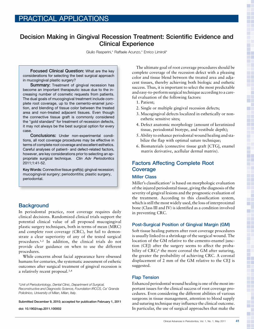
FIGURE 1 CAF procedure: flap design. Perform two horizontal beveledincisions (a), mesial and distal to the recession defect, and an intrasulcularincision (b). Execute two beveled oblique incisions (c) coming from the twohorizontal incisions, extending to the alveolar mucosa. Locate the twohorizontal incisions at a distance equal to the recession depth plus 1/2 mmfrom the tip of anatomic papillae (AP) to predefine the surgical papillae (SP).a ¼ horizontal incision; b ¼ intrasulcular incision; c ¼ vertical releasingincision; REC ¼ recession depth.
FIGURE 2 CAF procedure: suggested flap design in esthetic area. Whena single recession-type defect is present in the esthetic area, we suggestusing an envelope flap technique, avoiding vertical releasing incisions toreduce the probability of scar tissue formation. To facilitate the coronalrepositioning of the flap, make a horizontal incision that extendsmesiodistally to include three teeth. The horizontal incision of this modifiedenvelope technique consists of oblique submarginal incisions in theinterdental areas, which continue the intrasulcular incision at the recessiondefect. Locate the starting point of oblique incisions at a distance from thetip of the anatomic papilla equal to the recession depth plus 1/2 mm. Anumber of disadvantages of this surgical technique can be pointed out: theneed to involve healthy adjacent teeth in the procedure and the smallerdimension of the flap. 2a before surgery; 2b after surgery; 2c flap design.REC ¼ recession depth; SP ¼ surgical papillae.
P R A C T I C A L A P P L I C A T I O N S
42 Clinical Advances in Periodontics, Vol. 1, No. 1, May 2011 Decision Making in Gingival Recessions
preventing CRC. Thus, in such clinical conditions, the lineof root coverage may be considered the clinical CEJ, be-cause it may substitute for the anatomic CEJ when it isno longer clinically visible or when ideal conditions to ob-tain CRC are not fully represented.11,12
Preparation of Exposed Root SurfaceTo the best of our knowledge, no study has been reported inthe literature that shows one technique to be superior to allothers. The clinician may treat the exposed root surfacemechanically, by means of curets, sonic devices, polishingor rotary instruments, or chemically using tetracycline,sodium hypochlorite, or EDTA. According to our clinical
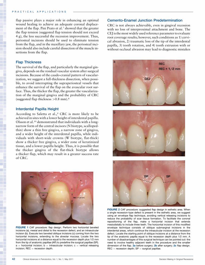
FIGURE 3 CAFþCTG procedure: suggested flap design and harvestingtechnique. Using a trap door technique (a) to harvest the CTG will allowa primary wound closure of the donor palatal site, reducing patientpostoperative morbidity. Secure the graft over the exposed root surfaceusing a resorbable sling suture passing through the connective tissue of theinterdental papilla. 3a CTG harvesting from palate; 3b suture of the graft; 3c6-month postoperative evaluation.
FIGURE 4 DPF procedure: flap design. Mucogingival defect affectingtooth #11. An inadequate amount of keratinized tissue is present apically tothe recession, and the presence of well-represented interdental papillasuggest a double papillae procedure. 4a baseline; 4b DPF; 4c 12-monthfollow-up.
P R A C T I C A L A P P L I C A T I O N S
Rasperini, Acunzo, Limiroli Clinical Advances in Periodontics, Vol. 1, No. 1, May 2011 43
experience, we suggest using simple root preparation proce-dures suchas scaling and root planingwith sonic devices andcurets. The need to flatten prominent roots may representa clinical indication for the use of rotary instruments.
Moreover, to avoid damaging any connective tissuefibers still embedded in cementum, it might be convenientto prepare the exposed root surface prior to raising the flap,especially if a mechanical root preparation procedure is tobe used.
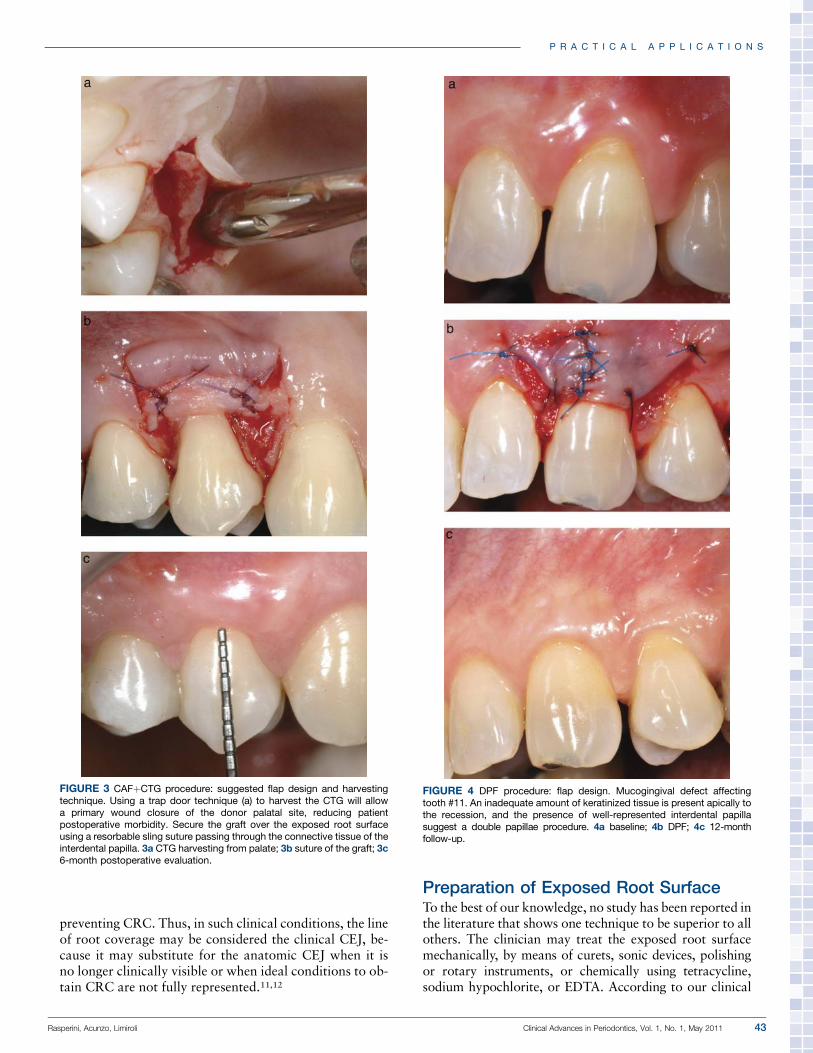
FIGURE 5 DPFþCTG procedure: surgical technique. To modify the qualityand amount of keratinized tissue over the exposed root surface, a DPF inconjunction with a CTG is performed. Use a trap door technique (Figure 3a)as described previously to harvest the CTG and secure the graft over theexposed root surface using a resorbable sling suture passing through theconnective tissue of the interdental papilla. 5a baseline; 5b CTG positionedon the root surface; 5c 12-month follow-up.
FIGURE 6 LAF procedure: flap design. 6a Flap design and areas needingto be deepithelized. An adequate amount of keratinized tissue is locateddistally to the canine; 6b the LAF plus CTG correctly repositioned upon theexposed root surface and stabilized with sutures; 6c 3-month follow-up.
P R A C T I C A L A P P L I C A T I O N S
44 Clinical Advances in Periodontics, Vol. 1, No. 1, May 2011 Decision Making in Gingival Recessions
Restorative Approach in MucogingivalTherapyGingival recession may be associated with dental abrasiondue to toothbrushing or cervical caries. In this situation,the lack of a definable anatomic CEJ may present clinicianswith difficulties during the diagnostic phase that preventcomplete coverage of the exposed root surface. A classifica-tionof suchdental defects has been recently proposedbyPiniPrato et al.13 In cases where there is an identifiable CEJ, wesuggest predetermining the lineof root coverage asdescribedbyZucchelli et al.11 and treating the portion of the tooth cor-onal to the CEJ using a restorative approach. To avoid dam-agingthegingivalmargin,wesuggestrestorationofthedentalabrasion prior to the surgical phase or during the surgery.
Treatment StrategyGingival recession treatment can no longer be consideredas a single treatment approach. In fact, there is evidenceto considermucogingival plastic surgery as amultifactorialtreatment approach comprising careful selection of pa-tients (see Decision Tree 1) and defects, different surgicaltechniques, many suturing approaches, and various typesof adjunctive materials. All the cited components shouldbe variously combined to develop different treatment strat-egies with different degrees of technical difficulties (seeDecision Tree 2).
Clinical Condition 1: Coronally Advanced Flap(CAF) – Table 1
Selection of surgical flapA distance fromGM tomucogingival junction (MGJ) of atleast 2 mm should be present to enhance the stability of thesurgical flap after suturing. A CAF procedure alone shouldbe performed when a thick and flat periodontal biotype ispresent to avoid a relapse. A moderate or deep vestibulewill allow coronal displacement of the flap without ten-sion; a shallow vestibule does not prevent the use of aCAF technique but requires an extensive partial-thickness
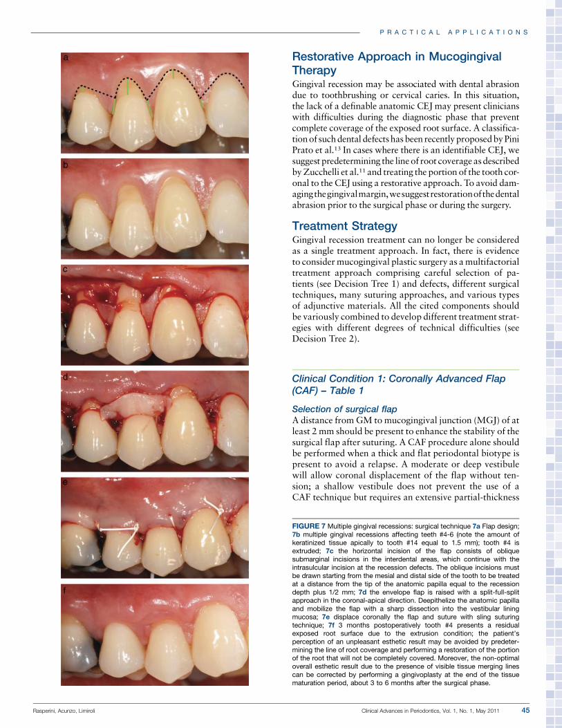
FIGURE 7 Multiple gingival recessions: surgical technique 7a Flap design;7b multiple gingival recessions affecting teeth #4-6 (note the amount ofkeratinized tissue apically to tooth #14 equal to 1.5 mm); tooth #4 isextruded; 7c the horizontal incision of the flap consists of obliquesubmarginal incisions in the interdental areas, which continue with theintrasulcular incision at the recession defects. The oblique incisions mustbe drawn starting from the mesial and distal side of the tooth to be treatedat a distance from the tip of the anatomic papilla equal to the recessiondepth plus 1/2 mm; 7d the envelope flap is raised with a split-full-splitapproach in the coronal-apical direction. Deepithelize the anatomic papillaand mobilize the flap with a sharp dissection into the vestibular liningmucosa; 7e displace coronally the flap and suture with sling suturingtechnique; 7f 3 months postoperatively tooth #4 presents a residualexposed root surface due to the extrusion condition; the patient’sperception of an unpleasant esthetic result may be avoided by predeter-mining the line of root coverage and performing a restoration of the portionof the root that will not be completely covered. Moreover, the non-optimaloverall esthetic result due to the presence of visible tissue merging linescan be corrected by performing a gingivoplasty at the end of the tissuematuration period, about 3 to 6 months after the surgical phase.
P R A C T I C A L A P P L I C A T I O N S
Rasperini, Acunzo, Limiroli Clinical Advances in Periodontics, Vol. 1, No. 1, May 2011 45
dissection apically to the MGJ to make the flap tensionfree.
Suggested surgical managementThe surgical procedure was originally described by Allenand Miller14 in 1989, and further modifications have beenproposed over the years. Perform a horizontal incision andtwo beveled and slightly divergent releasing incisions(Fig. 1). Using a small periosteal elevator, raise a full-thick-ness flap and treat the exposed root surface with thoroughscaling and root planing using curets and/or ultrasonic de-vices (Video 1: root surface conditioning by means of rootplaning). Deepithelize the anatomic papilla (Video 2: ana-tomic papilla deepithelization using a surgical blade [15c];Video 3: use scissors to remove all the epithelium when theroots are prominent) and expose the underlying connectivetissue. Extend the dissection of the flap apically to theMGJproceeding with a split-thickness approach (Video 4: re-lease residual muscle tension, keeping the surgical blade[15] parallel to the flap); pay close attention to releasingthe residual muscle tension as this will enhance the coronaldisplacement of the flap (Video 5: cover the recession defectonly when a completely passive coronal displacement ofthe flap can be achieved). Advance the flap coronally usinga sling suture technique and single interrupted sutures toclose the releasing incisions.
Surgical advice1. Locate the horizontal incision at a distance from the
tip of anatomic papilla equal to recession depthþ 1/2 mm (Fig. 1).
2. Avoid making releasing incisions across the MGJ dur-ing the initial phase of the surgical procedure; this willreduce postoperative swelling and pain.
3. Try to avoid releasing incisions when recession defectis located in esthetic area (Fig. 2).15
Clinical Condition 2: Coronally Advanced Flap þConnective Tissue Graft (CAFþCTG) – Table 2
Selection of surgical flapA distance from GM to MGJ of at least 2 mm should bepresent to enhance the stability of the surgical flap aftersuturing. A CAF procedure in conjunction with a CTG isthe technique of choice when a thin and scalloped peri-odontal biotype is present, so that both the amount andquality of marginal soft tissue may be appropriately trans-formed. In the case of a thick biotype, the placement ofCTG can create an impaired esthetic due to irregular gin-gival profile or scar tissue.4 A moderate or deep vestibulewill allow coronal displacement of the flap without ten-sion; a shallow vestibule does not prevent the use ofa CAFþCTG technique but requires an extensive partial-thickness dissection apical to theMGJ tomake the flap ten-sion free.
Suggested surgical managementLanger and Langer18 introduced the use of subepithelialCTGs for root coverage, and several modifications to theoriginal technique have been published over the years. Per-form the CAF procedure as described above. Harvest the
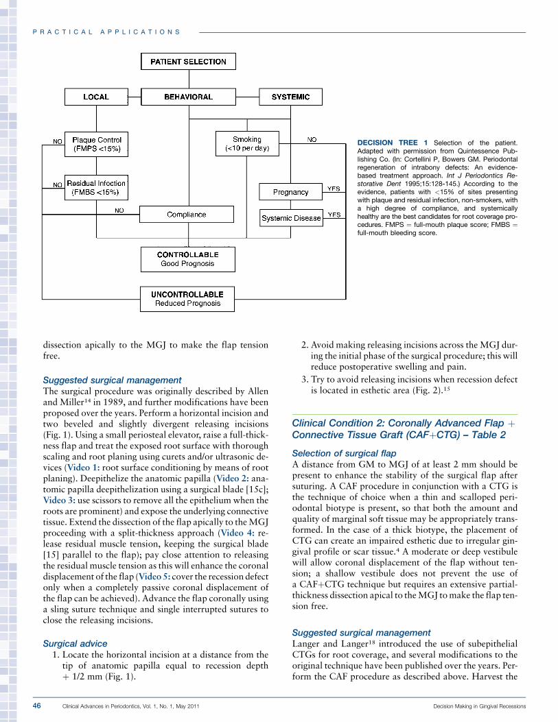
DECISION TREE 1 Selection of the patient.Adapted with permission from Quintessence Pub-lishing Co. (In: Cortellini P, Bowers GM. Periodontalregeneration of intrabony defects: An evidence-based treatment approach. Int J Periodontics Re-storative Dent 1995;15:128-145.) According to theevidence, patients with <15% of sites presentingwith plaque and residual infection, non-smokers, witha high degree of compliance, and systemicallyhealthy are the best candidates for root coverage pro-cedures. FMPS ¼ full-mouth plaque score; FMBS ¼full-mouth bleeding score.
P R A C T I C A L A P P L I C A T I O N S
46 Clinical Advances in Periodontics, Vol. 1, No. 1, May 2011 Decision Making in Gingival Recessions
CTG from the palate using a trapdoor technique (Fig. 3a);be sure to preserve a band of keratinized tissue at least 1to 2 mm from the palatal GM. To keep the graft moist,place it on gauze soaked in physiologic saline solution.Close the palatal wound with interrupted suture. Suturethe CTG at the recipient site using resorbable sling suturespassing through the interdental papilla connective tissue(Fig. 3b).
Surgical advice1. Locate the horizontal incision at a distance from the
tip of anatomic papilla equal to recession depthþ 1/2mm (Fig. 1).
2. Avoid making releasing incisions across theMGJ dur-ing the initial phase of the surgical procedure; this willreduce postoperative swelling and pain.
3. Try to avoid releasing incisions when recessiondefect is located in esthetic area (Fig. 2).15 A de-
epithelialized graft technique can be used instead ofa trap-door procedure to reduce the chair time andsimplify the harvesting procedure.
4. Close the palatal wound using collagen sponges (toenhance secondary intention wound healing) andcriss-cross suture technique after a deepithelializedgraft procedure.
Clinical Condition 3: Double Papillae Flap(DPF) – Table 3
Selection of surgical flapTo perform aDPF technique, an alternative keratinized tis-sue donor-site must be represented by adjacent interdentalpapillae. Periodontal biotype should be classified as thickand flat. This surgical technique is not affected by vestibule
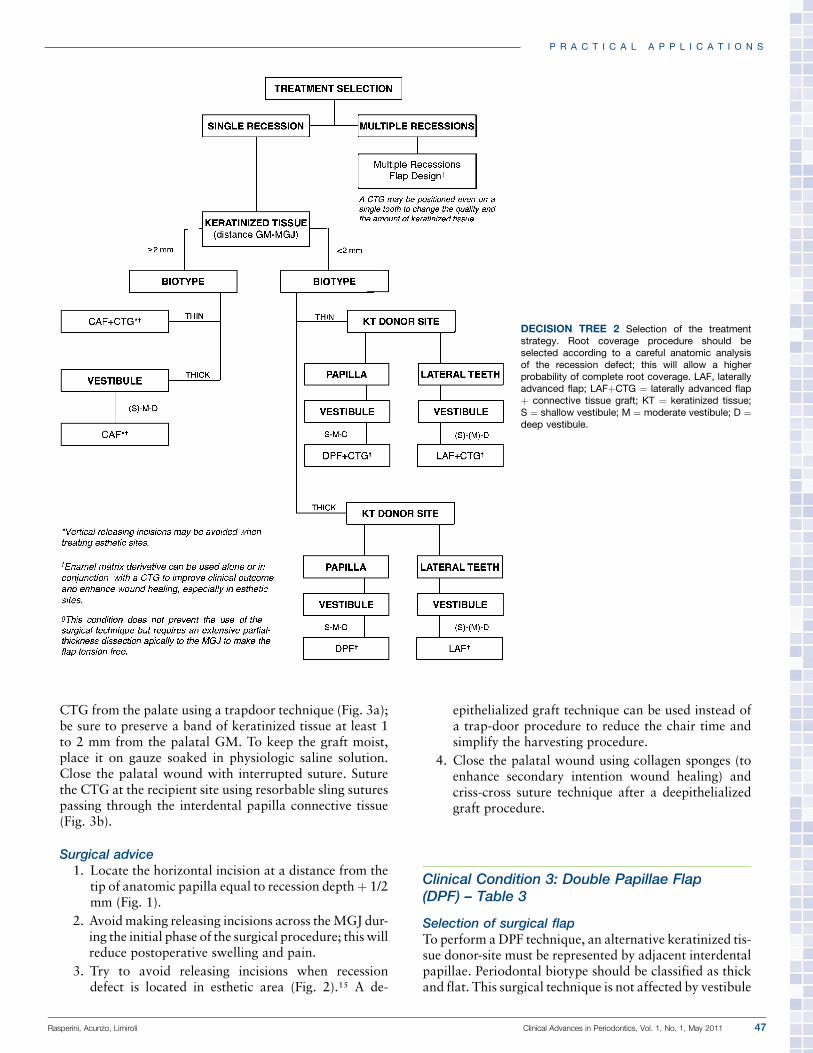
DECISION TREE 2 Selection of the treatmentstrategy. Root coverage procedure should beselected according to a careful anatomic analysisof the recession defect; this will allow a higherprobability of complete root coverage. LAF, laterallyadvanced flap; LAFþCTG ¼ laterally advanced flapþ connective tissue graft; KT ¼ keratinized tissue;S ¼ shallow vestibule; M ¼ moderate vestibule; D ¼deep vestibule.
P R A C T I C A L A P P L I C A T I O N S
Rasperini, Acunzo, Limiroli Clinical Advances in Periodontics, Vol. 1, No. 1, May 2011 47
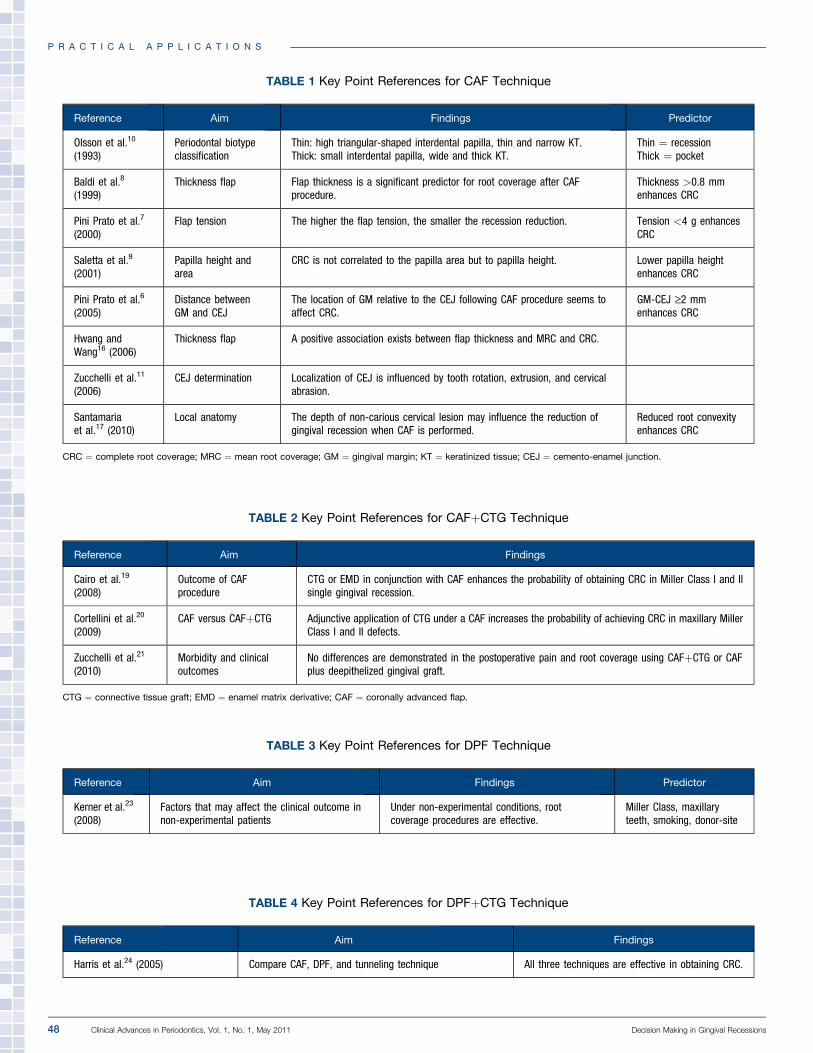
TABLE 1 Key Point References for CAF Technique
Reference Aim Findings Predictor
Olsson et al.10
(1993)Periodontal biotypeclassification
Thin: high triangular-shaped interdental papilla, thin and narrow KT.Thick: small interdental papilla, wide and thick KT.
Thin ¼ recessionThick ¼ pocket
Baldi et al.8
(1999)Thickness flap Flap thickness is a significant predictor for root coverage after CAF
procedure.Thickness >0.8 mmenhances CRC
Pini Prato et al.7
(2000)Flap tension The higher the flap tension, the smaller the recession reduction. Tension <4 g enhances
CRC
Saletta et al.9
(2001)Papilla height andarea
CRC is not correlated to the papilla area but to papilla height. Lower papilla heightenhances CRC
Pini Prato et al.6
(2005)Distance betweenGM and CEJ
The location of GM relative to the CEJ following CAF procedure seems toaffect CRC.
GM-CEJ ‡2 mmenhances CRC
Hwang andWang16 (2006)
Thickness flap A positive association exists between flap thickness and MRC and CRC.
Zucchelli et al.11
(2006)CEJ determination Localization of CEJ is influenced by tooth rotation, extrusion, and cervical
abrasion.
Santamariaet al.17 (2010)
Local anatomy The depth of non-carious cervical lesion may influence the reduction ofgingival recession when CAF is performed.
Reduced root convexityenhances CRC
CRC ¼ complete root coverage; MRC ¼ mean root coverage; GM ¼ gingival margin; KT ¼ keratinized tissue; CEJ ¼ cemento-enamel junction.
TABLE 2 Key Point References for CAFþCTG Technique
Reference Aim Findings
Cairo et al.19
(2008)Outcome of CAFprocedure
CTG or EMD in conjunction with CAF enhances the probability of obtaining CRC in Miller Class I and IIsingle gingival recession.
Cortellini et al.20
(2009)CAF versus CAFþCTG Adjunctive application of CTG under a CAF increases the probability of achieving CRC in maxillary Miller
Class I and II defects.
Zucchelli et al.21
(2010)Morbidity and clinicaloutcomes
No differences are demonstrated in the postoperative pain and root coverage using CAFþCTG or CAFplus deepithelized gingival graft.
CTG ¼ connective tissue graft; EMD ¼ enamel matrix derivative; CAF ¼ coronally advanced flap.
TABLE 3 Key Point References for DPF Technique
Reference Aim Findings Predictor
Kerner et al.23
(2008)Factors that may affect the clinical outcome innon-experimental patients
Under non-experimental conditions, rootcoverage procedures are effective.
Miller Class, maxillaryteeth, smoking, donor-site
TABLE 4 Key Point References for DPFþCTG Technique
Reference Aim Findings
Harris et al.24 (2005) Compare CAF, DPF, and tunneling technique All three techniques are effective in obtaining CRC.
P R A C T I C A L A P P L I C A T I O N S
48 Clinical Advances in Periodontics, Vol. 1, No. 1, May 2011 Decision Making in Gingival Recessions

depth due to the small coronal displacement required tocover the recession defect.
Suggested surgical managementCohen and Ross22 introduced the method in which bilateralinterdental papilla is used as donor tissue for localized rootcoverage. Perform a V-shaped incision at the buccal aspectof the involved tooth, with an internal bevel on one side oftheV-shaped incisionandanexternalbevelon theother.Makehorizontal andvertical incisions asdescribed for theCAFtech-nique, locating the horizontal incisions closer to the tip of in-terdental papilla as much as possible to includemore tissue inthe flap. Raise a full-thickness flap and condition the root sur-face by means of scaling and root planing using curets and/orsonic devices. Suture together the two surgical papillae withinterrupted sutures (Fig. 4b). Extend the dissection of the flapapically to the MGJ, proceeding with a split-thickness ap-proach (Video 4: release residual muscle tension, keepingthe surgical blade [15] parallel to the flap) and paying atten-tion to release the residual muscle tension (Video 5: cover therecession defect only when a completely passive coronal dis-placement of the flap can be achieved). Cover the recessiondefect using a sling suture technique anduse single interruptedsutures to close the releasing incisions (Fig. 4b).
Surgical advice1. Avoid making releasing incisions across the MGJ dur-
ing the initial phase of the surgical procedure; this willreduce postoperative swelling and pain.
2.Once the interdental papillae have been dissected, jointhem using interrupted sutures before proceedingwiththe next steps of the surgical procedure; this will makeflap manipulation simpler.
Clinical Condition 4: Double Papillae Flap þConnective Tissue Graft (DPFþCTG) – Table 4
Selection of surgical flapAs described for the DPF technique, an alternative kerati-nized tissue donor site must be represented by adjacent
interdental papillae to perform DPF in conjunction withCTG. Periodontal biotype should be classified as thinand scalloped. This surgical technique is not affected byvestibule depth due to the small coronal displacement re-quired to cover the recession defect.
Suggested surgical managementClean the root surface, perform the surgical flap, andharvestthe CTG as described previously (Fig. 5).
Surgical advice1. Avoid making releasing incisions across the MGJ dur-
ing the initial phase of the surgical procedure; this willreduce postoperative swelling and pain.
2.Once the interdental papillae have been dissected, jointhem using interrupted sutures before proceedingwiththe next steps of the surgical procedure; this will makeflap manipulation simpler.
3. A deepithelialized graft technique can be used insteadof a trap-door procedure to reduce the chair time andsimplify the harvesting procedure.
4. Close the palatalwound using collagen sponges (to en-hance secondary intention wound healing) and criss-cross suture technique after a deepithelialized graftprocedure.
Clinical Condition 5: Laterally AdvancedFlap (LAF) – Table 5
Selection of surgical flapTo perform a LAF technique, an alternative keratinized tis-sue donor site must be represented by adjacent teeth. Peri-odontal biotype should be classified as thick and flat. Thissurgical technique is not affected by vestibule depth due tothe small coronal displacement required to cover the reces-sion defect. However, a shallow or moderate vestibule mayrequire more surgical operator skill to obtain a completelytension-free flap; an inadequate dissection of periosteumand muscle insertions may lead to a relapse.
TABLE 6 Key Point References for Multiple Gingival Recessions Technique
Reference Aim Findings
Zucchelli and DeSanctis27 (2000)
Surgicaltechnique
The proposed surgical technique is effective for the treatment of multiple gingival recessions affecting teeth inesthetic areas of the mouth. This result may be achieved irrespective of both the number of recessionssimultaneously treated and the presence of minimal keratinized tissue prior to surgery.
Zucchelli and DeSanctis28 (2005)
Long-termoutcome
At the 5-year examination, 94% of the initially exposed root surfaces are still covered, and 85% of the treatedrecession defects showed CRC. CRC in all recessions is maintained in 15 of 22 patients (68%).
TABLE 5 Key Point References for LAF Technique
Reference Aim Findings
Zucchelli et al.26
(2004)Surgicaltechnique
The laterally moved CAF is very effective in treating isolated gingival recessions. The ideal gingival conditions mustbe present lateral to the defect to be treated.
P R A C T I C A L A P P L I C A T I O N S
Rasperini, Acunzo, Limiroli Clinical Advances in Periodontics, Vol. 1, No. 1, May 2011 49
Suggested surgical managementLAFs have been widely used since Grupe and Warren25 in-troduced this method for the treatment of localized gingi-val recession. In this procedure, the adjacent keratinizedgingiva is positioned laterally and the exposed root surfacecovered. Over the years, several further modifications ofthis technique have been proposed to avoid bone lossand gingival recession on the donor site, the most frequentadverse events related to this surgical procedure. RecentlyZucchelli et al.26 proposed a modified approach that ap-pears to be more reliable and safe (Fig. 6).
Surgical advice1. According to muscle insertion orientation, the LAF
should be preferably performed when the donor siteis localized mesial to the gingival recession defect.
2. When the flap is moved in the distal-mesial direction,another short horizontal incision should be performedat the most apical extension of the distal vertical re-leasing incision in order to facilitate mesial mobiliza-tion of the flap.
3. Use collagen sponges, stabilized with criss-cross su-tures, to promote wound healing of the keratinizedtissue donor site adjacent to the recession defect.
Clinical Condition 6: Laterally Advanced Flap þConnective Tissue Graft (LAFþCTG)
Selection of surgical flapAs described for the LAF technique, an alternative kerati-nized tissue donor-site must be available at adjacent teethto perform LAF in conjunction with CTG. The periodontalbiotype and vestibule depth for the LAFþCTG shouldbe the same as described for the laterally advanced flapalone.
Suggested surgical managementClean the root surface, perform the surgical flap, and har-vest the CTG as described previously.
Surgical advice1. According to muscle insertion orientation, the LAF
should be preferably performed when the donor siteis localized mesial to the gingival recession defect.
2. When the flap is moved in the distal-mesial direction,another short horizontal incision should be performedat the most apical extension of the distal vertical re-leasing incision in order to facilitate mesial mobiliza-tion of the flap.
3. Use collagen sponges, stabilized with criss-cross su-tures, to promote wound healing of the keratinizedtissue donor site adjacent to the recession defect.
4. A deepithelialized graft technique can be used insteadof a trap-door procedure to reduce the chair time andsimplify the harvesting procedure.
5. Close the palatalwound using collagen sponges (to en-hance secondary intention wound healing) and criss-
cross suture technique after a deepithelialized graftprocedure.
Clinical Condition 7: Multiple GingivalRecessions – Table 6
Another factor to consider in the surgical treatment of gin-gival recession is that mucogingival-type defects are veryseldom localized to a single tooth. More frequently, gingi-val recessions affect groups of adjacent teeth. Thus, tomin-imize the number of surgeries and to optimize the estheticresult, all of the contiguous recessions should be treated si-multaneously. Patient-related esthetic considerationswould suggest the use of a surgical technique that predict-ably obtains CRC in all present recessions by using the softtissue adjacent to the defects.
Suggested surgical managementZucchelli and De Sanctis27 proposed a new surgical ap-proach for the treatment of multiple recession defects(Fig. 7). This modified design of the envelope flap consistsof an oblique submarginal incision in the interdental area,which continues with an intrasulcular incision at the reces-sion defects.
Surgical advice1. When performing an envelope-type flap, avoid verti-
cal releasing incisions to help maintain adequateblood flow to the flap and reduce the formation of vis-ible white scars.
2. Use ‘‘split-full-split’’ flap elevation, with full thicknessfor that portion of the flap residing over the previouslyexposed root surface, to increase the potential toachieve CRC.
3. The absence of a wide zone of keratinized tissue apicalto the defects is not considered a limitation; a CTGmay be used at one single specific recession defect ifnecessary.
4. Suture the flap using a sling suture technique passingthrough the connective tissue of the anatomic papilla.
ConclusionsDue to an increasing public demand for cosmetic dentistry,the treatment of gingival recession has become an importanttherapeutic and esthetic issue for the contemporary peri-odontal practice.While the efficacy of usingCTGs to obtainfull coverage of root surface exposure is well supported inthe literature, this cannot be the onlyworthy treatment goal;surgeons must also use their skills to fulfill the demand forthe improved esthetics their patients expect.
Furthermore, even as CTG is considered the gold stan-dard treatment for single and multiple areas of recession,a simpler, less invasive approach, such as a CAF, may yieldan equally acceptable result. Each clinical situation mustbe evaluated to determine the most appropriate surgical
P R A C T I C A L A P P L I C A T I O N S
50 Clinical Advances in Periodontics, Vol. 1, No. 1, May 2011 Decision Making in Gingival Recessions

approach to achieve the esthetics expected by the patient.Therefore, to achieve the best clinical and esthetic success,a careful assessment of existing anatomic parameters, suchas the amount of keratinized tissue, the periodontal bio-type, and vestibule depth, is a vital part of the surgical de-cision-making process. n
AcknowledgmentThe authors did not receive any financial support for thisstudy.
CORRESPONDENCE:Dr. Giulio Rasperini, Via XX Settembre, 119 - 29121 Piacenza, Italy. E-mail:[email protected].
P R A C T I C A L A P P L I C A T I O N S
Rasperini, Acunzo, Limiroli Clinical Advances in Periodontics, Vol. 1, No. 1, May 2011 51
References1. Roccuzzo M, Bunino M, Needleman I, Sanz M. Periodontal plastic
surgery for treatment of localized gingival recessions: A systematic review.J Clin Periodontol 2002;29(Suppl. 3):178-194, discussion 195-196.
2. Oates TW, Robinson M, Gunsolley JC. Surgical therapies for thetreatment of gingival recession. A systematic review. Ann Periodontol2003;8:303-320.
3. Cairo F, Rotundo R, Miller PD, Pini Prato GP. Root coverage estheticscore: A system to evaluate the esthetic outcome of the treatment ofgingival recession through evaluation of clinical cases. J Periodontol2009;80:705-710.
4. Cairo F, Nieri M, Cattabriga M, et al. Root coverage esthetic score aftertreatment of gingival recession: An interrater agreement multicenterstudy. J Periodontol 2010;81:1752-1758.
5. Miller PD Jr. A classification of marginal tissue recession. Int JPeriodontics Restorative Dent 1985;5(2):8-13.
6. Pini Prato GP, Baldi C, Nieri M, et al. Coronally advanced flap: Thepost-surgical position of the gingival margin is an important factor forachieving complete root coverage. J Periodontol 2005;76:713-722.
7. Pini Prato G, Pagliaro U, Baldi C, et al. Coronally advanced flap procedurefor root coverage. Flap with tension versus flap without tension: Arandomized controlled clinical study. J Periodontol 2000;71:188-201.
8. BaldiC, Pini-PratoG, PagliaroU, et al. Coronally advanced flap procedurefor root coverage. Is flap thickness a relevant predictor to achieve rootcoverage? A 19-case series. J Periodontol 1999;70:1077-1084.
9. Saletta D, Pini Prato G, Pagliaro U, Baldi C, Mauri M, Nieri M.Coronally advanced flap procedure: Is the interdental papilla a prognos-tic factor for root coverage? J Periodontol 2001;72:760-766.
10. Olsson M, Lindhe J, Marinello CP. On the relationship between crownform and clinical features of the gingiva in adolescents. J ClinPeriodontol 1993;20:570-577.
11. Zucchelli G, Testori T, De Sanctis M. Clinical and anatomical factorslimiting treatment outcomes of gingival recession: A new method topredetermine the line of root coverage. J Periodontol 2006;77:714-721.
12. Zucchelli G, Mele M, Stefanini M, et al. Predetermination of rootcoverage. J Periodontol 2010;81:1019-1026.
13. Pini-Prato G, Franceschi D, Cairo F, Nieri M, Rotundo R. Classificationof dental surface defects in areas of gingival recession. J Periodontol2010;81:885-890.
14. Allen EP, Miller PD Jr. Coronal positioning of existing gingiva: Shortterm results in the treatment of shallow marginal tissue recession.J Periodontol 1989;60:316-319.
15. Zucchelli G, Mele M, Mazzotti C, Marzadori M, Montebugnoli L, DeSanctis M. Coronally advanced flap with and without vertical releasingincisions for the treatment of multiple gingival recessions: A comparativecontrolled randomized clinical trial. J Periodontol 2009;80:1083-1094.
16. Hwang D, Wang HL. Flap thickness as a predictor of root coverage: Asystematic review. J Periodontol 2006;77:1625-1634.
17. Santamaria MP, Ambrosano GM, Casati MZ, Nociti FH Jr. Sallum AW,Sallum EA. The influence of local anatomy on the outcome of treatmentof gingival recession associated with non-carious cervical lesions.J Periodontol 2010;81:1027-1034.
18. Langer B, Langer L. Subepithelial connective tissue graft technique forroot coverage. J Periodontol 1985;56:715-720.
19. Cairo F, Pagliaro U, Nieri M. Treatment of gingival recession withcoronally advanced flap procedures: A systematic review. J ClinPeriodontol 2008; 35(Suppl. 8):136-162.
20. Cortellini P, Tonetti M, Baldi C, et al. Does placement of a connectivetissue graft improve the outcomes of coronally advanced flap forcoverage of single gingival recessions in upper anterior teeth? A multi-center, randomized, double-blind, clinical trial. J Clin Periodontol 2009;36:68-79.
21. Zucchelli G, Mele M, Stefanini M, et al. Patient morbidity and rootcoverage outcome after subepithelial connective tissue and de-epithe-lialized grafts: A comparative randomized controlled clinical trial. J ClinPeriodontol 2010;37:728-738.
22. Cohen DW, Ross SE. The double papillae repositioned flap in periodon-tal therapy. J Periodontol 1968;39:65-70.
23. Kerner S, Borghetti A, Katsahian S, et al. A retrospective study of rootcoverage procedures using an image analysis system. J Clin Periodontol2008;35:346-355.
24. Harris RJ, Miller LH, Harris CR, Miller RJ. A comparison of threetechniques to obtain root coverage on mandibular incisors. J Periodontol2005;76:1758-1767.
25. Grupe J, Warren R. Repair of gingival defects by a sliding flap operation.J Periodontol 1956;27:92-95.
26. Zucchelli G, Cesari C, Amore C, Montebugnoli L, De Sanctis M.Laterally moved, coronally advanced flap: A modified surgical approachfor isolated recession-type defects. J Periodontol 2004;75:1734-1741.
27. Zucchelli G, De Sanctis M. Treatment of multiple recession-type defectsin patients with esthetic demands. J Periodontol 2000;71:1506-1514.
28. Zucchelli G, De Sanctis M. Long-term outcome following treatment ofmultiple Miller Class I and II recession defects in esthetic areas of themouth. J Periodontol 2005;76:2286-2292.
P R A C T I C A L A P P L I C A T I O N S
52 Clinical Advances in Periodontics, Vol. 1, No. 1, May 2011 Decision Making in Gingival Recessions