de novo thrombotic microangiopathy in renal transplant recipients: a comparison of hemolytic uremic...
TRANSCRIPT
De Novo Thrombotic Microangiopathy in Renal TransplantRecipients: A Comparison of Hemolytic Uremic SyndromeWith
Localized Renal Thrombotic Microangiopathy
Joshua Schwimmer, MD, Tibor A. Nadasdy, MD, PhD, Patrice F. Spitalnik, MD,Karen L. Kaplan, MD, and Martin S. Zand, MD, PhD
● Background: Thromboticmicroangiopathy (TMA) is awell-recognized and serious complication of renal transplan-tation, affecting 3% to 14% of patients administered calcineurin-inhibitor–based immunosuppression.Methods:Wereviewed 1,219 biopsy reports of 742 kidney and kidney-pancreas transplants performed during 15 years at ourcenter and found 21 biopsy-confirmed cases of TMA. Results: On presentation, the majority (62%) had systemicTMA with manifest hemolysis and thrombocytopenia, whereas a subset had TMA localized only to the graft (38%).There were no statistically significant differences in sex, type of transplant, age, race, or type of immunosuppres-sion. Patients with systemic TMA were more likely to be treated with plasma exchange (38% versus 13%; P < 0.05),more often required dialysis therapy (54% versus 0%; P � 0.01), and had a greater rate of graft loss (38% versus 0%;P < 0.05). No patient with the localized variant had TMA-related graft loss. Patients with localized TMA oftenresponded to reduction, conversion, or temporary discontinuation of calcineurin-inhibitor–based immunosuppres-sion therapy and did not routinely require plasma exchange for graft salvage. We compare our findings with theliterature regarding the prognosis of TMA. Conclusion: Classifying patients with post–renal transplantation TMAinto those with localized and systemic disease is clinically useful because each group has distinct characteristicsand clinical courses. Am J Kidney Dis 41:471-479.© 24 by the National Kidney Foundation, Inc.
INDEX WORDS: Thrombotic microangiopathy (TMA); hemolytic uremic syndrome (HUS); plasma exchange; cal-cineurin inhibitor; cyclosporine (CSA); tacrolimus (TAC); renal transplant; acute renal failure (ARF).
THROMBOTIC microangiopathy (TMA) isa well-recognized and serious complica-
tion of renal transplantation, affecting 3% to14% of patients on immunosuppression therapywith cyclosporine (CSA) or tacrolimus (TAC).1-6
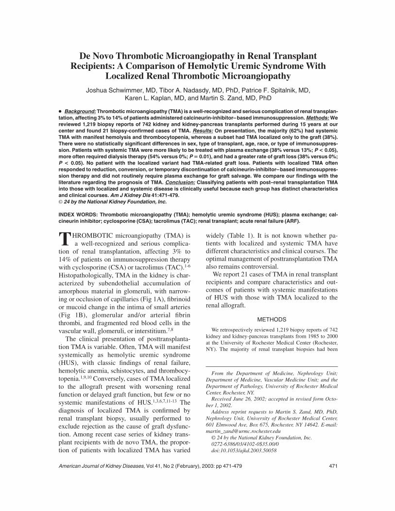
Histopathologically, TMA in the kidney is char-acterized by subendothelial accumulation ofamorphous material in glomeruli, with narrow-ing or occlusion of capillaries (Fig 1A), fibrinoidor mucoid change in the intima of small arteries(Fig 1B), glomerular and/or arterial fibrinthrombi, and fragmented red blood cells in thevascular wall, glomeruli, or interstitium.7,8
The clinical presentation of posttransplanta-tion TMA is variable. Often, TMA will manifestsystemically as hemolytic uremic syndrome(HUS), with classic findings of renal failure,hemolytic anemia, schistocytes, and thrombocy-topenia.1,9,10 Conversely, cases of TMA localizedto the allograft present with worsening renalfunction or delayed graft function, but few or nosystemic manifestations of HUS.1,3,6,7,11-13 Thediagnosis of localized TMA is confirmed byrenal transplant biopsy, usually performed toexclude rejection as the cause of graft dysfunc-tion. Among recent case series of kidney trans-plant recipients with de novo TMA, the propor-tion of patients with localized TMA has varied
widely (Table 1). It is not known whether pa-tients with localized and systemic TMA havedifferent characteristics and clinical courses. Theoptimal management of posttransplantation TMAalso remains controversial.We report 21 cases of TMA in renal transplant
recipients and compare characteristics and out-comes of patients with systemic manifestationsof HUS with those with TMA localized to therenal allograft.
METHODS
We retrospectively reviewed 1,219 biopsy reports of 742kidney and kidney-pancreas transplants from 1985 to 2000at the University of Rochester Medical Center (Rochester,NY). The majority of renal transplant biopsies had been
From the Department of Medicine, Nephrology Unit;Department of Medicine, Vascular Medicine Unit; and theDepartment of Pathology, University of Rochester MedicalCenter, Rochester, NY.
Received June 26, 2002; accepted in revised form Octo-ber 1, 2002.
Address reprint requests to Martin S. Zand, MD, PhD,Nephrology Unit, University of Rochester Medical Center,601 Elmwood Ave, Box 675, Rochester, NY 14642. E-mail:[email protected]
© 24 by the National Kidney Foundation, Inc.0272-6386/03/4102-0$35.00/0doi:10.1053/ajkd.2003.50058
American Journal of Kidney Diseases, Vol 41, No 2 (February), 2003: pp 471-479 471
performed to exclude rejection in patients with worseningrenal function or delayed graft function. Twenty-one caseswere selected based on the presence of TMA (Table 2). Forcomparison, Kaplan-Meier survival curves also were calcu-lated for the 721 transplants without a diagnosis of TMA.
Human Subjects ProtectionResearch data were coded such that subjects could not be
identified directly or through linked identifiers, in compli-ance with the Department of Health and Human ServicesRegulations for the Protection of Human Subjects (45 CFR46.101(b)(4)).
Criteria for Pathological Diagnosis of TMAA pathological diagnosis of TMAwas made based on the
presence of one or more of the following conditions: (1)arteriolar or arterial thrombi, (2) occlusion of glomerularcapillaries by amorphous material that corresponded to sub-endothelial accumulation of electron lucent to flocculent
deposits on electron microscopy, or (3) severe arterial orarteriolar endothelial widening with thickening of the glomer-ular capillaries (accumulation of subendothelial electronlucent material), particularly if associated with the presenceof fragmented red blood cells in glomeruli or arterioles. Allspecimens were examined using hematoxylin and eosin,Masson trichrome, periodic acid–Schiff, and methenaminesilver stains. In selected cases, electron microscopy wasperformed to confirm the accumulation of glomerular suben-dothelial lucent or flocculent material, or immunofluores-cence staining was performed to confirm fibrin deposition.A single pathologist reviewed all biopsy slides. Known
cases of thrombosis of large-size vessels or vascular orhumoral rejection with clinical and pathological features oflocalized TMAwere excluded because in these cases, micro-vascular pathological characteristics could be explained byischemia or acute cellular rejection alone. Cases that alsomet Banff 2000 criteria for acute cellular rejection were notexcluded. No patient had TMA or HUS as the pretransplan-tation renal disease or with a previous renal transplant.
Fig 1. Histopathologiccharacteristics of TMA in thetransplanted kidney. (A) Glo-merular capillaries occludedby amorphous hyalin mate-rial and fragmented redblood cells and (B) arterioleswith fibrinoid necrosis andintraluminal thrombi.
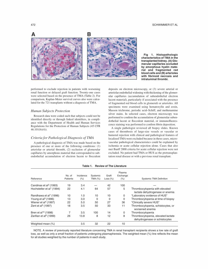
Table 1. Review of The Literature
ReferenceNo. ofPatients
Incidence(%)
SystemicTMA (%)
GraftLoss (%)
PlasmaExchange
(%) Systemic TMA Definition
Candinas et al2 (1993) 19 3.4 — 42 100 —Hochstetler et al1 (1994) 22 4.1 64 57 5 Thrombocytopenia with elevated
lactate dehydrogenase or anemiaRandhawa et al7 (1996) 10 1.0 10 10 0 “Laboratory evidence of HUS”Young et al3 (1996) 13 3.0 0 0 0 Thrombocytopenia at time of biopsyWiener et al4 (1997) 22 5.0 50 27 36 “Clinically severe HUS”Zent et al5 (1997) 18 3.5 50 33 11 Thrombocytopenia, schistocytes, or
worsened anemiaBren et al10 (1998) 7 3.5 100 14 0 ThrombocytopeniaZarifian et al6 (1999) 26 13.8 8 12 8 Thrombocytopenia, elevated lactate
dehydrogenase or schistocytes
Weighted mean (%) 3.5 32 22 10
NOTE. A review of previously reported literature concerning TMA in renal transplant recipients shows a low rate of graftloss, as well as only a small fraction of patients undergoing plasmapheresis. The weighted mean (%) line reflects the meanfor all studies weighted by the number of patients in each study.
SCHWIMMER ET AL472

Clinical Information Gathered by RetrospectiveReview of Medical Records
Recorded information included pretransplantation renaldisease; time to diagnosis of TMAposttransplantation; pres-ence of acute rejection on biopsy; immunosuppression attime of diagnosis of TMA; peak creatinine level and need fordialysis therapy during the episode of TMA; peak hemato-crit and platelet count posttransplantation; hematocrit andplatelet nadir attributed to TMA; presence of schistocytes onperipheral-blood smear; lactate dehydrogenase, total biliru-bin, and haptoglobin (when available) levels during theepisode of TMA; peak CSA and TAC levels within 10 daysbefore diagnosis; therapies for TMA; systolic and diastolicblood pressure the day of biopsy, and graft survival. Earlygraft loss related to TMA is defined as either nephrectomy orirreversible renal failure requiring dialysis therapy duringthe episode of TMA.Cases were divided into those with systemic TMA and
localized TMA. Systemic TMAis defined as thrombocytope-nia attributed to TMA (platelets �150 � 103/mL) withevidence of significant microangiopathic hemolysis (eitherschistocytes on peripheral-blood smear, haptoglobin �15mg/dL [0.15 g/L], lactate dehydrogenase �1,000 U/L, orrelative decrease in hematocrit �20%). Patients with anallograft biopsy diagnosis of TMA who lacked these sys-temic manifestations are defined as localized TMA.
Statistical AnalysisStatistical analysis was performed using the Statistica
software package (Statistica for Windows, 2000; StatSoftInc, Tulsa, OK). Comparisons of patients with localized andsystemic TMA were performed using Fisher’s exact t-test(two tailed) for categorical variables, Mann-Whitney U testfor continuous variables, and Pearson’s chi-squared test.Kaplan-Meier survival curves were compared using thelog-rank test.
RESULTS
The incidence of TMA in this case series was21 of 742 transplants (3%), consistent with theincidence in previous studies reviewed in Table 1(range, 1% to 13.8%; weighted mean, 3.5%).There were 13 cases (62%) of systemic TMAand8 cases (38%) of localized TMA. Patient data arelisted in Table 2, and patient demographics aresummarized in Table 3.TMA was attributed to TAC in 10 cases and
CSA in 9 cases (Table 2). There was no signifi-cant difference in proportions of systemic andlocalized TMA among patients treated with TACor CSA (Table 3). In 2 cases, the cause of TMAwas uncertain. In one of these patients (no. 11),
Table 2. Patient Data
Patient Age (y) Sex Race End-Stage Renal Disease DiagnosisType of
TransplantNo. of
Transplants
LossFromTMA
Follow-Up(mon)
Local vSystemic Cause
1 47 M C Type 1 diabetes CAD 1 No 55 L TAC2 24 F C Type 1 diabetes SKP 1 No 84 L TAC3 33 M AA Chronic glomerulonephritis CAD 1 No 94 L CSA4 57 M AA Type 2 diabetes LD 1 No 32 L TAC5 40 M C Type 1 diabetes CAD 1 No 26 L TAC6 42 M AA Hypertensive nephrosclerosis CAD 1 No 70 L CSA7 46 M C Renal cell carcinoma CAD 1 No 75 L CSA8 47 F C Polycystic LD 1 No 29 L Unknown9 38 F C Polycystic CAD 1 Yes 68 S CSA10 30 F C Reflux nephropathy CAD 1 No 26 S TAC11 28 M C Pauci-immune glomerulonephritis CAD 1 Yes 15 S Unknown12 49 M C Type 1 diabetes CAD 1 Yes 44 S TAC13 49 M AA Hypertensive nephrosclerosis CAD 1 Yes 27 S TAC14 56 F H Chronic glomerulonephritis CAD 1 No 137 S CSA15 43 F C Interstitial nephritis CAD 1 No 93 S CSA16 46 M C Chronic glomerulonephritis CAD 1 No 27 S TAC17 35 F C Renal cell carcinoma CAD 2 No 156 S CSA18 50 F C Polycystic LD 1 No 35 S TAC19 42 F C ImmunoglobulinA nephropathy CAD 1 No 23 S TAC20 31 F C Type 1 diabetes CAD 1 Yes 11 S CSA21 40 M C Chronic glomerulonephritis CAD 1 Yes 39 S CSA
Abbreviations: C, Caucasian; AA, African American; H, Hispanic; CAD, cadaveric transplant; LD, living donor; SKP,simultaneous kidney pancreas transplant; L, local; S, systemic.
POSTTRANSPLANT THROMBOTIC MICROANGIOPATHY 473

the allograft biopsy specimen showed TMA,immune complex deposition, and crescentic glo-merulonephritis. This case has been reportedpreviously.14 In the second patient (no. 8), TMAwas diagnosed in the setting of a history ofprevious thromboses, a known factor V Leidenmutation, and an episode of heparin-inducedthrombocytopenia. This patient underwent plasmaexchange given the prominent afferent arteriolar
and glomerular necrosis on biopsy. This was theonly case of localized TMA in this series treatedwith plasma exchange.Table 4 lists characteristics of patients with
localized and systemic TMA. Systemic TMAwas identified much earlier than localized TMA(21.5 � 19.0 versus 106.6 � 104.2 days; P �0.003). Patients with systemic and localized TMAdid not differ significantly with respect to age,
Table 3. Statistical Comparison of Patient Demographics
All (%) Local (%) Systemic (%) P (local v systemic)
No. of patients 21 8 (38) 13 (62)Age (y) 42.0 � 10.0 41.0 � 9.8 41.3 � 8.6 0.8743SexMen 11 (52) 6 (75) 5 (38) 0.1827Women 10 (48) 2 (25) 8 (62)
RaceCaucasian 15 (71) 5 (63) 10 (77) 0.4113African American 5 (24) 3 (38) 2 (15)Hispanic 1 (5) 0 (0) 1 (8)
Type of transplantCadaver 17 (81) 5 (63) 12 (92) 0.1951Living donor 3 (14) 2 (25) 1 (8)Kidney-pancreas 1 (5) 1 (13) 0 (0)
ImmunosuppressionTAC 11 (52) 4 (50) 7 (54) 0.983CSA 10 (48) 4 (50) 6 (46)
NOTE. There was no statistically significant difference in demographics between patients with local and systemic TMA.Values expressed as mean � SD or absolute number followed by percentage in parentheses.
Table 4. Comparison of Local and Systemic Posttransplantation TMA
Local (n � 8) Systemic (n � 13) P
Diagnosis (days posttransplantation) 106.6 � 104.2 21.5 � 19.0 0.0034*Pretransplant diabetic nephropathy (%) 50 15 0.0961Hematocrit (relative % decrease) 14.3 � 10.7 34.3 � 11.2 0.0007*Change in creatinine from nadir (mg/dL) 0.66 � 0.28 0.60 � 0.24 0.5960Platelet count nadir (� 1,000/mL) 224 � 113 66 � 30 0.0001*Lactate dehydrogenase peak (U/L) 659 � 578 1169 � 870 0.1834Total bilirubin peak (mg/dL) 0.49 � 0.16 1.16 � 0.44 0.0006*Haptoglobin nadir (mg/dL) 271 � 185 42.1 � 75.9 0.0237*Schistocytes (%) 13 62 0.0247*Acute rejection on biopsy (%) 50 31 0.3898Required dialysis (%) 0 54 0.0131*TMA-related graft loss (%) 0 39 0.0499*Plasma exhange (%) 13 38 0.3363Systolic blood pressure (mm Hg) 141 � 14 149 � 13 0.1368Diastolic blood pressure (mm Hg) 76 � 10 79 � 8 0.2539
NOTE. Numeric values expressed as mean � SD, and percentages for categorical values expressed with respect tonumber in either local or systemic TMA categories. To convert to SI units: creatinine (1 mg/dL � 88.4 �mol/L), bilirubin (1mg/dL � 17.1 �mol/L), haptoglobin (1 mg/dL � 0.01 g/L).*Statistically significant, P � 0.05.
SCHWIMMER ET AL474

sex, race, type of donor, incidence of hyperten-sion, and pretransplantation diagnosis. However,more patients with localized TMA had a historyof diabetic nephropathy as the primary renaldisease than patients with systemic TMA (50%versus 15%), but this did not reach statisticalsignificance (P � 0.096).
Patients with systemic TMA had a greaterincidence of both acute renal failure requiringdialysis therapy and graft loss. Of 13 patientswith systemic TMA, 7 patients (54%) requireddialysis therapy and 5 patients (38%) had TMA-related graft loss. Conversely, no patient withlocalized TMA required dialysis therapy (0%versus 54%; P � 0.013) or had graft loss (0%versus 38%; P � 0.0499). The need for hemodi-alysis therapy was a poor prognostic factor, asso-ciated with a 71% incidence of graft loss. Al-though early graft loss related to TMA wasgreater in the group with systemic TMA, long-term graft survival, including loss not related toTMA, was not significantly different betweengroups (Fig 2). Of the 2 patients with localizedTMAwho eventually lost their grafts, one had anepisode of biopsy-proven acute cellular rejec-tion, and the second had severe pyelonephritiswithout evidence of rejection. Both episodes ofgraft loss occurred long after treatment and reso-lution of TMA. Histopathologic examination ofthe allografts did not show evidence of TMA atthe time of nephrectomy, although analysis waslimited by widespread cellular rejection or bacte-rial infection.Minimization or temporary withdrawal of cal-
cineurin-inhibitor therapywas themajor therapeu-tic intervention in both the systemic- and local-TMA groups. Of the 8 patients with localizedTMA, 5 patients were treated without plasma-pheresis by decreasing the CSA or TAC dose, 2patients had no change in therapy, and only 1patient underwent plasma exchange. In the sys-temic-TMA group, 5 of 7 patients had CSAtherapy withdrawn, and 5 of 6 patients had TACtherapy withdrawn. Six of the 13 patients withsystemic TMA also were treated with OKT3.Patients with systemic TMAwere treated more
frequently with plasma exchange compared withpatients with localized TMA (38% versus 13%).However, in the group with TMA, there was nodifference in incidence of graft loss in those whounderwent plasma exchange (two of five grafts
lost) and those who did not (four of eight graftslost; P � not significant). Only one patient withlocalized TMA (13%) was treated with plasmaexchange, whereas the remainder (87%) im-proved with reduction or temporary discontinua-tion of CSA or TAC therapy alone.
DISCUSSION
Our data suggest that systemic and localizedTMA have different characteristics and clinicalcourses. Patients with systemic TMAwere signifi-cantly more likely to have renal failure requiringdialysis therapy and early graft loss. Despitethese differences, long-term graft survival ofgrafts with localized and systemic TMA wassimilarly poor (Fig 2B). Our findings are consis-tent with those of previous studies in which ahigh incidence of systemic TMA was associatedwith greater rates of early TMA-related graft loss(Table 1). Published case series composed primar-ily of systemic TMA (50% to 100%) have re-ported TMA-related graft loss rates of 14% to57%.1,4,5,10 Conversely, other series with a highproportion of patients with localized TMA (90%to 100%) noted lower rates of TMA-related graftloss (0% to 12%).3,6,7
Comparison of our findings with those ofother investigators is hampered by the absence ofa standard nomenclature for the syndrome ofposttransplantation TMA, which has been re-ferred to in the literature as posttransplantationHUS, thrombotic thrombocytopenic purpura, andTMA. We believe that the most appropriate termfor this spectrum of disorders is thrombotic mi-croangiopathy. However, this classification doesnot address the difficulty of finding localizedTMA in the setting of humoral or vascular rejec-tion. Some investigators have suggested an asso-ciation of acute vascular rejection with HUS.15 Inthis series, we excluded cases with findings ofboth TMA and acute humoral or vascular rejec-tion on biopsy, but reported them elsewhere.16
The classification, prognosis, and treatment ofthese cases in which TMA and acute humoralrejection are both present are topics for futurestudy.The uncertainty of what constitutes TMA-
induced graft loss is a major unavoidable draw-back to this and other retrospective analyses.Although early graft loss caused by TMA can bedefined easily given the temporal relationship of
POSTTRANSPLANT THROMBOTIC MICROANGIOPATHY 475

graft loss in the presence of an ongoing episodeof TMA and the absence of other factors, such ashumoral rejection, no pathognomonic featuresdistinguish graft loss occurring many monthsafter successful treatment of TMA. Severe vascu-lar damage is a hallmark of TMA and almostcertainly contributes to accelerated graft loss inthese cases. In future studies, it would be informa-tive to perform follow-up biopsies in patientswho appear to have successfully treated TMA toassess the extent of residual microvascular dam-
age.Although our data suggest that late graft lossin patients with localized TMA is not caused byrecurrence, such conclusion is problematic giventhe extensive damage from pyelonephritis orcellular rejection present in the two nephrectomyspecimens from patients with localized TMA.In our series, systemic TMA was diagnosed
earlier than localized TMA. It is unclear whetherthis indicates that systemic TMA occurs earlierafter transplantation or simply is associated withmore severe renal dysfunction leading to earlier
Fig 2. Kaplan-Meier graftsurvival in local versus sys-temic TMA. (A) A statisticallysignificant incidence of earlygraft loss caused by TMAoc-curred in systemic comparedwith localized TMA and his-toric controls (non–TMA-re-lated graft loss). (B) Graftloss from all causes showsthat the early favorable sur-vival trend in localized TMAis eventually lost, with nostatistically significant differ-ence between the twogroups (P � 0.1833; Cox’s Ftest). Graft survival in a co-hort of patients without evi-dence of TMA was substan-tially better than that in eitherlocalized or systemic TMA(P < 0.001; Cox’s F test).
SCHWIMMER ET AL476

diagnosis by renal biopsy. In our data, posttrans-plantation TMA often was localized to the kid-ney, with few or no systemic manifestations.Proportions of patients with localized and sys-temic TMA were 38% and 62%, respectively.Other investigators have reported a wide rangeof patients with systemic TMA (0% to 100%;Table 1). Reasons for this variability are notclear, but it is possible that proportions of local-ized and systemic TMA may vary at differentcenters or among physicians in relation to thresh-olds for performing renal biopsies. Additionalcases of mild or subclinical localized TMA maybe identified by more aggressive biopsy practicesfor renal allograft dysfunction. Our data indicateno difference in the absolute increase in creati-nine levels from nadir values between the local-TMA and systemic-TMA groups, making thisless likely as a cause of bias in biopsy ratesbetween groups. The effect of other factors (eg,type and degree of immunosuppression, ethnic-ity, delayed graft function) on the proportion ofcases of localized and systemic TMA at differentcenters is not known.We believe localized and systemic TMArepre-
sent a spectrum of severity of the same disorder,not two different disorders with distinct patho-physiological states. In this series, most cases ofTMA were readily classified as localized or sys-temic at the time of diagnosis, and cases oflocalized TMA never progressed to the systemicform. However, other investigators have re-ported cases in which TMA initially presented aslocalized to the kidney on biopsy, but subse-quently progressed over days to manifest sys-temic findings of HUS.13,17,18 This suggests thatlocalized and systemic TMA represent a spec-trum of the same disorder.The majority of de novo posttransplantation
cases of TMA appear related to the prothrom-botic effects of CSA or TAC,9,19,20 and this asso-ciation is highly significant in our series. Onepatient had the factor V Leiden mutation, aknown risk factor for TMA in nontransplantationindividuals and probably a risk factor for CSA-associated TMA after kidney or bone marrowtransplantation.21,22 However, we did not findother conditions proposed by some to triggerTMA, such as pretransplantation HUS,23-26 anti-cardiolipin antibodies,12 vascular rejection,2,27 or
cytomegalovirus1,28 or parvovirus B19 infec-tion.29
Temporary calcineurin-inhibitor withdrawalwas the mainstay of therapy for TMA in thisseries.10,30,31 Severe systemic cases of HUS fre-quently were treated with plasma exchange orinfusion.4,31-34 In a few cases, switching patientsto another calcineurin inhibitor resulted in recur-rence of TMA.35,36 In these patients, the substitu-tion of sirolimus for calcineurin inhibitors ormycophenolate mofetil–based immunosuppres-sion may be alternative therapies.36 In our series,two patients were treated with sirolimus in com-bination with mycophenolate mofetil with goodresults. It remains to be seen whether TMA alsooccurs with the same frequency in patients treatedwith calcineurin-inhibitor–free immunosuppres-sion regimens.Given the retrospective and uncontrolled na-
ture of our data, it is impossible to draw firmconclusions about the benefit of plasma ex-change in posttransplantation TMA. The compli-cation rate of plasma exchange may be as high as30%37; therefore, it would be useful to define asubset of patients with posttransplantation TMAin whom plasma exchange is indicated. Our datasuggest that patients with localized TMA re-spond to decreasing, changing, or temporarilydiscontinuing calcineurin-inhibitor immunosup-pressive therapy and do not routinely requireplasma exchange for graft salvage. In this study,89% of patients with localized TMA did notundergo plasma exchange, and no patient withlocalized TMAhad early TMA-related graft loss.Other investigators also reported low rates ofTMA-related graft loss (0% to 12%) in patientswith predominantly localized TMA despite theinfrequent use of plasma exchange (0% to8%).3,6,7 Conversely, reports of predominantlysystemic TMA report greater TMA-related graftloss rates (14% to 57%), even with high use ofplasma exchange.1,4,5,10
Comparison of our data with that of previousstudies has a number of important limitations.Definitions of localized and systemic TMAwereapplied retrospectively based on published pa-tient characteristics and differed among studies.Treatment strategies varied among studies, andfrom a retrospective review of other publisheddata, it is difficult to determine whether patientswith TMA-related graft loss had localized or
POSTTRANSPLANT THROMBOTIC MICROANGIOPATHY 477

systemic TMA. Despite these limitations, ourfindings are consistent with those of other inves-tigators in that: (1) localized TMA had a bettershort-term prognosis than systemic TMA; (2)renal function in almost all patients with local-ized TMA improved with reduction, conversion,or temporary discontinuation of calcineurin-inhibitor therapy; and (3) plasma exchange is notroutinely required for graft salvage in patientswith localized TMA.
REFERENCES1. Hochstetler LA, Flanigan MJ, Lager DJ: Transplant-
associated thrombotic microangiopathy: The role of IgGadministration as initial therapy. Am J Kidney Dis 23:444-450, 19942. Candinas D, Keusch G, Schlumpf R, et al: Prognostic
factors of hemolytic uremic syndrome in renal allografts.Transplant Proc 25:1041-1042, 19933. Young BA, Marsh CL, Alpers CE, Davis CL: Cyclo-
sporine-associated thrombotic microangiopathy/hemolyticuremic syndrome following kidney and kidney-pancreastransplantation. Am J Kidney Dis 28:561-571, 19964. Wiener Y, Nakhleh RE, Lee MW, et al: Prognostic
factors and early resumption of cyclosporin A in renalallograft recipients with thrombotic microangiopathy andhemolytic uremic syndrome. Clin Transplant 11:157-162,19975. Zent R, KatzA, Quaggin S, et al: Thrombotic microan-
giopathy in renal transplant recipients treated with cyclo-sporin A. Clin Nephrol 47:181-186, 19976. Zarifian A, Meleg-Smith S, O’Donovan R, Tesi RJ,
Batuman V: Cyclosporine-associated thrombotic microangi-opathy in renal allografts. Kidney Int 55:2457-2466, 19997. Randhawa PS, Tsamandas AC, Magnone M, et al:
Microvascular changes in renal allografts associated withFK506 (tacrolimus) therapy. Am J Surg Pathol 20:306-312,19968. Remuzzi G, Ruggenenti P, Bertani P: Thrombotic
microangiopathy, in Tisher CC, Brenner BM (eds): RenalPathology (ed 2). New York, NY, Lippincott Williams &Wilkins, 1994, pp 1154-11579. Remuzzi G, Bertani T: Renal vascular and thrombotic
effects of cyclosporine. Am J Kidney Dis 13:261-272, 198910. Bren AF, Kandus A, Buturovic J, et al: Cyclosporine-
related hemolytic-uremic syndrome in kidney graft recipi-ents: Clinical and histomorphologic evaluation. TransplantProc 30:1201-1203, 199811. Morozumi K, YoshidaA, Suganuma T, et al: Morpho-
logical analysis of glomerular lesions in renal transplantsimmunosuppressed with cyclosporine A (CYA): Has CYAinduced a new transplant glomerular lesion? Transplant Proc21:282-285, 198912. Baid S, Pascual M,WilliamsWW, et al: Renal throm-
botic microangiopathy associated with anticardiolipin anti-bodies in hepatitis C-positive renal allograft recipients. JAmSoc Nephrol 10:146-153, 199913. Trimarchi HM, Truong LD, Brennan S, Gonzalez JM,
Suki WN: FK506-associated thrombotic microangiopathy:
Report of two cases and review of the literature. Transplanta-tion 67:539-544, 199914. Gross M, Zand MS, Nadasdy T: Early renal allograft
loss in a patient with crescentic glomerulonephritis in thenative kidney. Am J Kidney Dis 37:202-209, 200115. Schlumpf R, Candinas D, Weder W, et al: Acute
vascular rejection with hemolytic uremic syndrome in kid-neys from non-heart-beating donors:Associated with second-ary grafts and early cyclosporine treatment? Transplant Proc25:1518-1521, 199316. Nadasdy T, Zand MS, Rabie S, et al: Overlap of acute
humoral rejection (AHR) and thrombotic microangiopathy(TMA): The value of C4D staining. J Am Soc Nephrol12:941A, 2001 (abstr)17. Pham PT, Peng A, Wilkinson AH, et al: Cyclosporine
and tacrolimus-associated thrombotic microangiopathy.Am JKidney Dis 36:844-850, 200018. Verpooten GA, Paulus GJ, Roels F, De Broe ME: De
novo occurrence of hemolytic-uremic syndrome in a cyclo-sporine-treated renal allograft patient. Transplant Proc 19:2943-2945, 198719. Curtis JJ: Renovascular elements of the cyclosporine
injury. Transplant Proc 28:2094-2096, 199620. Grupp C, Schmidt F, Braun F, et al: Haemolytic
uraemic syndrome (HUS) during treatment with cyclosporinA after renal transplantation—Is tacrolimus the answer?Nephrol Dial Transplant 13:1629-1631, 199821. Van den Berg-Wolf MG, Kootte AM, Weening JJ,
Paul LC: Recurrent hemolytic uremic syndrome in a renaltransplant recipient and review of the Leiden experience.Transplantation 45:248-251, 198822. Raife TJ, Lentz SR,Atkinson BS, Vesely SK, Hessner
MJ: Factor V Leiden: A genetic risk factor for thromboticmicroangiopathy in patients with normal von Willebrandfactor-cleaving protease activity. Blood 99:437-442, 200223. Schwarz A, Krause PH, Offermann G, Keller F:
Recurrent and de novo renal disease after kidney transplan-tation with or without cyclosporine A. Am J Kidney Dis17:524-531, 199124. Scantlebury VP, Shapiro R, McCauley J, et al: Renal
transplantation under cyclosporine and FK506 for hemolyticuremic syndrome. Transplant Proc 27:842-843, 199525. Miller RB, Burke BA, Schmidt WJ, et al: Recurrence
of haemolytic-uraemic syndrome in renal transplants: Asingle-centre report. Nephrol Dial Transplant 12:1425-1430,199726. Hariharan S, Adams MB, Brennan DC, et al: Recur-
rent and de novo glomerular disease after renal transplanta-tion:Areport fromRenalAllograft Disease Registry (RADR).Transplantation 68:635-641, 199927. Asaka M, Ishikawa I, Nakazawa T, et al: Hemolytic
uremic syndrome associated with influenzaA virus infectionin an adult renal allograft recipient: Case report and reviewof the literature. Nephron 84:258-266, 200028. Waiser J, Budde K, Rudolph B, Ortner MA, Neu-
mayer HH: De novo hemolytic uremic syndrome postrenaltransplant after cytomegalovirus infection. Am J Kidney Dis34:556-559, 199929. Murer L, Zacchello G, Bianchi D, et al: Thrombotic
microangiopathy associated with parvovirus B19 infection
SCHWIMMER ET AL478
after renal transplantation. J Am Soc Nephrol 11:1132-1137,200030. Buturovic J, Kandus A, Malovrh M, Bren A, Dri-
novec J: Cyclosporine-associated hemolytic uremic syn-drome in four renal allograft recipients: Resolution withoutspecific therapy. Transplant Proc 22:1726-1727, 199031. Katznelson S, Wilkinson A, Rosenthal TR, et al:
Cyclosporine-induced hemolytic uremic syndrome: Factorsthat obscure its diagnosis. Transplant Proc 26:2608-2609,199432. Venkat KK, Tkach D, Kupin W, et al: Reversal of
cyclosporine-associated hemolytic-uremic syndrome by plasmaexchange with fresh-frozen plasma replacement in renal trans-plant recipients. Transplant Proc 23:1256-1257, 199133. Franz M, Regele H, Schmaldienst S, et al: Posttrans-
plant hemolytic uremic syndrome in adult retransplanted
kidney graft recipients:Advantage of FK506 therapy? Trans-plantation 66:1258-1262, 199834. Kaplan AA: Therapeutic apheresis for renal disor-
ders. Ther Apher 3:25-30, 199935. Abraham KA, Little MA, Dorman AM, Walshe JJ:
Hemolytic-uremic syndrome in association with both cyclo-sporine and tacrolimus. Transpl Int 13:443-447, 200036. Said T, al-Mousawi M, Samhan M, Lao M: Cyclo-
sporin conversion to CellCept in a cadaveric renal allograftrecipient with hemolytic uremic syndrome. Transplant Proc31:3295-3297, 199937. Rizvi MA, Vesely SK, George JN, et al: Complica-
tions of plasma exchange in 71 consecutive patients treatedfor clinically suspected thrombotic thrombocytopenic pur-pura-hemolytic-uremic syndrome. Transfusion 40:896-901,2000
POSTTRANSPLANT THROMBOTIC MICROANGIOPATHY 479