cytotherapy (2009) vol. 11, no. 6, 706–715 an … (2009) vol. 11, no. 6, 706–715 © 2009 isct...
TRANSCRIPT

Cytotherapy (2009) Vol. 11, No. 6, 706–715
Cyt
othe
rapy
Dow
nloa
ded
from
info
rmah
ealth
care
.com
by
Mr
Paul
Her
chm
an o
n 02
/01/
12Fo
r pe
rson
al u
se o
nly.
An alternative method for the isolation of mesenchymal stromal cells derived from
lipoaspirate samplesLeandra S. Baptista1,2, Ronaldo J. F. C do Amaral1,3, Rosana B. V. Carias 4,
Marcelo Aniceto5, Cesar Claudio-da-Silva5 and Radovan Borojevic1,2,3
1APABCAM, Clementino Fraga Filho University Hospital, Federal University of Rio de Janeiro, RJ, Brazil, 2DIPRO, INMETRO, Rio de
Janeiro, RJ, Brazil, 3Institute of Biomedical Sciences, Federal University of Rio de Janeiro, RJ, Brazil, 4Excellion Biomedical Services, Petrópolis,
RJ, Brazil, and 5Medical Clinics Department, Clementino Fraga Filho University Hospital, Federal University of Rio de Janeiro, RJ, Brazil
Correspondence to: Leandra S. Baptista, APABCAM, Clementino Fraga Filho University Hospital, Federal University of Rio de Janeiro, Cidade Universitária, Ilha do Fundão, 21941–902 Rio de Janeiro, RJ, Brazil. E-mail: [email protected].
Background aims
Since initial methods were developed for isolating cells from adipose
tissue, little has been done to improve mesenchymal stromal cell
(MSC) yield. The aim of the present study was to isolate a population
of MSC from lipoaspirate samples without tissue digestion and to
assess the possibility of cryopreserving the freshly isolated cells.
Methods
A population of MSC was isolated from 13 patients’ lipoaspirate
samples by mechanical dissociation. Mechanically processed lipoaspi-
rate adipose tissue (MPLA) cells were characterized after in vitro cell expansion by morphologic analysis, expression of MSC surface
markers and differentiation assays.
Results
Mechanical dissociation yielded a large quantity of adherent MSC
both after standard and vibro-assisted liposuction. Preservation of
lipoaspirate samples at 4°C for 1 or 2 days until the mechanical
© 2009 ISCT
procedure did not change the MPLA cell content. It was possible to
store freshly isolated MPLA cells by cryopreservation without loss
of the MSC population. Adherent MPLA cells were negative for
CD45 and CD31 and positive for CD34, CD105, CD44 and
CD90. They also showed adipogenic, osteogenic and chondrogenic
potentials similar to MSC populations from other sources as already
described in the literature.
Conclusions
MSC can be isolated from human lipoaspirate samples by the
mechanical procedure described in this study with a signifi cant reduc-
tion in time and cost. Together with cryopreservation of freshly iso-
lated MPLA cells, this has made it easier to harvest and store MSC
for therapeutic applications such as soft-tissue augmentation and tis-
sue engineering.
Keywords
lipoaspirate, mesenchymal stromal cells, tissue engineering.
IntroductionThe methods for isolating cells from adipose tissue were initially proposed by Rodbell and colleagues in the mid-1960s [1]. They minced rat fat pads, washed them exten-sively to remove contaminating blood cells, incubated the tissue fragments with collagenase and centrifuged the digest, separating the fl oating population of mature adipo-cytes from the pelleted stromal cells. The population of stromal cells includes adipocytes, pre-adipocytes, endothe-lial cells, macrophages and fi broblasts [2]. Similar to the
bone marrow (BM) stroma, a subpopulation of multipo-tent mesenchymal stromal cells (MSC) also dwells in the adipose stroma. They are excellent candidates for tissue engineering of bone, cartilage, skin and other tissues [3].
In initial studies, fragments of human tissue were manually minced. The development of liposuction surgery simplifi ed the process of cell harvesting, as it generates fi nely minced tissue [4], allowing incubation of the fat sample directly with collagenase in order to isolate MSC. Liposuction procedures also improve the soft-tissue
DOI: 10.3109/14653240902981144
An alternative method for isolating MSC 707
Cyt
othe
rapy
Dow
nloa
ded
from
info
rmah
ealth
care
.com
by
Mr
Paul
Her
chm
an o
n 02
/01/
12Fo
r pe
rson
al u
se o
nly.
augmentation techniques using immediate autologous fat transfer. This is an attractive therapy, the success of which depends upon the techniques of harvesting, cleans-ing and reinjection of cells [5]. Recently, Yoshimura et al. [6] achieved optimal human MSC yield, resulting in subsequent formation of adipose tissue following implan-tation in immune-defi cient mice.
In tissue engineering, synthetic or tissue-derived scaf-folds can provide mechanical support to cells, promoting their differentiation, proliferation and tissue growth. MSC have been described as a better source for tissue engineer-ing than differentiated cells, such as chondrocytes or osteo-blasts. This approach requires large numbers of MSC. For example, an in vivo chondro-induction requires at least 107 cells/cm3 of scaffold [7]. Several donor sites can be used for harvesting MSC, BM and adipose tissue being the most frequent . The latter is easily harvested by liposuction and, in most patients, a large quantity of MSC can be obtained without harm to the donor [2]. Liposuction is frequently done for aesthetic reasons, allowing using MSC for reparative processes extemporaneously. They can be also cryopreserved and stored for future use.
The enzymatic digestion that is the current protocol for isolating MSC limits the volume of lipoaspirate that can be processed for two main reasons: diffi culties of the method per se when applied to large volumes, and the high cost of collagenase. The aim of the present study was to isolate a population of MSC from lipoaspirates with simultaneous red blood cell lysis without tissue digestion, in order to reduce both time and procedure costs. The mononuclear cell population obtained by mechanical procedure contains blood, endothelial and stromal cells. Cryopreservation was tested in this fresh mononuclear cell population to avoid in vitro expansion before a thera-peutic approach. The adherent cell fraction was character-ized after in vitro cell expansion by morphologic analysis and expression of MSC surface markers. The prolifera-tion profi le and adipogenic, osteogenic and chondrogenic potentials were monitored. Cells isolated in this study by the mechanical procedure showed MSC proprieties similar to lipoaspirate cells isolated by the enzymatic procedures of others [3] as well as our group [8].
MethodsHuman lipoaspirate sampling Lipoaspirates were harvested by vibro-assisted and standard liposuction from 13 healthy female donors that
underwent abdominal liposuction, after approval given by the research ethics committee of the University Hospital Clementino Fraga Filho, Federal University of Rio de Janeiro, RJ, Brazil. All donors signed an informed consent form. Adipose tissue samples were preserved at 4°C after surgery until the mechanical processing.
Isolation and culture of mechanically and enzymatically processed lipoaspirateAdipose tissue samples were distributed into tubes con-taining ACK buffer solution (1/1, v/v). The mixture was shaken and maintained at 37°C for 15 min for red blood cell lysis. Cells were separated from adipose tissue frag-ments, oil and debris by centrifugation at 900 g for 15 min at room temperature. The pellets obtained were resus-pended in fetal bovine serum (FBS; Cultilab, Campinas, SP, Brazil) supplemented with 10% dimethylsulfoxide (DMSO; Sigma Chemical Co., St Louis, MO, USA) for cryopreservation (107 cells/mL). The vials were kept over-night at –70°C, transferred and kept at –196°C until thawing. Alternatively, they were resuspended and plated into culture dishes (105 cells/cm2) in low-glucose Dulbec-co’s modifi ed Eagle’s medium (DMEM; LGC, Cotia, SP, Brazil) containing 20% FBS, 100 U/mL penicillin and 100 μg/mL streptomycin. Samples obtained by vibro-assisted and standard liposuction followed the same proto-col in order to compare the isolated cell populations.
For cell thawing, the vials were placed at 37°C until the ice had been thawed. Approximately 10 mL fresh DMEM was added to the cell suspension from each vial and centri-fuged at 400 g for 7 min. The cell pellets were resuspended in DMEM containing 20% FBS, 100 U/mL penicillin and 100 μg/mL streptomycin and plated into culture dishes (105 cells/cm2). Cultures of fresh and thawed cells were maintained at 37°C in a humid atmosphere containing 5% CO2, and the medium was changed every 3–5 days until cells reach pre-confl uence. Adherent cells were detached with 0.78 mm EDTA and 0.125% trypsin (Gibco BRL, Rockville, MD, USA) and cell suspension was centrifuged at 400 g for 7 min, counted and replated into culture dishes (104 cells/cm2). This was considered to be ‘one passage’. For monitoring cell proliferation, cells in the third and 10th passages were plated (104 cells/well; n � 3) in 24-well plates and cultured in DMEM containing 20% FBS for up to 2 weeks. At days 1, 3, 6, 9, 12 and 15, viable cells were quantifi ed using trypan blue staining, which allows dead cell exclusion.

708 L. S. Baptista et al.
Cyt
othe
rapy
Dow
nloa
ded
from
info
rmah
ealth
care
.com
by
Mr
Paul
Her
chm
an o
n 02
/01/
12Fo
r pe
rson
al u
se o
nly.
The same lipoaspirate samples underwent mechanical and enzymatic processes in order to compare the cell pop-ulation yield. Adipose tissue samples were incubated with collagenase IA (Sigma Chemical Co.). Cells were harvested by centrifugation and plated in tissue culture fl asks with low-glucose DMEM containing 20% FBS, 100 U/mL penicillin and 100 μg/mL streptomycin. Cultures were maintained at 37°C in a humid atmosphere with 5% CO2, and the medium was changed every 3–5 days until cells reach pre-confl uence.
Flow cytometry analysisCells were monitored for surface marker expression at the moment they were isolated, and at the fi rst and third passages (n � 3 for each analysis) using fl ow cytometry. Cells were washed with phosphate-buffered saline con-taining 3% bovine serum albumin (PBS-BSA 3%) and incubated for 30 min at 4°C with monoclonal antibodies conjugated with fl uorescent dies: CD14–phycoerythrin (PE), CD16–PE–Cy7, CD31–PE, CD34–APC, CD34–PerCP–Cy5.5, CD44–PE, CD45–fl uroescein isothiocya-nate (FITC), CD73–PE, CD90–APC (BD Biosciences, Franklin Lakes, NJ, USA) , CD105–FITC and CD105–PE (R&D Systems, Minneapolis, MN, USA). Subse-quently, cells were washed with PBS-BSA 3%. When necessary, the cell suspensions were incubated at room temperature in the absence of light for 10 min with FACS lysing solution (BD Biosciences). Flow cytometry analyzes were performed using a FACScanto (BD Biosciences) equipped with the software FACS Diva 4.0.
Cell differentiation assaysOsteogenic and adipogenic potentials were investigated using the appropriate inducing media (n = 3, for each assay), as described previously [3]. Briefl y, for adipogenic assays, cells were cultured in DMEM containing 20% FBS, 10 μm insulin (Biohulin, Montes Claros, MG, Brazil), 0.5 μm isobutylmethylxanthine (IBMX), 1 μm dexamethasone and 200 μm indomethacin (Sigma) for 14 days. For osteogenic assays, cells were cultured in DMEM containing 10% FBS, 5 � 10–6 m ascorbic acid, 0.01 μm dexamethasone and 10–2 m β-glycerolphosphate (Sigma) for 14 days.
Following lineage differentiation induction, cells were fi xed in 10% formaldehyde for 60 min. Adipogenic differ-entiation was assessed using Oil Red O staining (Sigma) as an indicator of intracellular lipid accumulation. Cells were pre-incubated in propylene glycol (Merck, Whitehouse
Station, NJ, USA), incubated in 0.5 mg/mL Oil Red O solution for 30 min at room temperature and excess staining removed with 85% propylene glycol. Osteogenic differentiation was assessed by a modifi ed Von Kossa’s method [3] as an indicator of extracellular calcium deposi-tion. Cells were rinsed with distilled water and then covered with a 2% silver nitrate solution (Sigma), in the absence of light, for 60 min. The cells were washed several times with distilled water and the reaction was developed under ultraviolet (UV) light for 30 min.
Chondrogenic potential was monitored in pellet cul-tures as described previously [9]. Briefl y, 1.5 � 105 cells were centrifuged at 400 g in 15-mL polypropylene conical tubes and the resulting pellets cultured for 21 days. To induce chondrogenic differentiation, cultures were grown in human MSC chondrogenic differentiation BulletKit supplemented with 10 ng/mL recombinant human trans-forming growth factor (TGF)-β3 (Cambrex, Charles City, IA, USA). Control cultures were grown in the same medium without dexamethasone and TGF-β3. After 21 days, pellet cultures were fi xed in 10% formaldehyde for 6 h and embedded routinely in paraffi n. Paraffi n sections were stained with periodic acid-Schiff (PAS) with nuclei coun-terstained by hematoxylin, or with alcian blue at pH 1.0 with nuclei counterstained by neutral red.
Statistical analysisUnpaired and non-parametric anova tests were performed when necessary to evaluate differences between groups. Results are expressed as mean � standard deviation (SD) and P-values less than 0.05 were considered statistically signifi cant.
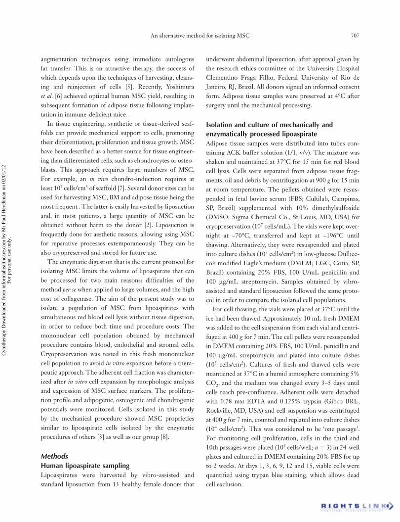
ResultsEx vivo isolation of MSC with a minor red blood cell contaminationMechanical dissociation with simultaneous lysis buffer treat-ment resulted in ex vivo cell suspensions, called mechani cally processed lipoaspirate adipose tissue (MPLA), that con-tained two different major mononuclear cell subpopulations, CD45� and CD45�. Cells positive for CD45, a pan-he-matopoietic marker, were also positive for CD16, CD14, CD31, surface markers of granulocytes, monocytes–mac-rophages and endothelial cells (Figure 1B–F). They were also positive for CD44, a cell-adhesion molecule that participates in a wide variety of cellular functions, inclu-ding lymphocyte activation, recirculation and homing,
An alternative method for isolating MSC 709
Cyt
othe
rapy
Dow
nloa
ded
from
info
rmah
ealth
care
.com
by
Mr
Paul
Her
chm
an o
n 02
/01/
12Fo
r pe
rson
al u
se o
nly.
hematopoiesis and tumor metastasis [10]. As CD45� cells present in lipoaspirate samples are peripheral blood con-taminants, they were removed after plating by washing, leaving a population of adherent cells with fi broblastoid morphology. In this study, this population was called adherent MPLA cells. The presence of macrophages in the adherent population was very low. Being resistant to trypsin, they were lost at replating, as shown by the absence of cells bearing macrophage markers at fl ow cytometry analysis. These adherent MPLA cells were negative for CD45 and CD73 and positive for CD44, CD31, CD90 and CD105 (Figure 1G–J), surface markers described in MSC popula-tions of different origins. They were also positive for CD34, which is reported only in adipose tissue-derived mesenchymal cells [11].
The mechanical process was accomplished in a reduced time compared with the enzymatic digestion, and it was
possible to isolate viable cells from lipoaspirate samples harvested by distinct liposuction procedures. The total number of MPLA cells harvested from the vibro-assisted lipoaspirates samples was superior to that obtained from the standard one. Adherent MPLA cells from these two samples showed no difference in adhesion and prolifera-tion (data not shown).
Lipoaspirate samples can be stored without damage to MPLA cellsIn order to monitor possible damage or loss of MPLA cells during their storage and transportation from surgery facilities to a cell-processing unit, lipoaspirate samples were stored at 4°C for up to 2 days in a lipoaspirate collection recipient containing approximately 10% fresh DMEM. Total MPLA cells were subsequently cultured in low-glucose DMEM supplemented with 20% FBS and
Figure 1. Phenotypic characterization of freshly isolated total MPLA cells. (A) FSC/SSC distribution of all MPLA cells that can be
subdivided into two subpopulations, P1 and P2. (B) P1 is basically constituted by blood cells (CD45+) gated in P3. The histogram of cells
within P3 is shown in (C–F), and outside P3 is shown in (G–L). Note that CD45– cells express MSC surface markers except CD73 (L).
Isotype controls are overlaid in all histograms.

710 L. S. Baptista et al.
Cyt
othe
rapy
Dow
nloa
ded
from
info
rmah
ealth
care
.com
by
Mr
Paul
Her
chm
an o
n 02
/01/
12Fo
r pe
rson
al u
se o
nly.
antibiotics at 37°C in 5% CO2 atmosphere. Although the number of total MPLA cells did not change, the number of adherent MPLA cells isolated from preserved samples (for 1–2 days) was reduced (not statistically signifi cant) compared with fresh samples (samples preserved at 4°C for up to 4 h) (Table I and Figure 2). This could be attrib-uted to a reduction of the total MPLA cell adhesion and/or viability, but no change in the isolated adherent cells morphology was observed. Lipoaspirate samples obtained by the enzymatic process, called here adherent MPLA cells, yielded a higher number of cells (Table II and Figure 2).
We also monitored the possibility of cryopreserving the total MPLA cells for a long time, without a previous in
vitro expansion. Immediately after the mechanical proce-dure, fresh MPLA cells were cryopreserved and, after thawing, morphologic (Figure 3A) and phenotype charac-teristics (Figure 3B) of the adherent fraction were the same as the non-cryopreserved MPLA cells.
Adherent MPLA cells showed in vitro MSC proprieties When adherent MPLA cells reached pre-confl uence, they were harvested and replated in low-glucose DMEM con-taining 20% FBS and antibiotics until the 10th passage. In
vitro expansion altered neither the MPLA cell prolifera-tion (Figure 4A) nor their spindle-shaped morphology (Figure 4B,C). Adherent expanded MPLA cells were neg-ative for CD45 (Figure 5A), as expected, as well as the mature endothelial cell surface marker CD31 (Figure 5B). They were positive for MSC-associated markers CD105 (Figure 5D), CD44 (Figure 5E) and CD90 (Figure 5F), which were up-regulated during in vitro expansion. At the third passage, CD34 expression was strongly reduced (Figure 5C). For differentiation assays, adherent MPLA cells were cultured under adipogenic- or osteogenic-inducing medium for 14 days. As expected, MPLA cells were able to accumulate lipid droplets (Figure 6B) and deposit extracellular calcium (Figure 6D), which were both observed under the microscope by specifi c stainings. MPLA cell pellets formed solid three-dimensional tissue structures that could be harvested and processed for histology. In the presence of TGF-β3, pellets grew in size, almost doubling the control cultures (Figure 6E,F). This growth could be attributed to cartilage matrix accumulation, as shown by alcian blue staining, which reacts mainly with sulfated proteoglycans (Figure 6H).
Discussion We have described a novel method of isolating MSC, derived from lipoaspirate samples, based on mechanical tissue dissociation. The usual enzymatic digestion proce-dure for adipose tissue consists of at least four main steps: washing, digestion, centrifugation and red blood cell lysis. This procedure generates tissue fragments that should be removed before cell plating through a 100–150 μm nylon mesh. Conversely, mechanical dissociation consists basically of two steps, dissociation of adipose tissue concomitantly with red blood cell lysis, followed
Figure 2. Graph representing the mean values of adherent MPLA
cells obtained from fresh and preserved lipoaspirate samples using
mechanical dissociation, and of PLA cells from fresh lipoaspirate samples
using enzymatic dissociation. No statistical signifi cance was detected
among the fresh and preserved lipoaspirate samples or between preserved
lipoaspirate samples (P � 0.05). Statistical signifi cance was only detected
among adherent MPLA and PLA cell numbers (enzymatic dissociation
versus fresh lipoaspirate sample using mechanical dissociation, P � 0.05;
enzymatic dissociation versus the preserved lipoaspirate samples for 1
and 2 days using mechanical dissociation, P � 0.01).
Table I. Number of total MPLA cells and adherent MPLA cells obtained from fresh and preserved lipoaspirate samples by mechanical dissociation. Data represent the mean ± SD of fi ve samples.
Number of MPLA cells per milliliter of Number of adherent MPLA cells per lipoaspirate sample (�104) milliliter of lipoaspirate sample (�104)
Fresh lipoaspirate sample 24.0�7.4 1.2�0.37Preserved lipoaspirate sample for 1 day 22.6�10.1 0.51�0.2Preserved lipoaspirate sample for 2 days 18.9�9.1 0.39�0.18

An alternative method for isolating MSC 711
Cyt
othe
rapy
Dow
nloa
ded
from
info
rmah
ealth
care
.com
by
Mr
Paul
Her
chm
an o
n 02
/01/
12Fo
r pe
rson
al u
se o
nly.
by centrifugation. There are no visible tissue fragments, and it is not necessary to the fi lter cell suspension. The ease of mechanical digestion reduces considerably both time and cost, and does not interfere with cell viability.
Compared with the enzymatic process, the mechanical process generates a signifi cantly lower number of adher-ent MPLA cells. However, compared with other studies, the MSC yield by enzymatic process [12] remains close to the adherent MPLA cells. Moreover, processes based on enzymatic digestion seem to be less reproducible, as there is no consensus in the literature regarding the MSC yield. We observed a large standard derivation among cell numbers isolated with the enzymatic diges-tion process. Adding reproducibility to the low costs, the mechanical process is proposed as a preferable method for large volumes of samples. The reproducibility of the mechanical process described here should be tested for a larger number of lipoaspirate samples and by other research groups.
This alternative procedure was also tested with lipoaspi-rate samples preserved at 4°C for up to 2 days. Recently, it has been shown that adipose tissue-derived stem cell yield from tissue digests is signifi cantly reduced by preservation at room temperature for 24 h and even by preservation at 4°C for 2–3 days [13]. In our study, preservation for 1–2 days did not make a difference in surface marker profi le or the number of total MPLA cells. Moreover, no signifi cant difference was observed among adherent MPLA cells isolated from fresh and preserved lipoaspirate samples, and after 2 weeks it was possible to obtain monolayers of MSC from all samples.
Liposuction surgery often generates large volumes of samples to be processed, so it is important not to waste them. In order to facilitate preservation of total MPLA cells for a long time, they were cryopreserved immediately after being isolated. It was possible to thaw them without loss of cell viability. Interestingly, another study has investigated a method for cryopreserving human adipose-derived stem cells isolated by an enzymatic process. Fresh human cells
Figure 3. Culture of thawed MPLA cells. (A) Adherent cells showed a typical morphology and (B) expression of mesenchymal surface
markers. (C) Isotype control. Bar size, 100 mm.
Table II. Number of total PLA cells and adherent PLA cells obtained from fresh lipoaspirate samples by enzymatic dissociation. Data represent the mean ± SD of fi ve samples.
Number of PLA cells per milliliter of Number of adherent PLA cells per lipoaspirate sample (�104) milliliter of lipoaspirate sample (�104)
Fresh lipoaspirate sample 58.4�17.8 8.5�6.7

712 L. S. Baptista et al.
Cyt
othe
rapy
Dow
nloa
ded
from
info
rmah
ealth
care
.com
by
Mr
Paul
Her
chm
an o
n 02
/01/
12Fo
r pe
rson
al u
se o
nly.
were cryopreserved using Me2SO as the cryoprotective agent at a density of 106 cells/mL [14], 10 times lower than the cell quantity cryopreserved in our study. Cryopreserva-tion is interesting because it reduces labor costs and avoids possible loss of cell viability and senescence after long-term cultures [15,16]. It is possible to thaw total fresh MPLA cells as needed for cell therapy approaches.
The yield of adipose tissue cells derived from liposuction procedures depends directly upon the surgical procedure. It has been reported for ultrasound-assisted liposuction that the number of MSC recovered from tissue digests is reduced, as is their proliferative capacity [17]. In the present study, MSC could be isolated from vibro-assisted liposuction, maintaining their biologic properties similar to the standard liposuction procedure. Vibro-assisted liposuction has already been reported to reduce the duration of surgery because of its large rate of aspiration [18]. Using a mechanical approach with vibro-assisted lipoaspirate samples, it was possible to obtain a large MSC population, not described for enzymatic processes.
The loss of cell viability caused by the use of enzymes such as collagenase during cell harvesting and processing has already been described in several protocols of primary cell cultures. Rodeheaver et al. [19] observed a loss of tubu-lar morphology and an increase in the rate of Na-K-ATPase activity in rabbit renal proximal tubules subjected to incu-bation with enzymes that were attributed to the disruption
Figure 4. The proliferation of adherent MPLA cells was evaluated
by trypan blue exclusion assay. (A) No considerable differences in cell
numbers generated by adherent MPLA cells in low and high passages.
Monolayer of adherent MPLA cells cultured in the presence of 20%
FBS in low (B) and high (C) passages. Bar size, 100 mm.
Figure 5. Surface marker profi le of adherent MPLA cells harvested after fi rst (green solid histograms) and third passage (blue solid histograms).
The population of adherent MPLA cells was negative for CD45 (A) and CD31 (B). Expression of CD34 (C) was down-regulated, unlike CD105
(D), CD44 (E) and CD90 (F) that were up-regulated at the third passage. Gray solid histograms represent negative controls (isotype controls).

An alternative method for isolating MSC 713
Cyt
othe
rapy
Dow
nloa
ded
from
info
rmah
ealth
care
.com
by
Mr
Paul
Her
chm
an o
n 02
/01/
12Fo
r pe
rson
al u
se o
nly.
of the tubular basement membrane. Recently, it has been shown that isolation of chondrocytes by enzymatic diges-tion disturbs their viability and differentiated phenotype [20]. We hypothesize that the described mechanical pro-cess of cell harvesting does not chemically destabilize or disrupt cell membranes, and for this reason it was possible to obtain a full cell yield even from vibro-assisted and not fresh (preserved) lipoaspirate samples. We believe that the process based on mechanical dissociation also explains the successful cryopreservation of freshly isolated cells.
Flow cytometry analysis revealed that the mechanical procedure was able to isolate CD45– cells with a profi le of surface markers typical of MSC [21], except for the absence of CD73 and presence of CD34. The lack of CD73 expression may refl ect a specifi c characteristic of MPLA cells. In addition, MSC when in culture adhere to plastic dishes, which can modulate their expression of surface markers [8]. With respect to CD34, several studies have described it as a surface marker for MSC from adipose tis-sue [8,11], in agreement with our study. We also found a
Figure 6. Adherent MPLA cells showed in vitro multipotentiality. Monolayers of cells were maintained under adipogenic or osteogenic
inducing media for 14 days (A–D). After this period, they were fi xed and stained with Oil Red O to show the lipid droplets, and with
the Von Kossa method to reveal calcium deposits. Control media (A, C); inducing media (B, D). Pellet cultures formed by adherent MPLA
cells under control (A, C) and chondrogenic (B, D) media for 21 days. (A, B) PAS staining with nuclei stained by hematoxylin. (C, D)
Alcian blue staining with nuclei stained by neutral red. Matrix accumulation is typical of cartilage. Bar size, 100 mm.

714 L. S. Baptista et al.
Cyt
othe
rapy
Dow
nloa
ded
from
info
rmah
ealth
care
.com
by
Mr
Paul
Her
chm
an o
n 02
/01/
12Fo
r pe
rson
al u
se o
nly.
slightly increased expression of MSC-associated markers and a down-regulation of CD34 by adherent MPLA cells after several passages in vitro, in accordance with pre-viously reported data [8,22]. Moreover, we observed the same surface marker profi le on adherent MPLA cells isolated from preserved lipoaspirate samples.
Differentiation assays to adipogenic, osteogenic and chondrogenic lineages support the idea that adherent MPLA cells from fresh and preserved lipoaspirate samples are a population of MSC. At third or fourth passages these cells were able to accumulate lipid droplets, calcium depos-its and cartilage matrix under appropriate inducing media in a manner similar to MSC from adipose tissue isolated by enzymatic digestion procedures [3,8].
Autologous progenitor cells, such as those derived from BM or adipose tissue, can be used clinically for regenerative cell therapy or for tissue engineering only when isolated in a reproducible manner and in suffi cient quantities. Despite the major differences between the enzymatic and mechanic processes, similar populations of MSC have been isolated. MPLA cells can be processed easily from lipoaspirate samples and provide a signifi cant quantity of adherent cells with minor time and costs for the procedure. Furthermore, MPLA proprieties support their use for diverse therapeutic applications. The possi-bility of cryopreservation of freshly isolated MPLA cells abrogates culture-associated changes found in cells after prolonged expansion, and provides the possibility of generating extemporaneously a large stock of cells using a relatively simple method.
AcknowledgmentsThis work was supported by CNPq and FAPERJ grants of the Brazilian Ministry of Science and Technology. We are grateful to our colleague Professor Valéria Mello-Coelho for critical review of the manuscript and to patients for donating and providing liposuction waste samples to this study.
Declaration of interest: The authors report no confl icts of interest. The authors alone are responsible for the content and writing of the paper.
ReferencesRodbell 1. M. Metabolism of isolated fat cells. II. The similar effects of phospholipase C (Clostridium perfringens alpha toxin) and of insulin on glucose and amino acid metabolism. J Biol Chem 1966;241:130–9.
Casteilla 2. L, Charrière G, Laharrague P, Cousin B, Planat-Benard V, Péricaud L, Chavoin JP. Tissus adipeux, chirurgie plastique et reconstructrice: le retour aux sources. Ann
Chirurg Plastiq Esthétiq 2004;49:409–18.Zuk P3. A, Zhu M, Mizuno H, Huang J, Futrell JW, Katz AJ, et al. Multilineage cells from human adipose tissue: implications for cell-based therapies. Tissue Eng
2001;7:211–28. Illouz Y4. G. Body contouring by lipolysis: a 5-year experience with over 3000 cases. Plast Reconstr Surg 1983;72:591–7.Mojallal 5. A, Foyatier JL. Historical review of the use of adipose tissue transfer in plastic and reconstructive surgery. Ann Chir Plast Esthet 2004;49:419–25. Yoshimura 6. K, Shigeura T, Matsumoto D, Sato T, Takaki Y, Aiba-Kojima E, et al. Characterization of freshly isolated and cultured cells derived from the fatty and fl uid portions of liposuction aspirates. J Cell Physiol 2006;208:64–76.Raghunath J, Salacinski HJ, Sales KM, Butler PE, 7. Seifalian AM. Advancing cartilage tissue engineering: the application of stem cell technology. Curr Opin Biotechnol 2005;16:503–9.Baptista LS, Pedrosa CGS, Silva KR, Otazú IB, Takyia CM, 8. Dutra HS, et al. Bone marrow and adipose tissue-derived mesenchymal stem cells: how close are they? J Stem Cells
2007;2. Johnstone B, Hering TM, Caplan AI, Goldberg VM, 9. Yoo JU. In vitro chondrogenesis of bone marrow-derived mesenchymal progenitor cells. Exp Cell Res 1998;238:265–72.Orian-Rousseau V, Ponta H. Adhesion proteins meet recep-10. tors: a common theme? Adv Cancer Res 2008;101:63–92.Planat-Benard V, Silvestre JS, Cousin B, André M, 11. Nibbelink M, Tamarat R, et al. Plasticity of human adipose lineage cells toward endothelial cells: physiological and therapeutic perspectives. Circulation 2004;109:656–63.Peroni 12. D, Scambi I, Pasini A, Lisi V, Bifari F, Krampera M, et al. Stem molecular signature of adipose-derived stromal cells. Exp Cell Res 2008;314:603–15.Matsumoto 13. D, Shigeura T, Sato K, Inoue K, Suga H, Kato H, et al. Infl uences of preservation at various temperatures on liposuction aspirates. Plast Reconstr Surg 2007;120:1510–7.Liu G, Zhou H, Li Y, Li G, Cui L, Liu W, Cao Y. Evaluation 14. of the viability and osteogenic differentiation of cryo-preserved human adipose-derived stem cells. Cryobiology 2008;57:18–24. Serakinci N, Guldberg P, Burns JS, Abdallah B, Schrødder H, 15. Jensen T, Kassem M. Adult human mesenchymal stem cell as a target for neoplastic transformation. Oncogene 2004;23:5095–8.Rubio 16. D, Garcia-Castro J, Martín MC, de la Fuente R, Cigudosa JC, Lloyd AC, Bernad A. Spontaneous human adult stem cell transformation. Cancer Res 2005;65:3035–9.Oedayrajsingh-Varma M17. J, van Ham SM, Knippenberg M, Helder MN, Klein-Nulend J, Schouten TE, et al. Adipose tissue-derived mesenchymal stem cell yield and growth
An alternative method for isolating MSC 715
Cyt
othe
rapy
Dow
nloa
ded
from
info
rmah
ealth
care
.com
by
Mr
Paul
Her
chm
an o
n 02
/01/
12Fo
r pe
rson
al u
se o
nly.
characteristics are affected by the tissue-harvesting proce-dure. Cytotherapy 2006;8:166–77.Viterbo F, Ochoa JS. Vibroliposuction: a study of rate of aspi-18. ration. Aesthetic Plast Surg 2002;26:118–22.Rodeheaver DP, Aleo MD, Schnellmann RG. Differences in 19. enzymatic and mechanical isolated rabbit renal proximal tubules: comparison in long-term incubation. In Vitro Cell
Dev Biol 1990;26:898–904.López C, Ajenjo N, Muñoz-Alonso MJ, Farde P, León J, 20. Gómez-Cimiano J. Determination of viability of human cartilage allografts by a rapid and quantitative method
not requiring cartilage digestion. Cell Transplant 2008;17:859–64.Dominici M, Le Blanc K, Mueller I, Slaper-Cortenbach I, 21. Marini F, Krause D, et al. Minimal criteria for defi ning multipotent mesenchymal stromal cells. The International Society for Cellular Therapy position statement. Cytotherapy 2006;8:315–7.Mitchell J22. B, McIntosh K, Zvonic S, Garrett S, Floyd ZE, Kloster A, et al. Immunophenotype of human adipose-derived cells: temporal changes in stromal-associated and stem cell-associated markers. Stem Cells 2006;24:376–85.