cytology analysis of urine among cigarette · pdf fileconclusion: on the basis of this...
TRANSCRIPT

Am. J. Biomed. Sci. 2016, 8(1), 56-67; doi: 10.5099/aj160100056 © 2016 by NWPII. All rights reserved 56
American Journal of Biomedical Sciences
ISSN: 1937-9080
nwpii.com/ajbms
Cytology Analysis of Urine among Cigarette Smokers
Ajileye Ayodeji Blessing 1*, Eze Gerald Ikechi
2 , Fasogbon Samuel Ayobami
3
1Department of Basic Medical Sciences, Achievers University, Owo, Ondo State.
2Department of Anatomy, University of Benin, Benin City, Edo State.
3Public Health In-vitro Diagnostic Control Laboratory, Medical Laboratory Science Council of Nigeria.
*Corresponding Author
Ajileye Ayodeji Blessing
Department of Basic Medical Sciences
Achievers University
PMB 1030, Owo
Ondo State, Nigeria
E-mail: [email protected]
Received: 22 February 2016; | Revised: 10 March 2016; | Accepted: 15 March 2016
Abstract
Introduction: This is a descriptive study carried out in Owo Town, Ondo State, during the period of
February (2015) to August (2015).
Aim: The main purpose of carrying out this research work is to evaluate the cytomorphological features of
urine smears (using papanicoulaou stain) among cigarette smokers in Owo town, Ondo State, Nigeria.
Materials and Method: 250 subjects were used for this research work, 200 subjects were cigarette smokers
while 50 subjects were non- cigarette smokers. The numbers of years of cigarette smoking were different and
the numbers of cigarette sticks smoked per day were also variable among the test group. Individuals with
urinary tract infection were not included in this research work and individuals with less than five (5) years of
cigarette smoking were also not included in this research work. From each urine sample collected, smears
were obtained from the sediments after centrifuging and were immediately fixed with a cytology-spray
fixative for at least 30 minutes, before staining smears with Papanicolaou stain.
Results and Discussion: The stained smears were examined under a light microscope and revealed a high
cellular turnover among 70% of the test group when compared with the control group which are non-
smokers, showing few normal urothelia cells. Enlargement in nuclear cytoplasm ratio, irregular nuclear
borders, necrosis, cluster of cells showing dysplastic changes, moderate haemorrhage, heavy infiltrates of
inflammatory cells, hyperchromatism, pleomorphysms and neoplastic transformation were among the
features observed in smears of the test group.
Conclusion: On the basis of this research work, cigarette smoking has been seen to be one of the leading
causes of renal diseases.
Keywords: Cytomorphological, cigarettes, papanicolaou, pleomorphysm.

Am. J. Biomed. Sci. 2016, 8(1), 56-67; doi: 10.5099/aj160100056 © 2016 by NWPII. All rights reserved 57
1. Introduction
Cytology is the study of free cells from
different organs of the body [1]. These cells may
have been shed by the body itself, aspirated by
tube or needle, or scraped or washed from tissue
surfaces. Effusions, secretions, aspirates and
scrapings are all used in diagnostic cytology [1].
Exfoliative cytology is the study of cells which
are shed spontaneously from epithelial surfaces
(e.g. vagina, oral mucosa, etc) of the body or
removed by physical means from various parts of
the body or cells that are found in body fluids as
effusions [2]. This spontaneous shedding is a
function of normal epithelium. The epithelium
surfaces undergo constant growth and so they
continue to shed worn out cells which are
replaced by new ones. However, malignant
tumour cells exfoliate more readily than those
from healthy tissues. The detection of malignant
cells in clinical specimens under microscopic
examination is the most important role of
diagnostic cytology. Diagnostic cytology is an art
and science of interpretation of cells that are
obtained from different tissues. Many factors are
attributed to the professional and social
acceptance of cytological technique, the
simplicity of the method and the reasonable
accuracy. Urine cytology comprises of large
proportion of non-gynecological specimens,
processed in most routine cytology laboratories.
Cytological examination of urine specimen is
simple, safe and cost effective inexpensive
method [2]. Cigarette smoking has been
identified as the most important source of
preventable morbidity and premature mortality
worldwide. Smoking-related diseases claim an
estimated 443,000 Nigerian lives each year,
including those affected indirectly such as babies
born prematurely due to prenatal maternal
smoking and victims of second hand exposure to
tobacco carcinogens [3]. Cigarette smoking
appears to be the single greatest risk factor for
bladder cancer [4], because cancer-causing
chemicals (carcinogens) in tobacco can become
concentrated in urine and eventually damage the
lining of the bladder. This damage can increase
the chances of a cancer-causing genetic mutation.
Cigarette smokers are at least twice more likely
to develop bladder cancer than the non-cigarette
smokers. The risk increases with the number of
cigarette sticks smoked per day and the number
of years an individual has been smoking [5].
There are approximately 600 ingredients in
cigarettes. When burned, they create more than
7,000 chemicals, at least 69 of these chemicals
are known to cause cancer and many are
poisonous. Many of these chemicals are also
found in consumer products, but these products
have warning labels, which warns the public
about the danger of the poisons in these products
[6]. Scientists have laid out the entire chemical
composition of human urine, revealing that more
than 3,000 compounds are found in the fluid and
base on previous studies, it was discovered that at
least 3,079 compounds can be detected in urine,
72 of these compounds are made by bacteria,
while 1,453 come from the body itself. Another
2,282 come from diet, drugs, cosmetics or
environmental exposure [7].
2. Material and Method
2.2 Study Area (Site)
This research work was conducted on human
populations who are cigarette smokers in Owo
town, Ondo State, Nigeria.
2.3 Duration of the Research Project
This research work took the duration of six
(6) months.
2.4 Sample Size Determination
Data from World Health Organisation (WHO)
shows that incidence rate of cigarette smokers in
men and women in Nigeria, is 18.1% [8].
Therefore in this study, sample size determination
shall employ P (reported prevalence of cigarette
smokers in Nigeria) = 0.181.
Sample size for this research work was
determined using:
n = Z2 P (1-P) [9]
d2
n = Required sample size
Z = Confidence level at 95% (standard value of
1.96)
P = Estimated prevalence (18.1%)

Am. J. Biomed. Sci. 2016, 8(1), 56-67; doi: 10.5099/aj160100056 © 2016 by NWPII. All rights reserved 58
d = Accepted error (5%)
n
n = 228
The minimum sample size that was used to
carry out this research work was 250 subjects;
200 subjects were cigarette smokers while 50
subjects were non-cigarette smokers.
2.5 Ethical clearance
Approval for this research work was
obtained from the Research Ethics Committee of
the Federal Medical Centre, Owo, Ondo State,
Nigeria.
2.6 Study Population
The study population was divided into
cigarette smokers (n = 200) and non-
cigarette/tobacco smokers (n = 50).
2.61 Exposed Subjects
Urine samples were gotten from cigarette
smokers in beer parlours, motor parks and in
other places where people do smoke cigarettes.
The subjects (Cigarette smokers) were first given
questionnaires to fill before giving them a sterile
universal (urinalysis) bottles and for those who
were unable to fill the questionnaires, assistance
was made in helping them to fill the
questionnaire.
2.62 Control (Non-exposed) Subjects
The control (non- smokers) subjects for this
study were both male and female subjects that are
not smoking any type of Tobacco.
2.7 Inclusion Criteria
Subjects for this study include people who
have been smoking cigarettes for at least 5 years.
Control subjects were apparently males and
females who are not Tobacco smokers. Such
subjects have no previous demographic and
medical history showing incidence of kidney
disease.
2.8 Exclusion Criteria
Subjects who have not been smoking
cigarettes for a period up to five (5) years at the
time of sample collection were not considered
suitable for this study and subjects for control
were individuals who have never smoked
cigarette. Subjects with history of any form of
kidney disease and subjects who take other type
of Tobacco were not considered as part of the
subjects (control group) for this research work.
2.9 Collection of samples
Urine samples were collected from smokers
in beer parlors, joints and in other places where
people do smoke. Questionnaires were first given
to the smokers to fill before giving them a sterile
universal (urinalysis) bottles and for those who
were unable to fill the questionnaire, assistance
was made in helping them to fill the
questionnaire. Urine samples that were used for
this research work were mainly collected from
200 cigarette smokers (test samples) and 50 non-
cigarette/tobacco smokers (control samples) in
Owo town, Ondo State, Nigeria.
2.10 Methods Urine samples were collected each inside a
clean sterile universal bottle from 200 cigarette
smokers (test group) and 50 non-cigarette
smokers (control group). Each of the urine
samples was treated as follows:
5-10mls of each of the urine samples was
transferred into a sterile clean test tubes, the test
tubes containing the urine samples were arranged
inside the centrifuge and were spun for 10minutes
at 1,500 rpm [revolution per minute], Following
centrifugation, the supernatant in the test tubes
containing the spun urine samples were poured
off, and the sediments left in the test tubes were
used to make smears on a clean frosted end slides.
The smears were immediately fixed with a
cytological spray fixative and were kept fixed for
at least 30 minutes before staining the smears
with Papanicolaou stain (Method Adopted from
[1]. The slides were analyzed with the aid of a
light microscope for cytomorphological changes.
The slides were viewed and captured on a Brunel
light microscope, 20 mega pixels (Brunel SP35
Digital Trinocular).

Am. J. Biomed. Sci. 2016, 8(1), 56-67; doi: 10.5099/aj160100056 © 2016 by NWPII. All rights reserved 59
2.11 Staining Procedures for Papanicolaou
Stain Smears were fixed with a cytological spray
fixative for at least 30 minutes.
Fixed smears were rinsed in descending
grades of alcohol (80%, 70%, 50%) and water for
10 seconds each.
Smears were stained in Harris Haematoxylin
for 4 minutes.
Smears were rinsed in tap water.
Smears were briefly differentiated in 1% acid
alcohol.
Smears were washed and blued in tap water
for 10 minutes.
Smears were transferred to 70% alcohol, and
then transferred to 95% alcohol for a few seconds.
Smears were stained in OG6 for 2 minutes.
Smears were rinsed in 2 changes of 95%
alcohol for few seconds each.
Smears were stained in EA 50 for 2 minutes.
Smears were rinsed in 2 changes of 95%
alcohol for a few seconds each.
Smears were dehydrated in absolute alcohol
for a few seconds.
Cleared in xylene and mounted with DPX
[10].
2.12 Statistical Analysis
All the information, results and data gotten
from this research work was analysed using SPSS
program; frequency distribution, pie-chart, bar-
chart and cross-tabulation values were calculated.
3. Results
Table 1: Sex of test group and control group.
Variables Test Control
Frequency Percentage (%) Frequency Percentage (%)
Male 177 87 40 80
Female 23 13 10 20
Total 200 100 50 100
Figure 1: Distribution of sex among the test group and the control group.

Am. J. Biomed. Sci. 2016, 8(1), 56-67; doi: 10.5099/aj160100056 © 2016 by NWPII. All rights reserved 60
Table 2: Age of test group.
Age Range (years) Frequency Percentage (%)
11-20 14 7
21-30 105 52
31-40 49 24
41-50 18 9
51-60 11 6
61-70 03 2
Total 200 100
Figure 2: Percentage of the test group by age.
Table 3: Age of test group and control group.
Age Range
(Years)
Test Group Control Group
Frequency Percentage (%) Frequency Percentage (%)
11-20 14 7 8 16
21-30 105 52 24 48
31-40 49 24 10 20
41-50 18 9 6 12
51-60 11 6 2 4
61-70 3 2 0 0
Total 200 100 50 100
Am. J. Biomed. Sci. 2016, 8(1), 56-67; doi: 10.5099/aj160100056 © 2016 by NWPII. All rights reserved 61
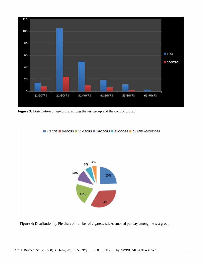
Figure 3: Distribution of age group among the test group and the control group.
Figure 4: Distribution by Pie chart of number of cigarette sticks smoked per day among the test group.

Am. J. Biomed. Sci. 2016, 8(1), 56-67; doi: 10.5099/aj160100056 © 2016 by NWPII. All rights reserved 62
Table 4: Number of Cigarettes smoked per day.
Cigarette Range (Cigarettes) Frequency Percentage (%)
<5 49 24
6-10 68 34
11-15 42 21
16-20 21 11
21-30 12 6
31 and above 08 4
Table 5: Different cell types among the Test and Control groups.
Cell Types Test Control
Total P-value
Present Absent Present Absent
Dysplastic cells 123 77 nil 50 250 0.000
Inflammatory Cells
Heavy infiltrates of inflammatory cells
140 60 nil 50 250 0.000
Mild infiltrates of Inflammatory cells
61 139 20 30 250 0.180
Red blood cells 40 160 nil 50 250 0.000
Cast 111 89 15 35 250 0.002
Power of significance = 0.05.

Am. J. Biomed. Sci. 2016, 8(1), 56-67; doi: 10.5099/aj160100056 © 2016 by NWPII. All rights reserved 63
Figure 5: (Control) Urine sample of a non-cigarette smoker showing [A] Intermediate Squamous cells. [B]
Superficial cells and [C] Mild Inflammatory cells (PAP X100).
Figure 6: (Control) Urine smear of a non-cigarette smoker showing [A] Superficial Squamous cells.
[B] Transitional cells [C] Cast and [D] Mild Inflammatory Cells (PAP X100).

Am. J. Biomed. Sci. 2016, 8(1), 56-67; doi: 10.5099/aj160100056 © 2016 by NWPII. All rights reserved 64
Figure 7: (Test) Urine smear of a cigarette smoker showing [A] A cluster of dysplastic epithelial cells and
[B] Heavy infiltrates of inflammatory cells (PAP X 100) .
Figure 8: (Test) Urine smear of a cigarette smoker showing [A] A cluster of dysplastic epithelial cells and
[B] Heavy infiltrates of inflammatory cells (PAP X 400).
Am. J. Biomed. Sci. 2016, 8(1), 56-67; doi: 10.5099/aj160100056 © 2016 by NWPII. All rights reserved 65
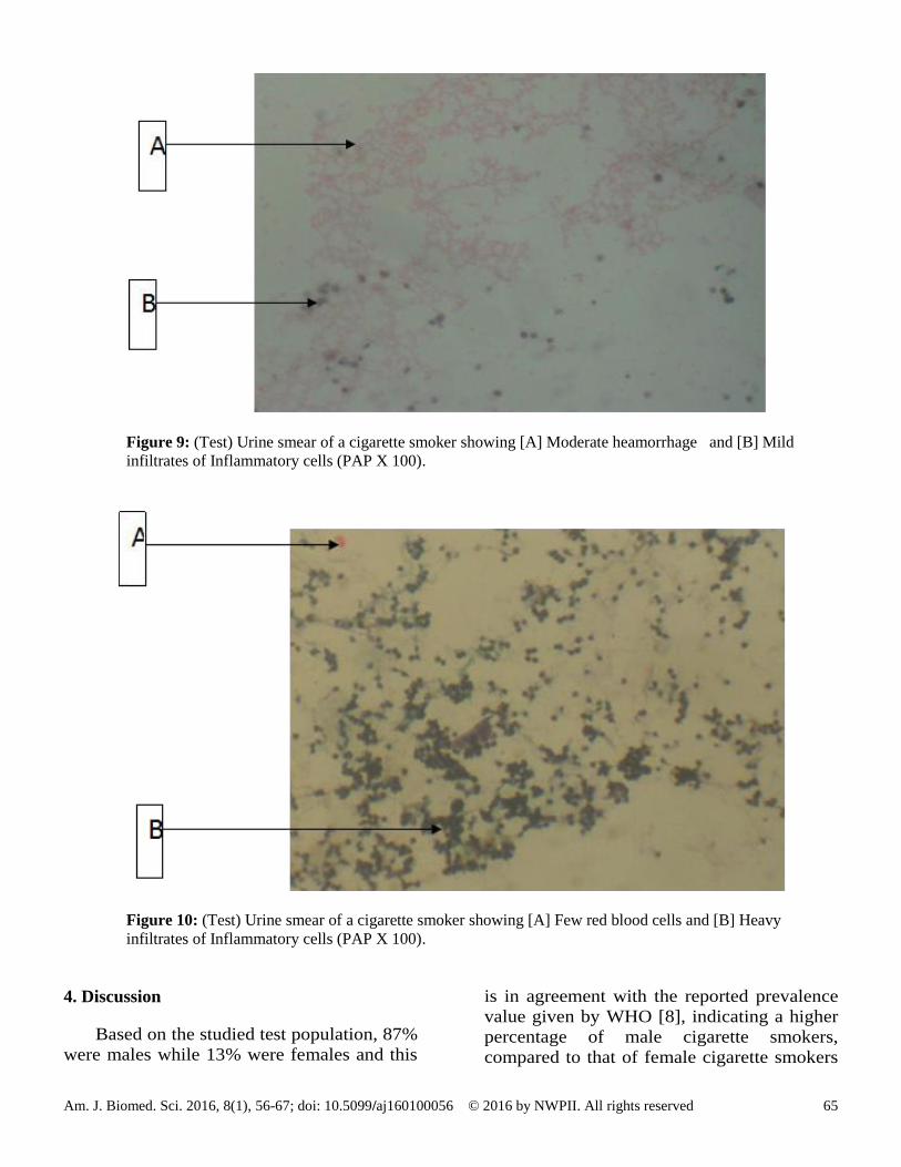
Figure 9: (Test) Urine smear of a cigarette smoker showing [A] Moderate heamorrhage and [B] Mild
infiltrates of Inflammatory cells (PAP X 100).
Figure 10: (Test) Urine smear of a cigarette smoker showing [A] Few red blood cells and [B] Heavy
infiltrates of Inflammatory cells (PAP X 100).
4. Discussion
Based on the studied test population, 87%
were males while 13% were females and this
is in agreement with the reported prevalence
value given by WHO [8], indicating a higher
percentage of male cigarette smokers,
compared to that of female cigarette smokers

Am. J. Biomed. Sci. 2016, 8(1), 56-67; doi: 10.5099/aj160100056 © 2016 by NWPII. All rights reserved 66
in the south/west geo-political region in
Nigeria. Their ages ranges from 11years to 70
years with a mean age of 41years. 59% of the
test group population ranges in ages between
11years to 30years which showed a high
percentage of youths involved in cigarette
smoking in Owo town, Ondo State, Nigeria.
The number of cigarette sticks smoked per
day among the test group varies widely with
34% smoking 6 to 10 cigarettes per day.
According to [11], the effects of cigarette
smoking on the body depends on the number
of years an individual smokes and the number
of cigarette sticks an individual smoked per
day, starting smoking earlier in life and
smoking cigarettes higher in tar increases the
risk of renal diseases. Few urothelial cells like
crystals, cast, necrotic debris, mild
inflammatory cells, transitional epithelium
and squamous epithelium were observed
consistently in most of the urine smears in the
control group who are non-cigarette smokers
which is in agreement with [12]. According to
[13], cigarette smoking has been one of the
leading causes of preventable death and a
major public health concern, cigarette
smoking causes most of the commonly
diseases affecting the heart, lungs and the
renal system [14]. In this research study, High
cellular turnover was detected among 140
(70%) of the test group, this is due to the fact
that when smokers inhale some of the
carcinogens (cancer causing chemicals) in
cigarettes, smokes are absorbed from the
lungs and get into the blood, and from the
blood, they are filtered by the kidneys and
they are concentrated in urine in the bladder,
thereby damaging the cells that line the inside
of the bladder, leading to high cellular
turnover and eventually causing bladder
cancer, depending on how often and how long
an individual continues to engage in cigarettes
smoking. Red blood cells were also detected
among 20% of the test group and this is due
to tumour diathesis and necrosis which
provokes bleeding in the urinary bladder
thereby causing injury to the renal system
and then leading to end stage renal disease, in
agreement with [15]. The distribution of
heavy infiltrates of inflammatory cells found
among 140 (70%) of the test group include;
neutrophils, eosinophils, lymphocytes,
macrophages and plasma cells, which is in
agreement with [16]. Cigarette smoke
contains a high percentage of tar which
increases the risk of having renal damage and
it also contains several carcinogenic pyrolytic
products that binds to DNA and can cause
many genetic mutations [11]. Other features
detected among the urine smears of cigarette
smokers include: cluster of cell showing
dysplastic changes, pleomorphisms,
hyperchromatism, increase in nuclear
cytoplasm ratio, irregular nuclear border and
many of the cells becoming transformed.
These cytological changes observed in the
urine smears of cigarette smokers (test group)
are the features of tumour diathesis which is
in agreement with [17]. This research work
detected significant differences between the
urine cytology of cigarette smokers and that
of non-cigarette smokers, indicating the
adverse health effects of cigarette smoke to
the renal system.
5. Conclusion
On the basis of this study and review of
relevant literatures, cigarette smoking
increases the risk of developing bladder
cancer, kidney cancer and tumour diathesis.
The consistent detection of high cellular
turnover, necrotic cells, cluster of cell
showing dysplastic changes, enlargement in
nuclear cytoplasm ratio, irregular nuclear
border, moderate haemorrhage, heavy
infiltrates of inflammatory cells,
hyperchromatism, pleomorphysms and
neoplastic transformation among the urine
smears of the test group, are caused as a result
of the harmful chemicals carcinogenic
substances present in cigarette smoke.

Am. J. Biomed. Sci. 2016, 8(1), 56-67; doi: 10.5099/aj160100056 © 2016 by NWPII. All rights reserved 67
References
1. Ochei, J. and Kolhatkar, A. (2005). Medical
Laboratory Science Theory and Practice,
London. Pp. 450-521.
2. Bancroft, J.D. (2002). Theory and Practice of
Histological Techniques, 4th Edition,
Churchill Livingstone London. Pp. 547-548.
3. Centers for Disease Control and Prevention
(CDC) (2008). Smoking-attributable
mortality, years of potential life lost, and
productivity losses--United States, MMWR
Morb. Mortal. Wkly. Rep. 57 (45): 1226–
1228.
4. Alberg, A. J., Kouzis, A. and Gerkinger, J. M,
(2007). A prospective cohort study of
bladder cancer risk in relation to active
cigarette smoking and household exposure to
secondhand cigarette smoke. Am J Epidemiol;
165:660. DOI: 10.1093/aje/kwk047
5. Kantor, A.F., Hartge, P., Hoover, R.N.,
Naryana, A.S., Sullivan, J.W. and Fraumeni,
J.R. (1984). Urinary Tract infection and Risk
of Bladder cancer.Uni. J. I.119: 510-515.
6. Dales, L.G., Friedman, G.D., Siegelaub, A.B.,
Seltzer, C.C. and Ury, H.K. (1978). Cigarette
smoking habits and urine characteristics. J.
Nephron. 20 (3): 163–170. DOI:
10.1159/000181215
7. Falide, M., Eckert, W.G. and Patterson, J.N.
(2010). A comparison of simple centrifuge
method and the Millipore Filter technique in
urinary cytology, Acta Cytol. 7 (4): 199-206.
8. World Health Organization (2008). The
Global Burden of Disease Geneva: World
Health Organization.
9. Naing, L., Winn, T. and Rusli, B.N. (2006).
Practical Issues in Calculating Sample Size
for Prevalence Studies. Archives of Orofacial
Sciences. 2(1):9-14.
10. Avwioro, O.G. (2002). Histochemistry and
Tissue Pathology. [1st edition] Clavarianum
Centre, Ibadan. Pp. 283.
11. Fowles, J., Dybing, E. and Dybing, H. (2003).
Application of toxicological risk assessment
principles to the chemical constituents of
cigarette smoke. Tob. Control. 12(4): 424–
430. DOI: 10.1136/tc.12.4.424
12. Feng, Z., Hu, W. and Tang, M.S. (2006).
Acrolein is a major cigarette related lung
cancer agent: preferential binding at p53
mutational hotspots and inhibition of DNA
repair. Proceedings of the National Academy
of Sciences of the United States of America.
103(42): 15404–15409. DOI:
10.1073/pnas.0607031103
13. Pendlebury, J.D., Wilson, R.J., Bano, S.,
Lumb, K.J., Schneider, J.M., Hasan, S.U. and
Wilson, B. (2008). Respiratory Control in
Neonatal Rats Exposed to Prenatal Cigarette
Smoke. American Journal of Respiratory
and Critical Care Medicine. 177(11): 1255–
1261. DOI: 10.1164/rccm.200711-1739OC
14. Hecht, S. S. (2011). Tobacco smoke
carcinogens and lung cancer. Chemical
carcinogenesis: Springer. Pp. 67.
15. Arcavi, L. and Benowitz, N.L. (2004).
Cigarette Smoking and Infection. Archives of
Internal Medicine. 164(20): 2206–2216.
DOI: 10.1001/archinte.164.20.2206
16. Sharf, E. and Rihab, I. (2008). Cytological
Pattern of Urine in Sudanese Tobacco
Smokers, Sudan University of Science and
Technology. J. SUST. 12(8): 133-140.
17. Shephard, E.A., Stapley, S., Neal, R.D, Rose,
P., Walter, F.M. and Hamilton WT. (2012).
Clinical features of Bladder cancer in
primary care. Br. J. Gen Pract. 62(602): 598-
604. DOI: 10.3399/bjgp12X653796