current biology review - · pdf filecurrent biology review ... here we examine various ways in...
TRANSCRIPT

Current Biology
Review
Marine Protists Are Not Just Big Bacteria
Patrick J. Keeling* and Javier del CampoBotany Department, University of British Columbia, 3529-6270 University Boulevard, Vancouver, BC, V6T 1Z4, Canada*Correspondence: [email protected]://dx.doi.org/10.1016/j.cub.2017.03.075
The study of marine microbial ecology has been completely transformed by molecular and genomic data:after centuries of relative neglect, genomics has revealed the surprising extent of microbial diversity andhow microbial processes transform ocean and global ecosystems. But the revolution is not complete: majorgaps in our understanding remain, and one obvious example is that microbial eukaryotes, or protists, are stilllargely neglected. Here we examine various ways in which protists might be better integrated into models ofmarine microbial ecology, what challenges this will present, and why understanding the limitations of ourtools is a significant concern. In part this is a technical challenge — eukaryotic genomes are more difficultto characterize — but eukaryotic adaptations are also more dependent on morphology and behaviour thanthey are on the metabolic diversity that typifies bacteria, and these cannot be inferred from genomic dataas readily as metabolism can be. We therefore cannot simply follow in the methodological footsteps ofbacterial ecology and hope for similar success. Understanding microbial eukaryotes will require differentapproaches, including greater emphasis on taxonomically and trophically diverse model systems. Molecularsequencing will continue to play a role, and advances in environmental sequence tag studies and single-cellmethods for genomic and transcriptomics offer particular promise.
IntroductionIn Essay Concerning Human Understanding (1689), John Locke
wrote, ‘‘It is of great use to the sailor to know the length of his
line, though he cannot with it fathom all the depths of the ocean.
It is well he knows that it is long enough to reach the bottom at
such places as are necessary to direct his voyage, and caution
him against running upon shoals that may ruin him.’’ Locke
was writing about understanding the tools of human thought,
but it is every bit as sensible to understand the limits of those
tools that contributed to the creation of a body of data as well,
because these affect our interpretation every bit as acutely.
The metaphor is especially apt when applied to the tangled
web of networks that makes up marine microbial ecosystems:
here is a problem where the uncharted waters still greatly
outweigh our understanding, and yet we are suddenly moving
so astonishingly quickly that the raw information available greatly
exceeds our conscious appreciation of both the strengths and
weaknesses of many tools used to create and analyse this infor-
mation, or how they affect our interpretation of it.
The problem is complex, but important, because microbial life
drives every one of the major biogeochemical cycles that make
the ocean so central to all other ecosystems on earth [1]. We
and our animal and plant cousins depend on these cycles and
sometimes also have strong effects on them — pulling them in
one direction or another by our activities — but the engines
driving nutrient and energy flow are all microbial processes [2].
Because microbial communities form the foundations of these
ecosystems, disrupting them can also have profound impacts
on the rest of the system, much in the same way that shifting
the foundation of a tall building may be amplified through the
structure in ways we cannot see until it is too late to mitigate
against the damage.
Despite their impact on a planetary scale, we know relatively
little about the composition of many of these microbial
Curren
communities and even less about how they interact and function
at the ecosystem level. Indeed, we probably know more about
some individual fish or marine mammalian species as we do,
collectively, about the tens of thousands of microbial species
whose activities allow those larger andmore charismatic species
to survive. The reasons for this are manifold, but broadly trace
back to two simple problems: microbes are small, and microbes
are diverse. The first of these probably should not come as a
surprise, but the microbial world is so small that in many ways
it lays even beyond our imagination, and size poses challenges
both of technical nature and in failing to command our attention.
For a microbial ecologist studying marine bacteria, for example,
the scale of the organism makes observation somewhat like a
traditional ecologist trying to study animals on the African
savanna from space: the vastness of the size difference makes
the problem qualitatively different. Because we can’t see them,
we also tend to ignore microbes, and observing their effects
demands greater attention, not less.
The second challenge is the vastness of the biological diversity
that we lump together with tags like ‘microplankton’, ‘nano-
plankton’, ‘picoplankton’, or even ‘the microbial world’ [1]. His-
torically, microbes have been relegated to an intellectual stew:
it was difficult to see much diversity with available tools, and
what we could see was even more difficult to interpret [3]. Bac-
terial diversity is largely not manifested at the level of structure,
but rather at the level ofmolecules, particularlymetabolism. Bac-
terial morphology afforded few characters that allowed us to
infer their evolutionary relationships, but their metabolic diversity
is so extreme that it had the opposite problem: metabolic varia-
tion evolves so rapidly that patterns of that variation shed little
light on how bacteria were related to one another or to eukary-
otes, or even how much evolutionary diversity bacteria encom-
pass. Microbial eukaryotes, by contrast, exhibit a great deal of
morphological diversity (e.g., see Figure 1), but even once the
t Biology 27, R541–R549, June 5, 2017 ª 2017 Elsevier Ltd. R541

Figure 1. Examples of morphological andtrophic complexity of marine microbialeukaryotes.Top row (left to right): the heterotrophic rhizariannanoflagellate Minorisa minuta, which is commonin coastal waters; a large heterotrophic radio-larian, Rhizoplegma, which are among the mostabundant protists in the world; a diatom, one ofthe most common autotrophic eukaryotes. Centrerow (left to right): the dinoflagellate Symbiodinum,a photosynthetic endosymbiont of corals (centre),which give corals their distinctive colours andmake reef-building possible; the gregarine api-complexan Lankesteria, which are well-studiedparasites of terrestrial animals (like us), but arealso abundant in the ocean and likely play asignificant role in marine animal populations.Bottom row (left to right): the photosyntheticdinoflagellate Ceratocorys, with its distinctivecellulose spines; the ciliate Euplotes, which is apredator but here appears green because it isfilled with prey algae (Duniella); the photosyntheticeuglenozoan Euglena, which is also green but heredue to its green secondary plastids. It also hasa visible red ‘eyespot’ which it uses to locatelight. All photos by the authors except: Rhizo-plegma (ª John Dolan), Symbiodinum and Cera-tocorys (courtesy of Nick Irwin), and Lankesteria(ª Sonja Rueckert).
Current Biology
Review
tools to look at the relevant scale were available (e.g. light
and electron microscopy) it also proved too much to reveal
patterns to help interpret this diversity or meaningfully relate it
to macroscopic life [4]. Moreover, only a tiny proportion of pro-
tists and prokaryotes are readily cultivable, so together our
inability to identify them, interpret their diversity, or cultivate
them to study them in detail all contributed to the lumping of
microbes into functional classes so broad as to be effectively
meaningless [5–7].
The breakthrough for understanding the scope of microbial
diversity was molecular biology, and phylogenetic trees based
on molecular sequence data in particular. Molecular trees gave
us our first real view of just how diverse the microbial world is,
and how microbes are related to more familiar animals, fungi,
and plants. The view was dramatic, but also problematic: in
molecular trees, microbial diversity outstrips all other biodiver-
sity by orders of magnitude: tiny creatures we thought were
the same species turned out to be as distant from one another
as humans are from chickens, or in some cases as humans are
R542 Current Biology 27, R541–R549, June 5, 2017
from pine trees [8]. This change is largely
a matter of perspective (things we think
are ‘important’ distinctions are just our
opinions), but is still an important prob-
lem when it comes to relating the some-
what more abstract diversity of microbial
life to specialists outside the field, to
whom microbes can still appear to lack
diversity.
Molecular phylogeny opened up the
‘black box’ of microbial diversity and
offered ways to manage it. The field of
microbial ecology is now rapidly growing
and our understanding is moving faster
than ever, largely based on using molecular tools to study micro-
bial diversity in the field, andmolecular phylogenetic frameworks
to interpret that data [1,2,6,7]. Communities of uncultivated
microbes can be examined collectively using environmental
‘tag’ sequencing, where a fragment of the small subunit ribo-
somal RNA (SSU rRNA) is sampled from an environment to
quickly identify most inhabitants. Similarly, various kinds of
‘meta-omics’ can tell us what genes are possessed by commu-
nities as a whole (metagenomics), which genes are being ex-
pressed (e.g., metatranscriptomics) [9,10] or proteins being
translated (metaproteomics) [11]. Advances have been made,
andmore are on the horizon, but before we celebrate the endless
bounty of a new age of molecular microbial ecology, we should
pause and consider the microbial eukaryotes.
Virtually every microbial ecosystem known in the oceans, in
freshwater, and on land includes eukaryotes (the exceptions be-
ing some of the most ‘extreme’ environments), where they play
some of the same roles as bacteria and archaea, but also have
some essential and unique ecological roles [12]. But, just as
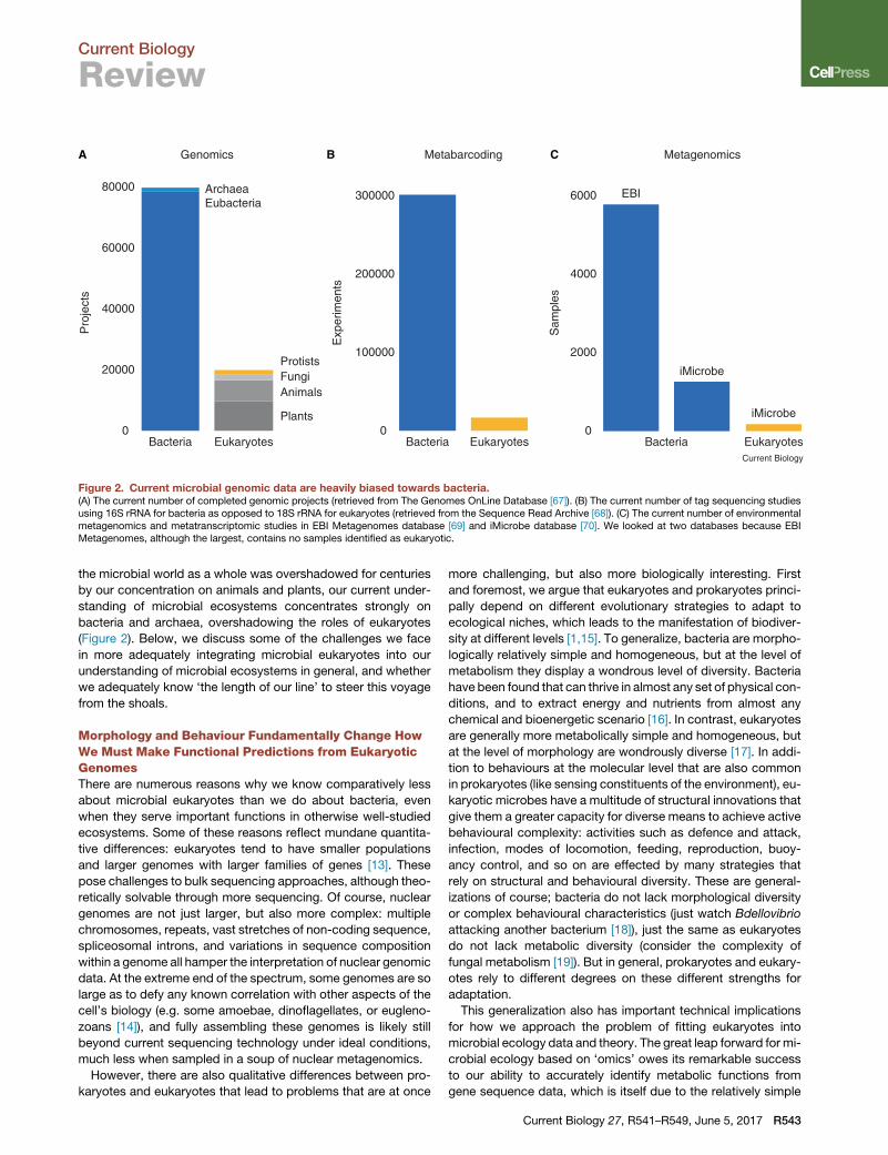
0
20000
40000
60000
80000
Bacteria Eukaryotes
Genomics
0
100000
200000
300000
Bacteria Eukaryotes
Metabarcoding
ProtistsFungiAnimals
ArchaeaEubacteria
Plants0
2000
4000
6000
Bacteria Eukaryotes
EBI
iMicrobe
Current Biology
iMicrobe
Metagenomics
Sam
ples
Exp
erim
ents
Pro
ject
s
A B C
Figure 2. Current microbial genomic data are heavily biased towards bacteria.(A) The current number of completed genomic projects (retrieved from The Genomes OnLine Database [67]). (B) The current number of tag sequencing studiesusing 16S rRNA for bacteria as opposed to 18S rRNA for eukaryotes (retrieved from the Sequence Read Archive [68]). (C) The current number of environmentalmetagenomics and metatranscriptomic studies in EBI Metagenomes database [69] and iMicrobe database [70]. We looked at two databases because EBIMetagenomes, although the largest, contains no samples identified as eukaryotic.
Current Biology
Review
the microbial world as a whole was overshadowed for centuries
by our concentration on animals and plants, our current under-
standing of microbial ecosystems concentrates strongly on
bacteria and archaea, overshadowing the roles of eukaryotes
(Figure 2). Below, we discuss some of the challenges we face
in more adequately integrating microbial eukaryotes into our
understanding of microbial ecosystems in general, and whether
we adequately know ‘the length of our line’ to steer this voyage
from the shoals.
Morphology and Behaviour Fundamentally Change HowWe Must Make Functional Predictions from EukaryoticGenomesThere are numerous reasons why we know comparatively less
about microbial eukaryotes than we do about bacteria, even
when they serve important functions in otherwise well-studied
ecosystems. Some of these reasons reflect mundane quantita-
tive differences: eukaryotes tend to have smaller populations
and larger genomes with larger families of genes [13]. These
pose challenges to bulk sequencing approaches, although theo-
retically solvable through more sequencing. Of course, nuclear
genomes are not just larger, but also more complex: multiple
chromosomes, repeats, vast stretches of non-coding sequence,
spliceosomal introns, and variations in sequence composition
within a genome all hamper the interpretation of nuclear genomic
data. At the extreme end of the spectrum, some genomes are so
large as to defy any known correlation with other aspects of the
cell’s biology (e.g. some amoebae, dinoflagellates, or eugleno-
zoans [14]), and fully assembling these genomes is likely still
beyond current sequencing technology under ideal conditions,
much less when sampled in a soup of nuclear metagenomics.
However, there are also qualitative differences between pro-
karyotes and eukaryotes that lead to problems that are at once
more challenging, but also more biologically interesting. First
and foremost, we argue that eukaryotes and prokaryotes princi-
pally depend on different evolutionary strategies to adapt to
ecological niches, which leads to the manifestation of biodiver-
sity at different levels [1,15]. To generalize, bacteria are morpho-
logically relatively simple and homogeneous, but at the level of
metabolism they display a wondrous level of diversity. Bacteria
have been found that can thrive in almost any set of physical con-
ditions, and to extract energy and nutrients from almost any
chemical and bioenergetic scenario [16]. In contrast, eukaryotes
are generally more metabolically simple and homogeneous, but
at the level of morphology are wondrously diverse [17]. In addi-
tion to behaviours at the molecular level that are also common
in prokaryotes (like sensing constituents of the environment), eu-
karyotic microbes have a multitude of structural innovations that
give them a greater capacity for diverse means to achieve active
behavioural complexity: activities such as defence and attack,
infection, modes of locomotion, feeding, reproduction, buoy-
ancy control, and so on are effected by many strategies that
rely on structural and behavioural diversity. These are general-
izations of course; bacteria do not lack morphological diversity
or complex behavioural characteristics (just watch Bdellovibrio
attacking another bacterium [18]), just the same as eukaryotes
do not lack metabolic diversity (consider the complexity of
fungal metabolism [19]). But in general, prokaryotes and eukary-
otes rely to different degrees on these different strengths for
adaptation.
This generalization also has important technical implications
for how we approach the problem of fitting eukaryotes into
microbial ecology data and theory. The great leap forward for mi-
crobial ecology based on ‘omics’ owes its remarkable success
to our ability to accurately identify metabolic functions from
gene sequence data, which is itself due to the relatively simple
Current Biology 27, R541–R549, June 5, 2017 R543

Current Biology
Review
modularity of metabolic proteins. Assuming our functional iden-
tification of metabolic genes is largely correct (an important
assumption to examine, but one that we will set aside here),
we have fundamental insights into how these enzymes work
that allows us to transfer those insights to new species as they
are investigated based on the further assumption that a given
enzyme will perform a similar reaction when plugged into an
otherwise coherent metabolic network. This gives us great
power to predict functional roles and interaction networks for
bacteria, because those networks are primarily based on meta-
bolism [20] (bacterial networks havemany other levels, but study
of their ecology has largely focused on metabolic networks).
Indeed, environmental microbiologists working on bacteria are
already verifying these predicted functions and interactions
in situ through the use of metaproteomics and metabolomics
[21,22]. The same well-tested genomic tools, environmental
sequence tags, metagenomics, and metatranscriptomics, are
now being applied to microbial eukaryotes, but is there any
expectation that they will be as successful? We argue there
are good reasons to think not [15].
To illustrate this question with something less abstract than
a protist, consider the case of the New Caledonian Crow. This
bird is known for its intelligence: it uses multi-step problem
solving and understands and uses abstract phenomena such
as displacement. Ask yourself, given its complete genome
sequence but no other information, could we infer its intelligence
and problem solving characteristics? The answer of course is
‘no’. Indeed, we argue further that without the benefit of a great
deal of context we would not even be able to conclude that it had
feathers and could fly, because there is little in the genome
to immediately suggest ‘birdness’ either. This is because we
cannot readily translate gene sequences into complex multi-
gene traits where much of the variation comes from expression
levels and regulation networks rather than more readily inter-
changeable modules like enzymes, and virtually everything to
do with structures and behaviour tends to be based upon such
traits. Or in other words, we lack the equivalent of the funda-
mental knowledge of enzyme function that allows us to interpret
metabolic genes in bacteria, and without that we cannot make
such detailed ecological and functional inferences from eukary-
otic genomes as we can for bacteria. Indeed, for eukaryotic traits
of interest it is even possible that any equivalent fundamental un-
derstanding will never exist. This is because the characteristics
appear to evolve more contingently and more often conver-
gently. In this case, similar ecological strategies can evolve
without a fundamentally homologous basis, and these would
be impossible to infer from one genome based on information
gleaned from another genome where a non-homologous system
evolved for the same purpose. For example, assume that the
structural basis for saprotrophy in oomycetes and fungi may
have evolved in each group independently from non-homolo-
gous components (or from non-specific cellular components
that have roles in many systems). If so, then this feeding mech-
anism could not be inferred from the genome of one lineage
even with a perfect understanding of the molecular basis of the
system in the other lineage. In cases where complexmorphology
and behaviour are underpinned by fundamentally homologous
traits, there is hope that a detailed understanding of model sys-
tems will one day allow us to infer these characteristics in other
R544 Current Biology 27, R541–R549, June 5, 2017
organisms based on the genome alone, but even then the infer-
ence will be much more complex than it is for metabolic path-
ways in bacteria.
While microbial eukaryotes may be small and have overlap-
ping ecological roles with bacteria, they are still eukaryotes after
all, and the same basic problem extends just as much to a
dinoflagellate as to a bird. Even with complete nuclear genome
sequences, therefore, our conclusions about ecological roles
of protists can be limited to ridiculously broad conclusions,
like ‘it’s photosynthetic’ [15]. We lack genomic flags for even
the broadest of defined ecological roles such as ‘parasitic’, or
‘predator’, and most likely such flags simply do not exist [23],
so it is not just a matter of learning to recognize them. Worse
still, the complexity of eukaryotic behaviour means that there
is overlap within a single species between the kinds of roles
we like to identify: for example, many or most ‘photosynthetic’
protists are actually better described as mixotrophs, because
they are also heterotrophic feeders, sometimes eating more
bacteria than the purely heterotrophic protists in the same envi-
ronment [24].
Thankfully, a solution to this problem does not require a
great leap of logic or theory, because the solution is already
common practice, albeit imperfectly applied. We routinely
make assumptions about what a little-studied species is like
and what its role in the environment is likely to be based on
what its close relatives are like and do. For example, with a
crow genome we would quickly infer it had feathers and could
fly because it was obvious from the sequence that it was
related to other feathered, flying animals, rather than because
we identified genes related to feathers or flying. This compara-
tive reasoning can be very powerful, but also depends heavily
on the quality and quantity of information one has from those
close relatives.
The first such requirement is a reference tree, which is neces-
sary to correctly identify the subject’s closest relatives. We
currently lack a well-supported reference tree spanning all
microbial eukaryotes, but we have most of the data one would
need to develop it, and this is a relatively straightforward problem
to solve that is currently underway for the most commonly used
tag sequence, SSU rRNA [25]. The second requirement is for a
large body of information about the biology of closely related
species. Obviously, you learn little about a species by identifying
a closely related species about which we also know nothing.
Moreover, the closer the relationship, the more likely the infer-
ences are to be true; our inferences about the crow might be
misleading if the nearest relative we could compare it to was a
crocodile.
This is a considerably more difficult problem that does not
readily lend itself to ‘high-throughput’ solutions, because what
this really means is we need a large number of relatively well-
studied model organisms scattered around each and every
major branch of the tree of eukaryotes. We need to go back to
nature and actually look at how these cells work and what they
are doing in the environment. By developing more model sys-
tems distributed through the tree of eukaryotes, and across
different ecological roles, we will massively improve our ability
to infer ecological roles of environmental samples because our
assumptions about roles based on sequence data will be
much more accurate [26,27].

Current Biology
Review
What Can We Learn from Environmental TagSequencing?As with bacteria, the first clone library studies of microbial eu-
karyotes revealed a picture of the marine diversity that was
different form the picture sketched out over the previous de-
cades by microscopy [28,29]. Two group of picoeukaryotes in
particular stood out: the parasitic MALV (Marine Alveolates, or
syndinians) and the bacterivorous MAST (Marine Stramenopiles)
lineages emerged as some of the most abundant organisms in
the sea, representing up to 50% of the sequences in seminal
molecular environmental protistolgy studies [30]. In addition to
these two groups, a myriad of other branches sprouted from
asmany parts of the eukaryotic tree, corresponding to additional
novel, uncultured lineages [31]. High-throughput tag sequencing
methods have now replaced clone libraries for the study of pro-
tist diversity in the environment [32,33] and further accelerated
these discoveries.
But to make full use of these data, our ability to interpret them
needs to keep pace with data generation. The reference data
discussed above form the foundation for how we interpret mo-
lecular tag sequencing from environmental samples, and we
need to understand how well the tags reflect the composition
of the communities that they are meant to represent. As with
bacteria, the current gene of choice to survey eukaryotic diver-
sity is the SSU rRNA. Next generation sequencing methods
require short fragments, so two different regions of the SSU
have been used to study eukaryotic diversity, V4 and V9 [34].
Each has known biases, some of which were predictable based
on the sequences of these regions: for example, V4 excludes eu-
glenozoans [35], and so the diversity of marine diplonemids
(a subgroup of euglenozoans) was not revealed until large sur-
veys using V9 were carried out [36]. Another major bias relates
to the amplification of sequence tags versus directly isolating
cells from the environment. When libraries of amplified and
cloned SSU fragments were compared with a database of the
same SSU fragment taken from large numbers of sorted single
cells from the same environment, the proportions of some taxa
were similar, but others were significantly different [37]. Some
of these differences related to particular taxa being over-repre-
sented in the amplified tags, but other differences were more
general and crossed taxonomic lines, the most significant being
the under-representation of heterotrophs as a whole in clone li-
braries. These biases are attributed to the copy number of
SSU rRNA genes in different genomes [37,38]. While bacteria
have between one and ten copies of the SSU in their genomes,
microbial eukaryotes can have a number of copies that differ
by many orders of magnitudes. Some groups, like dinoflagel-
lates, can have up to 12,000 copies [38] while others, like
MAST-4, have only 30 [39]. Differences in the SSU copy number
contribute to ‘abundance’ when analyzing DNA- or RNA-derived
tag data or metagenomes [35,40].
Other less obvious biases probably also exist, and under-
standing these and how they might affect major patterns is a
key problem. One approach to identifying cryptic biases is to
look for unrecognized patterns in the data and then ask whether
they reveal biological phenomena or methodological biases.
Normally such data are broken down by environmental criteria,
but one can look at patterns a priori, across taxa and between
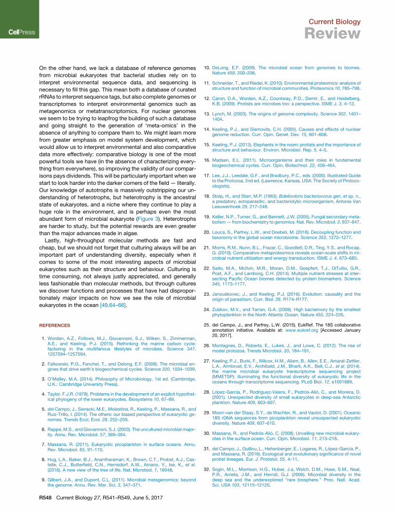
different methods and markers. As a simple example, here we
have re-analysed the complete set of microbial eukaryotic tag
V9 sequences from TaraOceans [41], the largest database of eu-
karyotic tags currently available. We examined the relative abun-
dance of the 25 most common individual operational taxonomic
units (OTUs; e.g., ‘species’) from the whole dataset, as well as
the relative abundance of the 10 most common OTUs within
12 well-defined and diverse protist lineages (dinoflagellates, ra-
diolarians, diatoms, diplonemids, fungi, pelagophytes, syndi-
nians, apicomplexans, ciliates, green algae, and bygirans, which
are heterotrophic stramenopiles). Across the whole data set, we
find that only 8 OTUs represent 50%of all reads (Figure 3). Taken
at face value, this means a small number of species are hyper-
abundant, which fits with the expected distribution [42]. Interest-
ingly, however, when we look at the structure of the read
distribution within each of the 12 common and diverse lineages,
we observe that not all the groups share the same pattern.
Instead, we see two different types of distributions — normal
vs jackpots. In the first instance, the ten most abundant OTUs
account for much of the data, but we see a gradual decline in
relative abundance and a significant proportion of the reads
from the entire group are distributed across less abundant
OTUs. This is seen for the radiolarians, haptophytes, ciliates,
green algae, bygirans (among them some of the abundant
MAST groups), and the fungi, where the most common taxa
are abundant, but no one taxon dominates. An extreme case is
the syndinians, where the distribution across the ten most abun-
dant OTUS is nearly linear. On the other extreme are lineages
dominated by a single ‘jackpot’ OTU. This is the case for the di-
atoms, diplonemids, pelagophytes, and the dinoflagellates,
where the jackpot is also the single most abundant OTU in the
whole data set.
We might therefore ask, is the global OTU structure real, or is
it an artifact of analytical pipelines and clustering methods?
What are these jackpot taxa, and if they are real what
does that tell us? Some of these questions have been
addressed in different data sets by re-clustering the data using
different methods [43], comparing the outcomes of different
markers from similar environments [34], or, as noted above,
using different levels of sampling from single cells to total
communities [37].
A better sense for these limitations is important because tag
sequencing guides our decisions as towhere we focus our atten-
tion: without any other information about these organisms, abun-
dance has become a proxy for importance, and without better
methods to estimate abundance in a high-throughput manner,
sequence abundance is the proxy for organismal abundance.
And even if our estimates of abundance are acceptably close,
its use as a proxy for importance is potentially misleading. We
seldom have information about process rates from these organ-
isms, so if, for example, rare organisms are more active than
common ones, abundance will not reflect ecological impact.
Going back to the savanna, if you did what we do with microbes
in the ocean, and scraped up a patch of East Africa and
counted the animals, you would find that there are lots of
gazelles but just a few lions, but we know that lions have a
huge impact on the systems. Interestingly, the results of our
attempts to count marine microbial eukaryotes has not given
what would perhaps be the most intuitive answer, that photo-
synthetic protists are more common: instead, heterotrophs
Current Biology 27, R541–R549, June 5, 2017 R545

Discoba
Radiolarians
Dinoflagell
ates
Apicom
plexa
Ciliates
Nucleariids
Choan
oflag
ellate
s
Parabasalids
Foraminifera
Cercozoa
Bigyr
a
Diatoms
Fungi
Anim
als
Red AlgaePlantsClorophytes
Centrohelids
Cryptophytes
Pelagophytes
Diplomonads
Syn
din
ian
s
Haptophytes
Total reads
Proportion of the 25 most abundant OTUs
Alveolates
Opisthokonts
StramenopilesExcavates
AmoebozoaArchaeplastids
Rhizaria
These 8 OTUs represent 50% of the reads
Current Biology
A
B
Tubulinea
Archamoebae
Figure 3. Relative abundance patterns ofthe most common microbial eukaryoticOTUs in Tara Oceans.(A) The relative abundance of the most commonoperational taxonomic units (OTUs) within 12well-defined and diverse protist lineages. For eachlineage, the total number of reads for the entiregroup is shown as a grey circle (the size shown toscale between lineages), and the ten most com-mon individual OTUs (e.g., species) are shown ascoloured circles of descending size. (B) The 25most abundant protist OTUs in the entire TaraOceans data set [41]. As above, the grey circlerepresents the size of the whole data set, while thecoloured circles represent individual OTUs, colourcoded according to lineage as in panel A. The firsteight OTUs account for over 50% of the totalnumber of reads in the entire data set.
Current Biology
Review
dominate at the level of both lineages and individual OTUs
(Figure 3), emphasizing the need to understand what these
counts really mean.
Remembering Cells in an Age of GenomesIn the absence of a wide diversity of model systems, environ-
mental tag data will be interpreted as best we can, and functional
data from potentially important but uncultivated species will be
sought by other means. The same problem was faced by bacte-
rial ecology years ago, but because of the different ways that
eukaryotic diversity is manifested, environmental protistology
will not be best served by simply following the trail blazed by
bacteriology in the direction of bulk environmental sequencing,
or meta-omics. Nevertheless, sequencing is relatively easy,
can be informative, and is still the obvious first tool to apply to
many questions. For many uncultivated protists, acquiring
detailed biological observations can be nearly impossible, so it
may be a question of acquiring the information that is available
from sequence data, or nothing at all. There are alsomany down-
stream benefits of large sequence data sets even if they cannot
be used to immediately infer detailed conclusions about the
ecological role of the organism, as argued above. So, the ques-
tion is not whether to sequence, but rather how to direct the tool
at eukaryotic diversity so it is most informative, and not allowing
our thinking to be restricted by treating sequencing as a panacea
that will solve all our problems.
One point to consider is that cells really do matter. The ways
that microbial eukaryotes rely on structure and behaviour to
R546 Current Biology 27, R541–R549, June 5, 2017
adapt are really only understood at the
cellular level (as opposed to the whole
community level), and even considering
metabolism alone, the partitioning of
functions within and between cells is
significant beyond the bulk metabolic
function of an environment because of
subcellular complexity (e.g., organelles)
and interactions like symbioses and
phagotrophic feeding. Happily, we are
in the midst of a technological break-
through, where methods developed for
high-throughput sequencing at the level
of single cells (primarily developed for
medical research [44]) are maturing to the point of becoming
widely applicable to uncultivated microbial species. Given the
diversity of uncultivated protists, there is an almost limitless
scope for sequencing: single-cell methods offer the chance to
retain a great deal of evolutionary context that allows us to
overcome many of the technical difficulties that are inherent in
analysing complex nuclear genomes.
Currently, single cell sequencing methods for protists include
two broad approaches: single cell genomics (SCG, which gener-
ates data sets often referred to as single amplified genomes, or
SAGs) [45], and single cell transcriptomics (SCT) [46]. As the
names imply, SCG aims to characterize the whole genome at
the DNA level, while SCT aims to characterize all the genes ex-
pressed as mRNA at the time the cell is collected. The benefits
and drawbacks are similar to those of traditional genomics and
transcriptomics. Acquiring a whole genome lends itself to a
more comprehensive picture of the cell’s potential, ideally
including even those genes that are expressed at lower levels
or under restricted conditions, as well as all the non-expressed
sequences. But whole genomes are also more challenging to
assemble (e.g., due to repeats) and interpret (e.g., without accu-
rate gene models, introns and exons can be hard to predict).
Also, current SCG technology does not generate complete ge-
nomes: in the few cases it has been applied to marine protists,
an estimated 10–25% of the genome is reported, which is only
an estimate because the genome size itself is impossible to pre-
dict. In part, this is inevitable because only one or two copies of
the genome exist in a single cell. SCG methods currently require

Current Biology
Review
an amplification step, which introduces biases and loss of infor-
mation; so even sequencing to a great depth will not necessarily
increase the coverage of the genome until amplification-inde-
pendent sequencing methods are routinely brought to bear on
single protist cells. Nevertheless, SCG-based studies of several
marine protists have already led to new insights into mysterious
and uncultivated marine protists that would not have been
possible from bulk environmental sequencing. For example,
the first description of the picobiliphyte protists as a new lineage
of marine algae was based on microscopy and SSU rRNA from
a single cell, leading to the conclusion that it was photosyn-
thetic [47]. But a subsequent SCG-based analysis of three
individual cells revealed they are not actually photosynthetic at
all, and that the original observation was probably a food alga in-
side a heterotrophic predator [48]: picobiliphytes became pico-
zoans [49]. Another recently discovered group are the marine
diplonemids: long known to exist but considered a rare and
ecologically uninteresting group, a subgroup of uncultured diplo-
nemids was unexpectedly found to be among the most abun-
dant marine heterotrophic eukaryotes in global marine surveys,
as well as being incredibly diverse [36]. Using manually isolated
cells and SCG analysis, their morphology was revealed along
with a number of interesting features about genome structure
and content that would not have been discernible from transcrip-
tome data [50]. Similarly, SCG surveys of uncultivated Marine
Alveoaltes (MALVs; unpublished) and Marine Stramenopiles
(MASTs) [51,52] have given the first look at their genome content
and the method has also been tested on the well-studied diatom
Thalassiosira [51] and the parasite Cryptosporidium parvum [53].
In the history of genomic technology, transcriptomes (formerly
called expressed sequence tags, or ESTs) arrived relatively late
as a means to quickly generate a large quantity of much more
easily interpretable data specifically from expressed mRNA.
The data are harder to generate, being derived frommore finicky
mRNA rather than from DNA, but because they are stripped of
the introns and intergenic regions the genes are more readily
assembled and analysed [27]. Transcriptomes are never a
comprehensive survey of the genome or all its genes, only those
that are expressed, andwith SCT thismeans the subset of genes
expressed at one time in a single cell, rather than an average
across a population of cells in culture. The method also involves
an amplification step, so bias and information loss also occur.
Nevertheless, the first applications to protists revealed a prom-
ising rate of gene recovery: in a controlled study (of relatively
large ciliates), SCT recovered between 80 and 100% of the
genes recovered in comparable culture-derived transcriptomes
from the same taxa, although with an apparent correlation be-
tween success and cell size [54]. It’s unclear whether the biases
introduced in SCT will be significant enough to challenge its
application to questions relating to the response of gene expres-
sion levels to environmental change, or its use on extremely
small eukaryotes [55], but for most uncultivated species at this
point the goal is simply to acquire as much gene sequence as
possible, and for this the method looks very promising.
Where it is possible to identify cells by morphology or some
other criteria, the effectiveness of both SCT and SCG can be
improved by pooling isolated cells or pooling data derived from
individual cells. This approach has been used to boost the quality
of SCG assemblies [48,51,52] and to extract taxon-specific data
from onemember of a complex culture (e.g. a predator that feeds
on a heterotrophic flagellate that itself feeds on bacteria [56]), but
it could be used to overcome problems inherent in the lack of
material that comes fromdealing with single cells, and small cells
in particular. At the extreme, pooling similar cells in very large
numbers can be achieved by fluorescence activated cell sorting
(FACS) technology. Given the appropriate sorting criteria, a rela-
tively large population of cells can be extracted from a complex
community using FACS. Ideally, the resulting population of cells
will include only close relatives, but in practice several taxa with
similar physical properties can be difficult to separate, resulting
in sorted populationswithmore than one taxon. Even still, sorting
can transform a complex community into a relatively simple one
and sorted populations can be large enough to avoid the primary
technical problems inherent in SCG and SCT [57–60]. Another
potentially more powerful emerging method is microfluidics
[61], which can separate complex communities into simplified
populations of similar cells based on a wide range of micro-
reactions, which offers more and potentially more specific
criteria to analyse individual species from complex protist com-
munities. In marine ecological studies of bacteria, another com-
mon approach is metagenome assembled genomes (MAGs),
where a genome, usually from an abundant organism, can be
assembled from bulk environmental sequence data [62,63].
This has not been applied to marine microbial eukaryotes, and
likely never will be very useful since there are few eukaryotic
metagenomic data sets and it seems doubtful that nuclear ge-
nomes could be assembled in this way. One could more easily
imagine metatranscriptome assembled transcriptomes, but in
this case the lack of physical linkage would make it difficult to
bin transcripts to individual organisms, so again it seems doubt-
ful such an approach would be more informative than a single
cell method.
ConclusionsWe live in an interesting time for the field of marine microbial
ecology. It has been understudied for years, but the growing
appreciation for the functional importance of marine microbial
communities has coincided with the development of new
genomic tools that have transformed it into a rapidly moving
and changing field where major discoveries are taking place at
a brisk pace.
But this does not mean we can simply sit back and reap the
rewards. Indeed, we argue the opposite it true: for a deep under-
standing of marine microbial eukaryotes, nowmore than ever we
need to examine where our tools are leading us and whether that
is the right direction. It may be difficult but necessary to wean
ourselves from the history of simply following the lead of environ-
mental bacteriology. Sequences will never tell us everything, but
for many microbial eukaryotes it may be currently the case that
genomics won’t tell us much at all about how they function in
and respond to the environment because we lack detailed func-
tional information from close relatives, which is necessary to
make well-supposed inferences about homologous functions.
Sequencing will still play a role so it is also important to ensure
it’s as effective as possible. On one hand, sequencing today is
an investment in the future for when the more hard-won data
from diverse model systems are available, since these data will
already be contextualized by sequences from many relatives.
Current Biology 27, R541–R549, June 5, 2017 R547
Current Biology
Review
On the other hand, we lack a database of reference genomes
from microbial eukaryotes that bacterial studies rely on to
interpret environmental sequence data, and sequencing is
necessary to fill this gap. This mean both a database of curated
rRNAs to interpret sequence tags, but also complete genomes or
transcriptomes to interpret environmental genomics such as
metagenomics or metatranscriptomics. For nuclear genomes
we seem to be trying to leapfrog the building of such a database
and going straight to the generation of ‘meta-omics’ in the
absence of anything to compare them to. We might learn more
from greater emphasis on model system development, which
would allow us to interpret environmental and also comparative
data more effectively: comparative biology is one of the most
powerful tools we have (in the absence of characterizing every-
thing from everywhere), so improving the validity of our compar-
isons pays dividends. This will be particularly important when we
start to look harder into the darker corners of the field — literally.
Our knowledge of autotrophs is massively outstripping our un-
derstanding of heterotrophs, but heterotrophy is the ancestral
state of eukaryotes, and a niche where they continue to play a
huge role in the environment, and is perhaps even the most
abundant form of microbial eukaryote (Figure 3). Heterotrophs
are harder to study, but the potential rewards are even greater
than the major advances made in algae.
Lastly, high-throughput molecular methods are fast and
cheap, but we should not forget that culturing always will be an
important part of understanding diversity, especially when it
comes to some of the most interesting aspects of microbial
eukaryotes such as their structure and behaviour. Culturing is
time consuming, not always justly appreciated, and generally
less fashionable than molecular methods, but through cultures
we discover functions and processes that have had dispropor-
tionately major impacts on how we see the role of microbial
eukaryotes in the ocean [49,64–66].
REFERENCES
1. Worden, A.Z., Follows, M.J., Giovannoni, S.J., Wilken, S., Zimmerman,A.E., and Keeling, P.J. (2015). Rethinking the marine carbon cycle:factoring in the multifarious lifestyles of microbes. Science 347,1257594–1257594.
2. Falkowski, P.G., Fenchel, T., and Delong, E.F. (2008). The microbial en-gines that drive earth’s biogeochemical cycles. Science 320, 1034–1039.
3. O’Malley, M.A. (2014). Philosophy of Microbiology, 1st ed. (Cambridge,U.K.: Cambridge University Press).
4. Taylor, F.J.R. (1978). Problems in the development of an explicit hypothet-ical phylogeny of the lower eukaryotes. Biosystems 10, 67–89.
5. del Campo, J., Sieracki, M.E., Molestina, R., Keeling, P., Massana, R., andRuiz-Trillo, I. (2014). The others: our biased perspective of eukaryotic ge-nomes. Trends Ecol. Evol. 29, 252–259.
6. Rapp�e,M.S., andGiovannoni, S.J. (2003). The unculturedmicrobial major-ity. Annu. Rev. Microbiol. 57, 369–394.
7. Massana, R. (2011). Eukaryotic picoplankton in surface oceans. Annu.Rev. Microbiol. 65, 91–110.
8. Hug, L.A., Baker, B.J., Anantharaman, K., Brown, C.T., Probst, A.J., Cas-telle, C.J., Butterfield, C.N., Hernsdorf, A.W., Amano, Y., Ise, K., et al.(2016). A new view of the tree of life. Nat. Microbiol. 1, 16048.
9. Gilbert, J.A., and Dupont, C.L. (2011). Microbial metagenomics: beyondthe genome. Annu. Rev. Mar. Sci. 3, 347–371.
R548 Current Biology 27, R541–R549, June 5, 2017
10. DeLong, E.F. (2009). The microbial ocean from genomes to biomes.Nature 459, 200–206.
11. Schneider, T., andRiedel, K. (2010). Environmental proteomics: analysis ofstructure and function of microbial communities. Proteomics 10, 785–798.
12. Caron, D.A., Worden, A.Z., Countway, P.D., Demir, E., and Heidelberg,K.B. (2009). Protists are microbes too: a perspective. ISME J. 3, 4–12.
13. Lynch, M. (2003). The origins of genome complexity. Science 302, 1401–1404.
14. Keeling, P.J., and Slamovits, C.H. (2005). Causes and effects of nucleargenome reduction. Curr. Opin. Genet. Dev. 15, 601–608.
15. Keeling, P.J. (2013). Elephants in the room: protists and the importance ofstructure and behaviour. Environ. Microbiol. Rep. 5, 4–5.
16. Madsen, E.L. (2011). Microorganisms and their roles in fundamentalbiogeochemical cycles. Curr. Opin. Biotechnol. 22, 456–464.
17. Lee, J.J., Leedale, G.F., and Bradbury, P.C., eds. (2000). Illustrated Guideto the Protozoa, 2nd ed. (Lawrence, Kansas, USA: The Society of Protozo-ologists).
18. Stolp, H., and Starr, M.P. (1963). Bdellovibrio bacteriovorus gen. et sp. n.,a predatory, ectoparasitic, and bacteriolytic microorganism. Antonie VanLeeuwenhoek 29, 217–248.
19. Keller, N.P., Turner, G., and Bennett, J.W. (2005). Fungal secondary meta-bolism— from biochemistry to genomics. Nat. Rev. Microbiol. 3, 937–947.
20. Louca, S., Parfrey, L.W., and Doebeli, M. (2016). Decoupling function andtaxonomy in the global ocean microbiome. Science 353, 1272–1277.
21. Morris, R.M., Nunn, B.L., Frazar, C., Goodlett, D.R., Ting, Y.S., and Rocap,G. (2010). Comparative metaproteomics reveals ocean-scale shifts in mi-crobial nutrient utilization and energy transduction. ISME J. 4, 673–685.
22. Saito, M.A., McIlvin, M.R., Moran, D.M., Goepfert, T.J., DiTullio, G.R.,Post, A.F., and Lamborg, C.H. (2014). Multiple nutrient stresses at inter-secting Pacific Ocean biomes detected by protein biomarkers. Science345, 1173–1177.
23. Janou�skovec, J., and Keeling, P.J. (2016). Evolution: causality and theorigin of parasitism. Curr. Biol. 26, R174–R177.
24. Zubkov, M.V., and Tarran, G.A. (2008). High bacterivory by the smallestphytoplankton in the North Atlantic Ocean. Nature 455, 224–226.
25. del Campo, J., and Parfrey, L.W. (2015). EukRef. The 18S collaborativeannotation initiative. Available at: www.eukref.org [Accessed January20, 2017].
26. Montagnes, D., Roberts, E., Luke�s, J., and Lowe, C. (2012). The rise ofmodel protozoa. Trends Microbiol. 20, 184–191.
27. Keeling, P.J., Burki, F., Wilcox, H.M., Allam, B., Allen, E.E., Amaral-Zettler,L.A., Armbrust, E.V., Archibald, J.M., Bharti, A.K., Bell, C.J., et al. (2014).the marine microbial eukaryote transcriptome sequencing project(MMETSP): illuminating the functional diversity of eukaryotic life in theoceans through transcriptome sequencing. PLoS Biol. 12, e1001889.
28. Lopez-Garcıa, P., Rodrıguez-Valera, F., Pedros-Alio, C., and Moreira, D.(2001). Unexpected diversity of small eukaryotes in deep-sea Antarcticplankton. Nature 409, 603–607.
29. Moon-van der Staay, S.Y., de Wachter, R., and Vaulot, D. (2001). Oceanic18S rDNA sequences from picoplankton reveal unsuspected eukaryoticdiversity. Nature 409, 607–610.
30. Massana, R., and Pedros-Alio, C. (2008). Unveiling new microbial eukary-otes in the surface ocean. Curr. Opin. Microbiol. 11, 213–218.
31. del Campo, J., Guillou, L., Hehenberger, E., Logares, R., Lopez-Garcıa, P.,and Massana, R. (2016). Ecological and evolutionary significance of novelprotist lineages. Eur. J. Protistol. 55, 4–11.
32. Sogin, M.L., Morrison, H.G., Huber, J.a, Welch, D.M., Huse, S.M., Neal,P.R., Arrieta, J.M., and Herndl, G.J. (2006). Microbial diversity in thedeep sea and the underexplored ‘‘rare biosphere.’’ Proc. Natl. Acad.Sci. USA 103, 12115–12120.

Current Biology
Review
33. Amaral-Zettler, L.A., McCliment, E.A., Ducklow, H.W., and Huse, S.M.(2009). A method for studying protistan diversity using massively parallelsequencing of V9 hypervariable regions of small-subunit ribosomal RNAgenes. PLoS One 4, e6372.
34. Stoeck, T., Bass, D., Nebel, M., Christen, R., Jones, M.D.M., Breiner,H.-W., and Richards, T.A. (2010). Multiple marker parallel tag environ-mental DNA sequencing reveals a highly complex eukaryotic communityin marine anoxic water. Mol. Ecol. 19, 21–31.
35. Massana, R., Gobet, A., Audic, S., Bass, D., Bittner, L., Boutte, C., Cham-bouvet, A., Christen, R., Claverie, J., Decelle, J., et al. (2015). Marine pro-tist diversity in European coastal waters and sediments as revealed byhigh-throughput sequencing. Environ. Microbiol. 17, 4035–4049.
36. Flegontova, O., Flegontov, P., Malviya, S., Audic, S., Wincker, P., de Var-gas, C., Bowler, C., Luke�s, J., and Horak, A. (2016). Extreme diversity ofdiplonemid eukaryotes in the ocean. Curr. Biol. 26, 3060–3065.
37. Heywood, J.L., Sieracki, M.E., Bellows, W., Poulton, N.J., and Stepanaus-kas, R. (2011). Capturing diversity of marine heterotrophic protists: onecell at a time. ISME J. 5, 674–684.
38. Zhu, F., Massana, R., Not, F., Marie, D., and Vaulot, D. (2005). Mapping ofpicoeucaryotes in marine ecosystems with quantitative PCR of the 18SrRNA gene. FEMS Microbiol. Ecol. 52, 79–92.
39. Rodrıguez-Martınez, R., Labrenz, M., del Campo, J., Forn, I., Jurgens, K.,and Massana, R. (2009). Distribution of the uncultured protist MAST-4 inthe Indian Ocean, Drake Passage and Mediterranean Sea assessed byreal-time quantitative PCR. Environ. Microbiol. 11, 397–408.
40. Not, F., del Campo, J., Balagu�e, V., de Vargas, C., and Massana, R. (2009).New insights into thediversityofmarinepicoeukaryotes.PLoSOne4, e7143.
41. de Vargas, C., Audic, S., Henry, N., Decelle, J.,Mah�e, F., Logares, R., Lara,E., Berney, C., Le Bescot, N., Probert, I., et al. (2015). Eukaryotic planktondiversity in the sunlit ocean. Science 348, 1261605–1261605.
42. Preston, F.W. (1948). The commonness, and rarity, of species. Ecology 29,254–283.
43. Forster, D., Dunthorn, M., Stoeck, T., and Mah�e, F. (2016). Comparison ofthree clustering approaches for detecting novel environmental microbialdiversity. PeerJ 4, e1692.
44. Gawad, C., Koh, W., and Quake, S.R. (2016). Single-cell genomesequencing: current state of the science. Nat. Rev. Genet. 17, 175–188.
45. Stepanauskas, R. (2012). Single cell genomics: An individual look at mi-crobes. Curr. Opin. Microbiol. 15, 613–620.
46. Saliba, A.E., Westermann, A.J., Gorski, S.A., and Vogel, J. (2014). Single-cell RNA-seq: advances and future challenges. Nucleic Acids Res. 42,8845–8860.
47. Not, F., Valentin, K., Romari, K., Lovejoy, C., Massana, R., Tobe, K., Vaulot,D., and Medlin, L.K. (2007). Picobiliphytes: a marine picoplanktonic algalgroup with unknown affinities to other eukaryotes. Science 315, 253–255.
48. Yoon, H.S., Price, D.C., Stepanauskas, R., Rajah, V.D., Sieracki, M.E., Wil-son, W.H., Yang, E.C., Duffy, S., and Bhattacharya, D. (2011). Single-cellgenomics reveals organismal interactions in uncultivated marine protists.Science 332, 714–717.
49. Seenivasan, R., Sausen, N., Medlin, L.K., and Melkonian, M. (2013). Pico-monas judraskeda Gen. Et Sp. Nov.: The first identified member of the pi-cozoa phylum Nov., a widespread group of picoeukaryotes, formerlyknown as ‘‘picobiliphytes.’’ PLoS One 8, e59565.
50. Gawryluk, R.M.R., del Campo, J., Okamoto, N., Strassert, J.F.H., Luke�s,J., Richards, T.A., Worden, A.Z., Santoro, A.E., and Keeling, P.J. (2016).Morphological Identification and single-cell genomics of marine diplone-mids. Curr. Biol. 26, 3053–3059.
51. Roy, R.S., Price, D.C., Schliep, A., Cai, G., Korobeynikov, A., Yoon, H.S.,Yang, E.C., and Bhattacharya, D. (2014). Single cell genome analysis of anuncultured heterotrophic stramenopile. Sci. Rep. 4, 4780.
52. Mangot, J., Logares, R., Sanchez, P., Latorre, F., Seeleuthner, Y., Mondy,S., Sieracki, M.E., Jaillon, O., Wincker, P., Vargas, C. de, et al. (2017).Accessing the genomic information of unculturable oceanic picoeukar-yotes by combining multiple single cells. Sci. Rep. 7, 41498.
53. Troell, K., Hallstrom, B., Divne, A., Alsmark, C., Arrighi, R., Huss,M., Beser,J., and Bertilsson, S. (2016). Cryptosporidium as a testbed for singlecell genome characterization of unicellular eukaryotes. BMC Genomics17, 471.
54. Kolisko, M., Boscaro, V., Burki, F., Lynn, D.H., and Keeling, P.J. (2014).Single-cell transcriptomics for microbial eukaryotes. Curr. Biol. 24,R1081–R1082.
55. Liu, Z., Hu, S.K., Campbell, V., Tatters, A.O., Heidelberg, K.B., and Caron,D.A. (2017). Single-cell transcriptomics of small microbial eukaryotes: lim-itations and potential. ISME J. 1–4.
56. Janou�skovec, J., Tikhonenkov, D.V., Burki, F., Howe, A.T., Kolısko, M.,Mylnikov, A.P., and Keeling, P.J. (2015). Factors mediating plastid depen-dency and the origins of parasitism in apicomplexans and their close rel-atives. Proc. Natl. Acad. Sci. USA 112, 10200–10207.
57. Worden, A.Z., Janou�skovec, J., Mcrose, D., Engman, A., Welsh, R.M.,Malfatti, S., Tringe, S.G., and Keeling, P.J. (2012). Global distribution ofa wild alga revealed by targeted metagenomics. Curr. Biol. 22, R675–R677.
58. Lepere, C., Demura, M., Kawachi, M., Romac, S., Probert, I., and Vaulot,D. (2011). Whole-genome amplification (WGA) of marine photosyntheticeukaryote populations. FEMS Microbiol. Ecol. 76, 513–523.
59. Cuvelier, M.L., Allen, A.E., Monier, A., McCrow, J.P., Messie, M., Tringe,S.G., Woyke, T., Welsh, R.M., Ishoey, T., Lee, J.-H., et al. (2010). Targetedmetagenomics and ecology of globally important uncultured eukaryoticphytoplankton. Proc. Natl. Acad. Sci. USA 107, 14679–14684.
60. Martinez-Garcia, M., Brazel, D., Poulton, N.J., Swan, B.K., Gomez, M.L.,Masland, D., Sieracki, M.E., and Stepanauskas, R. (2012). Unveilingin situ interactions between marine protists and bacteria through singlecell sequencing. ISME J. 6, 703–707.
61. Rusconi, R., Garren, M., and Stocker, R. (2014). Microfluidics expandingthe frontiers of microbial ecology. Annu. Rev. Biophys. 43, 65–91.
62. Albertsen,M., Hugenholtz, P., Skarshewski, A., Nielsen, K.L., Tyson, G.W.,and Nielsen, P.H. (2013). Genome sequences of rare, uncultured bacteriaobtained by differential coverage binning of multiple metagenomes. Nat.Biotechnol 31, 533–538.
63. Spang, A., Saw, J.H., Jørgensen, S.L., Zaremba-Niedzwiedzka, K., Mar-tijn, J., Lind, A.E., van Eijk, R., Schleper, C., Guy, L., and Ettema, T.J.G.(2015). Complex archaea that bridge the gap between prokaryotes andeukaryotes. Nature 521, 173–179.
64. Moore, R.B., Obornık, M., Janou�skovec, J., Chrudimsky, T., Vancova, M.,Green, D.H., Wright, S.W., Davies, N.W., Bolch, C.J.S., Heimann, K., et al.(2008). A photosynthetic alveolate closely related to apicomplexan para-sites. Nature 451, 959–963.
65. del Campo, J., Not, F., Forn, I., Sieracki, M.E., and Massana, R. (2013).Taming the smallest predators of the oceans. ISME J. 7, 351–358.
66. Shalchian-Tabrizi, K., Eikrem, W., Klaveness, D., Vaulot, D., Minge, M.A.,Le Gall, F., Romari, K., Throndsen, J., Botnen, A., Massana, R., et al.(2006). Telonemia, a new protist phylum with affinity to chromist lineages.Proc. R. Soc. B Biol. Sci. 273, 1833–1842.
67. Mukherjee, S., Stamatis, D., Bertsch, J., Ovchinnikova, G., Verezemska,O., Isbandi, M., Thomas, A.D., Ali, R., Sharma, K., Kyrpides, N.C., et al.(2017). Genomes OnLine Database (GOLD) v.6: data updates and featureenhancements. Nucleic Acids Res. 45, D446–D456.
68. Leinonen, R., Sugawara, H., and Shumway, M. (2011). The sequence readarchive. Nucleic Acids Res. 39, 2010–2012.
69. Mitchell, A., Bucchini, F., Cochrane, G.R., Denise, H., Ten Hoopen, P.,Fraser, M., Pesseat, S., Potter, S., Scheremetjew, M., Sterk, P., et al.(2016). EBI metagenomics in 2016-An expanding and evolving resourcefor the analysis and archiving of metagenomic data. Nucleic Acids Res.44, D595–D603.
70. Hurwitz, B.L., Youens-Clark, K., andWalls, R.L. (2017). iMicrobe. Availableat: http://imicrobe.us/ [Accessed January 20, 2017].
Current Biology 27, R541–R549, June 5, 2017 R549