crystal structure of hla-g: a nonclassical mhc class i ... · implemented in the amore package...
TRANSCRIPT

Crystal structure of HLA-G: A nonclassical MHC class Imolecule expressed at the fetal–maternal interfaceCraig S. Clements†‡, Lars Kjer-Nielsen‡§, Lyudmila Kostenko§, Hilary L. Hoare†, Michelle A. Dunstone†, Eric Moses¶,Katy Freed¶, Andrew G. Brooks§, Jamie Rossjohn†�††, and James McCluskey§�††
†Protein Crystallography Unit, Monash Centre for Synchrotron Science, Department of Biochemistry and Molecular Biology, School of Biomedical Sciences,Monash University, Clayton, Victoria 3800, Australia; and Departments of §Microbiology and Immunology and ¶Obstetrics and Gynaecology, Universityof Melbourne, Parkville, Victoria 3010, Australia
Communicated by Peter Doherty, University of Melbourne, Victoria, Australia, December 30, 2004 (received for review December 21, 2004)
HLA-G is a nonclassical major histocompatibility complex class I(MHC-I) molecule that is primarily expressed at the fetal–maternalinterface, where it is thought to play a role in protecting the fetusfrom the maternal immune response. HLA-G binds a limited rep-ertoire of peptides and interacts with the inhibitory leukocyteIg-like receptors LIR-1 and LIR-2 and possibly with certain naturalkiller cell receptors. To gain further insights into HLA-G function,we determined the 1.9-Å structure of a monomeric HLA-G com-plexed to a natural endogenous peptide ligand from histone H2A(RIIPRHLQL). An extensive network of contacts between the pep-tide and the antigen-binding cleft reveal a constrained mode ofbinding reminiscent of the nonclassical HLA-E molecule, therebyproviding a structural basis for the limited peptide repertoire ofHLA-G. The �3 domain of HLA-G, a candidate binding site for theLIR-1 and -2 inhibitory receptors, is structurally distinct from the �3domains of classical MHC-I molecules, providing a rationale for theobserved affinity differences for these ligands. The structural datasuggest a head-to-tail mode of dimerization, mediated by anintermolecular disulfide bond, that is consistent with the obser-vation of HLA-G dimers on the cell surface.
materno-fetal tolerance � crystallography � leukocyte Ig-like receptorrecognition � immunoreceptor
The class I major histocompatibility complex (MHC-I) encodesthe classical (class Ia) and nonclassical (class Ib) molecules
(1–3). These molecules contain a membrane-spanning heavy chain(hc) that is noncovalently associated with �2-microglobulin (�2M)(1). Peptide antigens are bound within the antigen-binding cleftformed by the �1 and �2 domains of the hc, whereas the �3 domaincan bind coreceptors (4). Class Ia molecules are characterized by asignificant number of polymorphisms, many of which are clusteredaround the antigen-binding cleft (1, 2), that determine bindingmotifs for individual MHC-I molecules (5). The class Ib HLA-E, -F,and -G genes show limited allelic variation (6, 7) compared withclass Ia genes (3, 6, 8). Although little is known about HLA-F,HLA-E interacts with CD94�NKG2A on the surface of naturalkiller cells, thereby inhibiting activation (9–11). A striking featureof HLA-G is its particular expression on the fetal trophoblast cellsthat invade the maternal endometrium during placenta formation(12–14). Although the precise in vivo function of HLA-G in theplacenta is unknown (15), indirect evidence suggests that it plays arole in maternal tolerance of the fetus by mediating protection fromthe deleterious effects of natural killer cells, cytotoxic T lympho-cytes, macrophages and mononuclear cells (14, 16–19). There isevidence that HLA-G interacts with the inhibitory receptors LIR-1(leukocyte Ig-like receptor 1 or ILT2) and LIR-2 (leukocyte Ig-likereceptor 2 or ILT4) that can be expressed by monocytes, macro-phages, cytotoxic T lymphocytes, and natural killer cells (20, 21).KIR2DL4 (killer Ig-like receptor 2DL4) also may interact withHLA-G, although the data are controversial (14, 22). LIR-1 and -2interact with the �3 domain of multiple class I molecules (14, 22).Interestingly, both LIR-1 and -2 bind to HLA-G with higher affinitythan to classical MHC molecules (23), suggesting that differences in
the �3 domain are important for LIR-1�2 affinity (24). However,the structural correlates of this difference in affinity have notpreviously been reported.
HLA-G mRNA transcripts produce up to seven differentisoforms of the HLA-G molecule (14, 25, 26), the functions ofwhich are unclear (15) but theoretically could involve modula-tion of leukocyte activity at the fetal–maternal interface (27, 28).Full-length HLA-G1 is expressed as a cell surface glycoprotein,some of which appears to be a disulfide-bonded homodimer thatis cross-linked by the cysteine position 42 (Cys-42) of the hc (29,30). HLA-G associates with the peptide-loading complex in theendoplasmic reticulum (30–32) but also binds some signalsequence-derived peptides in a transporter associated with an-tigen processing (TAP)-independent fashion (31). HLA-G bindsnonamer peptides from intracellular proteins with a definedpeptide-binding motif (13, 31, 33). The diversity of peptidesbound by HLA-G is restricted relative to class Ia molecules butdiversified relative to HLA-E (13, 31). In the placenta, theHLA-G peptide repertoire is even more restricted with a singlepeptide accounting for �15% of all recovered ligand (13). Thestructure of HLA-G explains its restricted peptide repertoire,provides a potential mode for disulfide-bonded dimerization,and suggests a structural basis for the higher affinity of LIR-1and -2 for HLA-G compared with other MHC-I molecules.
Materials and MethodsCloning, Expression, and Crystallization of HLA-G. Details pertainingto the cloning, expression, and crystallization of HLA-G will bepublished elsewhere (C.S.C., L.K.-N., L.K., J.R., and J.M., unpub-lished data). Briefly, the gene encoding HLA-G*0101 was clonedfrom the human choriocarcinoma cell line JEG-3 by using standardprotocols (32). Another form of HLA-G*0101 also was made, inwhich the codon for Cys-42 was changed to encode serine byQuikChange site-directed mutagenesis (Stratagene). The twoforms of HLA-G1 were expressed separately in BL21 Escherichiacoli, and inclusion body protein was prepared, refolded, and puri-fied essentially as described in ref. 34.
Data Collection, Structure Determination, and Refinement. A 1.9-Ådata set was collected from a flash-frozen crystal at the BioCarsbeamline (Advanced Photon Source, Chicago) by using a Quantum4 charge-coupled device detector. The data were processed andscaled by using the HKL package (HKL Research, Charlottesville,NC; Table 1). The crystal structure of the HLA–G�RIIPRHLQL
Abbreviations: �2M, �2-microglobulin; hc, heavy chain; LIR, leukocyte Ig-like receptor;MHC-I, MHC class I.
Data deposition: The atomic coordinates and structure factors have been deposited in theProtein Data Bank, www.pdb.org (PDB ID code 1YDP).
‡C.S.C. and L.K.-N. contributed equally to this work.
�J.R. and J.M. contributed equally to this work.
††To whom correspondence may be addressed. E-mail: [email protected] [email protected].
© 2005 by The National Academy of Sciences of the USA
3360–3365 � PNAS � March 1, 2005 � vol. 102 � no. 9 www.pnas.org�cgi�doi�10.1073�pnas.0409676102

complex was solved by using the molecular replacement method, asimplemented in the AMORE package (35), using the unligandedHLA-E structure (36) (Protein Data Bank ID code 1MEH) as thesearch model. For the search model, all differences were mutatedto alanine, water molecules were removed, and bound peptide wasremoved. Unbiased features in the initial electron density mapconfirmed the correctness of the molecular replacement solution.The progress of refinement was monitored by the Rfree value (4%of the data) with neither a � nor a low-resolution cutoff beingapplied to the data. The structure was refined by using thesimulated-annealing protocol implemented in CNS (Version 1.0; ref.37), interspersed with rounds of model building by using theprogram O (38). Tightly restrained individual B factor refinementwas used, and bulk solvent corrections were applied to the data set.H-bonds were located by using programs from the CCP4 package(contacts). The final 1.9-Å model, which comprises residues 2–276(hc), 1–99 (�2M), 1–9 (peptide), one cobalt ion, and 430 watermolecules, has an Rfac of 23.5% and Rfree of 26.4% (see Table 1 forstatistics). Coordinates have been deposited in the Protein DataBank (ID code 1YDP).
ResultsStructural Overview. We have determined, to 1.9-Å resolution, avariant of HLA-G in which Cys-42 of the hc was mutated to Ser toimprove the yield of correctly folded HLA-G. Diffracting crystals(0.2 � 0.2 � 0.2 mm) were obtained by using the hangingdrop vapor diffusion technique at 4°C by using 22% polyethyleneglycol (pH 6.8) as the precipitant. The crystals belong to spacegroup P3221 with unit cell dimensions a � b � 77.15 Å, andc � 151.72 Å. A Cys-42 represents a unique feature of HLA-Gwhen compared with other MHC-I molecules. HLA-G has apropensity to dimerize, by means of Cys-42 disulfide bondformation, on the cell surface (29). The crystal packing is notsuggestive of a higher-order oligomeric assembly within thelattice. Moreover, crystals of wild-type HLA-G also were grownunder identical conditions to that of the mutant and exhibitedidentical unit cell dimensions, revealing that HLA-G does notdimerize under these crystallization conditions (data notshown). However, the solvent-exposed position of residue 42 inthe HLA-G structure (Fig. 1A) suggests that an intermoleculardisulfide bond is possible.
The overall structure of HLA-G is similar to MHC-I molecules,namely, a hc comprising three domains (�1, �2, and �3) that isnoncovalently associated with �2M (Fig. 1). We compared thestructure of HLA-G with HLA-E and the murine nonclassical Qa-2,as well as representative structures of the classical HLA-A, -B, and-C alleles. HLA-G shares significant sequence similarity to Qa-2(78%), HLA-E (78%), HLA-A2 (82%), HLA-B44 (80%), andHLA-CW3 (84%) (Fig. 2). When comparing HLA-G with both
Table 1. Data collection and refinement statistics
Data collection statisticsTemperature, K 100X-ray source BioCars, APSDetector Quantum 4 CCDSpace group P3(2)21
Cell dimensions, Å 77.15, 77.15, 151.72Resolution, Å 1.9Total no. of observations 130133No. of unique observations 40,820Multiplicity 3.19Data completeness,* % 96.9 (86.6)Data � 2�I,* (%) 83.7 (57.8)I��I* 28.94 (3.07)Rmerge,*† % 4.0 (28)
Refinement statisticsNonhydrogen atoms
Protein 3,160Water 430Cobalt 1Chloride 2
Resolution, Å 50–1.9R factor‡ 23.5Rfree
‡ 26.4rmsd from ideality
Bond lengths, Å 0.005Bond angles, ° 1.26Impropers, ° 0.71Dihedrals, ° 24.66
Ramachandran plotMost favored 89.3Allowed regions 9.8
B-factors, Å2
Average main chain 46.12Average side chain 49.11Average water molecule 53.17Cobalt 45.76Chloride 49.9rmsd bonded Bs 2.0
APS, Advanced Photon Source; rmsd, rms deviation.*The values in parentheses are for the highest resolution bin (approximateinterval 0.1 Å).
†Rmerge � � Ihkl � �Ihkl� ��Ihkl.‡R factor � �hkl Fo � Fc ��hkl Fo for all data except for 4%, which wasused for the Rfree calculation.
Fig. 1. Overview of the structure of HLA-G. (A) The hc is shown in purple, �2Min blue, and the peptide in green. The position of the Cys-423 Ser mutationis labeled. (B) Conformation of bound peptide. The corresponding 2Fo � Fc
electron density is shown in mesh format. The peptide is in ball-and-stickformat with each amino acid labeled.
Clements et al. PNAS � March 1, 2005 � vol. 102 � no. 9 � 3361
IMM
UN
OLO
GY

classical and nonclassical MHC structures, there was no overtdifference in the juxtaposition of the domains of the hc and withrespect to the �2-m domain. The rms deviation for the pairwisesuperpositions between HLA-G and Qa-2, HLA-E, HLA-A2,
HLA-B44, and HLA-CW3 was 0.86, 0.94, 0.94, 0.89, and 1.21 Å overall atoms, respectively (see Table 2, which is published as supportinginformation on the PNAS web site). These pairwise structuralsuperpositions reveal that some of the most significant structural
Fig. 3. Comparison of MHC-I bound peptides. The conformations of peptides bound by HLA-G (purple), HLA-E (48) (red), HLA-A2 (49) (yellow), and Qa-2 (39)(green) are shown in a side view (A), top view (B), and a view showing the main chain interactions between the epitope and the HLA-G hc (C). Polar interactionsare depicted only (dashed lines); water molecules are shown in blue spheres.
Fig. 2. Sequence alignment of human HLA-G with HLA-E, -A2, -B44, and -CW3 as well as mouse Qa-2. The sequence alignment is divided into the three domains:�1, �2, and �3. Residues highlighted with red have 100% identity. Residues highlighted in yellow are conservatively substituted. The secondary structure of HLA-Gis illustrated directly above the sequence alignment. Orange arrows indicate �-strands, and blue cylinders indicate �-helices. Residues directly interacting withthe peptide are marked with a large green star, and residues interacting only via a water molecule are marked with a hollow circle (see Table 3). The loop indomain �3 involved in LIR-1�2 (ILT-2�4) binding is highlighted with a green box.
3362 � www.pnas.org�cgi�doi�10.1073�pnas.0409676102 Clements et al.
differences are clustered around the antigen-binding cleft and theLIR-1�2 binding site reflecting the sequence differences betweenHLA-G and the other MHC-I alleles (Fig. 2).
Constrained Peptide Conformation. The RIIPRHLQL peptide, anatural ligand of HLA-G from histone H2A protein (31), wasbound in a groove formed by the �1 and �2 helices with a floorformed by an antiparallel � sheet (Figs. 1B and 3). The electrondensity for the peptide, and the contacting residues, was unambig-uous. The peptide makes extensive polar and nonpolar contactswith HLA-G (see Table 3, which is published as supportinginformation on the PNAS web site), totaling 2 salt bridges, 17hydrogen (H) bonds, and 16 water-mediated H-bonds as well as alarge number of van der Waals contacts. The bound peptide adoptsan extended conformation, anchored at both the N and C terminiwith a bulged region, centered at Pro-4, that projects out of thebinding groove (Figs. 1B and 3).
Of these interactions, 13 H-bonds and 10 water-mediated H-bonds are mediated via the peptide backbone (Table 3 and Fig. 3C).The P1-Arg backbone is anchored by H-bonds to Tyr-7, Tyr-159,and Tyr-171 (Fig. 3C), a H-bonding network that is found in classIa MHC, as well as HLA-E (36) and Qa-2 (39). In addition, theGlu-63 H-bond to the P2 amide group, present in most class Iacomplexes, Qa-2 and HLA-E, is also present in HLA-G (Fig. 3C).The carboxylate of the P residue (the C-terminal amino acid) isalso anchored by a well conserved H-bonding network involvingTyr-84, Ser-143, and Lys-146 and water-mediated interactions withThr-73, Asn-77, and Thr-80 (Fig. 3C). Again, this interactionnetwork is generally well conserved in MHC-I complexes. There isnotable variation in the interactions with the P8 and P9 main chainbetween the MHC-I allotypes that is largely attributable to se-quence differences at positions 77 and 147 from the �1 and �2 helix,respectively (see Fig. 2, sequence alignment). In HLA-G, thesepositions are occupied by Asn-77 and Cys-147; in HLA-E, they are
occupied by Asn-77 and Ser-147, whereas in most class Ia structures(and Qa-2), position 147 is occupied by a Trp that H-bonds to themain chain at P8. The smaller residue at position 147 in HLA-Gdoes not interact with the peptide, analogous to that observed inHLA-E (36). Also resembling HLA-E, the Asn-77 in HLA-GH-bonds to the carbonyl of P-7 Leu and the amide of P-9 (Fig. 3C),whereas in MHC-Ia structures, the polymorphic residue at position77, only interacts with P-9 (40–42).
In addition to the tethering of the N and C termini, the centralregion of the peptide backbone also is involved in a significantnumber of interactions. For example, P4-Pro H-bonds to His-70, aninteraction that is unique when compared with the classical andnonclassical MHC-I structures (40) (Fig. 3C). Interestingly, theP4-Pro appears to play a significant structural role in maintainingthe bulged conformation of the peptide rather than direct speci-ficity-governing interactions with HLA-G, because the P4-Pro sidechain forms minimal contacts with the HLA-G (Figs. 1B and 3C).In addition, the main chain atoms of P3-Ile, P5-Arg, P6-His, andP7-Leu also are involved in interacting with HLA-G, either directlyor via a well ordered water-mediated network (Fig. 3C).
Collectively, these contacts along the length of the epitope arepartly responsible for the conformation of the epitope bound toHLA-G, which is atypical of MHC-Ia structures, and more remi-niscent of that observed in HLA-E (Fig. 3 A and B).
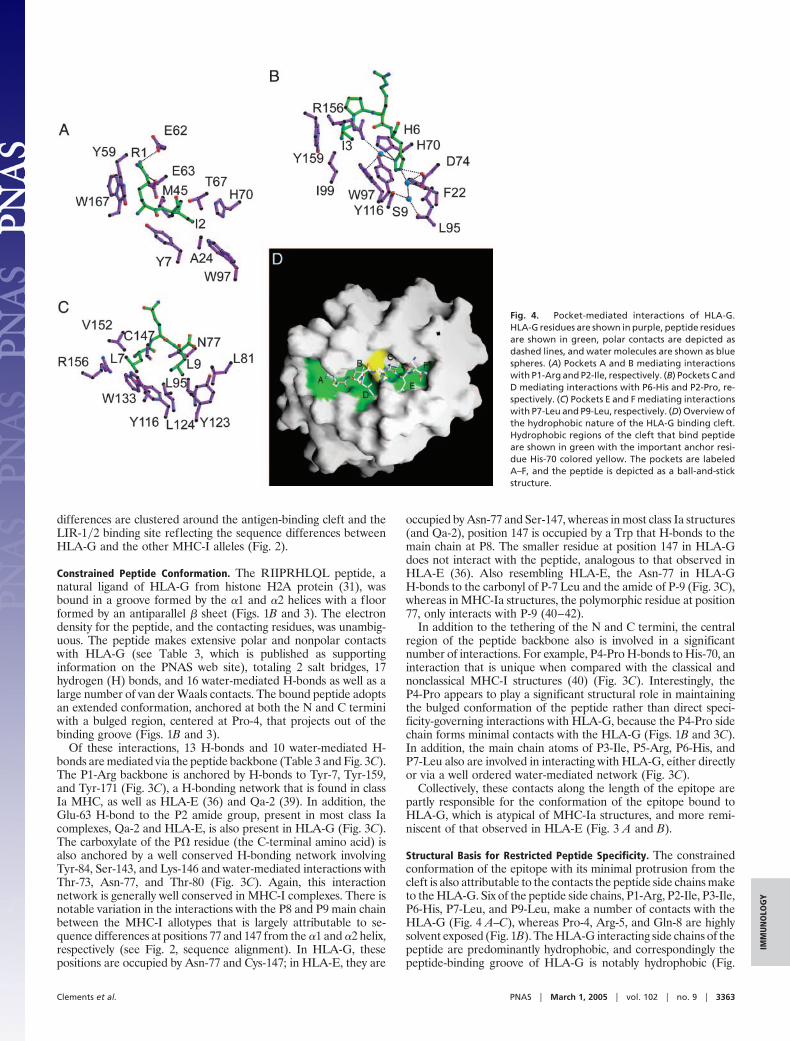
Structural Basis for Restricted Peptide Specificity. The constrainedconformation of the epitope with its minimal protrusion from thecleft is also attributable to the contacts the peptide side chains maketo the HLA-G. Six of the peptide side chains, P1-Arg, P2-Ile, P3-Ile,P6-His, P7-Leu, and P9-Leu, make a number of contacts with theHLA-G (Fig. 4 A–C), whereas Pro-4, Arg-5, and Gln-8 are highlysolvent exposed (Fig. 1B). The HLA-G interacting side chains of thepeptide are predominantly hydrophobic, and correspondingly thepeptide-binding groove of HLA-G is notably hydrophobic (Fig.
Fig. 4. Pocket-mediated interactions of HLA-G.HLA-G residues are shown in purple, peptide residuesare shown in green, polar contacts are depicted asdashed lines, and water molecules are shown as bluespheres. (A) Pockets A and B mediating interactionswith P1-Arg and P2-Ile, respectively. (B) Pockets C andD mediating interactions with P6-His and P2-Pro, re-spectively. (C) Pockets E and F mediating interactionswith P7-Leu and P9-Leu, respectively. (D) Overview ofthe hydrophobic nature of the HLA-G binding cleft.Hydrophobic regions of the cleft that bind peptideare shown in green with the important anchor resi-due His-70 colored yellow. The pockets are labeledA–F, and the peptide is depicted as a ball-and-stickstructure.
Clements et al. PNAS � March 1, 2005 � vol. 102 � no. 9 � 3363
IMM
UN
OLO
GY
4D). The peptide binding groove of HLA-G most closely resemblesthat of HLA-E, where the conformation of the bound peptides arevery similar (Fig. 3 A and B) (with rms deviation of 0.49 Å; see Table2); there is significantly more variation in the conformation of thebound peptide compared with the other MHC-I molecules (rmsdeviation 1.00–1.56 Å; see Table 2 and Fig. 3 A and B).
As with MHC-I molecules, the RIIPRHLQL peptide is accom-modated in the peptide-binding groove by a series of pockets (Fig.4). Pool sequencing of endogenous HLA-G peptide ligands hasidentified putative conserved anchor residues comprising a leucineat the C terminus and proline or small hydrophobic residue at P3,followed by a proline or glycine at P4 (13, 31, 33). Either leucine orisoleucine is found at P2 and there is a preference for a positivelycharged residue at P1 and an aromatic residue at P6. The restrictionof the P1 residue is explained by the interactions of the P1-Arg sidechain within the A pocket, which is closed at the N terminus of thepeptide by Trp-167, (Fig. 4A). The structure of this pocket is similarto both HLA-E (36) and HLA-A2 (43); however, in Qa-2 theresidue at position 167 is a serine leading to a wider open-endedpocket (39). The P1-Arg salt bridges to Glu-62 and Glu-63; more-over, the aliphatic moiety of the P1-Arg is further anchored by vander Waals interactions with Tyr-59 and Trp-167 (Fig. 4A).
The B pocket is deep and hydrophobic, a feature common to anumber of MHC-I molecules. The pocket is lined by Met-45,Ala-24, Tyr-7, Thr-67, His-70, and Trp-97 (Fig. 4A). The hydro-phobic characteristics of the B-pocket are consistent with theselection of a Leu�Ile peptide side chain at P2. In other MHC-Imolecules, hc residue 9 is occupied by larger side chains such ashistidine (HLA-E, HLA-B27, and Qa-2), phenylalanine (HLA-A2), and tyrosine (HLA-B44). However this residue is replaced bya Ser in HLA-G and as such does not participate in B-pocketinteractions, but instead lies at the floor of the C-pocket thataccommodates the P6-His (Figs. 2 and 4B). The substitution of aSer-9 creates a deeper pocket to accommodate the P6-His, whichpoints into the peptide-binding groove and is involved in a networkof interactions. The P6-His H-bonds to Asp-74 and His-70, formswater-mediated H bonds to Ser-9, Leu-95, Trp-97, Tyr-116, andArg-156, and forms van der Waals contacts with His-70 and Trp-97(Fig. 4B). The pocket also is lined by Phe-22 and Tyr-116, andbulkier aromatics at the P6-position also may favorably interact withthese residues. The P6-His�His-70 pairing is unusual; however, theextensive polar network, centered around P6-His, presumablyprovides a favorable environment for this His�His interaction tooccur (44). HLA-E also possesses an unusual His�His pairing(His-9�His-99) at the floor of the cleft that also is involved in anintricate H-bonding network (36). Analogous to HLA-E and Qa-2,the C pocket is more hydrophobic than the classical MHC-Imolecules, where Trp-97 replaces the charged Arg-97 found inmany class Ia structures. Trp-97 also lies at the base of the relativelyshallow D pocket in HLA-G, which accommodates the P3-Ile.HLA-G has a preference for proline or a small hydrophobic residueat P3, and the structural basis for this preference lies in hydrophobicinteractions being made with Tyr-159, Trp-97, and Ile-99. Thepresence of Ile-99 compared with tyrosine in Qa-2 and HLA-A2,or histidine in HLA-E, creates a somewhat deeper D pocket.
P7-Leu resides within the E pocket, making van der Waalscontacts with Tyr-116, Arg-156, Trp-133, Val-152, and Cys-147.Similar to HLA-E (Ser-147), the E pocket of HLA-G is deeper thanthe corresponding pocket in HLA-A2. However, the presence ofArg-156 in HLA-G serves to narrow this pocket, compared withother MHC-I molecules.
The P Leu anchor residue is firmly seated in the hydrophobicF pocket lined by Leu-81, Asn-77, Leu-95, Tyr-116, Tyr-123, andLeu-124, analogous to the ligation of the P Leu preferred byHLA-E. Leu-81 encloses this pocket at the C terminus of theHLA-G-bound peptide as occurs in HLA-A2 and HLA-E, whereasin Qa-2 this position is occupied by an alanine that provides a morespacious F pocket (36).
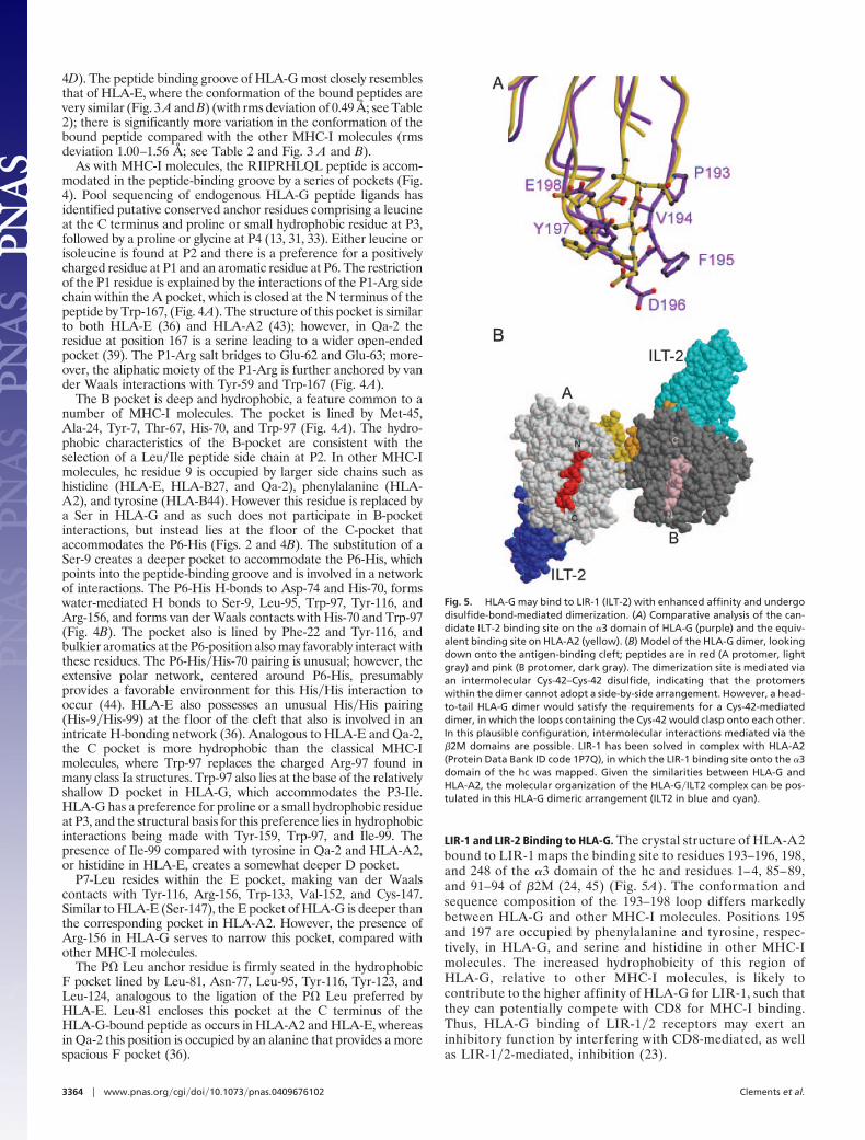
LIR-1 and LIR-2 Binding to HLA-G. The crystal structure of HLA-A2bound to LIR-1 maps the binding site to residues 193–196, 198,and 248 of the �3 domain of the hc and residues 1–4, 85–89,and 91–94 of �2M (24, 45) (Fig. 5A). The conformation andsequence composition of the 193–198 loop differs markedlybetween HLA-G and other MHC-I molecules. Positions 195and 197 are occupied by phenylalanine and tyrosine, respec-tively, in HLA-G, and serine and histidine in other MHC-Imolecules. The increased hydrophobicity of this region ofHLA-G, relative to other MHC-I molecules, is likely tocontribute to the higher affinity of HLA-G for LIR-1, such thatthey can potentially compete with CD8 for MHC-I binding.Thus, HLA-G binding of LIR-1�2 receptors may exert aninhibitory function by interfering with CD8-mediated, as wellas LIR-1�2-mediated, inhibition (23).
Fig. 5. HLA-G may bind to LIR-1 (ILT-2) with enhanced affinity and undergodisulfide-bond-mediated dimerization. (A) Comparative analysis of the can-didate ILT-2 binding site on the �3 domain of HLA-G (purple) and the equiv-alent binding site on HLA-A2 (yellow). (B) Model of the HLA-G dimer, lookingdown onto the antigen-binding cleft; peptides are in red (A protomer, lightgray) and pink (B protomer, dark gray). The dimerization site is mediated viaan intermolecular Cys-42–Cys-42 disulfide, indicating that the protomerswithin the dimer cannot adopt a side-by-side arrangement. However, a head-to-tail HLA-G dimer would satisfy the requirements for a Cys-42-mediateddimer, in which the loops containing the Cys-42 would clasp onto each other.In this plausible configuration, intermolecular interactions mediated via the�2M domains are possible. LIR-1 has been solved in complex with HLA-A2(Protein Data Bank ID code 1P7Q), in which the LIR-1 binding site onto the �3domain of the hc was mapped. Given the similarities between HLA-G andHLA-A2, the molecular organization of the HLA-G�ILT2 complex can be pos-tulated in this HLA-G dimeric arrangement (ILT2 in blue and cyan).
3364 � www.pnas.org�cgi�doi�10.1073�pnas.0409676102 Clements et al.

DiscussionA unique functional role for HLA-G is implied by its extraordinaryexpression in the placenta, unusual promoter elements (26), andlimited peptide repertoire (13, 31). HLA-G has the highest se-quence identity to HLA-Cw3 but nevertheless shows the highestdegree of overall structural-relatedness to the murine homologue,Qa-2 (46). Surprisingly, the peptide-binding groove of HLA-G mostclosely resembles HLA-E, with an extensive network of contactsrevealing the hybrid class Ia�Ib properties of the HLA-G molecule.This bonding network constrains the identity of peptides capable ofbinding HLA-G compared with classical MHC molecules in whichthe peptide sequence is fixed at only two or three anchor positions(40). As with HLA-E (36), the side chains of the residues atpositions P2, P3, P6, P7, and P9 lie within well defined pockets inthe peptide-binding groove; however, the P6 pocket of HLA-Gmust accommodate bulky side chains such as phenylalanine orhistidine. Two of the preferred side chains identified in HLA-Gligands, the positively charged P1 residue and Pro at P4, are solventexposed and appear to stabilize the HLA-G molecule rather thanform hc contacts directly involved in binding. HLA-G acquires mostof its peptide cargo after incorporation into the tapasin-dependent,peptide-loading complex containing the thiol oxido-reductaseER57, involved in disulfide bond rearrangements (30). This findingis consistent with the chaperone-assisted optimization of the con-strained ligand repertoire of HLA-G, but it also could reflect a rolefor ER57 in HLA-G dimerization (29).
HLA-G is proposed to contribute to immunological tolerance ofthe fetus, making the interaction of HLA-G with inhibitory natural
killer and LIR-1�2 receptors a matter of great interest. Although aninteraction between KIR2DL4 and HLA-G has been reported (22),the interaction is not essential for reproduction (47) and remainscontroversial (14).
HLA-G binds the inhibitory LIR-1�2 molecules via �2M and thehc �3 domain (24, 45). In HLA-G, positions 195 and 197 of the �3domain are occupied by phenylalanine and tyrosine, respectively,compared with serine and histidine in other MHC-I molecules. Thisaltered structure and increased hydrophobicity may be the basis forthe higher affinity of HLA-G for LIR-1 compared with otherMHC-I molecules. In addition, the capacity of HLA-G to formdisulfide-linked homodimers (29) might theoretically enhance theavidity of HLA-G for the LIR-1 and -2 receptors. In modeling thedisulfide-bonded HLA-G homodimer, the two HLA-G protomersare oriented head-to-tail, creating an interaction between adjacent�2M molecules that potentially could allow enhanced HLA-Gfunction through oligimerization of LIR-1�2 receptors (Fig. 5B).The data raise the possibility of a novel mode of interaction betweendimeric HLA-G complexes and LIR-1�2 or other immunomodu-latory receptors.
We thank the staff at BioCars (Advanced Photon Source, Chicago) forassistance with data collection. J.R. was supported by a Wellcome TrustSenior Research Fellowship in Biomedical Science in Australia, andC.S.C. was supported by a Monash University Research Fellowship. Thiswork was supported by the National Health and Medical ResearchCouncil, the Australian Research Council, and the Roche Organ Trans-plantation Research Foundation.
1. Margulies, D. & McCluskey, J. (2003) in Fundamental Immunology, ed. Paul,W. (Lippincott, Philadelphia).
2. Bjorkman, P. J. & Parham, P. (1990) Annu. Rev. Biochem. 59, 253–288.3. O’Callaghan, C. A. & Bell, J. I. (1998) Immunol. Rev. 163, 129–138.4. Gao, G. F., Willcox, B. E., Wyer, J. R., Boulter, J. M., O’Callaghan, C. A.,
Maenaka, K., Stuart, D. I., Jones, E. Y., Van Der Merwe, P. A., Bell, J. I. &Jakobsen, B. K. (2000) J. Biol. Chem. 275, 15232–15238.
5. Rammensee, H. G., Falk, K. & Rotzschke, O. (1993) Annu. Rev. Immunol. 11, 213–244.6. Robinson, J., Waller, M. J., Parham, P., de Groot, N., Bontrop, R., Kennedy,
L. J., Stoehr, P. & Marsh, S. G. (2003) Nucleic Acids Res. 31, 311–314.7. Shawar, S. M., Vyas, J. M., Rodgers, J. R. & Rich, R. R. (1994) Annu. Rev.
Immunol. 12, 839–880.8. Geraghty, D. E. (2002) Immunol. Rev. 190, 5–8.9. Braud, V. M., Allan, D. S., O’Callaghan, C. A., Soderstrom, K., D’Andrea, A.,
Ogg, G. S., Lazetic, S., Young, N. T., Bell, J. I., Phillips, J. H., et al. (1998)Nature 391, 795–799.
10. Borrego, F., Ulbrecht, M., Weiss, E. H., Coligan, J. E. & Brooks, A. G. (1998)J. Exp. Med. 187, 813–818.
11. Brooks, A. G., Borrego, F., Posch, P. E., Patamawenu, A., Scorzelli, C. J.,Ulbrecht, M., Weiss, E. H. & Coligan, J. E. (1999) J. Immunol. 162, 305–313.
12. Kovats, S., Main, E. K., Librach, C., Stubblebine, M., Fisher, S. J. & DeMars,R. (1990) Science 248, 220–223.
13. Ishitani, A., Sageshima, N., Lee, N., Dorofeeva, N., Hatake, K., Marquardt, H.& Geraghty, D. E. (2003) J. Immunol. 171, 1376–1384.
14. LeMaoult, J., Le Discorde, M., Rouas-Freiss, N., Moreau, P., Menier, C.,McCluskey, J. & Carosella, E. D. (2003) Tissue Antigens 62, 273–284.
15. Bainbridge, D., Ellis, S., Le Bouteiller, P. & Sargent, I. (2001) Trends Immunol.22, 548–552.
16. Mandelboim, O., Davis, D. M., Reyburn, H. T., Vales-Gomez, M., Sheu, E. G.,Pazmany, L. & Strominger, J. L. (1996) Science 274, 2097–2100.
17. Munz, C., Holmes, N., King, A., Loke, Y. W., Colonna, M., Schild, H. &Rammensee, H. G. (1997) J. Exp. Med. 185, 385–391.
18. Le Bouteiller, P. & Blaschitz, A. (1999) Immunol. Rev. 167, 233–244.19. Hofmeister, V. & Weiss, E. H. (2003) Semin. Cancer Biol. 13, 317–323.20. Colonna, M., Samaridis, J., Cella, M., Angman, L., Allen, R. L., O’Callaghan,
C. A., Dunbar, R., Ogg, G. S., Cerundolo, V. & Rolink, A. (1998) J. Immunol.160, 3096–3100.
21. Allan, D. S., Lepin, E. J., Braud, V. M., O’Callaghan, C. A. & McMichael, A. J.(2002) J. Immunol. Methods 268, 43–50.
22. Rajagopalan, S. & Long, E. O. (1999) J. Exp. Med. 189, 1093–1100.23. Shiroishi, M., Tsumoto, K., Amano, K., Shirakihara, Y., Colonna, M., Braud,
V. M., Allan, D. S., Makadzange, A., Rowland-Jones, S., Willcox, B., et al.(2003) Proc. Natl. Acad. Sci. USA 100, 8856–8861.
24. Chapman, T. L., Heikeman, A. P. & Bjorkman, P. J. (1999) Immunity 11, 603–613.25. Ishitani, A. & Geraghty, D. E. (1992) Proc. Natl. Acad. Sci. USA 89, 3947–3951.
26. Moreau, P., Paul, P., Rouas-Freiss, N., Kirszenbaum, M., Dausset, J. &Carosella, E. D. (1998) Am. J. Reprod. Immunol. 40, 136–144.
27. Hunt, J. S., Petroff, M. G., Morales, P., Sedlmayr, P., Geraghty, D. E. & Ober,C. (2000) Hum. Immunol. 61, 1113–1117.
28. Le Bouteiller, P., Legrand-Abravanel, F. & Solier, C. (2003) Placenta 24, Suppl.A, S10–S15.
29. Boyson, J. E., Erskine, R., Whitman, M. C., Chiu, M., Lau, J. M., Koopman,L. A., Valter, M. M., Angelisova, P., Horejsi, V. & Strominger, J. L. (2002) Proc.Natl. Acad. Sci. USA 99, 16180–16185.
30. Park, B. & Ahn, K. (2003) J. Biol. Chem. 278, 14337–14345.31. Lee, N., Malacko, A. R., Ishitani, A., Chen, M. C., Bajorath, J., Marquardt, H.
& Geraghty, D. E. (1995) Immunity 3, 591–600.32. Gobin, S. J., Wilson, L., Keijsers, V. & Van den Elsen, P. J. (1997) J. Immunol.
158, 3587–3592.33. Diehl, M., Munz, C., Keilholz, W., Stevanovic, S., Holmes, N., Loke, Y. W. &
Rammensee, H. G. (1996) Curr. Biol. 6, 305–314.34. Clements, C. S., Kjer-Nielsen, L., MacDonald, W. A., Brooks, A. G., Purcell,
A. W., McCluskey, J. & Rossjohn, J. (2002) Acta Crystallogr. D 58, 2131–2134.35. Navaza, J. (2001) Acta Crystallogr. D 57, 1367–1372.36. O’Callaghan, C. A., Tormo, J., Willcox, B. E., Braud, V. M., Jakobsen, B. K.,
Stuart, D. I., McMichael, A. J., Bell, J. I. & Jones, E. Y. (1998) Mol. Cell 1, 531–541.37. Brunger, A. T., Adams, P. D., Clore, G. M., DeLano, W. L., Gros, P.,
Grosse-Kunstleve, R. W., Jiang, J. S., Kuszewski, J., Nilges, M., Pannu, N. S.,et al. (1998) Acta Crystallogr. D 54, 905–921.
38. Jones, T. A., Zou, J.-Y., Cowan, S. W. & Kjeldgaard, M. (1991) Acta Crystallogr.A 47, 110–119.
39. He, X., Tabaczewski, P., Ho, J., Stroynowski, I. & Garcia, K. C. (2001) Structure(London) 9, 1213–1224.
40. Madden, D. R. (1995) Annu. Rev. Immunol. 13, 587–622.41. Kjer-Nielsen, L., Clements, C. S., Brooks, A. G., Purcell, A. W., Fontes, M. R.,
McCluskey, J. & Rossjohn, J. (2002) J. Immunol. 169, 5153–5160.42. Macdonald, W. A., Purcell, A. W., Mifsud, N. A., Ely, L. K., Williams, D. S.,
Chang, L., Gorman, J. J., Clements, C. S., Kjer-Nielsen, L., Koelle, D. M., etal. (2003) J. Exp. Med. 198, 679–691.
43. Madden, D. R., Garboczi, D. N. & Wiley, D. C. (1993) Cell 75, 693–708.44. Loewenthal, R., Sancho, J. & Fersht, A. R. (1992) J. Mol. Biol. 224, 759–770.45. Willcox, B. E., Thomas, L. M. & Bjorkman, P. J. (2003) Nat Immunol 4, 913–919.46. Comiskey, M., Goldstein, C. Y., De Fazio, S. R., Mammolenti, M., Newmark,
J. A. & Warner, C. M. (2003) Hum. Immunol. 64, 999–1004.47. Gomez-Lozano, N., de Pablo, R., Puente, S. & Vilches, C. (2003) Eur.
J. Immunol. 33, 639–644.48. Strong, R. K., Holmes, M. A., Li, P., Braun, L., Lee, N. & Geraghty, D. E.
(2003) J. Biol. Chem. 278, 5082–5090.49. Khan, A. R., Baker, B. M., Ghosh, P., Biddison, W. E. & Wiley, D. C. (2000)
J. Immunol. 164, 6398–6405.
Clements et al. PNAS � March 1, 2005 � vol. 102 � no. 9 � 3365
IMM
UN
OLO
GY