crystal structure of a 70s ribosome- trna complex reveals
TRANSCRIPT

Crystal Structure of a 70S Ribosome-tRNA Complex Reveals FunctionalInteractions and RearrangementsAndrei Korostelev,1 Sergei Trakhanov,1 Martin Laurberg,1 and Harry F. Noller1,*1Center for Molecular Biology of RNA and Department of Molecular, Cell and Developmental Biology, University of California, Santa
Cruz, Santa Cruz, CA 95064, USA*Contact: [email protected]
DOI 10.1016/j.cell.2006.08.032
SUMMARY
Our understanding of the mechanism of proteinsynthesis has undergone rapid progress in re-cent years as a result of low-resolution X-rayand cryo-EM structures of ribosome functionalcomplexes and high-resolution structures of ri-bosomal subunits and vacant ribosomes. Here,we present the crystal structure of the Thermusthermophilus 70S ribosome containing a modelmRNA and two tRNAs at 3.7 A resolution. Manystructural details of the interactions betweenthe ribosome, tRNA, and mRNA in the P and Esites and the ways in which tRNA structure isdistorted by its interactions with the ribosomeare seen. Differences between the conforma-tions of vacant and tRNA-bound 70S ribosomessuggest an induced fit of the ribosome structurein response to tRNA binding, including signifi-cant changes in the peptidyl-transferase cata-lytic site.
INTRODUCTION
Ribosomes are the ribonucleoprotein complexes respon-
sible for protein synthesis in all living cells. Detailed knowl-
edge of their molecular structure and the ways in which
they interact with mRNA, tRNA, and other functional
ligands is essential for understanding the mechanism of
this central process of gene expression. The smallest
ribosomes, from bacteria and archaea, have molecular
weights of about 2.5 MDa and are composed of about
60% RNA and 40% protein. Their small (30S) subunits
contain 16S rRNA (�1500 nucleotides) and about 20 pro-
teins; their large (50S) subunits contain 23S rRNA (�2900
nucleotides), 5S rRNA (�120 nucleotides), and more than
30 proteins. Unlike other polymerases, the mechanism of
action of ribosomes appears to be based on their RNA
(Green and Noller, 1997; Nissen et al., 2000; Noller et al.,
1992). The formidable structural complexity of the ribo-
some is compounded by its structural dynamics, which
Cell 12
underlie its functional capabilities. An important challenge,
therefore, is to understand the structure of the ribosome
not only at high resolution but in different complexes
representing its many different functional states.
In recent years, rapid progress has been made in the
determination of ribosome structures. Cryo-EM recon-
structions of ribosomes and many functional ribosome
complexes have provided low-resolution representations
of the ribosome trapped in different states of the transla-
tional process (Agrawal et al., 1999; Frank and Agrawal,
2000; Gao et al., 2003; Stark et al., 2000). These studies
have provided the first indications of large-scale molecu-
lar movements such as relative rotation of the two sub-
units during translocation (Frank and Agrawal, 2000). Until
recently, the most detailed views of the ribosome came
from all-atom crystal structures of the isolated 30S and
50S subunits from Thermus thermophilus, Haloarcula
marismortui, and Deinococcus radiodurans (Ban et al.,
2000; Harms et al., 2001; Wimberly et al., 2000), which
in some cases were cocrystals containing model tRNAs
and mRNAs (Ogle et al., 2001, 2002; Schmeing et al.,
2002, 2003), antibiotics (Brodersen et al., 2000; Hansen
et al., 2002; Pioletti et al., 2001), and translation factors
(Pioletti et al., 2001; Wilson et al., 2005). These structures
have not only revealed the three-dimensional structures of
the rRNAs, ribosomal proteins, and their mutual interac-
tions but have also led to proposals for the mechanisms
of aminoacyl-tRNA selection, catalysis of peptide bond
formation, and the modes of action of many antibiotics.
However, a complete understanding of protein synthesis
requires knowledge of the structure of functional com-
plexes of the whole ribosome, its true biological context.
The first crystal structures of complete ribosomes, from
T. thermophilus 70S complexes, provided a description of
the interactions between tRNA and the ribosome in the
A, P, and E sites and identified the molecular components
of the intersubunit bridges and the path of the mRNA (Yu-
supov et al., 2001; Yusupova et al., 2001). The structure
of a 70S complex containing a model threonyl-tRNA syn-
thetase mRNA and tRNAs bound to the A and P sites has
shown how structure in the 50 leader of the mRNA is able to
regulate initiation of translation (Jenner et al., 2005). More
recently, the structures of complexes containing the
6, 1065–1077, September 22, 2006 ª2006 Elsevier Inc. 1065

release factors RF1 and RF2 have provided insight into the
mechanism of translational termination (Petry et al., 2005).
So far, the structures of these 70S complexes have been
limited to resolutions of between 5.5 and 7 A, preventing
a more complete understanding of the molecular basis
of the interactions between the ribosome and its main
functional ligands as well as the ways in which they per-
turb each other’s structures during translation. In a recent
landmark paper, the structures of two conformational
states of the Escherichia coli 70S ribosome were solved
at 3.5 A resolution, representing the most detailed view
of the complete ribosome obtained so far and providing
new information about the structural basis of ribosome
dynamics (Schuwirth et al., 2005). The impact of these
structures on our understanding of protein synthesis is
nevertheless limited by the absence of mRNA and tRNA.
Here, we report the crystal structure of a complex of the
T. thermophilus 70S ribosome containing a model mRNA
and two tRNAs, bound to the P and E sites, at 3.7 A reso-
lution. The improved resolution enables visualization of the
nature of molecular interactions between the ribosome,
mRNA, and full-length tRNAs for the first time. Moreover,
it shows how the conformations of both the tRNAs and
the ribosome are perturbed by formation of the tRNA-
ribosome complex.
RESULTS AND DISCUSSION
Structure Determination
A complex of the Thermus thermophilus 70S ribosome
containing a 10 nucleotide model mRNA and two tRNAs
was crystallized in a new crystal form (Yusupov et al.,
2001; see also Figure S2 in the Supplemental Data avail-
able with this article online), belonging to the I422 space
group, with unit-cell dimensions a = b = 507.8 A, c =
689.5 A, that diffracts to 3.7 A (Table 1). Shortening of
the c axis by �110 A relative to the previous crystal form
(Yusupov et al., 2001) is mainly due to rearrangement of
one of the lattice contacts, in which the stalks of 5S
rRNA symmetry mates run parallel to each other rather
than stacking coaxially. An E. coli tRNAPhe was bound to
the P site, and an endogenous tRNA (most likely a hetero-
geneous mixture of T. thermophilus tRNAs) occupied the
E site.
Structure determination was done using the molecular
replacement method, followed by rigid-body refinement
using successively finer rigid-body groups. Finally, alter-
nating rounds of reciprocal- and real-space (Korostelev
et al., 2002) refinement, interspersed with manual refitting,
were used, resulting in a value for Rfree of 0.35 (see Exper-
imental Procedures). The positions of RNA bases, riboses,
and phosphates and the main chains of the proteins could
be assigned to features of the electron density map
(Figure 1C; Figures 2A–2C; Figure S1), with the exception
of proteins L7/L12, L10, L11, L31, and L33; positions 55-
160 of L1; the 30 end of the mRNA including the A codon;
and the very 50 and 30 ends of 16S rRNA, which were
disordered.
1066 Cell 126, 1065–1077, September 22, 2006 ª2006 Elsevier
The 70S P Site
The roles of the P site are to hold the peptidyl-tRNA tightly
to prevent loss of the nascent chain, to maintain the cor-
rect translational reading frame when the A site is vacant,
and to bind the initiator tRNA during initiation of protein
synthesis. In our complex, an E. coli tRNAPhe is held in po-
sition by numerous interactions with 16S and 23S rRNA
and three ribosomal proteins, in addition to base pairing
with a UUC codon (Figures 1A and 1B; Table S1). In the
30S P site, the main contacts with the anticodon stem-
loop (ASL) and codon are made by 16S rRNA, bolstered
by interactions with the C-terminal tail of protein S13, as
described below. In the 50S P site, the minor groove of
the P-tRNA D stem rests on the minor groove of helix 69
of 23S rRNA. The P-tRNA elbow contacts an extended
b hairpin of protein L5. Nucleotide 2 of its acceptor stem
interacts with the P stem (helix 80) of 23S rRNA. The N-ter-
minal end of protein L27, which approaches the end of
the acceptor stem, is well ordered in our electron density
map beginning only at position 9; its N-terminal tail is
disordered. The b hairpin at position 82 of protein L16
approaches within 10 A of the major-groove face of the
acceptor stem of P-tRNA at positions 2 and 64; although
not in direct contact, its position suggests that it could
interact transiently with the tRNA as it moves between
the A and P sites.
Ribosomal interactions with tRNA in the 30S P site con-
trast sharply with those in the 30S A site, which, in keeping
Table 1. Summary of Crystallographic Data andRefinement
Data Statistics
Unit-cell dimensions, A a = b = 507.8, c = 689.5
Resolution, A 3.7–75 (3.8)
Number of unique reflections 465,894 (33,259)
Completeness, % 99.3 (96.6)
Multiplicity 5.4 (4.1)
Mean I/s(I) 3.0 (1.5)
Rpim 0.149 (0.467)
Refinement Statistics
Resolution, A 3.7–30.0
Test-set (free) reflections, % 2.5
R/Rfree (CNS) 0.358/0.366
R/Rfree (REFMAC) 0.346/0.347
Deviation from ideal
bond lengths, A
0.013
Deviation from idealbond angles, �
1.36
All measured reflections were used in refinements with CNS
and REFMAC. Rpim denotes the precision-indicating mergingR factor (Weiss, 2001). Statistics for the highest resolution
shell are given in parentheses.
Inc.

Figure 1. The 30S Subunit P Site
(A) Stereo diagram showing details of interactions between 16S rRNA and the codon-anticodon helix.
(B) Interactions between the anticodon stem and the ribosome.
(C) Electron density map of the 30S P site (composite omit map contoured at 2.0s) showing the codon (yellow), anticodon (orange), and surrounding
features of 16S rRNA (cyan).
with its role in selecting the correct aminoacyl-tRNA, con-
tacts the tRNA and mRNA with only four nucleotides of
16S rRNA, three of which fit precisely into the minor
groove of the codon-anticodon helix (Ogle et al., 2001).
In the P site, eight nucleotides of the P site codon and
tRNA anticodon stem-loop (ASL) are fixed in position by
interactions with ten nucleotides of 16S rRNA, which in-
clude four of its eleven posttranscriptionally methylated
nucleotides (Guymon et al., 2006). In T. thermophilus ribo-
somes, these interactions are bolstered by interactions
with the extended C-terminal tail of protein S13. Four
universally conserved nucleotides contact the P codon
backbone of mRNA, forming H bonds with all three codon
nucleotides.
Phosphate 1 of the P codon is H bonded to the N1 and
N2 positions of G926. m3U1498 packs against the ribose-
phosphate backbone of nucleotides 1 and 2 of the P
codon, making an H bond from its O4 position to the 20
Cell 12
hydroxyl of codon nucleotide 2. The phosphate group of
codon nucleotide 3 H bonds with the 4-methylamino
group of m4Cm1402. No contacts are made between
the ribosome and the P codon bases. In addition to base
pairing with the P codon, the anticodon of the P site
tRNA is buttressed by stacking with base m5C1400 on
its wobble base (G34), as predicted more than 20 years
ago by Ofengand, Zimmermann, and coworkers (Prince
et al., 1982), and by packing of m22G966 against ribose
34 (Figure 1B). The sole protein contacts are with the
highly basic C-terminal tail of protein S13, at phosphates
31 and 36 (Figure 1B). The extensive packing of 16S
rRNA around the third base pair contrasts with what is ob-
served for the 30S A site, where minimal interactions
explain the prevalence of third-position wobble in the
genetic code (Ogle et al., 2001). In contrast, during trans-
lational initiation in the 30S P site, initiator tRNAfMet can
recognize AUG, GUG, or UUG as methionine start
6, 1065–1077, September 22, 2006 ª2006 Elsevier Inc. 1067

Figure 2. The Peptidyl-Transferase P
Site
(A) Stereoview of the electron density map
showing fitting of 23S rRNA features in the pep-
tidyl-transferase P site (composite omit map
contoured at 1.8s).
(B) Stereoview of superposition of 23S rRNA
from the H. marismortui 50S complex (PDB ID
code 1QVG) bound with a CCA oligonucleotide
(Schmeing et al., 2003) on the T. thermophilus
70S complex (blue) in the context of the elec-
tron density map, showing differences in the
positions of A2450 and A2451.
(C) Stereoview of the T. thermophilus 70S com-
plex showing fitting of 23S rRNA (blue) and
tRNA (orange) features in the electron density
map.
(D) Superposition of structures in (B) in a view
showing movement of A2450 and A2451 of
23S rRNA (blue) and A76 of P-tRNA (orange)
toward each other in the 70S complex, relative
to the 50S complex (magenta).
(E) Detailed view showing juxtaposition of ri-
bose 76 of P-tRNA (orange) with the protonated
C2063-A2450 base pair and A2451 of 23S
rRNA (blue).
codons—i.e., wobble is tolerated in the first rather than in
the third position. The contrasting architectures of the 30S
A and P sites may underlie their different rules for codon-
anticodon pairing.
Interactions with the anticodon stem involve both back-
bone and minor-groove interactions, focused on nucleo-
1068 Cell 126, 1065–1077, September 22, 2006 ª2006 Elsevier
tides 30 and 31 (Figure 1B). Extensive interactions, includ-
ing H bonding and van der Waals packing, are made
between the backbones of nucleotides 1229, 1230, and
1341 and the tRNA (Figure 1B). In the same region, bases
G1338 and A1339 are positioned in the minor groove of
the anticodon stem, poised to make potential type II and
Inc.

type I A-minor interactions with the 29-41 and 30-40 base
pairs, which would require movement of this region of the
head by �1.5 A. These interactions are believed to medi-
ate discrimination of initiator tRNA during formation of the
30S initiation complex (Dallas and Noller, 2001; Lancaster
and Noller, 2005). In their analysis of the structure of the
70S ribosome, Schuwirth et al. (2005) pointed out that
there is a structural barrier to movement of tRNA from
the P to E sites, created by the ridge containing G1338
and A1339 and the 790 loop, which are separated by
only �13 A in their structures. This barrier is maintained
in our structure, where the gap is reduced by only a fraction
of an angstrom, requiring movement of the head of the
30S subunit to permit translocation, as proposed by Schu-
wirth et al. The positions of the 16S rRNA P site nucleo-
tides in the vacant ribosome superimpose upon those in
the tRNA-containing complex around the codon-antico-
don interaction (with the exception of the flipped
m22G966), but the nucleotides that contact the anticodon
stem are displaced by several angstroms due to move-
ment of the head of the 30S subunit. The observed 16S
rRNA-tRNA contacts are in agreement with chemical
probing studies in which binding of tRNA to the P site pro-
tected G926, m2G966, G1338, A1339, and C1400 from at-
tack by the base-specific probes kethoxal and dimethyl
sulfate (Moazed and Noller, 1986, 1991); modification of
three of these bases (G926, m2G966, and G1338) was
shown to interfere with binding of tRNA to the E. coli
30S P site (von Ahsen and Noller, 1995).
The Peptidyl-Transferase Center
Studies on the mechanism of catalysis of peptide bond
formation by the ribosome have been influenced by
high-resolution structures of isolated 50S subunits bound
with oligonucleotide analogs of tRNA, including a con-
struct designed to mimic the acceptor ends of the A and
P site tRNAs joined together in the transition state of the
peptidyl-transferase reaction (Nissen et al., 2000; Schme-
ing et al., 2005a). These studies have shown that the cat-
alytic site is composed exclusively of RNA, a conclusion
that is also supported by our results. Surprisingly few dif-
ferences were observed for the positions of the 23S rRNA
nucleotides of the substrates or the surrounding features
of the peptidyl-transferase center (PTC) between the
structures of vacant 50S subunits and those of most
50S-substrate complexes or even the E. coli 70S ribo-
some, apart from A2602, whose orientation has been
found to be highly variable (Ban et al., 2000; Harms
et al., 2001; Schuwirth et al., 2005). The first indications
of structural mobility in the PTC come from a more recent
study, where it was found that the presence of a CCA-con-
taining A site substrate analog induces conformational
shifts of several nucleotides, including U2506, G2583,
and U2584-2585 as well as the substrate analog, resulting
in exposure of the carbonyl group of the peptidyl substrate
to attack by the aminoacyl group (Schmeing et al., 2005b).
In our structure, which represents the first detailed de-
scription of the interactions between a full-length P site
Cell 1
tRNA and a complete 70S ribosome, the state of our de-
acylated tRNA is expected to resemble most closely that
of a tRNA bound to the P site immediately following pep-
tide bond formation, with the difference that the A site of
our complex is not occupied by peptidyl-tRNA. When
compared with the structure of an H. marismortui 50S
complex containing a CCA oligonucleotide bound to the
P site (as well as to the A and E sites), our electron density
map (Figure 2A) shows significant movement of critical nu-
cleotides in the catalytic site (Figure 2B). It should be kept
in mind that the comparisons presented here need to take
into account possible phylogenetic structural differences
between T. thermophilus, E. coli, and H. marismortui ribo-
somes and the effects of different crystal lattice contacts
and crystallization conditions on ribosome conformation
as well as the different kinds of complexes that were
used. Nevertheless, detailed comparison of the structure
of the peptidyl-transferase region of our 70S complex
with those found for model complexes of the H. marismor-
tui 50S subunit and the vacant E. coli 70S ribosome shows
that, apart from movement of certain specific nucleotides
in the PTC, the positions of most nucleotides in the sur-
rounding structure (e.g., C2065 and U2449) are superim-
posable (Figure 2B).
One of the shifted nucleotides is A2451 (Figures 2B and
2D), previously implicated in the peptidyl-transferase cat-
alytic mechanism because of the proximity of its N3 to a
critical oxygen in the bound Yarus transition-state analog
(Nissen et al., 2000). In our structure, the N3 of A2451
moves toward the O30 position of ribose 76, resulting in
disruption of the A2451-G2102-G2482 base triple. Cou-
pled with this is a movement of the noncanonical A2450-
C2063 base pair and A76 of the P site tRNA toward
each other, bringing the A-C pair within H-bonding dis-
tance of the 20OH group of A76 (Figure 2E), which has
been shown to play an essential role in catalysis (Dorner
et al., 2003; Quiggle et al., 1981; Weinger et al., 2004).
This raises the possibility that the A-C pair, which is ex-
pected to have a pKa in the range of 6.0–6.5 (Cai and
Tinoco, 1996), acts as a proton donor/acceptor, by analogy
with the active-site histidines in many protein enzymes.
Studies of the pH-rate dependence of the peptidyl-trans-
ferase reaction identified a ribosomal group with an appar-
ent pKa of �7.5 (Fahnestock et al., 1970), and it was sug-
gested that this group could be the universally conserved
A2450-C2063 wobble pair (Katunin et al., 2002). Replace-
ment of the A2450-C2063 pair by an isosteric but un-
charged G-U wobble pair results in loss of the character-
istic pKa as well as a 200-fold or more decrease in the
catalytic rate using the puromycin reaction, suggesting
that the protonated A-C pair is responsible for the pH-
rate dependence and that peptidyl-transferase activity
depends on the uncharged (deprotonated) form (Hesslein
et al., 2004). However, more recent studies by the same
group using a full-length aminoacyl-tRNA as an A site
substrate have shown disappearance of the pH-rate de-
pendence in the pH range 6.0 to 9.0, suggesting that the
titratable group is important for positioning the model
26, 1065–1077, September 22, 2006 ª2006 Elsevier Inc. 1069
puromycin substrate and calling into question possible
acid-base catalysis by the ribosome (Bieling et al.,
2006). One caveat of these experiments is that binding
of full-length aminoacyl-tRNA substrate is rate limiting
and so could mask changes in rate for the chemical step
of the peptidyl-transferase reaction. In a second experi-
ment, where the a-amino group of the aminoacyl-tRNA
was replaced by hydroxyl, the pH-rate dependence was
also not observed; however, in this case, the rate of the re-
action was reduced by >50,000-fold, raising the possibility
that details of the catalytic mechanism might differ from
those of authentic peptide bond formation. Brunelle
et al. (2006) recently showed that use of (50) C-P puromy-
cin in place of puromycin as A site substrate eliminated the
pKa attributable to the ribosomal structural change, sug-
gesting that base pairing of C75 of the A site substrate
with G2553 triggers this change in a way that removes
its pH dependence. Although the potential role of the non-
canonical A2450-C2063 base pair in the peptidyl-transfer-
ase reaction remains an open question, the ability of this
critical region of the ribosome to undergo movement
needs to be taken into consideration in formulating mech-
anistic models involving the 50S P site.
The 70S E Site
The E (exit) site binds deacylated tRNA prior to its release
from the ribosome and has a virtually absolute specificity
for tRNA bearing a free ribose at its acceptor end. Its role is
thought to be to provide a favorable free-energy difference
for movement of tRNA out of the P site during transloca-
tion (Lill et al., 1989; Moazed and Noller, 1989b). Indeed,
destabilization of interactions between tRNA and the
50S E site has been found to inhibit translocation, most
likely by interfering with formation of the intermediate P/
E hybrid state (Feinberg and Joseph, 2001; McGarry
et al., 2005). In our complex, the E site of the 70S ribosome
is filled by an endogenous tRNA that most likely repre-
sents a heterogeneous mixture of tRNA species. Refine-
ment statistics indicate that the E site is fully occupied;
although the electron density map is somewhat weaker
for its D, T, and anticodon loops, density for the stems
of E-tRNA is as strong as that observed for the P-tRNA.
The weaker density for the loops and the heterogeneity
of the tRNAs in the E site make it difficult to interpret the
state of codon-anticodon pairing in the E site, an issue
of potential relevance to a proposed reciprocal allosteric
interaction between the ribosomal A and E sites (Nierhaus,
1990). A definitive answer to this question will thus have to
await the structure of a complex containing a cognate
tRNA bound in the E site.
The 70S E site holds the deacylated tRNA by interac-
tions with 16S rRNA, 23S rRNA, and three ribosomal pro-
teins (Table S1). In the 30S subunit, 16S rRNA interacts
with the E site ASL exclusively via backbone-backbone
interactions, explaining why no E site tRNA footprint was
observed for 16S rRNA using base-specific chemical
probes (Moazed and Noller, 1986, 1990). Ribose 694
and phosphate 695 pack against phosphate 39 of the
1070 Cell 126, 1065–1077, September 22, 2006 ª2006 Elsevie
E-tRNA anticodon stem, possibly mediated by a magne-
sium ion, and phosphate 1340 packs on ribose 35 in the
anticodon loop. The sole 30S protein interaction is with
the C-terminal a helix of protein S7, which contacts the
E-tRNA backbone at ribose 41. Guanine 693 of 16S
rRNA packs against ribose 1 of the E codon. The previ-
ously observed protection of G693 from kethoxal by bind-
ing of tRNA or an ASL to the 30S P site (Moazed and Nol-
ler, 1986, 1990) is likely due to contact between G693 and
the E codon that is induced upon formation of the flanking
P site codon-anticodon interaction.
In the 50S subunit, the elbow of E site tRNA contacts
both protein L1 and the head of the L1 stalk (helices 76-
78) of 23S rRNA, corresponding to a large-scale move-
ment of the stalk relative to the structures of the vacant
70S ribosome (Schuwirth et al., 2005) or the isolated 50S
subunit (Harms et al., 2001). Reversal of this movement
of the L1 stalk would allow release of the tightly seques-
tered deacylated tRNA from the ribosome (Figure 3A).
Contact between the tRNA and 23S rRNA involves stack-
ing of the tertiary G19-C56 base pair of tRNA on the
sheared G2112-A2169 tertiary pair of 23S rRNA (Fig-
ure 3B). This interaction accounts for the protection of
G2112 and A2169 from chemical probes by E site tRNA
(Moazed and Noller, 1989a). Protein L1 contacts the E-
tRNA elbow at J55 in the T loop with the tip of the b hairpin
around Lys167 and contacts the T stem backbone at po-
sitions 61-62 from the end of another b hairpin at Arg52
and Arg53. The contribution of the tRNA elbow contacts
to E site binding is clear from the greatly impaired E site
binding of ribosomes lacking L1 (L. Hoang and H.F.N.,
unpublished data).
The minor-groove surface at the end of the acceptor
stem of the E-tRNA makes extensive backbone-back-
bone contacts at nucleotides 70-71 with the minor groove
of helix 68 of 23S rRNA (Figure 3C). These include a ri-
bose-zipper interaction (Cate et al., 1996) between the
20OH of ribose 1851 of 23S rRNA and the base and ribose
of G71 of E-tRNA (Figure 3D). The functional importance of
this interaction was demonstrated by Feinberg and Jo-
seph (2001), who have shown that methylation of ribose
71 of P site tRNA blocks EF-G-dependent translocation.
Since no ribosomal contacts are made with ribose 71 in
the 70S P site, inhibition of translocation must be due to
the inability of the methylated deacylated tRNA to move
from the P/P into the P/E hybrid state. Protein L28 con-
tacts the backbone of the acceptor end of E-tRNA at po-
sitions 73 and 74 via basic side chains in its extended
b hairpin around residues 38-39 (Figure 4A). Although
L33 has been crosslinked to the 30 adenosine of E-tRNA
(Kirillov et al., 2002), we have not yet placed this protein
in our structure. Crucial to E site binding is the 30-terminal
adenosine of tRNA, which must be deacylated and un-
modified (Feinberg and Joseph, 2001; Lill et al., 1988). In
our structure, A76 is recognized and fixed in position by
extensive stacking and H-bonding interactions with 23S
rRNA (Figure 4A). As in the complex between the H. mar-
ismortui 50S subunit and a CCA-containing RNA hairpin
r Inc.
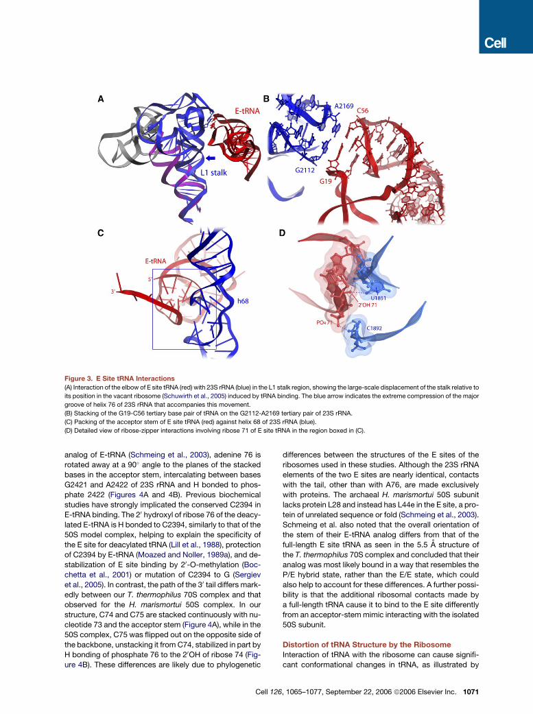
Figure 3. E Site tRNA Interactions
(A) Interaction of the elbow of E site tRNA (red) with 23S rRNA (blue) in the L1 stalk region, showing the large-scale displacement of the stalk relative to
its position in the vacant ribosome (Schuwirth et al., 2005) induced by tRNA binding. The blue arrow indicates the extreme compression of the major
groove of helix 76 of 23S rRNA that accompanies this movement.
(B) Stacking of the G19-C56 tertiary base pair of tRNA on the G2112-A2169 tertiary pair of 23S rRNA.
(C) Packing of the acceptor stem of E site tRNA (red) against helix 68 of 23S rRNA (blue).
(D) Detailed view of ribose-zipper interactions involving ribose 71 of E site tRNA in the region boxed in (C).
analog of E-tRNA (Schmeing et al., 2003), adenine 76 is
rotated away at a 90� angle to the planes of the stacked
bases in the acceptor stem, intercalating between bases
G2421 and A2422 of 23S rRNA and H bonded to phos-
phate 2422 (Figures 4A and 4B). Previous biochemical
studies have strongly implicated the conserved C2394 in
E-tRNA binding. The 20 hydroxyl of ribose 76 of the deacy-
lated E-tRNA is H bonded to C2394, similarly to that of the
50S model complex, helping to explain the specificity of
the E site for deacylated tRNA (Lill et al., 1988), protection
of C2394 by E-tRNA (Moazed and Noller, 1989a), and de-
stabilization of E site binding by 20-O-methylation (Boc-
chetta et al., 2001) or mutation of C2394 to G (Sergiev
et al., 2005). In contrast, the path of the 30 tail differs mark-
edly between our T. thermophilus 70S complex and that
observed for the H. marismortui 50S complex. In our
structure, C74 and C75 are stacked continuously with nu-
cleotide 73 and the acceptor stem (Figure 4A), while in the
50S complex, C75 was flipped out on the opposite side of
the backbone, unstacking it from C74, stabilized in part by
H bonding of phosphate 76 to the 20OH of ribose 74 (Fig-
ure 4B). These differences are likely due to phylogenetic
Cell 1
differences between the structures of the E sites of the
ribosomes used in these studies. Although the 23S rRNA
elements of the two E sites are nearly identical, contacts
with the tail, other than with A76, are made exclusively
with proteins. The archaeal H. marismortui 50S subunit
lacks protein L28 and instead has L44e in the E site, a pro-
tein of unrelated sequence or fold (Schmeing et al., 2003).
Schmeing et al. also noted that the overall orientation of
the stem of their E-tRNA analog differs from that of the
full-length E site tRNA as seen in the 5.5 A structure of
the T. thermophilus 70S complex and concluded that their
analog was most likely bound in a way that resembles the
P/E hybrid state, rather than the E/E state, which could
also help to account for these differences. A further possi-
bility is that the additional ribosomal contacts made by
a full-length tRNA cause it to bind to the E site differently
from an acceptor-stem mimic interacting with the isolated
50S subunit.
Distortion of tRNA Structure by the Ribosome
Interaction of tRNA with the ribosome can cause signifi-
cant conformational changes in tRNA, as illustrated by
26, 1065–1077, September 22, 2006 ª2006 Elsevier Inc. 1071

Figure 4. Interactions between the CCA Tail of tRNA and the 50S E Site
Comparison of the binding and conformations of (A) the CCA tail of tRNA to the E site of the T. thermophilus 70S complex and (B) a CCA-containing
RNA hairpin to the H. marismortui 50S E site (Schmeing et al., 2003).
cryo-EM images of the EF-Tu ternary complex bound to
the ribosome (Valle et al., 2003). In that study, tRNA was
visualized both in the A/T state, bound to the ribosome
in complex with EF-Tu, and in the A/A state. In the A/T
state, the anticodon arm of the tRNA is bent by more
than 30� to allow initial codon recognition, while in the
A/A state, the tRNA is bent by about 15�. The hinge point
was localized to the junction of the anticodon and D stems
at or near the purine-purine base pair between positions
26 and 44.
Our structure shows that binding of tRNA to the ribo-
somal P and E sites causes significant distortion of the
structure of tRNA when compared to the 2.0 A crystal
structure of yeast tRNAPhe (Jovine et al., 2000), which
was used as the initial model for our refinements. Root-
mean-square differences (rmsds) between backbone
phosphorus atoms of P- and E-tRNA with respect to those
of yeast tRNAPhe are 2.1 and 2.0 respectively. Both tRNAs
are kinked at the junction of the anticodon and D stems,
centered on the A26-G44 purine-purine base pair, bend-
ing the body of the tRNA relative to the ASL (Figures 5A–
5D). Kinking of the P-tRNA is most pronounced, causing
it to bend toward the large subunit by about 10� (Fig-
ure 5B). In addition, there is a partial unwinding of the D
stem relative to the anticodon stem, resulting in a rotation
of the body of the P-tRNA relative to the ASL by about 10�
around the axis of the anticodon stem, orienting it slightly
toward the A site (Figure 5A). The body of the E-tRNA also
rotates by about 10� relative to the ASL, but around an axis
drawn between the 26-44 base pair and its 30 acceptor
end, moving its elbow in the direction of the L1 stalk (Fig-
ures 5C and 5D). Interestingly, kinking has been observed
to be most extreme in the A/T state, decreasing incremen-
tally as the tRNA passes from the A/T through the A/A,
P/P, and E/E states. If kinking represents a higher-energy
state than the free state, as suggested by previous studies
(Auffinger et al., 1999; Ehrenberg et al., 1979; Friederich
et al., 1998), the energy to drive tRNA movement might
1072 Cell 126, 1065–1077, September 22, 2006 ª2006 Elsevier
derive in part from gradual relaxation of the conforma-
tional energy introduced in the initial A/T binding state by
EF-Tu and GTP (Valle et al., 2003). Another notable
change is seen in the anticodon loops, as suggested by
our previous 5.5 A structure (Yusupov et al., 2001); the
P-tRNA anticodon loop is wider and more rounded than
that of the isolated tRNA, while the apex of the E-tRNA
loop is narrow and more sharply kinked (Figure 5E).
Conformational Differences between Different
Ribosome Structures
Several large-scale rearrangements are revealed by com-
parison of the structure from the present crystal form with
the previous 5.5 A structure of a similar T. thermophilus
tRNA-mRNA-ribosome complex (Yusupov et al., 2001).
The rmsds for backbone phosphorus atoms of the 30S
and the 50S subunits between ribosomes from the two
crystal forms are 2.2 and 2.5 A, respectively. If the two
structures are superimposed on their 23S rRNAs, it can
be seen that, in our model, both the head and body of
the small subunit move relative to the large subunit (data
not shown). Viewed from the interface, the head rotates
counterclockwise and the body clockwise, each by about
2�, around an axis perpendicular to the interface plane,
closing the gap between the head and shoulder at 16S
rRNA nucleotides 422 and 1045 by �5 A. At the same
time, the top of the head moves toward the 50S subunit
by about 4 A. In the 50S subunit, the C-terminal domain
of L9, which makes a crystal contact with the 30S subunit
of a symmetry mate, is displaced by about 10 A down-
ward, away from the L1 stalk.
Changes in ribosome structure in response to tRNA
binding are suggested by comparison of our structure
with that of the vacant 70S ribosome (Schuwirth et al.,
2005). Our comparison is made with E. coli structure I,
whose structure is most similar to ours (Schuwirth et al.,
2005). Since both structures come from eubacterial ribo-
somes, in addition to the fact that structural features
Inc.

Figure 5. Distortion of tRNA Structure in
the P and E Sites
(A and B) P-tRNA.
(C and D) E-tRNA.
(E) Anticodon loop deformations showing the
differences between the loop conformations
of P-tRNA (orange), E-tRNA (red), and the crys-
tal structure of free tRNAPhe (Jovine et al.,
2000).
involved in ribosomal function are often universally con-
served, many functionally relevant conformational
changes are unlikely to be due to species differences. Dif-
ferences between the tRNA-ribosome complexes and va-
cant ribosomes are even more pronounced (rmsd values
for the 30S and 50S subunits of 3.0 and 2.6 A, respec-
tively). In the 30S subunit (Figure 6A), the most prominent
difference is seen for the head of the subunit, which moves
as a rigid body (Schuwirth et al., 2005), pivoting around the
neck (helix 27 of 16S rRNA) such that top of the head un-
dergoes a movement of about 15 A. As a result, the 1240
and 1340 regions of 16S rRNA clash strongly with the po-
sitions of the P- and E-tRNAs, as seen in our structure
(Figure 6C). This particular difference is unlikely to be at-
tributable simply to the presence or absence of tRNA
since cryo-EM reconstructions of vacant ribosomes do
not appear to undergo such movement (Frank and
Agrawal, 2000). Elsewhere in the small subunit, localized
differences are seen for individual helices, including the
‘‘spur’’ (helix 6), helix 23 in the right-hand side of the plat-
form, and in the middle of the penultimate stem (helix 44).
The latter difference is especially interesting because of its
location close to the axis of rotation for the relative move-
ment of the 30S and 50S subunits (Gao et al., 2003) and
because of the apparent localized mobility of the middle
of the penultimate stem.
In the 50S subunit, the most dramatic conformational
difference is found in the L1 stalk, which is displaced by
about 30 A away from the P site in the vacant ribosome,
reflecting a movement that is most likely coupled to re-
lease of tRNA from the E site. It opens even further in
the free 50S subunit (Harms et al., 2001), pivoting around
Cell 1
nucleotides G2100-U2189 of helix 76 by 30� with respect
to the structure of the 70S complex, resulting in displace-
ment of the top of the L1 stalk by about 40 A (Figure 3A and
Figure 6B). Movement of the L1 stalk between all three
structures can be ascribed to a localized flexibility be-
tween nucleotides 2187-2193 and 2096-2102. The major
groove of this part of helix 76 is dramatically compressed
in the 70S functional complex (Figure 3A), where the L1
stalk moves toward the interface to contact the elbow of
E site tRNA, as discussed above. The distorted region is
rich in G-U wobble pairs, which are present in nearly all
species. The presence of G-U pairs in double helices
has been shown to increase their deformability (Chang
et al., 1999; Ramos and Varani, 1997), in this case en-
abling large-scale movement of the L1 stalk.
Structural differences are also observed between our
structure and the E. coli structure in the interactions form-
ing two of the dozen intersubunit bridges that are respon-
sible for joining the 30S and 50S subunits. These are of
interest because of their role in enabling the intersubunit
movement that occurs during the translocation step of
protein synthesis (Gao et al., 2003). The RNA and protein
components of the bridges were first identified in lower-
resolution crystal structures of T. thermophilus 70S ribo-
somes (Cate et al., 1999; Culver et al., 1999; Yusupov
et al., 2001) and described in detail for the E. coli struc-
tures (Schuwirth et al., 2005). The bridges in the structure
presented here are very similar in detail to those described
by Schuwirth et al., with the exception of bridges B1a and
B1b, which can be explained by the different positions of
the head in the three structures. In bridge B1a, which con-
nects protein S13 in the head of the 30S subunit with the
26, 1065–1077, September 22, 2006 ª2006 Elsevier Inc. 1073

Figure 6. Conformational Variations between Different Ribosome Crystal Structures
(A and B) Comparison of (A) 16S and (B) 23S rRNAs in vacant E. coli (Schuwirth et al., 2005) and tRNA-occupied T. thermophilus ribosomes (this work).
Colors indicate the rmsd values for individual nucleotides between the two structures: 0–2 A, cyan (16S rRNA) or gray (23S rRNA); 2–4 A, yellow; 4–7 A,
gold; 7–10 A, orange; 10–14 A, red; >14 A, magenta.
(C) Comparison of the positions of features of the 30 major domain of 16S rRNA between the vacant E. coli 70S ribosome (magenta) (Schuwirth et al.,
2005) and the T. thermophilus tRNA-containing complex (cyan), showing clash between the 16S rRNA in the E. coli structure with P-tRNAs (orange)
and E-tRNAs (red) from the T. thermophilus complex.
A site finger (helix 38) of 23S rRNA and is disordered in
both E. coli structures, nucleotides 886-888 of 23S rRNA
make two separate contacts with S13 at positions 79-83
and 93-94. Bridge B1b, which connects the head of the
30S subunit with the central protuberance of the 50S sub-
unit via interactions between proteins S13 and L5, takes
two different forms in the two E. coli structures (Schuwirth
et al., 2005). Our structure has a third form (data not
shown), in which three different regions of S13 contact
L5, none of which are the same as those observed in the
E. coli structures.
Conclusion
The increased resolution of this 70S ribosome complex
provides a greatly enhanced view of the details of ribo-
1074 Cell 126, 1065–1077, September 22, 2006 ª2006 Elsevier I
some-tRNA interactions in the P and E sites. We can
now account for the high degree of phylogenetic conser-
vation of many of the features of ribosomes and tRNA,
and particularly how the unique structural capabilities of
RNA are exploited to create a functional ribosome. Our
structure also presents new evidence that both the ribo-
some and its RNA substrates are exquisitely dynamic mol-
ecules whose structural rearrangements are an integral
part of the mechanism of protein synthesis. In spite of
the rapid progress from structural, biochemical, biophys-
ical, and genetic studies of ribosomes during recent years,
we nevertheless lack a full understanding of the mecha-
nisms of the fundamental processes of tRNA selection
and accommodation, translocation, and catalysis of pep-
tide bond formation, not to mention many other important
nc.

functions. A detailed, static structure of a ribosome com-
plex is thus only a starting point for future studies that will
ultimately need to explain the molecular dynamics of
translation at atomic resolution.
EXPERIMENTAL PROCEDURES
Ribosomes were purified from Thermus thermophilus cells and crystal-
lized as described in the Supplemental Experimental Procedures. Dif-
fraction data were collected at beamline 12.3.1 at the Advanced Light
Source at Lawrence Berkeley National Laboratory using an X-ray
wavelength of 1.115879 A and an oscillation angle of 0.25�. They
were integrated with D*TREK (Pflugrath, 1999) and scaled with SCALA
(Evans, 2006). An all-atom energy-minimized model of the 70S T. Ther-
mophilus ribosome (Tung and Sanbonmatsu, 2004) was used as
a starting structure, with some modifications. Large-subunit ribosomal
protein models for L16, L18, L23, and L27 were added or replaced with
the structures of T. Thermophilus proteins determined recently by X-
ray crystallography and NMR (Nishimura et al., 2004; Ohman et al.,
2003; Wang et al., 2004; Woestenenk et al., 2002). Initial protein
models with unknown three-dimensional structures were obtained
by sequence alignment against homologous structures using T-
COFFEE (Notredame et al., 2000) and subsequent homology modeling
using Modeller (Fiser and Sali, 2003). The 2.0 A yeast tRNAPhe structure
(Jovine et al., 2000) was used as the starting model for P and E site
tRNAs. Molecular replacement and reciprocal-space torsion-angle
simulated annealing refinements were performed with CNS (Brunger
et al., 1998). O (Jones et al., 1991) and PyMOL (http://pymol.
sourceforge.net/) were used for manual refitting against composite
omit 2Fo � Fc maps. Stereochemically restrained real-space refine-
ment of refitted regions as well as of the entire model was carried
out using RSRef2000 (Korostelev et al., 2002). Composite omit maps
calculated using CNS (Brunger et al., 1998) were used to minimize
the effect of model bias (Hodel et al., 1992). Refinement of TLS param-
eters was performed using REFMAC (Winn et al., 2003) at the final
stage of structure determination and led to R/Rfree of 0.346/0.347.
The resolution limit of 3.7 A was initially determined empirically by
improvement in the quality of the electron density map and was con-
firmed statistically with the crossvalidated sA method of Ling et al.
(1998) as used by DeLaBarre and Brunger (2006) (Figure S2). Absence
of strong model bias is evident from the poor fit of the starting model to
the electron density map (Figure S3). Superposition of structures and
calculation of rmsd values were performed in PyMOL. Figures were
rendered using PyMOL and Ribbons (Carson, 1997).
Supplemental Data
Supplemental Data include Supplemental Experimental Procedures,
three figures, and one table and can be found with this article online
at http://www.cell.com/cgi/content/full/126/6/1065/DC1/.
ACKNOWLEDGMENTS
We thank W.G. Scott, A. Baucom, L. Lancaster, H. Asahara, C. Spie-
gel, and J. Nix for their help at various stages of this work; C. Gorringe
for computational support and rendering of figures; C. Chan for sup-
port at the UCB fermentation facility; and J. McCloskey for sharing
his unpublished posttranscriptional modification results. This work
was supported by grants from the NIH and the Agouron Institute and
a fellowship from the Danish Research Council to M.L. Beamline
12.3.1 at the ALS is supported by grants from the NIH and the US
Department of Energy.
Received: July 13, 2006
Revised: August 10, 2006
Accepted: August 30, 2006
Published online: September 7, 2006
Cell 1
REFERENCES
Agrawal, R.K., Heagle, A.B., Penczek, P., Grassucci, R.A., and Frank,
J. (1999). EF-G-dependent GTP hydrolysis induces translocation
accompanied by large conformational changes in the 70S ribosome.
Nat. Struct. Biol. 6, 643–647.
Auffinger, P., Louise-May, S., and Westhof, E. (1999). Molecular
dynamics simulations of solvated yeast tRNA(Asp). Biophys. J. 76,
50–64.
Ban, N., Nissen, P., Hansen, J., Moore, P.B., and Steitz, T.A. (2000).
The complete atomic structure of the large ribosomal subunit at 2.4
A resolution. Science 289, 905–920.
Bieling, P., Beringer, M., Adio, S., and Rodnina, M.V. (2006). Peptide
bond formation does not involve acid-base catalysis by ribosomal res-
idues. Nat. Struct. Mol. Biol. 13, 423–428.
Bocchetta, M., Xiong, L., Shah, S., and Mankin, A.S. (2001). Interac-
tions between 23S rRNA and tRNA in the ribosomal E site. RNA 7,
54–63.
Brodersen, D.E., Clemons, W.M., Jr., Carter, A.P., Morgan-Warren,
R.J., Wimberly, B.T., and Ramakrishnan, V. (2000). The structural basis
for the action of the antibiotics tetracycline, pactamycin, and hygrom-
ycin B on the 30S ribosomal subunit. Cell 103, 1143–1154.
Brunelle, J.L., Youngman, E.M., Sharma, D., and Green, R. (2006). The
interaction between C75 of tRNA and the A loop of the ribosome
stimulates peptidyl transferase activity. RNA 12, 33–39.
Brunger, A.T., Adams, P.D., Clore, G.M., DeLano, W.L., Gros, P.,
Grosse-Kunstleve, R.W., Jiang, J.S., Kuszewski, J., Nilges, M., Pannu,
N.S., et al. (1998). Crystallography & NMR system: A new software
suite for macromolecular structure determination. Acta Crystallogr. D
Biol. Crystallogr. 54, 905–921.
Cai, Z., and Tinoco, I., Jr. (1996). Solution structure of loop A from the
hairpin ribozyme from tobacco ringspot virus satellite. Biochemistry
35, 6026–6036.
Carson, M. (1997). Ribbons. Methods Enzymol. 277B, 493–505.
Cate, J.H., Gooding, A.R., Podell, E., Zhou, K., Golden, B.L., Szewc-
zak, A.A., Kundrot, C.E., Cech, T.R., and Doudna, J.A. (1996). RNA ter-
tiary structure mediation by adenosine platforms. Science 273, 1696–
1699.
Cate, J.H., Yusupov, M.M., Yusupova, G.Z., Earnest, T.N., and Noller,
H.F. (1999). X-ray crystal structures of 70S ribosome functional com-
plexes. Science 285, 2095–2104.
Chang, K.Y., Varani, G., Bhattacharya, S., Choi, H., and McClain, W.H.
(1999). Correlation of deformability at a tRNA recognition site and ami-
noacylation specificity. Proc. Natl. Acad. Sci. USA 96, 11764–11769.
Culver, G.M., Cate, J.H., Yusupova, G.Z., Yusupov, M.M., and Noller,
H.F. (1999). Identification of an RNA-protein bridge spanning the ribo-
somal subunit interface. Science 285, 2133–2136.
Dallas, A., and Noller, H.F. (2001). Interaction of translation initiation
factor 3 with the 30S ribosomal subunit. Mol. Cell 8, 855–864.
DeLaBarre, B., and Brunger, A.T. (2006). Considerations for the refine-
ment of low-resolution crystal structures. Acta Crystallogr. D Biol.
Crystallogr. 62, 923–932.
Dorner, S., Panuschka, C., Schmid, W., and Barta, A. (2003). Mononu-
cleotide derivatives as ribosomal P-site substrates reveal an important
contribution of the 20-OH to activity. Nucleic Acids Res. 31, 6536–
6542.
Ehrenberg, M., Rigler, R., and Wintermeyer, W. (1979). On the structure
and conformational dynamics of yeast phenylalanine-accepting trans-
fer ribonucleic acid in solution. Biochemistry 18, 4588–4599.
Evans, P. (2006). Scaling and assessment of data quality. Acta Crystal-
logr. D Biol. Crystallogr. 62, 72–82.
Fahnestock, S., Neumann, H., Shashoua, V., and Rich, A. (1970). Ribo-
some-catalyzed ester formation. Biochemistry 9, 2477–2483.
26, 1065–1077, September 22, 2006 ª2006 Elsevier Inc. 1075

Feinberg, J., and Joseph, S. (2001). Identification of molecular interac-
tions between P site tRNA and the ribosome essential for tanslocation.
Proc. Natl. Acad. Sci. USA 98, 11120–11125.
Fiser, A., and Sali, A. (2003). Modeller: generation and refinement of
homology-based protein structure models. Methods Enzymol. 374,
461–491.
Frank, J., and Agrawal, R.K. (2000). A ratchet-like inter-subunit reorga-
nization of the ribosome during translocation. Nature 406, 318–322.
Friederich, M.W., Vacano, E., and Hagerman, P.J. (1998). Global flex-
ibility of tertiary structure in RNA: yeast tRNAPhe as a model system.
Proc. Natl. Acad. Sci. USA 95, 3572–3577.
Gao, H., Sengupta, J., Valle, M., Korostelev, A., Eswar, N., Stagg, S.M.,
Van Roey, P., Agrawal, R.K., Harvey, S.C., Sali, A., et al. (2003). Study
of the structural dynamics of the E coli 70S ribosome using real-space
refinement. Cell 113, 789–801.
Green, R., and Noller, H.F. (1997). Ribosomes and Translation. Annu.
Rev. Biochem. 66, 679–716.
Guymon, R., Pomerantz, S.C., Crain, P.F., and McCloskey, J.A. (2006).
Influence of phylogeny on posttranscriptional modification of rRNA in
thermophilic prokaryotes: the complete modification map of 16S
rRNA of Thermus thermophilus. Biochemistry 45, 4888–4899.
Hansen, J.L., Ippolito, J.A., Ban, N., Nissen, P., Moore, P.B., and
Steitz, T.A. (2002). The structures of four macrolide antibiotics bound
to the large ribosomal subunit. Mol. Cell 10, 117–128.
Harms, J., Schluenzen, F., Zarivach, R., Bashan, A., Gat, S., Agmon, I.,
Bartels, H., Franceschi, F., and Yonath, A. (2001). High resolution
structure of the large ribosomal subunit from a mesophilic eubacte-
rium. Cell 107, 679–688.
Hesslein, A.E., Katunin, V.I., Beringer, M., Kosek, A.B., Rodnina, M.V.,
and Strobel, S.A. (2004). Exploration of the conserved A+C wobble pair
within the ribosomal peptidyl transferase center using affinity purified
mutant ribosomes. Nucleic Acids Res. 32, 3760–3770.
Hodel, A., Kim, S.-H., and Brunger, A.T. (1992). Model bias in macro-
molecular crystal structures. Acta Crystallogr. A48, 851–858.
Jenner, L., Romby, P., Rees, B., Schulze-Briese, C., Springer, M., Eh-
resmann, C., Ehresmann, B., Moras, D., Yusupova, G., and Yusupov,
M. (2005). Translational operator of mRNA on the ribosome: how re-
pressor proteins exclude ribosome binding. Science 308, 120–123.
Jones, T.A., Zou, J.Y., Cowan, S.W., and Kjeldgaard. (1991). Improved
methods for binding protein models in electron density maps and the
location of errors in these models. Acta Crystallogr. A 47, 110–119.
Jovine, L., Djordjevic, S., and Rhodes, D. (2000). The crystal structure
of yeast phenylalanine tRNA at 2.0 A resolution: cleavage by Mg(2+) in
15-year old crystals. J. Mol. Biol. 301, 401–414.
Katunin, V.I., Muth, G.W., Strobel, S.A., Wintermeyer, W., and Rod-
nina, M.V. (2002). Important contribution to catalysis of peptide bond
formation by a single ionizing group within the ribosome. Mol. Cell
10, 339–346.
Kirillov, S.V., Wower, J., Hixson, S.S., and Zimmermann, R.A. (2002).
Transit of tRNA through the Escherichia coli ribosome: cross-linking
of the 30 end of tRNA to ribosomal proteins at the P and E sites.
FEBS Lett. 514, 60–66.
Korostelev, A., Bertram, R., and Chapman, M.S. (2002). Simulated-
annealing real-space refinement as a tool in model building. Acta Crys-
tallogr. D Biol. Crystallogr. 58, 761–767.
Lancaster, L., and Noller, H.F. (2005). Involvement of 16S rRNA nucle-
otides G1338 and A1339 in discrimination of initiator tRNA. Mol. Cell
20, 623–632.
Lill, R., Lepier, A., Schwagele, F., Sprinzl, M., Vogt, H., and Winter-
meyer, W. (1988). Specific recognition of the 30-terminal adenosine
of tRNAPhe in the exit site of Escherichia coli ribosomes. J. Mol.
Biol. 203, 699–705.
1076 Cell 126, 1065–1077, September 22, 2006 ª2006 Elsevie
Lill, R., Robertson, J.M., and Wintermeyer, W. (1989). Binding of the 30
terminus of tRNA to 23S rRNA in the ribosomal exit site actively pro-
motes translocation. EMBO J. 8, 3933–3938.
Ling, H., Boodhoo, A., Hazes, B., Cummings, M.D., Armstrong, G.D.,
Brunton, J.L., and Read, R.J. (1998). Structure of the shiga-like toxin
I B-pentamer complexed with an analogue of its receptor Gb3. Bio-
chemistry 37, 1777–1788.
McGarry, K.G., Walker, S.E., Wang, H., and Fredrick, K. (2005). Desta-
bilization of the P site codon-anticodon helix results from movement of
tRNA into the P/E hybrid state within the ribosome. Mol. Cell 20, 613–
622.
Moazed, D., and Noller, H.F. (1986). Transfer RNA shields specific nu-
cleotides in 16S ribosomal RNA from attack by chemical probes. Cell
47, 985–994.
Moazed, D., and Noller, H.F. (1989a). Interaction of tRNA with 23S
rRNA in the ribosomal A, P, and E sites. Cell 57, 585–597.
Moazed, D., and Noller, H.F. (1989b). Intermediate states in the move-
ment of transfer RNA in the ribosome. Nature 342, 142–148.
Moazed, D., and Noller, H.F. (1990). Binding of tRNA to the ribosomal
A and P sites protects two distinct sets of nucleotides in 16S rRNA. J.
Mol. Biol. 211, 135–145.
Moazed, D., and Noller, H.F. (1991). Sites of interaction of the CCA end
of peptidyl-tRNA with 23S rRNA. Proc. Natl. Acad. Sci. USA 88, 3725–
3728.
Nierhaus, K.H. (1990). The allosteric three-site model for the ribosomal
elongation cycle: features and future. Biochemistry 29, 4997–5008.
Nishimura, M., Yoshida, T., Shirouzu, M., Terada, T., Kuramitsu, S.,
Yokoyama, S., Ohkubo, T., and Kobayashi, Y. (2004). Solution struc-
ture of ribosomal protein L16 from Thermus thermophilus HB8. J.
Mol. Biol. 344, 1369–1383.
Nissen, P., Hansen, J., Ban, N., Moore, P.B., and Steitz, T.A. (2000).
The structural basis of ribosome activity in peptide bond synthesis.
Science 289, 920–930.
Noller, H.F., Hoffarth, V., and Zimniak, L. (1992). Unusual resistance of
peptidyl transferase to protein extraction procedures. Science 256,
1416–1419.
Notredame, C., Higgins, D.G., and Heringa, J. (2000). T-Coffee: A novel
method for fast and accurate multiple sequence alignment. J. Mol.
Biol. 302, 205–217.
Ogle, J.M., Brodersen, D.E., Clemons, W.M., Tarry, M.J., Carter, A.P.,
and Ramakrishnan, V. (2001). Recognition of cognate transfer RNA by
the 30S ribosomal subunit. Science 292, 897–902.
Ogle, J.M., Murphy, F.V., Tarry, M.J., and Ramakrishnan, V. (2002). Se-
lection of tRNA by the ribosome requires a transition from an open to
a closed form. Cell 111, 721–732.
Ohman, A., Rak, A., Dontsova, M., Garber, M.B., and Hard, T. (2003).
NMR structure of the ribosomal protein L23 from Thermus thermophi-
lus. J. Biomol. NMR 26, 131–137.
Petry, S., Brodersen, D.E., Murphy, F.V., 4th, Dunham, C.M., Selmer,
M., Tarry, M.J., Kelley, A.C., and Ramakrishnan, V. (2005). Crystal
structures of the ribosome in complex with release factors RF1 and
RF2 bound to a cognate stop codon. Cell 123, 1255–1266.
Pflugrath, J.W. (1999). The finer things in X-ray diffraction data collec-
tion. Acta Crystallogr. D Biol. Crystallogr. 55, 1718–1725.
Pioletti, M., Schlunzen, F., Harms, J., Zarivach, R., Gluhmann, M.,
Avila, H., Bashan, A., Bartels, H., Auerbach, T., Jacobi, C., et al.
(2001). Crystal structures of complexes of the small ribosomal subunit
with tetracycline, edeine and IF3. EMBO J. 20, 1829–1839.
Prince, J.B., Taylor, B.H., Thurlow, D.L., Ofengand, J., and Zimmer-
mann, R.A. (1982). Covalent crosslinking of tRNA1Val to 16S RNA at
the ribosomal P site: identification of crosslinked residues. Proc.
Natl. Acad. Sci. USA 79, 5450–5454.
r Inc.

Quiggle, K., Kumar, G., Ott, T.W., Ryu, E.K., and Chladek, S. (1981).
Donor site of ribosomal peptidyltransferase: investigation of substrate
specificity using 20(30)-O-(N-acylaminoacyl)dinucleoside phosphates
as models of the 30 terminus of N-acylaminoacyl transfer ribonucleic
acid. Biochemistry 20, 3480–3485.
Ramos, A., and Varani, G. (1997). Structure of the acceptor stem of
Escherichia coli tRNA Ala: role of the G3.U70 base pair in synthetase
recognition. Nucleic Acids Res. 25, 2083–2090.
Schmeing, T.M., Seila, A.C., Hansen, J.L., Freeborn, B., Soukup, J.K.,
Scaringe, S.A., Strobel, S.A., Moore, P.B., and Steitz, T.A. (2002).
A pre-translocational intermediate in protein synthesis observed in
crystals of enzymatically active 50S subunits. Nat. Struct. Biol. 9,
225–230.
Schmeing, T.M., Moore, P.B., and Steitz, T.A. (2003). Structures of de-
acylated tRNA mimics bound to the E site of the large ribosomal sub-
unit. RNA 9, 1345–1352.
Schmeing, T.M., Huang, K.S., Kitchen, D.E., Strobel, S.A., and Steitz,
T.A. (2005a). Structural insights into the roles of water and the 20 hy-
droxyl of the P site tRNA in the peptidyl transferase reaction. Mol.
Cell 20, 437–448.
Schmeing, T.M., Huang, K.S., Strobel, S.A., and Steitz, T.A. (2005b).
An induced-fit mechanism to promote peptide bond formation and ex-
clude hydrolysis of peptidyl-tRNA. Nature 438, 520–524.
Schuwirth, B.S., Borovinskaya, M.A., Hau, C.W., Zhang, W., Vila-
Sanjurjo, A., Holton, J.M., and Cate, J.H. (2005). Structures of the
bacterial ribosome at 3.5 A resolution. Science 310, 827–834.
Sergiev, P.V., Lesnyak, D.V., Kiparisov, S.V., Burakovsky, D.E., Leo-
nov, A.A., Bogdanov, A.A., Brimacombe, R., and Dontsova, O.A.
(2005). Function of the ribosomal E-site: a mutagenesis study. Nucleic
Acids Res. 33, 6048–6056.
Stark, H., Rodnina, M.V., Wieden, H.J., van Heel, M., and Wintermeyer,
W. (2000). Large-scale movement of elongation factor G and extensive
conformational change of the ribosome during translocation. Cell 100,
301–309.
Tung, C.S., and Sanbonmatsu, K.Y. (2004). Atomic model of the Ther-
mus thermophilus 70S ribosome developed in silico. Biophys. J. 87,
2714–2722.
Valle, M., Zavialov, A., Li, W., Stagg, S.M., Sengupta, J., Nielsen, R.C.,
Nissen, P., Harvey, S.C., Ehrenberg, M., and Frank, J. (2003). Incorpo-
Cell 12
ration of aminoacyl-tRNA into the ribosome as seen by cryo-electron
microscopy. Nat. Struct. Biol. 10, 899–906.
von Ahsen, U., and Noller, H.F. (1995). Identification of bases in 16S
rRNA essential for tRNA binding at the 30S ribosomal P site. Science
267, 234–237.
Wang, H., Takemoto, C.H., Murayama, K., Sakai, H., Tatsuguchi, A.,
Terada, T., Shirouzu, M., Kuramitsu, S., and Yokoyama, S. (2004).
Crystal structure of ribosomal protein L27 from Thermus thermophilus
HB8. Protein Sci. 13, 2806–2810.
Weinger, J.S., Parnell, K.M., Dorner, S., Green, R., and Strobel, S.A.
(2004). Substrate-assisted catalysis of peptide bond formation by
the ribosome. Nat. Struct. Mol. Biol. 11, 1101–1106.
Weiss, M.S. (2001). Global indicators of X-ray data quality. J. Appl.
Crystallogr. 34, 130–135.
Wilson, D.N., Schluenzen, F., Harms, J.M., Yoshida, T., Ohkubo, T.,
Albrecht, R., Buerger, J., Kobayashi, Y., and Fucini, P. (2005). X-ray
crystallography study on ribosome recycling: the mechanism of bind-
ing and action of RRF on the 50S ribosomal subunit. EMBO J. 24,
251–260.
Wimberly, B.T., Brodersen, D.E., Clemons, W.M., Jr., Morgan-Warren,
R.J., Carter, A.P., Vonrhein, C., Hartsch, T., and Ramakrishnan, V.
(2000). Structure of the 30S ribosomal subunit. Nature 407, 327–339.
Winn, M.D., Murshudov, G.N., and Papiz, M.Z. (2003). Macromolecular
TLS refinement in REFMAC at moderate resolutions. Methods Enzy-
mol. 374, 300–321.
Woestenenk, E.A., Gongadze, G.M., Shcherbakov, D.V., Rak, A.V.,
Garber, M.B., Hard, T., and Berglund, H. (2002). The solution structure
of ribosomal protein L18 from Thermus thermophilus reveals a con-
served RNA-binding fold. Biochem. J. 363, 553–561.
Yusupov, M., Yusupova, G., Baucom, A., Lieberman, K., Earnest, T.N.,
Cate, J.H., and Noller, H.F. (2001). Crystal Structure of the Ribosome
at 5.5 A Resolution. Science 292, 883–896.
Yusupova, G.Z., Yusupov, M., Cate, J.H.D., and Noller, H.F. (2001).
The Path of Messenger RNA Through the Ribosome. Cell 106, 233–
241.
Accession Numbers
Coordinates and structure factors reported herein have been depos-
ited in the Protein Data Bank with the ID codes 2I1C and 1VS9.
6, 1065–1077, September 22, 2006 ª2006 Elsevier Inc. 1077