crusting of muzzle and perineum in a cavalier king charles spaniel author: ross bondeditor: david...
TRANSCRIPT

Crusting of muzzle and perineum in a Cavalier King Charles spaniel
Author: Ross BondAuthor: Ross Bond Editor: David LloydEditor: David Lloyd
© European Society of Veterinary Dermatology © European Society of Veterinary Dermatology
History | Signs | Differentials | Tests | Therapy | NotesHistory | Signs | Differentials | Tests | Therapy | Notes
Click to reveal the text on this screen
Click the forward arrow to jump to the next screen
History - 1
• 11 year-old entire male Cavalier King Charles spaniel
• Progressive skin disease of 3 weeks duration
• Owner reported reluctance to walk and interdigital dermatitis. Now “blisters” on perineum and scrotum
HistoryHistory
History | Signs | Differentials | Tests | Therapy | NotesHistory | Signs | Differentials | Tests | Therapy | Notes
History - 2
• Dog reportedly depressed
• Skin lesions progressively more severe
• Thirst and appetite considered normal
• Moderate pedal and perineal pruritus
HistoryHistory

History | Signs | Differentials | Tests | Therapy | NotesHistory | Signs | Differentials | Tests | Therapy | Notes
Peri-oral crusts and fissures
Clinical signs - 1
SignsSigns

History | Signs | Differentials | Tests | Therapy | NotesHistory | Signs | Differentials | Tests | Therapy | Notes
Clinical signs - 2
SignsSigns
Linear preputial lesion; erythema, erosion, crust
History | Signs | Differentials | Tests | Therapy | NotesHistory | Signs | Differentials | Tests | Therapy | Notes
Clinical signs - 3
SignsSigns
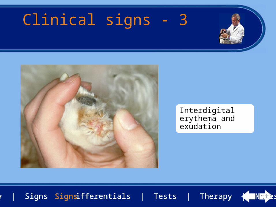
Interdigital erythema and exudation

History | Signs | Differentials | Tests | Therapy | NotesHistory | Signs | Differentials | Tests | Therapy | Notes
How would youapproach this case?
SignsSigns
• What are the next steps you would take?
• Make a list of your principle differential diagnoses
• List any samples you would collect
• List any tests you would perform to assist in making a definitive diagnosis

History | Signs | Differentials | Tests | Therapy | NotesHistory | Signs | Differentials | Tests | Therapy | Notes
Case investigation
DifferentialsDifferentials
• Principle differential diagnoses• Metabolic epidermal necrosis (superficial necrolytic
dermatitis, hepatocutaneous syndrome, necrolytic migratory erythema)
• Pemphigus foliaceus

History | Signs | Differentials | Tests | Therapy | NotesHistory | Signs | Differentials | Tests | Therapy | Notes
Tests - 1
TestsTests
• Diagnostic tests• Skin scrapings• Skin biopsy• Haematological and biochemical profiles• Urinalysis

History | Signs | Differentials | Tests | Therapy | NotesHistory | Signs | Differentials | Tests | Therapy | Notes
Tests - 2
TestsTests
• No evidence of parasites and fungal elements on microscopy
• Elevated alkaline phosphatase, alanine aminotransferase, glucose, cholesterol
• Mild lymphopenia and eosinopenia
• Urinalysis unremarkable

History | Signs | Differentials | Tests | Therapy | NotesHistory | Signs | Differentials | Tests | Therapy | Notes
Tests - 3
• Skin biopsies showed• compact diffuse parakeratosis and hydropic
degeneration of the upper epidermis• Mild acanthosis and sparse mononuclear cell infiltrate
in the upper dermis
TestsTests

History | Signs | Differentials | Tests | Therapy | NotesHistory | Signs | Differentials | Tests | Therapy | Notes
What is yourdiagnosis?
• Do the investigations permit a definitive diagnosis?
• Are there any additional investigations which you think may need to be done?
TestsTests

History | Signs | Differentials | Tests | Therapy | NotesHistory | Signs | Differentials | Tests | Therapy | Notes
Diagnosis
• Metabolic epidermal necrosis• Historical and clinical features suggestive, supported
by biopsy results• Laboratory tests support a metabolic disorder
TestsTests

History | Signs | Differentials | Tests | Therapy | NotesHistory | Signs | Differentials | Tests | Therapy | Notes
Further tests
• Post-prandial bile acids were elevated, consistent with hepatobiliary dysfunction
• Abdominal ultrasonography showed diffuse hepatic disease
TestsTests

History | Signs | Differentials | Tests | Therapy | NotesHistory | Signs | Differentials | Tests | Therapy | Notes
How would you deal with this case?
• What is your prognosis?
• How will you advise the owner?
• What treatment would you consider?
TherapyTherapy

History | Signs | Differentials | Tests | Therapy | NotesHistory | Signs | Differentials | Tests | Therapy | Notes
Prognosis
• Prognosis is poor• Many cases are difficult to manage and require
euthanasia, either because of the severe skin disease or due to hepatic disease or pancreatic neoplasia
TherapyTherapy

History | Signs | Differentials | Tests | Therapy | NotesHistory | Signs | Differentials | Tests | Therapy | Notes
Therapy - 1
TherapyTherapy
• Symptomatic therapy only
• Systemic and / or topical antibacterial therapy
• Nutritional supplementation with high protein diets are helpful in some cases

History | Signs | Differentials | Tests | Therapy | NotesHistory | Signs | Differentials | Tests | Therapy | Notes
Therapy - 2
• Glucocorticoids are generally contra-indicated due to the metabolic disease
• Specific therapy for hepatic or pancreatic disease is ideal but seldom possible
TherapyTherapy

History | Signs | Differentials | Tests | Therapy | NotesHistory | Signs | Differentials | Tests | Therapy | Notes
Comment - 1
NotesNotes
• Most dogs have associated hepatic disease (vacuolar alteration or cirrhosis); a few have pancreatic glucagonomas
• Some dogs become diabetic
• The cause of the hepatic and skin disease is unknown
• Skin lesions may reflect hypoaminoacidemia (present in this case)
History | Signs | Differentials | Tests | Therapy | NotesHistory | Signs | Differentials | Tests | Therapy | Notes
Comment - 2
NotesNotes
• The periorificial lesions plus footpad hyperkeratosis are the usual findings
• Often confused with autoimmune diseases
• Ultrasound may allow visualisation of pancreatic neoplasia or metastases in rare cases, and distinguish from liver disease
• Bile acid assays useful for assessment of hepatic function

History | Signs | Differentials | Tests | Therapy | NotesHistory | Signs | Differentials | Tests | Therapy | Notes
Review
NotesNotes
• If you would like to review this case, please use the navigation buttons below