crossmodal shaping of pain: a multisensory approach to nociception
TRANSCRIPT

TICS-1319; No. of Pages 9
Crossmodal shaping of pain: amultisensory approach to nociceptionDaniel Senkowski1, Marion Ho fle2, and Andreas K. Engel2
1 Department of Psychiatry and Psychotherapy, Charite - Universitatsmedizin Berlin, St. Hedwig Hospital, Große Hamburger Straße
5-11, 10115 Berlin, Germany2 Department of Neurophysiology and Pathophysiology, University Medical Center Hamburg-Eppendorf, Martinistraße 52, 20246
Hamburg, Germany
Review
Glossary
Crossmodal: this term is normally used to refer to situations in which the
presentation of a stimulus in one sensory modality is shown to influence the
perception, behavioral responses, or neural processing of a stimulus presented
in another sensory modality [77].
Defense response: mobilization and protection of the body accomplished by
attentional modifications such as alerting the organism and orienting it toward
the threatening event while prioritizing relevant sensory input.
Frequency bands: stimulus-induced and ongoing intrinsic oscillations are
usually categorized into five frequency bands: delta (2–3 Hz); theta (4–7 Hz);
alpha (8–12 Hz); beta (13–30 Hz); and gamma (>30 Hz).
Functional connectivity: quantifies the correlation between the activity of two
neuronal populations. Such correlations are assumed to indicate a functional
relation or the exchange of information between the recorded populations.
Noxious stimulus: a stimulus that is damaging or threatens damage to normal
tissues (IASP Taxonomy, http://www.iasp-pain.org).
Oscillations: rhythmic activity of neurons or neuronal populations. This
rhythmic activity is often band limited and characterized by dominant
frequencies. Oscillations can be quantified using spectral analysis.
Oscillatory power: a measure for the strength of an oscillatory signal. Power
scales with the square of the signal amplitude.
Peripersonal space: the spatial region surrounding the body that a person
regards as theirs psychologically. Unpleasant stimuli that enter the periperso-
Noxious stimuli in our environment are often accompa-nied by input from other sensory modalities that canaffect the processing of these stimuli and the perceptionof pain. Stimuli from these other modalities may distractus from pain and reduce its perceived strength. Alterna-tively, they can enhance the saliency of the painful input,leading to an increased pain experience. We discussfactors that influence the crossmodal shaping of painand highlight the important role of innocuous stimuli inperipersonal space. We propose that frequency-specificmodulations in local oscillatory power and in long-rangefunctional connectivity may serve as neural mechanismsunderlying the crossmodal shaping of pain. Finally, weprovide an outlook on future directions and clinicalimplications of this promising research field.
Pain, attention, and crossmodal processesA key function of pain is to protect our body from potentialharm and to facilitate the preparation of a defenseresponse [1]. Noxious stimuli can automatically captureour attention and it is likely that the salience network,which includes the anterior cingulate cortex (ACC) and theoperculoinsular cortex, plays a major role therein [2–4].Furthermore, top-down attention toward noxious stimulienhances the perception and facilitates the processing ofpain [5–9]. Thus, shifts in attention following or precedingnoxious stimuli may be important for the rapid preparationof a defense response (see Glossary).
Crossmodal processes, which are also known to interactwith attention [10], are another important factor that canbias the processing of pain stimuli. Several recent studiessuggested that crossmodal processes shape the perceptionof pain. For instance, an electroencephalography (EEG)study showed that a spatiotemporally aligned, task-irrele-vant visual stimulus enhances the perception and proces-sing of concurrent pain [11]. Crossmodal processes can alsohave diminishing effects on pain. For example, listening topreferred music [12], viewing pleasant pictures [13], orviewing their own body [14] reduces pain perception. These
1364-6613/
� 2014 Elsevier Ltd. All rights reserved. http://dx.doi.org/10.1016/j.tics.2014.03.005
Corresponding author: Senkowski, D. ([email protected]).Keywords: multisensory processing; pain processing; oscillatory activity; functionalconnectivity; salience network; attention network.
studies suggest that crossmodal processes can indeedmodulate the perception of pain.
In this review we examine in detail several factors thatinfluence the crossmodal shaping of pain and highlight theimportant role of innocuous stimuli in peripersonal space.Particular emphasis is placed on studies investigating therole of neural oscillations, as reflected in pain-relatedchanges in gamma-band, beta-band, alpha-band, and del-ta-band activity (GBA, BBA, ABA, and DBA) (Box 1).Neural oscillations and their dynamic coupling generallyseem to play an important role in multisensory processing[15–20]. Recent studies suggest that neural oscillationsmay also be involved in the crossmodal shaping of pain[11,12,21,22]. The interesting topic of how, in particular,tactile stimuli affect pain has been covered in an excellentrecent review [23] and is therefore only briefly discussedhere. In the reviewed studies, different dimensions ofpain have been assessed (Box 2). Rather than consideringthese separately, we use the term ‘pain perception’ when
nal space usually evoke a defense response.
Phase coherence: quantifies the consistency of the relative phase between two
simultaneously recorded signals that have the same frequency.
Stimulus saliency: the ability of a stimulus to capture attention in a bottom-up
fashion. A stimulus has a high saliency when it contrasts with its surrounding
stimuli. Salient stimuli activate a circumscribed set of cortical regions, the so-
called salience network.
Valence: characterizes a stimulus as pleasant (positive valence) or unpleasant
(negative valence) in the context of affect and emotion.
Trends in Cognitive Sciences xx (2014) 1–9 1

Box 1. Oscillatory power as a neural signature of pain
There is ample evidence for an involvement of neural oscillations in
the processing of noxious stimuli, such as electrical [6], temperature
[49], and laser [45–48]. Noxious laser stimuli, which have been
frequently used to investigate the neural mechanisms underlying
pain, modulate the power of oscillatory activity in different frequency
bands and at different latencies (Figure I). These different frequency
bands have been associated with specific cognitive and sensorimotor
functions [55,70,78]. In acute pain, a consistent finding is a positive
correlation between the magnitude of GBA in the somatosensory
cortex and pain perception [6,12,45,46]. Moreover, there is evidence
that GBA reflects the sensorimotor transformation of pain, implicating
a behavioral relevance for the initiation of a defense response [79].
However, GBA has also been linked to shifts in attention [80,81]. A
recent EEG study in which trains of consecutive laser stimuli were
presented at constant interstimulus intervals has addressed this
question [46]. Repetition of sensory stimuli reduces saliency and,
thus, repeatedly presented pain stimuli are less prone to capture
attention or enhance arousal. The study showed that repeated
stimulus presentation reduced laser-evoked potentials whereas pain
ratings and GBA remained constant. Importantly, for all stimulus
repetitions the amplitude of GBA correlated significantly with pain
perception. Another response pattern following noxious stimulation
is a suppression of BBA in the sensorimotor cortex [21,82]. Because
BBA also plays a role in motor processing, one possible interpretation
is that the pain-related suppression of BBA in the sensorimotor cortex
reflects the preparation of a defense response [83]. The observation
that painful stimulation increases beta-band coherence between
motor cortex and peripheral muscles supports this view [84]. In the
primary somatosensory cortex, pain-related suppression in BBA is
usually accompanied by suppression in ABA [82]. Moreover, pain-
related suppression in ABA has been related to increased excitability
of the sensorimotor system [47,85]. For example, pain-related ABA
suppression correlates positively with electromagnetic responses
over the primary somatosensory cortex to a non-painful electrical
stimulus [47]. A fourth response pattern following noxious stimula-
tion is an increase in DBA. Studies investigating the sources of pain-
related DBA suggested an involvement of the sensorimotor cortex
and the medial cingulate cortex [6,12]. Together, these studies
strongly suggest that neural oscillations play a role in the processing
of noxious stimuli.
Response (%)
-30
30
0 0.5 1.5 s1.00
5
10
δ
β
γ
Response (%)
-50
50
10
40
70
Response (%)
-10
10
50
70
90Hz
Beta (24-34 Hz) MI
z-Score
-7
-3
Gamma (70-80 Hz) SIISI
z-Score
3
7
Delta (2-6 Hz) SI MCC
z-Score
3
7
Central
Temporal
Parietal
(A) (B)
TRENDS in Cognitive Sciences
Figure I. Pain-related modulations in the power of oscillatory activity measured by magnetoencephalography (MEG). (A) Following a painful laser stimulus, enhanced
delta-band activity (DBA) (bottom panel) and gamma-band activity (GBA) (top panel) are observed. The third response component is longer-latency suppression of beta-
band activity (BBA) (middle panel). (B) The sources of GBA can be localized in the somatosensory cortices (SI, SII; top panel). The suppression of BBA is found in the
motor cortex (MI; middle panel) and the increase in DBA is observed in the somatosensory and medial cingulate cortex (MCC). Adapted, with permission, from [12]. The
figure has been reproduced with permission of the International Association for the Study of Pain. The figure may not be reproduced for any other purpose without
permission.
Review Trends in Cognitive Sciences xxx xxxx, Vol. xxx, No. x
TICS-1319; No. of Pages 9
describing study results regarding the subjective experi-ence of pain.
Factors influencing the crossmodal shaping of painStimulus intensity, temporal synchrony, and spatial
alignment
Studies using innocuous stimuli showed that multisensoryinteractions are most likely when the constituent inputsare spatially and temporally aligned and when they elicitweak neural responses by themselves [24,25]. The latterobservation has been termed the ‘principle of inverse
2
effectiveness’. Thus far, only a few studies have investigat-ed whether such known principles of multisensory integra-tion apply to the crossmodal shaping of pain. Given thatstimuli that elicit strong neural responses, such as noxiousstimuli, are usually less susceptible to crossmodal modula-tions, it seems conceivable that the crossmodal shaping ofpain may not strictly follow the principles of multisensoryintegration.
Using stimuli of different intensities, a recent EEGstudy examined the crossmodal influence of spatiotempo-rally aligned visual stimuli on the perception and proces-

Box 2. Dimensions of pain
Pain is a multifaceted experience comprising at least two compo-
nents [86–88]: (i) a sensory–discriminative component that reflects
intensity and spatiotemporal aspects; and (ii) an affective–motiva-
tional component that encodes the disturbing character of pain and
relates to emotion, arousal, and defense behavior. This unpleasant
experiential quality is immanent to pain. The two components of
pain perception seem to be processed in partially distinct networks
[87,89,90]. The sensory–discriminative component has been related
to activity in the primary somatosensory cortex [91], whereas the
affective–motivational component has been attributed to regions of
the salience network, including the ACC and the insular cortex [90].
Interestingly, responses of the autonomic nervous system have
primarily been related to the affective–motivational pain component
[38,88]. Experimental manipulations can differentially affect the two
components of pain [34,38,39]. For instance, attention seems to
primarily affect the sensory–discriminative component, whereas
emotional processes mainly influence the affective–motivational
component [34,92]. Behaviorally, these two main components of
pain perception can be monitored by subjective ratings of stimulus
intensity and stimulus unpleasantness, respectively. However,
subjective ratings of these two dimensions are often highly
correlated and therefore it has been discussed whether they
represent separate components of pain [93,94]. Yet the findings
that these components are differentially modulated by experimental
manipulations [11,33,38,89,92] suggest that they reflect, at least in
part, distinct aspects of the experience and processing of pain.
Review Trends in Cognitive Sciences xxx xxxx, Vol. xxx, No. x
TICS-1319; No. of Pages 9
sing of pain [11]. To examine multisensory interactions inthe local power of oscillatory activity, a nonlinear analysisapproach was used [26]. The crossmodal influence of visualstimuli on pain perception and pain-related neural oscilla-tions was stronger when pain stimuli were low comparedwith when they were high in intensity. The behavioral effectwas paralleled by a similar effect on BBA, which had atopography indicating involvement of the sensorimotor cor-tex (Figure 1A). Thus, this study suggests that the cross-modal shaping of pain follows the principle of inverseeffectiveness. Using magnetoencephalography (MEG), Inuiet al. [27] investigated the effect of temporal alignmentbetween vibrotactile and electrical pain stimuli on painperception and pain-induced event-related responses. Thestudy showed reduced pain perception and modulations ofpain-induced event-related responses when vibrotactile sti-muli and pain stimuli were presented simultaneously orwhen pain stimuli preceded vibrotactile stimuli by up to40 ms. Notably, the study also demonstrated that the laten-cy difference of event-related responses for these differentstimuli had the same magnitude; that is, the responselatencies were about 40 ms shorter for vibrotactile com-pared with noxious stimuli. Hence, this study suggests thatthe crossmodal shaping of pain is robust when vibrotactileand noxious inputs reach the cortex at about the same time.However, due to the close association between the corticalnetworks involved in the processing of these inputs, thecrossmodal shaping of pain through tactile stimuli may be aspecial case [23]. The question whether differences in re-sponse latencies also play a role in the modulation of pain byother sensory modalities remains to be addressed.
Another factor that could influence the crossmodalshaping of pain is spatial alignment. Van Ryckeghemet al. [28] examined pain perception and response speedto painful stimuli when visual stimuli that were spatiallyaligned or nonaligned with a pain stimulus were presented
at a short negative time lag. The study revealed higherpain perception and shorter response times for spatiallyaligned compared with spatially nonaligned visual–painstimuli. Taken together, the studies reviewed above sug-gest that the crossmodal shaping of pain follows the knownprinciples of multisensory integration.
Stimulus valence
The valence of stimuli surrounding us strongly modulatesthe efficiency and depth of sensory processing. Therefore, itis likely that the valence of other sensory stimuli influencestheir impact on pain perception. Studies investigating theeffect of pictures with emotional content on pain perceptionshowed pain-reducing effects for pleasant and -enhancingeffects for unpleasant pictures [29,30]. A MEG study in-vestigated the impact of facial expressions with neutral,positive (happy), and negative (angry and fearful) valenceon the perception and processing of pain [21]. Independentof their valence, faces with emotional expressions com-pared with faces with neutral expression led to increasedpain processing, as reflected by stronger modulation ofBBA in the sensorimotor cortex (Figure 1B). Interestingly,happy facial expressions also led to enhanced pain percep-tion, which seems to contradict findings of pain-relievingeffects for pictures with positive emotional content. Theauthors [21] suggested that a happy face that is presentedin combination with a pain stimulus may be interpreted asnegative; for example, as if this person might be laughingabout the observer.
Valence-specific modulations of pain are also found foremotional stimuli of other sensory modalities, such asaudition [12,31,32] and olfaction [33,34]. A recent MEGstudy investigated the perception and processing of pain inthe context of pleasant and unpleasant music [12]. Thestudy showed enhanced pain perception and stronger GBAin the somatosensory cortex when participants were lis-tening to unpleasant compared with pleasant music. Afunctional MRI (fMRI) study showed that smelling un-pleasant compared with pleasant odors is associated withenhanced pain perception and stronger hemodynamicresponses in a network comprising the ACC, the medialthalamus, and the somatosensory cortex [34]. Taken to-gether, these findings suggest that the valence of stimulishapes the perception and processing of pain in a similarmanner across modalities. Stimuli that are perceived aspleasant usually diminish the intensity of pain perceptionand reduce pain-related neural processing. Conversely,stimuli that are perceived as unpleasant enhance theperception and processing of pain. In conclusion, we pro-pose that the strength and direction of the crossmodalinfluences on pain by other inputs depend on the valenceof stimuli and follow integration principles that are ofgeneral relevance in multisensory perception.
Impact of stimuli in peripersonal space on painSensory stimuli in close proximity to one’s body may signifypotential threat and imminent pain. To prepare a defenseresponse, they must be analyzed rapidly with regard totheir threat value and potential harm for the body [35].Therefore, the influence of innocuous stimuli in periperso-nal space on pain may be particularly strong. Virtual
3

Angry Happy NeutralFearful
Le�
Posterior
PV m
inus
V18
Hz,
170
ms
Response (%)–35 35
Fearful Angry
Happy Neutral
48
52
56
60
Pain
ra�n
g
Fearful Angry Happy Neutral P-only
*****
* p < .05 ** p < .0001
Hand below screen
Gabor patch
Resp
onse
(%)
–40
40
Response (%)–25 25
PV m
inus
P+V
Response (%)–20 20
18 H
z, 3
00 m
s
Hz
0 0.2 0.4 s
10
20
30
10
20
30
P low
Vhi
ghP h
igh
V hig
h
PV m
inus
V18
Hz,
170
ms
(A)
(B)
TRENDS in Cognitive Sciences
Figure 1. Crossmodal effects of visual stimulus intensity and emotional facial expressions on pain. (A) Influence of visual input on pain stimuli of different intensities on
pain perception and pain-related oscillations in the electroencephalogram. Painful electrical stimuli (P) of different intensities (Plow, Phigh) were presented simultaneously
and spatially aligned with Gabor patches (V) of different contrast levels (Vlow, Vhigh) (left panel; only Vhigh is shown). The effect of visual stimulation was stronger for low-
compared with high-intensity pain stimuli, as reflected by enhanced modulation of subjective pain ratings (not illustrated) and enhanced suppression of pain-related beta-
band activity (BBA) power (right panel). Adapted from [11]. (B) Influence of emotional facial expressions on pain perception and pain-related oscillations in the
magnetoencephalogram. Painful electrical stimuli were presented simultaneously with facial expressions of different valence. Electrical stimuli were perceived as more
painful when combined with faces compared with pain stimulation alone. In augmenting pain rating, emotional faces were more effective than faces with a neutral
expression (upper left panel). The pain-related modulation of BBA power at sensors located over the contralateral sensorimotor cortex was stronger for faces with
emotional expressions compared with neutral faces (upper right panel). Source localization revealed that the increase in pain ratings was paralleled by a stronger pain-
related modulation of BBA in the sensorimotor cortex (lower panel). Adapted from [21].
Review Trends in Cognitive Sciences xxx xxxx, Vol. xxx, No. x
TICS-1319; No. of Pages 9
reality setups inducing the embodiment of artificial limbs(Box 3) are effective tools to study the crossmodal shapingof pain in peripersonal space [14,22,36–41]. A recent studydemonstrated that viewing a needle pricking a hand that isperceived as one’s own compared with viewing a Q-tip thattouches the hand enhances pain perception and anticipa-tory pupil dilation responses (Figure 2A) [38]. A follow-upEEG study showed that this effect is reflected by a strongeranticipatory suppression of ABA in the needle comparedwith the Q-tip condition [39]. Threat-related processesinduced by viewing a needle approaching one’s bodyreflecting expectation of pain are likely to have contributedto this effect [42]. Thus, visual input in peripersonal spacemodulates neural processes involved in predicting pain.
Recently, Martini et al. [37] investigated the effect of skincolor on pain (Figure 2B). In this experiment, the tempera-ture of a thermal heat stimulus was increased while parti-cipants were watching an avatar’s wrist perceived as their
4
own that concurrently changed its color toward red, green,or blue. Participants stopped the stimulation as soon as thethermal heat stimulus was felt as painful. The participants’pain perception was enhanced when the avatar’s wristbecame red compared with when it turned blue. Interest-ingly, the authors did not observe enhanced pain perceptionwhen a gray spot next to the participant’s wrist turned red.This shows that the effect of color change is confined to theembodied limb and suggests that the meaning attributed toa sensory cue affects the crossmodal shaping of pain.
Support for this assumption comes from a series ofstudies that used the mirror-box technique [43] to examinethe influence of viewing one’s body on pain [14,22,40,41].These studies demonstrated that viewing one’s own bodyhas a diminishing effect on pain perception. An fMRI studyrevealed that the pain-reducing effect of viewing one’s ownbody is associated with reduced hemodynamic responses insomatosensory areas, the insular cortex, the ACC, and

Box 3. Embodiment of artificial limbs in virtual reality set-
ups
In appropriate experimental settings, artificial limbs can be
perceived as if they belong to one’s own body; that is, they can
become integrated into the body schema. A prominent protocol to
create the illusion of embodiment of artificial limbs is the ‘rubber-
hand illusion’ [95]. In the rubber-hand illusion, participants do not
see their own hand but a rubber hand with the same posture and at
the same location where they would expect their own hand. When
artificial and real hands are synchronously stroked with a brush,
most participants get the impression of feeling the touch at the
viewed hand. Often, participants perceive the viewed hand as
belonging to their own body. The rubber-hand illusion can be
induced only when the observed and the felt touch happen
synchronously [95]. Interestingly, the rubber-hand illusion can also
be induced when pain stimuli instead of tactile stimuli are applied
[77]. In recent years there have been several modifications of the
classical rubber-hand paradigm. For instance, studies have used
virtual hands presented on a screen [38,39] or a head-mounted
display [37]. These setups allow well-controlled presentation of
innocuous and noxious stimuli. Besides the rubber-hand illusion,
the ‘mirror-box’ paradigm has been used to manipulate perception
of the body [14,40]. In the mirror-box paradigm, the participant’s
hand is hidden behind a mirror and thus, participants do not directly
look at the respective hand but see the reflection of the opposed
hand in the mirror superposing the actual hand at the viewed
location (Figure 2C in the main text). In this setup, participants get
the impression of looking at their hidden hand. Using the mirror-box
paradigm it is possible to manipulate what participants see in the
mirror; for instance, an object or the hand of the experimenter [14].
Both the rubber-hand and the mirror-box paradigms are well suited
for studying the crossmodal shaping of pain.
Review Trends in Cognitive Sciences xxx xxxx, Vol. xxx, No. x
TICS-1319; No. of Pages 9
more posterior brain areas that have been related to visualbody representation [41]. Furthermore, a recent EEGstudy demonstrated that the pain-relieving effect of view-ing one’s own body is reflected by an enhancement of pain-related BBA (Figure 2B) [22]. Taken together, these stud-ies indicate that visual stimuli in peripersonal space areparticularly prone to modulating the perception and pro-cessing of pain. Whether this is also the case for inputsfrom other sensory modalities, such as auditory stimuli,remains to be investigated.
Neural mechanisms underlying the crossmodal shapingof painThe last decade has seen an increasing number of studieshighlighting the role of neural oscillations in pain percep-tion [6,44–51]. Most of these studies showed pain-relatedchanges of local oscillatory power (Box 1). However, thereis some evidence that phase coherence of oscillatory signalsis also relevant for the functional interaction of pain-relat-ed brain regions [6,44,50,52,53]. For instance, an electro-corticography study investigated functional connectivity,as reflected in phase coherence, between the somatosenso-ry, medial frontal, and insular cortex during the processingof attended and unattended noxious stimuli [50]. In theattended compared with the unattended condition, anincrease in beta-band and alpha-band connectivity wasfound in this network. An additional analysis of directedconnectivity in the alpha band showed an influence of theprimary somatosensory cortex on the other networkregions, particularly in the attention condition [52]. UsingMEG, another study found enhanced interhemisphericconnectivity in the gamma band between sensors over
the left and right sensorimotor cortex for attended com-pared with unattended noxious stimuli [6]. We would liketo emphasize that the study of functional connectivity inEEG and MEG data requires great caution. Factors likethe signal-to-noise ratio, power of oscillatory signals, andvolume conduction can strongly influence the results andmust be thoroughly considered [54,55]. Taken together, thestudies suggest that local oscillatory signals and theirinteraction across regions are important for pain percep-tion and that attention can modulate both local power andfunctional connectivity.
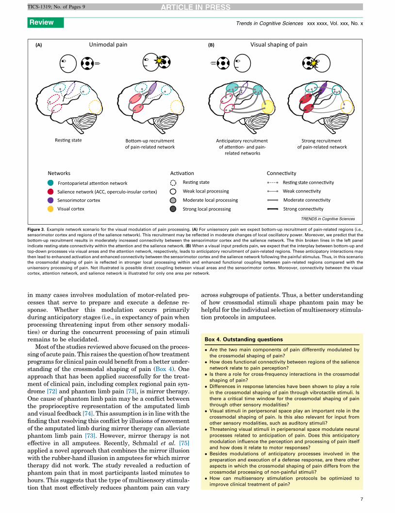
Likewise, the crossmodal processing of innocuous sti-muli involves modulations of both local oscillatory power[20,56] and long-range functional connectivity [19,57–59].This raises the question of whether similar mechanismsmediate the crossmodal shaping of pain. As discussedabove, the available data suggest that the crossmodalmodulation of pain perception is associated with localpower changes in various frequency bands [11,12,21,22].Given its importance for pain processing and for multisen-sory integration, it is tempting to hypothesize that long-range coupling of oscillatory signals may also be importantfor the crossmodal shaping of pain. Currently, however,there is a lack of studies examining this possibility directly.Figure 3 illustrates an example for a hypothetical networkscenario involving modulations of both local oscillationsand long-range neural connectivity. In cases where proces-sing of pain stimuli is facilitated by visual input, wehypothesize that pain-related regions and structures ofthe top-down attention network are recruited in an antici-patory manner due to crossmodal long-range interactionswith visual cortex. Compared with the processing of uni-sensory pain, which primarily occurs in a bottom-up man-ner, this may then lead to enhanced local oscillatoryactivity as well as to increased functional connectivitywithin pain-related regions.
Such scenarios are likely to involve both the frontopar-ietal attention [60] and the salience [3] network (Figure 3).Intracranial recordings in humans showed that pain-relat-ed responses in the sensorimotor cortex occur at latenciessimilar to those of responses in the operculoinsular [61] andcingulate [62] cortex, which are key structures of the sa-lience network. Numerous studies have shown activity inthe salience network after noxious stimulus presentation[2,4,63–65]. Interestingly, the salience network is also in-volved in the detection of salient stimuli across sensorymodalities [3,66,67]. Due to its multisensory function, itseems likely that the crossmodal shaping of pain involvesneural processing in the salience network. The saliencenetwork is closely linked to the frontoparietal attentionnetwork [60]. Support for an involvement of the frontopar-ietal attention network in the crossmodal shaping of paincomes from a recent fMRI study, which showed that pain-related modulations of hemodynamic responses by affectivepictures involve interactions between the insular cortex andother cortical structures, including the frontoparietal atten-tion network [68]. Another fMRI study demonstrated thathigher-order frontal structures and the ventral striatummediate predictive cue effects in the salience network andin the thalamus relating to pain perception [69]. Further-more, oscillatory activity, especially in the gamma band, is
5

10
20
30
0 4 12 s8
Hz
30
35
25
Hand Object
*
p < 0.05*
Inte
nsity
ra�n
g
Hand
min
us o
bjec
tO
bjec
t vie
w
10
20
30Hz
0 4 12 s8
Hand
vie
w
10
20
30
0 4 12 s8
Hz
Response (dB)–1.5 1.5
Response (dB)–3 3
* p < .05 ** p < .01
Pain
thre
shol
d (°
C)
Spot Blue Green Red
***
44
45*
Head-mountedvirtual reality system
Eye-tracker
Real hand below screen
0
.06
.04
.02
p < 0.05*NeedleQ-�p
p < 0.05Pupi
l dila
�on
(%)
0 2s 1
*
40
50
60
Ra�n
g
Unpleasant.Intensity
(A)
(B) (C)
TRENDS in Cognitive Sciences
Figure 2. Studies using virtual reality setups with artificial limbs. (A) Influence of viewing a needle pricking a hand that is perceived as one’s own on pain perception and
pupil dilation responses. Participants were presented with video clips depicting a needle prick or a Q-tip touch while receiving spatiotemporally aligned painful or non-
painful electrical stimuli (left panel). Pain unpleasantness ratings were enhanced when a needle prick compared with a Q-tip touch was presented (middle panel). This effect
was paralleled by an enhanced pupil dilation response (right panel). Adapted from [38]. The figure has been reproduced with permission of the International Association for
the Study of Pain. The figure may not be reproduced for any other purpose without permission. (B) Influence of skin color on pain perception. Participants wore a head-
mounted virtual reality system that showed an avatar arm at the same location as their real arm. Thermal heat stimuli were presented to the real arm while there was either
a grey dot next to the avatar arm that turned red or a color change of the skin toward blue, green, or red. The study revealed an enhancement in pain perception, as reflected
in reduced pain thresholds, when the skin turned red. Adapted from [37]. (C) Diminishing effect of viewing one’s own hand on pain perception and pain-related oscillations
on electroencephalography (EEG) using the mirror-box paradigm (Box 3). The participant’s left hand received painful thermal stimulation while the participant viewed the
reflection of their right hand with a fake thermode or a box superposing their stimulated left hand. Viewing the hand compared with viewing a box reduced pain perception,
which was paralleled by enhanced power of pain-related beta-band activity (BBA) over the sensorimotor cortex (right panel). Adapted from [22].
Review Trends in Cognitive Sciences xxx xxxx, Vol. xxx, No. x
TICS-1319; No. of Pages 9
likely to reflect attentional processing in the frontoparietalattention network [70]. Taking these studies together, wepropose that functional connectivity between pain-relatedregions and modulation of local oscillatory power are crucialneural mechanisms underlying the crossmodal shapingof pain.
Concluding remarks and outlookThe studies reviewed above show that innocuous stimulican modulate the processing and perception of pain. Dif-ferent factors, such as temporal and spatial alignment,stimulus intensity, and valence, have an impact on howother sensory stimuli shape pain perception. In this con-text, sensory stimuli in peripersonal space seem to be ofparticular relevance. At the neural level, oscillatory signalsmay be crucial for the crossmodal shaping of pain. Inagreement with the hypothesis that much, if not all, of
6
the neocortex is multisensory [71], we propose that innoc-uous stimuli can shape pain-related neural responses es-sentially at all processing stages.
Thus far, the data available suggest that the crossmodalshaping of pain follows, at least to a large extent, thegeneral principles of multisensory integration. Moreover,the studies reviewed above indicate that comparable mech-anisms, such as changes in unspecific arousal and modula-tions in spatial or feature-based attention, which havepreviously been related to the crossmodal processing ofinnocuous sensory stimuli, are also involved in the cross-modal shaping of pain. This raises the interesting questionof whether there are qualitative differences between thecrossmodal shaping of pain and the crossmodal processingof innocuous stimuli. Because a main function of acute painis to protect the body’s integrity and to prevent futuretissue injuries, it is likely that crossmodal shaping of pain
Unimodal pain(A) (B) Visual shaping of pain
Res�ng state
Moderate connec�vity
Strong connec�vity
Weak connec�vity
Networ ks Ac�va�on Connec�vityRes�ng state
Weak local processing
Strong local processing
Moderate local processing
Salience network (ACC, operculo-insular cor tex)
Visual cor tex
Sensorimotor cor tex
Frontoparietal a�en�on network Res�ng state connec�vity
An�cipatory recruitment of a�en�on- and pain-
related networks
Bo�om-up recruitmentof pain-related network
Strong recruitment of pain-related network
TRENDS in Cognitive Sciences
Figure 3. Example network scenario for the visual modulation of pain processing. (A) For unisensory pain we expect bottom-up recruitment of pain-related regions (i.e.,
sensorimotor cortex and regions of the salience network). This recruitment may be reflected in moderate changes of local oscillatory power. Moreover, we predict that the
bottom-up recruitment results in moderately increased connectivity between the sensorimotor cortex and the salience network. The thin broken lines in the left panel
indicate resting-state connectivity within the attention and the salience network. (B) When a visual input predicts pain, we expect that the interplay between bottom-up and
top-down processes via visual areas and the attention network, respectively, leads to anticipatory recruitment of pain-related regions. These anticipatory interactions may
then lead to enhanced activation and enhanced connectivity between the sensorimotor cortex and the salience network following the painful stimulus. Thus, in this scenario
the crossmodal shaping of pain is reflected in stronger local processing within and enhanced functional coupling between pain-related regions compared with the
unisensory processing of pain. Not illustrated is possible direct coupling between visual areas and the sensorimotor cortex. Moreover, connectivity between the visual
cortex, attention network, and salience network is illustrated for only one area per network.
Box 4. Outstanding questions
� Are the two main components of pain differently modulated by
the crossmodal shaping of pain?
� How does functional connectivity between regions of the salience
network relate to pain perception?
� Is there a role for cross-frequency interactions in the crossmodal
shaping of pain?
� Differences in response latencies have been shown to play a role
in the crossmodal shaping of pain through vibrotactile stimuli. Is
there a critical time window for the crossmodal shaping of pain
through other sensory modalities?
� Visual stimuli in peripersonal space play an important role in the
crossmodal shaping of pain. Is this also relevant for input from
other sensory modalities, such as auditory stimuli?
� Threatening visual stimuli in peripersonal space modulate neural
processes related to anticipation of pain. Does this anticipatory
modulation influence the perception and processing of pain itself
and how does it relate to motor responses?
� Besides modulations of anticipatory processes involved in the
preparation and execution of a defense response, are there other
aspects in which the crossmodal shaping of pain differs from the
crossmodal processing of non-painful stimuli?
� How can multisensory stimulation protocols be optimized to
improve clinical treatment of pain?
Review Trends in Cognitive Sciences xxx xxxx, Vol. xxx, No. x
TICS-1319; No. of Pages 9
in many cases involves modulation of motor-related pro-cesses that serve to prepare and execute a defense re-sponse. Whether this modulation occurs primarilyduring anticipatory stages (i.e., in expectancy of pain whenprocessing threatening input from other sensory modali-ties) or during the concurrent processing of pain stimuliremains to be elucidated.
Most of the studies reviewed above focused on the proces-sing of acute pain. This raises the question of how treatmentprograms for clinical pain could benefit from a better under-standing of the crossmodal shaping of pain (Box 4). Oneapproach that has been applied successfully for the treat-ment of clinical pain, including complex regional pain syn-drome [72] and phantom limb pain [73], is mirror therapy.One cause of phantom limb pain may be a conflict betweenthe proprioceptive representation of the amputated limband visual feedback [74]. This assumption is in line with thefinding that resolving this conflict by illusions of movementof the amputated limb during mirror therapy can alleviatephantom limb pain [73]. However, mirror therapy is noteffective in all amputees. Recently, Schmalzl et al. [75]applied a novel approach that combines the mirror illusionwith the rubber-hand illusion in amputees for which mirrortherapy did not work. The study revealed a reduction ofphantom pain that in most participants lasted minutes tohours. This suggests that the type of multisensory stimula-tion that most effectively reduces phantom pain can vary
across subgroups of patients. Thus, a better understandingof how crossmodal stimuli shape phantom pain may behelpful for the individual selection of multisensory stimula-tion protocols in amputees.
7

Review Trends in Cognitive Sciences xxx xxxx, Vol. xxx, No. x
TICS-1319; No. of Pages 9
An interesting recent finding is that visual distortion ofbody size modulates pain perception [40]. Using a virtualreality system including real-time videos that capture thehand from the same position as if viewed directly, Prestonand Newport [76] showed that, in patients with osteoar-thritis, stretching or shrinking the painful body part tem-porarily reduces pain. Multisensory virtual reality setupsmay be also incorporated in counter-conditioning programsfor blood-injection-injury phobia. In this regard, setups likethe one introduced by Hofle et al. [38,39] may be promising(Figure 2A). If needle prick clips were repeatedly pairedwith spatially and temporally aligned non-painful somato-sensory stimuli, one might expect a learning-induced re-duction of fear in subjects suffering from such phobias.Thus, multisensory stimulation protocols with virtuallimbs (Box 3) may represent a promising approach thatmight be useful in clinical treatment programs for pain. Aparticular challenge lies in the development of multisen-sory stimulation protocols that result in longer-lastingameliorating effects on pain. Taken together, researchon the crossmodal shaping of pain has clinical implicationsand provides interesting new insights into the relevanceand mechanisms of multisensory processing.
AcknowledgmentsThis work was supported by grants from the German ResearchFoundation (DFG) (SE 1859/1-2, D.S.; SFB TRR58, SFB 936, A.K.E.)and the European Union (ERC-2010-StG-20091209, D.S.; ERC-2010-AdG-269716, A.K.E.). The authors thank Ulrich Pomper and Julian Keilfor their helpful comments on this review.
References1 Donaldson, G.W. et al. (2003) Pain and the defense response: structural
equation modeling reveals a coordinated psychophysiological responseto increasing painful stimulation. Pain 102, 97–108
2 Legrain, V. et al. (2011) The pain matrix reloaded: a salience detectionsystem for the body. Prog. Neurobiol. 93, 111–124
3 Menon, V. and Uddin, L.Q. (2010) Saliency, switching, attention andcontrol: a network model of insula function. Brain Struct. Funct. 214,655–667
4 Iannetti, G.D. and Mouraux, A. (2010) From the neuromatrix to thepain matrix (and back). Exp. Brain Res. 205, 1–12
5 Kulkarni, B. et al. (2005) Attention to pain localization andunpleasantness discriminates the functions of the medial and lateralpain systems. Eur. J. Neurosci. 21, 3133–3142
6 Hauck, M. et al. (2007) Attention to painful stimulation enhancesgamma-band activity and synchronization in human sensorimotorcortex. J. Neurosci. 27, 9270–9277
7 Legrain, V. et al. (2005) Involuntary orientation of attention tounattended deviant nociceptive stimuli is modulated by concomitantvisual task difficulty. Evidence from laser evoked potentials. Clin.Neurophysiol. 116, 2165–2174
8 Spence, C. et al. (2002) Selective attention to pain: a psychophysicalinvestigation. Exp. Brain Res. 145, 395–402
9 Zampini, M. et al. (2007) ‘Prior entry’ for pain: attention speeds theperceptual processing of painful stimuli. Neurosci. Lett. 414, 75–79
10 Talsma, D. et al. (2010) The multifaceted interplay between attentionand multisensory integration. Trends Cogn. Sci. 14, 400–410
11 Pomper, U. et al. (2013) Crossmodal bias of visual input on painperception and pain-induced beta activity. Neuroimage 66, 469–478
12 Hauck, M. et al. (2013) The influence of music and music therapy on pain-induced neuronal oscillations measured by magnetencephalography.Pain 154, 539–547
13 Godinho, F. et al. (2008) Pain influences hedonic assessment of visualinputs. Eur. J. Neurosci. 27, 2219–2228
14 Longo, M.R. et al. (2009) Visually induced analgesia: seeing the bodyreduces pain. J. Neurosci. 29, 12125–12130
8
15 Hipp, J.F. et al. (2012) Large-scale cortical correlation structure ofspontaneous oscillatory activity. Nat. Neurosci. 15, 884–890
16 Schneider, T.R. et al. (2011) Gamma-band activity as a signature forcross-modal priming of auditory object recognition by active hapticexploration. J. Neurosci. 31, 2502–2510
17 Schepers, I.M. et al. (2013) Noise alters beta-band activity in superiortemporal cortex during audiovisual speech processing. Neuroimage 70,101–112
18 Lakatos, P. et al. (2007) Neuronal oscillations and multisensoryinteraction in primary auditory cortex. Neuron 53, 279–292
19 Kayser, C. and Logothetis, N.K. (2009) Directed interactions betweenauditory and superior temporal cortices and their role in sensoryintegration. Front. Integr. Neurosci. 3, 7
20 Senkowski, D. et al. (2008) Crossmodal binding through neuralcoherence: implications for multisensory processing. Trends Neurosci.31, 401–409
21 Senkowski, D. et al. (2011) Emotional facial expressions modulatepain-induced beta and gamma oscillations in sensorimotor cortex. J.Neurosci. 31, 14542–14550
22 Mancini, F. et al. (2012) Changes in cortical oscillations linked tomultisensory modulation of nociception. Eur. J. Neurosci. 37, 768–776
23 Haggard, P. et al. (2013) Spatial sensory organization and bodyrepresentation in pain perception. Curr. Biol. 23, R164–R176
24 Stein, B.E. and Meredith, M.A. (1993) The Merging of the Senses, MITPress
25 Stein, B.E. and Stanford, T.R. (2008) Multisensory integration: currentissues from the perspective of the single neuron. Nat. Rev. Neurosci. 9,255–266
26 Senkowski, D. et al. (2007) Multisensory processing and oscillatoryactivity: analyzing non-linear electrophysiological measures inhumans and simians. Exp. Brain Res. 177, 184–195
27 Inui, K. et al. (2006) Temporal analysis of cortical mechanisms for painrelief by tactile stimuli in humans. Cereb. Cortex 16, 355–365
28 Van Ryckeghem, D.M. et al. (2011) The role of spatial attention inattentional control over pain: an experimental investigation. Exp.Brain Res. 208, 269–275
29 Rhudy, J.L. et al. (2010) Habituation, sensitization, and emotionalvalence modulation of pain responses. Pain 148, 320–327
30 Roy, M. et al. (2009) Cerebral and spinal modulation of pain byemotions. Proc. Natl. Acad. Sci. U.S.A. 106, 20900–20905
31 Stancak, A. et al. (2013) Modulation of pain by emotional sounds: alaser-evoked potential study. Eur. J. Pain 17, 324–335
32 Roy, M. et al. (2008) Emotional valence contributes to music-inducedanalgesia. Pain 134, 140–147
33 Villemure, C. et al. (2003) Effects of odors on pain perception:deciphering the roles of emotion and attention. Pain 106, 101–108
34 Villemure, C. and Bushnell, M.C. (2009) Mood influences supraspinalpain processing separately from attention. J. Neurosci. 29, 705–715
35 Graziano, M.S. and Cooke, D.F. (2006) Parieto-frontal interactions,personal space, and defensive behavior. Neuropsychologia 44, 845–859
36 Mohan, R. et al. (2012) No pain relief with the rubber hand illusion.PLoS ONE 7, e52400
37 Martini, M. et al. (2013) What color is my arm? Changes in skin color ofan embodied virtual arm modulates pain threshold. Front. Hum.Neurosci. 7, 438
38 Hofle, M. et al. (2012) Viewing a needle pricking a hand that youperceive as yours enhances unpleasantness of pain. Pain 153, 1074–1081
39 Hofle, M. et al. (2013) Spectral signatures of viewing a needleapproaching one’s body when anticipating pain. Eur. J. Neurosci.38, 3089–3098
40 Mancini, F. et al. (2011) Visual distortion of body size modulates painperception. Psychol. Sci. 22, 325–330
41 Longo, M.R. et al. (2012) Linking pain and the body: neural correlates ofvisually induced analgesia. J. Neurosci. 32, 2601–2607
42 Ehrsson, H.H. et al. (2007) Threatening a rubber hand that you feel isyours elicits a cortical anxiety response. Proc. Natl. Acad. Sci. U.S.A.104, 9828–9833
43 Ramachandran, V.S. et al. (1995) Touching the phantom limb. Nature377, 489–490
44 Markman, T. et al. (2013) EEG analysis reveals widespread directedfunctional interactions related to a painful cutaneous laser stimulus. J.Neurophysiol. 110, 2440–2449

Review Trends in Cognitive Sciences xxx xxxx, Vol. xxx, No. x
TICS-1319; No. of Pages 9
45 Gross, J. et al. (2007) Gamma oscillations in human primarysomatosensory cortex reflect pain perception. PLoS Biol. 5, e133
46 Zhang, Z.G. et al. (2012) Gamma-band oscillations in the primarysomatosensory cortex – a direct and obligatory correlate ofsubjective pain intensity. J. Neurosci. 32, 7429–7438
47 Ploner, M. et al. (2006) Oscillatory activity reflects the excitability ofthe human somatosensory system. Neuroimage 32, 1231–1236
48 Tiemann, L. et al. (2010) Gamma oscillations as a neuronal correlate ofthe attentional effects of pain. Pain 150, 302–308
49 Shao, S. et al. (2012) Frequency–domain EEG source analysis for acutetonic cold pain perception. Clin. Neurophysiol. 123, 2042–2049
50 Ohara, S. et al. (2006) Analysis of synchrony demonstrates ‘painnetworks’ defined by rapidly switching, task-specific, functionalconnectivity between pain-related cortical structures. Pain 123,244–253
51 Ploner, M. et al. (2009) Functional integration within the human painsystem as revealed by Granger causality. Hum. Brain Mapp. 30, 4025–4032
52 Liu, C.C. et al. (2011) Attention to painful cutaneous laser stimuli evokesdirected functional connectivity between activity recorded directly fromhuman pain-related cortical structures. Pain 152, 664–675
53 Liu, C.C. et al. (2011) Attention to painful cutaneous laser stimulievokes directed functional interactions between human sensory andmodulatory pain-related cortical areas. Pain 152, 2781–2791
54 Schoffelen, J.M. and Gross, J. (2009) Source connectivity analysis withMEG and EEG. Hum. Brain Mapp. 30, 1857–1865
55 Siegel, M. et al. (2012) Spectral fingerprints of large-scale neuronalinteractions. Nat. Rev. Neurosci. 13, 121–134
56 Engel, A.K. et al. (2012) Multisensory integration through neuralcoherence. In The Neural Bases of Multisensory Processing (Murray,M.M. and Wallace, M.T., eds), pp. 115–130, CRC Press
57 Hipp, J.F. et al. (2011) Oscillatory synchronization in large-scalecortical networks predicts perception. Neuron 69, 387–396
58 Keil, J. et al. (2012) On the variability of the McGurk effect: audiovisualintegration depends on prestimulus brain states. Cereb. Cortex 22,221–231
59 Keil, J. et al. (2013) Prestimulus beta power and phase synchronyinfluence the sound-induced flash illusion. Cereb. Cortex http://dx.doi.org/10.1093/cercor/bhs409
60 Corbetta, M. and Shulman, G.L. (2002) Control of goal-directed andstimulus-driven attention in the brain. Nat. Rev. Neurosci. 3, 201–215
61 Frot, M. et al. (2013) Cortical representation of pain in primarysensory–motor areas (S1/M1) – a study using intracorticalrecordings in humans. Hum. Brain Mapp. 34, 2655–2668
62 Frot, M. et al. (2008) Parallel processing of nociceptive A-delta inputs inSII and midcingulate cortex in humans. J. Neurosci. 28, 944–952
63 Mouraux, A. and Iannetti, G.D. (2009) Nociceptive laser-evoked brainpotentials do not reflect nociceptive-specific neural activity. J.Neurophysiol. 101, 3258–3269
64 Iannetti, G.D. et al. (2008) Determinants of laser-evoked EEGresponses: pain perception or stimulus saliency? J. Neurophysiol.100, 815–828
65 Garcia-Larrea, L. (2012) The posterior insular–opercular region andthe search of a primary cortex for pain. Neurophysiol. Clin. 42, 299–313
66 Legrain, V. et al. (2012) Shielding cognition from nociception withworking memory. Cortex 49, 1922–1934
67 Zu Eulenburg, P. et al. (2013) Interoceptive and multimodal functionsof the operculo-insular cortex: tactile, nociceptive and vestibularrepresentations. Neuroimage 83, 75–86
68 Ploner, M. et al. (2011) Flexible cerebral connectivity patterns subservecontextual modulations of pain. Cereb. Cortex 21, 719–726
69 Atlas, L.Y. et al. (2010) Brain mediators of predictive cue effects onperceived pain. J. Neurosci. 30, 12964–12977
70 Fries, P. (2009) Neuronal gamma-band synchronization as afundamental process in cortical computation. Annu. Rev. Neurosci.32, 209–224
71 Ghazanfar, A.A. and Schroeder, C.E. (2006) Is neocortex essentiallymultisensory? Trends Cogn. Sci. 10, 278–285
72 Cacchio, A. et al. (2009) Mirror therapy for chronic complex regionalpain syndrome type 1 and stroke. N. Engl. J. Med. 361, 634–636
73 Chan, B.L. et al. (2007) Mirror therapy for phantom limb pain. N. Engl.J. Med. 357, 2206–2207
74 Ramachandran, V.S. and Hirstein, W. (1998) The perception ofphantom limbs. The D. O. Hebb lecture. Brain 121, 1603–1630
75 Schmalzl, L. et al. (2013) An alternative to traditional mirror therapy:illusory touch can reduce phantom pain when illusory movement doesnot. Clin. J. Pain 29, e10–e18
76 Preston, C. and Newport, R. (2011) Analgesic effects of multisensoryillusions in osteoarthritis. Rheumatology (Oxford) 50, 2314–2315
77 Spence, C. et al. (2009) Crossmodal processing. Exp. Brain Res. 198,107–111
78 Engel, A.K. and Fries, P. (2010) Beta-band oscillations – signalling thestatus quo? Curr. Opin. Neurobiol. 20, 156–165
79 Schulz, E. et al. (2012) Gamma oscillations are involved in thesensorimotor transformation of pain. J. Neurophysiol. 108, 1025–1031
80 Jensen, O. et al. (2007) Human gamma-frequency oscillationsassociated with attention and memory. Trends Neurosci. 30, 317–324
81 Siegel, M. et al. (2008) Neuronal synchronization along the dorsalvisual pathway reflects the focus of spatial attention. Neuron 60,709–719
82 Ploner, M. et al. (2006) Pain suppresses spontaneous brain rhythms.Cereb. Cortex 16, 537–540
83 Raij, T.T. et al. (2004) Modulation of motor-cortex oscillatory activity bypainful Adelta- and C-fiber stimuli. Neuroimage 23, 569–573
84 Stancak, A. et al. (2005) EEG source analysis and fMRI reveal twoelectrical sources in the fronto-parietal operculum duringsubepidermal finger stimulation. Neuroimage 25, 8–20
85 Hauck, M. et al. (2008) Role of synchronized oscillatory brain activityfor human pain perception. Rev. Neurosci. 19, 10
86 Auvray, M. et al. (2010) The sensory–discriminative and affective–motivational aspects of pain. Neurosci. Biobehav. Rev. 34, 214–223
87 Treede, R.D. et al. (1999) The cortical representation of pain. Pain 79,105–111
88 Rainville, P. et al. (1999) Dissociation of sensory and affectivedimensions of pain using hypnotic modulation. Pain 82, 159–171
89 Price, D.D. (2000) Psychological and neural mechanisms of theaffective dimension of pain. Science 288, 1769–1772
90 Rainville, P. et al. (1997) Pain affect encoded in human anteriorcingulate but not somatosensory cortex. Science 277, 968–971
91 Hofbauer, R.K. et al. (2001) Cortical representation of the sensorydimension of pain. J. Neurophysiol. 86, 402–411
92 Kenntner-Mabiala, R. et al. (2008) Distinct effects of attention andaffect on pain perception and somatosensory evoked potentials. Biol.Psychol. 78, 114–122
93 Chapman, C.R. et al. (2001) Sensory and affective dimensions of phasicpain are indistinguishable in the self-report and psychophysiology ofnormal laboratory subjects. J. Pain 2, 279–294
94 Fernandez, E. and Turk, D.C. (1994) Demand characteristicsunderlying differential ratings of sensory versus affectivecomponents of pain. J. Behav. Med. 17, 375–390
95 Botvinick, M. and Cohen, J. (1998) Rubber hands ‘feel’ touch that eyessee. Nature 391, 756
9