critical issues in the gross examination of the pancreas · critical issues in the gross...
TRANSCRIPT

1
Critical issues in the gross examination of the pancreas
Grace E. Kim, MDAssociate Professor
Humpty Dumpty sat on a wall.Humpty Dumpty had a great fall.
All the king’s horses and all the king’s men,couldn’t put Humpty Dumpty back together again.
http://games.multimedia.cx/wp-content/uploads/babes-in-toyland-humpty.jpg
Clinically important features in your pathology report about pancreatic adenocarcinoma
• Status of surgical margins– What should be sampled
• Status of lymph nodes– Where to look and how many must be found
• Site of origin• Special situations
– Arising from IPMN or MCN
Positive surgical margin is associated with very poor prognosis
• 30-80% of patients resected for cure have positive margins
• Median survival rate of patients with positive margin is the same or worse than that of patients with unresectable tumors
• Margin status is critical because those with close/positive retroperitoneal margin or bulky tumor (at least pT2) may be offered adjuvant chemoradiotherapy
J Gastrointest Surg 2000;4(6):567-79Ann Surg 2001;234(6):758-68
Int J Radiat Oncol Biol Phys 1997;39(1):39-49

2
Lymph node status
• One of the most important independent prognostic factors of survival is accurate staging of lymph nodes
• Predictive value of lymph node status is directly proportional to number of lymph nodes (up to 15 LNs) identified– Guidelines by the American College of
Surgeons will recommend examination of at least 12 lymph nodes for accurate staging of pN0 *
Arch Surg 2007;142:767-773Mod Pathol 2009;22:107-112
*Personal communcation
Pancreaticoduodenectomy
http://pathology.jhu.edu/pancreas/whipplePop.html
How to gross a pancreaticoduodenectomy specimen
• Orient the specimen• Remove surgical margins• Dissect the lymph nodes• Bivalve the pancreas

3
To orient the specimen
• Superior/proximal– Stomach, if present
• Inferior/distal– Duodenum/jejunum
• Anterior pancreatic surface bulges
• Posterior pancreatic surface is flat
Anterior view Pancreas bulges
Posterior view - Pancreas is flat Superior mesenteric vein to portal vein

4
Headof pancreas
Uncinate
SMASMV
PV
Anterior view
BodyTail
Neck
PancreaticoduodenectomyAnterior view
Headof pancreas
Uncinate
Neck
Vascular groove
PancreaticoduodenectomyMedial aspect
Vascular groove
Courtesy of Volkan Adsay, MD
Pancreatic resectionFind the pancreatic duct

5
Common bile duct
Posterior view How to gross a pancreaticoduodenectomy specimen
• Orient the specimen• Remove surgical margins
– Residual tumor• R0 no residual tumor
• R1 microscopic residual tumor
• R2 macroscopic residual tumor
• Dissect the lymph nodes• Bivalve the pancreas
Pancreatic pathologists agree these are surgical margins
• Common bile duct • Pancreatic resection
– CAP: Distal margin
– AJCC: Pancreatic neck
• Retroperitoneal/uncinate– CAP: Uncinate– AJCC: Retroperitoneal
• Stomach and duodenum
Retroperitoneal/uncinate marginPosterior view

6
Pancreatic pathologists agree the following are not surgical margins
• Anterior pancreatic surface covered by peritoneum– Anterior to the pancreas is
the omental bursa/lesser sac
• Indentation of superior mesenteric vessels– Also known as vascular
bed or vascular groove – Tumor involvement at this
site is considered unresectable
Pancreatic pathologists do not agree if the posterior surface is a surgical margin
• Peeled off the anterior surface of inferior vena cava
• Some consider this to be part of the retroperitoneal margin
• Palpate, if mass is near the posterior surface, ink and take perpendicular section Pancreas and duodenum
reflected to the left
How to gross a pancreaticoduodenectomy specimen
• Orient the specimen• Take surgical margins• Dissect the lymph nodes• Bivalve the pancreas
Dissect the lymph nodes
• Where are all these lymph nodes?
• American College of Surgeon will advocate identification of 12 lymph nodes

7
These are not true lymph node designations, but if you take soft tissue from these sites you will not miss any
• Anterior pancreatoduodenal surface• Anterior pancreatic surface• Peri-common bile duct• Superior aspect of the pancreatic head • Posterior pancreatoduodenal surface• Posterior pancreatic surface• Retroperitoneal/uncinate margin• Inferior aspect of the pancreatic
High yield areas for lymph nodes
• 66% of the lymph nodeswere located– Retroperitoneal/
uncinate margin • Range 0-15
– Peri-common bile duct• Range 0-3
– Anterior pancreatoduodenal
• Range 0-6
Mod Pathol 2009;107-112
Posterior view
Anterior view
Retroperitoneal/uncinate margin
Peri-common bile duct
Anterior pancreatoduodenal
Mod Pathol 2009;107-112
• Retroperitoneal/uncinate margin (26% of cases)
• Posterior pancreatoduodenal (26% of cases)
• Posterior pancreatic (18% of cases)
• Anterior pancreatic (18% of cases)
• Superior pancreatic (15% of cases)
• Inferior pancreatic (15% of cases)
• Anterior pancreatoduodenal (11% of cases)
• Peri-common bile duct (11% of cases)
Most likely site for positive lymph nodes
How to gross a pancreaticoduodenectomy specimen
• Orient the specimen• Take surgical margins• Dissect the lymph nodes• Bivalve the pancreas

8
Bivalved pancreas
Pancreatic duct Common bile duct
Ampullary adenocarcinoma
Pancreatic duct
Common bile duct
Section demonstrates relationship of tumor to ampulla and ducts
Pancreatic adenocarcinoma
Pancreatic duct
Common bile duct
9
Clinically important features in your pathology report about pancreatic
adenocarcinoma• Status of surgical margins
– Specifically take the retroperitoneal/uncinate margin
• Status of lymph nodes– Submit all peripancreatic soft tissue for lymph nodes
• Site of origin– If possible, bivalve the pancreas to examine the
relationship of tumor to ampulla, pancreatic duct, and common bile duct
Special situations
• Adenocarcinomas arising from cystic neoplasms– Intraductal papillary mucinous neoplasm
(IPMN)
– Mucinous cystic neoplasm (MCN)
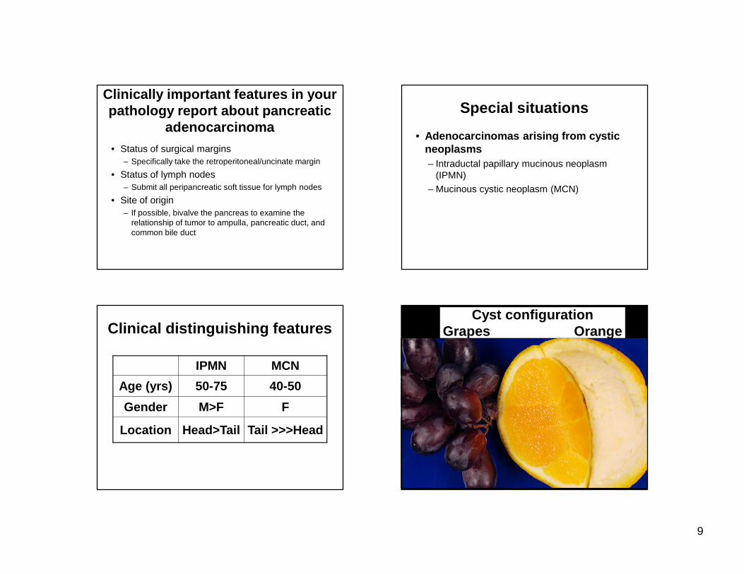
IPMN MCN
Age (yrs) 50-75 40-50
Gender M>F F
Location Head>Tail Tail >>>Head
Clinical distinguishing featuresCyst configuration
Grapes Orange

10
Mucin extrusion from ampulla of Vater is virtually diagnostic of intraductal papillary
mucinous neoplasm
Clue to the diagnosis Anatomic classificationTwo types of IPMN
Dilation of main duct > 1 cm
Cysts communication to main duct without
dilation
Tanaka M et al. Pancreatology 2006:6;17-32
Main duct Branch duct
IPMN main duct type
Pancreatic duct
IPMN main duct type
Pancreatic duct

11
Dilated pancreatic duct filled with papillary growth
IPMN branch duct type
Common bile duct
Pancreatic duct
Grape like cysts
IPMN branch duct type
Pancreatic duct
Grape like cysts
IPMN multifocal
Grape like cysts
Pancreatic duct

12
Pancreatic duct with filled with a fleshy mass
Common bile duct
IPMN main duct type with tiny focus of invasive carcinoma
Pancreatic duct
Your pathology report with IPMNshould contain the following
• If invasive carcinoma is present– X type invasive carcinoma, # cm, arising in
association with IPMN (# cm)
• If no invasive carcinoma– IPMN (# cm) with highest grade of dysplasia
• Main or branch duct type (correlate with radiology)• Epithelial subtype (intestinal, gastric/null,
pancreatobiliary, oncocytic)
Mucinous cystic neoplasm Mucinous cystic neoplasm
Pancreatic duct
13
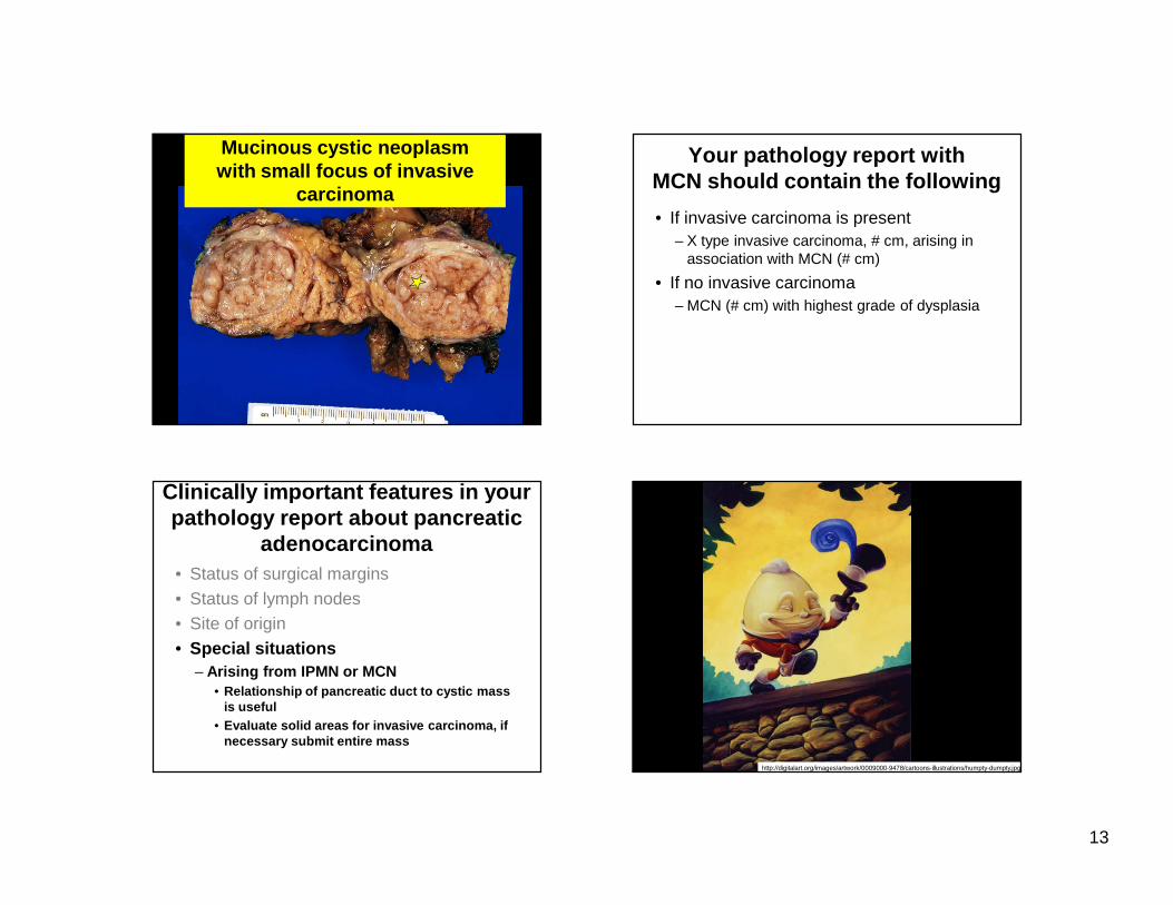
Solid fleshy cerebriform mass
Mucinous cystic neoplasm with small focus of invasive
carcinoma
Your pathology report with MCN should contain the following
• If invasive carcinoma is present– X type invasive carcinoma, # cm, arising in
association with MCN (# cm)
• If no invasive carcinoma– MCN (# cm) with highest grade of dysplasia
Clinically important features in your pathology report about pancreatic
adenocarcinoma• Status of surgical margins• Status of lymph nodes• Site of origin• Special situations
– Arising from IPMN or MCN• Relationship of pancreatic duct to cystic mass
is useful• Evaluate solid areas for invasive carcinoma, if
necessary submit entire mass
http://digitalart.org/images/artwork/0009000-9478/cartoons-illustrations/humpty-dumpty.jpg