coronary artery disease handouts - osu center for ... - coronary artery disease...svg to the lad,...
TRANSCRIPT

1
Coronary Artery DiseaseCoronary Artery Disease
John A Larry MDJohn A. Larry, MDAssociate Professor, Clinical Internal Medicine
Director of Cardiac RehabilitationSection Chief, OSU East Cardiovascular
MedicineThe Ohio State University
76 4 14
Coronary HeartDiseaseStroke
HF*
Hi h Bl d P
AHA StatisticsAHA Statistics
Percentage breakdown of deaths from cardiovascular diseases (United States:2004) * - Not a true underlying cause.
Source: NCHS and NHLBI.
52
17 High Blood Pressure
Diseases of theArteriesOther
Case PresentationCase Presentation• 79 year old gentleman underwent CABG 10
years ago: SVG to the LAD, SVG to the obtuse marginal branch, and SVG to the ramus intermedius vessel
• After presenting with a small non ST elevation MI 4 years ago, CATH revealed occlusion of the SVG to the LAD, 80% stenosis of the native LAD, as well as significant stenosis of the grafts to the ramus and OM vessels; the RCA was occluded and filled via collaterals.
• PCI with stent was performed to the native LAD, as well as the SVG to the ramus and OM vessels
Case PresentationCase Presentation• 3 years ago, he exhibited
unstable angina• Repeat cath demonstratedRepeat cath demonstrated
occlusion of the SVG to the ramus intermedius and medical therapy was recommended

2
Case PresentationCase Presentation• He had been doing well, exercising at a
very modest pace 3x a week.• 4-5 days prior to office visit, he noted
substernal chest tightness without exertional provocation radiation orexertional provocation, radiation, or associated symptoms, lasting 5-10 minutes, resolved with a single NTG on 2 occasions. Since that time, he walked some, up to 10 minutes at a slow pace without symptoms, and he has exhibited no recurrent chest pain.
Case PresentationCase Presentation• Current medications include
– ASA– Clopidogrel 75 mg daily– Metoprolol XL 25 mg daily– Isosorbide120 mg daily– Simvastatin 80 mg daily– Lisinopril 10 mg daily– SL NTG
Case PresentationCase Presentation• Exam
– Pulse 56, BP 138/60 right, 134/60 left, resp. rate 16
– JVP is normal. No carotid bruits are presentpresent
– Lungs are clear to auscultation and percussion
– PMI is nondisplaced. S1 and S2 are normal. A grade 1 systolic ejection murmur is noted. No gallops or rubs present
Case PresentationCase Presentation• Exam
– Abdomen is soft and nontender, with no organomegaly, aneurysm or bruits
– Extremities free of edema, distal l l blpulses are palpable.
3
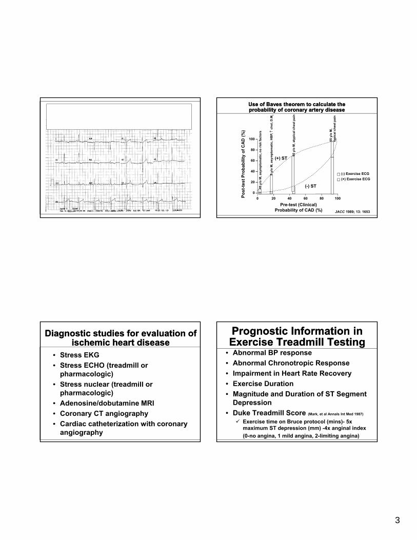
Diagnostic studies for evaluation of ischemic heart disease
Diagnostic studies for evaluation of ischemic heart disease
• Stress EKG• Stress ECHO (treadmill or
pharmacologic)St l (t d ill• Stress nuclear (treadmill or pharmacologic)
• Adenosine/dobutamine MRI • Coronary CT angiography• Cardiac catheterization with coronary
angiography
100 –
80 –
ty o
f CA
D (%
)
(+) STtic, n
o ris
k fa
ctor
s
mpt
omat
ic, H
BP,
↑ch
ol, D
.M.
55 y
/o M
, ty
pica
l che
st p
ain
Use of Baves theorem to calculate the probability of coronary artery disease Use of Baves theorem to calculate the probability of coronary artery disease
60 –
40 –
20 –
0 –0 20 40 60 80 100Po
st-te
st P
roba
bilit
Pre-test (Clinical)Probability of CAD (%)
(+) ST
(-) ST
JACC 1989; 13: 1653
45 y
/o M
, asy
mpt
omat
45 y
/o M
, asy
(-) Exercise ECG(+) Exercise ECG
Prognostic Information in Exercise Treadmill TestingPrognostic Information in Exercise Treadmill Testing
• Abnormal BP response• Abnormal Chronotropic Response• Impairment in Heart Rate Recovery
E i D ti• Exercise Duration• Magnitude and Duration of ST Segment
Depression• Duke Treadmill Score (Mark, et al Annals Int Med 1987)
Exercise time on Bruce protocol (mins)- 5x maximum ST depression (mm) -4x anginal index (0-no angina, 1 mild angina, 2-limiting angina)

4
Prognostic Data in Stress TestingPrognostic Data in Stress Testing
Circulation. 1998;98:1622-1630
High Risk Features in Stress/Dobutamine Echo
High Risk Features in Stress/Dobutamine Echo
• New or worsening wall motion abnormalities in multiple coronary territoriesterritories
• Peak wall motion score index >1.7• Drop in LVEF
Adverse Prognostic Features in Treadmill/Pharmacologic Nuclear
Imaging
Adverse Prognostic Features in Treadmill/Pharmacologic Nuclear
Imaging
• Multiple reversible perfusion defects in 2 or more coronary territories
• Quantitatively large myocardial perfusion defects
• Transient ischemic dilation of the LV• Lung uptake
Case PresentationCase Presentation• Pharmacologic nuclear study
ordered– His typical walking speed limited– HR independent study– Both issues raised concern a
treadmill study would not be adequate

5
Case PresentationCase Presentation• Pharmacologic nuclear study ordered
– Previous revascularization– By appropriateness criteria published
by the ACC/AHA imaging studyby the ACC/AHA, imaging study considered appropriate
– As an aside, pharmacologic nuclear study is preferred in patients with LBBB or ventricular paced rhythm
Case PresentationCase PresentationPharmacologic nuclear study findings:
Large, moderate to severe reversible perfusion defect in the inferoapical, entire lateral/inferolateral and basal and mid anterior/anterolateral walls, concerning for ischemiaconcerning for ischemia.
No scintigraphic evidence of prior injury.
He was referred for left heart catheterization with coronary and graft angiography.
Coronary Artery DiseaseCoronary Artery Disease
Richard J. Gumina, MD, PhDAssociate Professor, Cardiovascular MedicineDirector, Interventional Cardiovascular Research
The Ohio State University
Coronary Angiogram Video 1Coronary Angiogram Video 1

6
Coronary Angiogram Video 2Coronary Angiogram Video 2
Coronary Angiogram Video 3Coronary Angiogram Video 3
Revascularization OptionsRevascularization Options
• Indications for PCI
• Indications for Coronary Artery• Indications for Coronary Artery Bypass Graft Surgery
• Hybrid Revascularization Trial
ACCF/SCAI/STS/AATS/AHA/ASNC 2009 Appropriateness Criteria for Coronary Revascularization
A Report by the American College of Cardiology Foundation Appropriateness Criteria Task Force, Society for Cardiovascular Angiography and Interventions,
Society of Thoracic Surgeons, American Association for Thoracic Surgery, American Heart Association, and the American Society of Nuclear Cardiology Endorsed by the
American Society of Echocardiography, the Heart Failure Society of America, and the Society of Cardiovascular Computed Tomography
Revascularization OptionsAppropriateness Criteria
Revascularization OptionsAppropriateness Criteria
February 2009180 clinical scenarios
Appropriateness of revascularization and appropriateness of PCI or CABG individually
as the primary mode of revascularizationPatel, M. R. et al. J Am Coll Cardiol 2009;53:530-553
Manesh R. Patel, MD, Chair, Coronary Revascularization Writing Group, Gregory J. Dehmer, MD, FACC, FACP, FSCAI, FAHA, Coronary Revascularization Writing Group, John W. Hirshfeld, MD, Coronary Revascularization Writing Group , Peter K. Smith, MD, FACC, Coronary Revascularization Writing Group and John A. Spertus, MD,
MPH, FACC, Coronary Revascularization

7
Appropriateness Criteria: Low-Risk
Appropriateness Criteria: Low-Risk
• Low-risk treadmill score (≥ 5)• Normal or small myocardial perfusion
defect at rest or with stressdefect at rest or with stress• Normal stress echocardiographic wall
motion or no change of limited resting wall motion abnormalities during stress
Appropriateness Criteria: Intermediate Risk
Appropriateness Criteria: Intermediate Risk
• Mild/moderate resting left ventricular dysfunction (LVEF 35-49%)
• Intermediate-risk treadmill score (-11 to +5)• Stress induced moderate perfusion defect• Stress-induced moderate perfusion defect
without LV dilation or increased lung uptake (thallium-201)
• Limited stress echocardiographic ischemia with a wall motion abnormality only at higher doses of dobutamine involving ≤ 2 segments
Appropriateness Criteria: High Risk
Appropriateness Criteria: High Risk
• Severe resting left ventricular dysfunction (LVEF < 35%)
• High-risk treadmill score (≤ or equal to 11)• Severe exercise left ventricular
dysfunction (exercise LVEF < 35%)• Stress-induced multiple perfusion defect
(particularly if anterior)• Stress-induced multiple perfusion defects
of moderate size
Appropriateness Criteria: High Risk
Appropriateness Criteria: High Risk
• Large, fixed perfusion defect with LV dilation or increased lung uptake (thallium-201)
• Echocardiographic wall motion abnormality involving > 2segments developing with low dose dobutamine or at low heart rate (< 120)
• Stress echocardiographic evidence of extensive ischemia

8
Appropriateness Ratings by Risk Findings on Noninvasive Imaging Study and
Symptoms
Appropriateness Ratings by Risk Findings on Noninvasive Imaging Study and
SymptomsSymptoms
Asymptomatic to CCS Class IV
Medical therapyMinimal to maximal
Cornary anatomyChronic total occlusion 1 vessel1-2 Vessel without Proximal LAD1 Vessel disease2 Vessel Disease3 Vessel Disease
2-vessel CAD with proximal LAD stenosis
3-vessel CAD
Isolated Left Main Disease
Left Main disease and additional CAD
CABG No diabetesNormal LVEF
A A A A
Method of Revascularization of Advanced Method of Revascularization of Advanced Coronary Artery DiseaseCoronary Artery Disease
Modified from Patel, M. R. et al. J Am Coll Cardiol 2009;53:530-553
Diabetes A A A A
Depressed LVEF A A A A
PCI No diabetesNormal LVEF
A U I I
Diabetes A U I IDepressed LVEF A U I I
CABG indicates coronary artery bypass grafting; LAD, left anterior descending artery; LVEF, left ventricular ejection fraction; and PCI, percutaneous coronary intervention
2-vessel CAD with proximal LAD stenosis
3-vessel CAD
Isolated Left Main Disease
Left Main disease and additional CAD
CABG No diabetesNormal LVEF
A A A A
Syntax
Method of Revascularization of Advanced Method of Revascularization of Advanced Coronary Artery DiseaseCoronary Artery Disease
Modified from Patel, M. R. et al. J Am Coll Cardiol 2009;53:530-553
Diabetes A A A A
Depressed LVEF A A A A
PCI No diabetesNormal LVEF
A U I I
Diabetes A U I IDepressed LVEF A U I I
Syntax
CABG indicates coronary artery bypass grafting; LAD, left anterior descending artery; LVEF, left ventricular ejection fraction; and PCI, percutaneous coronary intervention
Original ArticlePercutaneous Coronary Intervention versus Coronary-Artery Bypass
Grafting for Severe Coronary Artery DiseasePatrick W. Serruys, M.D., Ph.D., Marie-Claude Morice, M.D., A. Pieter Kappetein, M.D., Ph.D.,
Antonio Colombo, M.D., David R. Holmes, M.D., Michael J. Mack, M.D., Elisabeth Ståhle, M.D., Ted E. Feldman, M.D., Marcel van den Brand, M.D., Eric J. Bass, B.A., Nic Van Dyck, R.N., Katrin
Leadley, M.D., Keith D. Dawkins, M.D., and Friedrich W. Mohr, M.D., Ph.D. for the SYNTAX Investigators
N Engl J Med 2009; 360:961-972. March 5, 2009
• Goal: To compare the safety and efficacy of CABG v. PCI
Syntax TrialSyntax Trial
with TAXUS DES in patients with 3 vessel disease or left main disease, who were eligible for either procedure.
• Hypothesis: DES-PCI would be non-inferior to CABG in the management of patients with 3VD and/or LM.• All patients in PCI arm received TAXUS DES.
1800 pts randomised (897 CABG, 903 PCI)Serruys PW et al. N Engl J Med 2009;360:961-972.

9
e R
ate
(%)
20
13.5
17.8
12.4
P=0.002
P=0.99
p<0.001
Rates of Outcomes among the Study Rates of Outcomes among the Study Patients, According to Treatment Patients, According to Treatment
Group at 12 monthsGroup at 12 months
Modified from Serruys PW et al. N Engl J Med 2009;360:961-972.
Cum
ulat
ive 10
0Death from Any Cause
Death from Any Cause, Stroke, or MI
Repeat RevascularizationMajor Adverse Cardiac or
Cerebrovascular Event
4.43.5
7.7 7.65.9p=0.37
PCI
CABG
Rates of Major Adverse Cardiac or Rates of Major Adverse Cardiac or CerebrovascularCerebrovascularEvents among the Study Patients, According to Events among the Study Patients, According to Treatment Group and SYNTAX Score Category.Treatment Group and SYNTAX Score Category.
High SYNTAX scores (≥33, indicating the most complex disease)
(%)
30 p<0.00123.4
Modified from Serruys PW et al. N Engl J Med 2009;360:961-972.
Cum
ulat
ive
Rat
e
20
0
10
10.9
“Redo” CABG Surgery “Redo” CABG Surgery -- considerationsconsiderations
Reoperative coronary artery bypass procedures: risk factors for early mortality and late survival
J.T. Christenson*, M. Schmuziger, F. SimonetThe Cardio6ascular Surgery Unit, Hoˆpital de la Tour, 1. a6. J.-D. Maillard,
CH-1217 Meyrin-Gene6a, SwitzerlandE J l f C di th i S 11 (1997) 129 133European Journal of Cardio-thoracic Surgery 11 (1997) 129–133
REDO CABG (n=594) Primary CABG (n=3148)
Risk Factor REDO CABG (n=594) Primary CABG (n=3148)P Odds P Odds
Emergent operation <0.001 2.12 0.001 1.92
Urgent operation 0.008 1.86 ----
CCS class 3 and 4 0.005 1.96 0.006 1.67
Independent rick factors for mortality. Multivariate logistic regression analysis
“Redo” CABG Surgery “Redo” CABG Surgery -- considerationsconsiderations
LVEF <40% 0.011 1.62 0.022 1.28
Multifocal vascular disease
0.007 1.77 0.014 1.73
Preoprerative renal insufficiency
0.012 1.48 ---- ----
IDDM 0.029 1.12 ---- ----
Age >65 years 0.028 1.13 0.011 1.08
Interval from primary CABG >1 year
0.012 1.81
Modified from: European Journal of Cardio-thoracic Surgery 11 (1997) 129–133

10
Simultaneous Hybrid Revascularization Versus
Off-Pump Coronary Artery Bypass for Multivessel
Coronary Artery Disease
Hybrid ApproachHybrid Approach
Coronary Artery Disease
Shengshou Hu, MD,* Qi Li, MD,* Peixian Gao, MD,* Hui Xiong, MD, Zhe Zheng, MD,Lihuan Li, MD, Bo Xu, MD, and Runlin Gao, MD
Departments of Surgery, Anesthesiology, and Cardiology, and Research Center for Cardiovascular Regenerative Medicine,
Ministry of Health of China, Cardiovascular Institute and Fuwai Hospital, Beijing, China
The Annals of Thoracic SurgeryVolume 91, Issue 2, February 2011,
Pages 432-438
Complication Hybrid (n = 104) No. (%)
OPCAB (n = 104) No. (%) p Value
MACCE 1 (1.0%) 10 (9.6%) 0.03
Death 0 1 (1.0%) 0.50
Myocardial infarction 0 0 1.00
Hybrid ApproachHybrid Approach
Neurologic event 0 5 (4.8%) 0.07
Repeat revascularization 1 (1.0%) 3 (2.9%) 0.34
Readmittance 9 (8.7%) 26 (25.0%) 0.001
Survival 104 (100%) 103 (99.0%) 0.50
MACCE = major adverse cardiac or cerebrovascular events; OPCAB = off-pump coronary artery bypass grafting.
Modified from: The Annals of Thoracic Surgery Volume 91, Issue 2, February 2011, Pages 432-438
• Underwent Redo-CABG
• Free RIMA to the left anterior
Our PatientOur Patient
descending artery
Coronary Artery DiseaseCoronary Artery Disease
John A Larry MDJohn A. Larry, MDAssociate Professor, Clinical Internal Medicine
Director of Cardiac RehabilitationSection Chief, OSU East Cardiovascular
MedicineThe Ohio State University

11
Importance of Dual Antiplatelet Therapy Post Drug Eluting Stent Implantation
Importance of Dual Antiplatelet Therapy Post Drug Eluting Stent Implantation
• AHA/ACC/SCAI/ACS/ADA Science Advisory 2007
• Because premature discontinuation of dual antiplatelet therapy greatlydual antiplatelet therapy greatly increases the risk of stent thrombosis, myocardial infarction, and death
• Dual antiplatelet therapy should be continued uninterrupted for one yearpost implantation of a drug eluting stent

12
Blood Pressure ControlBlood Pressure Control• Goal is less than 140/90 or• Less than 130/80 in patients with
diabetes or chronic kidney diseasey
• Initially, utilize B-blockers and ACE inhibitors and add additional therapy as needed

13
Lipid management goalsLipid management goals• Current secondary prevention
recommendations for lipid management recommend:
• LDL goal < 100, reasonable target of 70 /dLmg/dL
• Non HDL < 30 points above the LDL target• There may be need to consider additional
therapy beyond statin agents to achieve NCEP goals
Circulation 2006;113;2363-2372
OSUMC Comprehensive Lipid Management ClinicOSUMC Comprehensive Lipid Management Clinic
• Patients that may benefitDifficulty achieving NCEP lipid goals, intolerance to therapy, low HDL, elevated TG
• Appointments/Referrals- (614) 293-ROSS (7677) - Offices located at Ross ACC, OSU
East, Gahanna and Stoneridge (Dublin)
VaccinationVaccination
• Patient with CAD should receive appropriate vaccination forappropriate vaccination for influenza
Smoking CessationSmoking Cessation• Ask about tobacco use status at every visit. I (B)• Advise every tobacco user to quit. I (B)• Assess the tobacco user’s willingness to quit. I
(B)( )• Assist by counseling and developing a plan for
quitting. I (B)• Arrange follow-up, referral to special programs,
or pharmacotherapy I (B)• Urge avoidance of exposure to environmental
tobacco smoke at work and home. I (B)

14
Cardiac Rehab ProgramsCardiac Rehab Programs• Indications for Cardiac Rehab
– Angina with documented evidence of myocardial ischemia within 6 mos.
– MI within 12 months– PCI within 6 mos.– CABG within 6 mos.– Valve replacement/repair within 6 mos.– Heart transplant within 12 mos
• OSU Heart Center at Morehouse 293-6937• OSU East 257-3974
Effects of Cardiac Rehabilitation
Effects of Cardiac Rehabilitation
Outcome Mean 95% conf. intervals p-value
• Total Mortality* -20% (-7 to -32%) p=.005• Cardiac Mortality* -26% (-10 to -29%) p=.002
Nonfatal MI 21% ( 43 to 9%) p= 15• Nonfatal MI -21% (-43 to 9%) p=.15• CABG -13% (-35 to 16%) p=.4• PCI -19% (-51 to 34%) p=.4
Taylor, et al Am J of Medicine 2004; 116:682-97
20 30 g/dFiber50%–60% of total caloriesCarbohydrate (esp. complex carbs)25%–35% of total caloriesTotal fatUp to 20% of total caloriesMonounsaturated fatUp to 10% of total caloriesPolyunsaturated fat
<7% of total caloriesSaturated fat*Recommended IntakeNutrient
ATP III Dietary RecommendationsATP III Dietary Recommendations
*Trans fatty acids also raise LDL-C and should be kept at a low intakeNote: Regarding total calories, balance energy intake and expenditure to maintain
desirable body weight.
<200 mg/dCholesterol~15% of total caloriesProtein
20–30 g/dFiber
Expert Panel on Detection, Evaluation, and Treatment of High Blood Cholesterol in Adults. JAMA 2001;285:2486-97
ATP=Adult Treatment Panel
Chest PainClinic
Chest PainClinic
366-1279