cornell product left ventricular hypertrophy in...
TRANSCRIPT

Cornell Product Left Ventricular Hypertrophy inElectrocardiogram and the Risk of Stroke in a
General PopulationJoji Ishikawa, Shizukiyo Ishikawa, Tomoyuki Kabutoya, Tadao Gotoh, Kazunori Kayaba,
Joseph E. Schwartz, Thomas G. Pickering, Kazuyuki Shimada, Kazuomi Kario;for the Jichi Medical School Cohort Study Investigators Group
Abstract—Left ventricular hypertrophy (LVH), assessed by ECG, is associated with an increased risk for cardiovascularevents among hypertensive subjects. We evaluated the risks of LVH in a Japanese general population includingnormotensive and prehypertensive subjects. We measured ECG and blood pressure in 10 755 subjects at baseline. TheCornell product (CP) and Sokolow-Lyon (SL) voltage were calculated as markers of LVH (CP �2440 mm�ms and SLvoltage �38 mm). Follow-up was performed for 10 years, and the incidence of stroke and myocardial infarction wasevaluated. The prevalence of CP-LVH was 2.7% for normotensives, 5.2% for prehypertensives, and 11.0% forhypertensives, and the prevalence of SL-LVH was 5.0%, 8.2%, and 15.2%, respectively. In all of the subjects, CP-LVHand SL-LVH were both predictors of stroke (CP-LVH: hazard risk: 1.62, 95% CI: 1.19 to 2.20, P�0.002; SL-LVH:hazard risk: 1.29, 95% CI: 0.98 to 1.71, P�0.07) after adjustment for confounding factors but were not predictors ofmyocardial infarction. The adjusted hazard ratio of CP-LVH predicting stroke was especially high in the normotensives(hazard risk: 7.53; 95% CI: 3.39 to 16.77). In the normotensives, diabetes mellitus and hyperlipidemia were significantdeterminants of CP-LVH but not of SL-LVH. In all of the hypertensive subgroups (normotensives, prehypertensives,and hypertensives), the c-statistic for the equation predicting stroke increased when CP-LVH was added to the modelbut not when SL-LVH was added. In conclusion, both CP-LVH and SL-LVH are risk factors for stroke in the Japanesegeneral population. CP-LVH is related to glucose abnormality, and its predictive value for stroke is seen even innormotensives and prehypertensives. (Hypertension. 2009;53:28-34.)
Key Words: Cornell product � left ventricular hypertrophy � stroke event � cohort study
Left ventricular hypertrophy (LVH), a measure of hyper-tensive target organ damage in the heart, has been
reported to be associated with increased morbidity andmortality.1,2 LVH can be evaluated by echocardiography(Echo) and/or a 12-lead ECG. LVH defined by ECG (ECG-LVH) has been evaluated using standard voltage criteriareported by Sokolow and Lyon (SL)3 and more recently usingthe Cornell product (CP) criteria.4 LVH detected by CP(CP-LVH) is reported to have a higher sensitivity for thepresence of LVH evaluated by echocardiography (Echo-LVH) than the Sokolow-Lyon criteria (SL-LVH).5 CP-LVHand SL-LVH were independently associated with Echo-LVH6
and with stroke events, cardiovascular morbidity, and mor-tality7 in subjects with essential hypertension. These datashow that evaluation of CP-LVH is beneficial in Westernhypertensive subjects; however, in Japanese populations, theincidence of stroke is higher than that of ischemic heart
disease,8 and there are no data on the cardiovascular risk inJapanese subjects with CP-LVH.
In addition, hypertension is the major cause of LVH, andthe cardiovascular risks in normotensive and prehypertensivesubjects with CP-LVH and/or SL-LVH remain unknown. It isreported that hypertensive patients with diabetes mellitushave a higher prevalence and greater severity of LVH thanthose without diabetes mellitus.9,10 Okin et al11 reported thatdiabetes mellitus, per se, attenuates the regression of hyper-tensive LVH during antihypertensive treatments. However,there are no data examining whether cardiovascular riskfactors such as diabetes mellitus might contribute to anincrease of ECG-LVH in normotensive subjects or whetherLVH in normotensive and prehypertensive subjects can be arisk factor for cardiovascular events.
The purpose of the present study was to evaluate thecardiovascular risks of CP-LVH and SL-LVH in the Japanese
Received June 12, 2008; first decision July 4, 2008; revision accepted October 23, 2008.From the Division of Cardiovascular Medicine, Department of Internal Medicine (J.I., T.K., K.S., K. Kario), and Division of Community and Family
Medicine, Center for Community Medicine (S.I.), Jichi Medical University School of Medicine, Tochigi, Japan; Center for Behavioral CardiovascularHealth (J.I. J.E.S., T.G.P.), Division of General Medicine, Department of Medicine, Columbia University Medical Center, New York, NY; Wara NationalHealth Insurance Hospital (T.G.), Gifu, Japan; and Saitama Prefectural University (K. Kayaba), Saitama, Japan.
Correspondence to Joji Ishikawa, MD, PhD, Division of Cardiovascular Medicine, Department of Medicine, Jichi Medical University, School ofMedicine 3311-1, Yakushiji, Shimotsuke, Tochigi 329-0498, Japan. E-mail [email protected]
© 2008 American Heart Association, Inc.
Hypertension is available at http://hyper.ahajournals.org DOI: 10.1161/HYPERTENSIONAHA.108.118026
28
by guest on July 5, 2018http://hyper.ahajournals.org/
Dow
nloaded from
by guest on July 5, 2018http://hyper.ahajournals.org/
Dow
nloaded from
by guest on July 5, 2018http://hyper.ahajournals.org/
Dow
nloaded from
by guest on July 5, 2018http://hyper.ahajournals.org/
Dow
nloaded from
by guest on July 5, 2018http://hyper.ahajournals.org/
Dow
nloaded from
by guest on July 5, 2018http://hyper.ahajournals.org/
Dow
nloaded from
by guest on July 5, 2018http://hyper.ahajournals.org/
Dow
nloaded from
by guest on July 5, 2018http://hyper.ahajournals.org/
Dow
nloaded from

general population and to explore the differences in back-grounds and prognostic factors that predict the presence ofCP-LVH and SL-LVH, especially in normotensive and pre-hypertensive subjects.
Methods
SubjectsThe Jichi Medical School Cohort Study was begun in 1992, with theprimary aim of clarifying the risk factors for cardiovascular andcerebrovascular diseases in the Japanese general population. Thedetails of the protocol of the Jichi Medical School Cohort Study havebeen reported previously.12 Baseline data were collected betweenApril 1992 and July 1995 in 12 rural districts using a government-sponsored mass screening system. In each community, a localgovernment office sent personal invitations by mail to all of thesubjects in accordance with the health and medical service law forthe aged. The subjects for the mass screening examinations wereresidents aged 40 to 69 years in 8 areas (Iwaizumi, Tako, Kuze,Sakuma, Sakugi, Okawa, Ainoshima, and Akaike). Subjects includedthose aged �30 years in 1 area (Wara), and other age groups werealso included in 3 areas (Hokudan, Yamato, and Takasu). The totalnumber of subjects in the Jichi Medical School Cohort Study atbaseline was 12 490 (4911 men and 7579 women). The participationrate varied in each community (26.0% to 90.0%), and the overallparticipation rate of those invited to the mass screening examinationprogram was 65.4%.13
ECG Measurement and InterpretationECG was measured at a paper speed of 25 mm/s, at a gain of10 mm/mV (or 5 mm/mV), using ECG devices that the institutes had(FCP130-A9, FCP145-M4, and FCP270-M5, Fukuda Denshi, etc). Atrained person, who did not know the subjects’ backgrounds,measured ECGs at a central laboratory using a ruler with 0.01-mmgraduations. Both SL voltage (SV1�RV5) and Cornell voltage(RaVL�SV3, with 6 mm added for women)4,5 were measured. QRSduration was measured manually from lead II (or lead I or III if themeasurement of QRS duration was difficult from lead II) on a singleheart beat. CP was calculated as the product of Cornell voltage timesQRS duration afterward. SL-LVH was defined as �38 mm (3.8mV), and CP-LVH was defined as 2440 mm�ms according to aprevious report of the Losartan Intervention for Endpoint Reductionin Hypertension Study.7
Questionnaire and Other MeasurementsInformation about medical history and lifestyle was obtained with aquestionnaire at baseline. Age is the value at baseline. Smokingstatus was reported as current smoker, ex-smoker, or never smoked.Alcohol drinkers were defined as those who were reported consum-ing �20 g/d. Body mass index (BMI) was calculated as weight(kilograms)/height (meters squared). The systolic blood pressure(SBP) and diastolic blood pressure (DBP) at baseline were measuredusing a fully automated and validated upper arm cuff-oscillometricdevice, the BP203RV-II (Nippon Colin).14 Blood pressure wasmeasured once after resting for �5 minutes while seated. Hyperten-sion was defined as either a SBP/DBP of �140/90 mm Hg or takingantihypertensive medications. Prehypertension was defined as SBP/DBP 120/80 to 139/89 mm Hg. Normotension was defined asSBP/DBP �120/80 mm Hg. Diabetes mellitus was defined by afasting glucose level �7.0 mmol/L (126 mg/dL), a casual glucoselevel �11.1 mmol/L (200 mg/dL), or the use of an oral hypoglyce-mic agent or insulin. Impaired fasting glucose was defined as afasting glucose level of 110 to 125 mg/dL. Hyperlipidemia wasdefined as a total cholesterol level �5.7 mmol/L (220 mg/dL), atriglyceride level �1.7 mmol/L (150 mg/dL), or the use of an orallipid-lowering agent, according to the Japanese AtherosclerosisSociety Guidelines for Prevention of Atherosclerotic CardiovascularDiseases.
Informed ConsentThe internal review board of the Jichi Medical University School ofMedicine approved this study. Written informed consent for thestudy was obtained individually from all of the subjects during themass screening examination health checkup.
Follow-Up and Diagnostic CriteriaThe mass screening examination system was used to check thesubjects every year for 10 years. The details of follow-up are shownin the online supplemental data file (please see http://hyper.ahajournals.org). The diagnosis was determined independently by an end pointscommittee, which included radiologists, neurologists, and cardiolo-gists, in accordance with the World Health Organization Monitoringof Trends and Determinants in Cardiovascular Disease Project.15 Thedetails of diagnostic criteria are also shown in the online supplemen-tal data file.
Statistical AnalysisAmong the 12 490 subjects who were initially enrolled in the JichiMedical School Cohort Study, we analyzed 10 755 subjects who hadadequate follow-up, after excluding 1735 subjects with no ECGrecording (n�1285), immeasurable ECG findings (n�28), completeleft bundle branch block (n�20), complete right bundle branch block(n�189), atrial fibrillation (n�53), or no blood pressure data(n�160).
Data are shown as means�1 SDs for continuous variables and aspercentages for dichotomous variables. Differences in characteristicsbetween subjects with and without CP-LVH and SL-LVH wereevaluated using Student t test or �2 test. Because the prevalence ofLVH varied among the hypertensive subgroups (normotension,prehypertension, and hypertension), we performed analyses of char-acteristics of the subjects with CP-LVH in the hypertensive sub-groups. Determinants of CP-LVH and SL-LVH in all of the subjectsand separately for each hypertensive subgroup were evaluated usingmultivariate logistic regression analysis including age, gender, BMI,smoking status, alcohol drinking, SBP, antihypertensive medicationuse (only in the hypertensives), presence of hyperlipidemia, andstatus of diabetes mellitus. The incidence risks of stroke andmyocardial infarction in both unadjusted models and those adjustedfor significant covariates were evaluated using Cox regressionanalysis in the total sample and separately for each hypertensionsubgroup. The c-statistic was calculated, according to the method ofPencina and D’Agostino,16 for the baseline model that included theunmodifiable cardiovascular risk factors (age and gender) andthen for a series of models in which each cardiovascular riskfactor was added separately to the baseline model. Computersoftware SPSS 16.0 (SPSS Inc) and SAS 9.1 (SAS Institute) wereused for the analysis, and a P value �0.05 was consideredstatistically significant.
ResultsStudy SubjectsThe mean age was 55.6�11.2 years (men: 37.8%). Theaverage SBP/DBP was 130�21/78�12 mm Hg. The percent-ages of subjects with normotension, prehypertension, andhypertension were 32.9%, 32.6%, and 34.5%, respectively.The characteristics of the study subjects were as follows:history of stroke, 1.0%; history of myocardial infarction,0.5%; antihypertensive medication use, 11.1%; formersmoker, 12.4%; current smoker, 21.8%; alcohol drinker,27.6%; hyperlipidemia, 35.3%; impaired fasting glucose,2.5%; and diabetes mellitus, 3.6%. The prevalences of CP-LVH and SL-LVH in the full sample were 6.4% were 9.5%,respectively.
Ishikawa et al Cornell Product LVH and Stroke 29
by guest on July 5, 2018http://hyper.ahajournals.org/
Dow
nloaded from

Characteristics and Determinants of CP-LVHand SL-LVH in All SubjectsComparisons of subjects with and without CP-LVH andSL-LVH are shown in Table 1. Subjects with CP-LVH andthose with SL-LVH were older and had a higher prevalenceof hypertension and antihypertensive medication use; how-ever, there were differences in characteristics such as gender,BMI, and hyperlipidemia, and diabetes mellitus depended onwhether we grouped subjects by CP-LVH or SL-LVH.
In all of the subjects, significant determinants of bothCP-LVH and SL-LVH were SBP (CP-LVH: odds ratio [OR]:1.20 per 10 mm Hg, 95% CI: 1.15 to 1.25; SL-LVH: OR:1.26 per 10 mm Hg, 95% CI: 1.21 to 1.30) and antihyperten-sive medication use (CP-LVH: OR: 1.79, 95% CI: 1.45 to2.20; SL-LVH: OR: 1.71, 95% CI: 1.41 to 2.08). Additionaldeterminants of only CP-LVH were age (OR: 1.16 per 10years; 95% CI: 1.07 to 1.27) and higher BMI (OR: 1.03 per1 kg/m2; 95% CI: 1.00 to 1.06) and of only SL-LVH weremale gender (OR: 3.00; 95% CI: 2.47 to 3.65) and lower BMI(OR: 0.92 per 1 kg/m2; 95% CI: 0.89 to 0.93).
Characteristics and Determinants of Subjects WithCP-LVH and SL-LVH in the Hypertensive GroupsThe characteristics of subjects with and without CP-LVH andthose with and without SL-LVH in the 3 hypertensivesubgroups (normotensives, prehypertensives, and hyperten-
sives) are shown in the online supplemental data file (TablesS1 and S2).
In the normotensives, the significant determinants of bothCP-LVH and SL-LVH were male gender (CP-LVH: OR:2.15, 95% CI: 1.22 to 3.78; SL-LVH: OR: 3.55, 95% CI: 2.30to 5.48) and lower BMI (CP-LVH: OR: 0.90 per 1 kg/m2,95% CI: 0.83 to 0.98; SL-LVH: OR: 0.93 per 1 kg/m2, 95%CI: 0.87 to 0.99). The additional determinants of onlyCP-LVH were nonsmokers (ex-smokers: OR: 0.45, 95% CI:0.24 to 0.87; current smokers: OR: 0.24, 95% CI: 0.09 to0.66), the presence of hyperlipidemia (OR: 1.66; 95% CI:1.05 to 2.61), and diabetes mellitus (OR: 3.26; 95% CI: 1.24to 8.53), and that of only SL-LVH was age (OR: 1.15 per 10years; 95% CI: 1.01 to 1.32).
In the prehypertensives, the significant determinants ofboth CP-LVH and SL-LVH were SBP (CP-LVH: OR: 1.38per 10 mm Hg, 95% CI: 1.07 to 1.79; SL-LVH: OR: 1.27 per10 mm Hg, 95% CI 1.03 to 1.57). The significant determinantof only CP-LVH was age (OR: 1.22 per 10 years; 95% CI:1.04 to 1.42), and those of only SL-LVH were male gender(OR: 2.88; 95% CI: 2.00 to 4.14) and lower BMI (OR: 0.90per 1 kg/m2; 95% CI: 0.86 to 0.95).
In the hypertensives, the significant determinants of bothCP-LVH and SL-LVH were antihypertensive medication use(CP-LVH: OR: 1.51, 95% CI: 1.20 to 1.89; SL-LVH: OR:1.59, 95% CI: 1.28 to 1.96) and SBP (CP-LVH: OR: 1.07 per
Table 1. Characteristics of Subjects With/Without CP-LVH and SL-LVH
Characteristic
CP �2440 mm�ms SL Voltage �38 mm
LVH (�) (N�10 069) LVH (�) (N�686) P LVH (�) (N�9723) LVH (�) (N�1027) P
Age, y 55.4�11.2 58.7�9.7 �0.001 55.3�11.2 57.8�10.4 �0.001
Male, % 38.0 35.0 0.12 35.2 62.0 �0.001
BMI, kg/m2 23.1�3.1 23.9�3.4 �0.001 23.2�3.1 22.8�2.9 �0.001
Smokers 0.13 �0.001
Former, % 12.8 11.3 12.1 18.6
Current, % 22.6 20.2 21.4 32.9
Alcohol drinkers, % 27.7 26.1 0.36 27.7 26.4 0.36
History of stroke, % 0.9 2.2 0.002 0.9 2.3 �0.001
History of myocardial infarction, % 0.5 0.8 0.33 0.5 0.9 0.056
Hypertension, % 33.7 61.1 �0.001 33.3 56.2 �0.001
Antihypertensive therapy, % 10.6 25.5 �0.001 10.7 19.9 �0.001
SBP, mm Hg 129�21 141�22 �0.001 128�21 140�23 �0.001
DBP, mm Hg 77�12 83�13 �0.001 77�12 83�13 �0.001
Hyperlipidemia, % 35.0 43.2 �0.001 35.8 32.7 0.049
Total cholesterol, mg/dL 192�35 196�34 0.004 193�35 188�35 �0.001
Triglyceride, mg/dL 116�76 131�77 �0.001 117�77 115�68 0.41
Status of diabetes 0.056 0.078
Impaired fasting glucose, % 2.5 3.1 2.4 3.5
Diabetes, % 3.5 5.1 3.6 3.2
Blood glucose, mg/dL 103�26 112�34 �0.001 103�27 106�26 �0.001
SL-LVH, % 8.8 21.3 �0.001 . . . . . . . . .
CP-LVH, % . . . . . . . . . 5.6 14.2 �0.001
CP-LVH indicates LVH defined by CP (�2440 mm�ms); SL-LVH, LVH defined by SL (�38 mm); . . ., no data. P values were calculated using Student t test or �2
test. P value �0.05 was considered statistically significant.
30 Hypertension January 2009
by guest on July 5, 2018http://hyper.ahajournals.org/
Dow
nloaded from
10 mm Hg, 95% CI: 1.01 to 1.14; SL-LVH: OR: 1.17 per10 mm Hg; 95% CI: 1.11 to 1.24). The significant determi-nants of only CP-LVH were female gender (male: OR: 0.68;95% CI: 0.49 to 0.95) and higher BMI (OR: 1.04 per 1 kg/m2;95% CI: 1.00 to 1.07), and those of only SL-LVH were malegender (OR: 2.82; 95% CI: 2.14 to 3.71), lower BMI (OR:0.91 per 1 kg/m2; 95% CI: 0.88 to 0.94), nonsmokers (currentsmokers: OR: 0.71; 95% CI: 0.51 to 0.99), and absence ofimpaired fasting glucose or diabetes mellitus (diabetes: OR:0.53; 95% CI: 0.33 to 0.86).
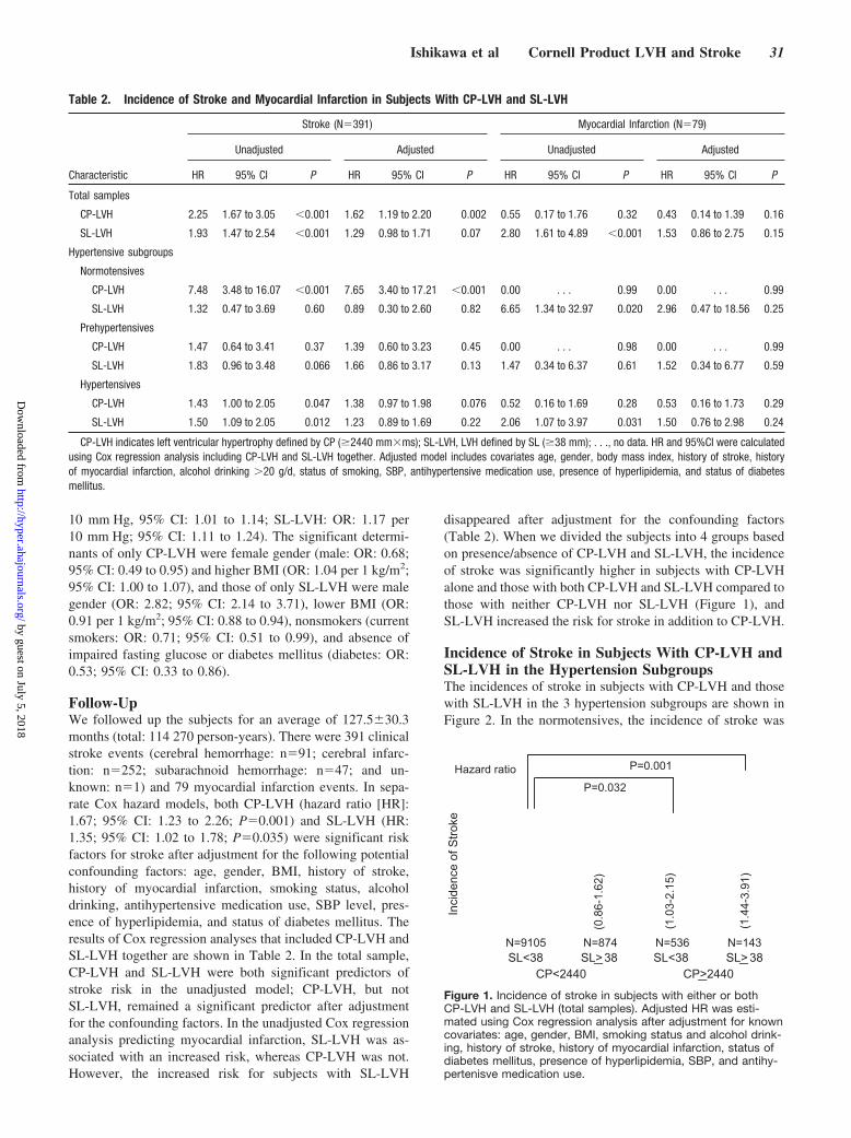
Follow-UpWe followed up the subjects for an average of 127.5�30.3months (total: 114 270 person-years). There were 391 clinicalstroke events (cerebral hemorrhage: n�91; cerebral infarc-tion: n�252; subarachnoid hemorrhage: n�47; and un-known: n�1) and 79 myocardial infarction events. In sepa-rate Cox hazard models, both CP-LVH (hazard ratio [HR]:1.67; 95% CI: 1.23 to 2.26; P�0.001) and SL-LVH (HR:1.35; 95% CI: 1.02 to 1.78; P�0.035) were significant riskfactors for stroke after adjustment for the following potentialconfounding factors: age, gender, BMI, history of stroke,history of myocardial infarction, smoking status, alcoholdrinking, antihypertensive medication use, SBP level, pres-ence of hyperlipidemia, and status of diabetes mellitus. Theresults of Cox regression analyses that included CP-LVH andSL-LVH together are shown in Table 2. In the total sample,CP-LVH and SL-LVH were both significant predictors ofstroke risk in the unadjusted model; CP-LVH, but notSL-LVH, remained a significant predictor after adjustmentfor the confounding factors. In the unadjusted Cox regressionanalysis predicting myocardial infarction, SL-LVH was as-sociated with an increased risk, whereas CP-LVH was not.However, the increased risk for subjects with SL-LVH
disappeared after adjustment for the confounding factors(Table 2). When we divided the subjects into 4 groups basedon presence/absence of CP-LVH and SL-LVH, the incidenceof stroke was significantly higher in subjects with CP-LVHalone and those with both CP-LVH and SL-LVH compared tothose with neither CP-LVH nor SL-LVH (Figure 1), andSL-LVH increased the risk for stroke in addition to CP-LVH.
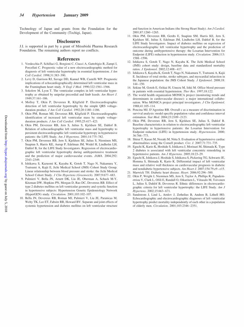
Incidence of Stroke in Subjects With CP-LVH andSL-LVH in the Hypertension SubgroupsThe incidences of stroke in subjects with CP-LVH and thosewith SL-LVH in the 3 hypertension subgroups are shown inFigure 2. In the normotensives, the incidence of stroke was
Table 2. Incidence of Stroke and Myocardial Infarction in Subjects With CP-LVH and SL-LVH
Characteristic
Stroke (N�391) Myocardial Infarction (N�79)
Unadjusted Adjusted Unadjusted Adjusted
HR 95% CI P HR 95% CI P HR 95% CI P HR 95% CI P
Total samples
CP-LVH 2.25 1.67 to 3.05 �0.001 1.62 1.19 to 2.20 0.002 0.55 0.17 to 1.76 0.32 0.43 0.14 to 1.39 0.16
SL-LVH 1.93 1.47 to 2.54 �0.001 1.29 0.98 to 1.71 0.07 2.80 1.61 to 4.89 �0.001 1.53 0.86 to 2.75 0.15
Hypertensive subgroups
Normotensives
CP-LVH 7.48 3.48 to 16.07 �0.001 7.65 3.40 to 17.21 �0.001 0.00 . . . 0.99 0.00 . . . 0.99
SL-LVH 1.32 0.47 to 3.69 0.60 0.89 0.30 to 2.60 0.82 6.65 1.34 to 32.97 0.020 2.96 0.47 to 18.56 0.25
Prehypertensives
CP-LVH 1.47 0.64 to 3.41 0.37 1.39 0.60 to 3.23 0.45 0.00 . . . 0.98 0.00 . . . 0.99
SL-LVH 1.83 0.96 to 3.48 0.066 1.66 0.86 to 3.17 0.13 1.47 0.34 to 6.37 0.61 1.52 0.34 to 6.77 0.59
Hypertensives
CP-LVH 1.43 1.00 to 2.05 0.047 1.38 0.97 to 1.98 0.076 0.52 0.16 to 1.69 0.28 0.53 0.16 to 1.73 0.29
SL-LVH 1.50 1.09 to 2.05 0.012 1.23 0.89 to 1.69 0.22 2.06 1.07 to 3.97 0.031 1.50 0.76 to 2.98 0.24
CP-LVH indicates left ventricular hypertrophy defined by CP (�2440 mm�ms); SL-LVH, LVH defined by SL (�38 mm); . . ., no data. HR and 95%CI were calculatedusing Cox regression analysis including CP-LVH and SL-LVH together. Adjusted model includes covariates age, gender, body mass index, history of stroke, historyof myocardial infarction, alcohol drinking �20 g/d, status of smoking, SBP, antihypertensive medication use, presence of hyperlipidemia, and status of diabetesmellitus.
CP<2440 CP>2440SL<38 SL<38SL>38 SL> 38
Hazard ratio P=0.001
P=0.032
(0.8
6-1.
62)
(1.0
3-2.
15)
(1.4
4-3.
91)
N=9105 N=874 N=536 N=143
Inci
denc
e of
Stro
ke
Figure 1. Incidence of stroke in subjects with either or bothCP-LVH and SL-LVH (total samples). Adjusted HR was esti-mated using Cox regression analysis after adjustment for knowncovariates: age, gender, BMI, smoking status and alcohol drink-ing, history of stroke, history of myocardial infarction, status ofdiabetes mellitus, presence of hyperlipidemia, SBP, and antihy-pertenisve medication use.
Ishikawa et al Cornell Product LVH and Stroke 31
by guest on July 5, 2018http://hyper.ahajournals.org/
Dow
nloaded from

higher in subjects with CP-LVH than in those with SL-LVH,although it was similar for subjects with CP-LVH andSL-LVH within prehypertensives and hypertensives. In sep-arate Cox regression analyses for the 3 groups, the HR forstroke events in subjects with CP-LVH was higher in thenormotensives (HR: 7.53; 95% CI: 3.39 to 16.77) than in theprehypertensives (HR: 1.49; 95% CI: 0.65 to 3.46) andhypertensives (HR: 1.41; 95% CI: 0.99 to 2.02; Figure 2A),after adjustment for age, gender, BMI, smoking status andalcohol drinking, history of stroke, history of myocardialinfarction, diabetes mellitus status, hyperlipidemia, and SBP.However, the HR for stroke events in subjects with SL-LVHwas not a significant predictor of stroke in subjects for any of thehypertensive groups (normotensives: HR: 1.15, 95% CI: 0.40 to3.32; prehypertensives: HR: 1.71, 95% CI: 0.89 to 3.25; hyper-tensives: HR: 1.26, 95% CI: 0.91 to 1.73; Figure 2B).
C-Statistics of LVH for Predicting Stroke Event inHypertensive GroupsThe c-statistics for the models that separately contain ECG-LVH and each of the other cardiovascular risk factors for theCox regression models predicting stroke in hypertensivegroups are shown in Table 3. The inclusion of CP-LVHincreased the c-statistic in all of the hypertensive groups, butthe inclusion of SL-LVH did not. Inclusion of CP-LVHresulted in similar c-statistic values to that of status of
diabetes mellitus in prehypertensives and resulted in thehighest c-statistic value in normotensives.
DiscussionBoth CP-LVH and SL-LVH were risk factors for the inci-dence of stroke in the Japanese general population. Screeningof ECG using CP-LVH, in addition to SL-LVH, is beneficialfor detecting individuals who are at high risk for stroke. Thepredictive value of CP-LVH was seen even in normotensivesubjects, where those with CP-LVH were more likely to havehyperlipidemia and diabetes mellitus. In normotensive sub-jects, the presence of CP-LVH is the strongest predictor offuture stroke events compared with the conventional cardio-vascular risk factors. In addition, in prehypertensive subjects,for whom antihypertensive medication is controversial, thepresence of CP-LVH independently predicted stroke as didthe status of diabetes mellitus, history of stroke, history ofmyocardial infarction, and smoking status.
CP-LVH and SL-LVH were related to different risk fac-tors, although they were both ECG markers of LVH. Okin etal17 reported in hypertensive patients for whom the presenceof CP-LVH was predominantly associated with higher BMI,whereas SL-LVH was predominantly related to lower BMIand male gender. The determinants of CP-LVH in the presentstudy of community-dwelling Japanese individuals were sim-
Figure 2. Incidence of stroke in subjectswith CP-LVH (A) and in subjects withSL-LVH (B) by hypertension subgroup.Adjusted HRs of subjects with CP-LVH (vsthose without CP-LVH) and of subjects withSL-LVH (vs those without SL-LVH) in eachhypertension subgroup were calculatedusing Cox regression analysis after adjust-ment for known covariates: age, gender,BMI, smoking status and alcohol drinking,history of stroke, history of myocardialinfarction, status of diabetes mellitus, pres-ence of hyperlipidemia, and SBP.
32 Hypertension January 2009
by guest on July 5, 2018http://hyper.ahajournals.org/
Dow
nloaded from

ilar to the results in the Losartan Intervention for EndpointReduction in Hypertension Study (hypertensive subjects).17
CP-LVH was related to metabolic factors (hyperlipidemiaand diabetes mellitus), especially in normotensive subjects,but SL-LVH was not. Okin et al6 reported that abnormal leftventricular geometry was more common in patients with onlyCP-LVH than in patients with only SL-LVH, and Shirai etal18 reported that CP-LVH is a better marker of relative wallthickness (RWT) than SL-LVH. We reported previously thathypertensive patients with type 2 diabetes mellitus hadgreater RWT (but not increased left ventricular mass index)than those without diabetes mellitus.19 Therefore, CP-LVHmight reflect increased RWT related to diabetes mellitus. Indiabetic patients, RWT is a better marker of cardiovascularevents than left ventricular mass index,20 and the higherpredictive value of CP-LVH for stroke might be derived fromthat of diabetes-associated RWT. It is reported that proteinglycation can cause myocardial damage in diabetes mellitus,and receptor binding of advanced glycation products inducesproinflammatory cytokines, inflammation, growth factor re-lease, and fibrosis.21 On the other hand, the presence ofdiabetes mellitus was a negative predictor of SL-LVH in thehypertensive group of the present study. Diabetic subjects aremore often overweight, which can reduce the SL productand, thus, SL-LVH on ECG. SL-LVH was more stronglyassociated with hypertension than with metabolic factorssuch as diabetes mellitus and hyperlipidemia comparedwith CP-LVH.
Evaluation of both CP-LVH and SL-LVH may be usefulfor detecting subjects at an increased risk of stroke events.The subjects with both CP-LVH and SL-LVH had an in-creased risk for stroke (Figure 1). In the present study, thepredictive value of SL-LVH for stroke lost the significancewhen we adjusted for CP-LVH and conventional risk factors(Table 2); however, the risks of both CP-LVH and SL-LVHwere significant when we preformed a parallel analysisexcluding the subjects with history of stroke and myocardialinfarction (data not shown).
In prehypertensives, the c-statistic increased when CP-LVH, history of stroke, history of myocardial infarction,status of diabetes, and smoking were each added to a baselinemodel that included only age and gender; however, thec-statistic did not increase when SBP was added. Therefore,a prehypertensive with these risk factors should be monitoredcautiously and may be a candidate for lifestyle modification(especially cessation of smoking and glycemic control ifneeded). Moreover, CP-LVH was the strongest predictor ofstroke in normotensives. Evaluation of CP-LVH may beuseful for detecting normotensive diabetic subjects at greatestrisk for stroke.
Study LimitationsThe specificity of ECG-LVH for detecting Echo-LVH ishigher than its sensitivity,5 and it is difficult to apply theresults of the present study to Echo-defined LVH. Okin et al22
reported that the use of ECG-LVH for detecting increased leftventricular mass index varies by race. Sundstrom et al23
reported that Echo-LVH and CP-LVH have independentpredictive values for mortality. ECG-LVH (“electronic”LVH) is clearly an imperfect proxy measure of Echo-LVH(structural LVH) for the prediction of cardiovascular events.Finally, there are no data with which to determine whether theadjustment of Cornell voltage (6 mm) for women is appro-priate for Japanese people.
PerspectivesBoth CP-LVH and SL-LVH predicted future stroke in aJapanese population. The prediction of stroke by the CP-LVHis high even in normotensive subjects, in whom CP-LVH isrelated to the presence of diabetes mellitus and hyperlipid-emia. In prehypertensives, CP-LVH predicts future strokeevents, as do history of stroke, history of myocardial infarc-tion, smoking status, and presence of diabetes mellitus.
Sources of FundingThis work was supported by a Grant-in-Aid for Scientific Researchfrom the Ministry of Education, Culture, Sports, Science and
Table 3. C-Statistics of Presence ECG-LVH for Stroke Event in Hypertensive Groups
Variable
Normotensives Prehypertensives Hypertensives
C-statistics 95% CI C-statistics 95% CI C-statistics 95% CI
Baseline model
Age and gender 0.808 0.799 0.817 0.737 0.729 0.744 0.688 0.680 0.696
Added variables
History of stroke 0.819 0.810 0.827 0.742 0.734 0.749 0.694 0.686 0.702
History of myocardial infarction 0.809 0.800 0.818 0.739 0.732 0.747 0.688 0.680 0.696
BMI 0.811 0.803 0.820 0.737 0.730 0.745 0.687 0.679 0.695
Smoking 0.808 0.799 0.816 0.744 0.736 0.752 0.690 0.682 0.698
Alcohol intake �20 g/d 0.809 0.800 0.817 0.737 0.729 0.744 0.686 0.678 0.694
Hyperlipidemia 0.807 0.798 0.815 0.737 0.729 0.745 0.687 0.679 0.695
Status of diabetes mellitus 0.818 0.810 0.827 0.740 0.733 0.748 0.693 0.686 0.701
SBP 0.813 0.804 0.821 0.737 0.729 0.745 0.690 0.682 0.698
Presence of SL-LVH 0.808 0.799 0.817 0.735 0.728 0.743 0.688 0.680 0.696
Presence of CP-LVH 0.820 0.812 0.828 0.740 0.732 0.747 0.693 0.685 0.701
Ishikawa et al Cornell Product LVH and Stroke 33
by guest on July 5, 2018http://hyper.ahajournals.org/
Dow
nloaded from
Technology of Japan and grants from the Foundation for theDevelopment of the Community (Tochigi, Japan).
DisclosuresJ.I. is supported in part by a grant of Mitsubishi Pharma ResearchFoundation. The remaining authors report no conflicts.
References1. Verdecchia P, Schillaci G, Borgioni C, Ciucci A, Gattobigio R, Zampi I,
Porcellati C. Prognostic value of a new electrocardiographic method fordiagnosis of left ventricular hypertrophy in essential hypertension. J AmColl Cardiol. 1998;31:383–390.
2. Levy D, Garrison RJ, Savage DD, Kannel WB, Castelli WP. Prognosticimplications of echocardiographically determined left ventricular mass inthe Framingham heart study. N Engl J Med. 1990;322:1561–1566.
3. Sokolow M, Lyon T. The ventricular complex in left ventricular hyper-trophy as obtained by unipolar precordial and limb leasds. Am Heart J.1949;37:161–186.
4. Molloy T, Okin P, Devereux R, Kligfield P. Electrocardiographicdetection of left ventricular hypertrophy by the simple QRS voltage-duration product. J Am Coll Cardiol. 1992;20:1180–1186.
5. Okin PM, Roman MJ, Devereux RB, Kligfield P. Electrocardiographicidentification of increased left ventricular mass by simple voltage-duration products. J Am Coll Cardiol. 1995;25:417–423.
6. Okin PM, Devereux RB, Jern S, Julius S, Kjeldsen SE, Dahlof B.Relation of echocardiographic left ventricular mass and hypertrophy topersistent electrocardiographic left ventricular hypertropy in hypertensivepatients: the LIFE Study. Am J Hypertens. 2001;14:775–782.
7. Okin PM, Devereux RB, Jern S, Kjeldsen SE, Julius S, Nieminen MS,Snapinn S, Harris KE, Aurup P, Edelman JM, Wedel H, Lindholm LH,Dahlof B, for the LIFE Study Investigators. Regression of electrocardio-graphic left ventricular hypertrophy during antihypertensive treatmentand the prediction of major cardiovascular events. JAMA. 2004;292:2343–2349.
8. Ishikawa S, Kazuomi K, Kayaba K, Gotoh T, Nago N, Nakamura Y,Tsutsumi A, Kajii E, Jichi Medical School (JMS) Cohort Study Group.Linear relationship between blood pressure and stroke: the Jichi MedicalSchool Cohort Study. J Clin Hypertens (Greenwich). 2007;9:677–683.
9. Palmieri V, Bella JN, Arnett DK, Liu JE, Oberman A, Schuck M-Y,Kitzman DW, Hopkins PN, Morgan D, Rao DC, Devereux RB. Effect oftype 2 diabetes mellitus on left ventricular geometry and systolic functionin hypertensive subjects: Hypertension Genetic Epidemiology Network(HyperGEN) study. Circulation. 2001;103:102–107.
10. Bella JN, Devereux RB, Roman MJ, Palmieri V, Liu JE, Paranicas M,Welty TK, Lee ET, Fabsitz RR, Howard BV. Separate and joint effects ofsystemic hypertension and diabetes mellitus on left ventricular structure
and function in American Indians (the Strong Heart Study). Am J Cardiol.2001;87:1260–1265.
11. Okin PM, Devereux RB, Gerdts E, Snapinn SM, Harris KE, Jern S,Kjeldsen SE, Julius S, Edelman JM, Lindholm LH, Dahlof B, for theLIFE Study Investigators. Impact of diabetes mellitus on regression ofelectrocardiographic left ventricular hypertrophy and the prediction ofoutcome during antihypertensive therapy: the Losartan Intervention forEndpoint (LIFE) reduction in hypertension study. Circulation. 2006;113:1588–1596.
12. Ishikawa S, Gotoh T, Nago N, Kayaba K. The Jichi Medical School(JMS) cohort study: design, baseline data and standardized mortalityratios. J Epidemiol. 2002;12:408–417.
13. Ishikawa S, Kayaba K, Gotoh T, Nago N, Nakamura Y, Tsutsumi A, KajiiE. Incidence of total stroke, stroke subtypes, and myocardial infarction inthe Japanese population: the JMS Cohort Study. J Epidemiol. 2008;18:144–150.
14. Sekine M, Gotoh E, Ochiai H, Umezu M, Ishii M. Office blood pressurein patients with essential hypertension. Ther Res. 1997;18:122.
15. The world health organization MONICA project (monitoring trends anddeterminants in cardiovascular disease): a major international collabo-ration. Who MONICA project principal investigators. J Clin Epidemiol.1988;41:105–114.
16. Pencina MJ, D’Agostino RB. Overall c as a measure of discrimination insurvival analysis: model specific population value and confidence intervalestimation. Stat Med. 2004;23:2109–2123.
17. Okin PM, Devereux RB, Jern S, Kjeldsen SE, Julius S, Dahlof B.Baseline characteristics in relation to electrocardiographic left ventricularhypertrophy in hypertensive patients: the Losartan Intervention forEndpoint reduction (LIFE) in hypertension study. Hypertension. 2000;36:766–773.
18. Shirai T, Kasao M, Nozaki M, Nitta S. Evaluation of hypertensive cardiacabnormalities using the Cornell product. Circ J. 2007;71:731–735.
19. Eguchi K, Kario K, Hoshide S, Ishikawa J, Morinari M, Shimada K. Type2 diabetes is associated with left ventricular concentric remodeling inhypertensive patients. Am J Hypertens. 2005;18:23–29.
20. Eguchi K, Ishikawa J, Hoshide S, Ishikawa S, Pickering TG, Schwartz JE,Homma S, Shimada K, Kario K. Differential impact of left ventricularmass and relative wall thickness on cardiovascular prognosis in diabeticand nondiabetic hypertensive subjects. Am Heart J. 2007;154:79.e9–e15.
21. Marwick TH. Diabetic heart disease. Heart. 2006;92:296–300.22. Okin P, Wright J, Nieminen MS, Jern S, Taylor A, Phillips R, Papadem-
etriou V, Clark L, Ofili E, Randall O, Oikarinen L, Viitasalo M, ToivonenL, Julius S, Dahlof B, Devereux R. Ethnic differences in electrocardio-graphic criteria for left ventricular hypertrophy: the LIFE Study. Am JHypertens. 2002;15:663–671.
23. Sundstrom J, Lind L, Arnlov J, Zethelius B, Andren B, Lithell HO.Echocardiographic and electrocardiographic diagnoses of left ventricularhypertrophy predict mortality independently of each other in a populationof elderly men. Circulation. 2001;103:2346–2351.
34 Hypertension January 2009
by guest on July 5, 2018http://hyper.ahajournals.org/
Dow
nloaded from

for the Jichi Medical School Cohort Study Investigators GroupJoseph E. Schwartz, Thomas G. Pickering, Kazuyuki Shimada and Kazuomi Kario
Joji Ishikawa, Shizukiyo Ishikawa, Tomoyuki Kabutoya, Tadao Gotoh, Kazunori Kayaba,Stroke in a General Population
Cornell Product Left Ventricular Hypertrophy in Electrocardiogram and the Risk of
Print ISSN: 0194-911X. Online ISSN: 1524-4563 Copyright © 2008 American Heart Association, Inc. All rights reserved.
is published by the American Heart Association, 7272 Greenville Avenue, Dallas, TX 75231Hypertension doi: 10.1161/HYPERTENSIONAHA.108.1180262009;53:28-34; originally published online November 17, 2008;Hypertension.
http://hyper.ahajournals.org/content/53/1/28World Wide Web at:
The online version of this article, along with updated information and services, is located on the
http://hyper.ahajournals.org/content/suppl/2008/11/17/HYPERTENSIONAHA.108.118026.DC1Data Supplement (unedited) at:
http://hyper.ahajournals.org//subscriptions/
is online at: Hypertension Information about subscribing to Subscriptions:
http://www.lww.com/reprints Information about reprints can be found online at: Reprints:
document. Permissions and Rights Question and Answer this process is available in the
click Request Permissions in the middle column of the Web page under Services. Further information aboutOffice. Once the online version of the published article for which permission is being requested is located,
can be obtained via RightsLink, a service of the Copyright Clearance Center, not the EditorialHypertensionin Requests for permissions to reproduce figures, tables, or portions of articles originally publishedPermissions:
by guest on July 5, 2018http://hyper.ahajournals.org/
Dow
nloaded from

ONLINE SUPPLEMENT
Cornell product left ventricular hypertrophy in electrocardiogram and the risk of
stroke in a general population
Joji Ishikawa, MD, PhD, Shizukiyo Ishikawa, MD, PhD, Tomoyuki Kabutoya, MD,
Tadao Gotoh, MD, PhD, Kazunori Kayaba, MD, PhD, Joseph E Schwartz, PhD,
Thomas G Pickering, MD, DPhil, Kazuyuki Shimada, MD, PhD, and Kazuomi
Kario MD, PhD: the JMS Cohort Study investigators group
Division of Cardiovascular Medicine, Department of Internal Medicine, Jichi
Medical University School of Medicine (J.I., T.K., K.S., K.Kario)
Center for Behavioral Cardiovascular Health, Division of General Medicine,
Department of Medicine, Columbia University Medical Center, New York, USA
(J.I. J.E.S., T.G.P.)
Division of Community and Family Medicine, Center for Community Medicine,
Jichi Medical University (S.I.)
Wara National Health Insurance Hospital (T.G.)

Saitama Prefectural University (K.Kayaba)
Address correspondence to: Joji Ishikawa, MD, PhD
Division of Cardiovascular Medicine, Department of Medicine, Jichi Medical
University, School of Medicine 3311-1, Yakushiji, Shimotsuke, Tochigi 329-0498,
JAPAN
Tel: +81-285-58-7344; FAX: +81-285-44-2132, E-mail: [email protected]

Follow-up
We asked the subjects directly whether they had suffered from a stroke or
cardiovascular disease since enrolling in the study. For the subjects who did not
undergo the annual screening examinations, we contacted the subjects or their
family by mail or phone to confirm whether the subjects had had cardiovascular
events or died. If a subject went to any medical facility at the time of a
cardiovascular events or death, a doctors or health nurse of the JMS Cohort
study visited the facility and checked the medical records. When an incident
case was suspected, we filled out forms and photocopied the brain computer
tomography or magnetic resonance imaging (when a cerebrovascular event was
suspected) and/or electrocardiograms (when myocardial infarction was
suspected). For the subjects who died that we were unable to contact during
the follow-up period, death certificates were collected at the public health
centers with permission of the Agency of General Affairs and the Ministry of
Health Labor and Welfare. Data on changes of residence during the study were
obtained from each municipal government annually.

Diagnostic criteria
The criterion for the diagnosis of stroke was the sudden onset of neurologic
deficit that persisted for >24 hours in the absence of any other disease process
that could explain the symptom. Stroke events included ischemic stroke
(cerebral infarction and cerebral embolism), hemorrhagic stroke (cerebral
hemorrhage and subarachnoid hemorrhage), and undefined type of stroke. We
excluded transient ischemic attacks in which the neurologic deficit cleared
completely within 24 hours from the onset of symptoms.
In subjects who were suspected of having a myocardial event, information
about the symptoms, ECG, cardiac enzymes and necropsy findings (if available)
were collected. The criteria for myocardial infarction were (a) definite ECG
findings (ST elevation), (b) typical or atypical or inadequately described
symptoms, together with probable ECG and abnormal enzymes, (c) typical
symptoms and abnormal enzymes with ischemic or non-codable ECG or ECG
not available, or (d) fatal cases, whether sudden or not, with gross appearance
of fresh myocardial infarction (MI) and/or recent coronary occlusion found at
necropsy.

Table S1: Characteristics of subjects with/without Cornell product LVH (in each hypertensive subgroup)
Normotension Prehypertension Hypertension Characteristic LVH (-) LVH (+) P LVH (-) LVH (+) P LVH (-) LVH (+) P
N=3441 N=94 N=3321 N=184 N=3307 N=408 Age, years 51.5+11.8 53.3+11.9 0.13 55.1+10.8 57.5+10.0 0.003 59.6+9.4 60.5+8.3 0.049 Male, % 32.5 37.2 0.33 40.3 34.2 0.11 41.3 34.8 0.011 BMI, kg/m2 22.1+2.7 21.7+2.6 0.24 23.2+3.0 23.5+3.5 0.21 24.0+3.2 24.5+3.4 0.004 Smokers 0.22 0.46 0.29 Former, % 10.4 5.4 13.0 11.9 15.3 12.5 Current, % 24.0 21.7 23.2 19.8 20.5 20.1 Alcohol drinkers 27.2 27.7 0.92 29.0 28.8 0.95 26.9 24.5 0.30 Antihypertensive therapy, % - - - - - - 32.2 42.6 <0.001SBP, mmHg 108+8 109+8 0.064 129+6 130+6 0.006 151+16 153+19 0.051 DBP, mmHg 66+7 67+7 0.088 77+6 78+7 0.35 88+10 90+11 0.029 Hyperlipidemia, % 24.6 34.0 0.036 36.8 37.2 0.92 44.0 48.0 0.12 Total cholesterol, mg/dl 185+34 190+33 0.16 194+35 194+31 0.88 199+35 199+36 0.95 Triglyceride, mg/dl 100+62 109+62 0.16 120+76 120+61 0.96 130+86 141+85 0.012 Status of diabetes 0.029 0.995 0.94 Impaired fasting glucose, % 1.4 0.0 2.2 2.2 3.9 4.2 Diabetes, % 1.8 5.3 3.1 3.3 5.6 5.9 Blood glucose, mg/dl 98+22 107+49 0.078 103+24 110+24 <0.001 107+30 113+34 0.001 Sokolow-Lyon voltage LVH, % 4.7 12.8 <0.001 7.7 17.9 <0.001 14.0 24.8 <0.001P values were calculated using non-paired t test or chi-square test.

Table S2: Characteristics of subjects with/without Sokolow-Lyon voltage LVH (in each hypertensive subgroup)
Normotension Prehypertension Hypertension Characteristic LVH (-) LVH (+) P LVH (-) LVH (+) P LVH (-) LVH (+) P
N=3358 N=175 N=3216 N=288 N=3149 N=564 Age, years 51.4+11.8 54.1+12.2 0.003 55.2+10.7 55.4+10.8 0.81 59.6+9.4 60.2+8.8 0.15 Male, % 31.0 63.4 <0.001 37.9 63.9 <0.001 37.1 60.6 <0.001BMI, kg/m2 22.1+2.7 21.6+2.5 0.030 23.3+3.0 22.4+2.7 <0.001 24.2+3.3 23.3+3.0 <0.001Smokers <0.001 <0.001 <0.001 Former, % 9.8 18.4 12.2 21.0 14.6 17.4 Current, % 23.4 35.6 22.1 33.0 18.4 32.0 Alcohol drinkers 27.5 22.3 0.13 29.2 26.7 0.38 26.5 27.5 0.63 Antihypertensive therapy, % - - - - - - 32.8 36.2 0.13 SBP, mmHg 108+8 109+7 0.003 129+6 129+6 0.067 150+16 155+19 <0.001DBP, mmHg 66+7 67+7 0.034 77+6 78+6 0.054 88+10 90+11 0.006 Hyperlipidemia, % 24.7 25.9 0.74 37.5 29.0 0.004 45.8 36.7 <0.001 Total cholesterol, mg/dl 185+34 183+33 0.35 194+34 186+36 <0.001 200+35 191+34 <0.001 Triglyceride, mg/dl 100+62 106+63 0.17 120+75 115+74 0.24 134+88 119+66 <0.001Status of diabetes 0.047 0.48 0.16 Impaired fasting glucose, % 1.2 3.4 2.1 3.1 4.0 3.7 Diabetes, % 1.9 1.7 3.2 2.8 5.9 3.9 Blood glucose, mg/dl 98+24 100+20 0.24 103+24 106+23 0.080 108+31 109+29 0.41 Cornell product LVH, % 2.4 6.9 <0.001 4.7 11.5 <0.001 9.7 17.9 <0.001P values were calculated using non-paired t test or chi-square test.