cornea and sclera - prime.edu.pkprime.edu.pk/4th_year_eye_lectures/cornea and... · anatomy and...
TRANSCRIPT
ANATOMY AND PHYSIOLOGY OF CORNEA AND SCLERA
DR. Faizur Rahman
Associate Professor
Peshawar Medical College
Peshawar

CORNEA
• Forms anterior 1/5th of the eyeball.
• Clear transparent tissue with smooth shining surface.
• Anterior surface is convex and elliptical.
• Horizontal diameter is 12mm and vertical diameter is 11.5mm.
• Posterior surface is concave and circular with diameter of 11.5mm.

CORNEA
• Thickness in center is 0.5mm and in periphery is 1mm.
• Radius of curvature of anterior surface is 7.8mm and posterior surface is 6.5mm.
• Refractive Power of cornea is 43D.
• Refractive index is 1.33.

Histology
It consists of five layers:
• Epithelium.
• Bowman’s membrane.
• Stroma.
• Descemet’s membrane.
• Endothelium.

Epithelium
• Non-keratinized Stratified Squamous: 50-90µm thick.
• Basal layer: single columnar, mitotically active, few organelles.
• Wing cells: 2or 3 layers of polygonal cells.
• Superficial cells: Flat cells, double layer. Desmosomal attachment, tight junctions microplicae & microvilli.
• Replaced every 7 days.

Bowman’s layer
• Acellular condensed collagen fibers.
• 8-14µm thickness.
• Not regeneratable.
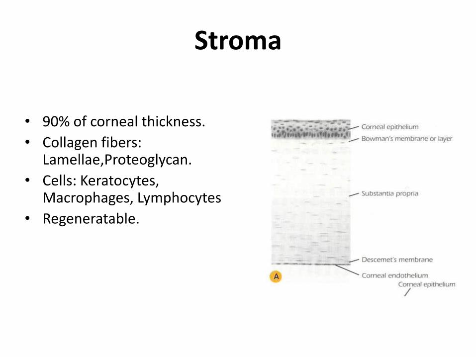
Stroma
• 90% of corneal thickness.
• Collagen fibers:Lamellae,Proteoglycan.
• Cells: Keratocytes, Macrophages, Lymphocytes
• Regeneratable.

Descemet’s membrane
• 10-12µm thick.
• Produced by Endothelium.
• Basement membrane.
• Ends at limbus - Schwalbe’s line.
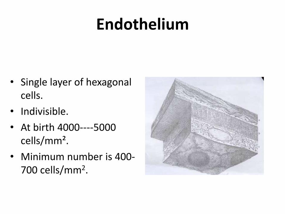
Endothelium
• Single layer of hexagonal cells.
• Indivisible.
• At birth 4000----5000 cells/mm².
• Minimum number is 400-700 cells/mm2.

BLOOD SUPPLY:• A vascular.
• Anterior ciliary vessels.
• No lymphatics.
NERVE SUPPLY: Sensory.• Ophthalmic division of trigeminal nerve---- Nasociliary
nerve ---- long ciliary nerve.
Limbus----Sclera----Precorneal plexus----Stromal plexus----Subepithelial plexus----Intraepithelial plexus.

Physiology
• Refracting surface.
• Protection of intraocular contents.
• Absorption of drugs.

Endothelium
• Barrier function:epithelium vs endothelium.
• Active pumps: Na/K ATP’ase.
Bicarbonate dependent.
Carbonic anhydrase.
• Passive diffusion: Supplementary.

Metabolism• Sources: Oxygen/Glucose.• Path ways:
Glycolitic pathway. Kreb’s cycles. Pentose pathway.
Transparency
• Regularity of stromal structures:
Fibers of regular diameter, arranged in lattice, interfibrilar distance less than a wavelength of light.
• Avascularity.
• Non-medulated nerve ending.
• Relative dehydration:
Active transport of electrolytes out of endothelium into AC – water follows (Na-K ATPase)
Evaporation from the corneal surface – little part.
Corneal hydration
• Stromal swelling pressure.
• Barrier function of Epithelium/Endothelium.
• Active pump.
• Evaporation from corneal surface.
• Intraocular pressure.
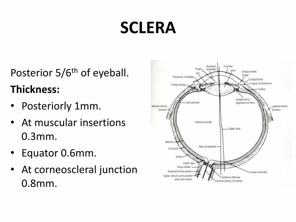
SCLERA
Posterior 5/6th of eyeball.
Thickness:
• Posteriorly 1mm.
• At muscular insertions 0.3mm.
• Equator 0.6mm.
• At corneoscleral junction 0.8mm.
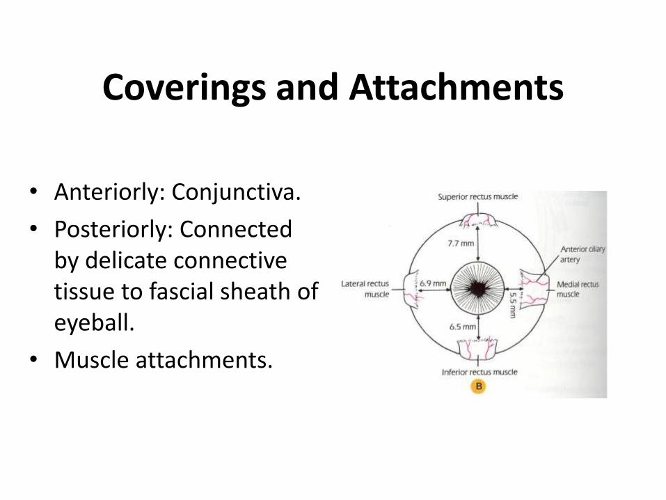
Coverings and Attachments
• Anteriorly: Conjunctiva.
• Posteriorly: Connected by delicate connective tissue to fascial sheath of eyeball.
• Muscle attachments.
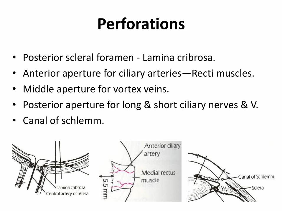
Perforations
• Posterior scleral foramen - Lamina cribrosa.
• Anterior aperture for ciliary arteries—Recti muscles.
• Middle aperture for vortex veins.
• Posterior aperture for long & short ciliary nerves & V.
• Canal of schlemm.

Episclera
• Outermost layer.
• Loose connective tissue—Tenon’s capsule.
• Rich blood supply—Anterior ciliary vessels.
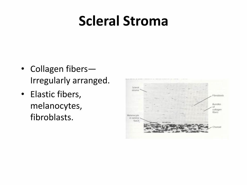
Scleral Stroma
• Collagen fibers—Irregularly arranged.
• Elastic fibers, melanocytes, fibroblasts.
Lamina Fusca
• Innermost layer contain collagen fibers and
melanocytes.
• Perichoroidal space.

Blood Supply:-
• Relatively avascular but anterior to the insertions of the recti muscles, the anterior ciliary arteries form a dense episcleral plexus. The posterior part of the sclera receives small branches from the long and short posterior ciliary arteries.
Nerve Supply:-
• Ciliary nerves—long and short.

Functions of Sclera
• Protects the intraocular contents from trauma
and mechanical displacement.
• Preserve the shape of the eyeball.
• Provide a rigid insertion for extraocular
muscles.
Embryology
• Corneal epithelium forms from the surface ectoderm.
• Bowman’s membrane is formed from mesenchyme.
• Stroma is formed from mesenchyme.
• Endothelium is derived from the neural crest.
• Descement’s membrane is synthesized by the endothelial cells.
Embryology
• Sclera is formed from a condensation of the
mesenchyme outside the optic cup.
• It first forms near the future insertion of the
rectus muscles.

Congenital disorders of cornea
• Megalocornea.
• Microcornea.
• Keratoglobus.
• Posterior keratoconus.
• Cornea plana.
• Sclerocornea.
• Dermoid & dermolipoma.
• Keractasia.

Microcornea• Very rare, hereditary, unilateral or bilateral• Corneal diameter is 10 mm or less• Shallow anterior chamber but other dimensions are normal
Associated systemic syndromesTurner, Ehlers-Danlos, Weill-Marchesani andWaardenburg
Ocular associationsGlaucoma, cataract, cornea plana, leukoma and iris abnormalities

Megalocornea
• Renal carcinoma and mental handicap
Systemic associations
• Marfan, Apert,
Ehlers-Danlos and
Down syndromes
• Osteogenesis imperfecta
• Very rare, hereditary, bilateral
• Corneal diameter 13 mmor more
• Very deep anteriorchamber
• High myopia andastigmatism
• Occasionally lenssubluxation

Keratoglobus
• Bilateral protrusion and thinning of entire cornea
• Associations - Leber congenital amaurosis and blue sclera
• Onset usually at birth

Sclerocornea• Very rare, usually bilateral
• Peripheral opacification and vascularization of cornea
• ‘Scleralization’ makes cornea appear smaller

Cornea plana• Very rare, bilateral severe decrease in corneal curvature• Hypermetropia and shallow anterior chamber
Ocular associationsGlaucoma, microcornea, microphthalmos and Petersanomaly

Keratectasia• Very rare, usually unilateral• Severe corneal opacification and protruberance• Probably caused by intrauterine keratitis
Slit-lamp biomicroscopy
• Diffuse illumination.
• Direct focal illumination.
• Lateral illumination.
• Retro-illumination.
• Specular reflaction.
• Sclerotic scatter.

Signs of corneal disease
A. Superficial:
• Punctate epithelial erosions.
Superior; Vernal disease, superior limbic keratoconjunctivitis, floppy eyelids poorly fitting contact lenses.
Interpalpebral; Dry eyes, diminished corneal sensation, exposure to UV.
Inferior; Lower lid margin disease, corneal exposure, rosacea, toxicity from drops.

Signs of corneal disease
• Punctate epithelial keratitis: viral infections.
• Epithelial oedema: Endothelial decompensation and acute high IOP.
• Fliaments: Keratoconjunctivitis sicca, superior limbic keratoconjunctivitis, recurrent erosion syndrome, eye patching, corneal exposure, diminished corneal sensation, HZO, midbrain strokes and essential blepharospasm.
• Pannus.
Signs of corneal disease
B. Stromal:
• Infiltrates: contact lens wear, marginal keratitis,
infectious keratitis.
• Oedema: Disciform keratitis, keratoconus, Fuchs
dystrophy, surgical damage.
• Vascularization.

Signs of corneal disease
C. Descement’s membrane:
• Breaks: Corneal enlargement, birth trauma,
keratoconus.
• Folds: Surgical trauma, ocular hypotony, stromal
inflammation and oedema.

THANK YOU