copyright 2009, john wiley & sons, inc. chapter 29: development and inheritance
TRANSCRIPT

Copyright 2009, John Wiley & Sons, Inc.
Chapter 29: Development and Inheritance


Copyright 2009, John Wiley & Sons, Inc.
Embryonic period i.e. first 8 weeks First week of development
Fertilization Genetic material from haploid sperm and haploid secondary
oocyte merges into single diploid zygote Normally occurs in uterine (fallopian) tubes Sperm undergo capacitation – series of functional changes
that prepare its plasma membrane to fuse with oocyte’s Sperm must penetrate coronoa radiata (granulosa cells) and
zona pellucida (clear glycoprotein layer between corona radiate and oocyte plasma membrane)
Acrosomal enzymes and strong movements help with penetration

Selected structures and events in fertilization

Copyright 2009, John Wiley & Sons, Inc.
First week of development (cont.)
Fusion of sperm cell and oocyte sets in motion events to block polyspermy – fertilization by more than one sperm Fast block to polyspermy – oocyte cell membrane
depolarizes so another sperm cannot fuse Also triggers exocytosis of secretory vesicles
Slow block to polyspermy – molecules released in exocytosis harden entire zona pellucida
Oocyte must complete meiosis Divides into ovum and polar body (disintegrates)

Copyright 2009, John Wiley & Sons, Inc.
First week of development (cont.)
Male pronucleus and female pronucleus fuse to form single diploid (2n) zygote with 46 chromosomes (23 pairs)
Dizygotic (fraternal) twins are produced by the release of 2 secondary oocytes and fertilization by separate sperm As genetically dissimilar as any other siblings
Monozygotic (identical) twins develop form a single fertilized ovum – they have exactly the same DNA Late separation results in conjoined twins

Copyright 2009, John Wiley & Sons, Inc.
First week of development (cont.)
Cleavage of zygote Rapid mitotic cell divisions after zygote forms First division begins 24 hours after fertilization and takes
6 hours Each succeeding division takes less time Blastomeres – progressively smaller cells produced by
cleavage Morula – solid sphere of cells
Still surrounded by zona pellucida About same size as original zygote

Copyright 2009, John Wiley & Sons, Inc.
First week of development (cont.)
Blastocyst formation Morula moves through uterine tubes toward uterus Day 4 or 5 reaches uterus Uterine milk – glycogen-rich secretions of endometrial glands
nourishes morula Blastocyst – at 32-cell stage, fluid collects and forms
blastocyst cavity or blastocoel 2 distinct cell populations
Embryoblast or inner cell mass – develops into embryo Trophoblast – outer layer that forms wall and will ultimately
develop into outer chorionic sac surrounding fetus and fetal portion of placenta
Day 5 “hatches” from zona pellucida

Copyright 2009, John Wiley & Sons, Inc.
Cleavage and the formation of the morula and blastocyst

Copyright 2009, John Wiley & Sons, Inc.
Implantation About 6 days after
fertilization attaches to endometrium
Orients inner cell mass toward endometrium
7 days after fertilization attaches more firmly and burrows in Endometrium becomes
more vascularized and glands enlarge
Decidua – modified portion of endometrium after implantation Regions named relative to
site of implantation

Copyright 2009, John Wiley & Sons, Inc.
Relationship of a blastocyst to the endometium of the uterus at implantation

Copyright 2009, John Wiley & Sons, Inc.
Summary of events associated with the first week of development

Copyright 2009, John Wiley & Sons, Inc.
Second week of development
Development of trophoblast About 8 days after fertilization, trophoblast develops into
2 layers in region of contact between blastocyst and endometrium Become part of chorion
Blastocyst becomes buried in endometrium and inner 1/3 of myometrium
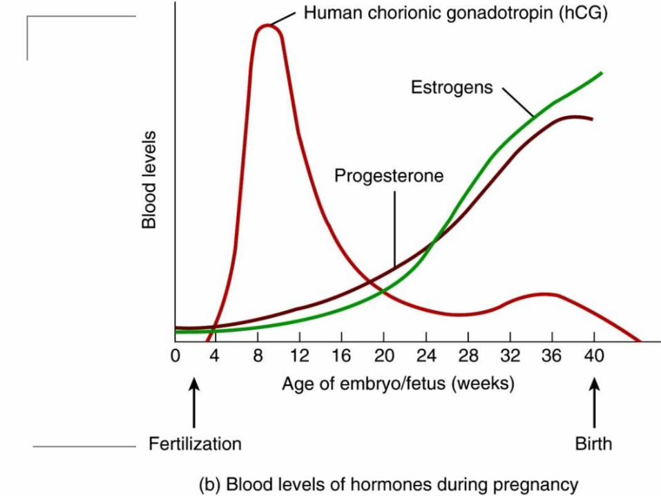
Secretes human chorionic gondadotropin (hCG) that maintains corpus luteum so it continues to secrete estrogens and progesterone Maintains uterine lining
Copyright 2009, John Wiley & Sons, Inc.
Second week of development (cont.)
Development of bilaminar embryonic disc Cells of embryoblast also differentiate into 2 layers
around 8 days after fertilization Hypoblast (primitive endoderm)
Mesoderm? Epiblast (primitive ectoderm)
Small cavity enlarges to form amniotic cavity Development of amnion
Amnion forms roof of amniotic cavity and epiblast forms floor
Amnion eventually surrounds entire embryo Amniotic cavity filled with amniotic fluid Fluid derived from maternal blood and later fetal urine

Principal events in the second week of development


Second week of development (cont.)
Development of yolk sac Also on 8th day after fertilization, cells at edge of
hypoblast migrate to cover inner surface of blastocyst wall
Form exocoelomic membrane Yolk sac – hypoblast and exocoelomic membrane
Relatively small and empty since nutrition derived from endometrium
Several important functions – supplies early nutrients, source of blood cells, contains primordial germ cells that migrate to gonads to form gametes, forms part of gut, functions as shock absorber, prevents desiccation

Second week of development (cont.) Development of sinusoids
9th day after fertilization, blastocyst completely embedded in endometrium
Syncytiotrophoblast expands and spaces (lacunae) develop 12th day – lacunae fuse to form lacunar networks Endometrial capillaries dilate to form maternal sinusoids Embryonic/maternal exchange
Development of extraembryonic coleom - about 12th day after fertilization Fuse to form single large cavity
Development of chorion Formed by extraembryonic mesoderm and 2 layers or trophoblast Becomes principal embryonic part of placenta Protect embryo from immune responses of mother Produces hCG Connecting (body) stalk connects bilaminar embryonic disc to
trophoblast – will become umbilical cord

Copyright 2009, John Wiley & Sons, Inc.
Third week of development
Begins 6 week period of rapid development and differentiation
Gastrulation 1st major event of 3rd week – about 15 days Bilaminar embryonic disc transforms into trilaminar
embryonic disc (Table 29.1 page 1190) Ectoderm (skin and nervous system), mesoderm (muscle,
bones, connective tissues, peritoneum), and endoderm (epithelial lining of GI tract, respiratory tract, and several other organs)
Involves rearrangement and migration of epiblast cells Primitive streak establishes head (primitive node) and tail ends

Copyright 2009, John Wiley & Sons, Inc.
Gastrulation

Copyright 2009, John Wiley & Sons, Inc.
Third week of development (cont.)
Gastrulation (cont.) 16 days after fertilization notochord forms – induces tissue to
become vertebral bodies (via induction) 2 depressions form
Oropharyngeal membrane will later break down to connect mouth to pharynx and GI tract
Cloacal membrane will later degenerate to form openings of anus, urinary and reproductive tracts
When cloacal membrane appears, wall of yolk sac forms allantois Extends into connecting stalk In most other mammals used for gas exchange and waste
removal – human placenta does this instead Does function in early formation of blood and blood vessels and
urinary bladder

Copyright 2009, John Wiley & Sons, Inc.
Development of the notochordal process

Copyright 2009, John Wiley & Sons, Inc.
Third week of development (cont.)
Neurulation Notochord also induces formation of neural plate Edges of plate elevate to form neural fold Neural folds fuse to form neural tube Develop into brain and spinal cord Neural crest cells give rise to spinal and cranial nerves and
ganglia, autonomic nervous system ganglia, CNS meninges, adrenal medullae and several skeletal and muscular components of head
Head end of neural tube develops into 3 primary brain vesicles Prosencephalon (forebrain), mesencephalon (midbrain), and
rhombencephalon (hindbrain)

Copyright 2009, John Wiley & Sons, Inc.
Neurulation and the development of somites

Copyright 2009, John Wiley & Sons, Inc.

Copyright 2009, John Wiley & Sons, Inc.
Third week of development (cont.)
Development of somites 42-44 pairs Mesoderm adjacent to notochord and neural tube forms
paired longitudinal columns of paraxial mesoderm Segment into paired, cube-shaped somites Number of somites can be correlated to age of embryo Each somite has 3 regions
Myotome – develops into skeletal muscles of neck, trunk and limbs Dermatome – develops into connective tissue and dermis Sclerotome - develops into vertebrae and ribs
Development of intraembryonic coelom Splits lateral plate mesoderm into
Splanchnic mesoderm – forms heart, blood vessels, smooth muscle and connective tissues of respiratory and digestive systems
Somatic mesoderm – gives rise to bones, ligaments, dermis of skin

Copyright 2009, John Wiley & Sons, Inc.
Neurulation and the development of somites

Copyright 2009, John Wiley & Sons, Inc.
Third week of development (cont.)
Development of cardiovascular system Angiogenesis – formation of blood vessels
Spaces develop in blood islands to form lumens of blood vessels Pluripotent stem cells form blood cells By end of 3rd week, heart forms and begins to beat
Development of chorionic villi and placenta Chorionic villi – fingerlike projections of chorion projecting into
endometrium Blood vessels in chorionic villi connect to embryonic heart
through body stalk (becomes umbilical cord) Maternal and fetal blood do not mix – diffusion only

Copyright 2009, John Wiley & Sons, Inc.
Development of chorionic villi

Copyright 2009, John Wiley & Sons, Inc.
Placentation Process of forming placenta
By beginning of 12th week has 2 parts Fetal portion formed by chorionic villi of chorion Maternal portion formed by decidua basalis of endometrium
Functionally allows oxygen and nutrients to diffuse from maternal to fetal blood while carbon dioxide and wastes diffuse from fetal to maternal blood
Not a protective barrier – allows microorganisms (HIV), drugs, alcohol to pass
Connection between embryo and placenta through umbilical cord 2 umbilical arteries carry deoxygenated fetal blood to placenta 1 umbilical veins carries oxygenated blood away from placenta
Afterbirth – placenta detaches from uterus

Copyright 2009, John Wiley & Sons, Inc.
Placenta and umbilical cord

Copyright 2009, John Wiley & Sons, Inc.
Fourth week of development
4th -8th week - all major organs develop Organogenesis – formation of body organs and
systems Embryo triples in size this week Converted from flat disc to 3D cylinder through
embryonic folding Main force is different rates of growth for different parts
Head fold brings heart and mouth into eventual adult position
Tail fold brings anus into eventual adult position Lateral folds for primitive gut – forerunner of GI tract

Copyright 2009, John Wiley & Sons, Inc.
Embryonic folding

Copyright 2009, John Wiley & Sons, Inc.
Embryonic folding

Copyright 2009, John Wiley & Sons, Inc.
4th week (cont.)
Somite and neural tube development
Pharyngeal (branchial) arches (5), clefts and pouches give rise to specific structures in head and neck 1st pharyngeal arch forms
jaw Otic placode – future
internal ear Upper and lower limb buds
appear – distinct tail

Copyright 2009, John Wiley & Sons, Inc.
5th – 8th weeks of development
During 5th week brain develops rapidly so head growth considerable
Limbs show substantial development by end of 6th week Heart now 4-chambered
8th week Digits of hands are short and webbed – by the end of the week
the webbing dies (apoptosis) Tail shorter and disappears by end of week Eyes open – eyelids come together and may fuse Auricles of ear visible External genitals begin to differentiate

Copyright 2009, John Wiley & Sons, Inc.
Fetal period
During this period, tissues and organs that developed during embryonic period grow and differentiate
Very few new structures appear Rate of body growth remarkable Fetus less vulnerable to damaging effect of
drugs, radiation, and microbes
Copyright 2009, John Wiley & Sons, Inc.
Summary of changes during embryonic and fetal development Table 29.2

Copyright 2009, John Wiley & Sons, Inc.
Summary of changes during embryonic and fetal development Table 29.2

Copyright 2009, John Wiley & Sons, Inc.
Summary of changes during embryonic and fetal development

Copyright 2009, John Wiley & Sons, Inc.
Summary of events of the embryonic and fetal periods
Copyright 2009, John Wiley & Sons, Inc.
Hormones during pregnancy Human chorionic somatomammotropin (hCS) or
human placental lactogen (hPL) produced by chorion Helps prepare mammary glands for lactation Regulates certain aspects of fetal and maternal
metabolism Corticotropin-releasing hormone (CRH) produced
by placenta In nonpregnant people secreted only by hypothalamus Though to be part of “clock” establishing timing of birth Increases secretion of cortisol needed for maturation of
fetal lungs and production of surfactant

Hormones during pregnancy
Copyright 2009, John Wiley & Sons, Inc.

Human Ovary
Copyright 2009, John Wiley & Sons, Inc.

Cat Ovary
Copyright 2009, John Wiley & Sons, Inc.
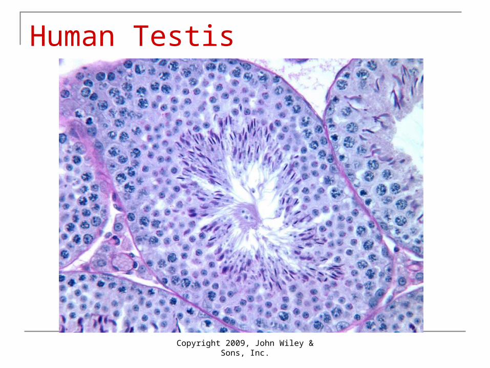
Human Testis
Copyright 2009, John Wiley & Sons, Inc.
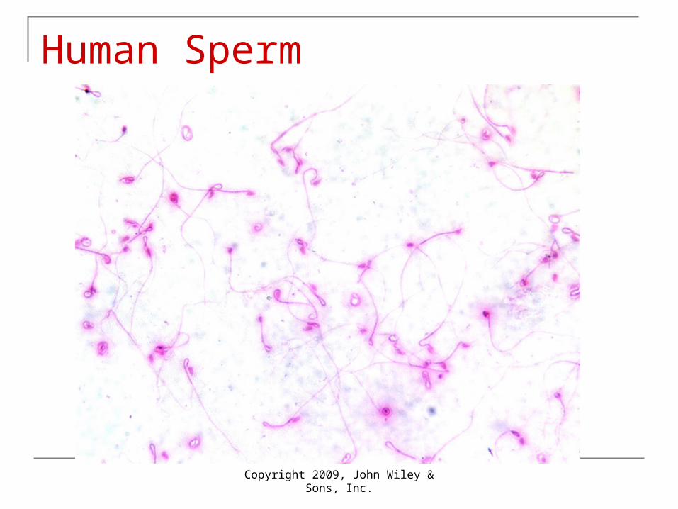
Human Sperm
Copyright 2009, John Wiley & Sons, Inc.

Human Chromosomes
Copyright 2009, John Wiley & Sons, Inc.

Copyright 2009, John Wiley & Sons, Inc.
Changes during pregnancy
By the end of a full-term pregnancy, uterus fills nearly the entire abdominal cavity
Physiological changes Weight gain due to fetus, amniotic fluid Increased storage of proteins, triglycerides and minerals Marked breast enlargement Lower back pain – lordosis
Changes in cardiovascular (30% SV) system due to increased maternal blood flow to placenta and increased metabolism
Respiratory functions change to meet added oxygen demands of fetus
Digestive system – increased appetite to meet energy demands of fetus
Urinary system – pressure on bladder can cause incontinence Increased renal filtering to eliminate wastes from fetus

Normal fetal location and position at the end of a full-term pregnancy

Copyright 2009, John Wiley & Sons, Inc.
Labor or parturition Process by which fetus expelled from uterus through vagina Onset determined by interactions between several placental and
fetal hormones Levels of estrogen must rise to overcome inhibiting effect of
progesterone on uterine contractions High levels of estrogens increase number of receptors for
oxytocin on uterine muscle fibers Oxytocin stimulates contractions Relaxin increases flexibility of pubic symphysis and dilates cervix
Control of labor through positive feedback cycle Contraction force fetal head into cervix which stretches Stimulated stretch receptors cause release of more oxytocin More oxytocin, more stretching Cycle broken when stretching stops as baby exits

Copyright 2009, John Wiley & Sons, Inc.
Stages of true labor
True labor begins when uterine contractions occur at regular intervals As interval shortens,
contractions intensify 3 stages
Copyright 2009, John Wiley & Sons, Inc.
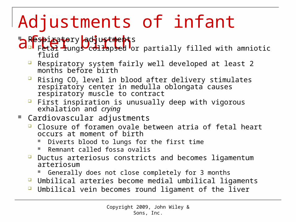
Adjustments of infant after birth Respiratory adjustments
Fetal lungs collapsed or partially filled with amniotic fluid Respiratory system fairly well developed at least 2 months before
birth Rising CO2 level in blood after delivery stimulates respiratory center
in medulla oblongata causes respiratory muscle to contract First inspiration is unusually deep with vigorous exhalation and crying
Cardiovascular adjustments Closure of foramen ovale between atria of fetal heart occurs at
moment of birth Diverts blood to lungs for the first time Remnant called fossa ovalis
Ductus arteriosus constricts and becomes ligamentum arteriosum Generally does not close completely for 3 months
Umbilical arteries become medial umbilical ligaments Umbilical vein becomes round ligament of the liver

Copyright 2009, John Wiley & Sons, Inc.
Physiology of lactation Secretion and ejection of milk from mammary
glands Prolactin – principal hormone promoting milk
synthesis and secretion Secreted by anterior pituitary Prolactin levels rise during pregnancy but progesterone
inhibits effects of prolactin After delivery, inhibition removed as estrogen and
progesterone levels fall Principal stimulus maintaining prolactin secretion is sucking
action of infant Impulses from stretch receptors decrease release of prolactin-
inhibiting hormone (PIH) and increases release of prolactin-releasing hormone (PRH) from hypothalamus

Copyright 2009, John Wiley & Sons, Inc.
The milk ejection reflex Oxytocin causes milk ejection
reflex Suckling, hearing baby cry,
touching mother’s genitals can initiate
Colostrum – before appearance of true milk on 4th day Contain important antibodies
Lactation often blocks ovarian cycles for few months after delivery
Primary benefit of breast-feeding is nutritional Other benefits also

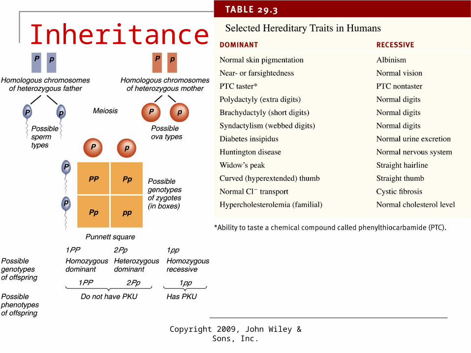
Inheritance
Copyright 2009, John Wiley & Sons, Inc.

Copyright 2009, John Wiley & Sons, Inc.
Inheritance
Passage of hereditary traits from one generation to the next
Genotype and phenotype Nuclei of all human cells except gametes contain 23 pairs
of chromosomes – diploid or 2n One chromosome from each pair came from father, other
member from mother Each chromosome contains homologous genes for same
traits Allele – alternative forms of a gene that code for the same
trait Mutation – permanent heritable change in allele that
produces a different variant

Copyright 2009, John Wiley & Sons, Inc.
Phenylketonuria or PKU example
Unable to manufacture enzyme phenylalanine hydroxylase Allele for function enzyme = P Allele that fails to produce functional enzyme = p Punnet square show possible combinations of alleles
between 2 parents Genotype – different combinations of genes Phenotype – expression of genetic makeup
PP – homozygous dominant – normal phenotype Pp – heterozygous – normal phenotype
1 dominant allele codes for enough enzyme Can pass recessive allele on to offspring – carrier
pp - homozygous recessive – PKU 2 recessive alleles make no functional enzyme
Copyright 2009, John Wiley & Sons, Inc.
Inheritance

Copyright 2009, John Wiley & Sons, Inc.

Copyright 2009, John Wiley & Sons, Inc.
Inheritance
Alleles that code for normal traits are not always dominant Huntington disease caused by dominant allele
Both homozygous dominant and heterozygous individuals get HD
Nondisjunction Error in cell division resulting in abnormal number of
chromosomes Aneuploid – chromosomes added or missing
Monosomic cell missing 1 chromosome (2n-1) Trisomic cell has additional chromosome (2n +1)
Down Syndrome – trisomy 21 – 3 21st chromosomes

Copyright 2009, John Wiley & Sons, Inc.
Variations of Dominant-recessive inheritance
Simple dominance-recessive Just described where dominant allele covers effect of
recessive allele Incomplete dominance
Neither allele dominant over other Heterozygote has intermediate phenotype Sickle-cell disease
Copyright 2009, John Wiley & Sons, Inc.
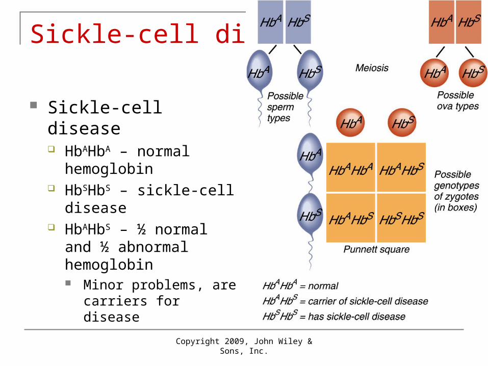
Sickle-cell disease
Sickle-cell disease HbAHbA – normal
hemoglobin HbSHbS – sickle-cell
disease HbAHbS – ½ normal and ½
abnormal hemoglobin Minor problems, are
carriers for disease

Copyright 2009, John Wiley & Sons, Inc.
Multiple-allele inheritance
Some genes have more than 2 alleles
ABO blood group IA produces A antigen IB produces B antigen i produces neither A and B are codominant
Both genes expressed equally in heterozygote
GenotypePhenotype
(blood type)
IA IA or IA i A
IB IB or IB i B
IA IB AB
Ii O

Copyright 2009, John Wiley & Sons, Inc.
Blood type inheritance

Copyright 2009, John Wiley & Sons, Inc.
Complex inheritance
Polygenic inheritance – most inherited traits not controlled by one gene
Complex inheritance – combined effects of many genes and environmental factors Skin color, hair color, height, metabolism rate, body build Even if a person inherits several genes for tallness, full
height can only be reached with adequate nutrition Neural tube deficits are more common if the mother lacks
adequate folic acid in the diet – environmental effect

Skin color is a complex trait
Depends on environmental conditions like sun exposure and nutrition and several genes
Additive effects of 3 genes plus environmental affect produces actual skin color

Copyright 2009, John Wiley & Sons, Inc.
Autosomes, sex chromosomes and sex determination
Karyotype shows 46 chromosomes arranged in pairs by size and centromere position
22 pairs are autosomes – same appearance in males and females
23rd pair are sex chromosomes XX = female XY = male
Copyright 2009, John Wiley & Sons, Inc.
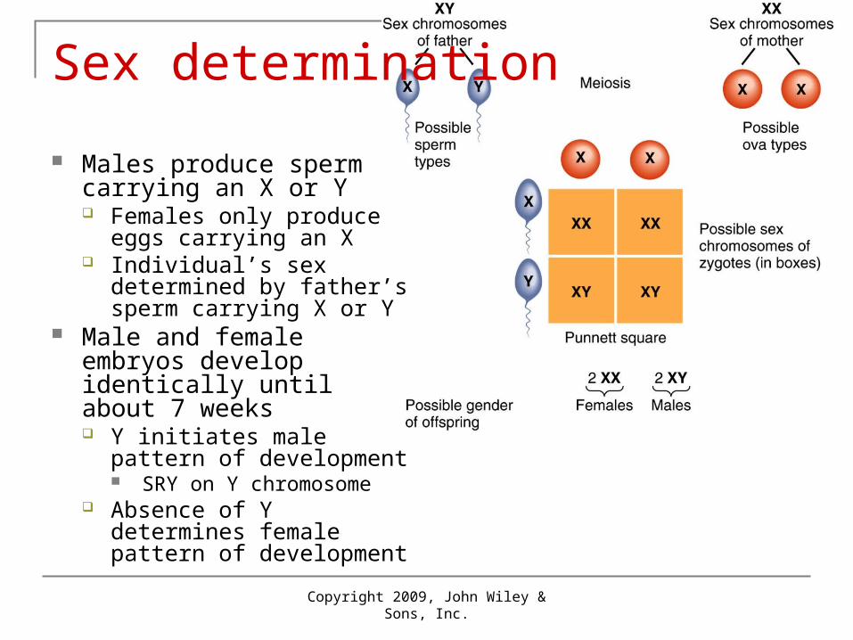
Sex determination
Males produce sperm carrying an X or Y Females only produce eggs
carrying an X Individual’s sex determined
by father’s sperm carrying X or Y
Male and female embryos develop identically until about 7 weeks Y initiates male pattern of
development SRY on Y chromosome
Absence of Y determines female pattern of development

Copyright 2009, John Wiley & Sons, Inc.
Sex-linked inheritance
Genes for these traits on the X but not the Y
Red-green colorblindness Most common type of
color blindness Red and green are seen
as same color Males have only one X
They express whatever they inherit from their mother
Genotype Phenotype
XCXC Normal female
XCXc Normal female (carrier)
XcXc Color blind female
XCY Normal male
XcYColor blind male

Inheritance of red-green color blindness