copper binding to the n terminally acetylated, naturally ... lewy bodies, containing the protein...
TRANSCRIPT

Copper Binding to the N‑Terminally Acetylated, Naturally OccurringForm of Alpha-Synuclein Induces Local Helical FoldingMarco C. Miotto,†,‡ Ariel A. Valiente-Gabioud,†,‡ Giulia Rossetti,§ Markus Zweckstetter,∥,⊥,#
Paolo Carloni,§ Philipp Selenko,∇ Christian Griesinger,∥ Andres Binolfi,*,†,‡,∇
and Claudio O. Fernandez*,†,‡
†Max Planck Laboratory for Structural Biology, Chemistry and Molecular Biophysics of Rosario and ‡Instituto de Investigaciones parael Descubrimiento de Farmacos de Rosario (IIDEFAR/CONICET-UNR), Universidad Nacional de Rosario, 27 de Febrero 210 bis,S2002LRK Rosario, Argentina§Computational Biophysics, German Research School for Simulation Sciences and Computational Biomedicine, Institute forAdvanced Simulations IAS-5, Forschungszentrum Julich, D-52425 Julich, Germany∥Department of NMR-based Structural Biology, Max Planck Institute for Biophysical Chemistry, Am Fassberg 11, D-37077Gottingen, Germany⊥Deutsches Zentrum fur Neurodegenerative Erkrankungen, 37077 Gottingen, Germany#Center for the Molecular Physiology of the Brain, University Medical Center, 37077 Gottingen, Germany∇Department of NMR-assisted Structural Biology, In-cell NMR, Leibniz Institute of Molecular Pharmacology, Robert-Roessle-Strasse10, 13125 Berlin, Germany
*S Supporting Information
ABSTRACT: Growing evidence supports a link betweenbrain copper homeostasis, the formation of alpha-synuclein(AS)-copper complexes, and the development of Parkin-son disease (PD). Recently it was demonstrated that thephysiological form of AS is N-terminally acetylated(AcAS). Here we used NMR spectroscopy to structurallycharacterize the interaction between Cu(I) and AcAS. Wefound that the formation of an AcAS−Cu(I) complex atthe N-terminal region stabilizes local conformations withα-helical secondary structure and restricted motility. Ourwork provides new evidence into the metallo-biology ofPD and opens new lines of research as the formation ofAcAS−Cu(I) complex might impact on AcAS membranebinding and aggregation.
Neurodegeneration in Parkinson disease (PD) is charac-terized by the progressive loss of dopaminergic neurons
in the substantia nigra and by the presence in multiple brainregions of amyloid fibrillar cytoplasmic aggregates, known asLewy bodies, containing the protein alpha-synuclein (AS).1
Although it remains unclear how AS initiates neuronal death,there is growing evidence that supports a role for ASaggregation in the pathological effects associated with PD.2
Protein−metal interactions play an important role in ASaggregation3,4 and might represent a link between thepathological processes of protein aggregation, oxidative damagein the brain, and neuronal cell loss.5−7 Indeed, the role ofcopper ions in AS amyloid assembly and neurodegenerationbecame a central question in the pathophysiology of PD.8−10
Recently, abundant evidence revealed that AS undergoes N-terminal acetylation in vivo (AcAS).11,12 From several in vitrostudies focused on the role of acetylation in AS, it was
demonstrated that the cotranslational modification induces amodest population of α-helical conformation for the first sixresidues and enhances the lipid binding properties of theprotein, whereas no significant differences were observed in thefibrillation kinetics between acetylated and nonacetylatedAS.13,14 In that direction, it was reported recently that N-terminal acetylation of AS abolishes Cu(II) binding at the high-affinity Met-1 site present in the nonacetylated protein.15
However, since copper ions are predominantly found in theirCu(I) state in the reducing environment of living cells, it is thecharacterization of the physiologically relevant AcAS−Cu(I)complexes that becomes fundamental to understand the impactof this interaction on protein conformation and aggregation.Accordingly, we report here the detailed structural character-ization of Cu(I) complexes with the full-length acetylated ASprotein.The details of Cu(I) binding to AcAS were explored at an
atomic resolution by NMR spectroscopy. We first recorded the1H−15N SOFAST-HMQC spectrum of Cu(II)−AcAS com-plexes (Supporting Information Figure 1), which confirmed thelack of binding of the metal ion to the Met-1 site at the N-terminus. Then, ascorbate was added as reducing agent togenerate the AcAS−Cu(I) complexes.16,17 The 1H−15NSOFAST-HMQC spectra of AcAS−Cu(I) complexes revealedthe occurrence of very large chemical shift changes in a discretenumber of residues located at the 1−15 segment of the N-terminal region, with smaller shift perturbations centered onthe amide group of His-50 and the region comprising residues115−130 that contains Met residues at positions 116 and 127(Figure 1).Then, we investigated the interaction at pH 7.4 and37 °C using direct 13C detection NMR methods,18 on the basis
Received: February 20, 2015Published: May 4, 2015
Communication
pubs.acs.org/JACS
© 2015 American Chemical Society 6444 DOI: 10.1021/jacs.5b01911J. Am. Chem. Soc. 2015, 137, 6444−6447

that these conditions of pH and temperature would resemblemore closely those found intracellularly. A similar profile ofchemical shift changes for the AcAS−Cu(I) interactions wasobtained from these studies after analysis of Cα and CO shiftperturbations (Supporting Information Figure 2). Inspection ofAcAS side chains through 1H−13C HSQC spectra revealed thatupon Cu(I) addition the most affected resonances corre-sponded to those of Hε−Cε and Hγ−Cγ correlations in Met-1and -5 and to a lesser extent to those belonging to residuesMet-116 and -127 and Hβ−Cβ from His-50 (SupportingInformation Figure 3).We have previously shown that Cu(I) binding to non-
acetylated AS occurs at three independent, noninteracting sitesand is mediated by coordination of sulfur atoms from Met-1/
Met-5 (site 1), the imidazole ring of His-50 (site 2), and sulfuratoms from Met-116/Met-127 residues (site 3).16,17,19 Theresults in Figure 1 and Supporting Information Figures 2 and 3demonstrate that these coordination modes are preserved uponacetylation of Met-1. From the experiments in Figure 1A, theNMR-derived apparent affinities for the Cu(I) complexes atsites 1−3 (Met-1/Met-5 (site 1), His-50 (site 2), and Met-116/Met-127 (site 3)) were Kd app1 = 12 ± 4 μM, Kd app2 = 50 ± 6μM, and Kd app3 > 200 μM, respectively (SupportingInformation Figure 4).Overall, the results presented here demonstrate that AcAS is
able to interact with Cu(I) with the same binding preferencesand affinity features as nonacetylated AS. Notably, the changesobserved in the chemical shifts of residues located in thevicinity of the high-affinity Cu(I) binding site were substantiallydifferent for the acetylated and nonacetylated proteins,16,19 afact that motivated us to evaluate the AcAS−Cu(I) complexesin terms of their conformational properties. NMR chemicalshifts are frequently used to probe the propensity ofintrinsically disordered proteins to populate different secondarystructure types. We then used 13Cα and 13Cβ secondarychemical shifts (SCS)20 and secondary structure propensityscores (SSP)21,22 to compare the secondary structure contentof AcAS and its Cu(I) complexed form. As previously reported,the SCS and SSP profiles for the acetylated form of the proteinshowed a slight increase in the population of α-helicalconformation near the N-terminus, strictly limited to the firstsix residues (Figure 2A,B).13,14 Surprisingly, we detected a largeincrease in α helix content in the first 15 residues of Cu(I)-bound AcAS, as evidenced by large positive deviations of Cαand negative deviations of Cβ chemical shifts. Assuming a ∼3ppm secondary chemical shift for an ideal α helix, our data isconsistent with α-helical conformations sampled ∼20 and∼70% of the time for the first 6 residues in AcAS and for thefirst 10 residues in the AcAS−Cu(I) form, respectively (Figure2A,B). Apart from chemical shifts, 3JHN−Hα couplings areparticularly reliable quantitative reporters of the time-averageddistribution of the backbone torsion angles, ϕ.23 Therefore, wemeasured a nearly complete set of 3JHN−Hα couplings in bothAcAS and AcAS−Cu(I) states. With the exception of theremarkable decrease in 3JHN−Hα for the first 10 residues of AcASupon Cu(I) binding, the values measured for the two forms ofthe protein were essentially indistinguishable. The observeddecreases, with 3JHN−Hα couplings ranging between 4.0 and 5.6Hz, are again indicative of a substantially increased α helixconformation in that segment of AcAS−Cu(I) relative to itsmetal-free form (Figure 2C). With an averaged 3JHN−Hα of 4 Hzexpected for an ideal α helix and a 3JHN−Hα of ∼7 Hz forrandom coil, the approximate increase in the level of α-helicalconformation is consistent with the one estimated by chemicalshifts. We also analyzed NOE-based data to evaluate theincrease of the α-helical content at the N-terminus of AcAS−Cu(I) relative to AcAS. From 3D 15N NOESY-HSQC editedexperiments, we were able to detect strong short-ranged NN(i −1,i) and medium-ranged αN(i − 3,i) 1H−1H NOE connectivitiesfor the first 10 residues of AcAS−Cu(I), which were absent inthe uncomplexed form of the protein (Supporting InformationFigure 5A,B). Added to this evidence, which is typicallyassociated with α-helical character,24,25 we also quantified theintensity ratios between intraresidue dαN(i,i) and sequentialdαN(i − 1,i) cross-peaks in both the metal-free and metal-complexed states of the protein.14 As shown in SupportingInformation Figure 5C, the increased values measured for the
Figure 1. NMR analysis of Cu(I) binding to AcAS. (A) Overlaid1H−15N SOFAST-HMQC spectra of AcAS in the absence andpresence of increasing Cu(I) concentrations. From blue to red, thetitration experiments represent the addition of 0, 0.25, 0.5, 1.0, 2.0, and5.0 equiv of Cu(I). Most-affected residues are labeled. (B) Differencesin the mean weighted chemical shift displacements (1H−15N MWCS)between free and Cu(I)-complexed AcAS at molar ratios of 1:1 (blue),2:1 (purple), and 5:1 (red). Gray box identifies the first 15 residues ofAcAS sequence. Experiments were recorded at 15 °C using AcAS (50μM) samples dissolved in buffer A.
Journal of the American Chemical Society Communication
DOI: 10.1021/jacs.5b01911J. Am. Chem. Soc. 2015, 137, 6444−6447
6445
dαN(i,i)/dαN(i − 1,i) ratios in the first residues of AcAS−Cu(I)relative to AcAS are also indicative of a helical signature at thatregion.14 Altogether, the changes in the three independentindicators of helical propensity are consistent with thetransition of a transient short α helix for the very N-terminalresidues in AcAS toward a longer, more structured and stable α-helical segment in its Cu(I) complexed state.Next, we characterized the dynamic properties of the Cu(I)-
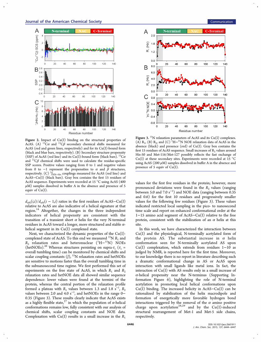
complexed state of AcAS. To this end we measured 15N R1 andR2 relaxation rates and heteronuclear (1H−15N) NOEs(hetNOEs).26 Whereas structures persisting on supra-τc (τc =overall tumbling time) can be monitored by chemical shifts andscalar coupling constants (J), 15N relaxation rates and hetNOEsare sensitive to motions faster than the overall tumbling time inthe subnanosecond time regime. We first performed this set ofexperiments on the free state of AcAS, in which R1 and R2relaxation rates and hetNOE data all showed similar sequencedependence: lower values were found at the termini of theprotein, whereas the central portion of the relaxation profileformed a plateau with R1 values between 1.3 and 1.8 s−1, R2values between 2.0 and 4.0 s−1, and hetNOEs in the range 0−0.35 (Figure 3). These results clearly indicate that AcAS existsas a highly flexible state,27 in which the population of α-helicalconformations remains low, fully consistent with our analysis ofchemical shifts, scalar coupling constants and NOE data.Complexation with Cu(I) results in a small increase in the R1
values for the first five residues in the protein; however, morepronounced deviations were found in the R2 values (rangingbetween 5.0 and 7.0 s−1) and NOE data (ranging between 0.35and 0.6) for the first 10 residues and progressively smallervalues for the following few residues (Figure 3). These valuesindicated restricted local sampling in the pico- to nanosecondtime scale and report on enhanced conformational order at the1−15 amino acid segment of AcAS−Cu(I) relative to the freeprotein, consistent with the stabilization of an α helix at thissite.In this work, we have characterized the interaction between
Cu(I) and the physiological, N-terminally acetylated form ofthe protein AS. The substantial increase in α helixconformation seen for N-terminally acetylated AS uponCu(I) complexation, which extends from residues 1−10 asjudged by NMR, is reported here for the first time. Moreover,to our knowledge there is no report in literature describing sucha dramatic conformational change in AS or AcAS uponinteraction with small ligands like metal ions. In fact, theinteraction of Cu(I) with AS results only in a small increase ofα-helical propensity near the N-terminus (Supporting In-formation Figure 6), highlighting the role of N-terminalacetylation in promoting local helical conformations uponCu(I) binding. The increased helicity in AcAS−Cu(I) can berationalized by stabilization of the helix macrodipole andformation of energetically more favorable hydrogen bondinteractions triggered by the removal of the α amino positivecharge upon acetylation28,29 and by the Cu(I)-inducedstructural rearrangement of Met-1 and Met-5 side chains,respectively.
Figure 2. Impact of Cu(I) binding on the structural properties ofAcAS. (A) 13Cα and 13Cβ secondary chemical shifts measured forAcAS (red and green lines, respectively) and for its Cu(I)-bound form(black and blue bars, respectively). (B) Secondary structure propensity(SSP) of AcAS (red line) and its Cu(I)-bound form (black bars). 13Cαand 13Cβ chemical shifts were used to calculate the residue-specificSSP scores. Positive values ranging from 0 to 1 and negative valuesfrom 0 to −1 represent the propensities to α and β structures,respectively. (C) 3JHN−Hα couplings measured for AcAS (red line) andAcAS−Cu(I) (black bars). Gray box contains the first 15 residues ofAcAS sequence. Experiments were recorded at 15 °C using AcAS (400μM) samples dissolved in buffer A in the absence and presence of 5equiv of Cu(I).
Figure 3. 15N relaxation parameters of AcAS and its Cu(I) complexes.(A) R1, (B) R2, and (C) 1H−15N NOE relaxation data of AcAS in theabsence (black) and presence (red) of Cu(I). Gray box contains thefirst 15 residues of AcAS sequence. Small increases of R2 values aroundHis-50 and Met-116/Met-127 possibly reflects the fast exchange ofCu(I) at these secondary sites. Experiments were recorded at 15 °Cusing AcAS (200 μM) samples dissolved in buffer A in the absence andpresence of 5 equiv of Cu(I).
Journal of the American Chemical Society Communication
DOI: 10.1021/jacs.5b01911J. Am. Chem. Soc. 2015, 137, 6444−6447
6446

Considering that copper concentrations can reach up to 300μM in synaptic vesicles30,31 and that AcAS is highly abundant(∼50 μM) in brain synaptosomes,32 our results suggest that anAcAS−Cu(I) complex with stabilized helically folded con-formations might exist in vivo. Linked to the fact that the Met-X3-Met motif at the N-terminus of AcAS resembles thosefound in helical copper transport proteins,33−35 the formationof AcAS−Cu(I) complex at site 1 might have physiologicallyrelevant implications in processes related to metal transport,membrane binding, or protein aggregation, which are enhancedby increased α-helical content at the N-terminus of theprotein.14,36,37 Overall, our findings open new avenues ofinvestigations into the metallobiology of PD, reshaping theconsideration of copper-mediated pathology in vivo.
■ ASSOCIATED CONTENT*S Supporting InformationNMR spectra of AcAS−Cu(II) complexes; 13C direct detectionNMR experiments; 1H−13C HSQC, binding curves, and NOEanalysis of AcAS−Cu(I) complexes under different conditions;and 13C SCS and SSP analysis for AS−Cu(I) complexes. TheSupporting Information is available free of charge on the ACSPublications website at DOI: 10.1021/jacs.5b01911.
■ AUTHOR INFORMATIONCorresponding Authors*[email protected]*[email protected] authors declare no competing financial interest.
■ ACKNOWLEDGMENTSWe thank Dr. Daniel Mulvihill for the N-acetylation B complexconstruct and Dr. Francois-Xavier Theillet for helpfuldiscussions. We acknowledge ANPCyT, FONCyT, CONICET,the Ministry of Education of Argentina, the Ministry of Healthof Argentina, Fundacion Medife, Fundacion Bunge y Born, theMax Planck Society, and the Alexander von HumboldtFoundation. C.O.F. is the Head of the Max Planck Laboratoryfor Structural Biology, Chemistry and Molecular Biophysics ofRosario (MPLbioR), associated with the Max Planck Institutefor Biophysical Chemistry (Gottingen).
■ REFERENCES(1) Goedert, M.; Spillantini, M. G.; Del Tredici, K.; Braak, H. Nat.Rev. Neurol. 2012, 9, 13.(2) Lashuel, H. A.; Overk, C. R.; Oueslati, A.; Masliah, E. Nat. Rev.Neurosci. 2013, 14, 38.(3) Binolfi, A.; Quintanar, L.; Bertoncini, C. W.; Griesinger, C.;Fernandez, C. O. Coord. Chem. Rev. 2012, 256, 2188.(4) Binolfi, A.; Rodriguez, E. E.; Valensin, D.; D’Amelio, N.; Ippoliti,E.; Obal, G.; Duran, R.; Magistrato, A.; Pritsch, O.; Zweckstetter, M.;Valensin, G.; Carloni, P.; Quintanar, L.; Griesinger, C.; Fernandez, C.O. Inorg. Chem. 2010, 49, 10668.(5) Bolognin, S.; Messori, L.; Zatta, P. NeuroMol. Med. 2009, 11, 223.(6) Brown, D. R. Metallomics 2011, 3, 226.(7) Gaggelli, E.; Kozlowski, H.; Valensin, D.; Valensin, G. Chem. Rev.2006, 106, 1995.(8) Gorell, J. M.; Johnson, C. C.; Rybicki, B. A.; Peterson, E. L.;Kortsha, G. X.; Brown, G. G.; Richardson, R. J. Neurotoxicology 1999,20, 239.(9) Kozlowski, H.; Brown, D.; Valensin, G. Metallochemistry ofNeurodegeneration: Biological, Chemical, and Genetic Aspects. RSCPublishing: Cambridge, U.K., 2007.
(10) Pall, H. S.; Blake, D. R.; Gutteridge, J. M.; Williams, A. C.;Lunec, J.; Hall, M.; Taylor, A. Lancet 1987, 330, 238.(11) Bartels, T.; Choi, J. G.; Selkoe, D. J. Nature 2011, 477, 107.(12) Fauvet, B.; Fares, M. B.; Samuel, F.; Dikiy, I.; Tandon, A.;Eliezer, D.; Lashuel, H. A. J. Biol. Chem. 2012, 287, 28243.(13) Kang, L.; Moriarty, G. M.; Woods, L. A.; Ashcroft, A. E.;Radford, S. E.; Baum, J. Protein Sci. 2012, 21, 911.(14) Maltsev, A. S.; Ying, J. F.; Bax, A. Biochemistry 2012, 51, 5004.(15) Moriarty, G. M.; Minetti, C. A.; Remeta, D. P.; Baum, J.Biochemistry 2014, 53, 2815.(16) Binolfi, A.; Valiente-Gabioud, A. A.; Duran, R.; Zweckstetter,M.; Griesinger, C.; Fernandez, C. O. J. Am. Chem. Soc. 2011, 133, 194.(17) Miotto, M. C.; Rodriguez, E. E.; Valiente-Gabioud, A. A.;Torres-Monserrat, V.; Binolfi, A.; Quintanar, L.; Zweckstetter, M.;Griesinger, C.; Fernandez, C. O. Inorg. Chem. 2014, 53, 4350.(18) Bermel, W.; Bertini, I.; Felli, I. C.; Pierattelli, R. J. Am. Chem.Soc. 2009, 131, 15339.(19) Miotto, M. C.; Binolfi, A.; Zweckstetter, M.; Griesinger, C.;Fernandez, C. O. J. Inorg. Biochem. 2014, 141, 208.(20) Wishart, D. S.; Sykes, B. D. Methods Enzymol. 1994, 239, 363.(21) Marsh, J. A.; Singh, V. K.; Jia, Z.; Forman-Kay, J. D. Protein Sci.2006, 15, 2795.(22) Tamiola, K.; Mulder, F. A. Biochem. Soc. Trans. 2012, 40, 1014.(23) Serrano, L. J. Mol. Biol. 1995, 254, 322.(24) Hill, R. B.; Flanagan, J. M.; Prestegard, J. H. Biochemistry 1995,34, 5587.(25) Wuthrich, K. NMR of Proteins and Nucleic Acids. Wiley-Interscience: New York, 1986.(26) Boehr, D. D.; Dyson, H. J.; Wright, P. E. Chem. Rev. 2006, 106,3055.(27) Bussell, R., Jr.; Eliezer, D. J. Biol. Chem. 2001, 276, 45996.(28) Hol, W. G. Adv. Biophys. 1985, 19, 133.(29) Tyanova, S.; Cox, J.; Olsen, J.; Mann, M.; Frishman, D. PLoSComput. Biol. 2013, 9, e1002842.(30) Hopt, A.; Korte, S.; Fink, H.; Panne, U.; Niessner, R.; Jahn, R.;Kretzschmar, H.; Herms, J. J. Neurosci. Methods 2003, 128, 159.(31) Scheiber, I. F.; Mercer, J. F.; Dringen, R. Prog. Neurobiol. 2014,116, 33.(32) Wilhelm, B. G.; Mandad, S.; Truckenbrodt, S.; Krohnert, K.;Schafer, C.; Rammner, B.; Koo, S. J.; Classen, G. A.; Krauss, M.;Haucke, V.; Urlaub, H.; Rizzoli, S. O. Science 2014, 344, 1023.(33) Guo, Y.; Smith, K.; Lee, J.; Thiele, D. J.; Petris, M. J. J. Biol.Chem. 2004, 279, 17428.(34) Ohrvik, H.; Nose, Y.; Wood, L. K.; Kim, B. E.; Gleber, S. C.;Ralle, M.; Thiele, D. J. Proc. Natl. Acad. Sci. U.S.A. 2013, 110, E4279.(35) Schushan, M.; Barkan, Y.; Haliloglu, T.; Ben-Tal, N. Proc. Natl.Acad. Sci. U.S.A. 2010, 107, 10908.(36) Anderson, V. L.; Ramlall, T. F.; Rospigliosi, C. C.; Webb, W. W.;Eliezer, D. Proc. Natl. Acad. Sci. U.S.A. 2010, 107, 18850.(37) Ghosh, D.; Singh, P. K.; Sahay, S.; Jha, N. N.; Jacob, R. S.; Sen,S.; Kumar, A.; Riek, R.; Maji, S. K. Sci. Rep. 2015, 5, 9228.
Journal of the American Chemical Society Communication
DOI: 10.1021/jacs.5b01911J. Am. Chem. Soc. 2015, 137, 6444−6447
6447