copd, nice and the national clinical strategy harold hosker airedale hospital march 2010
TRANSCRIPT

COPD, NICE and the National Clinical Strategy
Harold Hosker
Airedale Hospital
March 2010
Inflammation
• COPD is a disease state characterised by airflow limitation that is not fully reversible. The airflow limitation is usually both progressive and associated with an abnormal inflammatory response of the lungs to noxious particles or gases
• Chronic inflammation is caused by increased numbers of activated inflammatory cells, specifically neutrophils, which can damage lung structure and lead to mucosal oedema and airway narrowing

The impact of COPD • More than 30,000 deaths annually in the UK• UK – 900,000 diagnosed patients• Allowing for underdiagnosis, the true number of
patients with COPD in England and Wales is likely to be around 1.5 million
• Currently the fifth greatest cause of mortality worldwide – over 2.5 million deaths in 2000
• By 2020, COPD will be the third leading cause of mortality
• Exacerbations have an impact on patient quality of life and can be life-threatening

The typical COPD patient– Generally over 40 years
– A smoker or ex-smoker
– Presentation with:
» cough
» excessive sputum production
» shortness of breath / wheeze

COPD is a heterogeneous disease• Spectrum of clinical disease from ‘pink
puffer’ to ‘blue bloater’
• Several different pathological processes
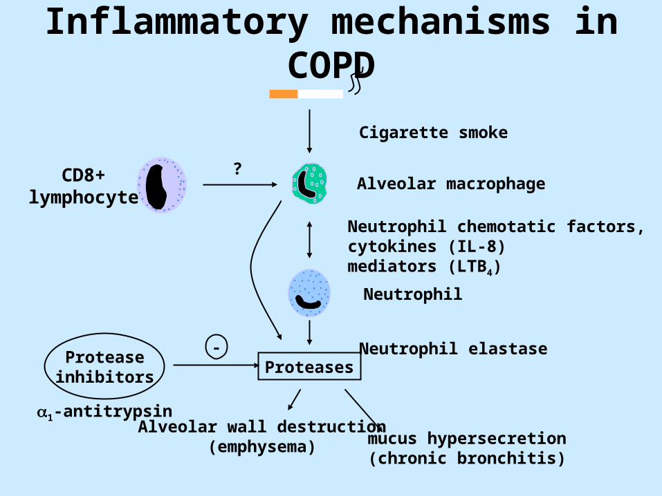
Inflammatory mechanisms in COPD
Alveolar macrophage
Cigarette smoke
Neutrophil chemotatic factors,cytokines (IL-8)mediators (LTB4)
Neutrophil
Proteases-
1-antitrypsin
Proteaseinhibitors
CD8+lymphocyte
Neutrophil elastase
mucus hypersecretion(chronic bronchitis)
Alveolar wall destruction(emphysema)
?

What is Spirometry?
Spirometry is a method of assessing lung function by measuring the volume of air the patient can expel from the lungs after a maximal expiration.
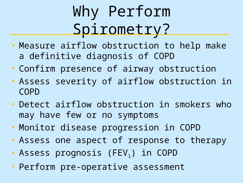
Why Perform Spirometry?
• Measure airflow obstruction to help make a definitive diagnosis of COPD
• Confirm presence of airway obstruction • Assess severity of airflow obstruction in COPD• Detect airflow obstruction in smokers who may have few
or no symptoms• Monitor disease progression in COPD• Assess one aspect of response to therapy
• Assess prognosis (FEV1) in COPD
• Perform pre-operative assessment

Spirometry – Additional Uses
• Make a diagnosis and assess severity in a range of other respiratory conditions (eg ILD, MND, G-B)
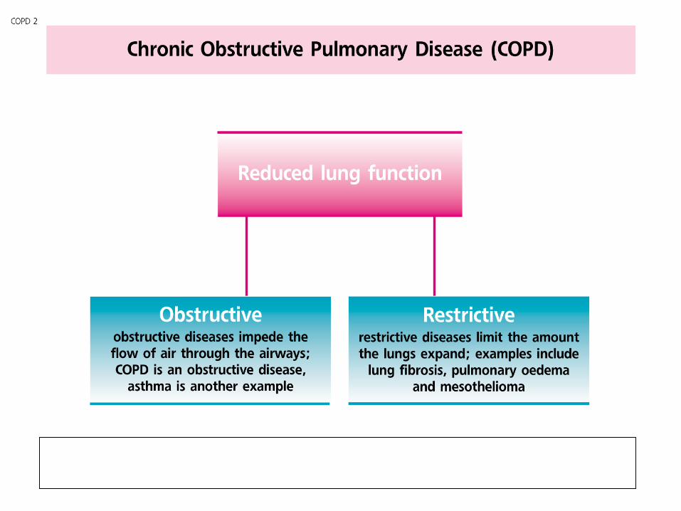
• Distinguish between obstruction and restriction as causes of breathlessness
• Screen workforces in occupational environments
• Assess fitness to dive
• Perform pre-employment screening in certain professions

Types of Spirometers
• Bellows spirometers:
Measure volume; mainly in lung function units
• Electronic desk top spirometers:
Measure flow and volume with real time display
• Small hand-held spirometers:
Inexpensive and quick to use but no print out

Volume Measuring Spirometer

Flow Measuring Spirometer

Small Hand-held Spirometers

Totallung
capacity
Tidal volume
Inspiratory reservevolume
Expiratory reservevolume
Residual volume
Inspiratory capacity
Vital capacity
Lung Volume Terminology

Standard Spirometric Indices
• FEV1 - Forced expiratory volume in one second:
The volume of air expired in the first second of the blow
• FVC - Forced vital capacity:
The total volume of air that can be forcibly exhaled in one breath
• FEV1/FVC ratio:
The fraction of air exhaled in the first second relative to the total volume exhaled
• VC - Vital capacity: A volume of a full breath exhaled in the patient’s own time and not
forced. Often slightly greater than the FVC, particularly in COPD

Normal Trace Showing FEV1 and FVC
1 2 3 4 5 6
1
2
3
4
Volu
me,
liters
Time, seconds
FVC5
1
FEV1 = 4L
FVC = 5L
FEV1/FVC = 0.8

Spirogram Patterns
• Normal
• Obstructive
• Restrictive
• Mixed Obstructive and Restrictive
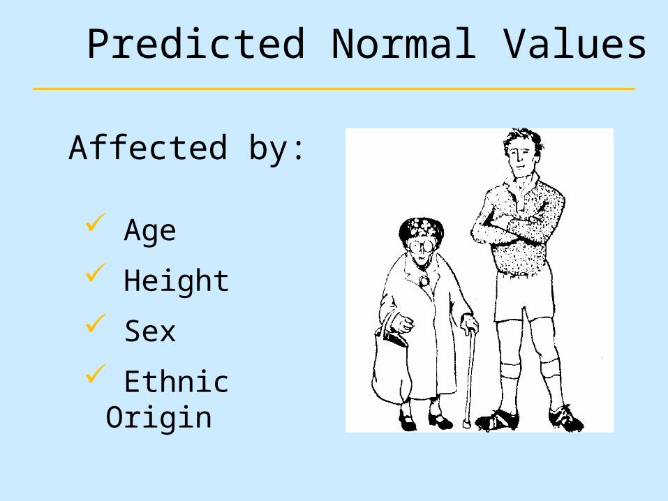
Predicted Normal Values
Age
Height
Sex
Ethnic Origin
Affected by:

Criteria for Normal Post-bronchodilator Spirometry
• FEV1: % predicted > 80%
• FVC: % predicted > 80%
• FEV1/FVC: > 0.7

Spirometry: Obstructive Disease
Volu
me,
liters
Time, seconds
5
4
3
2
1
1 2 3 4 5 6
FEV1 = 1.8L
FVC = 3.2L
FEV1/FVC = 0.56
Normal
Obstructive
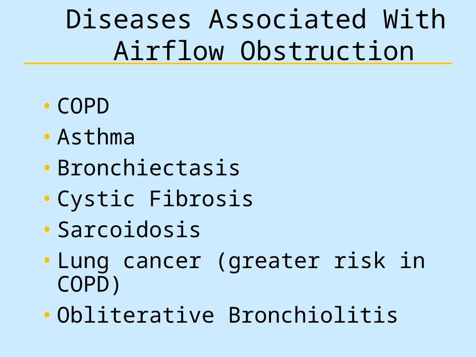
Diseases Associated With Airflow Obstruction
• COPD• Asthma• Bronchiectasis• Cystic Fibrosis• Sarcoidosis• Lung cancer (greater risk in COPD)• Obliterative Bronchiolitis

Volu
me,
liters
Time, seconds
FEV1 = 1.9L
FVC = 2.0L
FEV1/FVC = 0.95
1 2 3 4 5 6
5
4
3
2
1
Spirometry: Restrictive Disease
Normal
Restrictive

Diseases Associated with a Restrictive Defect
Pulmonary• Fibrosing lung diseases• Pneumoconioses• Pulmonary edema• Parenchymal lung tumors• Lobectomy or
pneumonectomy
Extrapulmonary• Thoracic cage deformity• Obesity• Pregnancy• Neuromuscular disorders• Fibrothorax
Spirometry
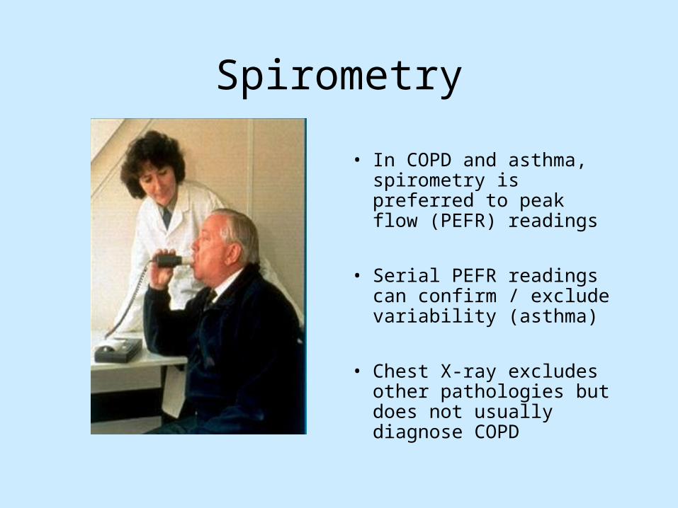
• In COPD and asthma, spirometry is preferred to peak flow (PEFR) readings
• Serial PEFR readings can confirm / exclude variability (asthma)
• Chest X-ray excludes other pathologies but does not usually diagnose COPD

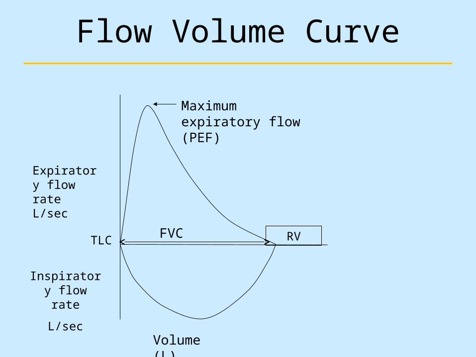
Flow Volume Curve
• Standard on most desk-top spirometers
• Adds more information than volume time curve
• Less understood but not too difficult to interpret
• Better at demonstrating mild airflow obstruction
Flow Volume Curve
Expiratory flow rateL/sec
Volume (L)
FVC
Maximum expiratory flow (PEF)
Inspiratory flow rate
L/sec
RVTLC
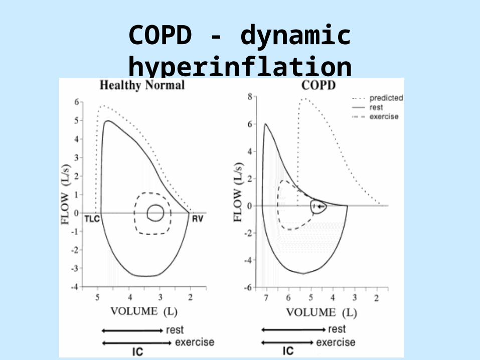
COPD - dynamic hyperinflation

COPD - exertional dyspnoea

Flow volume loop - severe COPD
2 4 6 8
2
4
6
8
10
12
-2
-4
-6
-8
Flow (l/s)
Volume (litres)
BaselinePost BD
Predicted

Flow volume loop - asthma
2 4 6 8
2
4
6
8
10
12
-2
-4
-6
-8
Flow (l/s)
Volume (litres)
BaselinePost BD
Predicted

Flow volume loop - restrictive (fibrosis)
2 4 6 8
2
4
6
8
10
12
-2
-4
-6
-8
Flow (l/s)
Volume (litres)
BaselinePost BD
Predicted

Flow volume loop - large airways obstruction (stridor)
2 4 6 8
2
4
6
8
10
12
-2
-4
-6
-8
Flow (l/s)
Volume (litres)
BaselinePost BD
Predicted
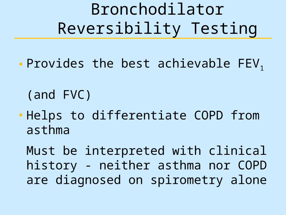
Bronchodilator Reversibility Testing
• Provides the best achievable FEV1 (and FVC)
• Helps to differentiate COPD from asthma
Must be interpreted with clinical history - neither asthma nor COPD are diagnosed on spirometry alone

Bronchodilator Reversibility Testing in COPD
Results
•An increase in FEV1 that is both greater than 200 ml and 12% above the pre-bronchodilator FEV1 (baseline value) is considered significant
•It is usually helpful to report the absolute change (in ml) as well as the % change from baseline to set the improvement in a clinical context

Equipment Maintenance
• Most spirometers need regular calibration to check accuracy
• Calibration is normally performed with a 3 litre syringe
• Some electronic spirometers do not require daily/weekly calibration
• Good equipment cleanliness and anti-infection control are important; check instruction manual
• Spirometers should be regularly serviced; check manufacturer’s recommendations

Other lung function tests
• Lung volumes– Helium dilution, body box (plethysmography)– TLC (RV derived)
• Gas transfer– Single breath carbon monoxide transfer– TLCO and KCO (=TLCO/Va)
• Mouth pressures– Indirect measure of muscle / diaphragm strength– Pimax, PEmax

Emphysema - the ‘pink puffer’

Damage to alveoli in emphysema
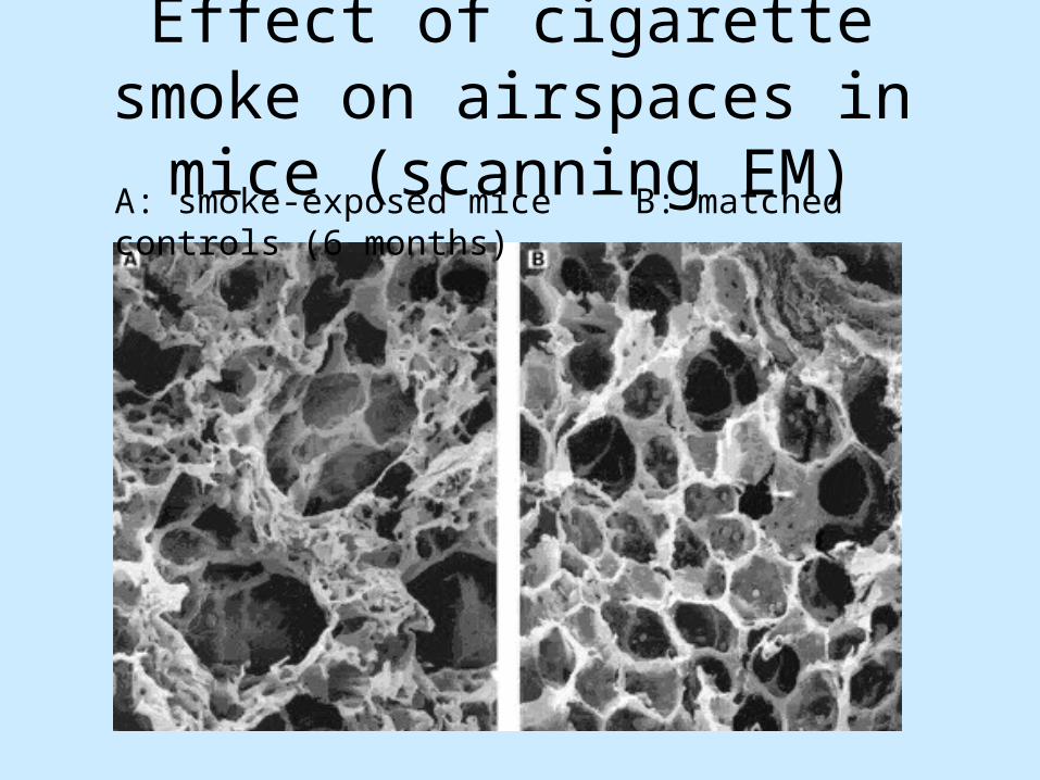
Effect of cigarette smoke on airspaces in mice (scanning EM)
A: smoke-exposed mice B: matched controls (6 months)

Pathological processes in emphysema
• Loss of alveolar surface area• Loss of lung elasticity• Hyperinflation causing mechanical
inefficiency• Muscle weakness / cachexia• Small airways collapse• Dynamic hyperinflation

Alveolar wall damage leading to small airways collapse in COPD

Chronic bronchitis: the ‘blue bloater’

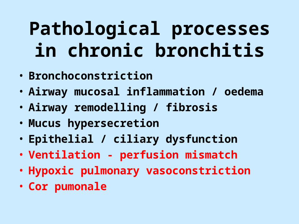
Pathological processes in chronic bronchitis
• Bronchoconstriction• Airway mucosal inflammation / oedema• Airway remodelling / fibrosis• Mucus hypersecretion• Epithelial / ciliary dysfunction• Ventilation - perfusion mismatch• Hypoxic pulmonary vasoconstriction• Cor pumonale

The role of exacerbations in disease progression
• Following an exacerbation, the likelihood of further exacerbation increases
• Exacerbations are closely associated with cumulative reduction in health status
• High frequency of COPD exacerbations is associated with a rapid decline in lung function and increased risk of hospitalisation
• Up to 70% of patients admitted to hospital with an exacerbation are re-admitted within a year
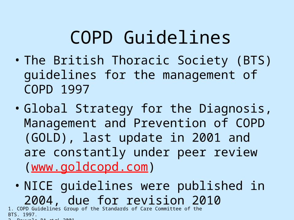
COPD Guidelines• The British Thoracic Society (BTS) guidelines
for the management of COPD 1997
• Global Strategy for the Diagnosis, Management and Prevention of COPD (GOLD), last update in 2001 and are constantly under peer review (www.goldcopd.com)
• NICE guidelines were published in 2004, due for revision 2010
1. COPD Guidelines Group of the Standards of Care Committee of the BTS. 1997.2. Pauwels RA et al. 2001.

NICE/BTS Recommendations
• Diagnose COPD
• Stop Smoking
• Effective inhaled therapy
• Pulmonary Rehabilitation
• NIV
• Manage exacerbations
• Multi-disciplinary working


Inhaled steroids in COPD
– No evidence of benefit in mild / moderate COPD
– Do not affect the rate of decline in FEV1
– Beneficial effect on quality of life and exacerbation rate in moderate / severe disease
– Dose response and long term safety in COPD are not known1
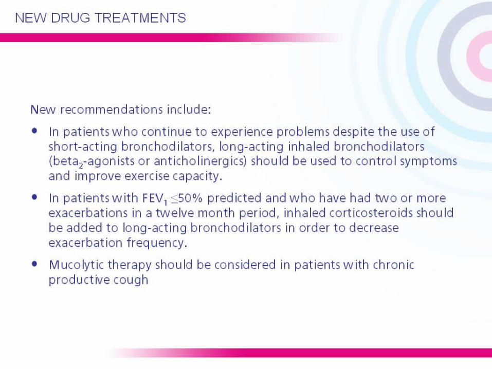
Drug therapy in COPD
• Combination therapy– Inhaled steroid plus long-acting B2 agonist
– Several studies show additive benefit using fluticasone / salmeterol or budesonide / eformoterol
– Benefits include FEV1 change, exacerbation rates, symptom scores and health status improvements
– New combinations (including triple combinations and ultra-long acting beta agonists) are on their way
– Roflumilast (PDE-4 inhibitor) due 2010

3-year study duration (6,112 patients)1
(sub-cut population 4,511 patients2)
1. Vestbo et al. Eur Respir J 20042. GSK Data on File SERTCODOF012
SeretideTM 500 Accuhaler™
Salmeterol 50mcg
Control group
2 week run-in
Fluticasone propionate 500 mcg
n=1,533
n=1,534
n=1,521
n=1,524
n=1,117
n=1,126
n=1,142
n=1,126
Full study Sub-cut data
1 Year 2 Years 3 Years
TORCH: study design

SALM FP
All-cause mortality at 3 years
Vertical bars are standard errors
18
16
14
12
10
8
6
4
2
0
Time to death (weeks)
Probability of death (%)
1524153315211534
1464148714811487
1399142614171409
1293133913161288
Placebo SALM/FP
Numberalive
0 12 24 36 48 60 72 84 96 108 120 132 144 156
0
4
8
12
16
20
24
28
32
36
40
44
48
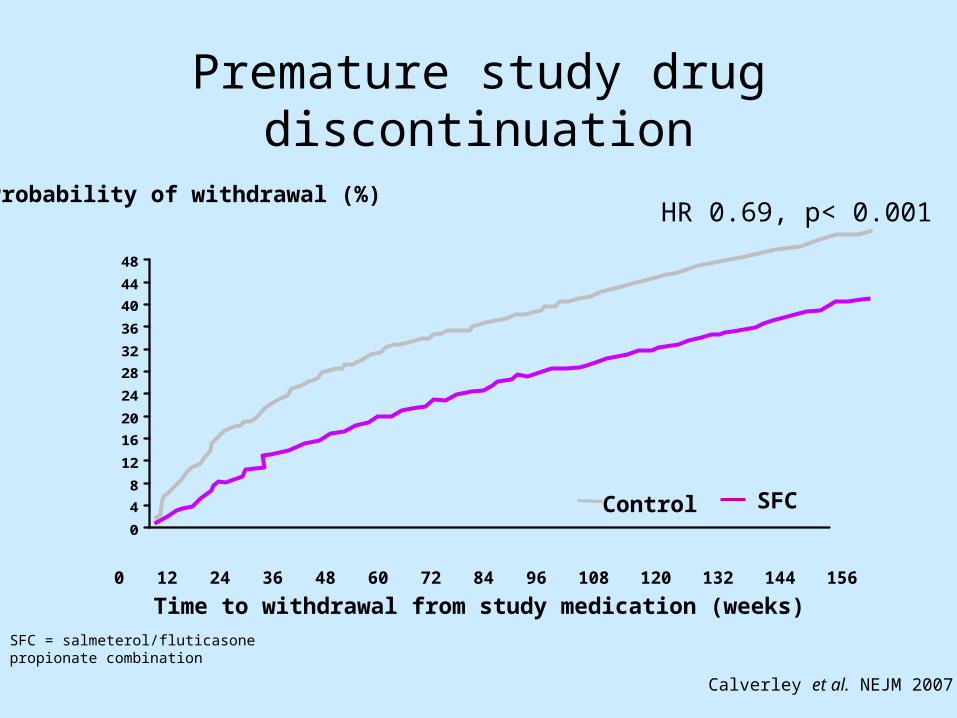
Premature study drug discontinuation
0 12 24 36 48 60 72 84 96 108 120 132 144 156
Probability of withdrawal (%)
Time to withdrawal from study medication (weeks)
Control SFC
Calverley et al. NEJM 2007
HR 0.69, p< 0.001
SFC = salmeterol/fluticasone propionate combination

Rate of moderate and severe exacerbations over three years
*p < 0.001 vs placebo; †p = 0.002 vs SALM; ‡p = 0.024 vs FP
Mean number of exacerbations/year
1.13
0.97*0.93*
0.85*†‡
25% reduction
0
0.2
0.4
0.6
0.8
1
1.2
Placebo SALM FP SALM/FP
Treatment
Calverley et al. NEJM 2007

UPLIFT trialNEJM October 2008
• A 4 year trial of tiotropium in COPD involving 5993 patients
• Real life study – all patients allowed to continue all other Rx except inhaled anti-cholinergics
• 46% patients were GOLD stage II (FEV1 50% – 80% predicted) ie mild disease
• 44% GOLD stage III, 8% GOLD stage IV• 60% patients on LABAs, 62% on ICS, with more than
70% on each by end of trial
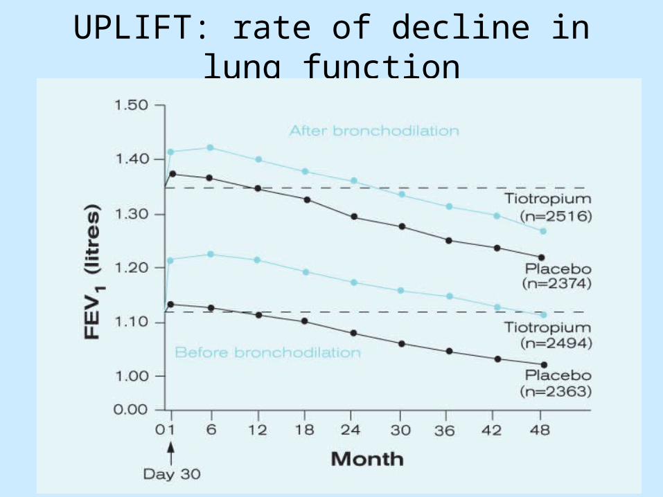
UPLIFT: rate of decline in lung function

Kaplan-Meier Estimates of the Probability of COPD Exacerbation and Death from Any Cause

UPLIFT trial exacerbations
• 14% reduced risk of exacerbations (time to 1st exacerbation)(p<0.001) and exacerbations leading to hospitalisations (p=0.002)
• But no difference in hospital days
• Seretide and Tiotropium both reduce exacerbations to a similar extent (INSPIRE study)

UPLIFT – safety of Tiotropium
• UPLIFT shows no increased risk of cardiac death with Tiotropium (significantly reduced risk – 27% risk reduction in CVS death, also stroke)
• 16% risk reduction in probability of death, p=0.034)
• No evidence of increased CVS risk from LABAs either

Case study – Margaret Margaret is 52 years old.
She works in an office and has two children
She has smoked around 25 cigarettes a day for 30 years
Her FEV1 is 65% predicted Margaret’s father died 4
years ago of ‘chest disease’ She has a productive cough
and finds she is breathless on exertion both at work and at leisure
Last winter she had a bad chest infection
She has tried various inhalers


The stages of COPD – NICE
Mild(FEV1
50–80%)
Moderate(FEV1
30–49%)
Severe(FEV1 <30%)
Breathlessness and exercise limitation
Prevention of exacerbations
Short- and long-acting bronchodilators
If still symptomatic consider a trial of combination long-acting
agonist and inhaled corticosteroid*
Consider adding theophylline
If still symptomatic despite maximum inhaled
bronchodilator consider referral for specialist
assessment
2-agonists/anticholinergics
In patients suffering two or more exacerbations per year
add inhaled corticosteroid usually in combination with long-acting bronchodilators
1. NICE Guideline No.12. Thorax 2004. *Discontinue if no benefit after 4 weeks

Diagnosis
Margaret has mild/moderate COPD
History
Has tried to quit smoking
Chest infection last winter
Prescribed various inhalers
FEV1 = 65% predicted normal
Symptoms
Breathlessness on exertion getting worse
Productive cough
What do Margaret’s symptoms stop her doing?
Enjoying leisure activities with her family
Playing bowls
Case study – Margaret (age 52)

Exacerbations
Managing Margaret’s COPD
Smoking
Breathlessness and exercise limitation
QoL

Exacerbations
Managing Margaret’s COPD
Smoking
Breathlessness and exercise limitation
QoL

Managing Margaret’s COPD symptoms
Smoking
Breathlessness and exercise limitation
QoL
Exacerbations
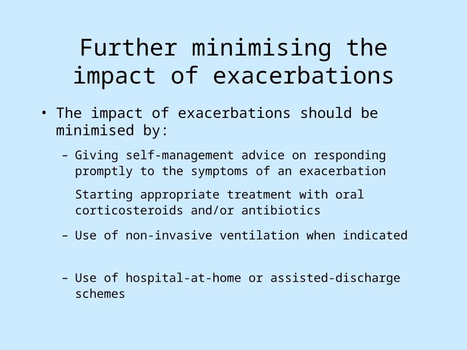
Further minimising the impact of exacerbations
• The impact of exacerbations should be minimised by:
– Giving self-management advice on responding promptly to the symptoms of an exacerbation
Starting appropriate treatment with oral corticosteroids and/or antibiotics
– Use of non-invasive ventilation when indicated
– Use of hospital-at-home or assisted-discharge schemes

Managing Margaret’s COPD
Smoking
Breathlessness and exercise limitation
QoL
Exacerbations
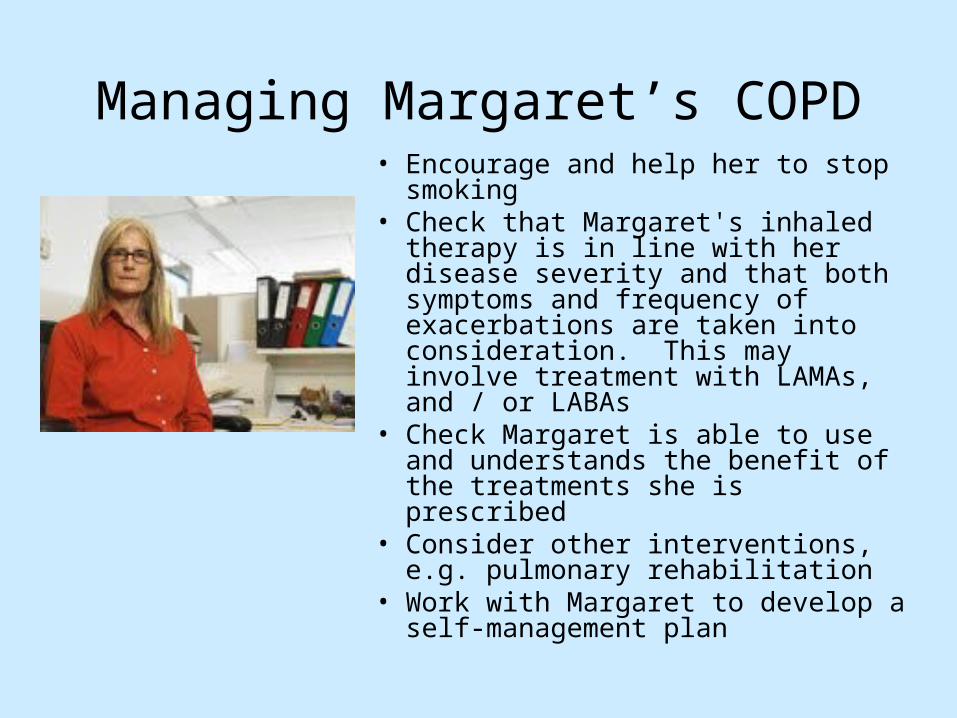
Managing Margaret’s COPD• Encourage and help her to stop
smoking• Check that Margaret's inhaled therapy
is in line with her disease severity and that both symptoms and frequency of exacerbations are taken into consideration. This may involve treatment with LAMAs, and / or LABAs
• Check Margaret is able to use and understands the benefit of the treatments she is prescribed
• Consider other interventions, e.g. pulmonary rehabilitation
• Work with Margaret to develop a self-management plan

Stopping smokingslows decline in lung function
FE
V1
(% o
f va
lue
at a
ge 2
5) 100
75
50
25
025 50 75
Never smoked or notsusceptible to smoke
Adapted from: Fletcher et al, Br Med J 1977.
Stopped at 65
Stopped at 45
Disability
Smoked regularlyand susceptible to
its effects
Death
Age (years)
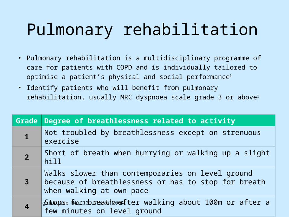
Pulmonary rehabilitation
• Pulmonary rehabilitation is a multidisciplinary programme of care for patients
with COPD and is individually tailored to optimise a patient’s physical and
social performance1
• Identify patients who will benefit from pulmonary rehabilitation, usually MRC
dyspnoea scale grade 3 or above1
1. NICE guideline No. 12. Thorax 2004.
Grade Degree of breathlessness related to activity
1 Not troubled by breathlessness except on strenuous exercise
2 Short of breath when hurrying or walking up a slight hill
3Walks slower than contemporaries on level ground because of breathlessness or has to stop for breath when walking at own pace
4Stops for breath after walking about 100m or after a few minutes on level ground
5 Too breathless to leave the house or breathless when dressing or undressing

Recognising an exacerbation
• Give written information on recognising worsening symptoms, such as:– You get much more breathless than you did
before (doing the same thing)– You produce more sputum than before– Your sputum becomes discoloured– You feel feverish or unwell– Cough gets worse
1. NICE guideline No. 12. Thorax 2004.
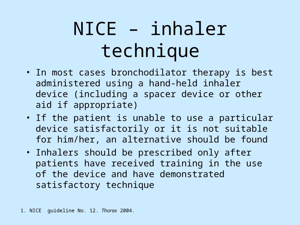
NICE – inhaler technique
• In most cases bronchodilator therapy is best administered using a hand-held inhaler device (including a spacer device or other aid if appropriate)
• If the patient is unable to use a particular device satisfactorily or it is not suitable for him/her, an alternative should be found
• Inhalers should be prescribed only after patients have received training in the use of the device and have demonstrated satisfactory technique
1. NICE guideline No. 12. Thorax 2004.

Case 2 - George
• 75 year old ex-steel worker • Ex-smoker, 45 pack years• Chronic productive cough• Gradually progressive SOB and wheeze over ten years• Regular antibiotics for bronchitis most winters• Limited exercise capacity• Difficulty shopping, playing with grandkids, walking to pub• 15 year history of of ‘asthma’ – bricanyl/beclomethasone• No family history of asthma/no pets

Points for discussion
• Diagnosis
• Impact on Patient
• What else do you need to know?– What do you need to ask?– Examination?– What investigations do you need?

How do we exclude asthma?
Spirometry FEV1 = 40% FEV1 /FVC = 50%


George
• 75 year old man
• No night-time wakening
• No day to day variability in symptoms
• No acute precipitants
• No diurnal variation in PEFR
Therefore no significant asthmatic element

Presentation sponsored by GlaxoSmithKline LM34524 LOM/SLK/06/28416/1


George
• 75 year old man• Moderate COPD• Enjoyed pulmonary rehabilitation• Started combination treatment of ICS and LABA• Also on LAMA (tiotropium)• Less frequent daily symptoms• Less exacerbations requiring antibiotics• Better exercise capacity, mild limitation

Case 3 - Stanley
• 76 yr old man• 10yr history of COPD• Ex-smoker, (50 pack years)• Current Rx
– ICS/LABA combination– Tiotropium– Theophylline– Mucolytic

Stanley
• Housebound– Unable to walk to pub
• Living in sitting room
• Sleeps in chair
• Weight loss
• Declines social services assistance
• DN noted ankle oedema

Points for discussion
• FEV1 = 35%
• FEV1/FVC = 50%
• What else do you need to know?– What would you ask?– Examination?– Investigations?


LTOT Assessment
• Surgery pulse oximetry– Sats <92%– Measure when COPD stable– repeated after 6 weeks
• Or refer for assessment (ABG)


National Clinical Strategy for COPD
• Started in 2005 (as an ‘NSF’) following pressure from BLF and BTS; originally scheduled for 2007 publication; then 2008; then launch in spring 2009; then winter 2009; now April 2010……..
• Currently the final document being shared with ministers• 12 week consultation from Dec 2009, including workshops
nationally• Focus on clinical pathways and managed clinical networks• Now includes asthma and home oxygen services (OSA to
follow later)
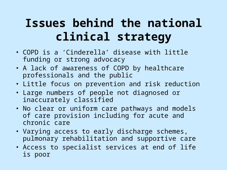
Issues behind the national clinical strategy
• COPD is a ‘Cinderella’ disease with little funding or strong advocacy
• A lack of awareness of COPD by healthcare professionals and the public
• Little focus on prevention and risk reduction• Large numbers of people not diagnosed or inaccurately
classified • No clear or uniform care pathways and models of care
provision including for acute and chronic care• Varying access to early discharge schemes, pulmonary
rehabilitation and supportive care• Access to specialist services at end of life is poor

National Clinical Strategy for COPD
• Launch end of Q1 2010• New DH Respiratory Programme Board• Variety of guidance / educational resources
etc• Need to reduce:
– Variation in care / outcomes– Unnecessary tests / duplication– Ineffective prescribing (25% of oxygen
prescriptions are ineffective)

National Clinical Strategy for COPD: national structure
• National Clinical Leads
• Respiratory Leads in each of 10 SHAs
• Trust Medical Directors (acute Trusts and PCTs)
– Ensure local leadership
– Promote local respiratory networks

National Clinical Strategy for COPD
• Guidelines on diagnostics and case-finding spirometry
• Competency framework for HCPs• COPD commissioning guidance• Metrics for primary and secondary care• Tariffs (reduce disincentives)
• DH – develop ‘Lung Improvement Programme’ to develop managed lung clinical networks, start national pilots, share information, resources etc
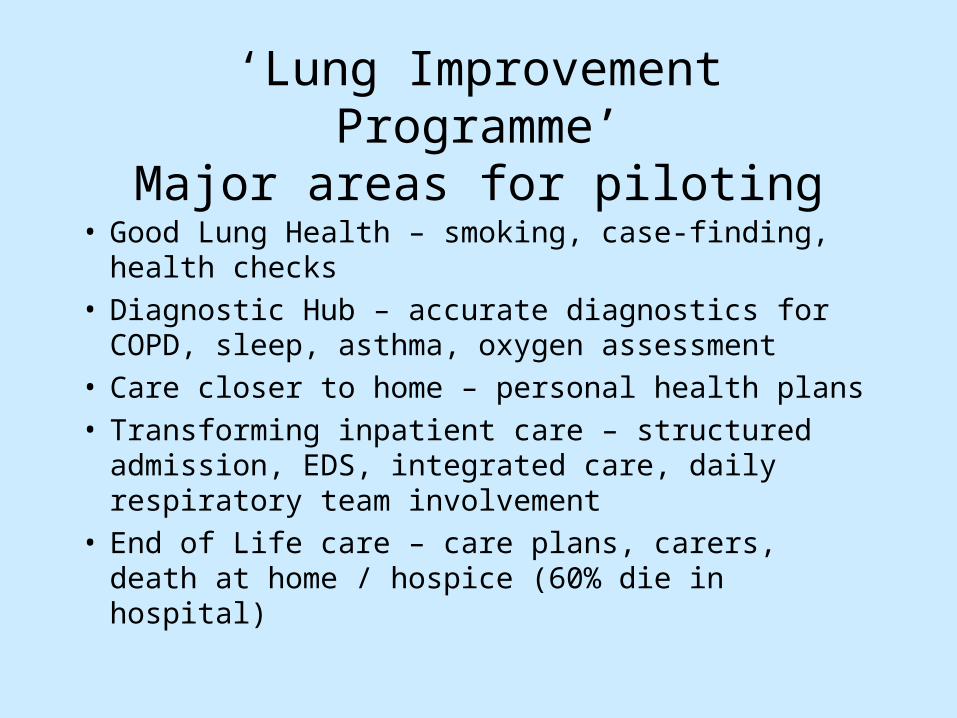
‘Lung Improvement Programme’Major areas for piloting
• Good Lung Health – smoking, case-finding, health checks
• Diagnostic Hub – accurate diagnostics for COPD, sleep, asthma, oxygen assessment
• Care closer to home – personal health plans• Transforming inpatient care – structured admission,
EDS, integrated care, daily respiratory team involvement
• End of Life care – care plans, carers, death at home / hospice (60% die in hospital)
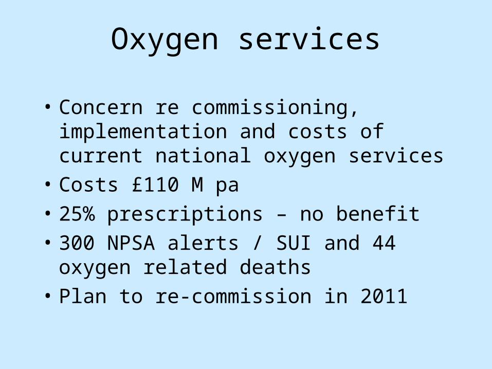
Oxygen services
• Concern re commissioning, implementation and costs of current national oxygen services
• Costs £110 M pa• 25% prescriptions – no benefit• 300 NPSA alerts / SUI and 44 oxygen related
deaths• Plan to re-commission in 2011

“Well, I guess I’ve taken up enough of your time…..”