conventional nuclear and pet imaging of suspected msk infections
TRANSCRIPT
Conventional Nuclear and PET Imaging of Suspected MSK infections
Niraj R. Patel, M.D.
January 12, 2011
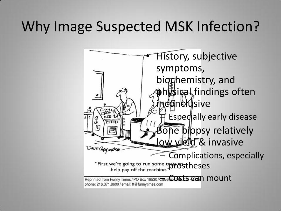
Why Image Suspected MSK Infection?
• History, subjective symptoms, biochemistry, and physical findings often inconclusive – Especially early disease
• Bone biopsy relatively low yield & invasive – Complications, especially
prostheses
– Costs can mount
MSK Infections
• Acute osteomyelitis
• Diabetic foot infection
• Spondylodiscitis
• Post-traumatic bone infection
• Inflected orthopedic prosthesis
• Chronic osteomyelitis
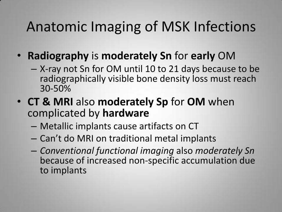
Anatomic Imaging of MSK Infections
• Radiography is moderately Sn for early OM – X-ray not Sn for OM until 10 to 21 days because to be
radiographically visible bone density loss must reach 30-50%
• CT & MRI also moderately Sp for OM when complicated by hardware – Metallic implants cause artifacts on CT – Can’t do MRI on traditional metal implants – Conventional functional imaging also moderately Sn
because of increased non-specific accumulation due to implants
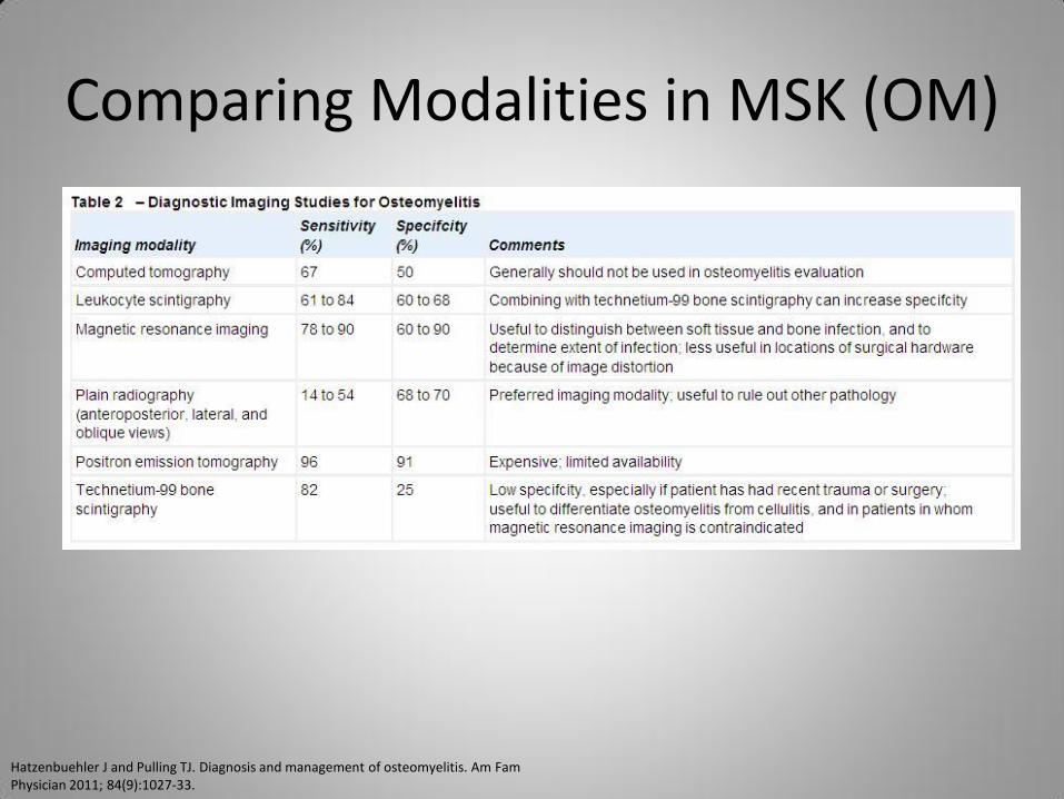
Comparing Modalities in MSK (OM)
Hatzenbuehler J and Pulling TJ. Diagnosis and management of osteomyelitis. Am Fam Physician 2011; 84(9):1027-33.
Conventional Nuclear Imaging of MSK Infections
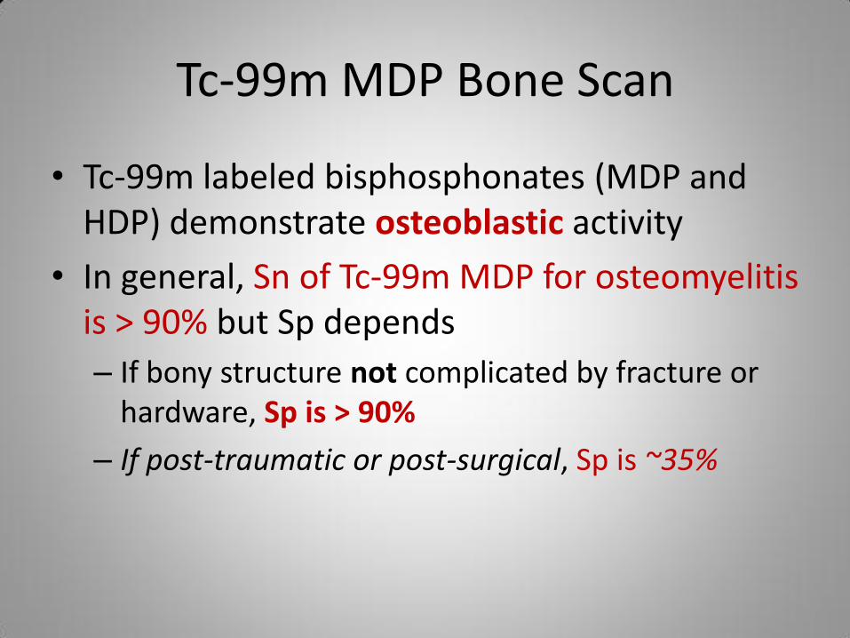
Tc-99m MDP Bone Scan
• Tc-99m labeled bisphosphonates (MDP and HDP) demonstrate osteoblastic activity
• In general, Sn of Tc-99m MDP for osteomyelitis is > 90% but Sp depends
– If bony structure not complicated by fracture or hardware, Sp is > 90%
– If post-traumatic or post-surgical, Sp is ~35%
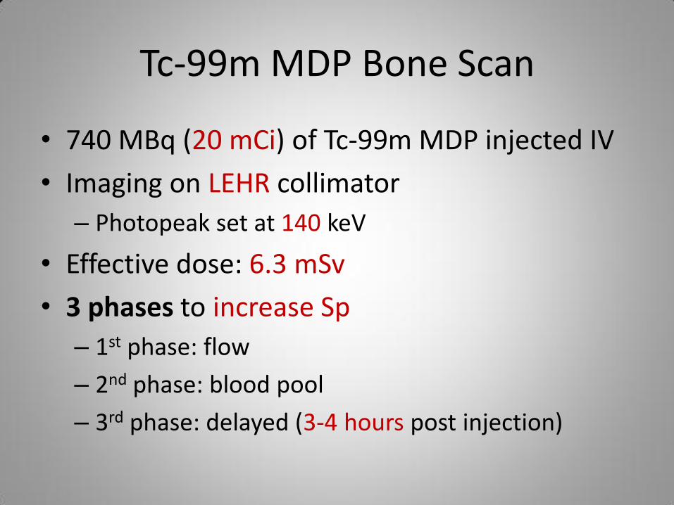
Tc-99m MDP Bone Scan
• 740 MBq (20 mCi) of Tc-99m MDP injected IV
• Imaging on LEHR collimator
– Photopeak set at 140 keV
• Effective dose: 6.3 mSv
• 3 phases to increase Sp
– 1st phase: flow
– 2nd phase: blood pool
– 3rd phase: delayed (3-4 hours post injection)
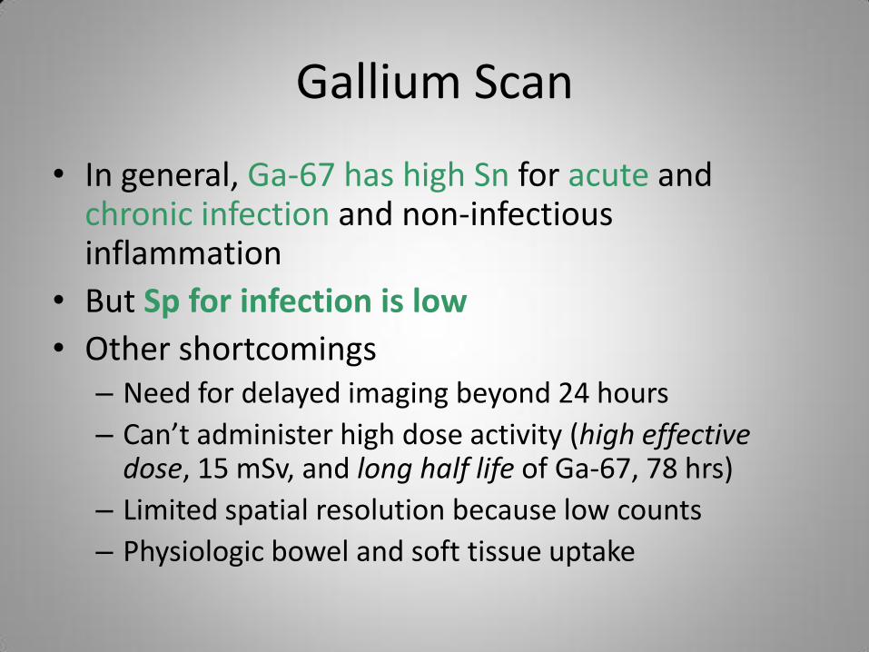
Gallium Scan
• In general, Ga-67 has high Sn for acute and chronic infection and non-infectious inflammation
• But Sp for infection is low
• Other shortcomings – Need for delayed imaging beyond 24 hours
– Can’t administer high dose activity (high effective dose, 15 mSv, and long half life of Ga-67, 78 hrs)
– Limited spatial resolution because low counts
– Physiologic bowel and soft tissue uptake
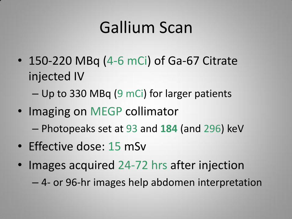
Gallium Scan
• 150-220 MBq (4-6 mCi) of Ga-67 Citrate injected IV
– Up to 330 MBq (9 mCi) for larger patients
• Imaging on MEGP collimator
– Photopeaks set at 93 and 184 (and 296) keV
• Effective dose: 15 mSv
• Images acquired 24-72 hrs after injection
– 4- or 96-hr images help abdomen interpretation
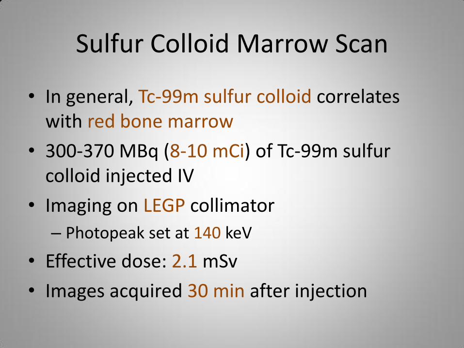
Sulfur Colloid Marrow Scan
• In general, Tc-99m sulfur colloid correlates with red bone marrow
• 300-370 MBq (8-10 mCi) of Tc-99m sulfur colloid injected IV
• Imaging on LEGP collimator
– Photopeak set at 140 keV
• Effective dose: 2.1 mSv
• Images acquired 30 min after injection
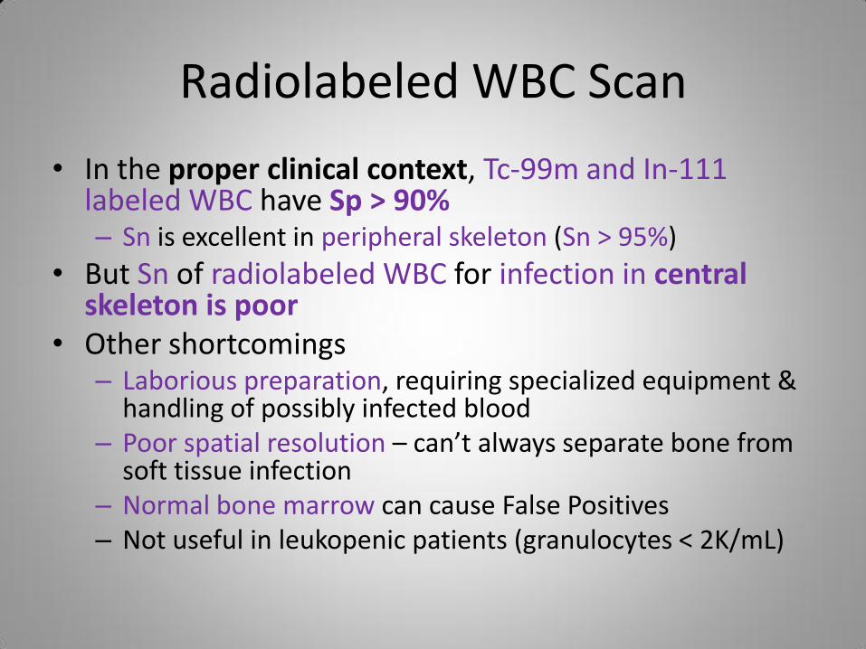
Radiolabeled WBC Scan
• In the proper clinical context, Tc-99m and In-111 labeled WBC have Sp > 90% – Sn is excellent in peripheral skeleton (Sn > 95%)
• But Sn of radiolabeled WBC for infection in central skeleton is poor
• Other shortcomings – Laborious preparation, requiring specialized equipment &
handling of possibly infected blood – Poor spatial resolution – can’t always separate bone from
soft tissue infection – Normal bone marrow can cause False Positives – Not useful in leukopenic patients (granulocytes < 2K/mL)
Radiolabeled WBC Scan
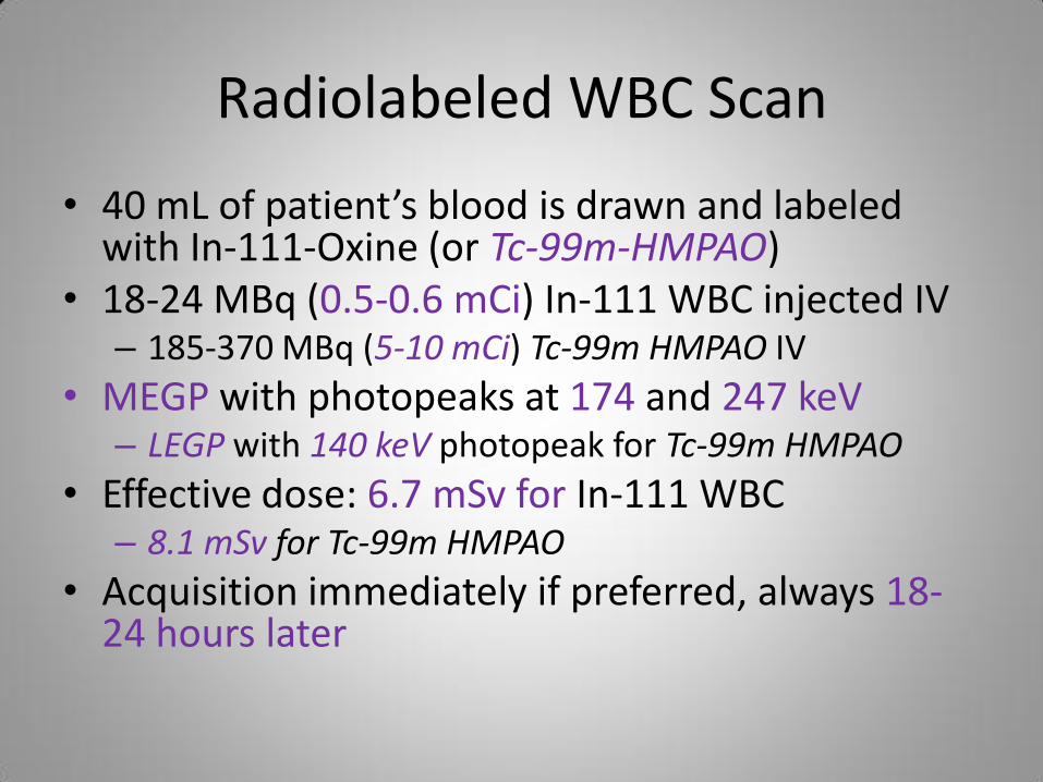
• 40 mL of patient’s blood is drawn and labeled with In-111-Oxine (or Tc-99m-HMPAO)
• 18-24 MBq (0.5-0.6 mCi) In-111 WBC injected IV – 185-370 MBq (5-10 mCi) Tc-99m HMPAO IV
• MEGP with photopeaks at 174 and 247 keV – LEGP with 140 keV photopeak for Tc-99m HMPAO
• Effective dose: 6.7 mSv for In-111 WBC – 8.1 mSv for Tc-99m HMPAO
• Acquisition immediately if preferred, always 18-24 hours later
Radiolabeled WBC Scan
Radiolabeled WBC Scan
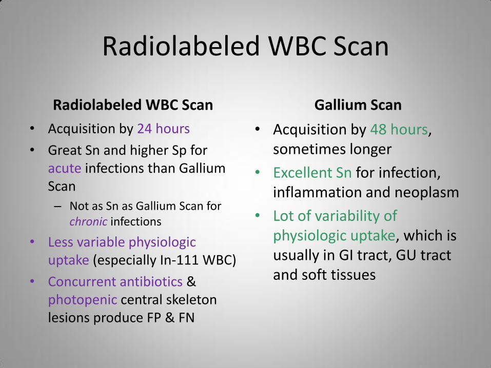
• Acquisition by 24 hours
• Great Sn and higher Sp for acute infections than Gallium Scan
– Not as Sn as Gallium Scan for chronic infections
• Less variable physiologic uptake (especially In-111 WBC)
• Concurrent antibiotics & photopenic central skeleton lesions produce FP & FN
Gallium Scan
• Acquisition by 48 hours, sometimes longer
• Excellent Sn for infection, inflammation and neoplasm
• Lot of variability of physiologic uptake, which is usually in GI tract, GU tract and soft tissues

Radiolabeled WBC Scan
In-111 WBC
• Does not concentrate in GI tract, GU tract or GB, thus better for abdominopelvic infections
• Longer half-life of In-111 (67 hours) allows better delayed imaging than Tc-99m
• Obligates us to use lower administered dose, causing grainier images
Tc-99m-HMPAO WBC • Labeling less stable than In-
111 WBC – Tracer in GIT, GUT and GB
• 6 h H.L. of Tc-99m leads to higher dose, thus more counts and better quality images
• Faster uptake in infection sites, thus better earlier imaging
• Better visualization of small anatomy
• Low absorbed radiation doses make it more suitable than In-111 for infants & children
Bone + Radiolabeled WBC Scan
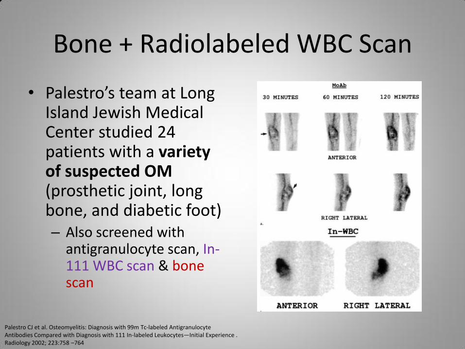
• Palestro’s team at Long Island Jewish Medical Center studied 24 patients with a variety of suspected OM (prosthetic joint, long bone, and diabetic foot) – Also screened with
antigranulocyte scan, In-111 WBC scan & bone scan
Palestro CJ et al. Osteomyelitis: Diagnosis with 99m Tc-labeled Antigranulocyte Antibodies Compared with Diagnosis with 111 In-labeled Leukocytes—Initial Experience . Radiology 2002; 223:758 –764
Bone + Radiolabeled WBC Scan
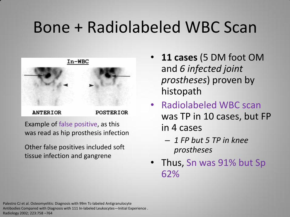
• 11 cases (5 DM foot OM and 6 infected joint prostheses) proven by histopath
• Radiolabeled WBC scan was TP in 10 cases, but FP in 4 cases – 1 FP but 5 TP in knee
prostheses
• Thus, Sn was 91% but Sp 62%
Example of false positive, as this was read as hip prosthesis infection
Other false positives included soft tissue infection and gangrene
Palestro CJ et al. Osteomyelitis: Diagnosis with 99m Tc-labeled Antigranulocyte Antibodies Compared with Diagnosis with 111 In-labeled Leukocytes—Initial Experience . Radiology 2002; 223:758 –764
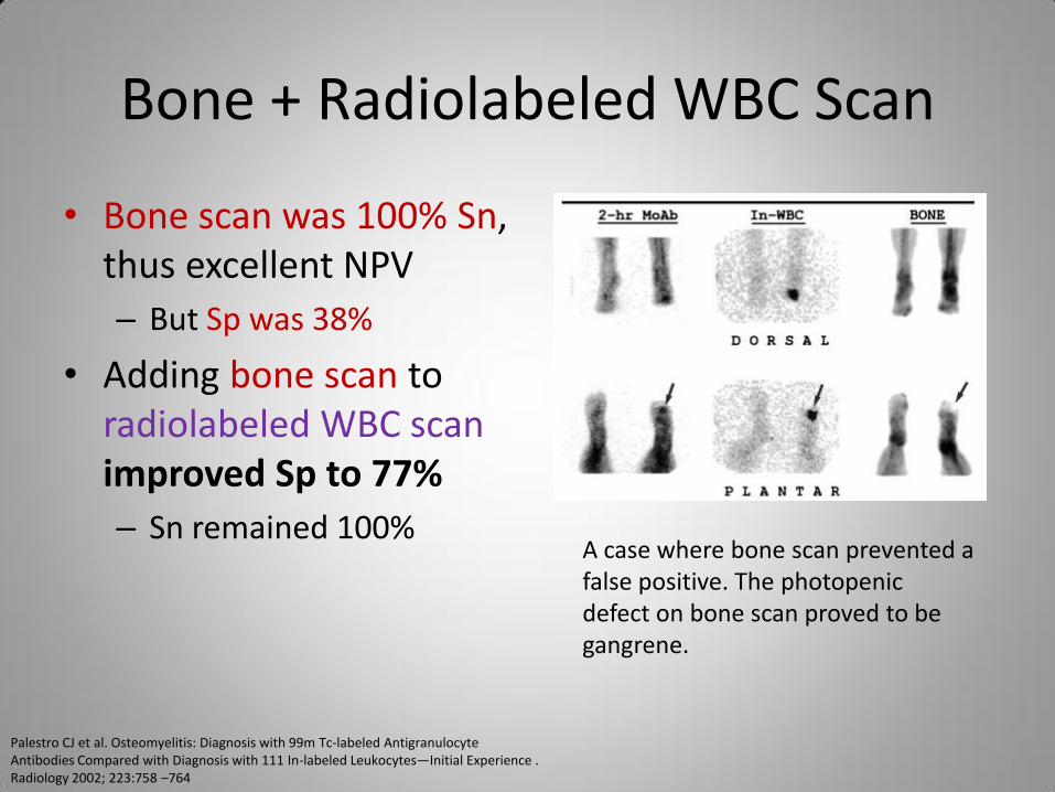
Bone + Radiolabeled WBC Scan
• Bone scan was 100% Sn, thus excellent NPV
– But Sp was 38%
• Adding bone scan to radiolabeled WBC scan improved Sp to 77%
– Sn remained 100% A case where bone scan prevented a false positive. The photopenic defect on bone scan proved to be gangrene.
Palestro CJ et al. Osteomyelitis: Diagnosis with 99m Tc-labeled Antigranulocyte Antibodies Compared with Diagnosis with 111 In-labeled Leukocytes—Initial Experience . Radiology 2002; 223:758 –764
Bone + Radiolabeled WBC Scan
• In diabetic foot, seems best to combine bone scan with radiolabeled WBC scan
• Bone scan is 100% Sn, but Sp low largely because of neuropathic joint disease
• In French study, 75 diabetics with 83 foot ulcers were scanned for suspected OM
Poirier JY et al. Diagnosis of Osteomyelitis in the Diabetic Foot with a Tc-99m HMPAO Leucocyte Scintigraphy Combined with a Tc-99m Bone Scintigraphy. Diabetes Metab (Paris) 2002, 28, 485-490.
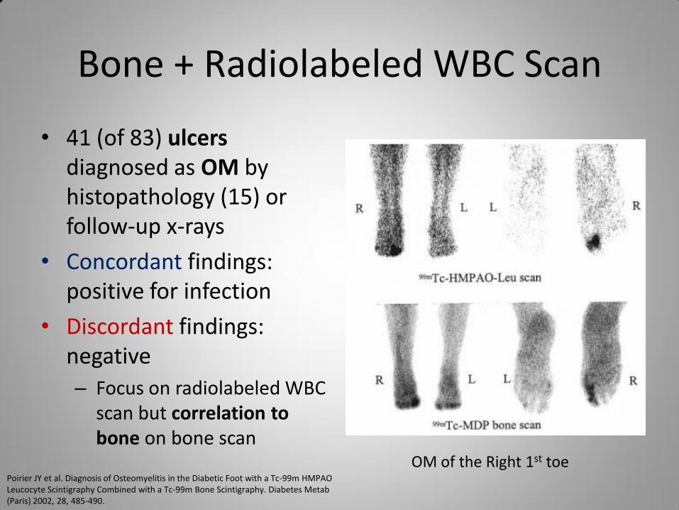
Bone + Radiolabeled WBC Scan
• 41 (of 83) ulcers diagnosed as OM by histopathology (15) or follow-up x-rays
• Concordant findings: positive for infection
• Discordant findings: negative
– Focus on radiolabeled WBC scan but correlation to bone on bone scan
Poirier JY et al. Diagnosis of Osteomyelitis in the Diabetic Foot with a Tc-99m HMPAO Leucocyte Scintigraphy Combined with a Tc-99m Bone Scintigraphy. Diabetes Metab (Paris) 2002, 28, 485-490.
OM of the Right 1st toe
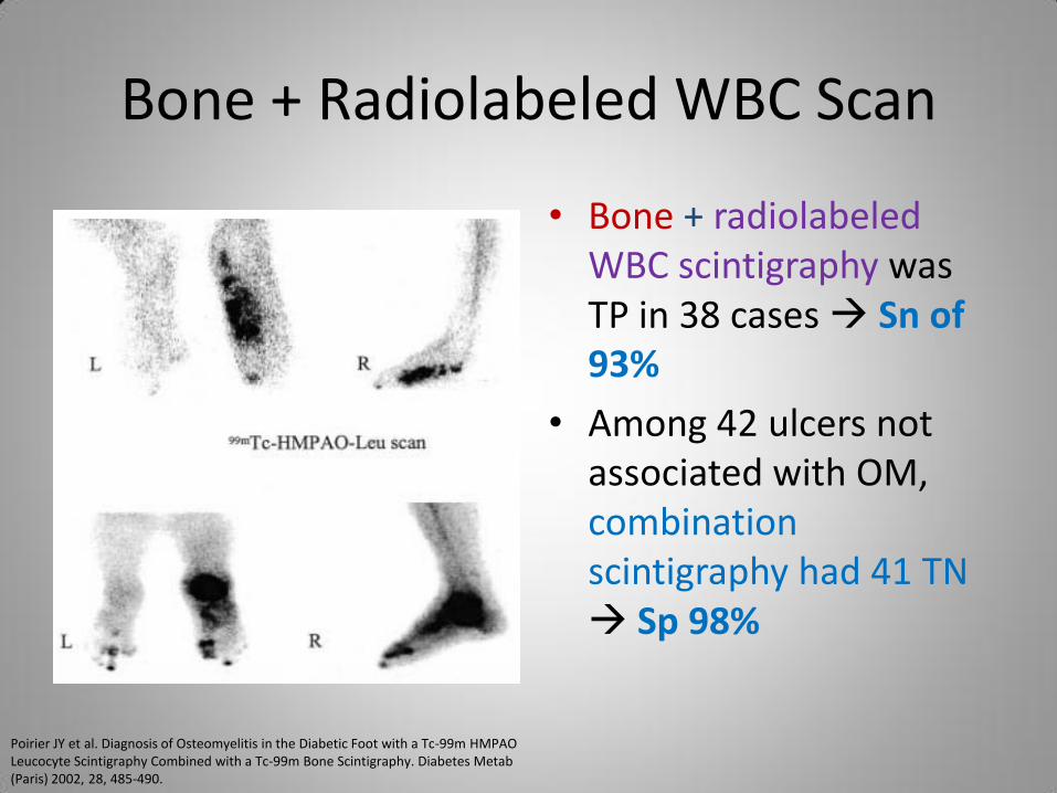
Bone + Radiolabeled WBC Scan
• Bone + radiolabeled WBC scintigraphy was TP in 38 cases Sn of 93%
• Among 42 ulcers not associated with OM, combination scintigraphy had 41 TN Sp 98%
Poirier JY et al. Diagnosis of Osteomyelitis in the Diabetic Foot with a Tc-99m HMPAO Leucocyte Scintigraphy Combined with a Tc-99m Bone Scintigraphy. Diabetes Metab (Paris) 2002, 28, 485-490.
Bone + Radiolabeled WBC Scan
• Bessette’s group in Milwaukee reviewed 32 patients with suspected sternal OM
• Group composed of 12 patients with biopsy-proven sternal OM
• All scanned with CT and radiolabeled WBC + bone scans
• CT positive for sternal OM in 7 patients - 5 bony erosions & 2 severe demin (Sn 58%)
• Combination scintigraphy positive in 11 patients (Sn 92%) – 1 case of FP due to
concurrent IV antibiotics which was not seen on radiolabeled WBC scan
Bessette PR et al. Evaluation of postoperative osteomyelitis of the sternum comparing CT and dual Tc-99m MDP bone and In-111 WBC SPECT. Clin Nucl Med. 1993 Mar;18(3):197-202.
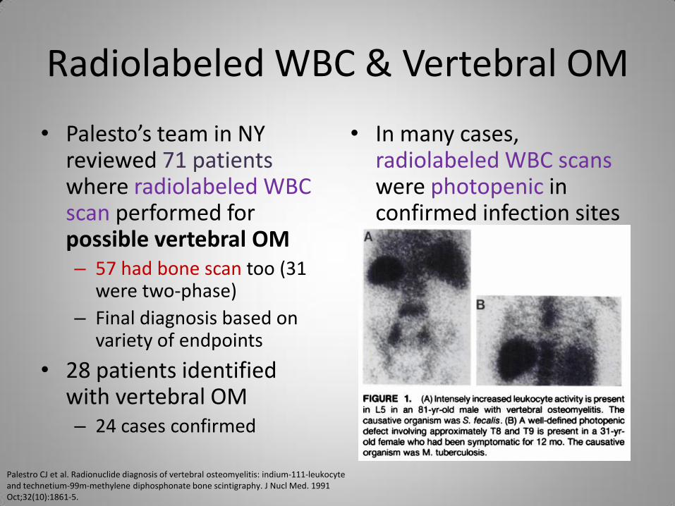
Radiolabeled WBC & Vertebral OM
• Palesto’s team in NY reviewed 71 patients where radiolabeled WBC scan performed for possible vertebral OM – 57 had bone scan too (31
were two-phase)
– Final diagnosis based on variety of endpoints
• 28 patients identified with vertebral OM – 24 cases confirmed
• In many cases, radiolabeled WBC scans were photopenic in confirmed infection sites
Palestro CJ et al. Radionuclide diagnosis of vertebral osteomyelitis: indium-111-leukocyte and technetium-99m-methylene diphosphonate bone scintigraphy. J Nucl Med. 1991 Oct;32(10):1861-5.
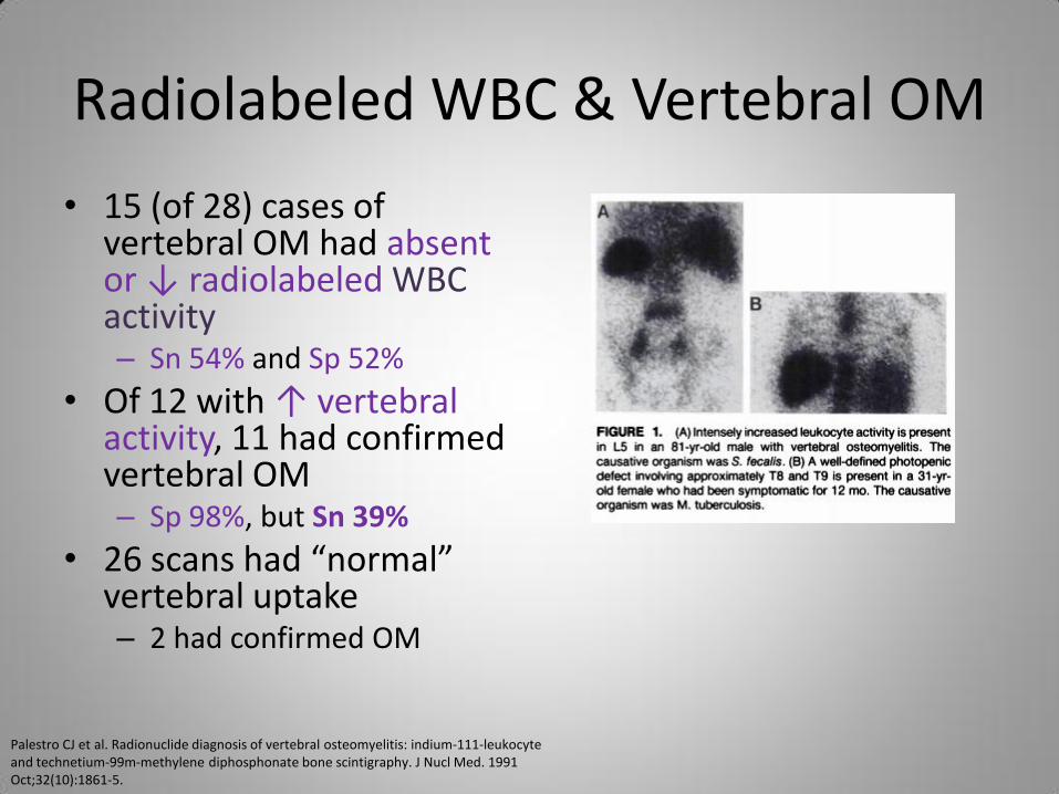
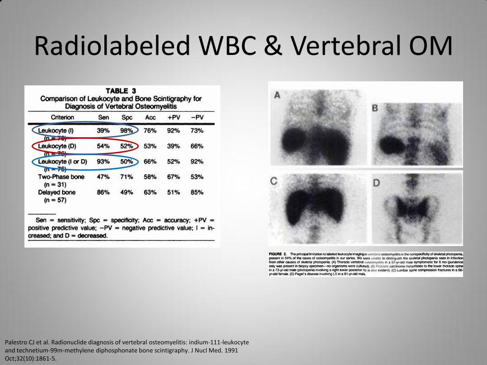
Radiolabeled WBC & Vertebral OM
• 15 (of 28) cases of vertebral OM had absent or ↓ radiolabeled WBC activity – Sn 54% and Sp 52%
• Of 12 with ↑ vertebral activity, 11 had confirmed vertebral OM – Sp 98%, but Sn 39%
• 26 scans had “normal” vertebral uptake – 2 had confirmed OM
Palestro CJ et al. Radionuclide diagnosis of vertebral osteomyelitis: indium-111-leukocyte and technetium-99m-methylene diphosphonate bone scintigraphy. J Nucl Med. 1991 Oct;32(10):1861-5.
Radiolabeled WBC & Vertebral OM
Palestro CJ et al. Radionuclide diagnosis of vertebral osteomyelitis: indium-111-leukocyte and technetium-99m-methylene diphosphonate bone scintigraphy. J Nucl Med. 1991 Oct;32(10):1861-5.
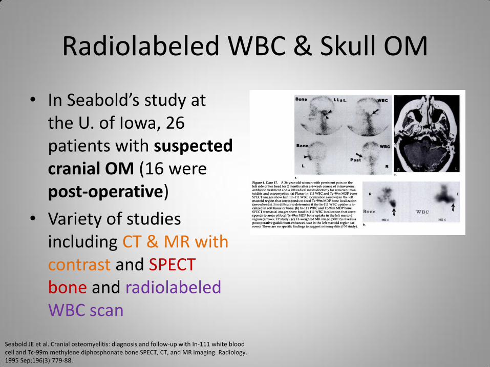
Radiolabeled WBC & Skull OM
• In Seabold’s study at the U. of Iowa, 26 patients with suspected cranial OM (16 were post-operative)
• Variety of studies including CT & MR with contrast and SPECT bone and radiolabeled WBC scan
Seabold JE et al. Cranial osteomyelitis: diagnosis and follow-up with In-111 white blood cell and Tc-99m methylene diphosphonate bone SPECT, CT, and MR imaging. Radiology. 1995 Sep;196(3):779-88.
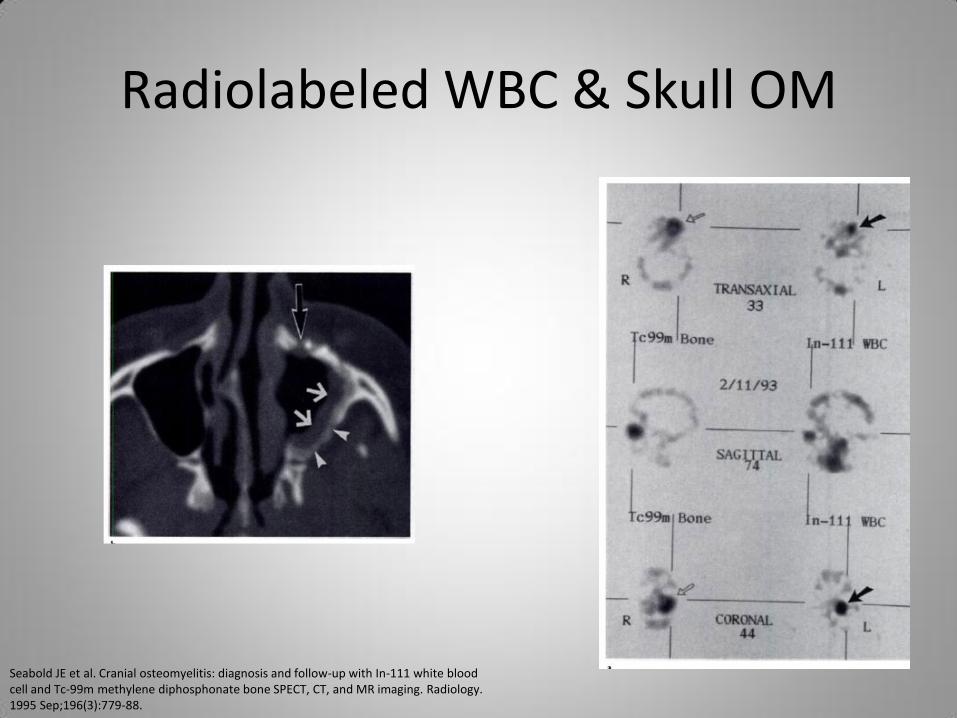
Radiolabeled WBC & Skull OM
Seabold JE et al. Cranial osteomyelitis: diagnosis and follow-up with In-111 white blood cell and Tc-99m methylene diphosphonate bone SPECT, CT, and MR imaging. Radiology. 1995 Sep;196(3):779-88.
Radiolabeled WBC & Skull OM
• If no prior intervention in skull (with minimal bone marrow), CT and bone scan most Sn for OM
• In skull base without prior intervention, MR and bone scan most Sn for OM
• MR is best imaging to assess extent of soft tissue involvement
• In skull with pre-existing abnormality (e.g., post-surgery), combined radiolabeled WBC & bone scans most Acc for OM
• Abnormal findings revert back to normal sooner with radiolabeled WBC scan vs MRI and CT in successfully treated patients
Seabold JE et al. Cranial osteomyelitis: diagnosis and follow-up with In-111 white blood cell and Tc-99m methylene diphosphonate bone SPECT, CT, and MR imaging. Radiology. 1995 Sep;196(3):779-88.
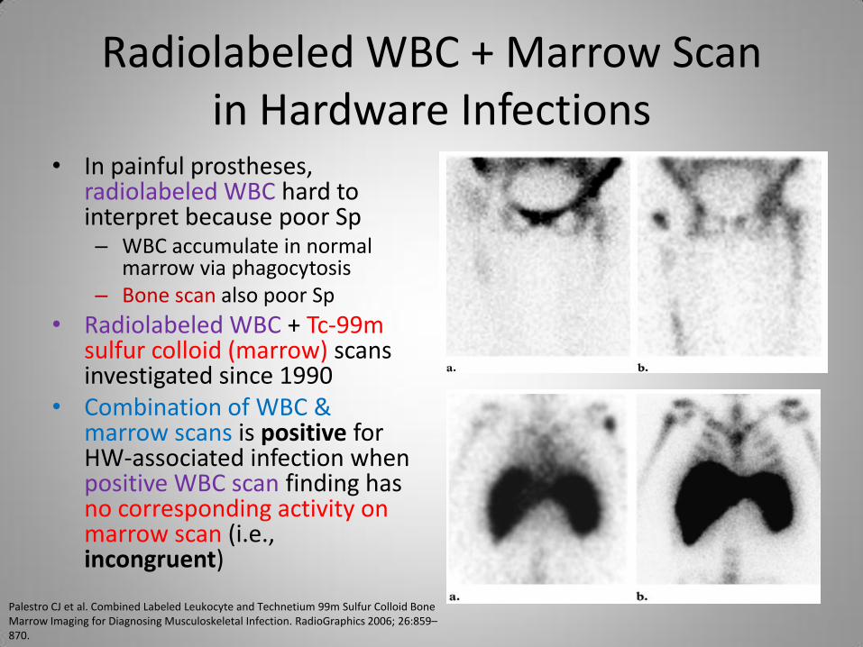
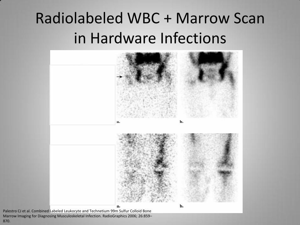
Radiolabeled WBC + Marrow Scan in Hardware Infections
• In painful prostheses, radiolabeled WBC hard to interpret because poor Sp – WBC accumulate in normal
marrow via phagocytosis – Bone scan also poor Sp
• Radiolabeled WBC + Tc-99m sulfur colloid (marrow) scans investigated since 1990
• Combination of WBC & marrow scans is positive for HW-associated infection when positive WBC scan finding has no corresponding activity on marrow scan (i.e., incongruent)
Palestro CJ et al. Combined Labeled Leukocyte and Technetium 99m Sulfur Colloid Bone Marrow Imaging for Diagnosing Musculoskeletal Infection. RadioGraphics 2006; 26:859–870.
Radiolabeled WBC + Marrow Scan in Hardware Infections
Palestro CJ et al. Combined Labeled Leukocyte and Technetium 99m Sulfur Colloid Bone Marrow Imaging for Diagnosing Musculoskeletal Infection. RadioGraphics 2006; 26:859–870.
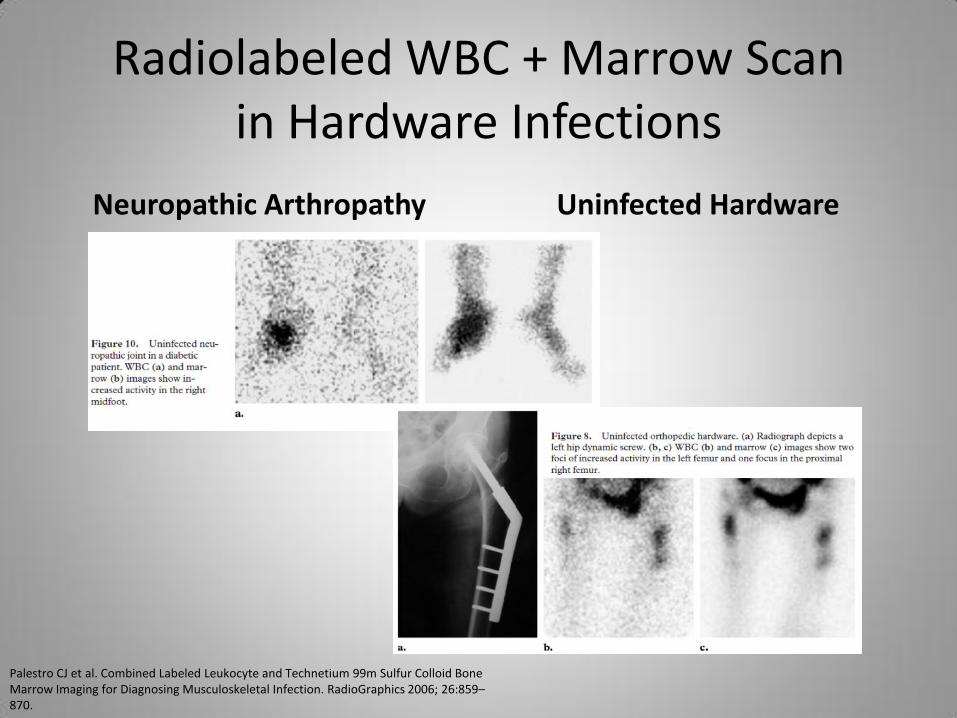
Radiolabeled WBC + Marrow Scan in Hardware Infections
Neuropathic Arthropathy Uninfected Hardware
Palestro CJ et al. Combined Labeled Leukocyte and Technetium 99m Sulfur Colloid Bone Marrow Imaging for Diagnosing Musculoskeletal Infection. RadioGraphics 2006; 26:859–870.
Radiolabeled WBC + Marrow Scan in Hardware Infections
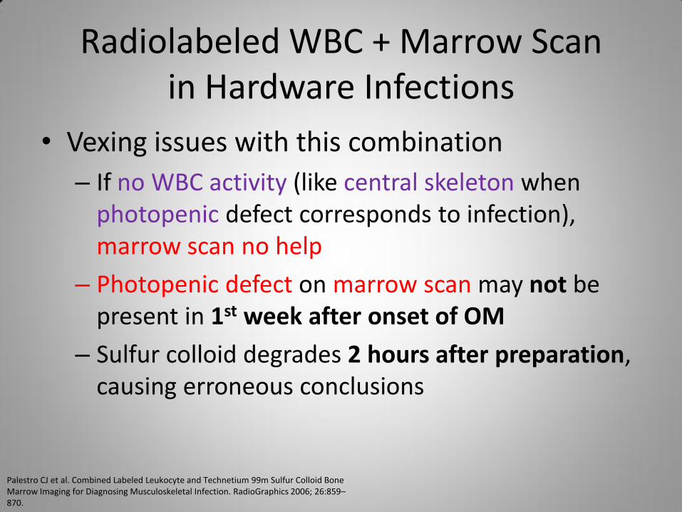
• Vexing issues with this combination
– If no WBC activity (like central skeleton when photopenic defect corresponds to infection), marrow scan no help
– Photopenic defect on marrow scan may not be present in 1st week after onset of OM
– Sulfur colloid degrades 2 hours after preparation, causing erroneous conclusions
Palestro CJ et al. Combined Labeled Leukocyte and Technetium 99m Sulfur Colloid Bone Marrow Imaging for Diagnosing Musculoskeletal Infection. RadioGraphics 2006; 26:859–870.
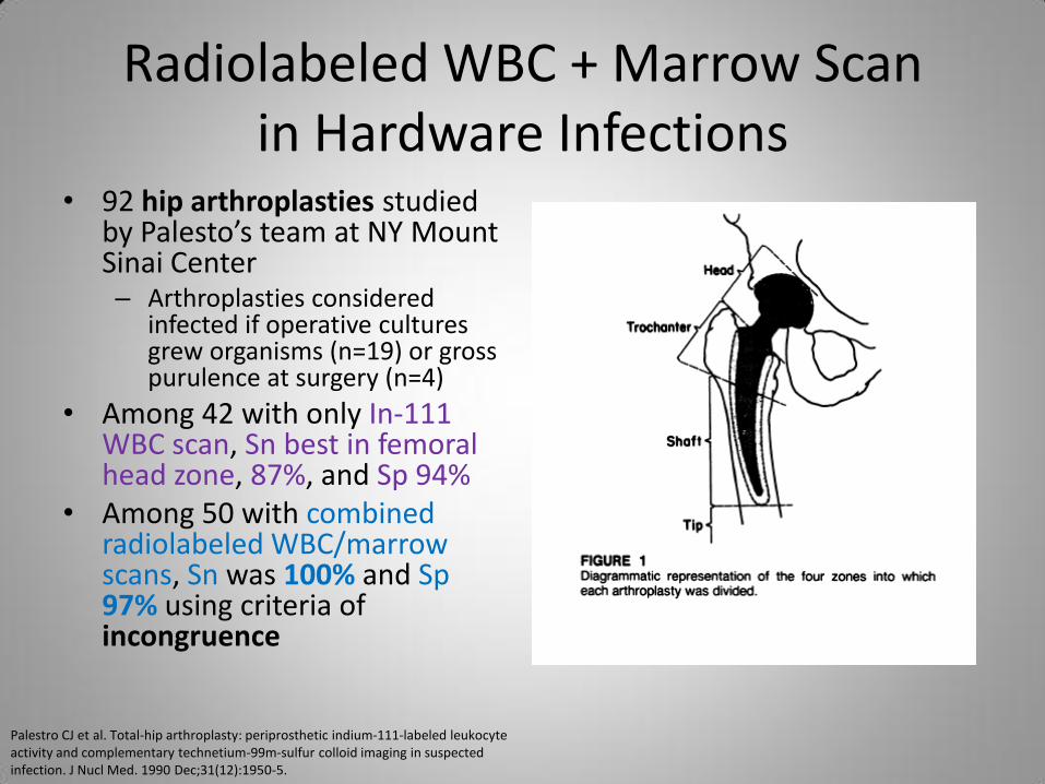
Radiolabeled WBC + Marrow Scan in Hardware Infections
• 92 hip arthroplasties studied by Palesto’s team at NY Mount Sinai Center – Arthroplasties considered
infected if operative cultures grew organisms (n=19) or gross purulence at surgery (n=4)
• Among 42 with only In-111 WBC scan, Sn best in femoral head zone, 87%, and Sp 94%
• Among 50 with combined radiolabeled WBC/marrow scans, Sn was 100% and Sp 97% using criteria of incongruence
Palestro CJ et al. Total-hip arthroplasty: periprosthetic indium-111-labeled leukocyte activity and complementary technetium-99m-sulfur colloid imaging in suspected infection. J Nucl Med. 1990 Dec;31(12):1950-5.
Radiolabeled WBC + Marrow Scan in Hardware Infections
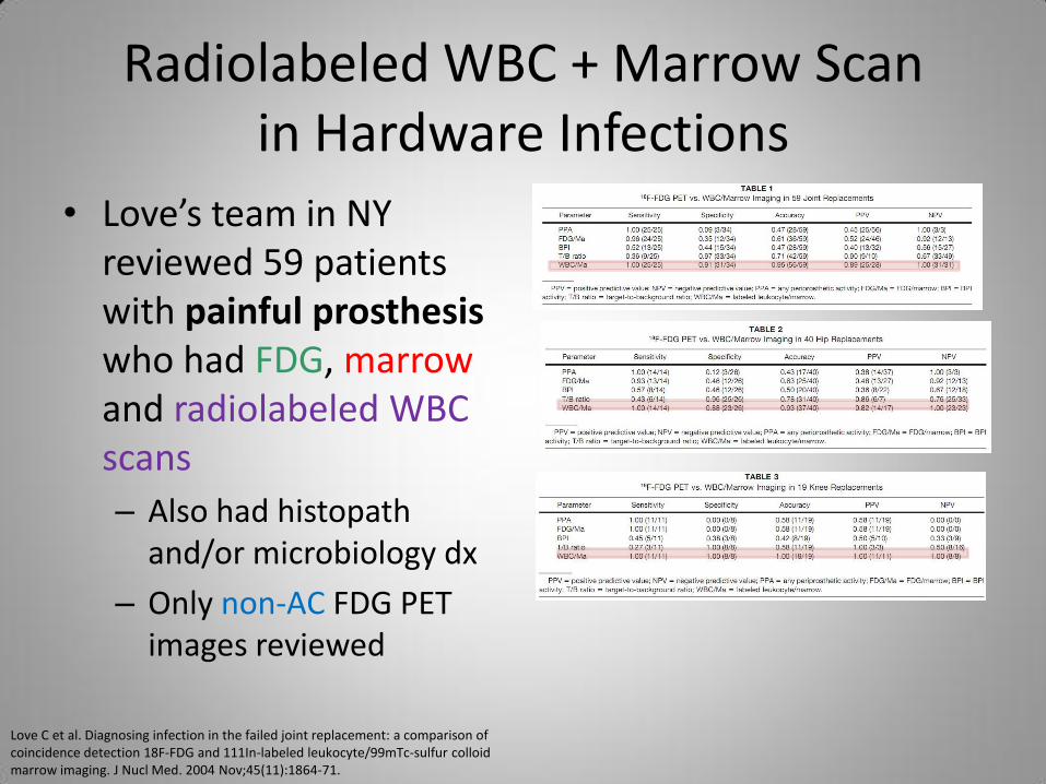
• Love’s team in NY reviewed 59 patients with painful prosthesis who had FDG, marrow and radiolabeled WBC scans
– Also had histopath and/or microbiology dx
– Only non-AC FDG PET images reviewed
Love C et al. Diagnosing infection in the failed joint replacement: a comparison of coincidence detection 18F-FDG and 111In-labeled leukocyte/99mTc-sulfur colloid marrow imaging. J Nucl Med. 2004 Nov;45(11):1864-71.
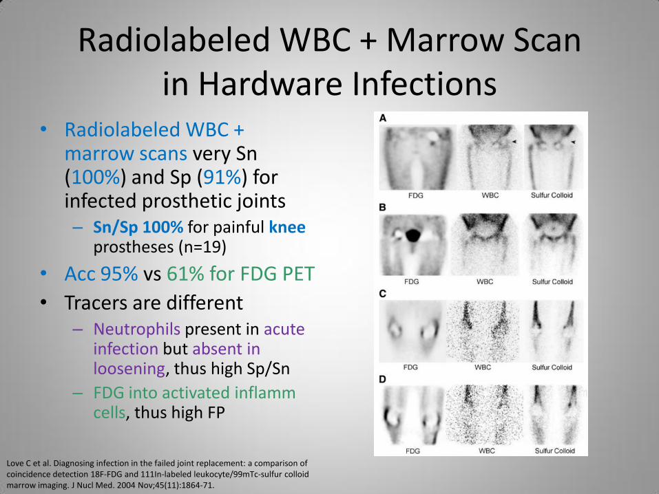
Radiolabeled WBC + Marrow Scan in Hardware Infections
• Radiolabeled WBC + marrow scans very Sn (100%) and Sp (91%) for infected prosthetic joints – Sn/Sp 100% for painful knee
prostheses (n=19)
• Acc 95% vs 61% for FDG PET
• Tracers are different – Neutrophils present in acute
infection but absent in loosening, thus high Sp/Sn
– FDG into activated inflamm cells, thus high FP
Love C et al. Diagnosing infection in the failed joint replacement: a comparison of coincidence detection 18F-FDG and 111In-labeled leukocyte/99mTc-sulfur colloid marrow imaging. J Nucl Med. 2004 Nov;45(11):1864-71.
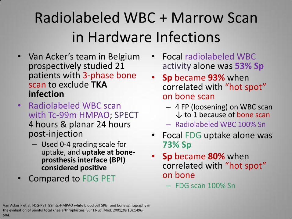
Radiolabeled WBC + Marrow Scan in Hardware Infections
• Van Acker’s team in Belgium prospectively studied 21 patients with 3-phase bone scan to exclude TKA infection
• Radiolabeled WBC scan with Tc-99m HMPAO; SPECT 4 hours & planar 24 hours post-injection – Used 0-4 grading scale for
uptake, and uptake at bone-prosthesis interface (BPI) considered positive
• Compared to FDG PET
• Focal radiolabeled WBC activity alone was 53% Sp
• Sp became 93% when correlated with “hot spot” on bone scan – 4 FP (loosening) on WBC scan
↓ to 1 because of bone scan – Radiolabeled WBC 100% Sn
• Focal FDG uptake alone was 73% Sp
• Sp became 80% when correlated with “hot spot” on bone – FDG scan 100% Sn
Van Acker F et al. FDG-PET, 99mtc-HMPAO white blood cell SPET and bone scintigraphy in the evaluation of painful total knee arthroplasties. Eur J Nucl Med. 2001;28(10):1496-504.
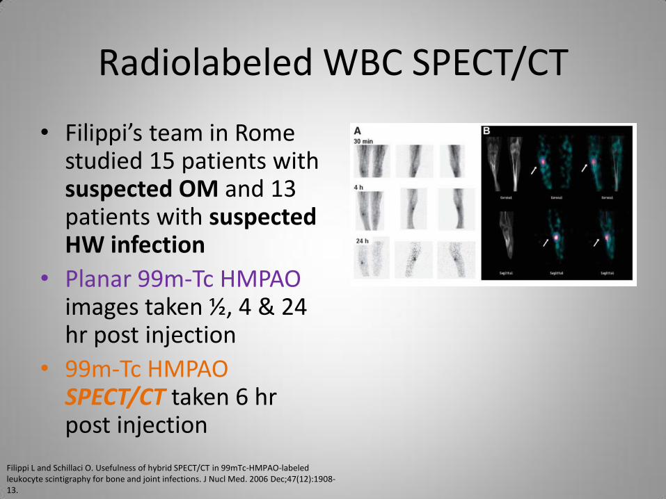
Radiolabeled WBC SPECT/CT
• Filippi’s team in Rome studied 15 patients with suspected OM and 13 patients with suspected HW infection
• Planar 99m-Tc HMPAO images taken ½, 4 & 24 hr post injection
• 99m-Tc HMPAO SPECT/CT taken 6 hr post injection
Filippi L and Schillaci O. Usefulness of hybrid SPECT/CT in 99mTc-HMPAO-labeled leukocyte scintigraphy for bone and joint infections. J Nucl Med. 2006 Dec;47(12):1908-13.
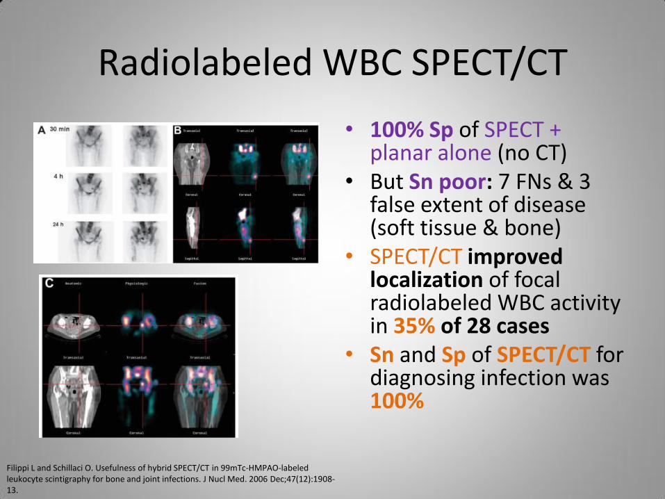
Radiolabeled WBC SPECT/CT
• 100% Sp of SPECT + planar alone (no CT)
• But Sn poor: 7 FNs & 3 false extent of disease (soft tissue & bone)
• SPECT/CT improved localization of focal radiolabeled WBC activity in 35% of 28 cases
• Sn and Sp of SPECT/CT for diagnosing infection was 100%
Filippi L and Schillaci O. Usefulness of hybrid SPECT/CT in 99mTc-HMPAO-labeled leukocyte scintigraphy for bone and joint infections. J Nucl Med. 2006 Dec;47(12):1908-13.
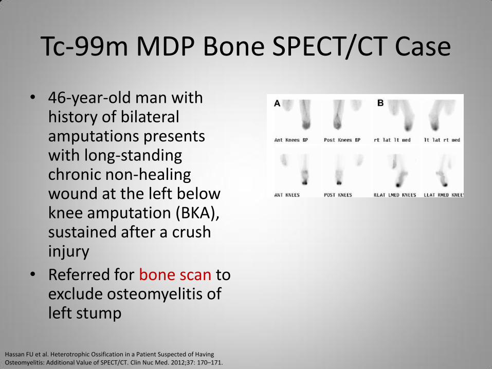
Tc-99m MDP Bone SPECT/CT Case
• 46-year-old man with history of bilateral amputations presents with long-standing chronic non-healing wound at the left below knee amputation (BKA), sustained after a crush injury
• Referred for bone scan to exclude osteomyelitis of left stump
Hassan FU et al. Heterotrophic Ossification in a Patient Suspected of Having Osteomyelitis: Additional Value of SPECT/CT. Clin Nuc Med. 2012;37: 170–171.
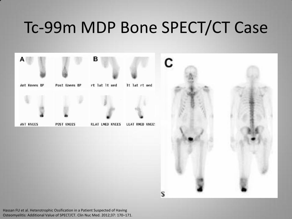
Tc-99m MDP Bone SPECT/CT Case
Hassan FU et al. Heterotrophic Ossification in a Patient Suspected of Having Osteomyelitis: Additional Value of SPECT/CT. Clin Nuc Med. 2012;37: 170–171.
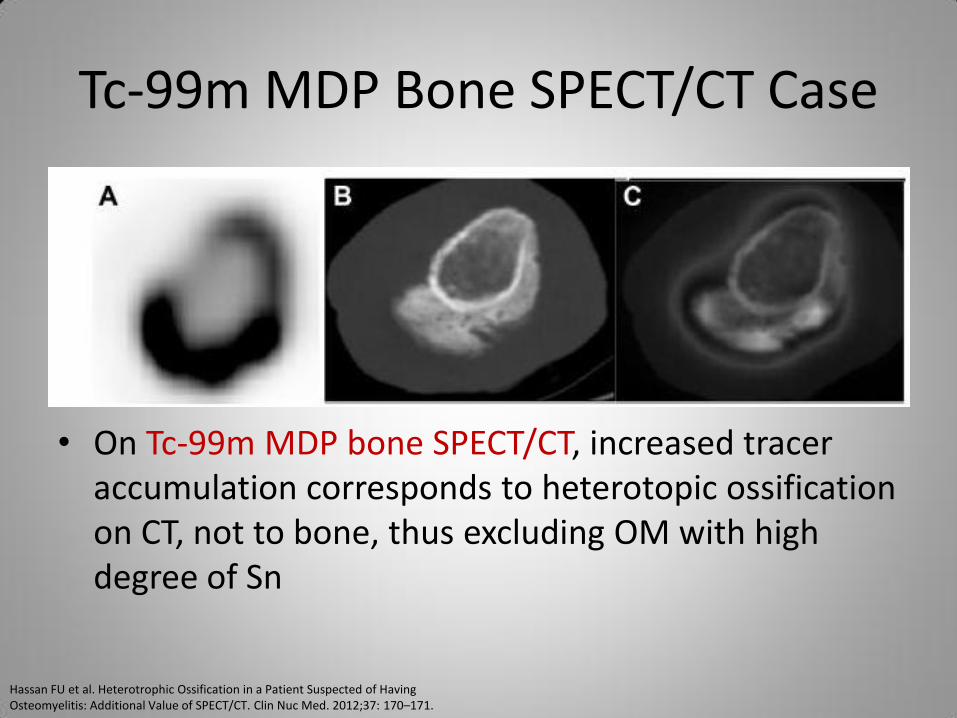
Tc-99m MDP Bone SPECT/CT Case
• On Tc-99m MDP bone SPECT/CT, increased tracer accumulation corresponds to heterotopic ossification on CT, not to bone, thus excluding OM with high degree of Sn
Hassan FU et al. Heterotrophic Ossification in a Patient Suspected of Having Osteomyelitis: Additional Value of SPECT/CT. Clin Nuc Med. 2012;37: 170–171.
Adding CT to SPECT
• Disadvantages – Lower count rate versus planar imaging (as it is
reconstructed and not truly tomographic imaging) – Lower spatial resolution than planar imaging – Takes longer to acquire good-quality images
• Advantages – Localizes “hot spot,” thus showing whether it is at site of
interest or outside of it – Regardless of tracer or disease entity, scintigraphy’s Sp
goes up when CT added – Improves inter-reader agreement & management of
patients
Horger M, Eschmann SM, Pfannenberg C, et al: Added value of SPECT/CT in patients suspected of having bone infection: preliminary results. Arch Orthop Trauma Surg . 2007; 127:211-221.
Horger M, Eschmann SM, Pfannenberg C, et al: The value of SPET/CT in chronic osteomyelitis. Eur J Nucl Med Mol Imaging 2003; 30:1665-1673.
Summary of Gamma Imaging of Suspected MSK Infections
• Bone Scan has excellent Sn but poor Sp
• Radiolabeled WBC Scan has good Sp & Sn in peripheral skeleton – Poor Sn in central skeleton
• Bone + Radiolabeled WBC scans improve Sp (and thus accuracy) – But in diabetic foot ulcer/OM, neuropathic joint
disease is problem
– Good Acc in infected knee & hip prostheses
Summary of Gamma Imaging of Suspected MSK Infections
• Radiolabeled WBC + Marrow scans improve accuracy
– Better in diabetic foot OM/ulcer
– Perhaps better Acc in infected knee prostheses
• Impact of SPECT and SPECT/CT
– Improves accuracy and localization
F-18 FDG PET Imaging of MSK Infections
F-18 FDG
• FDG enters cells via glucose transporters – Active transport mediated by GLUT 1-10, but primarily by
GLUT 1 & 3
– Active transport by Na+-glucose transporter (primary mechanism for kidney epithelial & intestinal cells)
– Passive diffusion is minor compared to active
• FDG gets phosphorylated and not further metabolized – 2’-FDG-6 phosphate not substrate for glycolytic pathway
or pentose-phosphate shunt
– Low initial concentrations of FDG in normal fasting heart & brain, but uptake increases over time
FDG & WBC
• Accumulates much more in activated (versus inactive) lymphocytes and especially neutrophils & macrophages – 24 hours after activation of WBC, increased de novo
synthesis of GLUT-1 & 3
– Blood glucose < 250 mg/dL, FDG uptake into inflammatory cells not impaired
• Radiolabeled WBCs are different, going wherever WBC are, activated or not – FDG relatively low accumulation in normal marrow
Forstrom LA et al. 18F-FDG labelling of human leukocytes. Nuc Med Comm. 2000; 21(7): 691-4.
Zhuang HM et al. Do high glucose levels have differential effect on FDG uptake in inflammatory and malignant disorders? Nuc Med Comm. 2001; 22(10): 1123-8.
Patient History & FDG PET
• Positive PET findings have to be interpreted in context of patient history
– FDG PET is a non-specific tracer
– Active inflammation present for months to year after orthopedic surgery
– Inflammation related to bone remodeling after fracture or surgical intervention
F-18 FDG PET/CT Scan
• 370-740 MBq (10-20 mCi) of F-18 FDG by IV
• Imaging on PET/CT scanner
– Low-dose CT for anatomic localization & attenuation correction
• Effective dose: 14.1 mSv
• Partial body PET/CT to reduce radiation dose
• Image acquisition starts about 45 min after injection and lasts 35 minutes (3-5 min for CT)
FDG PET & Chronic OM
• In acute OM, FDG PET adds no value over CT, MRI and combination scintigraphy – Conventional Imaging has > 90% accuracy
• FDG PET useful in chronic OM (COM), where these modalities are limited – COM: > 6 weeks of ongoing bone infection whether or
not signs & symptoms are present – Anatomic imaging: low Sp of X-ray, CT & MR (most
Sn?) for various reasons – Functional imaging: low Sp (bone & gallium scans) or
low Sn (radiolabeled WBC scan), also potential for suboptimal preparation, lots of time and costs
FDG PET & Chronic OM
• Zhuang’s team studied 22 patients at Penn
– 6 diagnosed with COM but no criteria defined (except 1 year follow-up)
• All 6 COM cases diagnosed by 3 readers using FDG PET (no CT), thus Sn 100%
– 2 patients had False Positive diagnosis by PET (both osteotomy-related inflammation), thus Sp not good
Zhuang H. Exclusion of Chronic Osteomyelitis With F-18 Fluorodeoxyglucose Positron Emission Tomographic Imaging. Clin Nucl Med. 2000; 25(4):281-4.
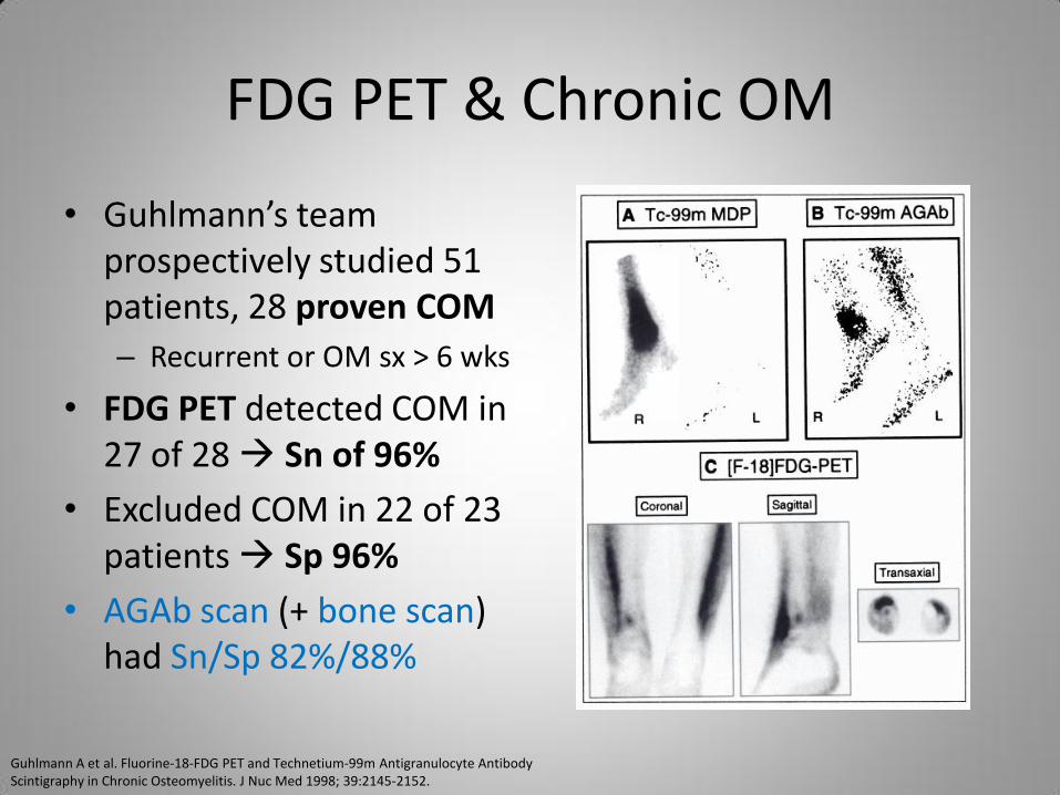
FDG PET & Chronic OM
• Guhlmann’s team prospectively studied 51 patients, 28 proven COM
– Recurrent or OM sx > 6 wks
• FDG PET detected COM in 27 of 28 Sn of 96%
• Excluded COM in 22 of 23 patients Sp 96%
• AGAb scan (+ bone scan) had Sn/Sp 82%/88%
Guhlmann A et al. Fluorine-18-FDG PET and Technetium-99m Antigranulocyte Antibody Scintigraphy in Chronic Osteomyelitis. J Nuc Med 1998; 39:2145-2152.
Case 1
• Man in his 50s complains of long standing central chest pain (“for years”) – PCP localizes it to sternum, which is TTP and
worse with exertion
– Remote history of trauma to sternum, but no surgical intervetion
– Labs were relatively normal
• Seen at VA in 2009 from OSH, then again in 2011
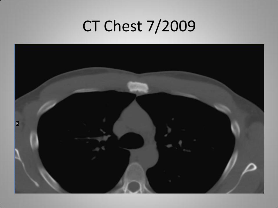
CT Chest 7/2009
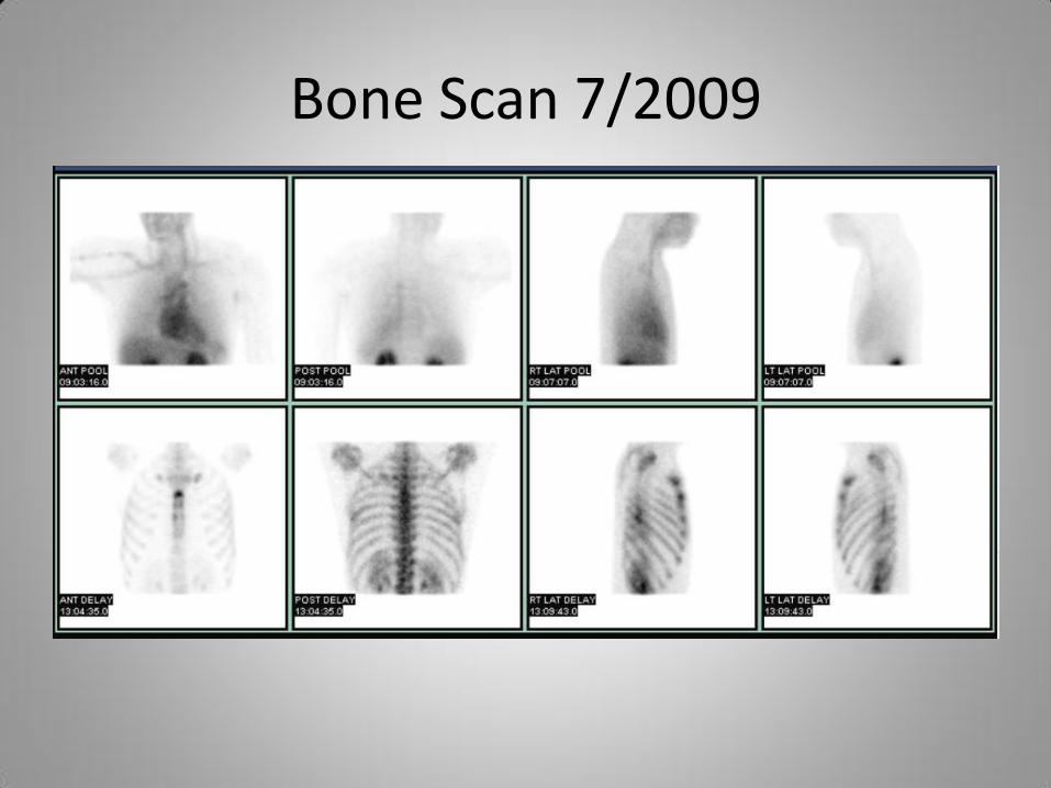
Bone Scan 7/2009
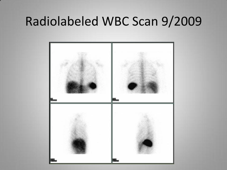
Radiolabeled WBC Scan 9/2009
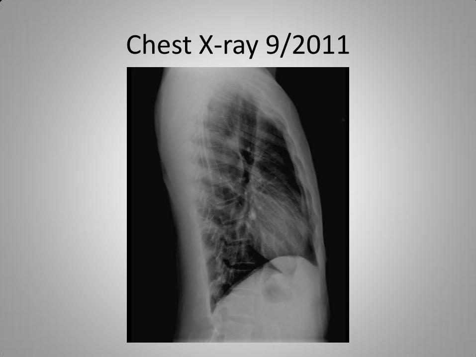
Chest X-ray 9/2011
Case 1
• PCP relied on negative WBC study – Recall that this has poor to moderate Sn for
chronic infections
• CT-guided aspiration biopsy was negative – Sensitivity of procedure is moderate (73-87%), on
lower end if on antibiotics
• New PCP reviewed reports and wants bone scan because patient’s sternal complains slightly worsening
Howard CB et al. Fine-Needle Bone Biopsy to Diagnose Osteonyelitis. J Bone Joint Surg 1994; 76-B:311-4.
Sutton PR et al. Diagnosing Osteomyelitis with Percutaneous Bone Biopsy in Patients with Diabetes and Bone Ulcerations. J of Gen Int Med 2008; 15(s2):6.
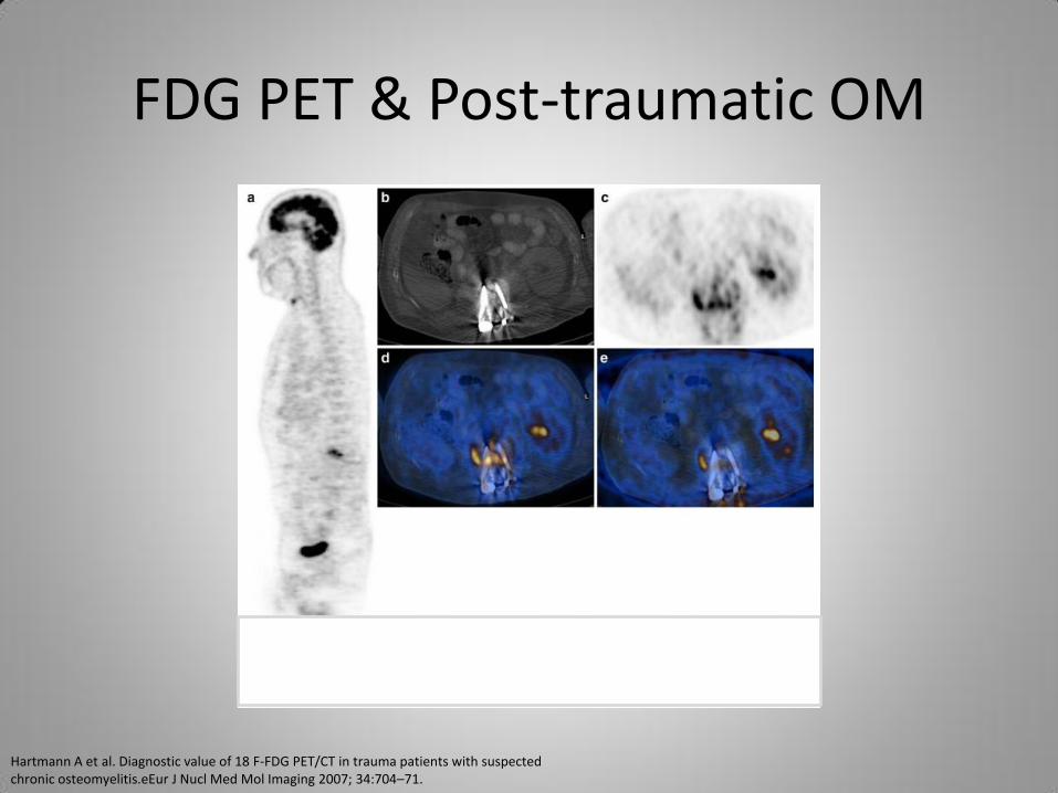
FDG PET & Post-traumatic OM
• Hartmann’s team in Switzerland reviewed 33 partial body FDG PET/CT in post-trauma patients suspected to have COM
• 18 had metallic implants
• Histopathology or culture was standard reference
• Radiolabeled WBC/Marrow scans study of choice for post-traumatic infection imaging – MRI & CT susceptible to
safety issue & beam-hardening artifact
– Bone scan susceptible to post-traumatic bone remodeling
Hartmann A et al. Diagnostic value of 18 F-FDG PET/CT in trauma patients with suspected chronic osteomyelitis.eEur J Nucl Med Mol Imaging 2007; 34:704–71.
FDG PET & Post-traumatic OM
Hartmann A et al. Diagnostic value of 18 F-FDG PET/CT in trauma patients with suspected chronic osteomyelitis.eEur J Nucl Med Mol Imaging 2007; 34:704–71.
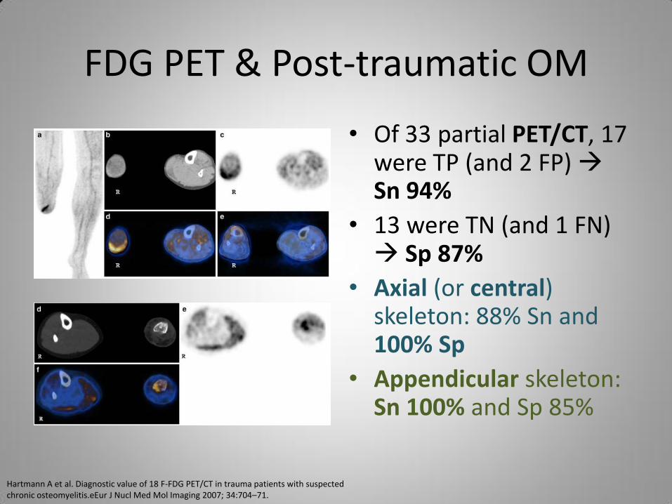
FDG PET & Post-traumatic OM
• Of 33 partial PET/CT, 17 were TP (and 2 FP) Sn 94%
• 13 were TN (and 1 FN) Sp 87%
• Axial (or central) skeleton: 88% Sn and 100% Sp
• Appendicular skeleton: Sn 100% and Sp 85%
Hartmann A et al. Diagnostic value of 18 F-FDG PET/CT in trauma patients with suspected chronic osteomyelitis.eEur J Nucl Med Mol Imaging 2007; 34:704–71.
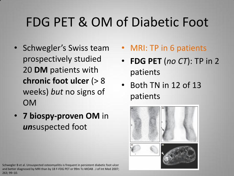
FDG PET & OM of Diabetic Foot
• Schwegler’s Swiss team prospectively studied 20 DM patients with chronic foot ulcer (> 8 weeks) but no signs of OM
• 7 biospy-proven OM in unsuspected foot
• MRI: TP in 6 patients
• FDG PET (no CT): TP in 2 patients
• Both TN in 12 of 13 patients
Schwegler B et al. Unsuspected osteomyelitis is frequent in persistent diabetic foot ulcer and better diagnosed by MRI than by 18 F-FDG PET or 99m Tc-MOAB . J of Int Med 2007; 263; 99–10.
FDG PET & OM of Diabetic Foot
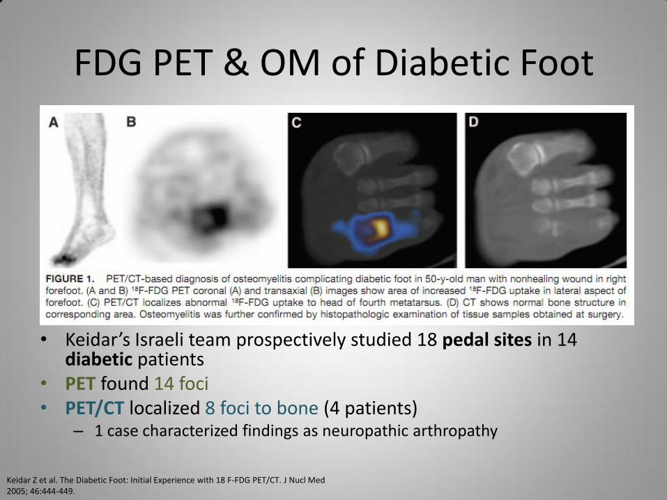
Keidar Z et al. The Diabetic Foot: Initial Experience with 18 F-FDG PET/CT. J Nucl Med 2005; 46:444-449.
• Keidar’s Israeli team prospectively studied 18 pedal sites in 14 diabetic patients
• PET found 14 foci • PET/CT localized 8 foci to bone (4 patients)
– 1 case characterized findings as neuropathic arthropathy
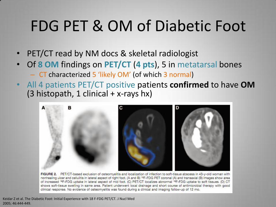
FDG PET & OM of Diabetic Foot
• PET/CT read by NM docs & skeletal radiologist • Of 8 OM findings on PET/CT (4 pts), 5 in metatarsal bones
– CT characterized 5 ‘likely OM’ (of which 3 normal)
• All 4 patients PET/CT positive patients confirmed to have OM (3 histopath, 1 clinical + x-rays hx)
Keidar Z et al. The Diabetic Foot: Initial Experience with 18 F-FDG PET/CT. J Nucl Med 2005; 46:444-449.
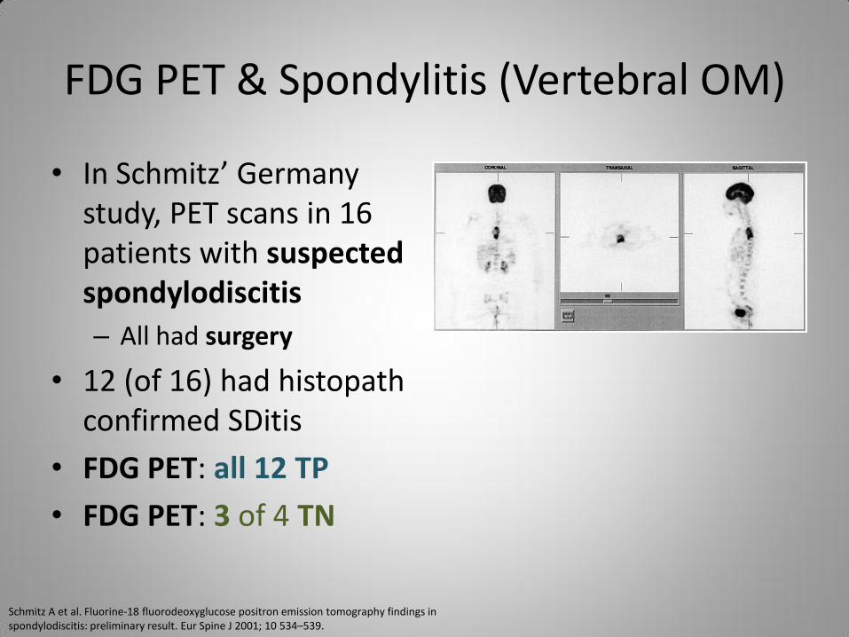
FDG PET & Spondylitis (Vertebral OM)
• In Schmitz’ Germany study, PET scans in 16 patients with suspected spondylodiscitis
– All had surgery
• 12 (of 16) had histopath confirmed SDitis
• FDG PET: all 12 TP
• FDG PET: 3 of 4 TN
Schmitz A et al. Fluorine-18 fluorodeoxyglucose positron emission tomography findings in spondylodiscitis: preliminary result. Eur Spine J 2001; 10 534–539.
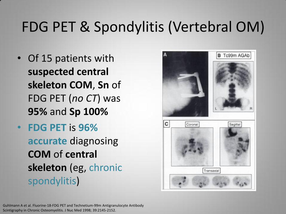
FDG PET & Spondylitis (Vertebral OM)
• Of 15 patients with suspected central skeleton COM, Sn of FDG PET (no CT) was 95% and Sp 100%
• FDG PET is 96% accurate diagnosing COM of central skeleton (eg, chronic spondylitis)
Guhlmann A et al. Fluorine-18-FDG PET and Technetium-99m Antigranulocyte Antibody Scintigraphy in Chronic Osteomyelitis. J Nuc Med 1998; 39:2145-2152.
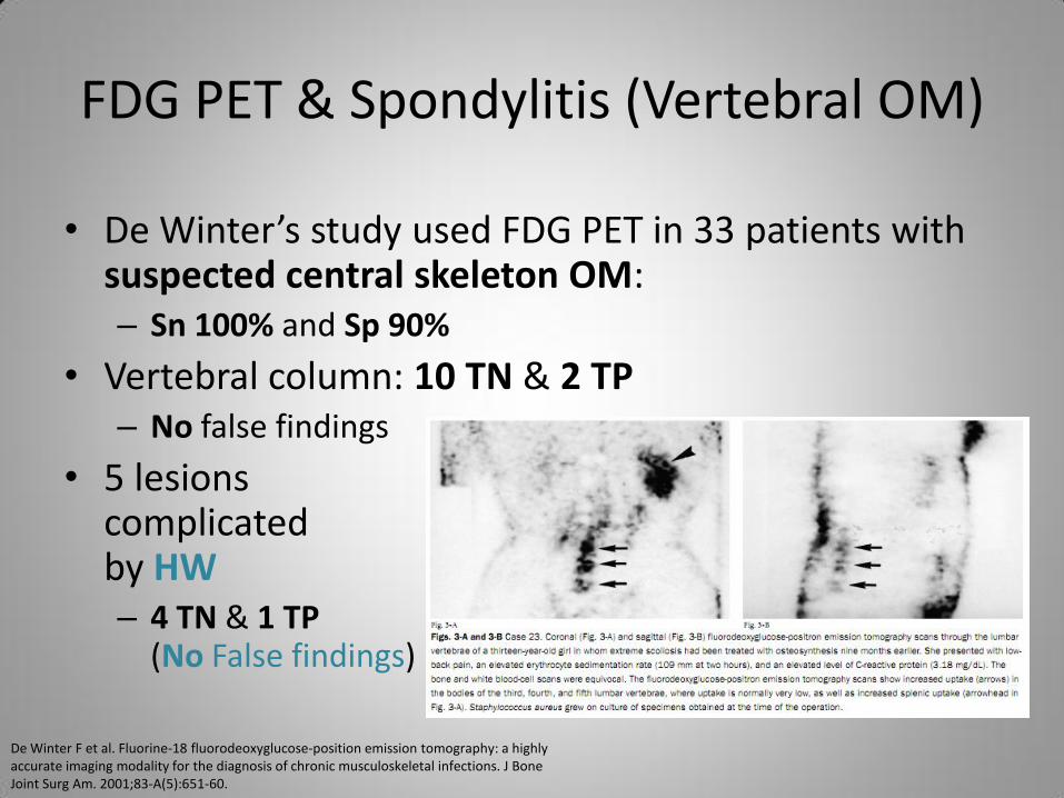
FDG PET & Spondylitis (Vertebral OM)
• De Winter’s study used FDG PET in 33 patients with suspected central skeleton OM: – Sn 100% and Sp 90%
• Vertebral column: 10 TN & 2 TP – No false findings
• 5 lesions complicated by HW – 4 TN & 1 TP
(No False findings)
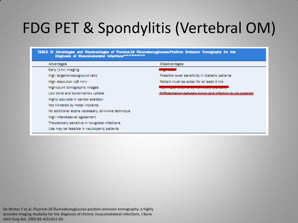
De Winter F et al. Fluorine-18 fluorodeoxyglucose-position emission tomography: a highly accurate imaging modality for the diagnosis of chronic musculoskeletal infections. J Bone Joint Surg Am. 2001;83-A(5):651-60.
FDG PET & Spondylitis (Vertebral OM)
De Winter F et al. Fluorine-18 fluorodeoxyglucose-position emission tomography: a highly accurate imaging modality for the diagnosis of chronic musculoskeletal infections. J Bone Joint Surg Am. 2001;83-A(5):651-60.
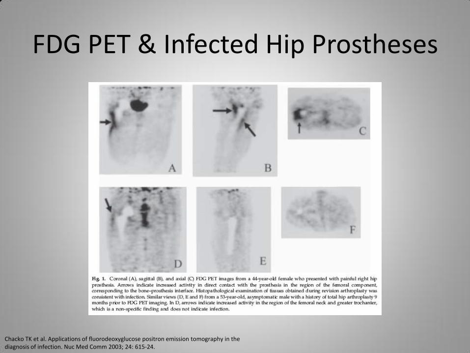
FDG PET & Infected Hip Prostheses
• In U. Penn study of 53 hip prosthesis, FDG PET had 11 TP of 12 pathology-proven infections (Sn 92%)
• Of 41 non-infected hip cases, PET was TN in 40 cases (Sp 98%)
• Criteria to call FDG-avid finding an infection: – Location, not intensity,
determining factor
– FDG uptake at Bone-Prosthesis Interface considered positive
– Other publications showed BPI FDG uptake increases Sp
– FDG uptake in soft tissue was non-specific
Chacko TK et al. Applications of fluorodeoxyglucose positron emission tomography in the diagnosis of infection. Nuc Med Comm 2003; 24: 615-24.
FDG PET & Infected Hip Prostheses
Chacko TK et al. Applications of fluorodeoxyglucose positron emission tomography in the diagnosis of infection. Nuc Med Comm 2003; 24: 615-24.
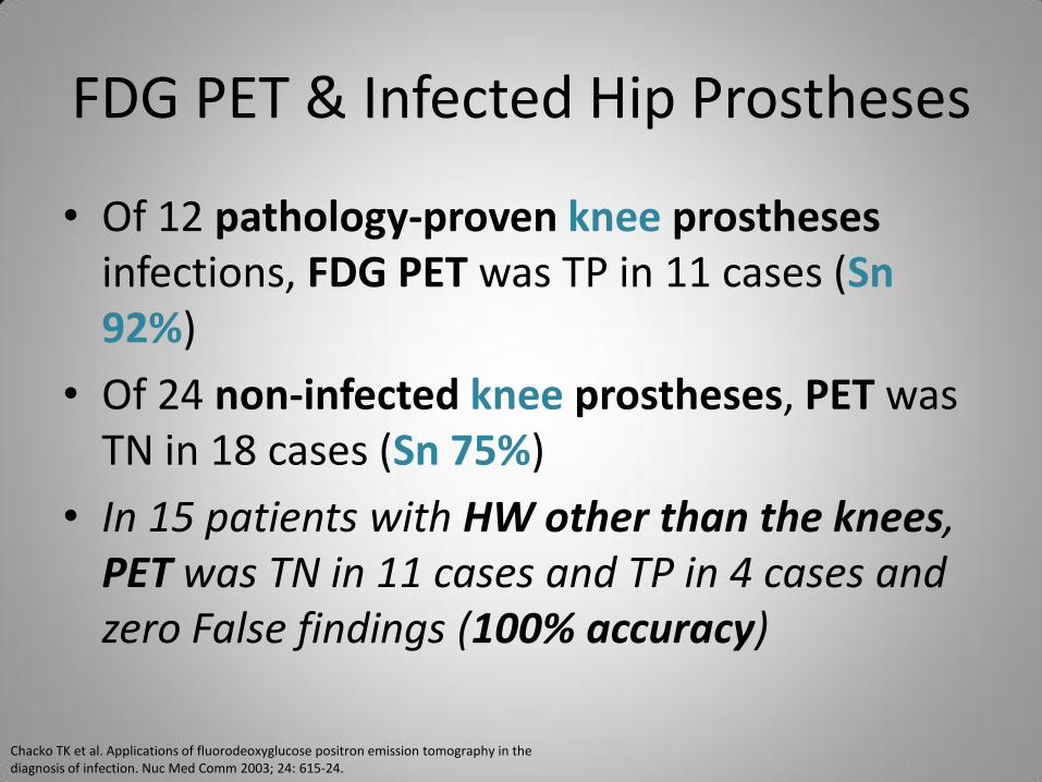
FDG PET & Infected Hip Prostheses
• Of 12 pathology-proven knee prostheses infections, FDG PET was TP in 11 cases (Sn 92%)
• Of 24 non-infected knee prostheses, PET was TN in 18 cases (Sn 75%)
• In 15 patients with HW other than the knees, PET was TN in 11 cases and TP in 4 cases and zero False findings (100% accuracy)
Chacko TK et al. Applications of fluorodeoxyglucose positron emission tomography in the diagnosis of infection. Nuc Med Comm 2003; 24: 615-24.
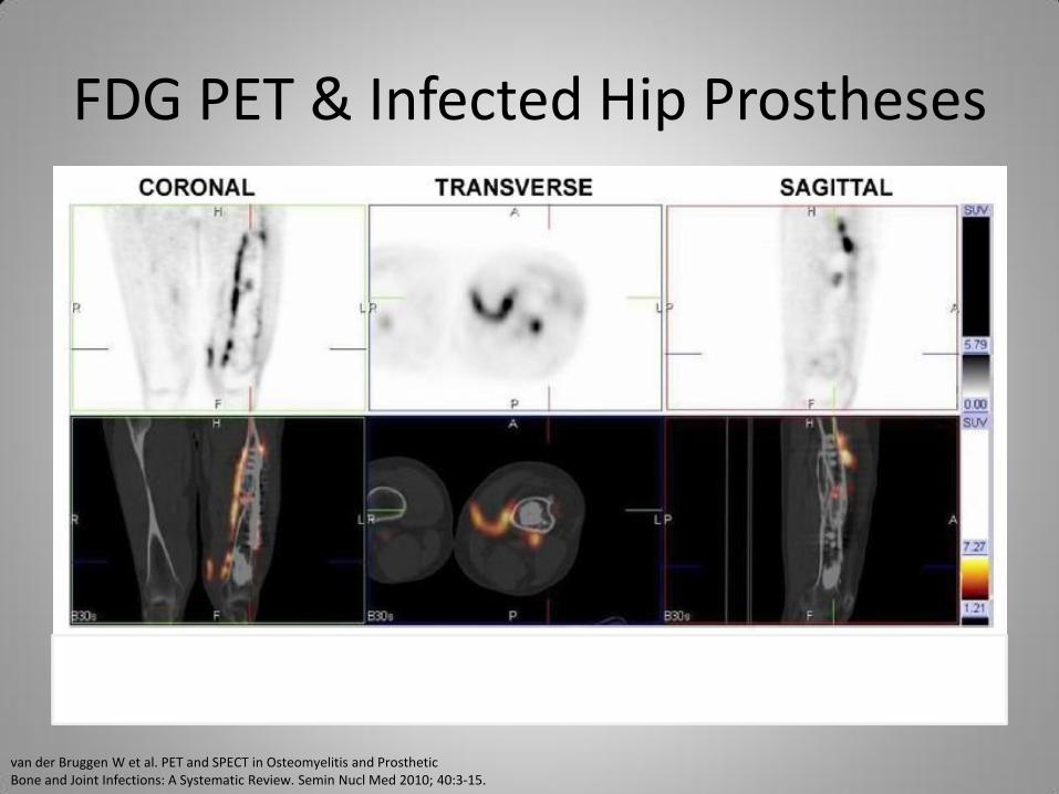
FDG PET & Infected Hip Prostheses
van der Bruggen W et al. PET and SPECT in Osteomyelitis and Prosthetic Bone and Joint Infections: A Systematic Review. Semin Nucl Med 2010; 40:3-15.
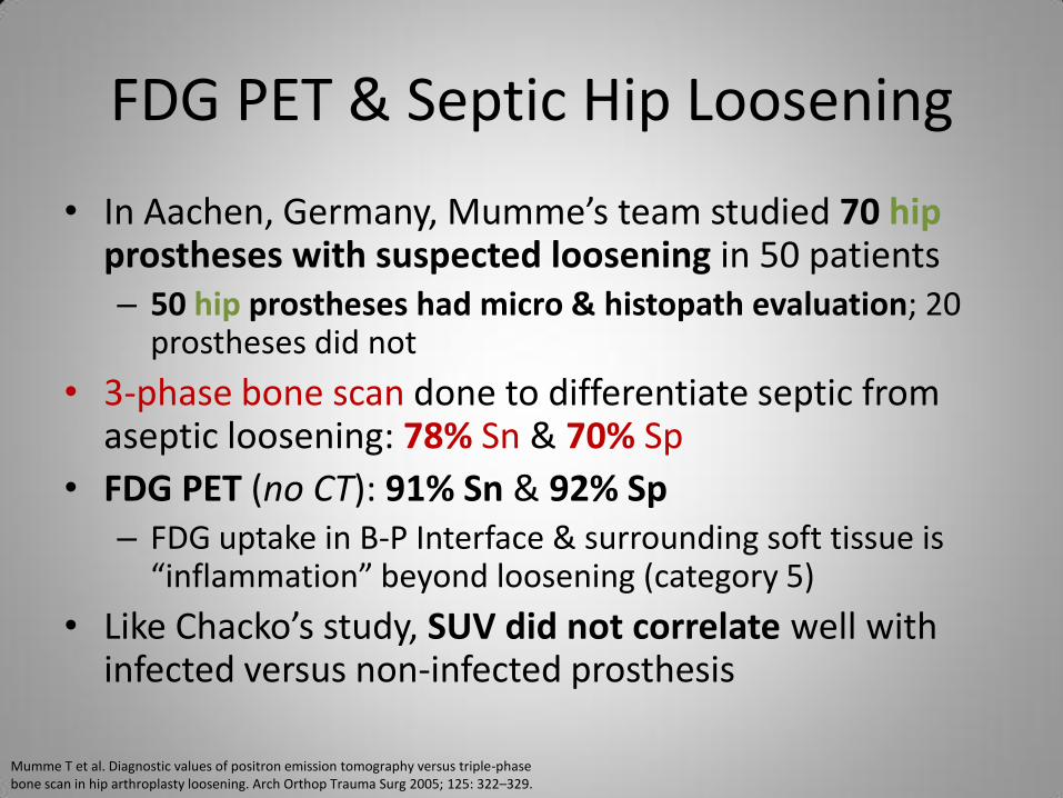
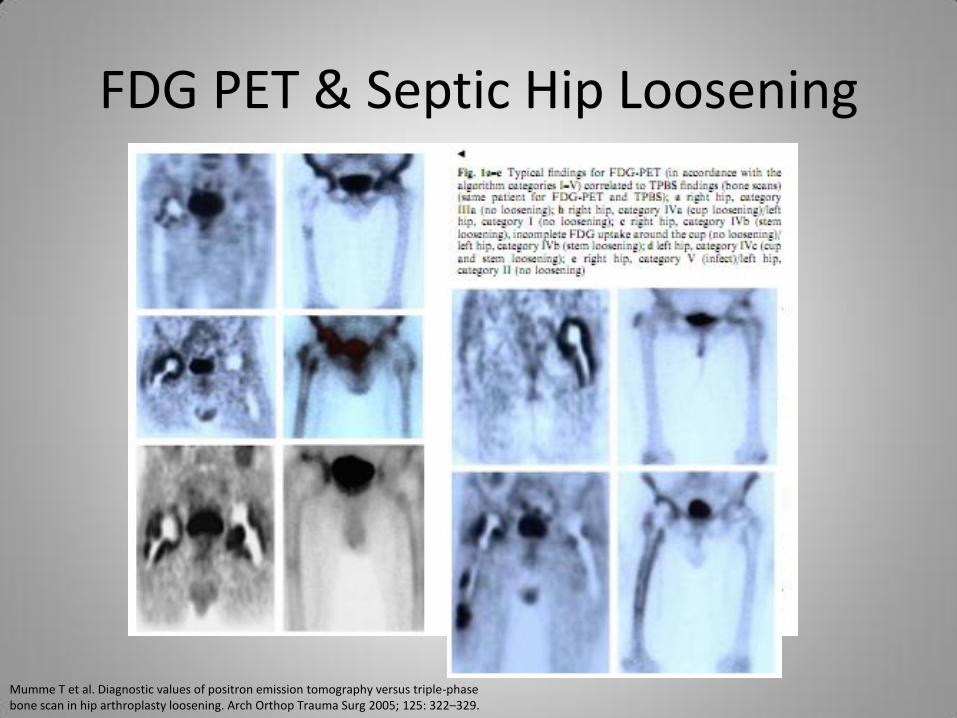
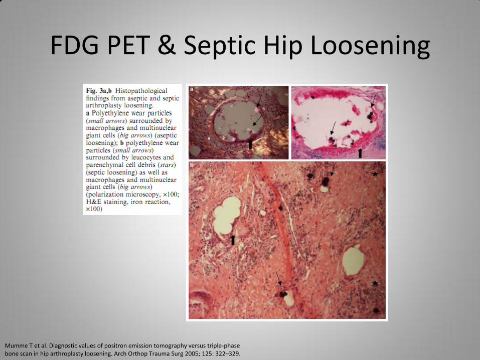
FDG PET & Septic Hip Loosening
• In Aachen, Germany, Mumme’s team studied 70 hip prostheses with suspected loosening in 50 patients – 50 hip prostheses had micro & histopath evaluation; 20
prostheses did not
• 3-phase bone scan done to differentiate septic from aseptic loosening: 78% Sn & 70% Sp
• FDG PET (no CT): 91% Sn & 92% Sp – FDG uptake in B-P Interface & surrounding soft tissue is
“inflammation” beyond loosening (category 5)
• Like Chacko’s study, SUV did not correlate well with infected versus non-infected prosthesis
Mumme T et al. Diagnostic values of positron emission tomography versus triple-phase bone scan in hip arthroplasty loosening. Arch Orthop Trauma Surg 2005; 125: 322–329.
FDG PET & Septic Hip Loosening
Mumme T et al. Diagnostic values of positron emission tomography versus triple-phase bone scan in hip arthroplasty loosening. Arch Orthop Trauma Surg 2005; 125: 322–329.
FDG PET & Septic Hip Loosening
Mumme T et al. Diagnostic values of positron emission tomography versus triple-phase bone scan in hip arthroplasty loosening. Arch Orthop Trauma Surg 2005; 125: 322–329.
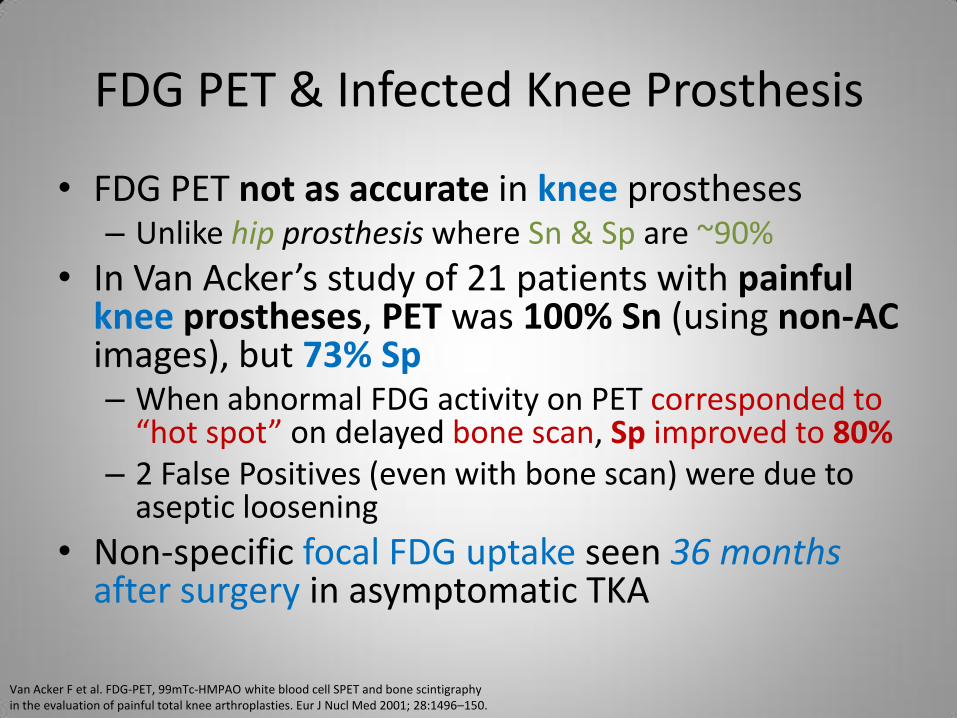
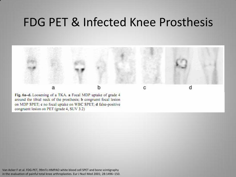
FDG PET & Infected Knee Prosthesis
• FDG PET not as accurate in knee prostheses – Unlike hip prosthesis where Sn & Sp are ~90%
• In Van Acker’s study of 21 patients with painful knee prostheses, PET was 100% Sn (using non-AC images), but 73% Sp – When abnormal FDG activity on PET corresponded to
“hot spot” on delayed bone scan, Sp improved to 80% – 2 False Positives (even with bone scan) were due to
aseptic loosening
• Non-specific focal FDG uptake seen 36 months after surgery in asymptomatic TKA
Van Acker F et al. FDG-PET, 99mTc-HMPAO white blood cell SPET and bone scintigraphy in the evaluation of painful total knee arthroplasties. Eur J Nucl Med 2001; 28:1496–150.
FDG PET & Infected Knee Prosthesis
Van Acker F et al. FDG-PET, 99mTc-HMPAO white blood cell SPET and bone scintigraphy in the evaluation of painful total knee arthroplasties. Eur J Nucl Med 2001; 28:1496–150.
FDG PET & Infected Knee Prosthesis
• Zhuang U. Penn team published that FDG PET better at diagnosing hip versus knee prosthesis infections – Knee: 10 TP of 11 knee prosthesis infections, but 7 FP
among 25 non-infected knees (Sn 91% & Sp 72%) – Hip: 9 TP of 10 hip prosthesis infections, and 3 FP
among 28 non-infected hips (Sn 90% & Sp 89%)
• In all 10 False Positive cases, surgery > 1 year before PET scan performed
• Study confirmed presence, not intensity, of FDG uptake at BPI is what best correlates to infection
Zhuang H et al. The Promising Role of 18 F-FDG PET in Detecting Infected Lower Limb Prosthesis Implants. J Nucl Med 2001; 42:44 – 4.
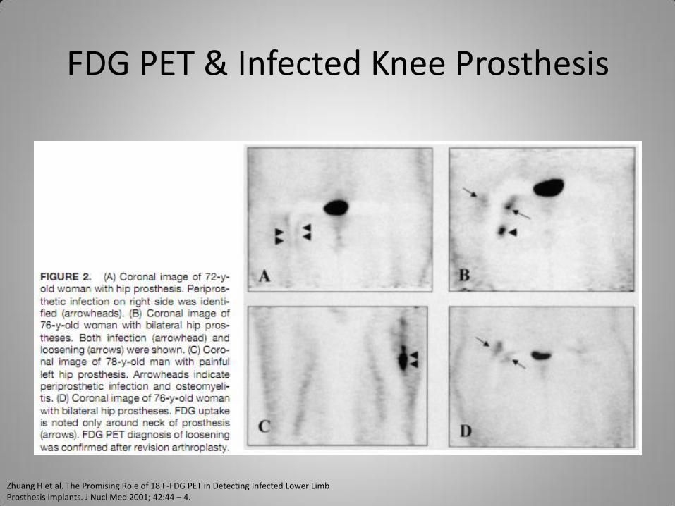
FDG PET & Infected Knee Prosthesis
Zhuang H et al. The Promising Role of 18 F-FDG PET in Detecting Infected Lower Limb Prosthesis Implants. J Nucl Med 2001; 42:44 – 4.
Case 2
• Man in his 60s complains of severe pain in L ankle
– Recent of minor blunt trauma
– Remote history of L ankle fracture s/p ORIF (“years ago,” at construction site)
• Seen at VA in late 2011 for ankle pain
– WBC (especially neutrophils) & ESR high
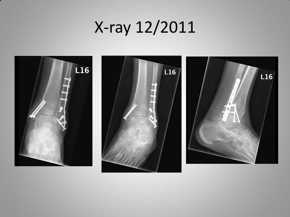
X-ray 12/2011
Case 2
• Orthopedic team diagnosed patient with infected HW in L ankle
– Plan to debride tissue and do HW Replacement
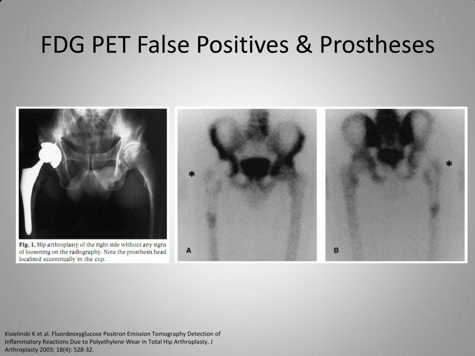
FDG PET False Positives & Prostheses
Kisielinski K et al. Fluordeoxyglucose Positron Emission Tomography Detection of Inflammatory Reactions Due to Polyethylene Wear in Total Hip Arthroplasty. J Arthroplasty 2003; 18(4): 528-32.
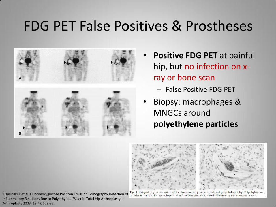
FDG PET False Positives & Prostheses
• Positive FDG PET at painful hip, but no infection on x-ray or bone scan – False Positive FDG PET
• Biopsy: macrophages & MNGCs around polyethylene particles
Kisielinski K et al. Fluordeoxyglucose Positron Emission Tomography Detection of Inflammatory Reactions Due to Polyethylene Wear in Total Hip Arthroplasty. J Arthroplasty 2003; 18(4): 528-32.
FDG PET False Positives & Other Cases
• Uncomplicated traumatic bone injury have abnormal FDG uptake up to 3 months post-trauma – Fracture healing involves bone remodeling
• Post-surgical bone could have abnormal FDG uptake (but low) greater than 1 year
• Higher FDG activity (SUVmax > 3) raised likelihood of infection – In 21 patients suspected with OM, PET “showed high uptake of
FDG within the infected tissue, with SUVs up to a maximum of 16.1. However, in fractures and pseudarthroses only very low FDG uptake, with SUVs of 0.2–1.1, was observed”
• CT (as in PET/CT) improves Sp (ie, reduces False Positives)
Kälicke T et al. Fluorine-18 fluorodeoxyglucose PET in infectious bone diseases: results of histologically confirmed cases. Eur J Nucl Med. 2000 May;27(5):524-8.
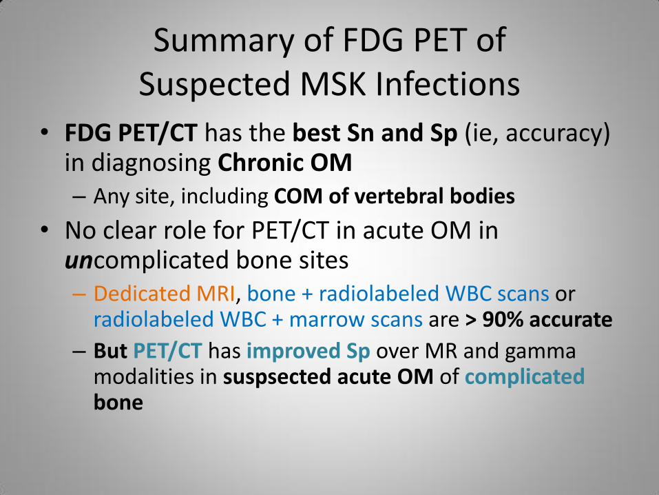
Summary of FDG PET of Suspected MSK Infections
• FDG PET/CT has the best Sn and Sp (ie, accuracy) in diagnosing Chronic OM – Any site, including COM of vertebral bodies
• No clear role for PET/CT in acute OM in uncomplicated bone sites – Dedicated MRI, bone + radiolabeled WBC scans or
radiolabeled WBC + marrow scans are > 90% accurate
– But PET/CT has improved Sp over MR and gamma modalities in suspsected acute OM of complicated bone
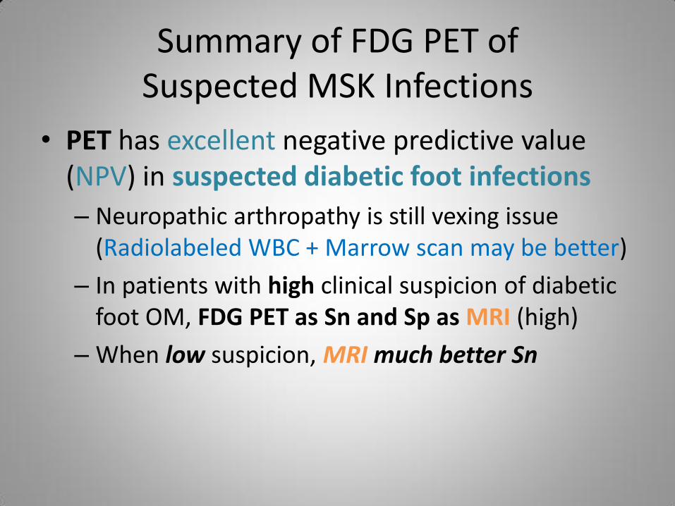
Summary of FDG PET of Suspected MSK Infections
• PET has excellent negative predictive value (NPV) in suspected diabetic foot infections
– Neuropathic arthropathy is still vexing issue (Radiolabeled WBC + Marrow scan may be better)
– In patients with high clinical suspicion of diabetic foot OM, FDG PET as Sn and Sp as MRI (high)
– When low suspicion, MRI much better Sn
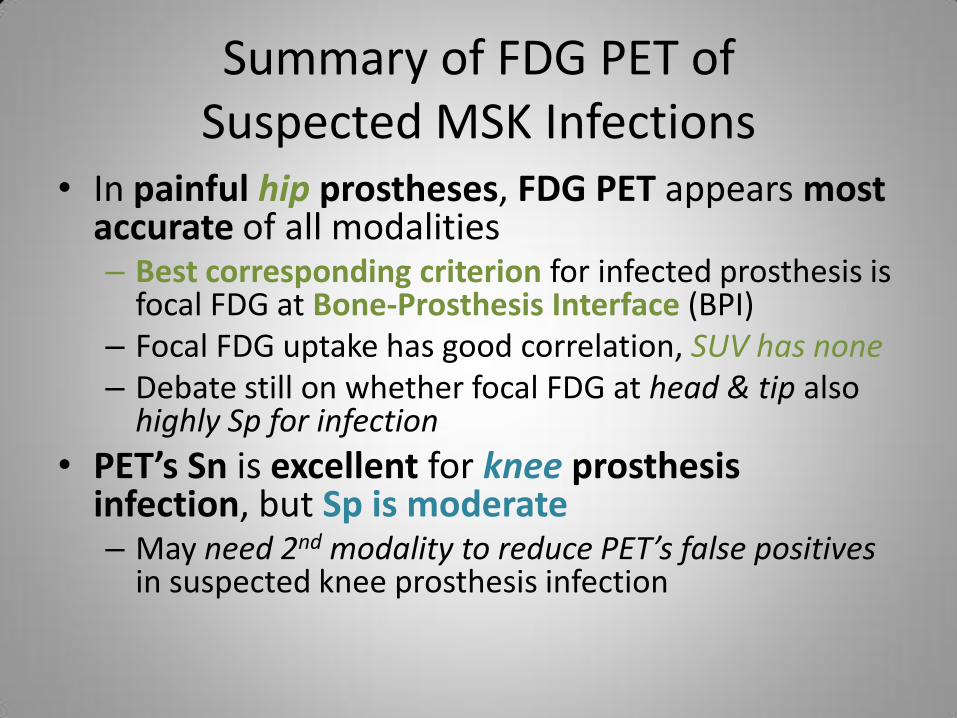
Summary of FDG PET of Suspected MSK Infections
• In painful hip prostheses, FDG PET appears most accurate of all modalities – Best corresponding criterion for infected prosthesis is
focal FDG at Bone-Prosthesis Interface (BPI) – Focal FDG uptake has good correlation, SUV has none – Debate still on whether focal FDG at head & tip also
highly Sp for infection
• PET’s Sn is excellent for knee prosthesis infection, but Sp is moderate – May need 2nd modality to reduce PET’s false positives
in suspected knee prosthesis infection
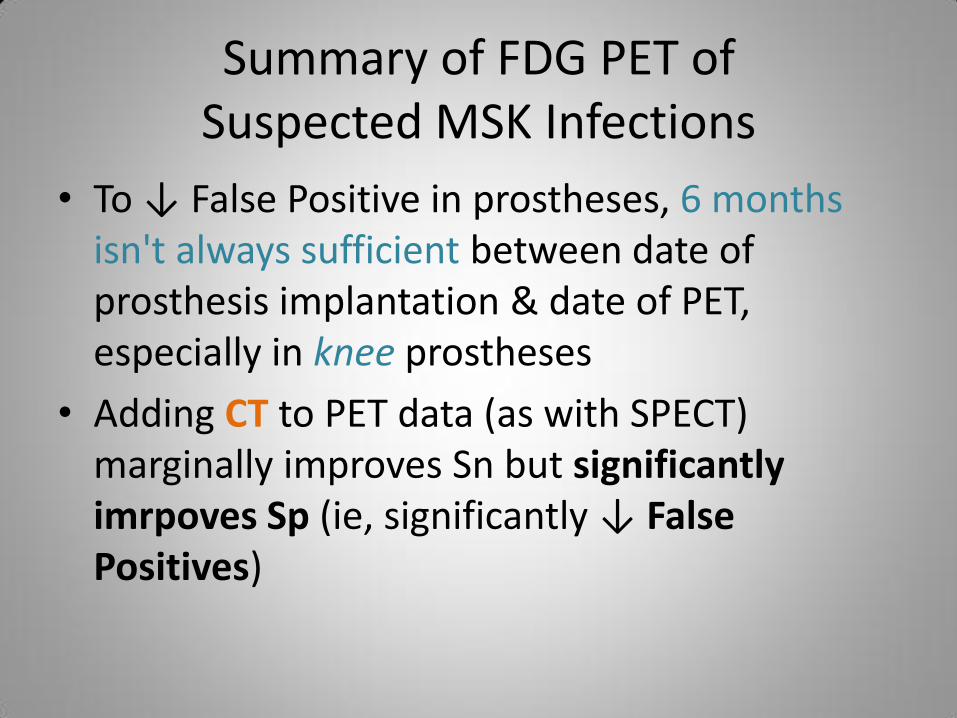
Summary of FDG PET of Suspected MSK Infections
• To ↓ False Positive in prostheses, 6 months isn't always sufficient between date of prosthesis implantation & date of PET, especially in knee prostheses
• Adding CT to PET data (as with SPECT) marginally improves Sn but significantly imrpoves Sp (ie, significantly ↓ False Positives)
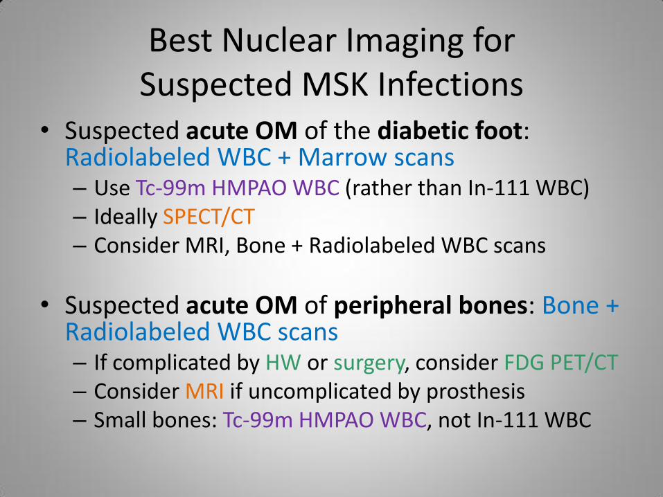
Best Nuclear Imaging for Suspected MSK Infections
• Suspected acute OM of the diabetic foot: Radiolabeled WBC + Marrow scans – Use Tc-99m HMPAO WBC (rather than In-111 WBC) – Ideally SPECT/CT – Consider MRI, Bone + Radiolabeled WBC scans
• Suspected acute OM of peripheral bones: Bone + Radiolabeled WBC scans – If complicated by HW or surgery, consider FDG PET/CT – Consider MRI if uncomplicated by prosthesis – Small bones: Tc-99m HMPAO WBC, not In-111 WBC
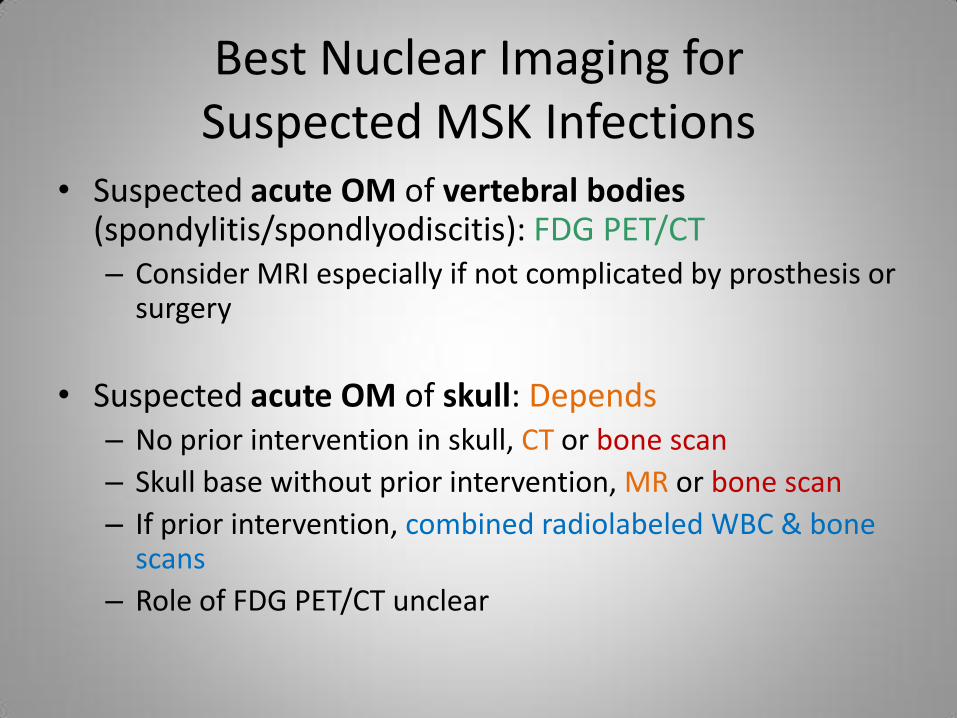
Best Nuclear Imaging for Suspected MSK Infections
• Suspected acute OM of vertebral bodies (spondylitis/spondlyodiscitis): FDG PET/CT – Consider MRI especially if not complicated by prosthesis or
surgery
• Suspected acute OM of skull: Depends – No prior intervention in skull, CT or bone scan
– Skull base without prior intervention, MR or bone scan
– If prior intervention, combined radiolabeled WBC & bone scans
– Role of FDG PET/CT unclear
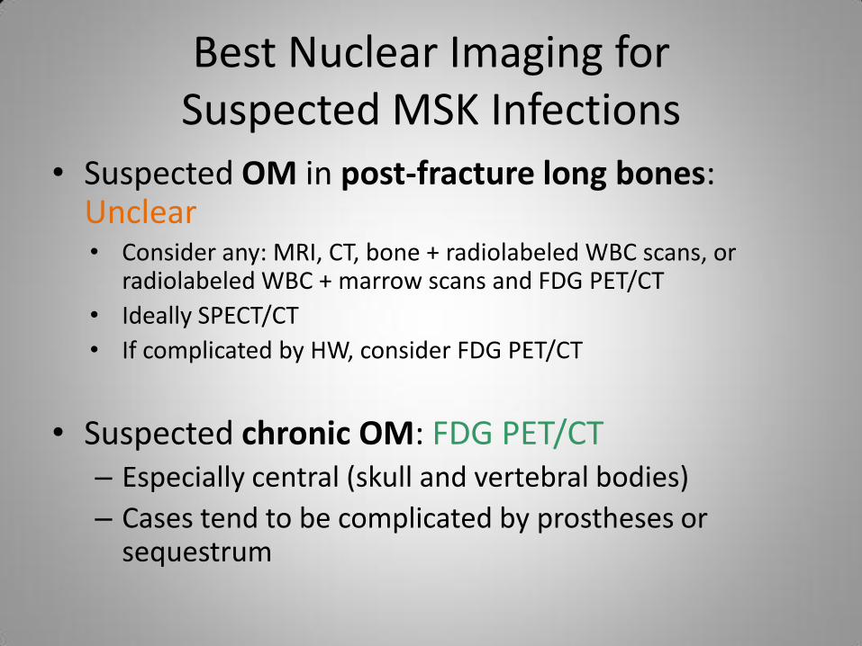
Best Nuclear Imaging for Suspected MSK Infections
• Suspected OM in post-fracture long bones: Unclear • Consider any: MRI, CT, bone + radiolabeled WBC scans, or
radiolabeled WBC + marrow scans and FDG PET/CT
• Ideally SPECT/CT
• If complicated by HW, consider FDG PET/CT
• Suspected chronic OM: FDG PET/CT – Especially central (skull and vertebral bodies)
– Cases tend to be complicated by prostheses or sequestrum
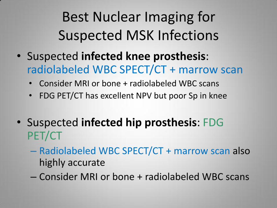
Best Nuclear Imaging for Suspected MSK Infections
• Suspected infected knee prosthesis: radiolabeled WBC SPECT/CT + marrow scan • Consider MRI or bone + radiolabeled WBC scans
• FDG PET/CT has excellent NPV but poor Sp in knee
• Suspected infected hip prosthesis: FDG PET/CT – Radiolabeled WBC SPECT/CT + marrow scan also
highly accurate
– Consider MRI or bone + radiolabeled WBC scans
Extended Bibliography & Thanks
• For summary of studies: van der Bruggen W et al. PET and SPECT in Osteomyelitis and Prosthetic Bone and Joint Infections: A Systematic Review. Sem. Nuc. Med. 2010; 40:3-15.
• For effective radiation doses: Mettler FA et al. Effective Doses in Radiology and Diagnostic Nuclear Medicine: A Catalog. Radiology 2008; 248(1): 254-63.
• For protocols: Society of Nuclear Medicine Procedure Guidelines Catalog, and Mettler FA and Guiberteau M. Essentials of Nuclear Medicine. Philadelphia: Saunders Elsevier, 2006.
• Special thanks: Dr. L. A. Tamara and Dr. G. S. Bhartur at M.E. DeBakey VAMC in Houston, Texas for use of FDG PET/CT exams during presentation. Also Dr. J. Wendt, Dr. W. Moore, Dr. P. Ford, and R. Srinivasan for general support.

The End