contraction and action potentials of frog heart muscles
TRANSCRIPT

Contraction and Action
Potentials of Frog Heart Muscles
Soaked in Sucrose Solution
W I L L I A M G. VAN D E R K L O O T and N I R A S. R U B I N
From the Department of Physiology and Biophysics, New York University School of Medicine, New York
ABSTRACT Isolated auricles or ventricles from the frog continue to contract, either spontaneously or when stimulated, for from 2 to 4 hours after they are placed in isotonic sucrose solution. After the muscles stop contracting in sucrose solution, contractility is partially restored when the muscles are placed in chloride Ringer's. However, contractility is usually not restored if the muscles are placed in sulfate Ringer's. Ventricles soaked in sucrose solution at 4-7°C continue to contract for 12 to 24 hours and during the first few hours in sucrose solution the contractions often are enhanced. Several types of experiment indicate that the sucrose solution does replace the Ringer's in the extracellular space. Auricles and ventricles also continue to conduct action potentials, with an overshoot, for from 30 to 360 minutes after being placed in sucrose solution. Muscles soaked in sucrose until they are inexcitable rapidly recover in chloride Ringer 's but often fail to recover in sulfate Ringer's. The results are discussed in relation to theories about the generation of the action potential in cardiac muscle, and the role of the extracellular fluid in contraction.
INTRODUCTION
Ringer's 0880) classic study of the ionic requirements for contraction by the frog heart is one of the cornerstones of physiology. Much of the subsequent work on the heart was designed to account for the special roles of the individual ions in excitation, conduction, and contraction. With this extensive back- ground on the necessity for balanced solutions of extracellular ions, it was a surprise when Singh, Sehra, and Singh (1945) reported that frog hearts con- tinued to contract spontaneously for about 2 hours in an isotonic sucrose solution and to remain electrically excitable for another 2 hours.
Singh (I 944) previously reported that smooth muscle from the frog stomach continues to contract for hours in isotonic or half-isotonic sucrose solutions. This report was confirmed (Kolodny and Van der Kloot, 1961). Smooth
35
The Journal of General Physiology
brought to you by COREView metadata, citation and similar papers at core.ac.uk
provided by PubMed Central

3 6 T H E J O U R N A L O F G E N E R A L P H Y S I O L O G Y • V O L U M E 46 . I962
muscles in sucrose solutions also con t inue to conduc t act ion potent ia ls and to genera te spike potent ia ls as j u d g e d f rom ext ra- and in t race l lu lar recordings. However , smooth muscles are difficult to s tudy wi th in t race l lu lar e lectrodes and, therefore, we t u rned to the frog hea r t as a more conven ien t p r e p a r a t i o n for invest igat ing the physiology of exci table tissues immersed in sucrose solutions. T h e results r epor ted here show tha t when immersed in sucrose solution the frog hea r t cont inues electrical and mechan ica l act ivi ty for a t least several hours. This p a p e r is conce rned wi th the changes in cont rac t i l i ty and in the act ion potent ia l occur r ing while the hear t is in sucrose solution and with the recovery of act ivi ty when the hea r t is rep laced in Ringer ' s solution.
M E T H O D S
Animals
The experiments were performed on isolated auricles or ventricles from the heart of Rana pipiens. The frogs Were obtained from a dealer and kept in running tap water at room temperature until use.
Solutions
The following solutions were used. Concentrations are given in mM. Chloride Ringer's: 121.0 Na +, 3.0 K +, 2.7 Ca ++, 4.0 HCO3-, and 125.4 C1-. Sulfate Ringer's: 121.0 Na +, 3.0 K +, 2.7 Ca ++, 64.7 So4", 64.7 sucrose. Isethionate Ringer's: 121.0 Na +, 3.0 K +, 2.7 Ca++, 4.2 SO4-, 121.0
CH~OHCH2SOs-. 18 mM Na + Ringer's: 18.0 Na +, 3.0 K +, 2.7 Ca ++, 26.4 Cl-, 206.0 sucrose. Sucrose solution: 240.0 sucrose. The conductivity of the sucrose solution was about
8.5 ~mho cm -I. All the solutions were prepared from reagent grade chemicals dissolved in dis-
tilled and subsequently deionized water (Illinois Water Treatment Company ex- change column).
Tension Recording
An auricle or a ventricle was tied in a bath, with a thread from the upper end of the muscle running to a force displacement transducer. The isometric contractions were recorded with a Grass inkwriter. A fine stream of 02 was constantly bubbled through the solution in the bath. Platinum stimulating electrodes were fixed just below and just above the muscle. Stimulating voltages varied widely depending on the conduc- tivity of the medium and the position of the electrodes in relation to the tissue. The muscle was stimulated with square waves 20 msec. long. Most of the experiments were carried out at room temperatures, between 20 and 30°C. Some experiments were made on muscles kept in the cold room at 4 to 7°C. All the results reported were re- produced in at least three experiments.

W. G. VAN D~.R KLOOT ANn N. S. Rtrsm Heart Muscle in Sucrose Solution 37
Electrical Recording
Intracellular recordings were made by using glass capillary micropipettes filled with 3 M KC1 with resistances varying from 10 to 80 megohms. The microelectrodes were flexibly mounted at the end of a thin platinum wire (Woodbury and Brady, 1956). A capacity compensated preamplifier was used. The muscles were stimulated by either of two systems. In the first method, two platinum electrodes, insulated to the tip with polyethylene, were placed about 1 mm apart on the surface of the tissue. In the second method, a polyethylene-jacketed cathode was inserted into the auricular or ventricular cavity, while a large platinum loop in the bath served as the anode. Stimuli were square waves usually of 3 re_see, duration; voltages were adjusted to be supra- maximal. The muscle bath contained 10 ml of solution which was stirred with a stream of 02 and was changed at least once every 10 minutes during an experiment. The measurements were made at room temperatures or in solutions cooled in an ice bath to about 7 to 10°C.
Workers on the frog heart often have measured low resting and action potentials, almost certainly because the thin, moving fibers are damaged during penetration. Therefore, mean values are seldom a true measure of the potentials generated by the tissue, and the highest values in any series of measurements are likely to be true. Nevertheless, some average values are presented to indicate the trends in the data.
R E S U L T S
Contractions in Ringer's and in Sucrose Solutions
T h e tension p roduced by e i ther an isolated auricle or ventr ic le soaking in Ringer ' s solution at r o o m t e m p e r a t u r e was first measured, while the muscle was s t imulated wi th single sup ramax ima l electr ical shocks. T h e Ringer ' s solution was then replaced by a sucrose solution and the tension exer ted by the muscle in response to single stimuli was r eco rded at 30 or at 60 minu te in ter- vals. Between st imulations the sucrose solution in the ba th was changed several times. T h e results are summar ized as T a b l e I. T h e r e is a progressive decl ine in the cont rac t ions of the cardiac muscle in sucrose solution. Nevertheless, the decl ine in the cont rac t ions is r a the r slow; after an hou r in sucrose solution the response is no t significantly di f ferent f rom tha t in Ringer ' s solution. T h e r e was a wide var ia t ion in the responses f rom one piece of hea r t to another . O n e im- po r t an t reason for the var iabi l i ty is u n d o u b t e d l y the occur rence of spon- taneous contract ions, which were c o m m o n in bo th auricles and ventricles while soaking in sucrose solution (Fig. 1 B). W h e n the muscle cont rac ts a n u m b e r of t imes in close succession, e i ther owing to spontaneous act ivi ty or to ex terna l s t imulat ion, the ampl i tude of the contract ions decreases, which will be discussed later. T h e rates of tension deve lopmen t and of re laxa t ion also change when the muscles are in sucrose solution; these changes will be dis-

38 THE JOURNAL OF GENERAL PHYSIOLOGY • VOLUME 46 • i 962
cussed in the section on contraction at low temperatures. While soaking in sucrose there often was an increase in the tension exerted by the unstimulated heart; after an hour or two in sucrose the muscles frequently maintained a steady contracture amounting to 30 to 40 per cent of the initial twitch tension. When the musdles were then replaced in Ringer's solution, the contracture dis- appeared within 3 minutes. The contracture of sucrose-soaked cardiac muscle is quite similar to the contracture of skeletal muscles in isotonic sucrose solu- tions (Fenn, 1931; Swift, Gordon, and Van der Kloot, 1960).
The ability of sucrose-soaked cardiac muscle to contract might simply mean that it takes a long time to wash the Ringer's from the extracellular space, so that some of the Ringer's remains as the extracellular fluid, perhaps only as a thin layer surrounding the muscle fibers. One way of checking on the ex-
T A B L E I
C O N T R A C T I O N O F C A R D I A C M U S C L E S
S O A K I N G I N S U C R O S E S O L U T I O N
A s u m m a r y of resu l t s f rom t h i r t y - s i x musc les
Time in sucro~ Auricle Ventricle
rain. Per cent of initial controxtion -4- BE Per tent of initial contraction 4- 8E
0 100 100
30 9O.8 4- 8 .8 - -
60 92.6 4- 10.2 90.5 4- 10.7 90 71.6 4- 13.4 - -
120 47.8 4- 11.7 87.3 4- 8 .7 150 28.7 4- 11.1 - -
180 16.4 4- 7 .8 - -
240 6.3 4- 3 .3 0
change between the fluid surrounding the muscle fibers and the solution in the bath, was to place muscles in isotonic NaC1. The contractions became weaker almost immediately, and within 7 to 10 minutes the contractions fell to a small fraction of the initial value. The isotonic sodium chloride solution was then replaced by sucrose solution. The result, shown as Fig. 1 A, was a prompt increase in the contractions. Similarly, hearts placed in isotonic K~SO, solu- tions promptly went into a contracture, which was rapidly reversed when the K2SO4 was replaced with sucrose solution. These experiments suggest that the extracellular fluid exchanges quite rapidly with the sucrose solution in the bath.
As was mentioned before, once the heart is in sucrose solution, frequent stimulations have a deleterious effect on contraction. For example, Fig. 2 A shows the contractions of a piece of ventricle which had been soaked in sucrose for 40 minutes and was then stimulated once every 5 seconds. The regularity in the decline of the contractions is emphasized in Fig. 3 in which the logarithm

W. G. VAN DER KLOOT AND N. S. RtraxN
A
Heart Muscle in Sucrose Solution 39
NaCI Sucrose unit
FZOURE 1. A, the spontaneous contractions of a frog ventricle after 4 minutes in iso- tonic NaCI. The NaC1 solution was then replaced with sucrose solution. Note the in- crease in the amplitude of the contractions. B, spontaneous contractions of a frog ventri- cle after 70 minutes of soaking in sucrose solution at room temperature. Calibrations, 0.5 gm and 10 sec.
B Aj , Fmt r~ 2. A, the contraction of a frog ventricle stimulated once every 5 seconds. Be- fore stimulation the muscle was soaked for 40 minutes in sucrose solution. B, the re- spome to a single stimulus by the same ventricle shown in Fig. 2 A. The ventricle was allowed to rest in sucrose solution for 5 minutes between the end of record A and the beginning of record B. Notice that after 5 minutes of rest the ventricle recovered much of its ability to contract. Calibrations, 0.5 gm and 20 sec.
of the r e l a t ive c o n t r a c t i o n a m p l i t u d e is p l o t t e d a g a i n s t the n u m b e r of s t i m u -
la t ions . Fig. 2 B shows t h a t w h e n the m u s c l e was a l l ow e d to r e m a i n i n a c t i v e
for 5 m i n u t e s a n d was t h e n s t i m u l a t e d once a g a i n , c o n t r a c t i l i t y h a d r e c o v e r e d
to close to the level a t t he b e g i n n i n g of the e x p e r i m e n t .
T h e s e resul ts a re s u r p r i s i n g l y s i m i l a r to the b e h a v i o r of the ton ic m u s c l e

4o THE JOURNAL OF GENERAL PHYSIOLOGY • VOLUME 4 6 • z962
fibers of the rectus abdominis which are soaked in isotonic sodium chloride and stimulated at regular intervals with acetylcholine. The contraction of the rectus also declines exponentially (Van der Kloot, Swift, and Rubin, un- published data). The contractility of the rectus can be restored by simply washing the muscle with a sucrose solution containing 3 mM CaCI~, which suggests that with each stimulation a fraction of the Ca ++ is lost.
This idea was tested on the heart by stimulating in a solution which con- tained 3 mM CaC12 in sucrose. When the Ca ++ solution was added, the muscle
Z
I--
r~ I-- Z 0
X
U_ 0
W
Z W
r r W
I 00 -
5 0 -
I0.
5-
OOOo0
00000000000000000000000
O00OO0 OOOO00
I I I i i
o ,b 2'o 4? STIMULUS NUMBER
FIGURE 3. The logarithm of the relative contractions of the muscle shown in Fig. 2 plotted as a function of the number of stimuli given at 5 second intervals. For discussion see text.
underwent a slight contracture; however, the phasic contractions were not improved and the response still declined with successive stimulations. Placing the heart in 3 mM KC1 or NaC1 also was without effect. Clearly, the addition of only one of the ions from Ringer's solution does not restore contractility.
When auricles or ventricles had been kept in sucrose solution until they con- tracted only feebly when stimulated, contractility was restored by soaking in Ringer's solution. Recovery was never complete; the best recovery we ever obtained was 50 per cent of the initial contraction. The muscles when replaced in Ringer's recovered gradually and recovery took 20 to 30 minutes. The com- parative slowness of the recovery suggested that it is not sufficient to refill the

W. G. VAN DER KLOOT AND N. S. l~uBm Heart Muscle in Sucrose Solution 41
extracellular space with Ringer's, since refilling might be expected to take only a few minutes. Perhaps some of the ions which leach from the cell during sucrose soaking also must be replaced before recovery reaches its peak. This idea was tested by taking muscles which were soaked in sucrose until they no longer contracted when stimulated and then placing them in a Ringer 's in which sulfate was the sole anion. Since cell membranes are relatively im- permeable to sulfate, there should be little replenishment of the intracellular contents when the muscle is soaked in sulfate Ringer's (auricles and ventricles contracted normally for hours while soaking in sulfate Ringer's). Six of the experiments were carried out on sucrose-soaked auricles. One auricle recovered contractility quite well in the sulfate Ringer's. The remaining five auricles re- covered scarcely at all. After 40 to 60 minutes in sulfate Ringer 's the average tension was less than 1 per cent of the initial value. The auricles were then transferred to chloride Ringer's, and after 30 minutes the average contraction was 23 per cent of the initial value.
Ventricles were studied in four similar experiments with identical results. None of the sucrose-soaked ventricles recovered to more than 5 per cent of the initial tension while soaking for 60 to 180 minutes in sulfate Ringer's. However, within 20 minutes after the transfer to chloride Ringer's, the tension de- veloped by the muscles averaged 20 per cent of the initial value. Similar results were obtained in two experiments in which isethionate Ringer's was used in place of sulfate Ringer's.
To be sure that the recovery in chloride Ringer 's did not depend on the prolonged presoaking in sulfate Ringer's, further experiments were performed on pairs of auricles or of ventricles. The paired muscles were soaked in sucrose for identical times. Then one muscle was placed in sulfate Ringer's, the other in chloride Ringer's. The muscles transferred to chloride Ringer's recovered as much as 50 per cent of the initial contractility within 20 minutes; there was scarcely any recovery in the sulfate Ringer's.
The results suggest that one of the reasons for the loss of contractility in sucrose solutions is that the muscle fibers lose chloride, potassium, sodium, or calcium which cannot be regained from a sulfate Ringer's. We at tempted to decide which of these ions was important by experiments in which auricles or ventricles were soaked in sucrose until the mechanical response was abolished. The muscles were then soaked for from 15 to 60 minutes in either 10 m~ KC1, 10 mM ,CaC12, or 120 mM NaC1 (made isotonic by adding sucrose). The muscles were finally washed for 15 minutes in isotonic sucrose solution and placed in sulfate Ringer's. In no case was the recovery in sulfate Ringer 's better than the recovery of controls which were not exposed to the salt solu- tions. Either no single ion is able to restore contractility to the heart, or the single salts were unable to penetrate to or remain at their active sites when applied in this way.

42 T H E J O U R N A L O F G E N E R A L P H Y S I O L O G Y • V O L U M E 4 6 • I 9 6 2
Contraction at 4 to 7°C
Exper iments also were per formed o n ventricles kept in a co ld r o o m at 4 to 7 °C. At the lower temperatures there was usual ly an increase in the contract i l i ty w h e n the heart was transferred f rom Ringer's to sucrose solution. T w o ex- amples are s h o w n as Fig. 4. T h e rates o f contrac t ion and of re laxat ion bo th c h a n g e d in musc les soaked in sucrose so lut ion at r o o m or at l ower t empera- tures: there was a progressive decrease in the rate o f tens ion d e v e l o p m e n t and
(.gLorrO') 3001
z
z z 200- _o I..- (_) < n,-' I.- z 0 (.9 u_ 0
IO0- w (..9
z w
n~ w n
FIGURE 4.
",o ~ - - - - - o . . . . - o_ - o - - ._o
I I I I I I I I
0 120 240 360 480 MINUTES
The contraction of two ventricles at 4 to 7°C as a function of the time in sucrose solution. The muscles were stimulated with supramaximal shocks once every 30 minutes.
an even m o r e m a r k e d s lowing of the re laxat ion. After 330 minute s in sucrose solut ion, it t ook three t imes as long to reach the m a x i m u m tens ion and a b o u t twenty- f ive t imes as l ong to re lax c o m p a r e d to the contrac t ion and re laxat ion of the same musc le in Ringer's .
A surprising feature o f the behav ior of the ventric le at l o w temperatures was that once contrac t ion dec l ined after pro longed soaking in sucrose, there was n o increase in the contrac t ions w h e n the musc l e was replaced in Ringer's . W h e n returned to Ringer's , the musc les c o n t i n u e d to contrac t for hours, pro-

W. G. VAN DER KLOOT AND N. S. RUI~IN Heart Muscle in Sucrose Solution 43
ducing almost the same tension which they exerted in sucrose. The Ringer's appears to preserve contraction at the level reached in sucrose solution, pre- venting further deterioration, but there is scarcely any reversal.
At low temperatures sucrose-soaked ventricles frequently contract spon- taneously. The entire ventricle contracted spontaneously in sucrose for as long as 300 minutes; parts of the ventricle continued to contract spontaneously for as long as 24 hours.
The behavior of auricles at 4 to 7 °C was different from that of the ventricles. Auricles never contracted more strongly when placed in sucrose at low tem- perature, contractile tension declined almost as rapidly as at room tempera- ture, and there was a pronounced recovery when the sucrose-soaked auricles were returned to Ringer's.
The Action Potential in Sucrose Solutions
Since pieces of the frog heart continue to contract spontaneously in sucrose solutions, some sort of propagated impulse also must be retained. The action potentials of a ventricle in Ringer's and after 90 to 120 minutes in sucrose solution are shown in Fig. 5. Some ventricles in sucrose solution continue to conduct action potentials, with an overshoot, for several hours. With further soaking in the sucrose solution, the amplitude of the action potentials declines, conduction stops, and only a small local response is obtained to a direct, supra- maximal stimulus. In our experience there is a considerable variation in the length of time the tissue must be soaked in sucrose solution before action potentials disappear. In one ventricle the action potentials were gone after 30 minutes; while in others action potentials with an overshoot persisted for 180 minutes.
~Ihe most obvious change in the shape of the action potential during sucrose soaking is a pronounced decrease in the rate of depolarization. Fig. 6 gives a closer look at this phase of the action potential. In Ringer's the maximum de- polarization rate was 31 v/see., which is close to the measurements of Ware, Bennett, and McIntyre (1957) and of Brady and Woodbury (1960). After 30 minutes in sucrose solution the maximum depolarization rate fell to 7 v/sec. ; after 60 minutes the maximum was about 1 v/see. Fig. 6 also shows that after soaking in sucrose solution the rapid depolarization at the beginning of the action potential was preceded by a long, gradual prepotential. ~Fhe prepo- tential is probably generated by local current flow between the excited and the resting regions of the muscle fiber. When the muscle is in sucrose, the local currents may flow for a longer time because the conduction rate is decreased owing to the increased resistance ot the extracellular fluid. We have not made detailed measurements of the change in the conduction rate, though in one series of observations after 30 minutes in sucrose solution the conduction rate fell to about 25 per cent of the rate in Ringer's.

44 THE JOURNAL OF GENERAL PHYSIOLOGY • VOLUME 46 • I962
FlCURE 5. Act ion potent ia ls elicited f rom a frog ventr ic le by electr ical s t imulat ion. A, in Ringer ' s . B, after 90 minutes in sucrose solution. C, af ter 120 minutes in sucrose solu-
tion. Ca l ib ra t ion pips on the zero po ten t ia l line, 10 m v and 100 msec.

W. G. VAN DER KLOOT AND N. S. RU~tN Heart Muscle in Sucrose Solution 45
FIGURE 6. The depolar izat ion phase of the action potent ia l recorded from a spontaneously con- t rac t ing frog ventricle. A, in Ringer 's . B and C, after 30 minutes in sucrose solution. D, after 60 minutes in sucrose solution. Cal ibrat ion, 10 mv and 100 msec.

46 T H E J O U R N A L O F G E N E R A L P H Y S I O L O G Y • V O L U M E 46 - I962
Fig. 6 B also shows another feature sometimes seen in the depolarization of the frog ventricle: a notch which interrupts the smooth course of the de- polarization. The notch was seen in some ventricular fibers in Ringer's and is more apparent when the muscle is in sucrose solution. Somewhat similar notches were recorded in papillary muscles by Hoffman and Crane field (1960) during recovery from a KCl-induced depolarization, in frog auricle by Wright and Ogata (1961), and in frog ventricle by Hoshiko and Sperelakis (1961), who believe that the notches are junctional potentials. Since the notch is greatly exaggerated when the ventricles are in sucrose solution, we believe that this is a favorable medium for analyzing further the upstroke of the action potential; our results on this point will be reported separately (Van der Kloot and Dane, data to be published).
Auricles also continued to conduct action potentials when soaked in sucrose; in fact, conduction usually persisted longer in auricles than in the ventricles. Action potentials with overshoots were recorded from auricles soaked in sucrose solution for as long as 360 minutes. In sucrose solution, the action potential of the auricle is lengthened because depolarization and repolarization are both slowed (Fig. 7).
Recovery of Muscles Soaked in Sucrose Solution
When an auricle or a ventricle which had been soaked in sucrose solution until action potentials could no longer be elicited was placed in chloride Ringer's, the normal action potential was rapidly restored. Recovery was usually com- plete within 5 minutes. If, after soaking for 30 to 40 minutes in Ringer's, the muscle was returned to sucrose solution, it took about the same length of time for the muscle to become inexcitable as in the first sucrose soak.
On the other hand, most of the sucrose-soaked muscles did not recover the ability to conduct action potentials if they were transferred to sulfate Ringer's. As an example, Table II summarizes an experiment on a ventricle. The muscle was first taken from chloride Ringer's and was placed in isotonic sodium chloride. There was little change in the action potential although the muscle stopped contracting. The muscle was then placed in isotonic sucrose solution, contractility returned, and action potentials were conducted without any strik- ing change in amplitude or duration. After 45 minutes in sucrose solution, an action potential was found in only two of the ten impalements, and the two action potentials were small and short. After 60 minutes in sucrose no action potentials were elicited, although the resting potentials were in the normal range. The muscle was then placed in sulfate Ringer's for 20 minutes. There was no change in the resting potential, and no recovery of excitability. But after 5 minutes in chloride Ringer's, the action potential was restored. The action potential then was only slightly shorter in duration than the action

W. G. VAN DER KLOOT AND N. S. Rtmm Heart Muscle in Sucrose Solution 47
potential at the beginning of the experiment. Muscles transferred directly from chloride Ringer's to sulfate Ringer's continued to conduct normal action potentials for many hours, so sulfate Ringer's by itself, does not inhibit the production of action potentials.
In performing these experiments the timing of the transfer from sucrose solution to sulfate Ringer's seems to be vitally important in determining the results. In some cases the ventricles recovered normal electrical activity when
\ ,o,
\
_..I
ii i i \
¢ ** \ *%,,, , , , , , , , , , , , , , , , , , ,
m m I w ~'J'm ~mta~'l I ° ~ °~ ,
i . _ _ u m m m ~ m u u ~ w
Fxo~Jm~ 7. Tracing of action potentials from an auricle. Solid line, in Ringer's, broken line, after 330 minutes in sucrose solution. Dashed line, after 390 minutes in sucrose solu- tion. Calibrations, 10 mv and 100 reset. Time calibration line indicates zero potential,
placed in sulfate Ringer's. We think that the ventricles recover when the sucrose soak is too short. We are unable to specify an exact time for trans- ferring a ventricle from sucrose to sulfate Ringer's without getting recovery of the action potential because the physiology of the heart varies with the season of the year and with the environmental temperature. In April, for example, the action potential was eliminated after 90 minutes in sucrose and there was no recovery in sulfate Ringer's. To obtain the same result in July, the muscle had to be soaked in sucrose solution for 240 minutes. The best general guide

48 T H E J O U R N A L O F G E N E R A L P H Y S I O L O G Y • V O L U M E 46 • x962
for repeating the experiment is to soak the muscle in sucrose until the action potential disappears, wait for an additional 30 minutes, and then transfer the muscle to sulfate Ringer's.
In two of the experiments, an intermediate result was obtained. When the sucrose-soaked ventricles were placed in sulfate Ringer's, action potentials could be elicited, but their amplitude and duration were decreased. When the muscles were transferred to chloride Ringer's, the normal amplitude and duration were restored (Fig. 8). In the experiment illustrated in Fig. 8, the mean values in sulfate Ringer's were : resting potential 66.8 4- 3.8 mv, action potential 58.8 -4- 3.8 mv, duration 431 4- 94 msec. In chloride Ringer's: resting potential 73.6 4-4- 8.4 mv, action potential 82.4 4- 14.4 mv, and duration 895 -4- 170 msec.
T A B L E I I
T R A N S M E M B R A N E E L E C T R I C A L A C T I V I T Y OF AN I S O L A T E D V E N T R I C L E
Treatment Resting potential Action potential Duration
(rag 4- SE)
In chloride Ringer ' s 55.7 4. 1.9 In isotonic NaC1 7.5 min. 59.3 4- 6.2 In i so tonic sucrose from 15-30 min. 62.5 4. 7.1 In i so tonic sucrose 30 rain. 68.5 4- 5.8 In i so tonic sucrose 45 rain. 54.1 4- 2.1 In isotonic sucrose 60 rain. 65.7 4- 5.5 In SO," Ringer ' s 20 min. 52.7 4. 6.0 In chloride Ringer ' s 5 rain. 68.8 4- 3.7
(my .4- 8£) (reset. ~ ~E)
79.5 -4- 11.7 764 4- 19 64.0 4- 5.6 966 4- 16 68.2 4- 7.3 871 4. 51 70.1 4. 6.0 467 4. 49
79.8 -4- 5.4 585 4- 7.0
* Small a n d short a c t i o n potent ia l s recorded in only two of ten penetra t ions .
When the sucrose-soaked ventricles did recover excitability in sulfate Ringer's, another observation was made. After recording action potentials in sulfate Ringer's, the ventricles were replaced in sucrose solution. Within 2 or 3 minutes the muscles became inexcitable. When returned again to sulfate Ringer's, there was rapid recovery, which was quickly lost when the ventricles were again placed in sucrose solution. These muscles could be switched be- tween sulfate Ringer's and sucrose solution, between excitability and inex- citability, for at least 5 cycles in close succession.
Another line of evidence also suggests that the extracellular fluid is readily exchanged during our experiments. When the muscles were placed in 60 m ~ K~SO, (made isotonic with sucrose) the resting potentials rapidly decreased and in less than 5 minutes the muscles became inexcitable and depolarized. The rapid recovery of sucrose-soaked muscles placed in Ringer's also points out that slow diffusion is unlikely to account for the persistence of the action potentials in sucrose solutions.

W. G. VAN DER KLOOT AND N. S. RUBIN Heart Muscle in Sucrose Solution 49
i
I
l
I
%'°''''o,.olmmm.,igm,
FIGURE 8. Tracings of auricular action potentials. Broken line, in sulfate Ringer's for 23 minutes after soaking in sucrose solution for 240 minutes. Solid line, 5 minutes after the auricle was transferred to chloride Ringer's. Calibration, 10 mv and 100 msec. (Time calibration line does not indicate zero potential base line.)
Sodium and the Action Potential
O n e surpris ing feature of the results is the abi l i ty of the hea r t musc le to
genera te ac t ion poten t ia l s in a m e d i u m which conta ins little sodium. Brady and W o o d b u r y (1960) r epor t ed tha t the ac t ion po ten t ia l of the frog ventr ic le
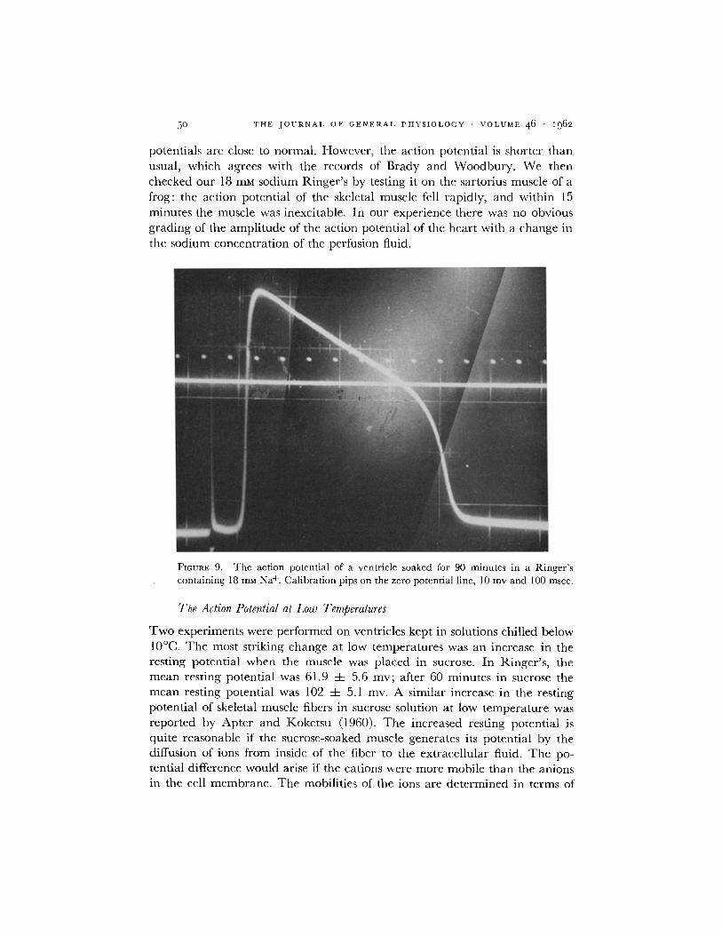
decreases in a m p l i t u d e when the sod ium concen t ra t ion in the R inge r ' s is re- duced. W e studied the electr ical act iv i ty of the ventr ic le in a R inge r ' s solution con ta in ing 18 mM Na+. Fig. 9 shows an ac t ion poten t ia l f rom a ventr ic le
soaked for 90 minu tes in the low sod ium Ringer ' s . T h e ac t ion and rest ing
5o T H E J O U R N A L O F G E N E R A L P H Y S I O L O G Y • V O L U M E 4 6 • 1962
potentials are close to normal . However , the act ion potent ia l is shorter lhan usual, which agrees with the records of Brady and Woodbury . We then checked our 18 mM sodium Ringer ' s by testing it on the sartorius muscle of a frog: the act ion potent ia l of the skeletal muscle fell rapidly, and within 15 minutes the muscle was inexcitable. In our exper ience there was no obvious grading of the ampl i tude of the act ion potent ia l of the hear t with a change in the sodium concent ra t ion of the perfusion fluid.
FIGURE 9. The action potential of a ventricle soaked for 90 minutes in a Ringer's containing 18 m u N a +. Calibration pips on the zero potential line, 10 mv and 100 msec.
The Action Potential at Low Temperatures
T w o exper iments were per formed on ventricles kept in solutions chilled below 10°C. T h e most striking change at low tempera tures was an increase in the resting potent ia l when the muscle was placed in sucrose. In Ringer 's , the m e a n resting potent ia l was 61.9 =k: 5.6 my; after 60 minutes in sucrose the m e a n resting potent ia l was 102 =t= 5.1 inv. A similar increase in the resting potent ia l of skeletal muscle fibers in sucrose solution at low t empera tu re was repor ted by Apter and Koketsu (1960). T h e increased resting potent ia l is quite reasonable if the sucrose-soaked muscle generates its potent ia l by the diffusion of ions f rom inside of the fiber to the extracel lular fluid. T h e po- tential difference would arise if the cations were more mobile than the anions in the cell membrane . T h e mobili t ies of the ions are de t e rmined in terms of

W. G. VAN DER KLOOT AND N. S. RuBm Heart Muscle in Sucrose Solution 51
conductance and transference number. As temperature falls, conductances de- crease. However, at lower temperatures, the difference between the trans- ference numbers of cations and of anions increases. Therefore, at lower tem- peratures differences in the mobilities of the cations and anions often become exaggerated so that diffusion potentials are increased (see Harned and Owen, 1958).
The changes in the action potential produced by lower temperatures are along expected lines. The action potential is greatly elongated; in Ringer's the mean duration was 3710 -4- 77 msec. The action potential in Ringer 's was 72.0 -4- 10.2 my, so the overshoot was about 10 my. After 1 hour in sucrose, the action potential was still 71.0 4- 5 my. Since there was an increased resting potential in sucrose solution, the potential across the membrane at the height of the action potential averaged - 3 1 my. The resting and the action po- tentials change in quite different fashions with a decrease in temperature.
A final point about the experiments at low temperature is that when the sucrose-soaked muscles were returned to Ringer's, recovery was slow. After 30 minutes in Ringer's, the mean action potential was 46.0 4- 5.2 my, while the mean resting potential was 55.0 4- 4.8 my. This result again suggests that the recovery of the sucrose-soaked muscles requires something more than the re- appearance of Ringer 's in the extracellular space.
D I S C U S S I O N
The Action Potential and the Sodium Hypothesis
Since the frog heart conducts action potentials for hours while in sucrose solution, it is important to consider this result in relation to the sodium mecha- nism for generating action potentials. In our experiments, auricles retained action potentials with an overshoot after 330 minutes of soaking in sucrose solution. If a sodium gradient is responsible for these action potentials, after 330 minutes in sucrose solution the electrochemical activity of sodium must still be greater outside of the fiber than inside. It might first be suggested that the fluid just outside of the fibers is protected by a barrier of some sort, so that the true extracellular fluid exchanges slowly with the sucrose solution. Four lines of evidence argue against this interpretation. (a) Pieces of cardiac muscle which are inexcitable after prolonged soaking in sucrose recover rapidly when replaced in Ringer 's and just as rapidly become inexcitable if they are quickly transferred back to sucrose solution. (b) Frog cardiac muscles depolarized in isotonic K~SO4 solutions rapidly repolarize when placed in sucrose solutions. (c) In the experiments in which sucrose-soaked ventricles placed in sulfate Ringer 's recovered the ability to conduct action potentials, excitability was promptly lost when the ventricles were then returned to sucrose solution. (d) Muscles which no longer contract after soaking in isotonic NaC1 quickly

52 T H E J O U R N A L O F G E N E R A L P H Y S I O L O G Y • V O L U M E 46 • I962
recover contractility when placed in isotonic sucrose solutions. These four lines of evidence all suggest that the sucrose solution rapidly diffuses to physio- logically active sites within the muscle. This conclusion will be supported by studies of the effiux into sucrose solution of Na 24 from " loaded" muscles (Van der Kloot and Dane, data to be published).
Therefore, if the sodium mechanism is operating in the frog heart muscle it must be working under special conditions. For example, it is not incon- ceivable that the action potential is generated across a membrane situated inside of the plasma membrane. A high sodium concentration might be main- tained by the active extrusion of sodium from the cell into the space between this hypothetical membrane and the plasma membrane. We do not regard this idea as likely, but there is now so much evidence for a sodium mechanism in other excitable tissues that apparent exceptions must be viewed with skep- ticism.
The experiments with sulfate or isethionate Ringer's suggest that after a ventricle is soaked in sucrose solution for a long time, the ability to conduct action potentials can be restored only by a Ringer's solution whose ions can actually penetrate into the muscle fibers. In ventricles soaked for a shorter time in sucrose solution, recovery can be obtained with sulfate Ringer's, per- haps because there is an exchange between diffusible cations remaining in the cell and the cations of the sulfate Ringer's. At present there is no way of de- ciding whether the ions must replenish the entire interior of the sucrose-soaked muscle cell, or merely restore the ions of some protected, superficial compart- ment.
Brady and Woodbury (1960) conclude that the sodium mechanism is re- sponsible for the action potential in the frog ventricle. However, we were unable to decrease the action potential markedly by placing ventricles in low sodium solutions. The reason for this discrepancy is unknown. A decrease in the external sodium concentration always does seem to lead to a decreased rate of depolarization. The action potential of the guinea pig heart also does not vary in the expected fashion with decreasing external sodium concentra- tions (Coraboeuf and Otsuka, 1956; D61~ze, 1959).
There is another aspect to consider in relation to the sodium mechanism in the generation of action potentials in frog cardiac muscle. The minimum transfer of change per unit area needed to produce the action potential will be AVC, where AV is the height of the action potential and C is the capaci- tance per unit area of the cell membrane. If the sodium mechanism is involved the minimum sodium inflow per gram of tissue will be given by:
2AVC ( 1 ) rF
where r is the radius of the muscle fiber. To maintain a steady state the

W. G. VAN I)ER KLOOT Am) N. S. RI.rBIN Heart Muscle in Sucrose Solution 53
sodium must then be transported out of the fiber. The secretory work (w) for the outward transfer of the minimum amount of sodium needed to generate the action potential will be:
2AVC W -- YaV,,a ( 2 )
Fr
where AVN, is the electrochemical potential difference of sodium ions across the membrane, which is approximately equal to AV. In the frog hearts, C is about 30 uf /cm 2 (Trautwein, Kuffier, and Edwards, 1956); V about 0.1 v, and r is about 5 X 10 -4 era. Therefore, the secretory work for transporting the sodium outward is about 2.9 × 10 -4 cal gm -1 impulse -1. The increased energy consumption of the frog heart when it is stimulated was calculated from the oxygen consumption data of Clark and White (1928). The values range from 3.2 × 10 -4 to 16.5 × 10 -4 cal gm -I impulse -1. Our calculations assume that the absolute minimum amount of sodium flows into the fiber and that the efficiency of the extrusion process is 100 per cent, instead of the 27 to 54 per cent efficiency found in frog's skin (Zerahn, 1956). It appears that a perilously large proportion of the energy produced by the tissue would have to be in- vested in active transport if the sodium mechanism is used.
An even more difficult situation is found in a mammalian C fiber with a radius of only 1.2 X 10 -4 cm. Assuming that the capacitance is 1 ~f/cm 2 and the AV is 0.1 v, the energy required for sodium extrusion is 0.38 × 10 -~ cal gm -~ impulse-k The minimum energy need for sodium transport is about the total energy production of C fibers stimulated at a rate of 10/see. (Larrabee and Bronk, 1952). Of course, the nerve might extrude most of the sodium during periods of rest. But the point is that with thin cells the sodium mecha- nism for generating an action potential is expensive. It is worth mentioning here that Oomura (1960) reports that the nodal membrane of frog nerve con- tinues to conduct an inward current in sucrose solution, so long as a slight hyperpolarizing current is applied to the membrane. And Koketsu, Cerf, and Nishi (1959) find that the dorsal root cell bodies when injected with tetra- e thylammonium ion continue to produce action potentials in a sucrose solution containing some CaC12.
If extracellular sodium is not supplying the current for generating an action potential in a tissue soaked in a sucrose solution, it seems possible that the current is produced by the outflow of an anion from the cell. An accounting of the anions which leave the cell during soaking in sucrose solutions should be a useful first step in exploring this possibility.
Contraction in Sucrose Solutions
In evaluating the parts played by the individual ions of Ringer's solution in supporting the beating ot the frog's heart there is considerable evidence point-
54 T H E J O U R N A L O F G E N E R A L P H Y S I O L O G Y • V O L U M E 4 6 • ~962
ing to a close relation between extraceUular calcium and contraction (Daly and Clark, 1921; Weidmann, 1959). Calcium moves into the stimulated cell (Niedergerke, 1959; Winegrad, 1961). And it is well established that the microinjection of calcium into a skeletal muscle fiber elicits a local contraction (Heilbrunn and Wiercinski, 1946, Niedergerke, 1955).
Since frog heart muscle can continue to contract for several hours in sucrose solution, it seems unlikely that calcium is moving down an electrochemical gradient from the sucrose solution into the cell. The results show that cardiac muscles undergo a contracture when calcium is added to the sucrose solution, so it is hard to believe that there is a store of "extracellular" calcium which is protected from diffusing into the external medium.
Contractility in ionic solutions depends on a ratio of C a / N a 2 in the external medium (Ltittgau and Niedergerke, 1958), which suggests that Ca ++ ions from the extracellular fluid are reversibly bound to anionic groups in the cell membrane. If the ratios of the cations on the membrane are changed, the properties of the membrane are altered. By this interpretation a cardiac muscle in sucrose solution retains relatively normal membrane properties because none of the ions is present in the extracellular fluid in high enough concentra- tion to displace some other ion which was bound to the membrane.
The other role of the Ringer's solution would be to preserve the intracellular ion concentrations. The intracellular ions are required either directly or in- directly for action potential generation and contractility. Although our results do not support the idea that the contractile machinery is activated by calcium which diffuses into the cell by moving down the electrochemical gradient, the possibility remains that the contraction is triggered by "membrane" calcium which is moved into the cell during stimulation and which is then replenished by calcium which moves out of the cell during rest. Nor do the results tell anything about the likelihood that calcium is released, by some indirect mechanism among the myofibrils of a stimulated muscle.
Another possibility, which cannot be casually dismissed, is that the con- traction of cardiac muscle in sucrose solution is elicited by a substitute, ab- normal mechanism. The results show that in sucrose solutions at 25°C con- traction markedly declines with repetitive stimulation. Fig. 3 suggests that the initial decline in contractility with successive stimulations is roughly a first order process. This relation can be interpreted by assuming that the sucrose- soaked muscle contains a store of some substance which is needed for con- traction. With each stimulation a set fraction of the store is dissipated, so the succeeding contraction is weaker. A steady, low level of contraction is reached when the rate of the replacement of the substance is equal to the rate of utilization. The nature of this hypothetical substance remains unknown, al- though the data strongly suggest that it is not Ca++.
The question of contractility in sucrose solutions might be profitably ex-

W. G. VAN Dr.R KLOOT AND N. S. Ruan~ Heart Muscle in Sucrose Solution 55
tended by comparing the ion contents and metabolism of ventricles soaked in sucrose solutions at 25 °C, where contraction disappears in a few hours, and at 4 to 7 °C, where contraction persists for as long as 24 hours.
Other Frog Muscles in Sucrose Solution
In conclusion, the behavior of cardiac muscles soaked in sucrose solution can be compared with that of other muscles of the frog. Smooth muscles from the stomach also continue to conduct action potentials and to contract for hours in sucrose solution (Kolodny and Van der Kloot, 1961). On the other hand, the twitch fibers of the skeletal muscles rapidly become inexcitable in sucrose solution. However, when the endplate of a muscle soaked in sucrose is stimu- lated with acetylcholine, there is frequently a localized hyperpolarization (Oomura and Tomita, 1960). The tonic (or slow) muscle fibers develop a sustained contracture in sucrose solution. When stimulated with acetylcholine, the tonic muscle fibers hyperpolarize and relax (Swift, Gordon, and Van der Kloot, 1960; Van der Kloot, Rubin, and Swift, data to be published). The pattern which underlies the behavior of different types of muscles when they are soaked in sucrose solution remains to be uncovered.
We are grateful to Dr. E. Ratinoff for assistance with many of the contraction experiments. This study was supported by research grant B-1870 from the Institute of Neurological Diseases and Blind- ness, Public Health Service. Received for publication, November 1, 1961.
B I B L I O G R A P H Y
1. APTER, J . T., and Kora~a'su, K., J. Cell. and Comp. Physiol., 1960, 56,123. 2. BRADY, A. J., and WOODBURX', J. W., J. Physiol., 1960, 154,385. 3. CLARa, A. J., and WHITE, A. C., J. Physiol., 1928, 66, 185. 4. CORABOEUF, E., and OTSUKA, M., Compt. rend. Soc. biol., 1956, 243,441. 5. DAn',', I. DEB., and ChARt<, A. J., J. Physiol,, 1921, 54,367. 6. D~.Li~zE, J., Circulation Research, 1959, 7, 411. 7. Fv.NN, W. 0 . , Am. or. Physiol., 1931, 97,635. 8. HARN~D, H. S., and OWEN, B. B., The Physical Chemistry of Electrolyte Solu-
tions, New York, Reinhold Publishing Corp., 3rd edition, 1958. 9. I-tEILBRU~N, L. V., and WmRcmSZi, F. J., Biol. Bull., 1946, 91,237.
I0. HOFFMAN, B. F., and CRANEmELI), P. F., Electrophysiology of the Heart, New York, McGraw-Hill Publishing Co., Inc., 1960.
11. HosI-uKo, T., and SP~RELAKIS, N., Am. J. Physiol., 1961, 201,873. 12. Korma'su, K., C_~RF, J. A., and NISHI, S., J. Neurophysiol., 1959, 22,693. 13. Konormv, R. L., and VAN m~.m KLOOa', W. G., Nature, 1961, 190,786. 14. LARRABV.~., M. G., and BRONK, D. W., ColdSpring Harbor Symp. Quimt. Biol., 1952,
17,245. 15. L/STTGAU, H. C., and NmDERO~.RraZ, R., J. Physiol., 1958, 143,486. 16. NmDv.mOERra~, R., J. Physiol., 1955, 128, 12P.

56 THE J O U R N A L OF G E N E R A L P H Y S I O L O G Y • VOLUME 46 • i962
17. N I E D E R G E R K E , R . , Experientia, 1959, 15,128. 18. GO,aURA, Y., Seitai no Kagaku, 1960, 11,276. 19. OO~URA, Y., and TOYOTA, T., in Electrical Activity of Silagle Cells, Tokyo,
Igakushoin, Hongo, 1960, 181. 20. RINGER, S., J. Physiol., 1880, 3,380. 21. SmGH, I., Current Sc., India, 1944, 10,251. 22. Small, I,, SEHRA, K. B., and SmOH, S. I., Current Sc., India, 1945, 14,152. 23. SWIFT, M. R., GORDON, H. P., and VAN DER KLOOT, W. G., Proc. Nat. Acad. Sc.,
1960, 46, 1415. 24. TRAUTWEm, W., KUFFLER, S., and EDWARDS, C., J. Gen. Physiol., 1956, 40,135. 25. WARE, F., JR., BENNETT, A. L., and MCINTYPa~, A. R., Am. J. Physiol., 1957, 190,
194. 26. WEm~A~N, S., Experientia, 1959, 15,128. 27. WXNEGRAD, S., Circulation, 1961, 24,523. 28. WOODBURY, J. W., and BRADY, A. J., Science, 1956, 12.3,100. 29. WRIGHT, E. B., and OGATA, M., Am. J. Physiol., 1961, 201, 1101. 30. ZERAHN, K., Acta Physiol. &and., 1956, 36,300.