contractility of the cell rear drives invasion of breast ... · contractility of the cell rear...
TRANSCRIPT
Contractility of the cell rear drives invasion of breasttumor cells in 3D MatrigelRenaud Poinclouxa,b,1, Olivier Collina,c,2, Floria Lizárragaa,b, Maryse Romaoa,d, Marcel Debraye, Matthieu Piela,c,and Philippe Chavriera,b,3
aResearch Center, Institut Curie, F-75248 Paris, France; bMembrane and Cytoskeleton Dynamics, cSystems Cell Biology of Cell Polarity and Cell Division, anddStructure and Membrane Compartments, Centre National de la Recherche Scientifique, Unité Mixte de Recherche 144, 75248 Paris Cedex 05, France; andeDépartement de Santé Publique et Biostatistique, Faculté des Sciences Pharmaceutiques et Biologiques, Université Paris-Descartes, 75006 Paris, France
Edited by Joan S. Brugge, Harvard Medical School, Boston, MA, and approved December 21, 2010 (received for review August 18, 2010)
Cancer cells use different modes of migration, including integrin-dependent mesenchymal migration of elongated cells alongelements of the 3D matrix as opposed to low-adhesion-, contraction-based amoeboid motility of rounded cells. We report that MDA-MB-231 human breast adenocarcinoma cells invade 3D Matrigelwith a characteristic rounded morphology and with F-actin andmyosin-IIa accumulating at the cell rear in a uropod-like structure.MDA-MB-231 cells display neither lamellipodia nor bleb extensionsat the leading edge and do not require Arp2/3 complex activityfor 3D invasion in Matrigel. Accumulation of phospho-MLC andblebbing activity were restricted to the uropod as reporters ofactomyosin contractility, and velocimetric analysis of fluorescentbeads embedded within the 3D matrix showed that pulling forcesexerted to the matrix are restricted to the side and rear of cells.Inhibition of actomyosin contractility or β1 integrin function inter-feres with uropod formation, matrix deformation, and invasionthrough Matrigel. These findings support a model whereby acto-myosin-based uropod contractility generates traction forces on theβ1 integrin adhesion system to drive cell propulsion within the 3Dmatrix, with no contribution of lamellipodia extension or blebbingto movement.
3D migration | integrin | invasion
During metastasis, tumor cells encounter various extracellularmatrix (ECM) environments with distinct composition and
architecture, including basement membrane (BM) and intersti-tial collagen networks (1–3). Although basic mechanisms of cellmotility on 2D substrata are generally well understood, much lessis known regarding the mechanics of cell migration in 3D matrixenvironments (1, 4–7). Recent evidence supports the conclusionsthat 2D and 3D migration can differ considerably and that dif-ferent types of 3D motility exist depending on the biophysicalproperties of the 3D environment (viscoelasticity, confinement,porosity) (8–15). Understanding the mechanics of cancer cellmigration in these different 3D environments is therefore ofparamount importance.Schematically, two modes of 3Dmigration of invasive cells have
been described; the models differ by the dependency on actomy-osin contractility, requirement for integrin adhesion, and matrixremodeling. In the mesenchymal mode, which has some parallelwith 2D migration of fibroblasts (1), invasive cells are elongatedand require pericellular matrix proteolysis for extending Rac-dependent F-actin-based leading pseudopodia in an integrin-dependent manner (16–20). In contrast, some carcinoma cellsinvade with a low-adhesion amoeboid mode of migration, char-acterized by a round morphology, no stable intrinsic cell polarity,and a required high level of RhoA/ROCK-driven actomyosin con-tractility (17, 18). Amoeboid cell motility, which is typically asso-ciated with bleb formation, is independent of proteases, and it isgenerally thought that by forming blebs, tumor cells can squeezethemselves through preexisting voids in the matrix (5, 21). In vivo,bleb-basedmotility has been implicated in directional migration ofprimordial germ cells in zebrafish embryo (22). Depending onlevels of RhoA, Rac, and Cdc42, matrix degradation capacity, andarchitecture of the ECM, carcinoma cells can perform different
modes ofmotility in 3Dand,hence, are thought to adapt to variableenvironmental clues (17, 18, 20, 23–25).Several methods have been developed for visualizing and an-
alyzing forces generated by cells during 2D migration. Thesestudies revealed that propulsive forces that are produced bymyosin II on the F-actin network, which is linked to adhesionsites underneath the protruding lamellipodium, support migra-tion (26, 27). However, mechanics underlying 2D and 3D mi-gration differ quite significantly (8–11, 13–15), and forcerepartition is generally unknown for cells migrating in 3D.Tracking of embedded beads within the matrix has been recentlyused to follow matrix deformations during mesenchymal migra-tion of fibrosarcoma cells in a 3D type I collagen network (28).In the present study, we address the mechanics of 3D migra-
tion of highly invasive MDA-MB-231 human breast cancer cellsin Matrigel, a reconstituted BM matrix, by tracking micrometer-scale movements of fluorescent microbeads seeded in the gel.We found that MDA-MB-231 cells invade and migrate in 3DMatrigel with a striking round morphology, and they displayneither lamellipodial extension nor blebbing at the leading edge,while cells form an F-actin/myosin II-rich uropod as a generatorof contractile forces at the cell rear. Actomyosin-based contra-ctility at the uropod, which is transmitted to and exerts tractionforces on the ECM through β1 integrins, powers migration withinthe 3D matrix.
ResultsMechanics of MDA-MB-231 Rounded Cell 3D Migration. Highly met-astatic MDA-MB-231 cells stably expressing mCherry-Lifeact,which allows visualization of F-actin dynamics with minimal in-terference with actin assembly (29, 30), were seeded in 3DMatrigel matrix together with fluorescent microbeads (0.2-μmdiameter) and followed by time-lapse fluorescence microscopy.MDA-MB-231 cells moved in random directions in 3D Matrigelwith an average speed of 3.2 ± 0.14 μm/h during motile phase,similar to published data (Table S1; refs. 11 and 18). Motile cellsexhibited a rounded morphology with no sign of membrane ex-tension at the front and a striking accumulation of F-actin at thecell pole opposite to the direction of movement (Figs. 1A and 2Aand Movies S1 and S2). By analogy with the rear of migratingleukocytes, we refer to the F-actin back of the cells as the uropod
Author contributions: R.P., M.P., and P.C. designed research; R.P., F.L., and M.R. per-formed research; R.P., O.C., and M.D. analyzed data; and R.P. and P.C. wrote the paper.
The authors declare no conflict of interest.
This article is a PNAS direct submission.1Present address: Centre National de la Recherche Scientifique, Institut de Pharmacologieet de Biologie Structurale, and Université de Toulouse, Université Paul Sabatier, 31077Toulouse, France.
2Present address: Ecole Normale Supérieure, Centre National de la Recherche Scientifique,Unité Mixte de Recherche 8541, Functional Imaging of Transcription, 75230 Paris Cedex05, France.
3To whom correspondence should be addressed. E-mail: [email protected].
This article contains supporting information online at www.pnas.org/lookup/suppl/doi:10.1073/pnas.1010396108/-/DCSupplemental.
www.pnas.org/cgi/doi/10.1073/pnas.1010396108 PNAS | February 1, 2011 | vol. 108 | no. 5 | 1943–1948
CELL
BIOLO
GY

(31), although in contrast to leukocytes, MDA-MB-231 cells’uropod does not protrude.In contrast to the absence of microbead movement in cell-free
regions of the matrix, indicating that beads are trapped in themeshwork, beads in the vicinity of motile cells underwent move-ment. Displacements of these beads reflect remodeling and de-formation of the matrix induced by cell migration (Fig. 1A andA″and Movie S1). To provide a micrometer-scale quantitative de-scription of matrix remodeling by invading cells, velocity fieldswere estimated from time-lapse image series of microbeads byusing particle image velocimetry (PIV) analysis. Individual ve-locity fields of several cells were averaged (see Methods), re-vealing that Matrigel was pushed away in front of migrating cellswith a ∼3.0 μm/h maximum velocity, similar to the migrationspeed of the cells (Fig. 1B, region 1, and Fig. S1B). In addition,the displacement field displayed two vortex-like structures origi-nating from the poles perpendicular to the migration axis (Fig.1B, region 2) and converging toward the cell rear (region 3). Axialvorticity field computed from average velocity field also revealedhigh shear motion in region 2, consistent with high tractions ofthe matrix exerted by migrating cells in 3D (Fig. S1A).Cell movement was accompanied by the accumulation of
beads at the cell rear (Fig. S2 A and B and Movie S2). Similarly,when MDA-MB-231 cells were plated atop a thick layer ofMatrigel, they progressively invaded through the matrix witha round morphology with the matrix pulled on top of the cell andgenerating a track of modified ECM behind it (Fig. 1C; ref. 30).Of note, the matrix dome around invading cells was not causedby matrix retraction during scanning electron microscopy samplepreparation, as it was also observed with lived unfixed MDA-MB-231 cells plated atop Matrigel and visualized with seededfluorescent microbeads (Fig. S2C). In addition, we noticed thepresence of intense membrane blebbing activity at the free rearof invading cells (Fig. 1C and below; ref. 30).At the ultrastructural level, electron microscopy on thin sec-
tions of MDA-MB-231 cells embedded within 3D Matrigel con-
firmed extensive blebbing activity at one pole of the cell (Fig. S3).In addition, in comparison with normal porosity of Matrigel(Fig. S3A Inset 1, away from the cell), progressive reduction ofthe pore size and densification of the matrix was observed on thesides of the cell (Fig. S3A Inset 2), becoming maximal behindthe cell (Fig. S3A Inset 3). Gel densification is in agreement withthe observed accumulation of microbeads at the cell rear. Inaddition—possibly as a consequence of high shear forces (Fig.S1A) and gel cracking—voids in Matrigel were clearly observed inthe immediate vicinity of the uropod (Fig. S3 A’ and B), whichcould contribute to some extent to the settling of beads. Com-paction of the gel was also visible at the front of the motile cellpushing on the matrix (Fig. S3A Inset 1).Of note, inhibition of matrix metalloproteinase (MMP) ac-
tivity had a modest inhibitory effect on the speed of migration ofMDA-MB-231 cells in 3D Matrigel (∼15%; see Fig. 3E andTable S1), indicating no prominent contribution of MMP-basedmatrix degradation to this type of movement. Matrigel is a vis-coelastic meshwork of matrix proteins of high elastic modulus(11). The observed voids in the gel, high shear forces, and pro-gressive gel densification from the front to the rear rather sup-port the view that cells move within this viscoelastic material bypulling on and pushing Matrigel aside.
Rounded MDA-MB-231 Cells Migrating in 3D Matrigel Are Polarized.High-resolution confocal spinning disk imaging of fixed cells in3D Matrigel confirmed the accumulation of F-actin at the cellrear and in evenly distributed, finger-like cortical structures, aswell as the absence of membrane extension and F-actin accu-mulation at the front (Fig. 2B and Movie S3). Filament bundlesradiating from the uropod toward the cell front were visible (Fig. 2B and C, arrows, and Movies S3 and S4). Although they migratedwith a round morphology, MDA-MB-231 cells in 3D Matrigel
Fig. 1. Matrix displacements during MDA-MB-231 rounded cell 3D migra-tion in Matrigel. (A) MDA-MB-231 cells stably expressing mCherry-Lifeactseeded in Matrigel containing fluorescent microbeads (green) were imagedevery 15 min (Movie S1). Shown is the last frame of mCh-Lifeact (red)superimposed on a 2-h time projection of the bead sequence (green).Dashed line indicates the position of the cell at the beginning of the time-lapse series. (A′ and A″) Bead positions at the back and front of the cell,respectively, with the first two frames (T0 and T15’) color-coded in green,subsequent five frames (T30’ to T90’) in blue, and last two frames (T105’ andT120’) of the time series in red. (B) PIV analysis of matrix displacement aroundthe cells. Individual velocity fields of eight cells were averaged. The masscenter of cells is depicted with a red dot. Red arrow corresponds to themean velocity vector of the cells. The amplitude of the bead velocity is color-coded, and direction of displacements is depicted with arrows. Matrix ispushed at the front (region 1), while it is pulled on the sides and at the back(regions 2 and 3, respectively). (C) Scanning electron micrographs of MDA-MB-231 cells invading a thick layer of Matrigel at indicated time after plat-ing. (Scale bars, 10 μm.)
Fig. 2. F-actin dynamics during MDA-MB-231 rounded cell 3D migration.MDA-MB-231 cells expressing mCh-Lifeact were seeded in Matrigel con-taining fluorescent microbeads (green in F) and imaged by wide-field (A) orconfocal spinning disk (B–F) microscopy. (A) Selected frames from a time-lapse sequence with the white line showing the position of the rear of thecell at the beginning of the sequence. Arrowheads point to accumulation ofF-actin at the cell rear in a uropod-like structure. (B) Z-projection of confocalsections (see Movie S3 for 3D reconstruction) showing the accumulation of F-actin at the uropod (arrowhead), in finger-like cortical extensions (asterisks),and in bundles radiating from the uropod toward the cortex (arrows). (C)Selected frame of a time-lapse series (Movie S4) showing intense blebbingactivity restricted to the uropod region (arrowheads). Arrows point to radialF-actin bundles. (D and E) Selected frame (D) of a time-lapse series (MovieS6) showing the dashed line used for kymographic analysis of corticalmovements in E (tags 1–3 are positioned on the x axis of the kymograph, aswell as the cortical structure highlighted in F, shown by a red arrowhead). (F)Selected frames from the time series as in D and E (Movie S6). The regiondepicted is boxed in D. Images show F-actin labeled with mCh-Lifeact (red)and microbeads (green). Arrowheads point to parallel rearward movementof a cortical actin structure (red arrowhead) and microbeads (green arrow-head). Asterisks, position of the objects at time 0. (Scale bars, 10 μm.)
1944 | www.pnas.org/cgi/doi/10.1073/pnas.1010396108 Poincloux et al.

were highly polarized with the nucleus positioned at the back andthe Golgi apparatus localized at the cell front (Fig. S4 A and B).The Arp2/3 complex is the main lamellipodial actin nucleating
activity and is critical for 2D cell migration. Interestingly, si-lencing of the actin nucleating Arp2/3 complex by means ofsiRNA targeting of the p34-Arc subunit (causing also reduction
of p16-Arc; see Fig. 3B) had no effect on vertical invasion ofMDA-MB-231 cells when plated atop a thick layer of Matrigel(Fig. 3D; for statistics see Table S2) or on the migration speedand uropod formation in cells seeded in 3DMatrigel (Fig. 3E andTables S1 and S3). In contrast—confirming our previous obser-vation that another class of actin nucleators, the Diaphanous-
Fig. 3. Inhibition of RhoA-ROCK-Myosin II and β1 integrinimpairs uropod formation and invasion in Matrigel. (A–C)MDA-MB-231 cells were treated with the indicated siRNAstargeting RhoA (A), the p34-Arc (ARPC2) subunit of theArp2/3 actin nucleating complex (B), or β1 integrin (C). Celllysates were prepared after 72 h, and immunoblottinganalysis was performed with indicated antibodies. Ofnote, knockdown of p34-Arc leads to decreased expres-sion of p16-Arc (B). (D) Quantification of cell invasion bymCh-Lifeact MDA-MB-231 cells plated atop of a thick layerof Matrigel and treated with indicated siRNAs or drugs.Cell invasion was determined by analyzing the proportionof cells buried in Matrigel after 14 h from low magnifi-cation scanning electron micrographs. (E) Average mi-gration speed (filled bars) and percentage of cells withuropod (open bars) in MDA-MB-231 cells seeded inMatrigel and treated with indicated siRNA and drugs.Asterisks indicate statistically significant differences com-pared with control cell populations (Tables S1–S3).
Fig. 4. Actomyosin-based contractility is required for MDA-MB-231 cell invasion. (A and B) MDA-MB-231 cells seeded inMatrigel for 4 h, fixed, and stained for F-actin and myosin IIA(A) or pS-MLC (B). Arrowheads point to accumulation ofmyosin IIA and pS-MLC at F-actin positive uropod. Averagednormalized intensity profiles from 15 cells are shown. Posi-tion 0 corresponds to maximum normalized F-actin intensity(see SI Methods for details). For clarity, SEMs are not rep-resented (< ±0.08 normalized intensity arbitrary unit). (C)Scanning electron micrographs of siRhoA-, Y27632-, orblebbistatin (bleb)-treated MDA-MB-231 cells seeded atopof Matrigel and fixed after 14 h. (D and E) Individual velocityfields of 13 Y27632-treated cells (D) or 4 blebbistatin-treatedcells (E) were averaged as in Fig. 1B. Amplitude of the beadvelocity is color-coded, and direction of displacements isdepicted with arrows. (Scale bars, 10 μm.)
Poincloux et al. PNAS | February 1, 2011 | vol. 108 | no. 5 | 1945
CELL
BIOLO
GY

related formins (DRFs), are required for MDA-MB-231 cellvertical invasion in Matrigel (30)—we found that a general in-hibitor of FH2-domain containing formins (SMIFH2; ref. 32) ledsimilarly to a ∼35% reduction of migration speed in 3D Matrigeland concomitant decrease of uropod formation (Fig. 3E). Thus,our data indicate that DRFs, but not the Arp2/3 complex, areimplicated in 3D cell invasion and migration of MDA-MB-231cells in Matrigel.Live imaging also confirmed extensive blebbing activity at the
cell rear (Fig. 2C, arrowhead, and Movies S4 and S5). In par-ticular, large blebs were frequently observed at the edge of theuropod, which may correspond to a region of weakness of thecell cortex (Movie S5; ref. 5). Kymograph analysis of actin cor-tical structures revealed retrograde movement of the cortex to-ward the uropod during migration (Fig. 2 D and E and MovieS6). The retrograde flow of cortical F-actin was high close to theuropod region (Fig. 2E, arrowhead 2) and nearly zero at theopposite front side of the cell (arrowheads 1 and 3). Further-more, rearward movement of the beads paralleled that of thecortex as an indication that the two were coupled (Fig. 2F), inagreement with observed high shear forces (Fig. S1A). Interest-ingly, the presence of an F-actin rich uropod-like structure wasalso observed when cells were plated on top of Matrigel (Fig.S4C and Movie S7), further indicating that vertical invasion ofcells seeded atop or invasion of cells within Matrigel proceedthrough the same mechanism.
RhoA-ROCK-Myosin II–Mediated Contractility at the Uropod IsRequired for 3D Migration. The presence of blebs and conver-gent retrograde movement of the cell cortex were indicative ofcell contraction. Immunolocalization analysis showed that myo-sin IIA heavy chain and the active (phospho-Ser19) form ofmyosin light chain (pS-MLC) were strongly accumulated aturopod together with F-actin (representative cells are shown inFig. 4 A and B and Fig. S5). The functional contribution of ac-
tomyosin contractility for MDA-MB-231 cell invasion withinMatrigel was assessed by inhibition of myosin II or its upstreamactivators. Pharmacological inhibition of myosin II with bleb-bistatin resulted in a strong decrease of vertical invasion capacity(Fig. 3D) and a concomitant and correlated reduction of mi-gration speed and uropod formation in cells seeded in Matrigel(Fig. 3E and Tables S1–S3). Similarly, inhibition of RhoA byRNAi silencing (Fig. 3A) or treatment with Clostridium botuli-num C3 exoenzyme, inhibition of ROCK kinase with Y27632compound, or myosin II inhibition by blebbistatin led to corre-lated reduction of invasion capacity and uropod formation byMDA-MB-231 cells (Fig. 3 D and E and Tables S1–S3). In thesecells, F-actin and pS-MLC accumulation, characteristic of uro-pod structure, was no longer visible (Fig. S5).In addition, cells inhibited for RhoA, ROCK, or myosin II
activity remained spread on the matrix when plated atopMatrigel, in contrast to the rounded invasive morphology ofcontrol MDA-MB-231 cells (compare Figs. 4C and 1C). PIVanalysis of Y27632-treated samples showed isotropic low-forcepulling of the matrix by the cells (Fig. 4D, Fig. S1B, and MovieS8), which did not support cell migration (Fig. 3E and Table S1).Finally, myosin II inhibition by blebbistatin almost complete-ly abolished cell-induced matrix movements (Fig. 4E, Fig. S1B,and Movie S9). Together, these data demonstrate that RhoA,ROCK, and myosin II are required for formation of and con-traction at uropod, and they suggest that uropod contractility isessential for MDA-MB-231 cell migration in 3D Matrigel.
β1 Integrin Is Required for Contraction at Uropod and Cell 3DMigration in Matrigel. Contractile retrograde forces can supportforward movement if coupled to the surrounding matrix throughreceptor (integrin)-mediated force transduction (4). We thereforeassessed the involvement of β1 integrin in matrix remodeling and3D migration of MDA-MB-231 cells in Matrigel. β1 integrin waspresent at the cell cortex and enriched at the uropod (Fig. 5A),
Fig. 5. Role of β1 integrin in 3D migration of MDA-MB-231cells in Matrigel. (A) MDA-MB-231 cells were incubated for 4 hin Matrigel, fixed, and stained for F-actin and β1 integrin.Arrowheads point to accumulation of β1 integrin at F-actin–rich uropod. Averaged normalized intensity profiles from 15cells are shown as in Fig. 4 A and B. (B) Scanning electronmicrographs of an MDA-MB-231 cell plated atop a thick layerof Matrigel in the presence of β1 integrin blocking antibody(4B4) for 14 h before fixation. (C) MDA-MB-231 cell seeded inMatrigel for 4 h in the presence of 4B4 antibody and stainedfor F-actin (green) and pS-MLC (red). Nucleus was labeledwith DAPI (blue). Cells adopt irregular shapes with F-actin andpS-MLC accumulations and blebbing activity. (D) mCh-LifeactMDA-MB-231 cells seeded in Matrigel in the presence of 4B4mAb were imaged during 4 h by wide-field video microscopy.Shown is the superimposition of the 4-h time projection ofthe bead sequence and the last frame of mCh-Lifeact se-quence. Asterisk points to region of cell detachment to thematrix. (E) Individual velocity fields of seven 4B4-treated cellswere averaged as in Fig. 1B. Amplitude of the bead velocity iscolor-coded, and direction of displacements is depicted witharrows. (Scale bars, 10 μm.)
1946 | www.pnas.org/cgi/doi/10.1073/pnas.1010396108 Poincloux et al.
although we cannot rule out the possibility that membrane ac-cumulation at uropod resulted in apparent increased β1 staining.Addition of anti-human β1 integrin-blocking antibody 4B4 or si-lencing of β1 with two independent siRNAs (Fig. 3C) resulted ina significant 45–65% reduction of migration speed in 3DMatrigelcorrelated with impaired uropod formation (Fig. 3E and TablesS1–S3). In addition, 4B4-treated cells cultured atop the Matrigellayer were impaired in their vertical invasion capacity (Fig. 3D)and remained rounded and loosely attached to the surface ofMatrigel, contrasting with the phenotype of control cells orMDA-MB-231 cells with impaired RhoA or myosin II activity(ref. 30; Fig. 5B; compare with Figs. 1C and 3C).When seeded in microbead-containing Matrigel, 4B4-treated
cells displayed saltatory movements, leading to matrix de-formation and cortex/matrix detachment at the cell rear as vi-sualized by dark regions surrounding the cells (Fig. 5D andMovies S10 and S11). These effects were correlated with strongcell constriction and blebbing activity localized in the middleplane of the cells where F-actin and pS-MLC accumulation wasvisible (Movie S11 and Fig. 5C, arrows). In contrast, inhibition ofαV integrins with a nonpeptide RGD mimetic (compoundS36578-2) had no effect on the migration of MDA-MB-231 cellsin 3D Matrigel or on uropod formation (Fig. 3E). Together,these results indicate that β1 integrin binding to the ECM con-tributes to the mechanism of uropod formation, positioning ofmyosin II activation at uropod, and invasion of MDA-MB-231metastatic breast cancer cells in Matrigel.
DiscussionWe report on the mechanics of rounded-cell migration of MDA-MB-231 breast cancer cells invading through a reconstituted BM.Features of rounded-cell migration include the formation of anactomyosin contractile uropod as a generator of tensional forcesto the matrix through β1 adhesion receptors and the absence oflamellipodial extensions and blebs as leading edge protrusions.
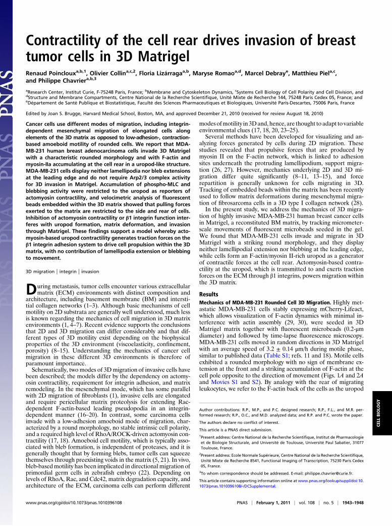
Model for MDA-MB-231 Rounded-Cell Invasion Through Matrigel.Using velocimetric analysis of seeded microbeads, we reporta unique comprehensive analysis of matrix remodeling inducedby the migration of rounded breast cancer cells within 3DMatrigel. In addition, two-color imaging of F-actin corticalstructures and microbeads with high spatial and temporal reso-lution demonstrated tight coupling to rearward movement of thecell cortex. Based on these analyses, we propose the followingmodel for MDA-MB-231 rounded-cell migration (depicted inFig. 6A). An actomyosin cytoskeleton assembled at the cell rearin a uropod-like structure contracts (ellipsoid spring), and trac-tion forces are transmitted to the surrounding matrix via β1integrin transmembrane adhesion receptors connected to the cellcortex and to radially extending F-actin bundles (arrows).Ligands for β1 integrins in Matrigel include its major componentslaminin and type IV collagen and are represented by crosses atthe end of integrins. Contractile convergent retrograde forcespull on the matrix in the rearward direction, generating forwardmovement of the cell that pushes the matrix at the front (grayarea). Cell movement induces high shear motion in the matrixand matrix remodeling (dotted area).Invasion of MDA-MB-231 cells seeded atop the reconstituted
BMproceeds through the samemechanism (Fig. 6B). Inhibition ofactomyosin activity leads to spreading of the cells on the surface ofthe matrix through integrin engagement, while invasion is blocked(Fig. 6C). Cells blunted for β1 integrin function have minimalcontact with the matrix and do not invade (Fig. 6D). Of note, thecontraction- and adhesion-based 3Dmigration of rounded cells wedescribe here appears to be distinct from the adhesion-in-dependent amoeboid migration of leukocytes in 3D (6).
Mechanics of Rounded-Cell Migration. A striking observation of thepresent study is that there were neither lamellipodial extensionsnor blebs as leading protrusions during rounded-cell movement.Blebbing is generally viewed as the driving mechanism for
amoeboid motion by allowing cells to squeeze themselves throughpreexisting voids in the matrix (5, 21). Here, based on observationof hundreds of cells by optical and electron microscopy techni-ques, blebs were restricted to the rear of the cells where acto-myosin contractility is highest. Beside the role of actomyosin ingenerating cortical tension underlying the mechanism of blebformation (5, 33), it is also possible that bleb extension was fa-cilitated by voids forming behind cells moving in the 3D matrix orat the free rear of cells invading atop Matrigel. Indeed, in cellswith blunted β1 integrin function, large blebs formed in voidsgenerated by cell-matrix detachment associated with sites of ac-tomyosin-based cell constriction (Movies S10 and S11). In addi-tion, in a control situation, matrix rigidity/porosity and pushingforce of the moving cells on the matrix may oppose to bleb ex-tension at the front. Therefore, our data support the conclusionthat blebbing is simply an epiphenomenon caused by strong ac-tivation of cortical contractility at the rear and cannot contributeper se to rounded breast tumor cell movement that appears todepend solely on actomyosin contractility.Our data indicate that lamellipodial extension and Arp2/3
complex, the main lamellipodial actin nucleating activity, are dis-pensable for MDA-MB-231 cell invasion and migration in Matri-gel (Fig. 3). In contrast, formins of the DRF family contribute bothto vertical invasion and migration of MDA-MB-231 cells in 3D
Fig. 6. Model of breast cancer round cell invasion and migration in 3DMatrigel. (A) MDA-MB-231 cells invade through Matrigel maintaining aspheroid shape. Actomyosin contractility is restricted to the cell rear (ellipsoidspring), generating tractional forces that are transmitted to the matrixthrough radial F-actin bundles (arrows) and β1 integrins bound to their matrixligands (cross at the extremity of integrins). Gray and dotted areas representregions of matrix compression and remodeling induced by cell movement,respectively. (B) Vertical invasion of cells atop a thick layer of Matrigel. (C)Inhibition of actomyosin contractility leads to cells spreading on the surface ofMatrigel. (D) Inhibition of β1 integrin binding to its ligand(s) in the matrix(closed circle at the extremity of integrins) results in spheroid cells havingminimal contact with the matrix and unable to invade. See text for details.
Poincloux et al. PNAS | February 1, 2011 | vol. 108 | no. 5 | 1947
CELL
BIOLO
GY
Matrigel (ref. 30 and this study). Interestingly, actin filamentbundles, which are connected to the substrate via focal adhesions,have been shown to assemble through DRF1-driven actin poly-merization in cells plated on a 2D substratum (34). This findingleaves open the possibility that DRFs may also play a role inrounded-cell invasion at the level of tensional force transmission tothe cortex by controlling the formation of radial F-actin bundlesconnecting the cortex to the uropod. Somewhat contrasting withthe differential implication of the two classes of actin nucleatorsduring rounded-cell invasion, both Arp2/3 complex and formin(DRF) activities are required for invadopodia formation and ma-trix proteolysis by invasive tumor cells on a rigid substrate (30, 35).Together, these data support the conclusion of at least partiallydistinct mechanisms underlying the processes of invadopodialdegradation of a rigid matrix and invasion of reconstituted BM.Nonmuscle myosin II and the actomyosin contractile appara-
tus comprise a generic force-generating system required for re-traction of the trailing edge of the moving cell, for biomechanicalcoupling to actin assembly, lamellipodial protrusion, and adhe-sion at the cell front (36, 37). Myosin II similarly plays criticalroles in tumor cell invasion in the elongated and amoeboid mi-gration modes through upstream MRCK or ROCK regulatorypathways, respectively (18, 23, 24, 38, 39). Here, we characterizethe mechanism of breast cancer cell invasion in Matrigel, whichis based on activation of the RhoA/ROCK/Myosin II cascade atthe cell rear and transmission of traction forces to the cortex,whereas blebbing is only a contractility reporter with no contri-bution to migration per se.
MethodsSee SI Methods for details on cell culture, inhibitors, siRNA treatments,antibodies, and indirect immunofluorescence analysis.
Analysis of Matrix Displacements by PIV. See SI Methods for details.
Quantification of Cell Migration and Uropod Formation. Cells were trackedmanually with ImageJ software to measure migration speeds. Dividing, ag-gregated, and out-of-focus cells were excluded from quantifications. Uropodwas defined as polarized accumulation of actin filaments decorated withmCh-Lifeact that persisted for at least 2 h over a 4-h sequence.
Scanning Electron and Transmission Electron Microscopy. See SI Methodsfor details.
Statistical Analyses. Statistical analyses were performed with SPSS (Version12.0). See the legends of Tables S1–S3 for details.
ACKNOWLEDGMENTS. We thank the PICT-IBiSA imaging facility of theInstitut Curie for help with image acquisition and processing; Drs. G. Raposo(Curie Institute) and M.-C. Prévost (Ultrastructural Microscopy Platform ofPasteur Institute) for help with transmission and scanning electron micros-copy, respectively; Drs. E. Dérivery, M. Heuzé, S. Miserey-Lenkei, M. H. Verl-hac, G. C. Tucker, C. Albiges-Rizo, and M. Bornens for providing reagents;Drs. P. Silberzan and A. Buguin for helpful discussion during this study; andDr. C. Sykes for critical reading of the manuscript. This work was supportedby grants from Ligue Nationale contre le Cancer “Equipe Labellisée 2008”and Agence Nationale de la Recherche (ANR-08-BLAN-0111) and core fund-ing support from Institut Curie and Centre National de la Recherche Scien-tifique (to P.C.). R.P. was supported by grants from Association pour laRecherche contre le Cancer (ARC) and Institut National du Cancer (INCa).F.L. and O.C. were supported by postdoctoral fellowship from Fondationpour la Recherche Médicale (FRM) and Institut Curie.
1. Friedl P, Wolf K (2003) Tumour-cell invasion and migration: Diversity and escapemechanisms. Nat Rev Cancer 3:362–374.
2. Rowe RG, Weiss SJ (2008) Breaching the basement membrane: Who, when and how?Trends Cell Biol 18:560–574.
3. Poincloux R, Lizárraga F, Chavrier P (2009) Matrix invasion by tumour cells: A focus onMT1-MMP trafficking to invadopodia. J Cell Sci 122:3015–3024.
4. Lauffenburger DA, Horwitz AF (1996) Cell migration: A physically integratedmolecular process. Cell 84:359–369.
5. Charras G, Paluch E (2008) Blebs lead the way: how to migrate without lamellipodia.Nat Rev Mol Cell Biol 9:730–736.
6. Lämmermann T, Sixt M (2009) Mechanical modes of ‘amoeboid’ cell migration. CurrOpin Cell Biol 21:636–644.
7. Petrie RJ, Doyle AD, Yamada KM (2009) Random versus directionally persistent cellmigration. Nat Rev Mol Cell Biol 10:538–549.
8. Cukierman E, Pankov R, Stevens DR, Yamada KM (2001) Taking cell-matrix adhesionsto the third dimension. Science 294:1708–1712.
9. Beningo KA, Dembo M, Wang YL (2004) Responses of fibroblasts to anchorage ofdorsal extracellular matrix receptors. Proc Natl Acad Sci USA 101:18024–18029.
10. Even-Ram S, Yamada KM (2005) Cell migration in 3D matrix. Curr Opin Cell Biol 17:524–532.
11. Zaman MH, et al. (2006) Migration of tumor cells in 3D matrices is governed by matrixstiffness along with cell-matrix adhesion and proteolysis. Proc Natl Acad Sci USA 103:10889–10894.
12. Faure-André G, et al. (2008) Regulation of dendritic cell migration by CD74, the MHCclass II-associated invariant chain. Science 322:1705–1710.
13. Doyle AD, Wang FW, Matsumoto K, Yamada KM (2009) One-dimensional topographyunderlies three-dimensional fibrillar cell migration. J Cell Biol 184:481–490.
14. Packard BZ, Artym VV, Komoriya A, Yamada KM (2009) Direct visualization ofprotease activity on cells migrating in three-dimensions. Matrix Biol 28:3–10.
15. Sabeh F, Shimizu-Hirota R, Weiss SJ (2009) Protease-dependent versus -independentcancer cell invasion programs: Three-dimensional amoeboid movement revisited. JCell Biol 185:11–19.
16. Sabeh F, et al. (2004) Tumor cell traffic through the extracellular matrix is controlledby the membrane-anchored collagenase MT1-MMP. J Cell Biol 167:769–781.
17. Wolf K, et al. (2003) Compensation mechanism in tumor cell migration: mesenchymal-amoeboid transition after blocking of pericellular proteolysis. J Cell Biol 160:267–277.
18. Sahai E, Marshall CJ (2003) Differing modes of tumour cell invasion have distinctrequirements for Rho/ROCK signalling and extracellular proteolysis. Nat Cell Biol 5:711–719.
19. Wolf K, et al. (2007) Multi-step pericellular proteolysis controls the transition fromindividual to collective cancer cell invasion. Nat Cell Biol 9:893–904.
20. Sanz-Moreno V, et al. (2008) Rac activation and inactivation control plasticity oftumor cell movement. Cell 135:510–523.
21. Fackler OT, Grosse R (2008) Cell motility through plasma membrane blebbing. J CellBiol 181:879–884.
22. Blaser H, et al. (2006) Migration of zebrafish primordial germ cells: a role for myosincontraction and cytoplasmic flow. Dev Cell 11:613–627.
23. Wilkinson S, Paterson HF, Marshall CJ (2005) Cdc42-MRCK and Rho-ROCK signallingcooperate in myosin phosphorylation and cell invasion. Nat Cell Biol 7:255–261.
24. Wyckoff JB, Pinner SE, Gschmeissner S, Condeelis JS, Sahai E (2006) ROCK- and myosin-dependent matrix deformation enables protease-independent tumor-cell invasion invivo. Curr Biol 16:1515–1523.
25. Gadea G, Sanz-Moreno V, Self A, Godi A, Marshall CJ (2008) DOCK10-mediated Cdc42activation is necessary for amoeboid invasion of melanoma cells. Curr Biol 18:1456–1465.
26. Munevar S, Wang YL, Dembo M (2001) Distinct roles of frontal and rear cell-substrateadhesions in fibroblast migration. Mol Biol Cell 12:3947–3954.
27. Gupton SL, Waterman-Storer CM (2006) Spatiotemporal feedback betweenactomyosin and focal-adhesion systems optimizes rapid cell migration. Cell 125:1361–1374.
28. Bloom RJ, George JP, Celedon A, Sun SX, Wirtz D (2008) Mapping local matrixremodeling induced by a migrating tumor cell using three-dimensional multiple-particle tracking. Biophys J 95:4077–4088.
29. Riedl J, et al. (2008) Lifeact: a versatile marker to visualize F-actin. Nat Methods 5:605–607.
30. Lizárraga F, et al. (2009) Diaphanous-related formins are required for invadopodiaformation and invasion of breast tumor cells. Cancer Res 69:2792–2800.
31. Sánchez-Madrid F, Serrador JM (2009) Bringing up the rear: Defining the roles of theuropod. Nat Rev Mol Cell Biol 10:353–359.
32. Rizvi SA, et al. (2009) Identification and characterization of a small molecule inhibitorof formin-mediated actin assembly. Chem Biol 16:1158–1168.
33. Tinevez JY, et al. (2009) Role of cortical tension in bleb growth. Proc Natl Acad Sci USA106:18581–18586.
34. Hotulainen P, Lappalainen P (2006) Stress fibers are generated by two distinct actinassembly mechanisms in motile cells. J Cell Biol 173:383–394.
35. Yamaguchi H, et al. (2005) Molecular mechanisms of invadopodium formation: Therole of the N-WASP-Arp2/3 complex pathway and cofilin. J Cell Biol 168:441–452.
36. Giannone G, et al. (2007) Lamellipodial actin mechanically links myosin activity withadhesion-site formation. Cell 128:561–575.
37. Vicente-Manzanares M, Ma X, Adelstein RS, Horwitz AR (2009) Non-muscle myosin IItakes centre stage in cell adhesion and migration. Nat Rev Mol Cell Biol 10:778–790.
38. Itoh K, et al. (1999) An essential part for Rho-associated kinase in the transcellularinvasion of tumor cells. Nat Med 5:221–225.
39. Dulyaninova NG, House RP, Betapudi V, Bresnick AR (2007) Myosin-IIA heavy-chainphosphorylation regulates the motility of MDA-MB-231 carcinoma cells. Mol Biol Cell18:3144–3155.
1948 | www.pnas.org/cgi/doi/10.1073/pnas.1010396108 Poincloux et al.