continuing education radionuclide gastroesophageal motor...
TRANSCRIPT

CONTINUING EDUCATION
Radionuclide Gastroesophageal Motor Studies*
Giuliano Mariani, MD1; Giuseppe Boni, MD1; Marco Barreca, MD2; Massimo Bellini, MD3; Bruno Fattori, MD4;Abedallatif AlSharif, MD1; Mariano Grosso, MD1; Cristina Stasi, MD3; Francesco Costa, MD3;Marco Anselmino, MD2; Santino Marchi, MD3; Domenico Rubello, MD5; and H. William Strauss, MD6
1Regional Center of Nuclear Medicine, University of Pisa Medical School, Pisa, Italy; 2Fourth Division of General Surgery,Regional Center for Diseases of the Esophagus, “S. Chiara” University Hospital, Pisa, Italy; 3Gastroenterology Unit, Departmentof Internal Medicine, University of Pisa Medical School, Pisa, Italy; 4Otolaryngology Unit, Department of Neuroscience, Universityof Pisa Medical School, Pisa, Italy; 5Nuclear Medicine Service, “S. Maria della Misericordia” Hospital of Rovigo, Rovigo, Italy;and 6Division of Nuclear Medicine, Memorial Sloan-Kettering Cancer Center, New York, New York
Disorders of the upper digestive tract have a high impact onmodern society, in terms of both direct and indirect health carecosts and of social burden. The most common presentingsymptom is either dysphagia or dyspepsia. Discriminating spe-cific diagnoses within this wide group of diseases requiressound clinical judgment and application of procedures to dis-tinguish organic from nonorganic disease and to further char-acterize the functional or motility disturbance of nonorganicdiseases. Non–radionuclide-based diagnostic techniques in-clude both noninvasive tests (upper gastrointestinal barium se-ries, ultrasonography, and breath test for gastric emptying) andinvasive procedures (fiberoptic endoscopy, esophagogas-troduodenoscopy, pharyngeal manometry, stationary esopha-geal manometry, 24-h pH monitoring, esophageal biliary refluxmonitoring, multichannel intraluminal impedance, and electro-gastrography). Some of these techniques are not well toleratedby patients or not widely available. Radionuclide transit/empty-ing scintigraphy provides a means of characterizing exquisitefunctional abnormalities with a set of low-cost procedures thatare easy to perform and widely available, entail a low radiationburden, closely reflect the physiology of the tract under evalu-ation, are well tolerated and require minimum cooperation bypatients, and provide quantitative data for better intersubjectcomparison and for monitoring response to therapy. Despite therelatively low degree of standardization both in the scintigraphictechnique per se and in image processing, these methods haveshown excellent diagnostic performance in several function ormotility disorders of the upper digestive tract. Dynamic scintig-raphy with a radioactive liquid or semisolid bolus provides im-portant information on both the oropharyngeal and the esoph-ageal phases of swallowing, thus representing a usefulcomplement or even a valid alternative to conventional invasivetests (such as stationary esophageal manometry) for evaluatingabnormalities of oropharyngoesophageal transit. Clinical appli-cations of esophageal transit scintigraphy include disorderssuch as nutcracker esophagus, esophageal spasm, noncardiac
chest pain of presumed esophageal origin, achalasia, esopha-geal involvement of scleroderma, and gastroesophageal refluxand monitoring of response to therapy (either medical or surgi-cal treatment of disease—for example, organic disease such asesophageal cancer). Scintigraphy with a radiolabeled test mealrepresents the gold standard for evaluating gastric emptying,whereas more recent radionuclide methods include dynamicantral scintigraphy and gastric SPECT for assessing gastricaccommodation. Clinical applications of gastric-emptying scin-tigraphy include, among others, evaluation of patients with dys-pepsia and evaluation of gastric function in various systemicdiseases affecting gastric emptying. The present review in-cludes the proposal of clinical algorithms for evaluating patientswith the main disorders of the upper digestive tract. Thesealgorithms, originally derived from available literature, havebeen developed on the basis of a vast clinical experience inconjunction with the specialists more deeply involved in the careof patients with such disorders (medical and surgical gastroen-terologists and nuclear medicine physicians). The role of radio-nuclide gastroesophageal motor studies is clearly identified inthe various steps of patients’ management, from the initial di-agnostic approach to functional characterization to postopera-tive follow-up or monitoring of medical therapy.
Key Words: upper digestive tract; radionuclide transit studies;quantitative parameters; diagnosis and monitoring; functionaldisorders; motility disorders; clinical algorithms
J Nucl Med 2004; 45:1004–1028
Disorders of the upper gastrointestinal tract are preva-lent in all countries. These diseases reduce the quality of lifeand often require long-term medication for control of symp-toms (1–3). In the late 1980s, about 12% of the U.S.population (approximately 34 million people) reported atleast one chronic digestive disease. Diagnosis and treatmentof these disorders caused 1.8 hospitalizations per 1,000people (4). The estimated incidence of gastroesophagealreflux disease (GERD) is about 18,600,000 cases per year inthe United States. For comparison, peptic ulcer disease hasan incidence of 6,730,000 cases per year and Barrett’sesophagus has an incidence of 808,000. In an unselected
Received Apr. 5, 2004; accepted Apr. 5, 2004.For correspondence or reprints contact: Giuliano Mariani, MD, Regional
Center of Nuclear Medicine, University of Pisa Medical School, Via Roma 67,I-56126 Pisa, Italy.
E-mail: [email protected]*NOTE: FOR CE CREDIT, YOU CAN ACCESS THIS ACTIVITY THROUGH
THE SNM WEB SITE (http://www.snm.org/ce_online) THROUGH JUNE 2005.
1004 THE JOURNAL OF NUCLEAR MEDICINE • Vol. 45 • No. 6 • June 2004
by on June 14, 2018. For personal use only. jnm.snmjournals.org Downloaded from

population the overall incidence of GERD and dyspepticsymptoms is about 45% in the United States and 30% inCanada, with the associated deterioration in the quality oflife (1).
In European countries, GERD and dyspeptic symptomsoccur in about 30% of the population. There are markedregional differences in the incidence of these disorders,ranging from 16% in Belgium and Norway to 41% inFrance (3,5).
The global cost of upper gastrointestinal tract diseases isdifficult to calculate because, in addition to the direct costs,there are the additional costs of reduced work performance,absence from work, premature retirement, and other factors.These costs are estimated to add about 30% to the direct costs(hospital stays, medical procedures, physician charges, reha-bilitation and retraining procedures, and drugs) (6). In theUnited States alone such global costs easily reach tens ofbillions of dollars each year (2), with European countriesfollowing closely (3). The cost of drugs for disorders of theupper gastrointestinal tract is more than €1.5 billion in theUnited Kingdom and $6.5 billion in the United States (2,3).Substantial savings can occur by optimizing diagnosis andtreatment. Optimal treatment requires accurate characterizationof the underlying disorder. Radionuclide methods are wellsuited for evaluating this group of diseases.
Upper gastrointestinal disease often has symptoms ofdysphagia (difficulty swallowing) and dyspepsia (pain ordiscomfort in the upper abdomen). These symptoms arefrequently due to impairment of motor function. Radionu-clide gastroesophageal motor studies are well suited foridentifying and characterizing these disorders semiquantita-tively and for monitoring the efficacy of therapy. Under-standing the anatomy and physiology of the upper gastro-intestinal tract helps in selecting the procedure most suitablefor each patient and in interpreting the results.
ANATOMY
The adult esophagus is a hollow muscular canal thatvaries in length with patient height but averages about 25cm, with a diameter of about 2.5 cm. It begins in the neckat the lower border of the cricoid cartilage, extends throughthe diaphragm, and ends at the cardia of the stomach. Theesophagus is perfused by branches from the inferior thyroidartery, descending aorta, left gastric branch of the celiacartery, and inferior phrenic artery of the abdominal aorta.There is both superior and inferior lymphatic drainage.Muscular activity is under the control of the autonomicnervous system, with parasympathetic innervation throughthe vagus and sympathetic innervation through pregangli-onic fibers originating at T1–T10, mostly between T4 andT6 (Fig. 1). The stomach is a hollow muscular viscus about30 cm long and 15 cm wide, with a capacity of about 1 L inthe resting state to a maximum of about 5 L. The mucosa ofthe stomach has more than 35,000 gastric glands, producingmucus, hydrochloric acid, and digestive enzymes. Bloodvessels to the stomach arise from the common hepatic, leftgastric, and splenic arteries to form 2 vascular arcades,which perfuse the greater and lesser curvature of the stom-ach. Lymph drains from the stomach to the left and rightgastric nodes and the subpyloric and omental nodes, whichdrain to the hepatic and celiac nodes. The stomach has bothsympathetic and parasympathetic innervation, which con-trols both the secretory and the motor activity of the organ.
PATHOPHYSIOLOGY OF MOTOR FUNCTION
Oropharyngeal Motor FunctionThe normal esophagus propels a bolus of solid or liquid
from the mouth to the stomach. Liquids require approxi-mately 2 s to traverse the esophagus, whereas solids cantake about 9 s. The process begins with swallowing. Of the
FIGURE 1. (A) Anatomic drawing showsthe complex afferent and efferent neuro-regulation of deglutition (sensory fibers inblack, motor fibers in different colors) andcranial nerves participating in the system.(B) Summed image from dynamic record-ing of oropharyngoesophageal radionu-clide transit study (liquid bolus) allows clearidentification of the various anatomic re-gions. Upper arrow indicates posteriormouth sphincter, lower arrow indicates up-per esophageal sphincter, and space be-tween arrows is pharyngeal region. Duringscintigraphic acquisition, patient stoodfacing collimator surface for an anteriorview of the chest, with head and neck tiltedleft. (Modified from (235).)
RADIONUCLIDE GASTROESOPHAGEAL MOTOR STUDIES • Mariani et al. 1005
by on June 14, 2018. For personal use only. jnm.snmjournals.org Downloaded from

6 phases that constitute swallowing, only the first phase istotally voluntary: positioning of the bolus on the tongue’scentral groove and propulsion of the bolus toward andthrough the faucial arches (oral phase) (7). The subsequentphases are involuntary and are initiated by elevation andretraction of soft palate, with closure of the nasopharynxassociated with simultaneous opening of the posteriormouth sphincter (oropharyngeal phase). The next 2 steps(proximal and distal pharyngeal phases) take the bolus intothe hypopharynx (8). The most important and delicate taskof these 2 phases is avoiding passage of food through thelarynx into the respiratory tree (9). Aspiration is avoidedthrough a complex, multistep process involving a reflexmechanism consisting of sensitive afferences and articu-lated motor efferences (10). The pharyngoesophageal phaseis marked by progression of the bolus through the striatedcricopharyngeus muscle, or upper esophageal sphincter,whose function is to avoid reflux of the esophageal contentback into the pharynx (11,12). Although the first phases ofswallowing are concentrated in a relatively short spaceimmediately posterior and inferior to the mouth, their phys-iologic coordination requires complex interaction of mus-cular and nervous structures. Coordinating this process re-quires participation of the IV, V, IX, X, and XII cranialnerves and of sympathetic fibers originating mostly atT4–T6 (Fig. 1). Oropharyngeal dysphagia can be caused bydegenerative disorders of the central nervous system (Par-kinson’s disease, multiple sclerosis, and supranuclearpalsy), of the spinal motor neuron (amyotrophic lateralsclerosis), of the neuromuscular junction (myasthenia gra-vis), or of striated muscle per se (polymyositis and musculardystrophy) (13,14).
Esophageal Motor FunctionThe last step of swallowing (the esophageal phase) in-
volves the traveling of a much longer distance by the bolusyet involves a much simpler peristaltic mechanism aided bygravity. The primary peristaltic pump originating in thepharynx propagates through the inner (circular) and theouter (longitudinal) muscular layers of the esophagus atabout 4 cm/s. Coordination of the primary peristaltic pumpwith postswallowing relaxation of the lower esophagealsphincter (LES) lets the bolus enter the stomach (15). Dis-orders of peristalsis can be caused by derangement of motorinnervation provided by the vagus nerve for the striatedmuscle fibers, derangement of the sympathetic and para-sympathetic innervation provided for the smooth muscle,and derangement of the myenteric plexus (Table 1) (16).
Functional esophageal disorders are a heterogeneousgroup of chronic disturbances often complicated by associ-ation with psychiatric or psychologic disorders, as deter-mined on the basis of a lack of identifiable structural ormetabolic damage (Table 2). After irritable bowel syn-drome, esophageal disorders are the second most frequentfunctional gastrointestinal disorder and are usually not as-
sociated with GERD or other structural, metabolic, or motordisorders (17,18).
Achalasia is a primary esophageal motility disorder ofunknown cause. The disease results in partial or absence ofrelaxation of the LES and loss of esophageal-body peristal-sis. It is characterized by progressive neuronal degenerationin the myenteric plexus, with a paucity of ganglion cells, thepresence of neural fibrosis, and some degree of chronicinflammation at histology. There is also a significant de-crease in the synthesis of nitric oxide, a mediator of relax-ation in the LES. Dysphagia and varying degrees of regur-gitation, weight loss, and chest pain are the most commonclinical presentation. Achalasia may present at any time,with the highest incidence occurring between 20 and 50 y ofage. Most patients seen today in a surgical practice have haddilation or botulinum toxin injections before. Nevertheless,having undergone previous therapies should not be regardedas proof that a patient has achalasia. In fact, one can confusethe clinical manifestations of achalasia with those of otheresophageal disorders; thus, patients must have, in additionto dysphagia and regurgitation, the radiologic and mano-metric findings consistent with achalasia.
GERD is common among the populations of industrial-ized countries (19). Heartburn, the most common symptomof gastroesophageal reflux, occurs daily in approximately10% of the population and is the major reason for theconsumption of antacids in our society. GERD can beoverlooked for several reasons: First, the most commonsymptoms, heartburn and regurgitation, occur in only half ofthese patients; second, because of the natural history of thedisease and the frequency of its spontaneous remission,many patients will not seek medical advice; and third, thereis no diagnostic standard for GERD.
Symptoms of GERD are divided into 2 categories: esoph-ageal, which includes typical esophageal reflux complaints(heartburn, regurgitation, and dysphagia) and noncardiacchest pain, and extraesophageal, which includes pulmonary(asthma and recurrent aspiration pneumonia), otolaryngo-logic (hoarseness, chronic dry cough, chronic sore throat,and globus sensation), and dyspeptic symptoms (upper ab-dominal pain, nausea, vomiting, and bloating).
The work-up of patients with gastroesophageal symptomsshould include confirmation of the presence of pathologicgastroesophageal reflux and exclusion of other motility dis-orders or lesions of the esophagus and stomach, quantifica-tion of the severity of reflux, and clear definition of theanatomy of the esophagus and gastroesophageal junction.
Gastric Motility and EmptyingGastric Motor Function. Motor functions of the stomach
result from a complex interaction of muscular and neuralactivity in physiologically distinct regions, integrated withfeedback regulation from the small bowel (20). The stomachcan be divided into 3 functional regions: the proximalstomach (cardia, fundus, and proximal body), the distalstomach (distal body and antrum), and the pylorus.
1006 THE JOURNAL OF NUCLEAR MEDICINE • Vol. 45 • No. 6 • June 2004
by on June 14, 2018. For personal use only. jnm.snmjournals.org Downloaded from

The proximal stomach regulates gastric emptying by ac-commodating and storing food and by regulating intragas-tric pressure and tonic propulsion of chyme into the distalstomach. Smooth muscles of the proximal stomach do notexhibit rhythmic fluctuations in their membrane potentialbut, rather, are in a state of continual partial contraction, ortone. This motor activity of the proximal stomach maintainsa stable intragastric pressure even after consumption of alarge meal; in fact, the stomach can accommodate up to 2 Lof fluid with less than a 10–mm Hg increase in intragastricpressure.
This property is mediated by 2 neurally mediated re-flexes: receptive relaxation (vagovagal reflex reducing gas-tric tone in response to swallowing) and gastric accommo-dation. The latter reflex is elicited in response to gastricdistension and is mediated by stimulation of mechanorecep-tors in the gastric wall.
In contrast to the proximal stomach, the distal stomachexhibits electrical and contractile activity characterized byrhythmic oscillation in the membrane potential, accompa-nied by phasic, rather than tonic, motor activity. This reg-ular rhythmic depolarization, known as pacesetter potentials(21,22), originates in the interstitial cells of Cajal (network
of specialized cells extending from the corpus to the distalantrum) and initiates gastric peristalsis. The gastric pace-setter potential (which propagates in both distal and circum-ferential directions but not proximally to the fundus) has abaseline frequency of 3 cycles per minute and is modulatedby parasympathetic (vagal) and sympathetic innervation, inaddition to the intrinsic enteric neurons organized as themyenteric and submucosal plexuses (23). Motor activity ofthe distal stomach results in mixing, grinding, and tritura-tion of solid food (ingested food is propelled distally byantral contractions, only to be repelled back into the moreproximal stomach) and also regulates gastric emptying. Forequivalent volumes of ingested food, the amplitude of pha-sic contractions is more intense in response to particulatethan to homogenized material. Additional neural and hor-monal factors modulate postprandial gastric motility.
The pylorus is a specialized region of the stomach at thejunction of the antrum with the duodenal bulb, which acts asa sieve, regulating outflow of intraluminal gastric content.Because of the thickness of the smooth muscle layers andthe presence of highly redundant mucosa, the pylorus acts asa mechanical stricture preventing the passage of large par-ticles.
TABLE 1Classification of Gastroesophageal Motor Disorders
Demonstrable abnormality Clinical entity Associated disorder
1. Esophageal dysmotilityCategory 1: Well-defined entities
1.1.1 Excessive acid exposure GERD Scleroderma, diabetes mellitus1.1.2 Manometric pattern of achalasia Achalasia Chagas’ disease, enteric neuropathy1.1.3 Spastic manometric pattern Esophageal spasm Diabetes mellitus, enteric neuropathy
Category 2: Entities with variable dysfunction-symptomrelationship
1.2.1 High-amplitude peristalsis Nutcracker esophagus Enteric neuropathy1.2.2 Low-amplitude peristalsis
Failed peristalsisLow-amplitude simultaneous contractions
Ineffective esophagealmotility
Scleroderma, enteric myopathy, diabetesmellitus, amyloidosis, GERD
1.2.3 Low LES pressure Hypotensive LES Scleroderma, diabetes mellitus, GERD1.2.4 Incomplete LES relaxation LES dysrelaxation After fundoplication
Category 3: Questionable entities1.3.1 High LES pressure Hypertensive LES
Category 4: Entities associated with behavioraldisorders
1.4.1 Forced regurgitation Rumination syndrome Anorexia nervosa (purging type), bulimia nervosa(purging type)
1.4.2 Excessive air swallowingExcessive belching
Aerophagia GERD
2. Gastric dysmotilityCategory 1: Well-defined entities
2.1.1 Accelerated gastric emptying Dumping syndrome After resection dumping, or vagotomy dumpingCategory 2: Entities with variable dysfunction-symptom
relationship2.2.1 Delayed gastric emptying Gastroparesis GERD, diabetes mellitus, scleroderma, after
vagotomy, enteric neuropathy, entericmyopathy, anorexia nervosa (restricting type)
Modified from (39).
RADIONUCLIDE GASTROESOPHAGEAL MOTOR STUDIES • Mariani et al. 1007
by on June 14, 2018. For personal use only. jnm.snmjournals.org Downloaded from

Gastric Emptying. Because the stomach handles solidsand liquids differently, liquid gastric emptying and solidgastric emptying can be specifically described.
Inert liquids, such as water, empty exponentially; thus,the volume of fluid emptied into the duodenum in a giventime is a constant fraction of the volume remaining in thestomach. The emptying rate is modified by volume, osmo-lality, pH, caloric density, and nutrient content of the liquid.Although interesting from the physiologic point of view,evaluation of liquid emptying per se is of little if any clinicalsignificance (20,24).
After ingestion of a meal, solids are kept in the stomachto be ground and triturated into fine particles. During thisphase, there is no emptying. This interval has been calledthe lag phase. In addition to being reduced into a finelydispersed suspension of particles, the solid material is con-verted into chyme because of extensive contact with gastricacid and peptic enzymes. When the solid particles are �1–2mm in diameter, a linear emptying phase commences duringwhich the chyme is slowly delivered to the duodenum(25–29). Physical and nutritional properties may modify therate of delivery of solid food to the small bowel by modu-lating the duration of the initial lag phase. Larger particlesprolong the lag phase, whereas evenly dispersed suspen-sions have a relatively short lag phase; however, after theinitial lag phase a homogenized meal and a nonhomog-enized meal empty at a similar rate (30). The emptying rateis also affected by the caloric content of the meal and itscomposition of total fats, triglycerides, and carbohydrates(26,27). Although the volume of liquid ingested with the
solid food modifies the rate at which the solid food isdelivered to the intestine, the liquid component is emptiedmore rapidly than the solid component, suggesting that thestomach can distinguish between the 2 phases when presentsimultaneously (31). The gastric-emptying rate can also bemodified by additional factors such as age, sex, menstrualcycle, and time of day (32–36). Although variable, the lagphase of gastric emptying is prolonged in some diseasestates, such as diabetic gastroparesis (37), and is shortenedafter antrectomy and pyloroplasty or after administration ofmetoclopramide and domperidone (38).
Gastric neuromuscular disorders, such as visceral hyper-sensitivity, gastric dysrhythmias, gastric dysrelaxation, an-tral hypomotility, pylorospasm, and gastroparesis, can causedysmotility-like dyspepsia characterized by various symp-toms such as early satiety, fullness, abdominal discomfort,bloating, nausea, and vomiting (23,39). The other possibleface of the dyspepsia syndrome is represented by ulcer-likesymptoms, whose predominant feature is pain centered inthe upper abdomen.
When considering a patient with either dysmotility-likedyspepsia or ulcer-like dyspepsia, one should keep in mindthat dyspepsia can either be the expression of a well-definedorganic condition (peptic ulcer, GERD, malignancy, hepa-tobiliary disease, or side-effects of drugs) or have no asso-ciation with definite structural or biochemical disorders(functional dyspepsia). Therefore, functional dyspepsiashould always be considered after other organic conditionshave been excluded (40,41).
TABLE 2Classification of Functional Gastroesophageal Disorders
Disorder Definition
Esophageal disordersA1. Globus Sensation of a lump, something stuck, or tightness in the throatA2. Rumination syndrome Regurgitation of recently ingested food into the mouth with
subsequent remastication and reswallowing or spitting out, inthe absence of structural disease
A3. Functional chest pain of presumed esophageal origin Episodes of chest pain, usually midline, of visceral qualityA4. Functional heartburn Episodic retrosternal burning in the absence of pathologic
gastroesophageal reflux, pathology-based motility disorders,or structural explanations
A5. Functional dysphagia Sensation of abnormal bolus transit through the esophagealbody, in the absence of structural abnormality, pathologicreflux, or pathology-based motility disturbance
A6. Unspecified functional esophageal disorder
Gastroduodenal disordersB1. Functional dyspepsia
B1a. Ulcer-like dyspepsiaB1b. Dysmotility-like dyspepsiaB1c. Unspecified (nonspecific) dyspepsia
B2. AerophagiaB3. Functional vomiting
Modified from (17).
1008 THE JOURNAL OF NUCLEAR MEDICINE • Vol. 45 • No. 6 • June 2004
by on June 14, 2018. For personal use only. jnm.snmjournals.org Downloaded from

NON–NUCLEAR MEDICINE EVALUATIONS
Upper Gastrointestinal SeriesUpper gastrointestinal radiography with barium contrast
medium allows examination of the esophagus, stomach, andduodenum, whereas special protocols have been developedto evaluate the oropharyngeal phase of swallowing. This isthe procedure most patients who complain of dysphagia stillundergo before any other testing. On the other hand, currentdiagnostic protocols call for esophagogastroduodenoscopyas a first-line approach for patients who complain of heart-burn or regurgitation, chest pain, or epigastric pain or haveunexplained vomiting or severe indigestion. This leaves amore limited role than in the past for an upper gastrointes-tinal series for these symptoms. Barium radiography is nowconsidered a second-line diagnostic test whenever endos-copy reveals no obvious abnormalities explaining thesesymptoms.
Different barium suspensions (solid, semisolid, or liquid)can be used to characterize the parameters of swallowing.Although capable of identifying the major categories ofdysfunction in patients with pharyngoesophageal dysphagia(42), the examination cannot estimate muscle fatigue, mea-sure pharyngeal contractile forces, or estimate the intraboluspressure during swallowing (12).
The esophagogram should include upright double-con-trast views with a high-density barium suspension to assessmucosal disease, and prone single-contrast views with alow-density barium suspension to assess distensibility andmotility. Barium swallows allow a dynamic evaluation ofesophageal motility and transit of the barium boluses fromthe mouth to the stomach through the entire esophagus.Cine- and videoradiography (possibly recorded as digitalvideofluorography) help in evaluating functional disordersof the pharyngoesophageal and the esophageal phases ofswallowing. Particular attention is paid to evaluation of thegastroesophageal junction and hiatus through varying of thepatient position (e.g., oblique, standing, and supine).
An upper gastrointestinal series should always includeevaluation of the stomach and duodenum, even if symptomssuggest a primarily esophageal disorder.
UltrasonographyThe oral phase of swallowing can be evaluated by posi-
tioning the ultrasonography probe under the chin to thehyoid region, using transverse and longitudinal scans tovisualize the tongue and mouth floor both at rest and duringswallowing of a bolus (43,44). This procedure can be usedto identify various abnormalities, such as inability to keepthe bolus in the mouth, lack of backward propulsion of thebolus, and asymmetric contraction of the tongue.
Although a simple ultrasonography examination can giveuseful information on visceral wall thickness, a functionalevaluation is needed for patients with upper gastrointestinaldisorders to gain data on esophageal and gastric motility. Aswith other diagnostic ultrasonographic applications, the testis highly dependent on the operator’s skill; in addition, the
test is relatively time consuming because it requires re-peated and prolonged observations (45,46).
The proximal (cervical) tract of the esophagus and itsdistal tract (gastroesophageal junction) can be explored inchildren. Ultrasonography can detect gastroesophageal re-flux in children up to 5 y of age with 100% sensitivity and87.5% specificity (47).
Even though air in the gastrointestinal tract and a thickabdominal wall can interfere with visualization of the fundicand antral regions, ultrasonography can evaluate gastricvolume and emptying and transpyloric flow (45,48). A highcorrelation has been found between gastric-emptying valuesevaluated by ultrasonographic procedures and those derivedby radionuclide-based procedures (49). In addition, ultra-sonography can measure gastric area and volume and de-tects gastric contraction and distension (50).
Functional ultrasonography of the stomach is indicatedmainly for evaluation of patients with dysmotility-like dys-pepsia (45), evaluation of both dyspeptic and nondyspepticpatients with chronic disease potentially causing delayedgastric emptying (e.g., diabetes mellitus, systemic sclerosis,and myotonic dystrophy) (51,52), monitoring of the effectsof pharmacologic and nonpharmacologic agents potentiallyaffecting gastrointestinal motility (53), and evaluation ofnewborns with suspected hypertrophic pyloric stenosis (54).
Breath Test for Gastric EmptyingThe use of breath tests in gastroenterology has become
increasingly popular since stable isotopes such as 13C wereintroduced. Mass spectrometers are required to measure theconcentration of these nuclides in expired air. These instru-ments are now widely available (at relatively low cost) andeasy to operate. This technologic evolution has caused the�-emitter 14C to largely be replaced by the mass isotope 13C.This change avoids the need for extensive isotope inven-tories and record keeping, eliminates radiation exposure forboth patients and personnel, and allows gastroenterologiststo perform these procedures in their own environment.
Combined with different food substrates, octanoic acid(solid meal) or acetate (liquid meal) labeled with 13C areused to assess gastric emptying. The underlying concept isthat these compounds pass unabsorbed through the stomachto the duodenum, where they are quickly absorbed. Afterabsorption in the duodenum, portal circulation transportsthe 13C-labeled substrate to the liver, where fast metabolicdegradation produces 13CO2, which is excreted with exhaledair. Breath is therefore tested for enrichment with13CO2 atregular intervals for up to 6 h, thus deriving the basicparameters of 13CO2 appearance in the breath (beginningof gastric emptying) and time-related enrichment (a ris-ing curve whose slope is related to the gastric-emptyingrate) (55,56).
The test is safe and noninvasive, can be performed onchildren and during pregnancy, and can be repeated when-ever necessary, as when monitoring the efficacy of therapyor assessing the effects of drugs on gastric emptying (56).
RADIONUCLIDE GASTROESOPHAGEAL MOTOR STUDIES • Mariani et al. 1009
by on June 14, 2018. For personal use only. jnm.snmjournals.org Downloaded from

Because the breath test is not an imaging technique, infor-mation is not provided about intragastric distribution of thedifferent phases of the meal. Moreover, considering gastricemptying as the sole limiting step of the delivery of 13CO2
to the breath can be misleading (thus reducing the reliabilityof the test) in certain conditions such as malabsorption; indiseases of the pancreas, liver, or lungs; or in the presenceof visceral hemodynamic changes (e.g., physical exercise)(46,56).
Fiberoptic EndoscopySmall, dedicated fiberoptic endoscopes are available to
directly visualize all mucosal surfaces of the nasopharynx,pharynx, and larynx. Nasoendoscopy is minimally invasive,repeatable, and easy to perform and allows bedside evalu-ation for nonambulatory patients. Although this techniquecan evaluate both the motor and the sensorial components ofswallowing, it can examine only the pharyngeal phase, withthe additional limitation of a swallowing blackout (57).
A recent variant of the technique uses endoscopic deliv-ery of pulsated air on the laryngeal mucosa to estimate theadductional reflex contraction of vocal folds or the patient’sability to discriminate increasing pressure (58).
EsophagogastroduodenoscopyThis is the procedure that most gastroenterologists and
surgeons choose when evaluating patients with gastroesoph-ageal disorders, as it permits direct visualization of theesophageal, gastric, and duodenal mucosa and permits tis-sue biopsies of suggestive lesions. Endoscopy is indicatedfor any patient complaining of dysphagia or dyspepsia,generally as a first-line diagnostic procedure. More rarely(depending on the choice of the general practitioner andlocal logistics), endoscopy is performed after an upper gas-trointestinal series revealing no obvious abnormalities.
The gastroesophageal flap valve is easily seen duringesophagogastroduodenoscopy, thus revealing esophagitisand abnormal hiatal and paraesophageal hernias. Esopha-geal diverticula can also be visualized during endoscopy.This invasive technique is not always well accepted bypatients and requires a relatively long learning curve for theoperator. A consensus has been reached on the grading ofmucosal damage and esophagitis, thus minimizing interob-server variability and subjectivity in reporting results (59).However, about 55% of patients with typical GERD-relatedsymptoms do not have gross esophagitis, although carefulendoscopic examination may detect, in about 12% of pa-tients with GERD, the presence of intestinal metaplasia(Barrett’s esophagus, a condition associated with a 40-foldincreased probability of development of esophageal adeno-carcinoma).
Pharyngeal ManometryA transnasally positioned manometric probe permits es-
timation of the rate of upper esophageal sphincter relaxationand the strength of pharyngeal contraction while also mea-suring the duration of these 2 events (60). Manometry can
be performed at the time of videofluoroscopy (manofluorog-raphy) for better positioning of the pressure sensor and fordistinguishing between recordings of intrabolus pressureand recordings from within a closed lumen (8).
Pharyngeal manometry is more difficult to perform thanthe esophageal test because of the extreme longitudinal andradial asymmetry of intraluminal pressures recorded fromwithin the pharynx during deglutition (61). Moreover,movements during the pharyngeal phase of swallowingfrequently displace the pressure sensor, thus making thetechnique difficult to standardize (62).
Stationary Esophageal ManometryEsophageal manometry is usually performed on patients
with dysphagia in whom endoscopy or a barium swallowstudy has excluded obvious structural abnormalities. Thedynamic pressure measurement is especially useful in thediagnosis of primary esophageal motility disorders such asachalasia, diffuse esophageal spasm, nutcracker esophagus,or hypertensive LES. It is also useful in the characterizationof esophageal disorders secondary to systemic diseases suchas scleroderma, dermatomyositis, and polymyositis.
Although esophageal manometry is rarely indicated inpatients with typical GERD-like symptoms, it is crucial forcorrect placement of the probe for esophageal pH testing inpatients with atypical symptoms or unresponsive to propermedical therapy and in patients being considered for anti-reflux surgery. Esophageal manometry may identify a de-fective LES, suggesting the diagnosis of GERD, and pro-vides valuable information on peristaltic function (thusallowing selection of the most appropriate antireflux proce-dure).
Ambulatory pH MonitoringA pH electrode at the end of a catheter is placed in the
esophagus 5 cm above the upper limit of the LES forprolonged monitoring of esophageal pH. The pH electrodeis attached to a battery-operated ambulatory device that canrecord data for up to 24–48 h. This device records pH overa circadian cycle to identify the frequency and duration ofesophageal mucosa exposure to acid and the ability of theesophagus to clear gastric reflux and allows correlation ofthe intensity and duration of symptoms with reflux episodes.
Esophageal exposure to gastric juice is evaluated as 6major components contributing to a combined score: thepercentage of the total time, upright time, and supine timethat esophageal pH drops below 4; the total number ofreflux episodes per day; the number of episodes � 5 min;and the duration of the longest episode (63,64).
Biliary RefluxThe combination of bile and hydrochloric acid has a
noxious effect on esophageal mucosa. An ambulatory bile-reflux monitor (Bilitec 2000; Medtronic) has been devel-oped to detect the presence of bile reflux in the esophagus(65,66). Using bilirubin as a marker for bile, this spectro-photometric system records the frequency and duration of
1010 THE JOURNAL OF NUCLEAR MEDICINE • Vol. 45 • No. 6 • June 2004
by on June 14, 2018. For personal use only. jnm.snmjournals.org Downloaded from

bile exposure in either the stomach or the esophagus over a24-h period. Combined with 24-h pH testing, the bile-refluxdetector gives a more complete profile of a patient’s reflux,thus identifying patients at greater risk for developing com-plications such as Barrett’s esophagus. Nevertheless, refluxalone of bile in the esophagus cannot be considered themain risk factor for such complications.
Reflux of duodenal content into the stomach, possiblycausing alkaline gastritis and intestinal metaplasia in thegastric mucosa, occurs in up to 40%–50% of patients whoundergo cholecystectomy (Fig. 2). This condition can alsobe secondary to gastric surgery that alters the function of thepylorus. As already emphasized, reflux of duodenal andgastric contents into the esophagus can play a role in thepathogenesis of Barrett’s esophagus.
Multichannel Intraluminal Impedance (MII)In this new technique for evaluating esophageal function
and gastroesophageal reflux, a change in resistance to alter-nating current between 2 metal electrodes is produced bythe presence of a bolus inside the esophageal lumen (67,68).Combined MII and esophageal manometry provide simul-taneous information on intraluminal pressure changes andbolus movement, whereas combined MII and pH monitor-ing allow detection of reflux episodes irrespective of theirpH values (i.e., acid vs. nonacid reflux). Combined MII andpH testing shift the gastroesophageal reflux testing para-digm, in that reflux events are no longer detected solely bypH changes. In fact, reflux presence, distribution, and clear-ance are detected primarily by MII and are characterizedsimply as acid or nonacid on the basis of pH change and asliquid, gas, or mixed on the basis of MII.
On the other hand, combined MII and esophageal ma-nometry identify those patients with abnormal esophagealmotility on manometry who actually have a clinically im-portant defect in esophageal function. In fact, whereas ma-nometry is an indirect measure of esophageal function,impedance makes it possible to follow the bolus movement.
Impedance technology has great potential for identifyingthe relative roles of acid and nonacid reflux, such as inpatients who continue to have reflux-related symptoms eventhough their acid is being successfully suppressed or con-
trolled with traditional acid-suppressant therapy. This tech-nique can also help in assessing patients with reflux-relatedrespiratory problems and in understanding dysphagia and itsmanometric and clinical correlates.
ElectrogastrographyBy using mucosal, serosal, or cutaneous electrodes, this
technique evaluates the fasting and postprandial myoelec-trical activity generated by the gastric antrum (69). Cutane-ous electrogastrography is most frequently used clinicallythrough the positioning of 3 or 4 electrodes on the epigas-trium (46). The normal gastric pacesetter potentials rangefrom 2.5 to 3.75 cycles per minute (cpm). Either brady-gastria (�2.5 cpm), tachygastria (3.75–10 cpm), or brady-tachygastric arrhythmia (mixed pattern) is detectable (69).Such gastric dysrhythmias can be observed in dysmotility-like dyspepsia, idiopathic gastroparesis, diabetes mellitus,nausea of pregnancy, and motion sickness and after drugintake or gastric surgery (69), whereas increased wave am-plitude has been reported in gastroparesis due to mechanicalobstruction (70). Electrogastrography has a complementaryrole in evaluating patients with dyspeptic symptoms, inpredicting gastroparesis, and in assessing gastric motorfunction in patients with GERD or with chronic constipationor atypical upper-gastrointestinal symptoms (46).
ESOPHAGEAL TRANSIT SCINTIGRAPHY
Esophageal scintigraphy was first introduced by Kazemin 1972 (71). Esophageal scintigraphy performed with alarge-field �-camera views the entire esophagus. The im-ages can quantitate rates of emptying and reflux in allsegments simultaneously (72,73). Using esophageal ma-nometry as the gold standard, sensitivities and specificitiesup to 95% and 96%, respectively, have been reported(74,75), whereas some investigators found esophageal scin-tigraphy to be more sensitive than esophageal manometryitself or contrast radiology (76,77). Such discrepant resultsare probably due to the application of different protocols forimaging and data analysis. Relevant differences includebolus consistency, volume and temperature, patient pop-ulation, single or multiple swallows, display methods,quantitative parameters, and thresholds used in decisionmaking (75).
Because esophageal scintigraphy is not standardized, wewill describe various approaches at reaching an acceptabledegree of standardization.
Scintigraphy ProcedurePatient Preparation. Esophageal scintigraphy should be
performed after a fast of at least 3 h but preferably over-night. The patient should rehearse the procedure with apractice swallow using unlabeled plain water. This ap-proach helps educate patients about the procedure to en-courage their enthusiastic participation.
Image Acquisition. A large-field-of-view �-camera fittedwith a low-energy, general-purpose collimator is adequate.
FIGURE 2. Delayed abdom-inal hepatobiliary scan (45 minafter injection, when radioac-tivity has almost cleared fromhepatocellular compartment)obtained with a 99mTc-iminodi-acetic acid analog for a pa-tient with persisting dyspepsiaafter endoscopic cholecys-tectomy. Reflux of radioactivebile from duodenum intostomach is obvious (arrows),possibly explaining persis-tence of symptoms.
RADIONUCLIDE GASTROESOPHAGEAL MOTOR STUDIES • Mariani et al. 1011
by on June 14, 2018. For personal use only. jnm.snmjournals.org Downloaded from

Because high temporal resolution is preferred for quantita-tive studies, a high-sensitivity collimator should be usedwhenever available. Dynamic images in either a 64 � 64 or128 � 128 matrix must be acquired in a rapid sequence.Because many of the events occur in a short time, imagesshould be acquired at 4–10 frames per second for 60 s. Thedata can be summed if necessary, but high temporal reso-lution is necessary to identify brief episodes of reflux ordelays in transit through segments of the esophagus.
Although esophageal scintigraphy can be performed withthe patient supine or upright, it is more “physiologic” tostudy patients who are seated and upright. Obviously, quan-titative parameters derived from the study (transit time andretention indices) vary according to the position adopted(78 – 81). Although the upright position mimics the phys-iologic condition of swallowing and esophageal empty-ing, by eliminating the effects of gravity the supineposition may allow easier demonstration of esophagealmotility disorders (82).
Both the anterior and the posterior views have been usedfor esophageal scintigraphy. The ideal view should mini-mize tissue attenuation and keep it constant along the entirelength of the esophagus. In a cross-section of the body, theesophagus is situated rather anteriorly at its most proximalportion, with a posterior-to-anterior depth ratio of 2.3; itmoves posteriorly at its mid portion, where it lies behind theheart, and anteriorly again at its distal extreme. In theanterior view, tissue attenuation is initially relatively lowbut increases significantly after the middle third, especiallybecause of interposition of the heart. In the posterior view,although tissue attenuation is more significant, it is moreuniform along the whole esophageal length. This is espe-cially advantageous because accurate monitoring is possiblewhen the counting rate is relatively independent of distri-bution within the organ (83). On the other hand, anteriorimaging is preferred when the oral and pharyngeal phasesare being evaluated, because the mouth and pharynx can bebrought closer to the detector surface. External markers onthe cricoid cartilage may help improve anatomic localiza-tion of the pharynx. An optimum solution uses a dual-head�-camera with the patient positioned to allow simultaneousrecording of anterior and posterior views. The correspond-ing pixels in each view are combined to create a geometricmean image, which is used for analysis.
Radioactive Bolus. Any radiopharmaceutical that is notabsorbed by the gastrointestinal tract, including 99mTc-sulfurcolloid, 99mTc-nanocolloid, and 99mTc-diethylenetriamine-pentaacetic acid (DTPA), can be used to prepare a radioac-tive bolus for esophageal scintigraphy. A dose of at least7–11 MBq (200–300 �Ci) of the 99mTc-labeled agent isusually mixed with water or juice (to form a radioactiveliquid bolus) or with a semisolid medium. Most esophagealscintigraphic studies have been performed using a liquidbolus, whereas few studies have used a semisolid bolus(75,84), probably because of the difficulty in reaching aconsensus on the viscosity and type of semisolid bolus. In
our experience, homogenized baby paste or “gelified water”(available for patients with swallowing difficulties, e.g., inthe rehabilitation phase after pharyngolaryngeal surgery) isan optimal viscous semisolid meal, provided attention ispaid to keep the bolus viscosity constant and to avoidpossible fragmentation.
The pharyngeal ejection force is sufficient to propel theliquid bolus to the gastroesophageal junction, leaving minorwork to be done by peristalsis. A more viscous, semisolidbolus is propelled only to the proximal half of the esopha-gus, thus requiring more intense peristalsis to completetransport over the distal half (85). Therefore, a semisolidbolus is more challenging for assessing esophageal transit,thus resulting in increased sensitivity of the test. When aliquid bolus was compared with a semisolid bolus (homog-enized baby meal), the latter showed higher sensitivity atcomparable levels of specificity, thus suggesting a prefer-ence for semisolid over liquid boluses (75).
Solid boluses consisting of radiolabeled gelatin capsulesor chicken liver cubes have been proposed, but such bolusescan remain in the esophagus for as long as 2 h despiterepeated dry swallows, even in subjects with normal esoph-ageal function. Esophageal scintigraphy performed with asolid gelatin bolus ingested with water demonstrated abnor-malities in half the patients with dysphagia and normalesophageal manometry, barium radiology, and pH studies,thus resulting in high sensitivity for the test. The clinicalsignificance of this finding, however, is uncertain (77,86–88).
The volume of the bolus also requires standardization;healthy individuals can easily ingest a liquid 20-mL bolus ina single swallow (89); failing to pass a liquid bolus smallerthan 20 mL from the mouth to the esophagus in a singledeglutition might reflect impaired oropharyngeal swallow-ing (90). Esophageal transit may vary substantially accord-ing to the bolus volume, as 10-mL boluses were shown totravel more quickly than 20-mL boluses in the upright butnot in the supine position (91). In addition, the larger bolusincreased the swallow interval required to reestablish nor-mal peristaltic progression of a second bolus (92).
The Swallow (Single or Multiple Swallow, Dry or WetSwallow). There is intrasubject variability in repetitiveswallows (76,93). Aberrant swallows (which can occurin healthy subjects) may hamper distinction of normalfrom abnormal findings, especially in borderline cases(79,85,94,95). Barium swallow studies have shown that upto 5 swallows are needed to maximize sensitivity for de-tecting abnormal swallows (96). Mughal et al. found poorsensitivity (44%) and low specificity (71%) for esophagealscintigraphy performed with a single swallow test, com-pared with manometry analyzing at least 10 swallows (74).In a similar manner, scintigraphy is expected to yield opti-mal results if performed with a corresponding number ofswallows (84). Two approaches have been proposed toovercome this problem.
In the first approach, the patient is asked to first performa wet (liquid bolus) swallow, followed 30 s later by a series
1012 THE JOURNAL OF NUCLEAR MEDICINE • Vol. 45 • No. 6 • June 2004
by on June 14, 2018. For personal use only. jnm.snmjournals.org Downloaded from

of 40 dry swallows at 15- to 30-s intervals. Esophagealretention at 10 min is considered an indirect index of esoph-ageal clearance (93). Klein demonstrated that an acquisitionspanning only 4 swallows (1 wet and 3 subsequent dryswallows) over 75 s is enough to reliably estimate theesophageal residual fraction (79,97).
In the second approach, the patient is asked to perform 6independent wet swallows at 30-s intervals; this procedureprovides sufficient data to establish an accurate diagnosis ofesophageal abnormality (84,98). A summed image of all 6swallows is used to calculate both time parameters (meantime, mean transit time, and transit time) and retentionparameters at particular time points (75). In this multiple-swallow approach, it is important to pay attention to theinterval between swallows, because a second swallow in�4 s inhibits the peristaltic wave, and a second swallowbetween 3 and 8 s may arrest the swallow in the striatedmuscle. Regular peristaltic waves are elicited at swallowintervals � 10–15 s (92).
In adults, esophageal transit scintigraphy should be com-pleted by acquiring a high-temporal-resolution dynamic se-quence over 120 s, during which the patient is asked toperform 4–5 Valsalva maneuvers. In this manner, it ispossible to detect the presence of gastroesophageal refluxbecause of the increased intraabdominal pressure.
Image ProcessingFew studies have evaluated patients with oropharyngeal
dysphagia with a radiolabeled swallow test; most of thesestudies concerned patients with neuromuscular dysphagia,and the analysis was based on the oral and pharyngealtransit times and their corresponding indices (99,100). Be-cause of the high interobserver variability in the calculationof transit times (G. Mariani et al., unpublished data, 2003),we believe that simple retention indices obtained after sin-gle swallows are a reliable means of assessing oropharyn-geal dysphagia.
Visual Analysis and Image Display. Reviewing the re-corded sequence in the cine mode depicts the dynamics ofswallowing. This procedure helps to identify aberrant pat-terns, such as oral or pharyngeal retention, bolus fragmen-tation, premature swallows resulting in deglutition inhibi-tion, gastroesophageal reflux, tracheal aspiration, andabnormal esophageal events.
The adynamic pattern is characterized by slow progres-sion (or even stopping) of the bolus along the esophagus,but with a craniocaudal direction basically maintained. Thispattern is observed in patients with achalasia or sclero-derma.
The incoordinate oscillatory pattern is characterized byrandom disorganized movement up and down the esopha-gus, as occurs in patients with diffuse esophageal spasm orelderly patients or simply as a passive response to respira-tion, rather than an esophageal muscular activity(76,79,101). This visual pattern corresponds to multiple
peaks of the time–activity curves in all esophageal segmentsas determined by quantitative analysis.
Svedberg developed an elegant method for presentingdynamic data in a single image with 1 temporal dimensionand 1 spatial dimension (swallowing occurs in a craniocau-dal direction without any lateral motion) (102). This con-densed dynamic image displays the profiles of the swallow-ing event side by side on the y-axis, along with time on thex-axis. This method displays the whole deglutition event ina single image, and when multiple swallows are performeda summed, condensed image can be generated, thus provid-ing the pattern that more precisely identifies the predomi-nant dysfunction (75,84).
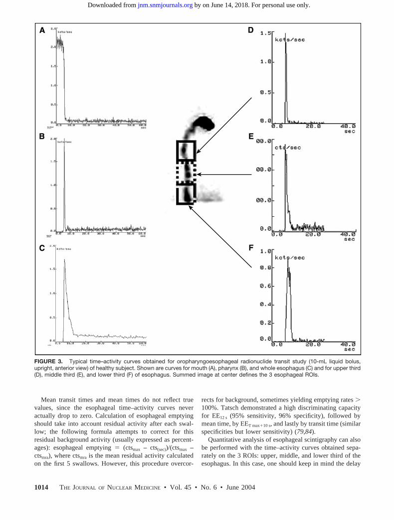
Quantitative Parameters. The main goal of quantitativeanalysis is to quantify retention and measure the rate ofesophageal transit. Most quantitative approaches use multi-ple-swallow techniques to overcome problems related to theintraindividual variability of a single wet swallow. Usually3 regions of interest (ROIs) are drawn, encompassing theupper, middle, and lower thirds of the esophagus. The datacan be summed for a total esophageal measurement. Thegastric fundus should be identified and excluded from thelower third region, as oscillations linked to respiratorymovements interfere with the analysis (Fig. 3).
Based on 1 wet (15 mL of water labeled with 99mTc-sulfurcolloid) and 3 successive dry swallows, Klein and Waldcalculated the fourth-swallow residual activity fraction. Afraction � 19.8% in the posterior view (or 13.1% in theanterior view) is considered abnormal. They also proposeda mathematic 2-compartment model to calculate the meantransit time, by separating the time–activity curve of the firstswallow into a rapid component (major fraction of the bolusthat has traveled the esophagus) and a slow component(residual fraction of the bolus clearing with subsequentswallows). The mean transit time is calculated as the ratio ofthe area under the fast component to the maximum height.Although this index is elegant, it did not prove to bediagnostically meaningful (97).
An alternative approach is based on multiple independentswallows (6 swallows are adequate for a reliable scinti-graphic study). The individual swallows of a given esoph-ageal scintigraphy study are normalized to their correspond-ing starting points, arranged consecutively, and thencondensed in a sum image. Curves are generated by plottingthe counting-rate columns assembled in each image. Thefollowing indices have been suggested (75,84,98):
● Transit time: lag time from the starting point untilactivity falls to �10% of peak activity;
● Mean transit time: (�cts)/ctsmax;● Mean time: (�cts(t)x t)/�cts (t);● Esophageal emptying (EE) at 10 s after Tmax as fraction
of peak activity: EET max�10 s; and● Esophageal emptying at 12 s after swallow as fraction
of peak activity: EE12 s.
RADIONUCLIDE GASTROESOPHAGEAL MOTOR STUDIES • Mariani et al. 1013
by on June 14, 2018. For personal use only. jnm.snmjournals.org Downloaded from
Mean transit times and mean times do not reflect truevalues, since the esophageal time–activity curves neveractually drop to zero. Calculation of esophageal emptyingshould take into account residual activity after each swal-low; the following formula attempts to correct for thisresidual background activity (usually expressed as percent-ages): esophageal emptying (ctsmax – cts(sec))/(ctsmax –ctsmra), where ctsmra is the mean residual activity calculatedon the first 5 swallows. However, this procedure overcor-
rects for background, sometimes yielding emptying rates �100%. Tatsch demonstrated a high discriminating capacityfor EE12 s (95% sensitivity, 96% specificity), followed bymean time, by EET max�10 s, and lastly by transit time (similarspecificities but lower sensitivity) (79,84).
Quantitative analysis of esophageal scintigraphy can alsobe performed with the time–activity curves obtained sepa-rately on the 3 ROIs: upper, middle, and lower third of theesophagus. In this case, one should keep in mind the delay
FIGURE 3. Typical time–activity curves obtained for oropharyngoesophageal radionuclide transit study (10-mL liquid bolus,upright, anterior view) of healthy subject. Shown are curves for mouth (A), pharynx (B), and whole esophagus (C) and for upper third(D), middle third (E), and lower third (F) of esophagus. Summed image at center defines the 3 esophageal ROIs.
1014 THE JOURNAL OF NUCLEAR MEDICINE • Vol. 45 • No. 6 • June 2004
by on June 14, 2018. For personal use only. jnm.snmjournals.org Downloaded from

in clearance at the distal third that is due to the time lagrequired for relaxation of the LES (76). This procedure canconstitute a second-line approach reserved for patients inwhom cine-mode visual analysis or condensed-dynamic-image analysis has detected abnormalities in the overallesophagus, so that the specific abnormal segments can beidentified.
Regional-transit-time topography has recently been de-veloped. The method, which is based on visualization of theesophageal bolus transit as profiles connecting pixels con-taining the same number of counts, produces a multicontourplot and calculates relative local transit times along theesophagus. The counting rate measured at a certain cross-section is inversely proportional to the velocity of flow anddirectly proportional to transit time. The relative local tran-sit times so obtained concisely describe the local kinetics ofbolus transit along the esophagus, with a longitudinal reso-lution of about 25% of the esophagus (103,104).
General Clinical ApplicationThe wide range in sensitivity and specificity reported for
esophageal scintigraphy can at least in part be attributed tothe different disorders evaluated. The potential clinical roleof esophageal scintigraphy (including the oropharyngealphase of swallowing) in patient management is better ap-preciated in light of the pathophysiologic changes underly-ing esophageal motility disorders (Figs. 4 and 5).
Nutcracker Esophagus. The term nutcracker esophaguswas coined by Dalton et al. to describe the conditions underwhich patients with noncardiac chest pain or dysphagiaexhibit peristaltic waves in the distal esophagus with meanamplitudes exceeding the normal values by more than 2 SDs(105). These high-amplitude waves may not interfere withesophageal clearance or strictly correlate with episodes ofdysphagia or chest pain or may result in mild distal esophagealretention with reflux. Furthermore, symptoms may not respondto drugs that modulate the peristaltic wave pressure (106).
The clinical consequences of an isolated hypertensiveLES remain unclear. Three groups of patients fall under the
term isolated hypertensive LES: those with abnormally el-evated resting LES, those with exaggerated contraction ofLES after relaxation, and those with incomplete LES relax-ation. Although the first 2 conditions can even be asymp-tomatic, incomplete LES relaxation usually interferes withesophageal emptying; this disorder is thus better termedatypical disorder of LES relaxation (with impaired esoph-ageal clearance) rather than isolated hypertensive LES(105,107,108).
No definite diagnostic benefit of esophageal scintigraphyhas been demonstrated in patients with normal peristalsis,even in the presence of nutcracker esophagus or isolatedhypertensive LES (109,110). On the other hand, abnormalesophageal scintigraphic patterns, such as dysmotility orgastroesophageal reflux, were identified in 89% of patientswith atypical chest pain, which in some cases was associ-ated with nutcracker esophagus despite the fact that thesepatients did not complain of dysphagia (111).
Diffuse Esophageal Spasm. Diffuse esophageal spasm ischaracterized by uncoordinated esophageal contractions thatcan interfere with esophageal clearance (Fig. 6). Differentmanometric criteria have been proposed for diagnosing thiscondition, including either the presence of spontaneous andrepetitive contractions or the presence of high-amplitude orprolonged contractions. The key diagnostic criterion is thepresence of simultaneous contractions induced by wet swal-lows. Besides the discrepancies in manometric criteria, dif-fuse esophageal spasm is an intermittent phenomenon that isevident manometrically in �10% (but ��100%) of wetswallows (106,112). Therefore, in patients with this condi-tion, esophageal scintigraphy should include multiple wetswallows, visual analysis of bolus dynamics (showing cha-otic bolus movements), and definition of the time–activitycurves (showing multiple peaks after a single swallow).
Achalasia. Classic achalasia is caused by degeneration ofneurons in the wall of the esophagus. These neurons areinvolved in the production of nitric oxide, which relaxesesophageal smooth muscle, therefore causing the basal LES
FIGURE 4. Examples of radionuclideswallow studies of patients with oropha-ryngeal dysphagia. (A) Time–activity curvefor ROI drawn over oral region of patientwith amyotrophic lateral sclerosis after pa-tient swallowed liquid bolus (10 mL, up-right, anterior view) shows significant re-tention and markedly delayed clearance ofactivity from mouth, with double swallow(piecemeal deglutition because entire bo-lus could not be swallowed at once). (B)Static image of patient with severe oropha-ryngeal dysphagia after total thyroidec-tomy (damage of upper left laryngealnerve). Dynamic recording of the swallow-
ing of a liquid bolus (10 mL, upright) was impossible because of wide movements due to coughing. Aspiration in trachea is obvious.(C) Semisolid bolus (10 mL, upright, anterior view) did not cause gross aspiration, thus permitting dynamic recording. Time–activitycurve obtained for ROI drawn over pharyngeal region on completion of semisolid transit study shows markedly delayed, irregular,and incomplete clearance of radioactivity from pharynx.
RADIONUCLIDE GASTROESOPHAGEAL MOTOR STUDIES • Mariani et al. 1015
by on June 14, 2018. For personal use only. jnm.snmjournals.org Downloaded from

pressure to rise (106,113–116). The result is impairedesophageal clearance and delayed transit times. Esophagealscintigraphy readily detects the delay in transit (Fig. 7), witha sensitivity close to 100% (74). Distal esophageal retention
does not clear after the patient assumes an upright positionor drinks a glass of water. Scintigraphic evaluation ofesophageal clearance is widely accepted as the method ofchoice in the follow-up of patients with achalasia, for mon-itoring the efficacy of treatment (myotomy, pneumatic di-lation, or botulinum toxin injections). To optimize sensitiv-ity for this condition, the patient should be upright whenstudied and should swallow a bolus of unlabeled water afterthe radiolabeled swallow (117–122).
Scleroderma. Fibrosis and vascular obliteration of theesophageal muscle and its innervation are the underlyingcauses of ineffective esophageal motility in patients withscleroderma. Esophageal scintigraphy can detect esopha-geal involvement in patients with asymptomatic disease,showing a typical pattern of retention of radioactivity in thelower esophagus, with clearing after the patient is upright ordrinks a glass of water. As an indicator of dysmotility inboth early and advanced disease, esophageal scintigraphyhas a higher sensitivity than do manometry and bariumswallows (123–127).
Table 3 compares the diagnostic performance of esoph-ageal scintigraphy and videoesophagography (upper gastro-intestinal series with barium swallows) in a variety of dis-orders of the esophagus. A general conclusion is thatesophageal scintigraphy, which is easy to perform andyields quantitative parameters reflecting pathophysiologicprocesses, is especially suitable for serial studies to monitorthe efficacy of medical or surgical treatment of variousdisorders (achalasia, gastroesophageal reflux, scleroderma,and hiatal hernia). Esophageal scintigraphy also plays animportant role in the assessment of a patient’s subjectivesensation of difficult swallowing, offering objective evi-dence of dysphagia and therefore stimulating further inves-tigation (e.g., in psychiatric patients) (128), since no clini-cally significant motor disorder is likely to be missed (129).
The reliability of transit scintigraphy for diagnosingesophageal cancer is too low to warrant its routine use fordiagnosis (130,131). However, it is particularly useful for itsability to assess the effect of stricture and of dysmotility ina single test (73). Furthermore, transit scintigraphy has aconsiderable role after esophagectomy. Transit scintigraphyis ideal for assessing swallowing function and gastric emp-tying after replacement of the esophagus (e.g., with a por-tion of the stomach formed into a tube to reestablish a routebetween the pharynx and stomach) (132–136). In patientswith inoperable esophageal cancer being treated only pal-liatively (such as through insertion of a stent to palliatedysphagia), an esophageal transit study can easily be per-formed to evaluate, noninvasively, restoration of near-nor-mal patency (73).
Application to Pediatric GERD
Regurgitation and vomiting are common symptoms ininfants with GERD, probably because of the limitedcapacity of the esophagus in this age group. In infants, it
FIGURE 5. Radionuclide esophageal transit study (15-mL semi-solid bolus, upright, anterior view) of patient with amyotrophiclateral sclerosis. Shown are time–activity curves for upper third (A),middle third (B), and lower third (C) of esophagus. Summed imageat left defines the 3 esophageal ROIs. All 3 portions of the esoph-agus clear initially with a normal-appearing pattern, but there ismarked retention of radioactivity, mainly in mid esophagus but alsosomewhat in proximal third, and slow subsequent passage ofradioactive bolus to lower third. A similar scintigraphic pattern withdelayed clearance of esophageal radioactivity is also seen in pa-tients with achalasia or with scleroderma, but with retention indistal rather than middle portion of esophagus (Fig. 7).
1016 THE JOURNAL OF NUCLEAR MEDICINE • Vol. 45 • No. 6 • June 2004
by on June 14, 2018. For personal use only. jnm.snmjournals.org Downloaded from

is difficult to discriminate pathologic from physiologicreflux. If the event is within physiologic levels, the infantthrives well and usually responds to simple measuressuch as nursing in an upright position or thickening of thefood.
Most infants with severe reflux have symptoms by theage of 2 mo; the condition usually has a benign course, andabout 60% of these babies are free of symptoms by the ageof 18 mo. However, about 30% have persistent symptomsuntil approximately 4 y of age, 5% develop esophageal
FIGURE 6. Radionuclide esophagealtransit study (10-mL liquid bolus, upright,anterior view) of patient with esophagealdysphagia. (A) One-second frames fromdynamic recording show irregular move-ments of radioactive bolus up and downthe esophagus. (B) Time–activity curve forROI drawn over pharynx shows the normalefficiency of a single swallow without re-gurgitation. (C) Time–activity curve for ROIdrawn over entire esophagus shows inco-ordinate clearance of radioactivity, the typ-ical pattern for a patient with diffuse esoph-ageal spasm. Review of the dynamicrecording in cine mode excluded gastro-esophageal reflux as an alternative causeof the irregular, multiple-peak pattern in theesophageal time–activity curve.
FIGURE 7. Radionuclide esophagealtransit study (15-mL liquid bolus, supine,posterior view, 1 wet swallow followed 30 slater by 3 dry swallows at 15-s intervals) ofa patient with achalasia. (A) Summed im-age of whole esophagus, with ROI. (B)Time–activity curve shows marked reten-tion of radioactivity in the esophagus.Marked esophageal retention of radioactiv-ity remained virtually unchanged after pa-tient stood upright for some minutes. Incontrast, esophageal scintigraphy of pa-tients with scleroderma shows similar pat-tern of radioactivity retention, which, how-ever, clears after they stand.
RADIONUCLIDE GASTROESOPHAGEAL MOTOR STUDIES • Mariani et al. 1017
by on June 14, 2018. For personal use only. jnm.snmjournals.org Downloaded from

strictures, and another 5% die if they do not receive ade-quate treatment.
Clinical presentation of severe gastroesophageal reflux inchildren includes regurgitation or vomiting and failure tothrive. The spectrum of complications includes recurrentinfections due to aspiration, asthma, apnea, cough, andstridor (137,138), along with the sequelae of esophagitis,including anemia, esophageal stricture, esophageal dysmo-tility (139), Sandifer’s syndrome, and even sudden infantdeath. Primary respiratory disorders may predispose to gas-troesophageal reflux as a result of increased intraabdominalpressure caused by coughing. Contrary to common beliefs,crying does not exacerbate gastroesophageal reflux (140).
Most conventional tests for diagnosing gastroesophagealreflux in adults are not suitable in infants and childrenbecause of the radiation dose or because they require sub-stantial cooperation from the patient (141). In particular, abarium upper-gastrointestinal series (with either intermittentimaging or cineradiography) uses a nonphysiologic testmeal and requires provocative measures such as abdominalcompression. Other diagnostic tests are more or less inva-sive, such as the acid infusion test (during which the patientis asked to communicate symptoms), intraluminal pH mon-itoring, esophageal manometry, upper digestive endoscopy,and tracheal aspiration (searching for lipid-laden macro-phages to confirm aspiration of milk) (142).
The radionuclide method that has been adapted for use ininfants and children (143) is performed at the time ofscheduled feeding. 99mTc-Sulfur colloid (3.7–18.5 MBq[100–500 �Ci]) mixed with milk or formula is used to labelthe liquid meal at a concentration of �1.85 MBq/mL (�50�Ci/mL). About half the regular meal is labeled, leaving the
remainder unlabeled for completing the feeding study. Ifpossible, the scintigraphic study includes the initial swal-lowing phase and esophageal transit. After the radiolabeledhalf of the meal has been given, the remaining, unlabeled,portion is given to wash radioactivity from the mouth,pharynx, and esophagus. The baby is then burped andplaced supine for imaging of the chest and gastric area(reflux in infants is more likely to occur in the supine thanin the prone position) (144). According to age and cooper-ation, the child can be positioned either lying on the imagingtable or lying on the �-camera head. A high-sensitivitycollimator should be used to maximize radioactive counts,because the activity during an episode of reflux can be lowor in a small volume.
Sequential images should be recorded continuously forabout 5–10 s per image for at least 60 min, since about 25%of reflux episodes can be missed when limiting the acqui-sition to 30 min. Although dynamic acquisition at 20, 30, oreven 60 s per frame (64 � 64 matrix) is usually adequate toidentify gastroesophageal reflux, using shorter framing(e.g., 5 s per frame) can help in detecting brief refluxepisodes and in evaluating clearance of reflux from theesophagus. At the end of dynamic acquisition, 5-min staticimages of the anterior and posterior lung fields are obtained,taking care to exclude the high gastric radioactivity. Anadditional procedure normally applied only to adults is toprogressively increase abdominal pressure, as is done dur-ing a conventional barium radiography study. If aspirationis suspected, static imaging should be repeated at about 2and 24 h.
For data analysis, ROIs are manually defined over thelower, middle, and upper esophagus and the oropharynx togenerate time–activity curves, in which reflux episodes arecharacterized as sharp spikes. The dynamic acquisitionshould be reviewed in the cine mode to identify movementsof the body, which can simulate reflux spikes in the time–activity curve.
Gastroesophageal reflux can be quantitated using differ-ent indices, which usually consider the volume of eachepisode, the frequency of the episodes, and the rate of refluxclearance from the esophagus. A simple approach is toexpress esophageal activity (either in selected frames fromdynamic recording or in the static images) as a fraction ofgastric activity (145). This approach clearly discriminatesadult patients with gastroesophageal reflux from those with-out reflux (11.7% 1.8% vs. 2.7% 0.3%). When themean value of esophageal activity was expressed as a frac-tion of initial gastric activity, patients with peptic esophagi-tis had significantly different values from those of controls(3.66% 0.81% vs. 0.66% 0.12%) (146). One can alsocalculate an overall reflux index based on the ratio ofsummed activities in each episode to gastric activity overthe entire 60 min of the dynamic study (recorded at 20 s perframe) (147). If higher than 5%, this index denotes thepresence of reflux.
TABLE 3Comparative Diagnostic Performance of Esophageal
Scintigraphy vs. Videoesophagography in Patients withDysphagia Caused by Various Esophageal Disorders
DisorderSensitivity
(%)Specificity
(%)
Positivepredictivevalue (%)
Esophageal transitscintigraphy
Achalasia 91 98 95Diffuse esophageal spasm 33 99 67Scleroderma 75 99 75LES dysfunction 25 99 67NSEMD 71 76 48
VideoesophagographyAchalasia 100 98 96Diffuse esophageal spasm 67 100 100Scleroderma 75 100 100LES dysfunction 25 98 50NSEMD 62 85 57
NSEMD nonspecific esophageal motility disorder.Data are from (234).
1018 THE JOURNAL OF NUCLEAR MEDICINE • Vol. 45 • No. 6 • June 2004
by on June 14, 2018. For personal use only. jnm.snmjournals.org Downloaded from

The sensitivity of scintigraphy in detecting gastroesoph-ageal reflux has been reported to be 75%–100%, dependingon the protocol used (148). These values are consistentlyhigher than those of conventional barium studies or manom-etry.
GASTRIC-EMPTYING SCINTIGRAPHY
Since its introduction by Griffith et al. in 1966 (149),gastric scintigraphy has evolved to include an evaluation ofcompartmental or antral motility, and more recently toSPECT to evaluate postprandial gastric accommodation.
As a physiologic, quantitative, noninvasive test, gastric-emptying scintigraphy is well suited for evaluating patientsbefore and after medical or surgical treatment. This proce-dure is now widely considered the gold standard for eval-uating gastric emptying.
Scintigraphy ProcedurePatients fast for at least 8 h before a gastric-emptying
study. In addition, it is preferable to study women during thefirst 10 d of the menstrual cycle, to avoid possible hormonaleffects on gastrointestinal motility. Any medication thatpotentially interferes with gastric motility (narcotic analge-sics, anticholinergics, antidepressants, calcium channelblockers, gastric acid suppressants, aluminum-containingantacids, or somatostatin) is discontinued for an appropriateperiod (depending on specific pharmacokinetics), unless thescintigraphic test is performed specifically to assess theeffect of such drugs on gastric motility. Tobacco and alco-hol is also withheld for at least 24 h.
The first radiopharmaceutical described for gastric-emp-tying studies was chicken liver labeled by injecting sulfurcolloid into the chicken. The liver was harvested, cooked,and administered to the patient. An advantage of this ap-proach was the stability of the label. However, this proce-dure is not practical for routine use (150). A more conve-nient alternative is to mix 11–18 MBq (300–500 �Ci) of99mTc-sulfur colloid with eggs or a fat-free egg substitute(EggBeaters; ConAgra Foods, Inc.) to obtain a stable labelfor the solid phase (Table 4) (151). Besides the radiolabeledcomponent, the test meal typically contains other unlabeledproducts because a caloric content of at least 200–300 kcalis needed for complete activation of digestive functions
(152). In a multicenter study, a standardized, low-fat mealshowed excellent reproducibility for gastric-emptying stud-ies. The meal consisted of fat-free egg substitute (Egg-Beaters) served with 2 slices of bread, 30 g of strawberryjam, and 120 mL of water (150). Standardizing the con-tent of the meal, the proportion of liquid and solid, theamount of fat, the number of calories, and the rate ofconsumption is important for establishing a reproducibleresponse (153,154).
111In-DTPA is usually used for liquid-phase studies, as itis chemically inert and does not bind to the solid phase ofthe meal, thus allowing simultaneous assessment of bothsolid and liquid emptying.
The radiolabeled meal is ingested within 10 min, understandardized environmental conditions (ambient noise, am-bient light, and patient comfort). If any portion of the mealis not eaten, the uneaten amount is recorded.
Imaging can be performed while the patient is eitherstanding, sitting, or supine, provided the position does notchange during the study. Because movement of solids fromthe posteriorly located fundus to the more anteriorly locatedantrum results in a nonuniform attenuation of the gastricactivity, anterior and posterior imaging with subsequentcalculation of the geometric mean is suggested to correct forthis attenuation nonuniformity; alternatively, a single leftanterior oblique view can be used (155–159). Images areacquired in at least a 64 � 64 pixel matrix. Continuous datarecording at a framing rate of 30–60 s per image for at least90 min is generally recommended (160) (up to 2–3 h forlarger-volume meals or meals with a higher calorie, fat,carbohydrate, or protein content). Alternatively, data re-corded at discrete 15-min intervals (or even at 0, 60, 120,and 180 min after meal ingestion) have been shown toprovide reliable gastric-emptying data. However, with thisapproach no information is available on the lag phase, andrapid gastric emptying (dumping) may not be fully charac-terized (32,161). Images recorded at 4 h may be necessaryto characterize delayed gastric emptying in cases with bor-derline 2-h emptying (162).
The evaluation of indigestible solids (fibers or pellets)requires prolonged patient observation and provides infor-mation on the interdigestive period rather than postprandialmotility (32).
Data Analysis and Quantitative ParametersAfter the set of geometric mean images has been created,
the full study is initially reviewed in cine mode to detectpatient motion. After the cine review, it is helpful to add theimages from the serial acquisition to define the borders ofthe stomach and the adjacent loops of bowel. ROIs aredrawn around the gastric area, adjacent esophagus, duode-num, and jejunum. The curves are corrected for radioactivedecay.
The percentage of gastric retention at a specific time point(usually 2, 3, or even 4 h) is calculated with respect to theimmediate postingestion value at the zero time point. Be-
TABLE 4Stability of the Radiolabeled Solid In Vitro
Solid meal% bound in
gastric juice (3 h)
99mTc chicken liver (in vivo) 9899mTc-Sulfur colloid whole egg 8299mTc-Sulfur colloid white egg �9599mTc surface-labeled chicken liver 8499mTc-chelex resin 98 (24 h)
Data are from (151).
RADIONUCLIDE GASTROESOPHAGEAL MOTOR STUDIES • Mariani et al. 1019
by on June 14, 2018. For personal use only. jnm.snmjournals.org Downloaded from

cause this parameter does not have a normal, gaussian-typedistribution, the median and 90th or 95th percentile, ratherthan the mean and SD, are used to define normal values(32).
When continuous imaging is recorded, half-emptyingtime (T50) is determined as the time it takes to reach half thepeak counts, whereas when static, discrete-interval imagesare acquired, the data points should first be analyzed bynonlinear least-squares fit. Because solid gastric emptyingfollows a sigmoidal rather than a simple exponential curve,at least 2 parameters are required for an optimal description.At least 2 computational procedures have proven capable ofadequately describing the pattern of biphasic solid gastricemptying: the Elashoff power exponential function, y(t) (e–kt)� (163), and a modified power exponential function,y(t) (1 � [1 � ekt]�) (25), where y(t) is the gastricretained activity at time (t), k is the gastric-emptying rate,and � is the intercept on the y-axis back-extrapolated fromthe terminal portion of the curve.
When the Elashoff function is applied, T50 (ln 21/�)/k,whereas when the modified power exponential function isapplied, T50 �ln ([1 � 0.5]1/�)/k.
The lag phase represents the time required for transfer ofsolid food from the fundus to the antrum and then for theantrum to grind it into small particles (suspended in thegastric fluid) that can begin to empty into the duodenum(164). Various methods have been proposed for evaluatingthis parameter. With continuous imaging, it is relativelyeasy to visually define the transition point in the gastrictime–activity curve: the point of first appearance of duode-nal activity or the time of maximal antral filling. When dataare acquired as serial static images, the lag phase canarbitrarily be calculated as a given percentage of drop(5%–10%) from peak counts; however, this approach ishighly dependent on the slope of the second phase (165). Inmathematic terms, the lag time corresponds to the inflectionpoint of the gastric time–activity curve, where the secondderivative equals zero. When the Elashoff function is ap-plied, lag time ([� � 1]/�)1/�/k, whereas when the mod-ified power exponential function is applied, lag time �/k.
The lag phase calculated by the modified power-expo-nential function correlates poorly with the data derived byother methods, and the accuracy of such an estimate isgreatly limited by the rate of temporal sampling. It hastherefore been suggested that the lag phase calculated bythis method possibly represents a more complex phenomenonthan just emptying of solid food into the duodenum (163).
Dynamic Antral ScintigraphyAlthough manometry is the most accurate method for
assessing gastric motor function, this invasive procedure isnot well accepted by patients and is not widely available(166). Real-time ultrasound evaluation of gastric motorfunction is often hampered by poor image quality becauseof echogenic food particles (167). Simultaneous scinti-graphic evaluation of both gastric motility and gastric emp-
tying optimizes the characterization of gastric function withminimal additional cost, time, and technical effort.
Movements of the stomach wall induce changes in thedistribution of radioactive food within the stomach. There-fore, dynamic scintigraphy as performed for a gastric-emp-tying study can evaluate gastric motility as well. Becauseantral hypomotility (reduction of phasic contractions in thedistal stomach) has been reported for a variety of dyspepticsyndromes, almost all studies have evaluated only antralmotility (168–175). The most common procedure is to adda short, high-definition dynamic acquisition (1–2 s perframe for 4–5 min) at the end of a standard gastric-empty-ing study, performed either with dynamic imaging or withserial static imaging. Data analysis is performed after atten-uation correction, to generate time–activity curves for ROIsdefined on the proximal, middle, and distal antral regions.Further data processing includes elimination of artifacts dueto patient movement or to translational movement of thestomach (performed by baseline restoration of raw datausing a polynomial fit) and elimination of background noise(performed by an autocorrelation function). A refined Fou-rier transform is then performed on antral time–activitycurves to determine the amplitude and frequency of antralcontractions.
Consistent with manometric (176), real-time ultrasono-graphic, and electrogastrographic estimates (177), in severalscintigraphic studies the frequency of antral contractionshas been found to be about 3 per minute (127,167–173).This normal frequency does not seem to change signifi-cantly in patients with diabetes, gastritis, functional dyspep-sia, progressive systemic sclerosis, or pyloric stenosis, ei-ther after administration of gastric acid suppressants or aftergastric surgery. In contrast, the amplitude of antral contrac-tions seems to discriminate various patient groups. In func-tional dyspepsia, paradoxically increased amplitude hasbeen reported, possibly explained by predominantly ineffi-cient (nonexpulsive) antral contractility or by pyloric dys-motility (170), whereas decreased amplitude has been ob-served in diabetic patients (168) and in patients whopreviously underwent Billroth type I or type II gastrectomyor truncal vagotomy (178).
Unfortunately, variability in the definition of the antralROIs, as well as in the normalization and filtering steps,hampers the reproducibility of measurement of antral func-tion. Two methods avoiding the autocorrelation functionhave therefore been proposed to improve reproducibility.Both involve a postsynchronization method using either asingle distal antral ROI (179) or Fourier analysis of con-densed images of the whole stomach (180).
Gastric SPECTDyspeptic patients frequently have defective gastric ac-
commodation contributing to postprandial symptoms suchas early satiety, distension, and nausea. This condition hasbeen reported in functional dyspepsia, postfundoplicationdyspepsia, postvagotomy gastric surgery, and neuropathic
1020 THE JOURNAL OF NUCLEAR MEDICINE • Vol. 45 • No. 6 • June 2004
by on June 14, 2018. For personal use only. jnm.snmjournals.org Downloaded from

diabetic gastroparesis (181–186). Although proximal gastricaccommodation can be measured by a barostatic balloon,and real-time ultrasonography can measure antral accom-modation, neither of these 2 methods can evaluate whole-stomach accommodation. SPECT has been used to measuregastric accommodation in response to a meal, based on thewell-known ability of both the parietal (oxyntic) and thenonparietal (mucous) gastric cells to take up and excrete99mTc-pertechnetate (187).
The procedure is as follows: A 370–740 MBq (10–20mCi) dose of 99mTc-pertechnetate is injected intravenouslyafter an overnight fast. Dynamic tomographic acquisition isperformed starting 10 min after injection, using a multiorbitmode; 6° images are acquired in a 128 � 128 matrix at 3 sper step (total acquisition time, about 180 s). After 2 initialorbits have been recorded as baseline, the patient drinks thetest meal while supine, with the aid of a straw. Immediatelyafterward, 7 additional orbits are acquired over about 21min to evaluate gastric accommodation to the meal chal-lenge.
A semiautomated segmentation algorithm is used to re-construct the stomach. Total gastric volume is measuredduring fasting, during the 3- to 12-min postprandial period,and during the 12- to 21-min postprandial period. Theprocedure has recently been simplified by acquiring 3 com-plete 360° orbits every 10 min (6° steps, 64 � 64 matrix,10 s per step) (188). Intra- and interobserver coefficients ofvariation in estimating fasting and postprandial gastric vol-umes are 8%–9% and 12%–13%, respectively. The greatestpostprandial volume increase was in the proximal stomach(about 2- to 4-fold), with no significant difference betweenearly (3–12 min) and late (12–21 min) volume changes.
The method has recently been validated by a phantomstudy using a balloon filled with 99mTc-DTPA, and changesin gastric volume measured in response to a meal proved to
be as accurate as those derived by the barostat balloontechnique, with no effects of age (at least in adults) andbody mass index on the fasting or postprandial gastricvolume ratio (189). Gastric SPECT has also demonstratedimpaired accommodation in patients with idiopathic non-ulcer dyspepsia and in patients after fundoplication(187,190,191).
Clinical ApplicationGastric scintigraphy (Fig. 8) is generally indicated in
several clinical conditions when morphologic investigationsfail to reveal the cause of dyspepsia, such as postprandialnausea, vomiting, upper abdominal discomfort, early satietyand bloating, suspected gastroparesis, poor diabetic control,and severe reflux esophagitis unresponsive to medical treat-ment (46,158).
The pathophysiologic basis of functional dyspepsia hasvariously been ascribed to delayed gastric emptying ofsolids (192,193), failure of fundic relaxation (184), visceralhypersensitivity (40), and Helicobacter pylori infection(194,195). Although more than 40% of patients with func-tional dyspepsia have delayed solid gastric emptying,whether such an abnormality explains the clinical symptomsremains controversial (190,196–199). However, some cor-relation has been found between delayed gastric emptyingand relevant postprandial fullness or severe vomiting andwith being female (191), the total symptom score beinghigher in those patients with delayed gastric emptying(200). In diabetic patients, independent predictors of de-layed gastric emptying included abdominal bloating, thefemale sex, and high blood glucose levels and body massindex (201).
Another controversial issue concerns the effect of proki-netic drugs, inducing accelerated gastric emptying, onsymptom relief. Surprisingly few studies have evaluated
FIGURE 8. Examples of gastric-emptying curves (corrected for physical radioisotopic decay) obtained by sequential imaging atdiscrete intervals after ingestion of radiolabeled solid meal. (A) Results for healthy volunteer (in our institution, T1/2 values of gastrictime–activity curves for healthy subjects range from 110 to 120 min). Curve is characterized by distinct lag phase lasting about20–30 min before emptying begins. (B) Results for patient with gastroesophageal reflux; this curve shows virtually no lag phase andshows markedly delayed gastric emptying, as observed in an average 40% of these patients (236,237). On the basis of this finding,the surgical treatment chosen was fundoplication and pyloroplasty, instead of isolated fundoplication, which would have inducedpostoperative symptoms (e.g., bloating) with no obvious lag phase. (C) Gastric-emptying curve obtained for same patient aftersurgical treatment demonstrates slightly accelerated gastric emptying (due to pyloroplasty), with reappearance of a discernible lagphase.
RADIONUCLIDE GASTROESOPHAGEAL MOTOR STUDIES • Mariani et al. 1021
by on June 14, 2018. For personal use only. jnm.snmjournals.org Downloaded from

whether baseline gastric emptying is a predictor of theefficacy of these agents or whether accelerated gastric emp-tying after therapy is a predictor of symptom improvement(202). Helicobacter pylori infection in patients with nonul-cer dyspepsia is usually not associated with delayed gastricemptying (203–205), although both gastric emptying andsymptom scores have been reported to improve after Heli-cobacter pylori eradication (206).
Delayed gastric emptying, most likely due to autonomicneuropathy (resulting in a prolonged lag phase (168)), hasbeen identified in 27%–58% of diabetic patients; however,hyperglycemia further delays gastric emptying, and delayedgastric emptying itself may contribute to poor glycemiccontrol (207). Delayed gastric emptying is also associatedwith various systemic syndromes (Parkinson’s disease,amyloidosis, myotonic dystrophy, polymyositis, HIV infec-tion, or cytomegalovirus infection) and with local condi-tions such as vagotomy without pyloroplasty, and gastro-esophageal reflux (208–215).
As expected, accelerated gastric emptying is found afterpyloroplasty or after surgical treatment of reflux (216–219).
Another clinical application of gastric scintigraphy ispostsurgical evaluation of the gastroesophageal junction.Achalasia is the esophageal disorder whose treatment hasbeen most substantially affected by the development ofminimally invasive surgery—that is, a laparoscopic ap-proach to Heller’s myotomy (220)—widely reported toyield favorable results in up to 90% of these patients (221–224). Because the support structures of the antireflux mech-anism, such as the phrenoesophageal ligament and the LES,are sacrificed during laparoscopic dissection of the esopha-gus and myotomy, an antireflux procedure is also required(225–227). Residual dysphagia can be linked to persistentesophageal narrowing due to incomplete myotomy. Esoph-ageal transit scintigraphy is well suited as an addition toesophageal manometry and 24-h pH monitoring for fol-low-up and monitoring of this complication.
The efficacy of medical therapy in patients with GERDseems to wane with time, and for many patients the dose ofproton pump inhibitors needs to be readjusted to controlsymptom relapse. However, surgical therapy for GERD hasseen dramatic changes over the last decade, making it muchmore accessible and less invasive than in the past (228). Thegoal of effective antireflux surgery is to restore the compe-tency of the cardia. Incompetence of the gastroesophagealjunction has a multifactorial etiology, including such abnor-malities as transient relaxation of the LES (relaxation notassociated with a swallow), primary hypotension of theLES, gastric-emptying abnormalities, and absence of a flapvalve. The presence of hiatal hernia exacerbates reflux byimpairing esophageal emptying and LES function. An idealoperation, thus, is one that addresses each of these aspects.The most effective way (shown to yield favorable results inabout 90% of patients) to permanently restore the compe-tency of the cardia is to create some form of plication over
the esophagus just proximal to the cardioesophageal junc-tion, according to the following principles. First, the wrapmust be constructed over the esophagus, just proximal to thegastroesophageal junction, and must be fixed to the esoph-agus to remain in that position permanently. Second, thewrap must be constructed without tension using the fundusof the stomach and leaving enough space to accommodate a42- to 50-French bougie inside the esophagus. Third, a totalwrap, such as the total fundoplication described by Nissen,should measure about 2 cm in length in its anterior aspect.Fourth, the wrapped portion of the esophagus must liebelow the diaphragm without tension. Finally, the diaphrag-matic hiatus must be gently closed around the esophagus,above the wrap.
Esophageal transit scintigraphy and, above all, gastric-emptying scintigraphy are well suited for following uppatients who undergo fundoplication because of GERD(Fig. 8) (229).
PROPOSED ALGORITHMS
Scintigraphic evaluation of the upper digestive tract witha transit study is simple, safe, quick, physiologic, and wellaccepted by patients. By quantitatively or semiquantita-tively measuring serial changes or response to therapy, thistechnique noninvasively provides clinically relevant infor-mation on various motility disorders and can modify clinicalmanagement of patients. Nevertheless, there is a certainreluctance to rely on this set of procedures, although withsome variance between esophageal transit scintigraphy (per-haps because of the paucity of information available on itsreproducibility) (230) and gastric-emptying studies (widelyrecognized as the gold standard for this physiologic param-eter). Thus, we can agree that transit scintigraphy, especiallyesophageal transit scintigraphy, is generally underused de-spite its recognized clinical value (73).
Here, we propose diagnostic and clinical algorithms forevaluating patients with some selected functional or motilitydisorders of the upper digestive tract. Although originallyderived from published literature, these algorithms havebeen modified for this review on the basis of wide clinicalexperience and a consensus developed during extensivediscussions with specialists in nuclear medicine and inmedical and surgical gastroenterology.
Figure 9 shows a flowchart of a possible diagnosticalgorithm for patients presenting with dysphagia of eitheroropharyngeal or esophageal origin. The left side of thefigure concerns oropharyngeal dysphagia, whose evaluationis usually quite straightforward. In these patients, clinicalevaluation is usually initiated by the otolaryngology spe-cialist and must include careful neurologic assessment.Evaluation of the swallowing function with a barium transitstudy is usually the first imaging procedure, usefully com-pleted with a radionuclide esophageal transit study thatprovides a more physiologic, quantitative assessment tobetter characterize the underlying disorder. After a specific
1022 THE JOURNAL OF NUCLEAR MEDICINE • Vol. 45 • No. 6 • June 2004
by on June 14, 2018. For personal use only. jnm.snmjournals.org Downloaded from

diagnosis has been established and the best treatment cho-sen, esophageal transit scintigraphy is especially useful formonitoring the disease and its response to treatment.
The right side of Figure 9 concerns esophageal dyspha-gia. In these patients, whether upper gastrointestinal endos-copy (the most common occurrence, solid arrow) or a bar-ium swallow (less frequent occurrence, dotted arrow) isperformed first varies according to local logistics. Whenupper gastrointestinal endoscopy is performed first, a bar-ium swallow study is generally performed anyway in asubsequent phase of the patient’s characterization. Whenupper gastrointestinal endoscopy reveals organic disease(e.g., esophageal stricture or tumor), treatment is usuallysurgical, with an ensuing certain role for transit esophagealscintigraphy as a simple, noninvasive test for assessingrestoration of swallowing function. When endoscopy ex-cludes organic disease, esophageal manometry is usuallyrecognized as the gold standard for functional characteriza-tion of the swallowing disorder. Variously combined withthe other nonradionuclide tests, transit esophageal scintig-raphy is helpful for quantitative characterization of thefunctional disorder and especially for subsequent monitor-ing of the response to therapy (18).
As shown in Figure 10, the diagnostic work-up of patientswith symptoms suggesting GERD (with the earlier, mildervariant of nonerosive reflux disease) is particularly complex(231). In these patients, esophageal transit scintigraphy has
a pivotal role in a relatively late phase of the clinicalalgorithm, as a means of stratifying patients for treatment orfor full reevaluation after failure of medical therapy. In bothinstances, scintigraphy again provides useful information
FIGURE 11. Proposed diagnostic–therapeutic algorithm forpatients presenting with recurrent noncardiac chest pain. Pos-itive pH-metry is prolonged exposure of the esophagus to a pHof �4 for �5% of the total time. Negative pH-metry is exposureof the esophagus to a pH of �4 for �5% of the total time. GI gastrointestinal.
FIGURE 9. Proposed diagnostic–therapeutic algorithm forpatients with dysphagia of either oropharyngeal or esophagealorigin. Oropharyngeal dysphagia is defined as the occurrence ofchoking, coughing, and nasal regurgitation in association withthe initiation of a swallow. Esophageal dysphagia is defined asa sensation of food stuck in the esophagus a few seconds afterswallowing. Barium swallow for suspected oropharyngeal dys-phagia is best performed as cineradiography. ENT - CNS otolaryngologic and neurologic evaluation; GI gastrointestinal.
FIGURE 10. Proposed diagnostic–therapeutic algorithm forpatients with symptoms suggestive of GERD. Alarm symptomsare defined as pain, dysphagia, anemia, weight loss, fever, andhematemesis. Positive pH-metry is prolonged exposure of theesophagus to a pH of �4 for �5% of the total time. NegativepH-metry is exposure of the esophagus to a pH of �4 for �5%of the total time. A positive symptom index is �50% of thesymptoms arising simultaneously with drops of esophageal pHto �4, even if extending over �5% of the total time; a negativesymptom index is �50% of the same parameter. Full reevalu-ation is based on the entire set of diagnostic tests available,such as manometry, bilimetry, scintigraphy, upper gastrointes-tinal series, or multichannel impedance. GI gastrointestinal;H2-blockers histamine-2 receptor inhibiting/antagonist drugs;NERD nonerosive reflux disease; PPI proton pump inhibitors.
RADIONUCLIDE GASTROESOPHAGEAL MOTOR STUDIES • Mariani et al. 1023
by on June 14, 2018. For personal use only. jnm.snmjournals.org Downloaded from

either for monitoring response to therapy (including surgicalprocedures) or for better characterizing the diagnosis.
Esophageal transit scintigraphy has a pivotal role in de-termining whether patients with noncardiac chest pain haveachalasia or other (spastic) motility disorders or have func-tional chest pain of presumed esophageal origin (Fig. 11)(229). These 2 classes of disease benefit from markedlydifferent therapeutic approaches. Treatment of achalasiais based on administration of nitrates, endoscopic infil-tration with botulinum toxin, endoscopic pneumatic di-lation, or surgical (extramucosal) myotomy. Functionalchest pain of presumed esophageal origin is treated withantidepressant drugs, psychotherapy, or behavioral ther-apy.
As shown in Fig. 12, the diagnostic and managementapproach to patients presenting with dyspeptic symptomscan also be quite complex (232,233). Gastric-emptyingscintigraphy is important for diagnosing functional dyspep-sia in patients with negative results on upper gastrointestinalendoscopy, either after failure of therapy for ulcer-likesymptoms or in the presence of certain conditions increas-ing the risk for organic disease of the stomach.
REFERENCES
1. Frank L, Kleinman L, Ganoczy D, et al. Upper gastrointestinal symptoms inNorth America: prevalence and relationship to healthcare utilization and qualityof life. Dig Dis Sci. 2000;45:809–818.
2. Sandler RS, Everhart JE, Donowitz M, et al. The burden of selected digestivediseases in the United States. Gastroenterology. 2002;122:1500–1511.
3. Mahmood Z, McNamara D. Gastroesophageal reflux disease and ulcer disease.Aliment Pharmacol Ther. 2003;18:S31–S37.
4. Everhart JE. Overview. In: Everhart JE, ed. Digestive Diseases in the UnitedStates: Epidemiology and Impact. Washington DC: National Institute of Dia-betes, Digestive and Kidney Disease; 1994:3–50. NIH Publication 94-1447.
5. Penston JG, Pounder RE. A survey of dyspepsia in Great Britain. AlimentPharmacol Ther. 1996;10:83–89.
6. Jones RH. Clinical economics review: gastrointestinal disease in primary care.Aliment Pharmacol Ther. 1996;10:233–239.
7. Kahrilas PJ, Lin S, Chen J, Logemann JA. Oropharyngeal accommodation toswallow volume. Gastroenterology. 1996;111:297–306.
8. Kahrilas PJ, Logemann JA, Lin S, Ergun GA. Pharyngeal clearance duringswallowing: a combined manometric and videofluoroscopic study. Gastroenter-ology. 1992;103:128–136.
9. Logemann JA, Kahrilas PJ, Cheng J, et al. Closure mechanisms of the laryngealvestibule during swallow. Am J Physiol. 1992;262:G338–G344.
10. Dua KS, Ren J, Bardan E, Xie P, Shaker R. Coordination of deglutitive functionand pharyngeal bolus transit during normal eating. Gastroenterology. 1997;112:73–83.
11. Jacob P, Kahrilas PJ, Logemann JA, Shah V, Ha T. Upper esophageal sphincteropening and modulation during swallowing. Gastroenterology. 1989;97:1469–1478.
12. Cook IJ, Kahrilas PJ. American Gastroenterological Association technical re-view on management of oropharyngeal dysphagia. Gastroenterology. 1999;116:455–478.
13. Dray TG, Hillel AD, Miller RM. Dysphagia caused by neurologic deficits.Otolaryngol Clin North Am. 1998;31:507–524.
14. Lind CD. Dysphagia: evaluation and treatment. Gastroenterol Clin North Am.2003;32:553–575.
15. Bonapace ES, Parkman HP. Normal esophageal physiology. In: Brandt LJ, ed.Clinical Practice of Gastroenterology. Philadelphia, PA: Churchill Livingstone;1999:2–12.
16. Stendal C. Anatomy of digestive system. In: Stendal C, ed. Practical Guide toGastrointestinal Function Testing. Oxford, UK: Blackwell Science; 1997:1–25.
17. Clouse RE, Richter JE, Heading RC, Janssens J, Wilson JA. Functional esoph-ageal disorders. Gut. 1999;45:31–36.
18. Dekel R, Fass R. Current perspectives on the diagnosis and treatment offunctional esophageal disorders. Curr Gastroenterol Rep. 2003;5:314–322.
19. Graham DY. The changing epidemiology of GERD: geography and Helicobac-ter pylori. Am J Gastroenterol. 2003;98:1462–1470.
20. Hasler WL. The physiology of gastric motility and gastric emptying. In:Yamada T, Hasler WL, Alpers DH, Owyang C, Powell DW, Silverstein FE, eds.Textbook of Gastroenterology. 2nd ed. Philadelphia, PA: Lippincott; 1995:181–206.
21. Huisinga JD. Physiology and pathophysiology of interstitial cells of Cajal: frombench to bedside. II. Gastric motility lessons from mutant mice on slow wavesand innervation. Am J Physiol. 2001;281:G1129–G1134.
22. Kim TW, Beckett EAH, Hanna R, et al. Regulation of pacemaker frequency ingastric antrum. J Physiol (London). 2002;538:145–157.
23. Koch KL. Diagnosis and treatment of neuromuscular disorders of the stomach.Curr Gastroenterol Rep. 2003;5:323–330.
24. Urbain J, Chakes ND. Recent advances in gastric emptying scintigraphy [re-view]. Semin Nucl Med. 1995;25:318–325.
25. Siegel JA, Urbain JL, Adler LP, et al. Biphasic nature of gastric emptying? Gut.1989;29:85–89.
26. Moore JG, Christian PE, Colmena RE. Gastric emptying of varying meal weightand composition in man: evaluation by dual liquid, solid phase isotopic method.Dig Dis Sci. 1981;26:16–22.
27. Collins PJ, Horowitz M, Cook DJ, et al. Gastric emptying in normal subjects: areproducible technique using a single scintillation camera and a computersystem. Gut. 1983;24:1117–1125.
28. Mayer EA, Thomson JB, Jehn D, et al. Gastric emptying and sieving of solidfood and pancreatic and biliary secretions after solid meals in patients withnonresective ulcer surgery. Gastroenterology. 1984;87:1264–1271.
29. Meyer JH, Thomson JB, Cohen MB, et al. Sieving of food by canine stomachand sieving after gastric surgery. Gastroenterology. 1979;76:804–813.
30. Malagelada JR, Go VLW, Summerskill WHJ. Different gastric, pancreatic andbiliary response to solid-liquid or homogenized meals. Dig Dis Sci. 1979;24:101–110.
31. Kroop HS, Long WB, Alavi A. Effect of water and fat on gastric emptying ofsolid meals. Gastroenterology. 1979;77:997–1000.
32. Tougas J, Chen Y, Coates G, et al. Standardization of a simplified scintigraphicmethodology for assessment of gastric emptying in a multicenter setting. Am JGastroenterol. 2000;95:78–86.
33. Dtatz Fl, Christian PE, Moore J. Gender related differences in gastric emptying.J Nucl Med. 1987;28:1204–1207.
34. Hutson WR, Roehrkasse RL, Wald A. Influence of gender and menopause ongastric emptying and motility. Gastroenterology. 1989;96:11–17.
35. Knight LC, Parkman HP, Brown KL, et al. Delayed gastric emptying and
FIGURE 12. Proposed diagnostic–therapeutic algorithm forpatients presenting with dyspeptic symptoms (epigastric pain orupper-abdominal discomfort). Alarm symptoms are defined aspain, dysphagia, anemia, weight loss, fever, and hematemesis.The breath test is a test for gastric emptying. Organic disease isdefined as the presence of tumor or other gross anatomicabnormality. EGG electrogastrogram; GI gastrointestinal;NSAIDs nonsteroidal antiinflammatory drugs.
1024 THE JOURNAL OF NUCLEAR MEDICINE • Vol. 45 • No. 6 • June 2004
by on June 14, 2018. For personal use only. jnm.snmjournals.org Downloaded from

decreased antral contractility in normal pre-menopausal women compared withmen. Am J Gastroenterol. 1997;92:967–975.
36. Bennik R, Peeters M, Van den Maegdenbergh V, et al. Comparison of total andcompartmental gastric emptying and antral motility between healthy men andwomen. Eur J Nucl Med. 1998;25:1293–1299.
37. Loo FD, Palmer DW, Soergel KH. Gastric emptying in patients with diabetesmellitus. Gastroenterology. 1984;86:485–494.
38. Horowitz M, Harding PE, Chatterton BE. Acute and chronic effects of dom-peridone on gastric emptying in diabetic gastropathy. Dig Dis Sci. 1985;30:301–309.
39. Wingate D, Hongo M, Kellow J, Lindberg G, Smout A. Disorders of gastroin-testinal motility: towards a new classification. J Gastroenterol Hepatol. 2002;17(suppl):S1–S14.
40. Talley NJ, Stanghellini V, Heading RC, Koch KL, Malagelada JR, Tytgat GNJ.Functional gastroduodenal disorders: a working team report for Rome II con-sensus on functional gastrointestinal disorders. Gut. 1999;45:37–42.
41. Bytzer P, Talley NJ. Dyspepsia. Ann Intern Med. 2001;134:815–822.42. Martin-Harris B, Logemann JA, McMahon S, et al. Clinical utility of the
modified barium swallow. Dysphagia. 2000;15:136–141.43. Shawker TH, Sonies BC, Stone M, Baum BJ. Real-time ultrasound visualization
of tongue movement during swallowing. J Clin Ultrasound. 1983;11:485–490.44. Maniere-Ezvan A, Duval JM, Darnauld P. Ultrasonic assessment of the anatomy
and function of the tongue. Surg Radiol Anat. 1993;15:55–61.45. Portincasa P, Colecchia A, Di Ciaula A, et al. Standards for diagnosis of
gastrointestinal motility disorders. Section: ultrasonography. A position state-ment from the Gruppo Italiano di Studio Motilita Apparato Digerente (GIS-MAD). Digest Liver Dis. 2000;32:160–172.
46. Camilleri M, Hasler WL, Parkman HP, Quigley EMM, Soffer E. Measurementof gastrointestinal motility in the GI laboratory. Gastroenterology. 1998;115:747–762.
47. Riccabona M, Maurer U, Lackner H, Uray E, Ring E. The role of sonographyin the evaluation of gastro-esophageal reflux: correlation to pH-metry. EurJ Pediatrics. 1992;151:655–657.
48. Bolondi L, Bortolotti M, Santi V, Caletti T, Gaiani S, Labo G. Measurement ofgastric emptying time by real-time ultrasonography. Gastroenterology. 1985;89:752–759.
49. Benini L, Sembenini C, Heading RC, et al. Simultaneous measurement ofgastric emptying of a solid meal by ultrasound and by scintigraphy. Am JGastroenterol. 1999;94:2861–2865.
50. Berstad A, Hausken T, Gilja OH, Hveem K, Nesje LB, Odegaard S. Ultrasonog-raphy of the human stomach. Scand J Gastroenterol. 1996;220:75–82.
51. McCallum RW. Delayed gastric emptying: an important consideration in themanagement of diabetes. Gastroenterol Dis Today. 1997;6:1–11.
52. Bellini M, Alduini P, Costa F, et al. Gastric emptying in myotonic dystrophicpatients. Digest Liver Dis. 2002;34:484–488.
53. Hausken T, Berstad A. Wide gastric antrum in patients with non-ulcer dyspep-sia: effect of cisapride. Scand J Gastroenterol. 1992;27:427–432.
54. Haider N, Spicer R, Grier D. Ultrasound diagnosis of infantile hypertrophicpyloric stenosis: determinants of pyloric length and the effect of prematurity.Clin Radiol. 2002;57:136–139.
55. Ghoos YF, Maes BD, Geypens BJ, et al. Measurement of gastric emptying rateof solids by means of a carbon-labeled octanoic acid breath test. Gastroenter-ology. 1993;104:1640–1647.
56. Kim DY, Camilleri M. Stable isotope breath test and gastric emptying. In:Schuster M, Crowell MD, Koch KL, eds. Schuster Atlas of GastrointestinalMotility in Health and Disease. Hamilton, Canada: BC Decker, Inc.; 2002:203–218.
57. Kaye G, Zorowitz R, Baredes S. Role of flexible laryngoscope in evaluatingaspiration. Ann Otol Rhinol Laryngol. 1997;106:705–709.
58. Aviv JE, Kaplan ST, Thomson JE, et al. The safety of flexible endoscopicevaluation of swallowing with sensory testing (FEESST): an analysis of 500consecutive evaluations. Dysphagia. 2000;15:39–44.
59. Lundell LR, Dent J, Bennett JR, et al. Endoscopic assessment of oesophagitis:clinical and functional correlates and further validation of the Los Angelesclassification. Gut. 1999;45:172–180.
60. Castell JA, Castell DO, Duranceau A, Topart P. Manometric characteristics ofthe pharynx, upper esophageal sphincter, esophagus, and lower esophagealsphincter in patients with oculopharyngeal dystrophy. Dysphagia. 1995;10:22–26.
61. Kahrilas PJ, Clouse RE, Hogan WJ. American Gastroenterological Associationtechnical review on the clinical use of esophageal manometry. Gastroenterol-ogy. 1994;107:1865–1884.
62. Ergun GA, Kahrilas PJ, Logemann JA. Interpretation of pharyngeal manometricrecordings: limitations and variability. Dis Esophagus. 1993;6:11–16.
63. Johnson LF, DeMeester TR. Twenty-four hour pH monitoring of the distalesophagus: a quantitative measure of gastroesophageal reflux. Am J Gastroen-terol. 1974;62:325–332.
64. DeMeester TR, Johnson LF. The evaluation of objective measurements ofgastroesophageal reflux and their contribution to patient management. Surg ClinNorth Am. 1976;56:39–53.
65. Caldwell MT, Byrne PJ, Brazil N, et al. An ambulatory bile reflux monitoringsystem: an in vitro appraisal. Physiol Meas. 1994;15:57–65.
66. Vaezi MF, Lacamera RG, Richter JE. Validation studies of Bilitec 2000: anambulatory duodenogastric reflux monitoring system. Am J Physiol. 1994;267(Pt 1):G1050–G1057.
67. Shay SS, Bomeli S, Richter J. Multichannel intraluminal impedance accuratelydetects fasting, recumbent reflux events and their clearing. Am J Physiol.2002;283:G376–G383.
68. Castell DO, Vela M. Combined multichannel intraluminal impedance andpH-metry: an evolving technique to measure type and proximal extent ofgastroesophageal reflux. Am J Med. 2001;111(suppl 8A):157S–159S.
69. Kock KL. Electrogastrography. In: Schuster M, Crowell MD, Koch KL, eds.Schuster Atlas of Gastrointestinal Motility in Health and Disease. Hamilton,Canada: BC Decker; 2002:185–201.
70. Brzana RJ, Bingaman S, Koch KL. Gastric myoelectrical activity in patientswith gastric outlet obstruction and idiopathic gastroparesis. Am J Gastroenterol.1998;93:1083–1089.
71. Kazem I. A new scintigraphic technique for the study of the esophagus. AJR.1972;115:681–688.
72. Klein HA. Esophageal transit scintigraphy. Semin Nucl Med. 1995;25:306–317.73. Stacey B, Patel P. Oesophageal scintigraphy for the investigation of dysphagia:
in and out of favour—underused when available. Eur J Nucl Med Mol Imaging.2002;29:1216–1220.
74. Mughal MM, Marples M, Bancewicz J. Scintigraphic assessment of esophagealmotility: what does it show and how reliable is it? Gut. 1986;27:946–953.
75. Tatsch K, Voderholzer WA, Weiss MJ, Schrottle W, Hahn K. Re-appraisal ofquantitative esophageal scintigraphy by optimizing results with ROC analyses.J Nucl Med. 1996;37:1799–1805.
76. Russel CO. Radionuclide esophageal transit: a sensitive screening test foresophageal dysfunction. Gastroenterology. 1981;80:887–892.
77. Kjellen G, Svedberg JB, Tibbling L. Solid bolus transit by esophageal scintig-raphy in patients with dysphagia and normal manometry and radiology. Dig DisSci. 1984;29:1–5.
78. Sand A, Ham H, Piepsz A. Esophageal transit patterns in healthy subjects. NuclMed Commun. 1986;7:741–745.
79. Klein HA, Wald HA. Normal variation in radionuclide esophageal transitstudies. Eur J Nucl Med. 1987;13:115–120.
80. Jorgensen F. Esophageal scintigraphy: reproducibility and normal ranges. J NuclMed. 1992;33:2106–2109.
81. Chang FY, Lee CT, Yeh CL, Lee SD, Chu LS. Correlation of esophagealmanometry and radionuclide esophageal transit in normal subjects. ChinJ Physiol. 1995;38:43–46.
82. Lamki L. Radionuclide esophageal transit study: the effect of body posture. ClinNucl Med. 1985;10:108–110.
83. Klein HA. The effect of projection in esophageal transit scintigraphy. Clin NuclMed. 1990;3:157–162.
84. Tatsch K. Multiple swallow test for quantitative and qualitative evaluation ofesophageal motility disorders. J Nucl Med. 1991;32:1365–1370.
85. Buthpitiya AG, Stroud D, Russell CO. Pharyngeal pump and esophageal transit.Dig Dis Sci. 1987;32:1244–1248.
86. Fisher RS, Malmud LS, Applegate G, Rock E, Lorber SH. Effect of boluscomposition on esophageal transit: concise communication. J Nucl Med. 1982;23:878–882.
87. Eriksen CA, Holdsworth RJ, Sutton D, Kennedy N, Cuschieri A. The solid bolusesophageal egg transit test: its manometric interpretation and usefulness as ascreening test. Br J Surg. 1987;74:1130–1133.
88. Kjellen G, Svedberg JB, Tibbling L. Computerized scintigraphy of esophagealbolus transit in asthmatics. Int J Nucl Med Biol. 1981;8:153–158.
89. Ertekin C, Aydogdu I, Yuceyar N. Piecemeal deglutition and dysphagia limit innormal subjects and in patients with swallowing disorders. J Neurol NeurosurgPsychiatry. 1996;61:491–496.
90. Argon M, Ssecil Y, Duygum U. The value of scintigraphy in the evaluation oforopharyngeal dysphagia. Eur J Nucl Med Mol Imaging. 2004;31:94–98.
91. Ham HR, Piepsz A, Georges B, Verelst J, Guillaume M, Cadranel S. Quanti-tation of esophageal transit by means of 81mKr. Eur J Nucl Med. 1984;9:362–365.
92. Vanek AW, Dominant NE. Responses of human esophagus to paired swallows.Gastroenterology. 1987;92:643–650.
RADIONUCLIDE GASTROESOPHAGEAL MOTOR STUDIES • Mariani et al. 1025
by on June 14, 2018. For personal use only. jnm.snmjournals.org Downloaded from

93. Tolin RD, Malmud LS, Reilley J, Fisher RS. Esophageal scintigraphy toquantitate esophageal transit (quantitation of esophageal transit). Gastroenter-ology. 1979;76:1402–1408.
94. Ham HR, Georges B, Froideville JL, Piepsz A. Esophageal transit of liquid:effect of single or multiple swallows. Nucl Med Commun. 1985;6:263–267.
95. Carette S, Lacourciere Y, Lavoie S, Halle P. Radionuclide esophageal transit inprogressive systemic sclerosis. J Rheumatol. 1985;12:478–481.
96. Ott DJ, Chen YM, Hewson EG, et al. Esophageal motility: assessment withsynchronous video tape fluoroscopy and manometry. Radiology. 1989;173:419–422.
97. Klein HA, Wald A. Computer analysis of radionuclide esophageal transitstudies. J Nucl Med. 1984;25:957–964.
98. Bartlett RJV. Reproducibility of esophageal transit studies: several single swal-lows must be performed. Nucl Med Commun. 1987:8;317–326.
99. Taillefer R, Duranceau AC. Manometric and radionuclide assessment of pha-ryngeal emptying before and after cricopharyngeal myotomy in patients withoculopharyngeal muscular dystrophy. J Thorac Cardiovasc Surg. 1988;95:868–875.
100. Galli J, Valenza V, Plaudetti G. An oro-pharyngeal-esophageal study of deglu-tition. Acta Otolaryngol Ital. 1996;16:412–419.
101. Klein HA. Esophageal transit scintigraphy. In: Freeman LM, Weissman HS,eds. Nuclear Medicine Annual, 1988. New York, NY: Raven Press; 1989:79–124.
102. Svedberg JB. The bolus transport diagram: a functional display method appliedto esophageal studies. Clin Phys Physiol Meas. 1982;3:267–272.
103. Stier AW. Image processing in esophageal scintigraphy: topography of transittimes. Dis Esophagus. 2000;13:152–160.
104. Stier AW, Stein HJ, Allescher HD, Feith M, Schwaiger M. Scintigraphic studyof local esophageal bolus transit: differences between patients with Barrett’sesophagus and healthy controls. Gut. 2002;50:159–164.
105. Dalton CB, Castell DO, Richter JE. The changing faces of nutcracker esopha-gus. Am J Gastroenterol. 1988;83:623–628.
106. Spechler SJ, Castell DO. Classification of esophageal motility disorders [re-view]. Gut. 2001;49:145–151.
107. Bassotti G, Alunni G, Cocchieri M, Pelli MA, Morelli A. Isolated hypertensivelower esophageal sphincter: clinical and manometric aspects of an uncommonesophageal motor abnormality. J Clin Gastroenterol. 1992;14:285–287.
108. Garrett JM, Godwin DH. Gastroesophageal hypercontracting sphincter: mano-metric and clinical characteristics. JAMA. 1969;208:992–998.
109. de Caestecker JS, Blackwell JN, Adam RD, Hannan WJ, Brown J, Heading RC.Clinical value of radionuclide esophageal transit measurement. Gut. 1986;27:659–666.
110. Holloway RH, Lange RC, Plankey MW, McCallum RW. Detection of esoph-ageal motor disorders by radionuclide transit studies: a reappraisal. Dig Dis Sci.1989;34:905–912.
111. Howarth D, Oldfield G, Booker J, Tan P. Esophageal dysfunction in patientswith atypical chest pain investigated with esophageal scintigraphy and myocar-dial perfusion imaging: an outcome study. J Nucl Cardiol. 2003;10:490–497.
112. Richter JE, Castell DO. Diffuse esophageal spasm: a reappraisal. Ann InternMed. 1984;100:242–245.
113. Reynolds JC, Parkman HP. Achalasia. Gastroenterol Clin North Am. 1989;18:223–255.
114. Goldblum JR, Whyte RI, Orringer MB, Appelman HD. Achalasia: a morpho-logic study of 42 resected specimens. Am J Surg Pathol. 1994;18:327–337.
115. Holloway RH, Dodds WJ, Helm JF, Hogan WJ, Dent J, Arndorfer RC. Integrityof cholinergic innervation of lower esophageal sphincter in achalasia. Gastro-enterology. 1986;90:924–929.
116. Singaram C. Nature of neuronal loss in human achalasia [abstract]. Gastroen-terology. 1996;110:A259.
117. Kim CH, Cameron AJ, Hsu JJ, et al. Achalasia: prospective evaluation ofrelationship between lower esophageal sphincter pressure, esophageal diameterand symptoms in response to pneumatic dilatation. Mayo Clin Proc. 1993;68:1067–1073.
118. Robertson CS, Hardy JG, Atkinson M. Qualitative assessment of response totherapy in achalasia of the cardia. Gut. 1989;30:768–773.
119. Bonavina L, Nosadini A, Bardini R, Baessato M, Peracchia A. Primary treat-ment of esophageal achalasia: long term results of myotomy and Dor fundop-lication. Arch Surg. 1992;127:222–226.
120. Pasricha PJ, Ravich WJ, Hendrix TR, Sostre S, Jones B, Kalloo AN. Intra-sphincteric botulinum toxin for treatment of achalasia. N Engl J Med. 1995;322:774–778.
121. Cohen S, Parkman HP. Treatment of achalasia: from whale bone to botulinumtoxin. N Engl J Med. 1995;322:815–816.
122. Chawda SJ, Watura R, Adams H, Smith PM. A comparison of barium swallow
and erect esophageal transit scintigraphy following balloon dilatation for acha-lasia. Dis Esophagus. 1998;11:181–187.
123. Klein HA. Receiver operating characteristic surfaces for esophageal transitscintigraphy in systemic sclerosis [abstract]. Eur J Nucl Med. 1991;16(suppl):S31.
124. Edenbrandt L, Theander E, Hogstrom M, Scheja A, Akesson A, Palmer J.Esophageal scintigraphy in normal subjects and patients with systemic sclerosis.J Nucl Med. 1995:1533–1537.
125. Geatti O, Shapiro B, Fig LM, et al. Radiolabelled semisolid test meal clearanceof esophageal involvement in scleroderma and Sjogren syndrome. Am J PhysiolImaging. 1991;6:65–73.
126. Kaye SA, Siraj QH, Agnew J, Hilson A, Black CM. Detection of early asymp-tomatic esophageal dysfunction in systemic sclerosis using a new scintigraphicgrading method. J Rheumatol. 1996;23:297–301.
127. Cozzi F, Zucchetta P, Durigon N, et al. Esophageal dysmotility in sclerodermapatients with different forms of disease and ANA patterns: a scintigraphic studyin 100 cases. Reumatismo. 2003;55:86–92.
128. Roland J, Dhaenen H, Ham HR, et al. Oesophageal motility disorders in patientswith psychiatric disorders. Eur J Nucl Med. 1996;23:1583–1587.
129. Taillefer R, Jadliwalla M, Pellerin E, et al. Radionuclide esophageal transitstudy in the detection of esophageal motor dysfunction: comparison with mo-tility studies (manometry). J Nucl Med. 1990;31:1921–1926.
130. Menin R, Fisher RS. Return of esophageal peristalsis in achalasia secondary togastric cancer. Dig Dis. Sci. 1981;26:1038–1044.
131. Halpert RD, Spickler E, Feczko PJ. Dysphagia in patients with gastric cancerand a normal esophagogram. Radiology. 1985;154:589–591.
132. Casson AG, Powe J, Inculet R, Finley R. Functional results of gastric interpo-sition following total esophagectomy. Clin Nucl Med. 1991;16:918–922.
133. Foltynova V, Brousil J, Velatova A, Rehak F, Vavrik J, Pafko P. Swallowingfunction and gastric emptying in patients undergoing replacement of the esoph-agus. Hepatogastroenterology. 1993;40:48–51.
134. Yamashita Y, Hirai T, Mukaida H, et al. Finger bougie method compared withpyloroplasty in the gastric replacement of the esophagus. Surg Today. 1999;29:107–110.
135. Johansson J, Sloth M, Bajc M, Walther B. Radioisotope evaluation of theesophageal remnant and the gastric conduit after gastric pull-up esophagectomy.Surgery. 1999;125:297–303.
136. Gawad KA, Hosch SB, Bumann D, et al. How important is the route ofreconstruction after esophagectomy: a prospective randomized study. Am JGastroenterol. 1999;94:1490–1496.
137. Orenstein SR, Orenstein DM, Whitington PF. Gastroesophageal reflux causingstridor. Chest. 198;84:301–302.
138. Klotz SD, Moeller RK, Hiatal hernia and intractable bronchial asthma. AnnAllergy. 1971;29:325–328.
139. Rode H, Millav AJW, Brown RA, Cywes S. Reflux strictures of the esophagusin children. J Pediatric Surg. 1991;27:462–465.
140. Orenstein SR. Controversies in pediatric gastroesophageal reflux. J PediatrGastroenterol Nutr. 1992;14:338–348.
141. Creteur V, Thoeni RF, Federle MP, et al. The role of single and double-contrastradiography in the diagnosis of reflux esophagitis. Radiology. 1983;147:71–75.
142. Williams HE, Freeman M. Milk inhalation pneumonia: the significance of fatfilled macrophages in tracheal secretions. Aust Paediatr J. 1973;9:286–288.
143. Rudd TG, Christie DL. Demonstration of gastroesophageal reflux in children byradionuclide gastroesophagography. Radiology. 1979;131:483–486.
144. Blumenthal I, Lealman GT. Effect of posture on gastro-oesophageal reflux in thenewborn. Arch Dis Child. 1982;57:555–556.
145. Fisher RS, Malmud LS, Roberts GS, Lobis IF. Gastroesophageal (GE) scinti-scanning to detect and quantitate GE reflux. Gastroenterology. 1976;70:301–308.
146. Devos PG, Forget P, DeRoo M, Eggermont E. Scintigraphic evaluation ofgastrointestinal reflux (GER) in children [abstract]. J Nucl Med. 1979;20(suppl):636.
147. Piepsz A, Georges B, Perlmutter N, Rodesch P, Cadranel S. Gastroesophagealscintiscanning in children. Pediatr Radiol. 1981;11:71–74.
148. Arasu TS, Wyllie R, Fitzgerald JF, et al. Gastroesophageal reflux in infants andchildren: comparative accuracy of diagnostic methods. J Pediatr. 1980;96:798–803.
149. Griffith GH, Owen GM, Kirkman S. Measurement of rate of gastric emptyingusing chromium-51. Lancet. 1966;1(7449):1244–1245.
150. Meyer JH, MacGregor IL, Gueller R, Martin P, Cavalieri R. 99mTc-Taggedchicken liver as a marker of solid food in the human stomach. Am J Dig Dis.1976;21:296–304.
151. Knight LC. Radiopharmacy aspects of gastrointestinal imaging. In: Henkin RE,
1026 THE JOURNAL OF NUCLEAR MEDICINE • Vol. 45 • No. 6 • June 2004
by on June 14, 2018. For personal use only. jnm.snmjournals.org Downloaded from

Boles MA, Dillehay GL, et al, eds. Nuclear Medicine. Vol 2. St. Louis, MO:Mosby;1996:922–932.
152. Stanghellini V, Tosetti C, Corinaldesi R. Standards for non-invasive methodsfor gastrointestinal motility: scintigraphy—a position statement from theGruppo Italiano di Studio Motilita Apparato Digerente (GISMSAD). DigestLiver Dis. 2000;32:447–452.
153. Hunt JN, Stubbs DE. The volume and energy content of meals as determinantsof gastric emptying. Am J Physiol. 1975;245:209–225.
154. Collins PJ, Horowitz M, Cook DJ, et al. Effects of meal size and correctiontechniques on gastric emptying time: studies with 2 tracers and opposed detec-tors. J Nucl Med. 1980;21:883–885.
155. Maurer AH, Knight LC, Charkes ND, et al. Comparison of left anterior obliqueand geometric mean gastric emptying. J Nucl Med. 1991;32:2176–2180.
156. Collins PJ, Horowitz M, Shearman DJC, et al. Correction for tissue attenuationin radionuclide gastric emptying studies: a comparison of a lateral image methodand a geometric mean method. Br J Radiol. 1984;57:689–695.
157. Moore JG, Christian PE, Taylot AT, et al. Gastric emptying measurements:delayed and complex emptying patterns without appropriate correction. J NuclMed. 1985;27:1206–1210.
158. Meyer JH, VanDeventer G, Graham LS, et al. Error and corrections withscintigraphic measurement of gastric emptying of solid foods. J Nucl Med.1989;24:197–203.
159. Fahey FH, Ziessman HA, Collen MJ, et al. Left anterior oblique projection andpeak to scatter ratio for attenuation compensation of gastric emptying studies.J Nucl Med. 1989;30:233–239.
160. Donohoe K, Maurer A, Ziessman H, et al. Procedure Guideline for GastricEmptying and Motility. Reston, VA: Society of Nuclear Medicine; 1999.
161. Halkar KR, Paszkowski A, Jones M. Two point, timesaving method for mea-surement of gastric emptying with diagnostic accuracy comparable to that of theconventional method. Radiology. 1999;213:599–602.
162. Guo JP, Fisher RS, Parkman HP, et al. Extending gastric scintigraphy from 2 to4 hours detects more patients with gastroparesis. Dig Dis Sci. 2001;46:24–29.
163. Elashoff JD, Reedy TJ, Meyer JH. Analysis of gastric emptying data. Gastro-enterology. 1982;83:1306–1312.
164. Maurer AH, Knight LC, Krevsky B. Proper definitions for lag phase in gastricemptying of solid foods. J Nucl Med. 1992;33:465–456.
165. Ziessman HA, Atkins FB, Vemulakonda US. Lag phase quantification of solidgastric emptying. J Nucl Med. 1996;24:1639–1642.
166. Malagelada JR. Gastric motility disorders and their clinical implications. ScandJ Gastroenterol. 1989;24(suppl):29–35.
167. King PM, Adam RM, Pryde A, et al. Relationships of human antroduodenalmotility and transpyloric fluid movement: non invasive observations in real timeultrasound. Gut. 1984;24:1384–1391.
168. Ham HR, Muls V, Cadiere G-B, et al. Radionuclide study of regional gastricmotility. Nucl Med Commun. 1995;16:827–833.
169. Urbain JLC, Van Custem E, Sirgel JA, et al. Visualization and characterizationof gastric contractions using a radionuclide technique. Am J Physiol. 1990;259:G1062–G1067.
170. Urbain JLC, Vekemans MC, Bouillon R, et al. Characterization of gastric antralmotility disturbances in diabetes using the scintigraphic technique. J Nucl Med.1993;34:576–581.
171. Stacher G, Bergmann H. Scintigraphic quantification of gastrointestinal motoractivity and transport: esophagus and stomach. Eur J Nucl Med. 1992;19:815–823.
172. Urbain JLC, Vekemans MC, Parkman H, et al. Dynamic antral scintigraphy tocharacterize antral motility in functional dyspepsia. J Nucl Med. 1995;36:1579–1586.
173. Knight LC, Parkman HP, Kevin LB, et al. Delayed gastric emptying anddecreased antral contractility in normal premenopausal women compared withmen. Am J Gastroenterol. 1997;92:968–975.
174. Parkman HP, Trate DM, Knight LC, et al. Cholinergic effects on human gastricmotility. Gut. 1999;45:346–354.
175. Parkman HP, Urbaine JLC, Knight LC, et al. Effects of gastric acid suppressantson human gastric motility. Gut. 1998;42:243–250.
176. Jones K, Edelbroek M, Horowitz M, et al. Evaluation of antral motility inhumans using manometry and scintigraphy. Gut. 1995;37:643–648.
177. Ricci DA, Mc Callum RW. Diagnosis and treatment of delayed gastric empty-ing. Adv Intern Med. 1988;33:357–384.
178. Fich A, Neri M, Camilleri M. Stasis syndromes following gastric surgery:clinical and motility features of 60 symptomatic patients. J Clin Gastroenterol.1990;12:505–512.
179. Le Rest C, Parrein B, Morin V, et al. Post-synchronized data to estimate antralmotility. Nucl Med Commun. 2001;22:291–303.
180. Linke R, Munzing W, Hahn K, Tatsch K. Evaluation of gastric motility byFourier analysis of condensed images. Eur J Nucl Med. 2000;27:1531–1537.
181. Aspiroz F, Malagelada JR. Gastric tone measured by electronic barostat inhealth and postsurgical gastroparesis. Gastroenterology. 1987;92:934–943.
182. Samson JM, Salet GA, Roelofs GP, et al. Compliance of the proximal stomachand dyspeptic symptoms in patients with type I diabetes mellitus. Dig Dis Sci.1995;40:2037–2042.
183. Salet GA, Samson JM, Roelofs GP, et al. Responses to gastric distension infunctional dyspepsia. Gut. 1998;42:823–829.
184. Wijnhoven BP, Salet Ga, Roelofs JM. Function of the proximal stomach afterNissen fundoplication. Br J Surg. 1998;85:267–271.
185. Saslow SB, Thumshirn M, Camilleri M, et al. Influence of H. pylori infection ongastric motor and sensory function in asymptomatic volunteers. Dig Dis Sci.1998;43:258–264.
186. Tack J, Piessevaux H, Coulie B, Caenepeel P, Janssens J. Role of impairedgastric accommodation to a meal in functional dyspepsia. Gastroenterology.1998;115:1586–1588.
187. Kuiken SD, Samson M, Camilleri M, et al. Development of a test to measuregastric accommodation in humans. Am J Physiol. 1999;277:G1217–G1221.
188. Liau S-S, Camilleri M, Kim D-Y, et al. Pharmacological modulation of humangastric volumes demonstrated noninvasively using SPECT imaging. Neurogas-troenterol Mot. 2001;13:533–542.
189. Bouras EP, Delgado-Aros S, Camilleri M, et al. SPECT imaging of the stomach:comparison with barostat, and effects of sex, age and body mass index, andfundoplication. Gut. 2002;51:781–786.
190. Kim D-Y, Delgado-Aros S, Camilleri M, et al. Noninvasive measurement ofgastric accommodation in patients with idiopathic nonulcer dyspepsia. Am JGastroenterol. 2001;96:3099–3105.
191. Choi M, Kim B, Choo K. Measurement of gastric accommodation and emptyingof a solid meal by magnetic resonance imaging [abstract]. Gastroenterology.2000;118:A389.
192. Stanghellini V, Tosetti C, Paternico A, et al. Predominant symptoms identifydifferent subgroups in functional dyspepsia. Am J Gastroenterol. 1999;94:2080–2085.
193. Stanghellini V, Tosetti C, Paternico A, et al. Risk indicators of delayed gastricemptying of solids in patients with functional dyspepsia. Gastroenterology.1996;110:1036–1042.
194. Talley NJ, Vakil N, Ballard ED II, Fennerty MB. Absence of benefit oferadicating H. pylori infection in patients with nonulcer dyspepsia. N EnglJ Med. 1999;341:1106–1111.
195. Quartero AO, de Wit NJ, Lodder AC, et al. Disturbed solid phase gastricemptying in functional dyspepsia: a meta-analysis. Dig Dis Sci. 1998;43:2028–2033.
196. Talley NJ, Weaver AL, Tesmer DL, et al. Lack of association between gastricemptying of solids and symptoms in nonulcer dyspepsia. J Clin Gastroenterol.1989;105:1378–1386.
197. Waldron B, Cumlen PT, Kumar R, et al. Evidence of hypomotility in nonulcerdyspepsia: a prospective multifactorial study. Gut. 1991;32:246–251.
198. Scott AM, Kellow JE, Shutter B, et al. Intragastric distribution and gastricemptying of solids and liquids: lack of influence of symptom subgroups and H.pylori associated gastritis. Dig Dis Sci. 1993;38:2247–2254.
199. Caballero-Plasencia AM, Muros Navarro MC, et al. Dyspeptic symptoms andgastric emptying of solids in patients with functional dyspepsia: role of H. pyloriinfection. Scand J Gastroenterol. 1995;30:745–751.
200. Parkman HP, Miller MA, Trate D, et al. Electrogastrography and gastricemptying scintigraphy are complementary in assessment of dyspepsia. J ClinGastroenterol. 1997;24:214–219.
201. Jones KL, Russo A, Stevens JE, Wishart JM, Berry MK, Horowitz M. Predictorsof delayed gastric emptying in diabetes. Diabetes Care. 2001;24:1264–1269.
202. van Zanten SJOV, Jones M, Verlinden M, et al. Efficacy of cisapride anddomperidone in functional (nonulcer) dyspepsia: a meta-analysis. Am J Gastro-enterol. 2001;96:689–699.
203. Rhee PL, Kim YH, Son HJ, et al. Lack of association of Helicobacter pyloriinfection with gastric hypersensitivity or delayed gastric emptying in functionaldyspepsia. Am J Gastroenterol. 1999;94:3165–3169.
204. Minocha A, Mokshagundam S, Gallo SH, Rahal PS. Alterations in uppergastrointestinal motility in Helicobacter pylori-positive nonulcer dyspepsia.Am J Gastroenterol. 1994;89:1797–1800.
205. Jones KL, Wishart JM, Berry M, et al. Helicobacter pylori infection is notassociated with delayed gastric emptying or upper gastrointestinal symptoms indiabetes mellitus. Dig Dis Sci. 2002;47:704–709.
206. Miyaji H, Azuma T, Ito S, et al. The effect of H. pylori eradication therapy ongastric antral myoelectrical activity and gastric emptying in patients with non-ulcer dyspepsia. Aliment Pharmacol Ther. 1999;13:1303–1309.
RADIONUCLIDE GASTROESOPHAGEAL MOTOR STUDIES • Mariani et al. 1027
by on June 14, 2018. For personal use only. jnm.snmjournals.org Downloaded from

207. Talley NJ. Diabetic gastropathy and prokinetics. Am J Gastroenterol. 2003;98:264–271.
208. Shih WJ, DeLand FH, Domstad PA, Stahr BJ, Powell RD, Yoneda K. Scinti-graphic findings in primary amyloidosis of heart and stomach. Clin Nucl Med.1985;10:466–467.
209. Vinograd I, Udassin R, Beilin B, Neuman A, Maayan C, Nissan S. The surgicalmanagement of children with familial dysautonomia. J Pediatr Surg. 1985;20:632–636.
210. Barohn RJ, Levine EJ, Olson JO, Mendell JR. Gastric hypomotility in Du-chenne’s muscular dystrophy. N Engl J Med. 1988;319:15–18.
211. Horowitz M, McNeil JD, Maddern GJ, Collins PJ, Shearman DJ. Abnormalitiesof gastric and esophageal emptying in polymyositis and dermatomyositis. Gas-troenterology. 1986;90:434–439.
212. Oh JJ, Kim CH. Gastroparesis after presumed viral illness: clinical and labora-tory features and natural history. Mayo Clin Proc. 1990;56:636–642.
213. Summers GE, Hocking MP. Preoperative and postoperative motility disorders ofthe stomach. Surg Clin North Am. 1992;72:467–486.
214. Rieu PN, Van Kroonenburgh MJ, Jansen JB, Joosten HJ, Lamers CB. Gastricemptying after partial gastrectomy without vagotomy with primary Roux-en-Yor Billroth II anastomosis. Nucl Med Commun. 1989;10:715–722.
215. Buckles D, Sarosiek I, McMillin C, et al. Delayed gastric emptying in gastro-esophageal reflux disease: reassessment with new methods and symptomaticcorrelations. Am J Med Sci. 2004;327:1–4.
216. Vu MK, Ringers J, Arndt JW, Lamers CB, Masclee AA. Prospective study ofthe effect of laparoscopic hemifundoplication on motor and sensory function ofthe proximal stomach. Br J Surg. 2000;87:338–343.
217. Viljakka M, Saali K, Koskinen M, et al. Antireflux surgery enhances gastricemptying. Arch Surg. 1999;134:18–21.
218. Samuk I, Afriat R, Horne T, Bistritzer T, Barr J, Vinograd I. Dumping syndromefollowing Nissen fundoplication, diagnosis, and treatment. J Pediatr Gastroen-terol Nutr. 1996;23:235–240.
219. Jian R, Lemann M, Flourie B, Rain JJ, Rambaud JC. Clinical relevance ofscintigraphic measurement of gastric emptying of a solid-liquid meal in thedumping syndrome. Hepatogastroenterology. 1992;39:17–21.
220. Shimi S, Nathanson LK, Cuschieri A. Laparoscopic cardiomyotomy for acha-lasia. J R Coll Surg Edinb. 1991;36:152–154.
221. Pellegrini CA, Liechter R, Patti M, et al. Thoracoscopic esophageal myotomy inthe treatment of achalasia. Ann Thorac Surg. 1993;56:680–682.
222. Swanstrom LL, Pennings J. Laparoscopic esophagomyotomy for achalasia. SurgEndosc. 1995;9:286–292.
223. Delgado F, Bluffer JM, Martinez-Abad M, et al. Laparoscopic treatment ofesophageal achalasia. Surg Laparosc Endosc. 1996;6:83–90.
224. Morino M, Rebecchi F, Festa V, Garrone C. Laparoscopic Heller cardiomyo-tomy with intraoperative manometry in the management of oesophageal acha-lasia. Int Surg. 1995;80:332–335.
225. Kumar V, Shimi SM, Cuschieri A. Does laparoscopic cardiomyotomy requirean antireflux procedure? Endoscopy. 1998;30:8–11.
226. Shoenut JP, Duerksen D. Yaffe CS. A prospective assessment of gastroesoph-ageal reflux before and after treatment of achalasia patients: pneumatic dilationversus transthoracic limited myotomy. Am J Gastroenterol. 1997;92:1109–1112.
227. Aguilar-Paiz LA, Valdovinos-Diaz MA, Flores-Soto C, et al. Prospective eval-uation of gastroesophageal reflux in patients with achalasia treated with pneu-matic dilatation, thoracic or abdominal myotomy. Rev Invest Clin. 1999;51:345–350.
228. Pellegrini CA. Therapy for gastroesophageal reflux disease: the new kid on theblock. Am J Surg. 1995;180:585–587.
229. Boni G, Anselmino M, Suriano S, et al. Gastric emptying studies to evaluate theeffect of laparoscopic fundoplication in patients with gastro-esophageal refluxdisease [abstract]. J Nucl Med. 2002;43(suppl 5):162p.
230. Van den Hoogen FH, Buijs WC, Boerbooms AM, van de Putte LB, CorstensFH. Quantitative esophageal scintigraphy: how reproducible is this test? [letter].J Nucl Med. 1997;38:1665.
231. Dent J, Brun J, Fendrick AM, et al. An evidence-based appraisal of refluxdisease management: the Genval Workshop report. Gut. 1999;44:S1–S16.
232. Current European concepts in the management of Helicobacter pylori infection:the Maastricht consensus report. European Helicobacter Pylori Study Group.Gut. 1997;41:8–13.
233. Talley NJ. Dyspepsia: how to manage and how to treat? [review]. AlimentPharmacol Ther. 2002;16(suppl 4):95–104.
234. Parkman HP, Maurer AH, Caroline DF, et al. Optimal evaluation of patientswith nonobstructive esophageal dysphagia: manometry, scintigraphy, or video-esophagography? Dig Dis Sci. 1996;41:1355–1368.
235. Netter FH. The CIBA Collection of Medical Illustrations. Vol. 3. Part I. NewYork, NY: CIBA Pharmaceutical Products, Inc.; 1959:78.
236. Maddern GJ, Chatterton BE, Collins PJ, Horowitz H, Shearman DJ, JamiesonGG. Solid and liquid gastric emptying in patients with gastro-oesophagealreflux. Br J Surg. 1985;72:344–347.
237. Lundell L, Myers JC, Jamieson GG. Is motility impaired in the entire uppergastrointestinal tract in patients with gastro-oesophageal reflux disease? ScandJ Gastroenterol. 1996;31:131–135.
1028 THE JOURNAL OF NUCLEAR MEDICINE • Vol. 45 • No. 6 • June 2004
by on June 14, 2018. For personal use only. jnm.snmjournals.org Downloaded from

2004;45:1004-1028.J Nucl Med. Cristina Stasi, Francesco Costa, Marco Anselmino, Santino Marchi, Domenico Rubello and H. William StraussGiuliano Mariani, Giuseppe Boni, Marco Barreca, Massimo Bellini, Bruno Fattori, Abedallatif AlSharif, Mariano Grosso, Radionuclide Gastroesophageal Motor Studies*
http://jnm.snmjournals.org/content/45/6/1004This article and updated information are available at:
http://jnm.snmjournals.org/site/subscriptions/online.xhtml
Information about subscriptions to JNM can be found at:
http://jnm.snmjournals.org/site/misc/permission.xhtmlInformation about reproducing figures, tables, or other portions of this article can be found online at:
(Print ISSN: 0161-5505, Online ISSN: 2159-662X)1850 Samuel Morse Drive, Reston, VA 20190.SNMMI | Society of Nuclear Medicine and Molecular Imaging
is published monthly.The Journal of Nuclear Medicine
© Copyright 2004 SNMMI; all rights reserved.
by on June 14, 2018. For personal use only. jnm.snmjournals.org Downloaded from