contextualizing experience - estudo geral: home and... · contextualizing experience: ......
TRANSCRIPT

Ana Raquel Oliveira Martins
Contextualizing experience: plasticity of subcortical and cortical
systems for controlling auditory perception
Doctoral Thesis in the area of Biosciences, specialty of Neuroscience, supervised by Dr. Robert C. Froemke and Dr. Carlos Duarte,and presented to the Department of Life Sciences of the School of Science and Technology,University of Coimbra.
September 2014
Contextualizing experience:
plasticity of subcortical and cortical
systems for controlling auditory
perception
Ana Raquel Oliveira Martins, PharmD
Tese apresentada à Faculdade de Ciências e Tecnologia da Universidade
de Coimbra comvista à obtenção do grau de Doutor em Biociências,
especialidade em Neurosciencias.
Thesis presented to the Faculty of Sciences and Technology of the
University of Coimbra as a requirement for the PhD degree in
Biosciences, specialty of Neuroscience.
Coimbra, September 2014
(Cover illustration by Shari Ross)

This work was conducted entirely in New York, at the Skirball
Institute of Biomolecular Medicine, part of New York University
School of Medicine, in the laboratory of Dr Robert C. Froemke.
The work was fully was supervised by Dr Robert C. Froemke at New
York University. Dr Carlos B. Duarte was the project supervisor
at University of Coimbra.
Support for this work was provided by the Portuguese Foundation
for Science and Technology, Skirball Institute of Biomolecular
Medicine and NIH.


Acknowledgements
This work would not have been possible without the help and
support of many special people over the years.
Rob, my mentor. He was the person who took me in, into the world
of science, and took a chance on me. Who has always been there
through the ups and downs, making sure my needs in and outside
of lab were met, to provide me with a stable life outside of my
home country. Rob passed on to me a totally new and exciting way
to look at science and who has always nurtured my intellectual
attraction towards the "weird things" in science, while keeping
me en route.
Froemke Lab, and its dynamic population. Throughout the years
the lab has grown and changed but the culture has, in essence,
remained the same. It has been a very special and exceptional
opportunity, to grow as a graduate student in this culture of
achieving, of going for the hard questions (because those are
the interesting ones), of "figuring it out". Special thanks to
Natalya Zaika, for her unwavering support and willingness to
help with behavior and histology experiments. Ioana Carcea, for
her mentorship and patient teachings. Also Ugomma Ueze, an
extremely talented undergraduate student in the lab, for her
immense help with tissue histological processing. I especially
want to acknowledge Bianca Jones (Marlin), for her friendship
and constant readiness to provide highly supportive
commiserating hugs- because graduate school, like life, isn't
always fun.
The strong female figures I had the pleasure of interacting with
throughout my graduate career, for every meaningful conversation
that increased my state of awareness about the world. Because
awareness is the first step for change.
My family, who always gave me the space and means to pursue my
interest, academic and others. Whom during my time in New York,
took care of my pending matters and errands in Portugal, and
whom through thick and thin have always been there for me. To my
father, whom I didn't have the change to truly get to know, but
that I always keep close to my heart.
Andrea Gomez, who stuck with me through the good and bad.
Through the valid downs, and the overreacted ones, being my rock
and giving me perspective always. Andrea, without whom I
wouldn't have made it through as successfully, and importantly
as balanced. Andrea, with whom I expect to have many charged
scientific discussions for a very long time.
Finally, I want to acknowledge the rats. Without their
contribution this work, with which I hope to add a meaningful
advance in the field, would not have been possible. I always did
my very best to make sure each of you added a something to the
growing body of knowledge, no matter how tired and how late. I
can only hope my respect for you was good enough.

"In any field, find the strangest thing and then explore it."
- John A. Wheeler
Published work
Martins AR and Froemke RC (under revisions for Nature
Neuroscience) Long-term modification of Noradrenergic
circuitry prolongs cortical synaptic receptive field
plasticity.
Froemke RC, Carcea I, Barker AJ, Yuan K, Seybold BA, Martins
AR, Zaika N, Bernstein H, Wachs M, Levis PA, Polley DB,
Merzenich MM, Schreiner CE (2013). Long term modification of
cortical synapses improves sensory perception. Nature
Neuroscience, 16, 79-88.
Froemke RC, Martins AR (2011). Spectrotemporal dynamics of
auditory cortical synaptic receptive field plasticity. Hearing
Research, 279, 149-61


Table of contents
Chapter I. Introduction: An Overview of Synaptic and Functional
Plasticity in the Auditory Cortex.............................. 1
Preface ...................................................... 1
Auditory cortex: Primary Auditory Cortex as a Model for Changes
in Cortical Networks ......................................... 2
Receptive Field Plasticity in Primary Auditory Cortex:
Inhibition Regulation and the Case of Nucleus Basalis ........ 5
Receptive Field Plasticity in Primary Auditory Cortex: The
unsolved case of Noradrenaline and Noradrenergic Locus
Coeruleus ................................................... 10
Conclusion .................................................. 15
Chapter II. Locus Coeruleus Activation-Induced Primary Auditory
Cortex Plasticity............................................. 17
Preface ..................................................... 17
Introduction ................................................ 17
Electrophysiological recording of Layer 5 Pyramidal Neurons of
the Rat Primary Auditory Cortex in vivo ..................... 20
Primary Auditory Cortex Plasticity induced by Activation of
Locus Coeruleus ............................................. 25

Conclusion .................................................. 44
Chapter III. Locus Coeruleus plasticity....................... 46
Preface ..................................................... 46
Introduction ................................................ 47
Electrophysiological Recording of Locus Coeruleus Neurons in
the Rat Brainstem ........................................... 48
Locus Coeuruleus Plasticity ................................. 50
Conclusion .................................................. 59
Chapter IV. Perceptual consequences of Locus Coeruleus Pairing 60
Preface ..................................................... 60
Introduction ................................................ 60
Behavior Training and Chronic Implantation of Adult Rats .... 62
Perceptual Consequences of Locus Coeruleus Pairing .......... 64
Conclusion .................................................. 70
Chapter V. Conclusion: Locus Coeruleus Plasticity controls
duration of Auditory Cortical Plasticity...................... 73
Discussion .................................................. 73
Future Directions ........................................... 76
References.................................................... 78

Abstract
The cerebral cortex is plastic and represents the world
according to the significance of sensory stimuli. However,
cortical networks are embedded within complex neural circuits
that include neuromodulatory systems such as the noradrenergic
locus coeruleus, which contextualize incoming stimuli taking
into account internal state and behavioral relevance. While
noradrenalin and other neuromodulators are important for
cortical plasticity, it is unknown how subcortical
neuromodulatory neurons themselves might respond and adapt to
changes in sensory input providing differential neuromodulatory
control based on past experience.
Here we examine how noradrenergic the noradrenergic locus
coeruleus interacts with cortical circuits and enables
plasticity.
In vivo whole-cell recordings were made from primary auditory
cortical neurons, and pure tones of varying frequencies were
presented to the animal to characterize auditory responses.
Pairing tones with locus coeruleus activation greatly increased
synaptic and spiking responses, elevating and flattening tuning
curves across frequencies. After tens of minutes, tuning curve
structure returned, leaving the paired frequency selectively
enhanced. Multiple recordings after a single episode of pairing
demonstrated that tuning changes stabilized after 3-6 hours and
persisted for the duration of our recordings, for 11+ hours.
Pairing noradrenalin iontophoresis in cortex with a tone,
increased synaptic and spiking responses, but was not enough for
the long term duration of tuning changes to be apparent.
Moreover, blocking noradrenalin receptors only during pairing
temporarily blocked cortical effects, but keeping receptors
blocked continuously after pairing prevented expression of long-
term changes. This suggested that release of noradrenalin from
locus coeruleus neurons might persist for hours post-pairing. To
determine how locus coeruleus neurons might be modified by
experience, in vivo whole-cell recordings were obtained from
neurons within the locus itself. These recordings showed that
pairing induced long-term changes in the local noradrenergic
circuitry. Although previously unresponsive to sounds, after
pairing, locus coeruleus neurons developed and maintained
synaptic and spiking auditory responses to paired stimuli with
short latency (~30-50 msec). These changes were prevented by
infusion of APV (NMDA receptor blocker) in locus coeruleus,
suggesting the occurrence of NMDA receptor-dependent plasticity.
Preventing plasticity in the locus coeruleus reduced the
duration of cortical plasticity (from 11+ hours to ~4 hours).
Furthermore, to investigate the perceptual consequences of

locus coeruleus stimulation in this way, rats were implanted
with locus coeruleus stimulation electrodes and trained to
respond to a target tone, for a food reward. Notably locus
coeruleus pairing increased detection of quiet tones, after a
single pairing episode, lasting for days or even months in some
animals.
Our results demonstrate that synapses within locus coeruleus are
highly plastic and that plasticity of subcortical
neuromodulation has a major impact on the dynamics of cortical
plasticity, creating persistent changes in sensory perception.
Keywords: Neuromodulation, Locus coeruleus, Noradrenaline,
primary auditory cortex

Resumo
O cortex cerebral tem a capacidade de mudar, exibindo
plasticidade que lhe permite representar o mundo de acordo com a
significancia dos estimulos sensoriais que o rodeiam. Contudo,
as redes neuronais corticais fazem parte de circuitos que tambem
incluem sistemas neuromodulatorios subcorticais, como o Locus
Coeuruleus noradrenergico, que contextualizam os estimulos tendo
em conta a importancia do mesmo e o estado interno do cerebro
que inclui experiencias passadas. Enquanto a noradrenalina e
outros nueromoduladores sao importantes para plasticidade
cortical, e ainda desconhecido como os neuronios dos sistemas
neuromoduladores podem tambem exibir plasticidade e mudar e
adapatar-se ao ambiente, fornecendo contexto diferencial,
baseando-se me experiencias passadas.
Neste trabalho examinam-se os mecanismos pelos quais o Locus
Coeruleus noradrenergico interage com os circuitos corticais e
permite a occurencia de plasticidade.
Gravacoes electrofisiologicas intracellulares in vivo foram
feitas em neuronios do cortex auditivo primario, e "tons puros"
de frequencia variada foram apresentados em ordem pseudo-
aleatoria de modo a caraterizar repostas neuronais auditivas.
Apresentacao simultanea de tons com estimulacao do Locus
Coeruleus aumentou substancialmente as respostas sinapticas e
potenciais de accao em neuronios do cortex auditivo.
Inicialmente observou-se uma robusta elevacao das curvas de
resposta a estimulos auditivos, com um aumento de resposta a
toda as frequencias e uma perda da preferencia de frequencia (o
maximo da curva de resposta) que caracteriza estes neuronios.
Apos umas dezenas de minutos, a estrutura da curva de resposta
neuronal regressa, e uma preferencia de frequencia e visivel,
mas o maximo da curva e agora e diferente do inicial,
correspondendo agora a frequencia apresentada durante
estimulacao do Locus Coeruleus. Multiplas gravacoes, apos um
unico episodio de estimulacao, demonstrou que as curvas
estabilizam em 3-6h e o novo maximo persiste durante a duracao
da experiencia, por 11+h. Apresentar uma frequencia com
iontoforese de noradrenalina no cortex, aumenta as respostas
sinapticas e os potenciais de accao, mas nao e suficiente para a
notavel longa duracao das mudancas nas respostas auditivas
neuronais, vistas com estimulacao directa do Locus Coeruleus.
Alem disto, bloquear receptores noradrenergicos no cortex
durante a estimulacao do Locus Coeruleus, apenas bloqueia os
efeitos corticais temporariamente observando-se apos algumas

horas mudanca nas curvas de reposta neuronais semelhantes as
observadas com estimulacao do Locus na ausencia de bloqueador.
No entanto, manter os receptores continuamente bloqueados,
atraves da aplicacao constante de bloqueador, apos estimulacao
do Locus Coeurleus, previne totalmente a expressao das mudancas
de longa duracao no cortex. Isto sugere que o Locus Coeruleus
pode libertar nradrenalina constantemente durante horas apos
estimulacao. Para determinar se os neuronios do Locus Coeruleus
podem de facto ser modificados apos estimulacao de forma a
permitir alteracoes na libertacao de noradrenalina em respota ao
tom apresentado durante a estimulacao, gravacoes intracellulares
neuronais foram efectuadas no proprio Locus. estas gravcoes
mostraram que estimulacao e apresentacao de tom de facto induz
mudancas de longa duracao no circuito noradrenergico local.
Inicialmente nao respondendo a apresentaco passiva de tons, apos
estimulacao os neuronios do Locus Coeruleus desenvolvem e mantem
uma resposta a tons de curta latencia (~30-50 mseg). Esta
mudanca foi eficazmente prevenida pela aplicacao da droga APV
(bloqueador de receptores NMDA) no Locus Coeruleus antes da
estimulacao, sugerindo que a emergencia de respostas no Locus e
dependente de receptores NMDA. Prevenir a ocorrencia de
plasticidade no Locus Coeruleus deste modo reduziu a duracao dos
efeitos corticais (de 11h para ~4h).
Alem disto para investigar as consequencias comportamentais e
perceptivas da estimulacao do Locus Coeruleus desta maneira,
ratos foram implantados com electrodos de estimulacao no Locus,
e treinados para responder a um tom alvo por uma recompensa.
Estimulacao do Locus Coeruleus nestas condicoes aumentou a
deteccao de tons silenciosos, tendo este efeito sido mantido,
apos um unico episodio de estimulacao, por dias e ate alguns
meses em certos animais.
Os nossos resultados demonstram que as sinapses no Locus
Coeurleus sao altamente plasticas e que a plasticidade de
sistemas neuromoduladores subcorticais tem um impacto notavel na
plasticidade cortical e percepcao sensorial.
Palavras chave: Neuromodulacao, Locus coeruleus, Noradrenalina,
cortex auditivo primario

1
Chapter I. Introduction: An Overview
of Synaptic and Functional Plasticity
in the Auditory Cortex
Preface
The nervous system must dynamically represent sensory
information in order for animals to perceive and navigate a
complex, dynamic external environment. Plasticity of neural
circuits involves interaction of cortical networks with
subcortical neuromodulatory centers, necessary for appropriate
contextualization of incoming stimuli. This interaction of
cortical and subcortical elements is essential for a dynamic and
appropriate sensory representation and subsequent behavioral
response. However how it is that cortical networks and
subcortical neuromodulatory systems interact to create these
successful sensory representations is still a subject of active
study.
This introductory chapter describes the current status of the
literature on how receptive field plasticity in the auditory
cortex allows cortical networks to organize around salient
features of the sensory environment, and how the release of

2
neuromodulators, focusing on noradrenaline, contributes to those
changes.
Portions of this Chapter have been published in Hearing Research
(Froemke R.C. and Martins A.R.O., 2011).
Auditory cortex: Primary Auditory Cortex as a Model for
Changes in Cortical Networks
In the auditory system, neurons are tuned to various acoustic
properties and parameters such as sound frequency and intensity.
The receptive fields and tuning preferences of auditory cells to
these variables can adapt and change depending on the forms of
sensory experience during neonatal development and throughout
life.
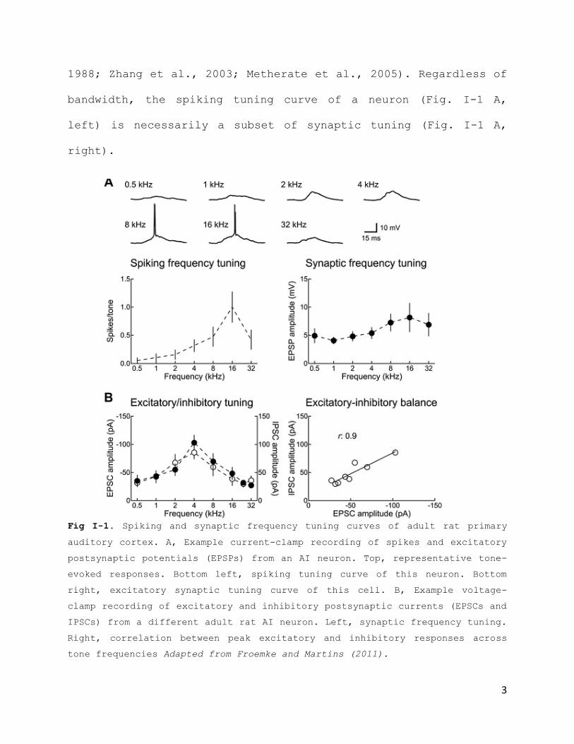
Primary auditory cortical neurons are generally tuned to sound
frequency (Fig. I-1 A). While many neurons have a clear
preference for pure tones of a specific frequency (the ‘best
frequency’), the tuning curve width, and overall response rates
depend strongly on sound level. Neurons in adult rat primary
auditory cortex, for example, exhibit broad sub- and
suprathreshold tuning at high intensities, potentially spanning
much of the total cochlear frequency range (Sally and Kelly,
3
1988; Zhang et al., 2003; Metherate et al., 2005). Regardless of
bandwidth, the spiking tuning curve of a neuron (Fig. I-1 A,
left) is necessarily a subset of synaptic tuning (Fig. I-1 A,
right).
Fig I-1. Spiking and synaptic frequency tuning curves of adult rat primary
auditory cortex. A, Example current-clamp recording of spikes and excitatory
postsynaptic potentials (EPSPs) from an AI neuron. Top, representative tone-
evoked responses. Bottom left, spiking tuning curve of this neuron. Bottom
right, excitatory synaptic tuning curve of this cell. B, Example voltage-
clamp recording of excitatory and inhibitory postsynaptic currents (EPSCs and
IPSCs) from a different adult rat AI neuron. Left, synaptic frequency tuning.
Right, correlation between peak excitatory and inhibitory responses across
tone frequencies Adapted from Froemke and Martins (2011).
4
As the entire receptive field of a particular neuron can be
difficult or impossible to completely characterize in high
detail, here frequency tuning preference in the rodent primary
auditory cortex will be used as a model for investigating the
phenomenology, mechanisms, and functional consequences of
synaptic receptive field plasticity in general. While these
changes can be registered at the suprathreshold spiking level,
using extracellular recording techniques (Bakin and Weinberger,
1996; Rasmusson D.D. and Dykes R.W., 1988; Kilgard MP. and
Merzenich M.M., 1998), this plasticity is ultimately due to the
adjustment of the subthreshold events that lead to spike
generation and collectively determine the tuning preferences and
therefore tuning curves of cortical neurons (Losonczy A. et al.,
2008; Feldman D.E., 2009; Dorrn et al., 2010). To a first
approximation, the organization of suprathreshold spiking tuning
curves is governed by the strengths and kinetics of excitatory
and inhibitory synapses, (Monier C. et al., 2003; Wehr M. and
Zador A.M., 2003; Zhang L.I. et al., 2003; Dorrn A.L. et al.,
2010). For this reason, data discussed throughout this thesis
refer to the synaptic basis of receptive field plasticity,
although bearing in mind that other factors that influence
postsynaptic integration- directly or indirectly- also play
important roles in shaping the tuning properties of cortical
neurons (Häusser M. and Mel B., 2003).
5
Receptive Field Plasticity in Primary Auditory Cortex:
Inhibition Regulation and the Case of Nucleus Basalis
Changes in the patterns of acoustic input, including sensory
deprivation and repetitive tonal presentation, can lead to
enduring changes in neuronal tonal preferences. However, the
strength and endurance of cortical changes through simple
manipulation of external acoustic environment is highly
dependent on developmental period (Chang E.F. et al., 2005; de
Villers-Sidani E. et al., 2007; Razak K.A. and Fuzessery Z.M.,
2007; Insanally M.N. et al., 2009; Sanes D.H. and Bao S., 2009;
Popescu M.V. and Polley,D.B., 2010). After a certain
developmental stage, and especially in adult life, long-term
cortical plasticity seems to depend more on stimulus history and
internal state factors like arousal state and motivation. This
behavioral context is conveyed by co-activation of subcortical
neuromodulatory nuclei such as the cholinergic nucleus basalis
(Rasmusson D.D., 2000; Weinberger N.M., 2007).and encoded as
stimulus salience level in cortical networks. In primary
auditory cortex, co-release of neuromodulators with tonal
presentation translates into a change in tonal response
preference. Regardless of developmental stage, the state of the
literature to the present indicates that regulation of the
6
relationship between inhibitory and excitatory inputs appears to
be a general mechanism by which changes in sensory experience
and neuromodulatory state can remodel cortical receptive fields
(Letzkus J.J. et al., 2011; Kuhlman S.J. et al., 2013; Froemke
R.C. et al., 2007, Fig. I-2 ; Dorrn A.L. et al., 2010, Fig. I-
3). Extracellular recording studies in vivo have shown that
pairing pure tones of a specific frequency with electrical
stimulation of nucleus basalis induce large, long-lasting
enhancements of spontaneous and tone-evoked spiking (Bakin and
Weinberger, 1996; Rasmusson and Dykes, 1988; Kilgard and
Merzenich, 1998). Whole-cell recordings in vivo have revealed
the mechanisms by which stimulation of the nucleus basalis
neuromodulatory system activates cortical networks and enables
receptive field plasticity (Froemke R.C. et al., 2007; Letzkus
et al., 2011). In these, in vivo whole-cell voltage-clamp
recordings were performed from neurons in adult rat primary
auditory cortex (Fig. I-2). Excitatory and inhibitory synaptic
frequency tuning profiles were initially measured, followed by
pairing of tones of a specific non-preferred frequency with
electrical stimulation of nucleus basalis. After the start of
pairing, there was a large suppression of inhibitory events
evoked by the paired tone, followed by a gradual enhancement of
tone-evoked excitation. These changes persisted 20 minutes or
more after the end of the pairing procedure.
7
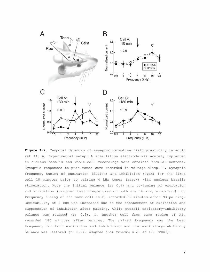
Figure I-2. Temporal dynamics of synaptic receptive field plasticity in adult
rat AI. A, Experimental setup. A stimulation electrode was acutely implanted
in nucleus basalis and whole-cell recordings were obtained from AI neurons.
Synaptic responses to pure tones were recorded in voltage-clamp. B, Synaptic
frequency tuning of excitation (filled) and inhibition (open) for the first
cell 10 minutes prior to pairing 4 kHz tones (arrow) with nucleus basalis
stimulation. Note the initial balance (r: 0.9) and co-tuning of excitation
and inhibition (original best frequencies of both are 16 kHz, arrowhead). C,
Frequency tuning of the same cell in B, recorded 30 minutes after NB pairing.
Excitability at 4 kHz was increased due to the enhancement of excitation and
suppression of inhibition after pairing, while overall excitatory-inhibitory
balance was reduced (r: 0.3). D, Another cell from same region of AI,
recorded 180 minutes after pairing. The paired frequency was the best
frequency for both excitation and inhibition, and the excitatory-inhibitory
balance was restored (r: 0.9). Adapted from Froemke R.C. et al. (2007).

8
Furthermore, during development, changes to inhibitory and
excitatory responses seem to be underlying previous findings
observed in young animals from manipulating acoustic
environment, by using patterned stimulation (Fig. I-3).
Figure I-3. Patterned stimulation improved excitatory–inhibitory coupling by
coordinated synaptic modifications across multiple inputs. a, Synaptic
modifications at the presented tone frequency spread to other inputs within
one octave (excitation one octave from presented frequency: 21.6±6.7%,
n=12,P<0.01; inhibition: 36.0±12.5%,P<0.02), but not two or more octaves away
(P>0.3).**P<0.01;*P<0.05.b, After patterned stimulation, responses at
original best frequency were reduced (excitation: -34.8±6.4%,n=12,P<0.0003;
inhibition:-22.7±6.1%,P<0.004). Adapted from Dorrn A.L. et al. (2010).
9
Eventually, these dynamic changes in inhibitory and excitatory
balance lead to wide-scale changes to many synapses throughout
cortical networks. These changes are coordinated to enhance the
representations of newly-significant stimuli, possibly for
improved signal processing (Froemke R.C. and Carcea I. et al.,
2013).
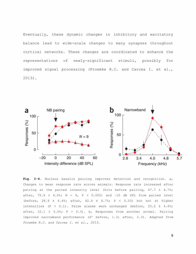
Fig. I-4. Nucleus basalis pairing improves detection and recognition. a,
Changes to mean response rate across animals. Response rate increased after
pairing at the paired intensity level (hits before pairing, 47.7 ± 4.7%;
after, 70.6 ± 4.0%; N = 9, P < 0.002) and −10 dB SPL from paired level
(before, 28.9 ± 6.4%; after, 42.4 ± 6.7%; P < 0.03) but not at higher
intensities (P > 0.1). False alarms were unchanged (before, 25.2 ± 4.6%;
after, 22.1 ± 5.5%; P > 0.3). b, Responses from another animal. Pairing
improved narrowband performance (d′ before, 1.3; after, 2.3). Adapted from
Froemke R.C. and Carcea I. et al., 2013.
10
Receptive Field Plasticity in Primary Auditory Cortex:
The Unsolved Case of Noradrenaline and Noradrenergic
Locus Coeruleus
The behavioral context of an incoming stimulus is conveyed by
activation of subcortical neuromodulatory systems, like the
cholinergic nucleus basalis or the noradrenergic locus
coeruleus.
Noradrenergic and cholinergic systems while different
functionally, seem to have a similar developmental
trajectory/origin, with Locus coeruleus and Nucleus basalis
axons growing and developing closely in the brain during
development and both systems taking part in mechanisms important
for plasticity (Bear M.F. & Singer W, 1986).
The mechanisms underlying Nucleus basalis stimulation -induced
auditory cortical plasticity have been addressed in the previous
chapter, so the question arises of how it is that noradrenergic
Locus coeruleus interacts with and refines auditory cortical
networks.
The noradrenergic Locus coeruleus is a small nucleus of
electrotonically-coupled noradrenergic neurons in the brainstem,
with an extensive axonal network reaching all areas of the brain

11
with the notable exception of basal ganglia. Locus coeruleus
dendrites group in a spatially distinct area in the brainstem,
the sub coeruleus area, right bellow the cell bodies. The sub
coeruleus area is a quaint place with a little studied local
inhibitory network, and receives most of the top-down inputs
from higher brain areas. The Locus coeruleus cell bodies receive
inputs mostly from local brainstem centers (including Raphe
Nucleus). The excitatory or inhibitory nature of most of the
inputs onto the locus coeruleus and subcoeruleus area remains
mostly unknown. Interestingly, Locus coeruleus axons have direct
synapses on astrocytic endfeet, and the role of noradrenaline in
the regulation of blood brain barrier and astrocytic activity is
an active field of study (Foote et al., 1983a; Foote et al.,
1983b; Loughlin S.E., et al, 1986; Morrison J.H. and Foote S.L.,
1986; Simpson K.L. et al, 1997; Waterhouse B.D. and Berridge
C.W. , 2003; Sara S.J., 2009).
The locus coeruleus is the sole source of noradrenaline in the
central nervous system, but similar to other neuromodulatory
systems, it also releases small amounts of other factors, like
vasopressin, somatostatin, neuropeptide Y, enkephalin,
neurotensin, CRH and galanin. Galanin, a hyperpolarizing
neuropeptide, is of particular interest since it has been
estimated that 80% of Locus coeruleus neurons colocalize galanin
12
and noradrenaline. However only half of the Locus coeruleus
neurons that project to either cortical or sub-cortical
somatosensory circuits contain galanin (Waterhouse B.D. and
Berridge C.W. , 2003)
Noradrenaline is important for learning, synaptic plasticity,
and modification of sensory representations and is released
throughout the brain, including the auditory cortex, by locus
coeruleus during periods of arousal or anxiety (Bear, M.F. &
Singer, W, 1986; Berridge, C.W., 2008; Sara S.J., 2009; Carter,
M.E. et al, 2009; Bush, D.E. et al., 2010; Constantinople, C.M.
and Bruno, R.M, 2011). Locus coeruleus has two modes of firing:
tonic (range between 0-5Hz, in mammals; full silence of 0Hz
seems to happen specifically during REM sleep) and phasic
(bursts in the range of 5-20Hz have been reported, in mammals).
The tonic mode controls general states of arousal and sleep-wake
cycle, while the phasic mode is elicited by behaviorally-
relevant salient stimuli, as well as top-down decision- and
response-related signals from prefrontal cortical regions
(Berridge and Waterhouse 2003; Aston-Jones and Bloom 1981b,
2005a,b; Devilbiss D.M and Waterhousse B.D., 2010). It is
hypothesized that locus coeruleus plays a major role in
adjusting gains of cortical synapses (Aston-Jones G et al.,
1994; Usher M., et al., 1999, Yu A.J. and Dayan P., 2005, Kuo,

13
S.P. and Trussel L.O., 2011). This occurs when locus coeruleus
neurons are activated by behaviorally significant stimuli
(Aston-Jones G. and Cohen J.D., 2005; Berridge, C.W., 2008; Sara
S.J., 2009; Carter, M.E. et al, 2009). However, it is unknown
how locus coeruleus neurons and noradrenaline interact with and
refine cortical circuits.
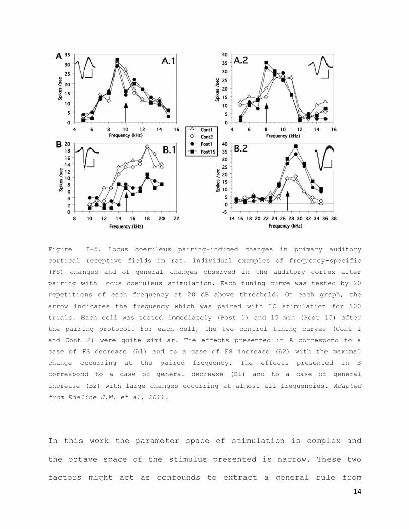
Work done by the Edeline and Manunta (Edeline J.M. et al., 2011)
has showed that stimulation of Locus coeruleus changes auditory
cortical receptive fields in a very heterogeneous way (Fig. I-
5). In their study, specific increases and decreases in the
paired tone frequency were observed, and also increases or
decreases across all tones. A general rule is drawn that the
distance between the initial preferred frequency and the paired
frequency is critical for the occurrence of frequency specific
effects. These types of effects would happen when the paired
frequency and the initial preferred frequency were spectrally
very close (<1/4 of an octave).
14
Figure I-5. Locus coeruleus pairing-induced changes in primary auditory
cortical receptive fields in rat. Individual examples of frequency-specific
(FS) changes and of general changes observed in the auditory cortex after
pairing with locus coeruleus stimulation. Each tuning curve was tested by 20
repetitions of each frequency at 20 dB above threshold. On each graph, the
arrow indicates the frequency which was paired with LC stimulation for 100
trials. Each cell was tested immediately (Post 1) and 15 min (Post 15) after
the pairing protocol. For each cell, the two control tuning curves (Cont 1
and Cont 2) were quite similar. The effects presented in A correspond to a
case of FS decrease (A1) and to a case of FS increase (A2) with the maximal
change occurring at the paired frequency. The effects presented in B
correspond to a case of general decrease (B1) and to a case of general
increase (B2) with large changes occurring at almost all frequencies. Adapted
from Edeline J.M. et al, 2011.
In this work the parameter space of stimulation is complex and
the octave space of the stimulus presented is narrow. These two
factors might act as confounds to extract a general rule from
15
the results- the octave spacing might be too narrow to see
consistency, and stimulation parameters might sit at an
ambiguous level, where the Locus is not robustly "tipping the
scale" one way or another.
This created the need to explore unequivocal, high frequency,
behaviorally-relevant LC firing in order to clarify the effects
of Locus coeruleus stimulation in cortical networks.
Conclusion
This chapter described the current state of the literature in
primary auditory cortex plasticity, and the contribution of
neuromodulator release for synaptic cortical changes and
adaptation. In the rest of this thesis, I focus on studying the
noradrenergic Locus coeruleus and the neuromodulator
noradrenaline in the context of auditory cortical plasticity and
auditory perception in adult rats. I chose to study locus
coeruleus due to the importance of noradrenergic neuromodulation
in normal arousal states and behavior output, and the relatively
limited body of literature on the contribution of noradrenalin
to plasticity of neural networks. Despite all the work done to
understand how noradrenaline and the noradrenergic locus
coeruleus interact with and change auditory cortical networks,
16
consistency is lacking and a general rule for this interaction
is still unable to be drawn.
Furthermore, it remains unexplored how neuromodulator systems,
and in particular the noradrenergic Locus coeruleus, can be an
active element of the circuit, adapting and providing
differential modulatory control based on past experience and the
needs of the current environmental context.

17
Chapter II. Locus Coeruleus
Activation-Induced Primary Auditory
Cortex Plasticity
Preface
Sensory cortical networks are plastic, their receptive fields
dynamically adapting in response to changes activity or sensory
experience. Receptive field plasticity in the auditory cortex,
driven by environmental statistics combined with
neuromodulation, allows cortical networks to re-organize around
salient acoustic features of the external environment. Here I
describe how stimulation of Locus coeruleus paired with a
passive tonal presentation re-organizes tonal response receptive
fields in primary auditory cortex.
Most of this chapter has been submitted for publication in
Nature Neuroscience. The Nucleus basalis experiment, depicted in
Fig. II-5 was entirely performed by Dr Robert C. Froemke.
.
Introduction
Receptive fields of sensory cortical neurons are highly
structured. The spatial arrangement and strength of synaptic

18
inputs contribute to the organization of receptive fields, which
relay the perception of the external world (Hubel D.H. and
Wiesel T.N., 1962; Hirsch and Martinez, 2006; Huberman et al.,
2008; Ye et al., 2010). Receptive field plasticity in the
auditory cortex allows cortical re-organization around salient
features of the sensory environment, and depends on the patterns
of electrical activity (Fregnac Y., et al, 1988; Talwar S.K., et
al, 2001; Meliza C.D. and Dan Y., 2006; Jacob V., et al, 2007),
sensory experience (katz L.C. and Shatz C.J., 1996; Buonomano
D.V. and Merzenich M.M, 1998; Fritz J.S. egt al,2003; Feldman
D.E. and Brecht M., 2005; Dan Y. and Poo M.M., 2006; de Villers-
Sidani, E., et al, 2007; Li Y., et al, 2008; Dahmen J.C., et al,
2008; Dorrn A.L., et al, 201) and engagement of neuromodulatory
systems such as the cholinergic Nucleus basalis and the
noradrenergic Locus Coeruleus.
Previous work in vivo extracellular recording work done on
cholinergic Nucleus basalis modulation of primary auditory
cortical receptive fields found that the have shown that pairing
pure tones of a specific frequency with electrical stimulation
of nucleus basalis induces large, long-lasting enhancements of
spontaneous and tone-evoked spiking (Bakin and Weinberger, 1996;
Rasmusson and Dykes, 1988;Kilgard and Merzenich, 1998). But the
mechanism underlying this change is spiking remianed unknown

19
until postdoctoral work done by my mentor, Dr Robert C. Froemke,
shed light on the synaptic mechanism of this. In Froemke et al.,
2007, the synaptic mechanism by which stimulation of the nucleus
basalis neuromodulatory system activates cortical networks,
enabling receptive field plasticity is revealed. By performing
in vivo whole-cell voltage clamp recordings from layer 5
pyramidal neurons in primary auditory cortex, this work showed
that Nucleus basalis stimulation and pairing with a tone, leads
to a disinhibition of the response to the paired tone, by
temporarily de-coupling excitatory and inhibitory tuning
profiles specifically at the paired tone frequency, creating
this "synaptic memory trace" of disinhibition to the tone paired
( Fig. I-2, Froemke R.C. et al., 2007) .
However, how do other neuromodulatory systems interact with and
change cortical networks? Is there a general rule to be drawn?
Do they each have their own set of different rules to
contextualize stimuli? We decided to address these questions by
looking at the noradrenergic Locus coeruleus system, given the
parallelisms with the cholinergic basalis, in its general role
on attention and arousal, and the lack of coherent literature on
the cortical effects of Locus coeruleus stimulation.
As a step towards understanding how noradrenergic Locus
coeruleus stimulation affects receptive field plasticity, in
20
this chapter I perform in vivo intracellular recordings from
100+ cortical neurons in Layer 5 of primary auditory cortex,
looking at early and long-term changes brought about by pairing
stimulation of the Locus with passive tonal presentation.
Methods: Electrophysiological Recordings of Layer 5
Pyramidal Neurons of the Rat Primary Auditory Cortex In
Vivo
Here I will describe the methods used for performing my
electrophysiological studies in vivo. All procedures were
approved under NYU IACUC protocols. Experiments were carried out
in a sound-attenuating chamber. Female Sprague-Dawley rats 3-5
months old were anesthetized with ketamine (1.2 ml/kg) and
dexmedetomidine (1.0 ml/kg). A bipolar stimulation electrode was
implanted in the right locus coeruleus using stereotaxic
coordinates (from lambda, in mm: 3.6 posterior, 1.2 lateral,
5.6-6 ventral). Location was verified during procedures by
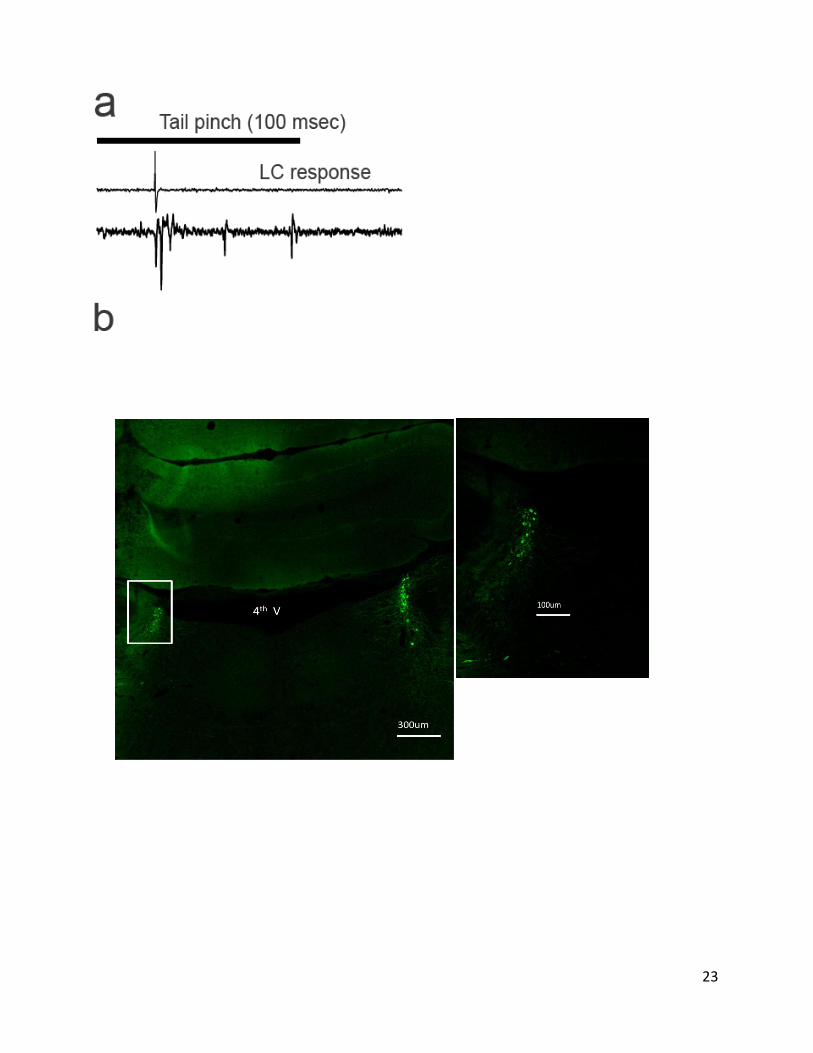
measuring responses to noxious stimuli (tail pinch, Fig. II-1a)
and other electrophysiological criteria (spontaneous rates, Fig.
II-1d), and afterwards using histological methods (Fig. II-
1b,c). To determine the effective activation radius of locus
coeruleus stimulation in Figure II-1e, local field potentials
(LFPs) were recorded with a tungsten electrode (0.5-1 M)
21
lowered to 5500-6000 µm below the cerebellar surface. Several
penetrations were made at different distances from the
stimulation electrode (100-2000 µm). LFPs were digitized at 20
kHz and bandpass filtered between 1-100 Hz.
A craniotomy was performed over the right temporal lobe and the
right auditory cortex was exposed. Pure tones (10-80 dB SPL,
0.5-32 kHz, 50 msec, 3 msec cosine on/offramps) were delivered
in pseudo-random sequence at 0.5-1 Hz. AI location was
determined by mapping multiunit responses 500-700 µm below the
surface using tungsten electrodes. In vivo whole-cell recordings
from AI neurons were made with a Multiclamp 700B amplifier
(Molecular Devices). For current-clamp recordings, patch
pipettes (5-9 MΩ) contained (in mM): 135 K-Gluconate, 5 NaCl, 5
MgATP, 0.3 GTP, 10 phosphocreatine, 10 HEPES, pH 7.3. For
voltage-clamp recordings, pipettes contained: 125 Cs-gluconate,
5 TEACl, 4 MgATP, 0.3 GTP, 10 phosphocreatine, 10 HEPES, 0.5
EGTA, 3.5 QX-314, 2 CsCl, pH 7.2. In some cases, 1% biocytin
(Sigma) was added to the internal solution for post-hoc recovery
of recorded neurons (Fig II-1d). Recordings from AI neurons were
obtained from cells located 400-1200 µm below the pial surface.
Resting potential of AI neurons: –62.6±11.7 mV; Rs: 21.9±12.2
M ; Ri: 106.0±55.3 MΩ. Data were excluded if Rs changed >30% or
Ri changed >50% from values measured during baseline (as locus

22
coeruleus stimulation modestly but significantly increased Ri,
in AI current-clamp recordings from 126.6±68.4 MΩto 135.6±75.1
MΩ, n=21, p<0.03). Data were filtered at 5 kHz, digitized at 20
kHz, and analyzed with Clampfit 10 (Molecular Devices).
For noradrenaline iontophoresis in Fig. II-7a, double-barreled
iontophoresis pipettes (30 MΩ) contained noradrenaline (1 mM)
and were placed ~700 µm below the pial surface, ~250-300 µ m
from the recording pipette. For experiments of Fig. II-76b,c,
phentolamine (1 mM in saline) was topically applied to AI.
Animals were perfused with 4% paraformaldehyde, brains
recovered, and embedded in Optimal Cutting Temperature compound
prior to freezing at –80°C. Afterwards, 40 µm thick slices were
cut from the brainstem and stained using standard
immunohistochemistry histological methods. Staining for tyrosine
hydroxylase (primary antibody 1:1000, Aves Labs; secondary
antibody, DYL488 anti-chicken, 1:500, Life Technologies Labs)
was co-localized with biocytin staining revealed with Alexa
Fluor 555 conjugated Streptavidin (1:100, Life Technologies
Labs).
23

24

25
Figure II-1. Recording from locus coeruleus. a, Noxious stimuli evoke phasic
spike bursts in rat locus coeruleus. Top, brief tail pinch (100 msec
duration) evoked phasic spiking in locus coeruleus, as measured through the
stimulation electrode. b, example post hoc histological recovery of electrode
placement in Locus coeruleus. Boxed area ex exemplifying correct electrode
placement in the Locus amplified on the side. c, example locus coeruleus
neurons filled with biocytin during whole-cell recording in vivo and stained
for tyrosine hydroxylase. d, Whole-cell current-clamp (top) and cell-attached
(bottom) recordings from single neurons in locus coeruleus in vivo. Shown are
traces of spontaneous spiking activity. e, Local field potentials (LFPs)
measured at distances from locus coeruleus stimulation electrode. LFPs were
only observed <500 µm.
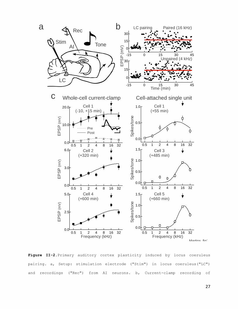
Primary Auditory Cortex Plasticity induced by
Activation of Locus Coeruleus
A major output of locus coeruleus is the cerebral cortex, where
noradrenergic modulation controls sensory processing and
behavior (Aston-Jones G and Cohen J.D., 2005; Berridge C.W.,
2008; Sara S.J., 2009). To determine how locus coeruleus pairing
affected cortical auditory representations, recordings were
made in vivo, whole-cell from 91 (51 current-clamp, 40 voltage-
clamp) and cell-attached from 50 primary auditory cortex cells
in 49 adult rats implanted with stimulation electrodes in locus
coeruleus(Fig. II-2a). Baseline responses to pure tones were
recorded from primary auditory cortical neurons, locus coeruleus
26
pairing was performed, and responses measured as long as
recordings remained stable. When the first recording ended, 1-7
more recordings were sequentially made from that cortical
location to document the dynamics of post-pairing modification
over 12 hours.
One set of recordings demonstrating the cortical effects of
locus coeruleus pairing is shown in Fig. II- 2b,c. Recordings
from five neurons from the same region of AI initially tuned to
4 kHz, for 11 hours after pairing. The first cell had a peak in
the synaptic tuning curve at 4 kHz. The paired frequency was 16
kHz; after pairing, large increases were observed in responses
to all frequencies (Fig. 2b) and the peak shifted to the paired
frequency (Fig. II- 2c, upper left, black line). This recording
then re-sealed and cell-attached spiking responses were
measured. It was observed that the paired 16 kHz tone was peak
of the spiking tuning curve (Fig. II-2c, lower left). Over the
next 10 hours, four more recordings were obtained (Fig. II-2c
middle, right). Tuning width recovered but 16 kHz remained best
frequency.
27
Figure II-2.Primary auditory cortex plasticity induced by locus coeruleus
pairing. a, Setup: stimulation electrode ("Stim") in locus coeruleus("LC")
and recordings ("Rec") from AI neurons. b, Current-clamp recording of
a
LC
AIStim
Tone
Rec
c
Frequency (kHz)
EP
SP
(mV
)
Cell 1(-10, +15 min)
Pre
PostS
pik
es/t
on
e
Frequency (kHz)
Cell 1(+55 min)
Cell 2(+320 min)
Cell 3(+485 min)
Cell 4(+600 min)
Cell 5(+660 min)
Whole-cell current-clamp Cell-attached single unit
Sp
ike
s/t
one
Sp
ike
s/t
one
EP
SP
(mV
)E
PS
P(m
V)
0.0
0.5
1.0
0.5 1 2 4 8 16 320.0
10.0
20.0
0.5 1 2 4 8 16 32
Martins_fig2
0.0
3.0
6.0
0.5 1 2 4 8 16 32
0.0
0.5
1.0
1.5
0.5 1 2 4 8 16 32
0.0
2.5
5.0
0.5 1 2 4 8 16 32
0.0
0.5
1.0
1.5
0.5 1 2 4 8 16 32
b Paired (16 kHz)
Time (min)
EP
SP
(mV
)
LC pairing
Unpaired (4 kHz)
0
15
30
0 15-15 30 45
0
15
30
0 15-15 30 45
28
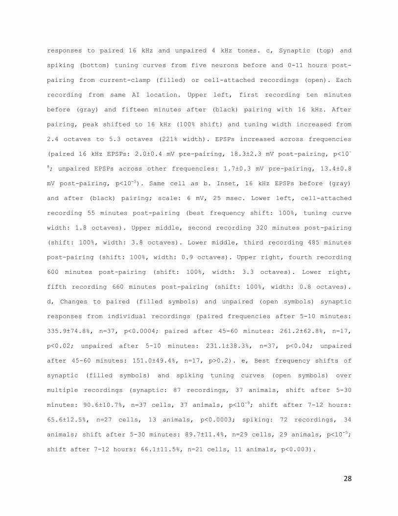
responses to paired 16 kHz and unpaired 4 kHz tones. c, Synaptic (top) and
spiking (bottom) tuning curves from five neurons before and 0-11 hours post-
pairing from current-clamp (filled) or cell-attached recordings (open). Each
recording from same AI location. Upper left, first recording ten minutes
before (gray) and fifteen minutes after (black) pairing with 16 kHz. After
pairing, peak shifted to 16 kHz (100% shift) and tuning width increased from
2.4 octaves to 5.3 octaves (221% width). EPSPs increased across frequencies
(paired 16 kHz EPSPs: 2.0±0.4 mV pre-pairing, 18.3±2.3 mV post-pairing, p<10-
8; unpaired EPSPs across other frequencies: 1.7±0.3 mV pre-pairing, 13.4±0.8
mV post-pairing, p<10-5). Same cell as b. Inset, 16 kHz EPSPs before (gray)
and after (black) pairing; scale: 6 mV, 25 msec. Lower left, cell-attached
recording 55 minutes post-pairing (best frequency shift: 100%, tuning curve
width: 1.8 octaves). Upper middle, second recording 320 minutes post-pairing
(shift: 100%, width: 3.8 octaves). Lower middle, third recording 485 minutes
post-pairing (shift: 100%, width: 0.9 octaves). Upper right, fourth recording
600 minutes post-pairing (shift: 100%, width: 3.3 octaves). Lower right,
fifth recording 660 minutes post-pairing (shift: 100%, width: 0.8 octaves).
d, Changes to paired (filled symbols) and unpaired (open symbols) synaptic
responses from individual recordings (paired frequencies after 5-10 minutes:
335.9±74.8%, n=37, p<0.0004; paired after 45-60 minutes: 261.2±62.8%, n=17,
p<0.02; unpaired after 5-10 minutes: 231.1±38.3%, n=37, p<0.04; unpaired
after 45-60 minutes: 151.0±49.4%, n=17, p>0.2). e, Best frequency shifts of
synaptic (filled symbols) and spiking tuning curves (open symbols) over
multiple recordings (synaptic: 87 recordings, 37 animals, shift after 5-30
minutes: 90.6±10.7%, n=37 cells, 37 animals, p<10-9; shift after 7-12 hours:
65.6±12.5%, n=27 cells, 13 animals, p<0.0003; spiking: 72 recordings, 34
animals; shift after 5-30 minutes: 89.7±11.4%, n=29 cells, 29 animals, p<10-5;
shift after 7-12 hours: 66.1±11.5%, n=21 cells, 11 animals, p<0.003).

29
Over 141 primary auditory cortical recordings, three
general features of cortical plasticity induced by locus
coeruleus pairing were apparent: 1) large increases in tone-
evoked responses to all stimuli; 2) shifts in peak towards
paired inputs; and 3) return of average tuning width over
several hours, with maintained peak preference at paired inputs
(Fig.II-3). In individual neurons, responses at paired inputs
were substantially larger 45-60 minutes after pairing (Fig. II-
2d, filled; Fig.II-3a). Responses to unpaired inputs were also
enhanced after pairing, but returned towards original levels 45-
60 minutes post-pairing (Fig. II-2d, open; Fig. II-32b). Similar
changes were also observed for spiking responses (Fig. II-2e,
open; Fig.II-3e,f) and intensity profiles (FigII-4).

30
Figure II-3. Changes to synaptic and spiking tuning curves after locus
coeruleus pairing. a, Changes to paired inputs from individual recordings.
Squares, current-clamp (EPSP amplitude 5-10 minutes post-pairing:
367.9±124.7%, n=21, p<0.05; 45-60 minutes: 341.3±80.8%, n=7, p<0.03).
Triangles, voltage-clamp (5-10 minutes: 296.9±56.4%, n=16, p<0.004; 45-60
minutes: 197.1±53.8%, n=10, p<0.06). Circles, all recordings (5-10 minutes:

31
335.9±74.8%, n=37, p<0.0004; 45-60 minutes: 261.2±62.8%, n=17, p<0.02). b,
Changes to unpaired inputs from individual recordings for current-clamp (5-10
minutes: 253.3±59.4%, n=21, p<0.02; 45-60 minutes: 168±40.3%, n=7, p>0.1),
voltage-clamp (5-10 minutes: 205.8±39.1%, n=16, p<0.03; 45-60 minutes:
139.1±47.4%, n=10, p>0.3) and all recordings (5-10 minutes: 231.1±38.3%,
n=37, p<0.04; 45-60 minutes: 151.0±49.4%, n=17, p>0.2). Same recordings as a.
c, Best frequency shift of synaptic tuning curves over multiple recordings.
Circles, current-clamp (total: 47 cells, 21 animals; best frequency shift 5-
30 minutes post-pairing: 96.6±10.4%, n=21 cells, 21 animals, p<10-7; shift 7-
12 hours post-pairing: 74.4±15.5%, n=15 cells, 7 animals, p<0.003).
Triangles, voltage-clamp (total: 40 cells, 16 animals; 5-30 minutes:
82.8±20.8%, n=16 cells, 16 animals, p<0.002; 7-12 hours: 55.5±20.9%, n=12
cells, 6 animals, p<0.05). Circles, all recordings (total: 87 recordings, 37
animals; 5-30 minutes: 90.6±10.7%, n=37 cells, 37 animals, p<10-9; 7-12 hours:
65.6±12.5%, n=27 cells, 13 animals, p<0.0003). d, Synaptic tuning curve width
over multiple recordings. Values normalized to baseline width. Squares,
current-clamp (total: 47 cells, 21 animals, baseline width: 3.7 octaves;
width 5-30 minutes post-pairing: 154.3±17.6%, n=21 cells, 21 animals, p<0.02;
width 7-12 hours post-pairing: 106.1±9.7%, n=15 cells, 7 animals, p>0.5).
Triangles, voltage-clamp (total: 40 recordings, 16 animals, average baseline
width: 4.0 octaves; 5-30 minutes: 129.0±23.8%, n=16 cells, 16 animals,
p<0.004; 7-12 hours: 106.6±14.6%, n=12 cells, 6 animals, p>0.2). Circles, all
recordings (total: 87 recordings, 37 animals; 5-30 minutes: 146.6±14.8%, n=37
cells, 37 animals, p<0.0002; 7-12 hours: 103.3±7.8%, n=27 cells, 13 animals,
p>0.1). Same recordings as c. e, Best frequency shifts of spiking tuning
curves over multiple recordings. Filled squares, current-clamp (total: 22
cells, 13 animals; shift in best frequency 5-30 minutes after pairing:
92.0±16.0%, n=13 cells, 13 animals, p<0.003; shift 7-12 hours after pairing:
37.5±23.9%, n=4 cells, 2 animals). Open squares, cell-attached recordings
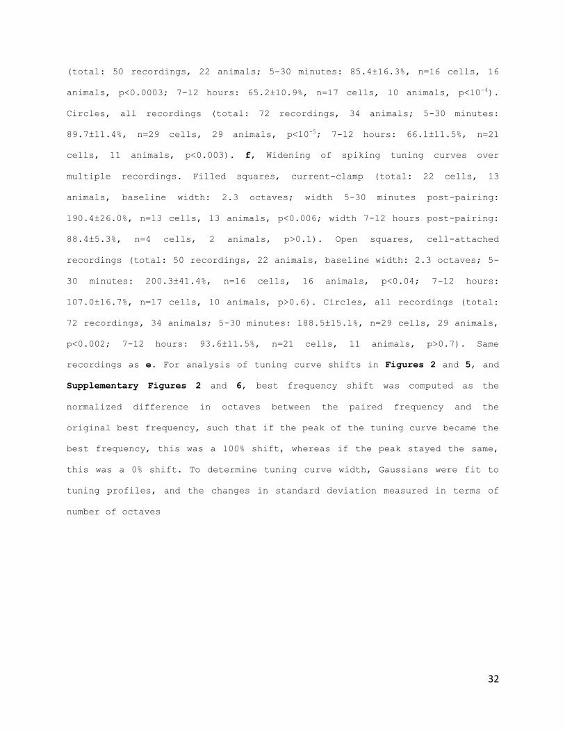
32
(total: 50 recordings, 22 animals; 5-30 minutes: 85.4±16.3%, n=16 cells, 16
animals, p<0.0003; 7-12 hours: 65.2±10.9%, n=17 cells, 10 animals, p<10-4).
Circles, all recordings (total: 72 recordings, 34 animals; 5-30 minutes:
89.7±11.4%, n=29 cells, 29 animals, p<10-5; 7-12 hours: 66.1±11.5%, n=21
cells, 11 animals, p<0.003). f, Widening of spiking tuning curves over
multiple recordings. Filled squares, current-clamp (total: 22 cells, 13
animals, baseline width: 2.3 octaves; width 5-30 minutes post-pairing:
190.4±26.0%, n=13 cells, 13 animals, p<0.006; width 7-12 hours post-pairing:
88.4±5.3%, n=4 cells, 2 animals, p>0.1). Open squares, cell-attached
recordings (total: 50 recordings, 22 animals, baseline width: 2.3 octaves; 5-
30 minutes: 200.3±41.4%, n=16 cells, 16 animals, p<0.04; 7-12 hours:
107.0±16.7%, n=17 cells, 10 animals, p>0.6). Circles, all recordings (total:
72 recordings, 34 animals; 5-30 minutes: 188.5±15.1%, n=29 cells, 29 animals,
p<0.002; 7-12 hours: 93.6±11.5%, n=21 cells, 11 animals, p>0.7). Same
recordings as e. For analysis of tuning curve shifts in Figures 2 and 5, and
Supplementary Figures 2 and 6, best frequency shift was computed as the
normalized difference in octaves between the paired frequency and the
original best frequency, such that if the peak of the tuning curve became the
best frequency, this was a 100% shift, whereas if the peak stayed the same,
this was a 0% shift. To determine tuning curve width, Gaussians were fit to
tuning profiles, and the changes in standard deviation measured in terms of
number of octaves

33
Figure II-4. Locus coeruleus pairing modifies intensity tuning of AI
neurons. a, Example current-clamp recording showing synaptic intensity tuning
curves (measured at 8 kHz) before and after locus coeruleus pairing at a
lower intensity (50 dB SPL). b, Example current-clamp recording showing
spiking intensity tuning curves (at 8 kHz) before and after locus coeruleus
pairing at a higher intensity (70 dB SPL). c, Summary of changes to evoked
synaptic and spiking responses after pairing. Left, EPSPs evoked by the
paired intensity before and after pairing (pre: 1.0±0.5 mV, post: 5.0±1.7 mV;
n=6, p<0.04). Right, tone-evoked spikes before and after pairing (pre:
0.05±0.03 spikes/tone, post: 0.35±0.11 spikes/tone; n=6, p<0.02).
34
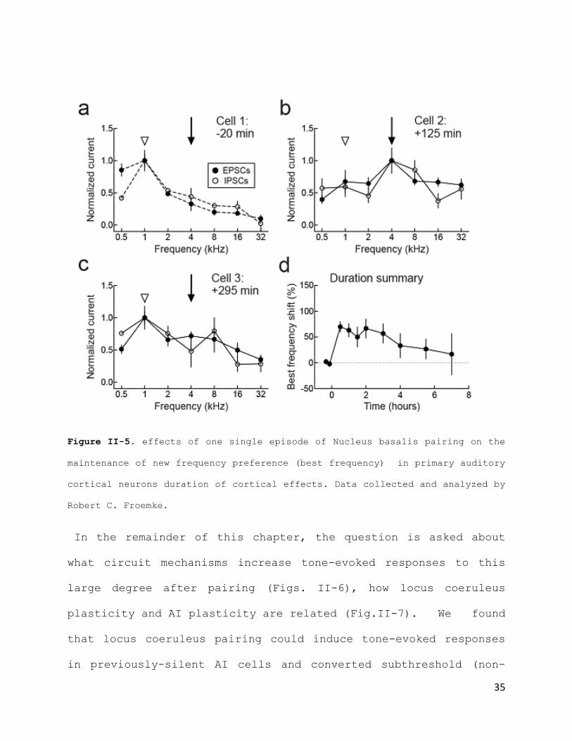
Importantly, these data revealed that the duration of cortical
frequency preference changes induced by Locus coeruleus pairing
were incredibly long lasting. In fact, one single episode of
pairing, lasting only 1-2min, seemed to allow an unusually
enduring maintenance of the new frequency preference for the
duration of our recordings (~11h+). Noteworthy is the
dissimilarity between this finding and the maintenance of
cortical effects observed for Nucleus basalis Pairing (Fig. II-
5), where the new frequency preference is only maintained for a
period of ~5h, before a return to baseline.
35
Figure II-5. effects of one single episode of Nucleus basalis pairing on the
maintenance of new frequency preference (best frequency) in primary auditory
cortical neurons duration of cortical effects. Data collected and analyzed by
Robert C. Froemke.
In the remainder of this chapter, the question is asked about
what circuit mechanisms increase tone-evoked responses to this
large degree after pairing (Figs. II-6), how locus coeruleus
plasticity and AI plasticity are related (Fig.II-7). We found
that locus coeruleus pairing could induce tone-evoked responses
in previously-silent AI cells and converted subthreshold (non-

36
spiking) responses into suprathreshold (spiking) responses (Fig.
II-6a-d). Interestingly, tone-evoked responses were not detected
in 9/37 AI neurons before pairing. In 6/9 non-responsive cells,
pairing rapidly induced responses that were maintained for the
recording duration. Moreover, paired tones initially evoked
spikes only in 3/21 current-clamp and 9/12 cell-attached
recordings. After pairing, spikes were evoked in 13/21 current-
clamp and 12/12 cell-attached recordings. These results indicate
that pairing increases the size of the cell assembly encoding
paired tones, amplifying AI output to environmental stimuli
linked with Locus coeruleus activation.
37
i

38
FigureII-6. Cortical circuit mechanisms of enhanced AI responses after locus
coeruleus pairing. a, Voltage-clamp recording showing new tone-evoked
responses during pairing. Inset, responses before (gray) and during pairing
(black); scale: 150 pA, 10 msec. b, Current-clamp recording showing sub- to
suprathreshold tone-evoked responses. Dashed line, spike threshold; responses
above this evoked 1+ action potentials. Inset, responses before (gray) and
during pairing (black); scale: 5 mV, 10 msec. c, Synaptic changes. Left,
voltage-clamp recordings pre/post pairing (pre: –15.7±2.8 pA, post: –31.3±6.2
pA; n=16, p<0.007). 10/16 recordings had significant tone-evoked responses
before pairing, increased to 14/16 post-pairing (p<0.05). Right, current-
clamp recordings (pre: 2.0±0.4 mV, post: 5.1±1.1 mV; n=21, p<0.003). 18/21
recordings had significant tone-evoked responses before pairing, increased to
20/21 post-pairing. d, Spiking changes. Left, current-clamp recordings before
and after pairing (pre:0.05±0.03 spikes/tone, post: 0.25±0.07 spikes/tone;
n=21, p<0.005). 4/21 recordings had 1+ tone-evoked spikes before pairing,
increased to 13/21 after pairing. Right, cell-attached recordings (pre:
0.5±0.2 spikes/tone, post: 0.9±0.3 spikes/tone; n=12, p<0.05). 9/12
recordings had 1+ tone-evoked spikes before pairing and 12/12 post-pairing.
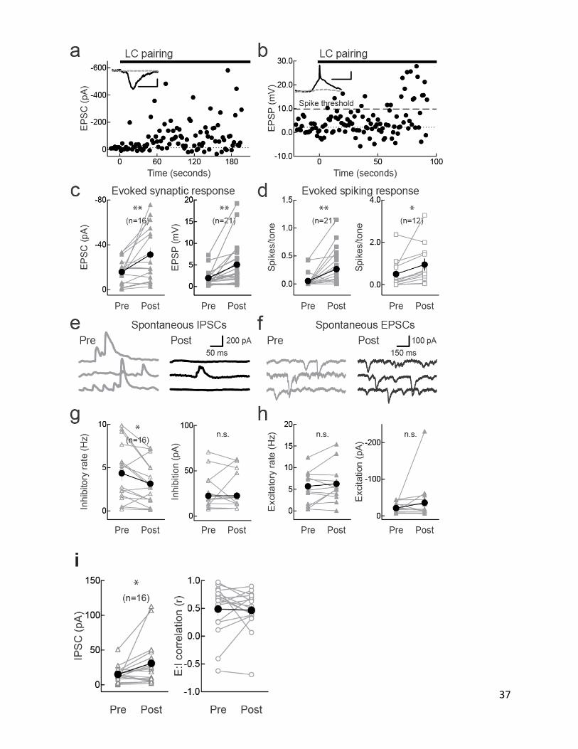
e, Pairing decreased spontaneous IPSC rate (pre: 9.2 Hz, post: 5.0 Hz) but
not amplitude (pre: 70.8 pA, post: 61.3 pA). f, Pairing did not affect
spontaneous EPSC rate (pre: 12.4 Hz, post: 15.4 Hz) or amplitude (pre: –44.3
pA, post: –56.7 pA). Same recording as e. g, Spontaneous IPSCs. Left, IPSC
rate pre/post-pairing (pre: 4.3±0.8 Hz, post: 3.1±0.6 Hz; n=16, p<0.02).
Right, amplitude (pre: 24.6±4.7 pA, post: 24.7±4.9 pA; n=16, p>0.9). 'n.s.',
non-significant. h, Spontaneous EPSCs. Left, EPSC rate pre/post-pairing (pre:
5.7±1.0 Hz, post: 6.3±1.1 Hz; n=16, p>0.3). Right, amplitude (pre: –21.1±3.0
pA, post: –35.7±13.7 pA; n=16, p>0.2). i, Summary of changes to tone-evoked
IPSCs and excitatory-inhibitory correlation after pairing. Left, voltage-
clamp recordings of IPSCs evoked by paired tones before and after pairing
39
(pre: 15.1±3.1 pA, post: 31.0±8.6 pA; n=16, p<0.05, Student's paired two-
tailed t-test). Right, linear correlation coefficient r of excitation and
inhibition for all presented tones before and after pairing was unchanged
(pre: 0.5±0.1, post: 0.5±0.1 mV; n=21, p>0.8).
The neuromodulator acetylcholine reduces evoked inhibition to
shift cortical excitatory-inhibitory balance in favor of
excitation Thus we next asked if locus coeruleus stimulation had
a similar effect, making voltage-clamp recordings from AI
neurons to assess changes to inhibitory postsynaptic currents
(IPSCs). Pairing greatly increased tone-evoked EPSCs and IPSCs
(Fig. II-6) but decreased tonic inhibition by reducing
spontaneous IPSC rate while spontaneous IPSC amplitudes and
spontaneous EPSCs ( Fig. II-6f-h) were unaffected. These
findings show that locus coeruleus activation seems to
specifically decrease tonic (spontaneous) inhibition rather than
phasic (stimulus-evoked) inhibition. This provides a basic gain
control mechanism by which responses to any incoming stimuli
would be transiently enhanced after noradrenergic modulation, as
reduction of spontaneous inhibition would affect all subsequent
inputs, paired and unpaired. In this manner, locus coeruleus may
increase broadband sensory processing in novel or hazardous
environments, where one or more of many environmental cues are
important for behavioral performance. Furthermore, these results

40
also suggest that spontaneous and tone-evoked inhibition are
under distinct forms of neuromodulatory control.
Next, the pharmacology of pairing-induced AI changes was
examined, to connect these forms of subcortical and cortical
plasticity. First, tones were paired with noradrenaline
iontophoresis locally in AI instead of locus coeruleus
stimulation (Fig. II-7a). While 'noradrenaline pairing'
increased responses and shifted tuning curves, these changes
lasted <1 hour (Fig. II-7d). Noradrenaline, paired with sensory
input, is not by itself sufficient for the long-lasting changes
to AI responses observed with locus coeruleus pairing.
The need for noradrenegic receptor activation during
pairing was examined, by topically applying the alpha-adrenergic
receptor antagonist phentolamine (1 mM). Phentolamine initially
blocked effects of pairing, but minutes afterward AI tuning
curves shifted towards the paired frequency, resulting in
enduring changes similar to those induced by locus coeruleus
pairing (Fig. II-7b,d, 'Phento during').
Surprisingly, when phentolamine was applied to AI starting ~30
minutes post-pairing, these changes were diminished within an
hour and AI tuning curves shifted back to their original best
frequency in the presence of phentolamine (Fig. II-7c,d, 'Phento
after'). These findings reveal two important features of

41
neuromodulatory plasticity. First, alpha-noradrenergic receptor
activation is required to maintain long-lasting cortical changes
after locus coeruleus pairing, suggesting that locus coeruleus
plasticity and enhanced noradrenaline release is required for
long-term modification of AI tuning curves. Second, this
modulatory control over cortical plasticity must occur within AI
itself, despite potential for plasticity to be induced in other
brain areas beyond AI. This is because local AI application of
noradrenergic receptor antagonist prevented tuning curve shifts.
Thus plasticity of locus coeruleus directly controls AI
plasticity: each time paired tones are presented after pairing,
newly-responsive locus coeruleus neurons release noradrenaline
into AI, maintaining changes to cortical representations in a
selective and powerful manner.

42
Figure II-7. Noradrenergic receptor activation is required for AI plasticity
during and after pairing. a, Noradrenaline pairing does not induce long-term
changes to AI tuning curves. Example synaptic tuning curves from two neurons
recorded in current-clamp from the same AI before and after pairing 1 kHz
tones (arrow) with AI noradrenaline iontophoresis (1 mM) instead of locus
coeruleus pairing. In the first cell, the best frequency (open arrowhead)
shifted from 2 kHz (light gray dashed line) to the paired 1 kHz input for
minutes after cortical noradrenaline pairing (dark gray solid line). However,
the best frequency had returned to 2 kHz as measured in a second cell 6 hours
after noradrenaline pairing (black solid line). b, Phentolamine during
pairing does not prevent long-term changes to AI tuning curves. Spiking
tuning curves from one cell-attached recording before and after locus
ba
Frequency (kHz)
Noradrenaline pairingN
orm
aliz
ed
EP
SP
dc Phentolamine after LC pairing Peak shift
Be
st
fre
qu
en
cy s
hif
t(%
)
Frequency (kHz)
Phentolamine during LC pairing
No
rma
lize
d s
pik
ing
-5 min+10 min+60 min
Frequency (kHz)
Norm
aliz
ed
sp
ikin
g
-5 min+10 min+395 min
*
+10 min> +60 min
NE pairing
(N = 4)
Phento during
(N = 3)
Phento after
(N = 4)
****
0.5 1 2 4 8 16 320.0
0.5
1.0
1.5
0.5 1 2 4 8 16 320.0
0.5
1.0
1.5
-50
0
50
100
150
Martins_fig5
0.5 1 2 4 8 16 320.0
0.5
1.0
1.5-5 min+10 min+480 min
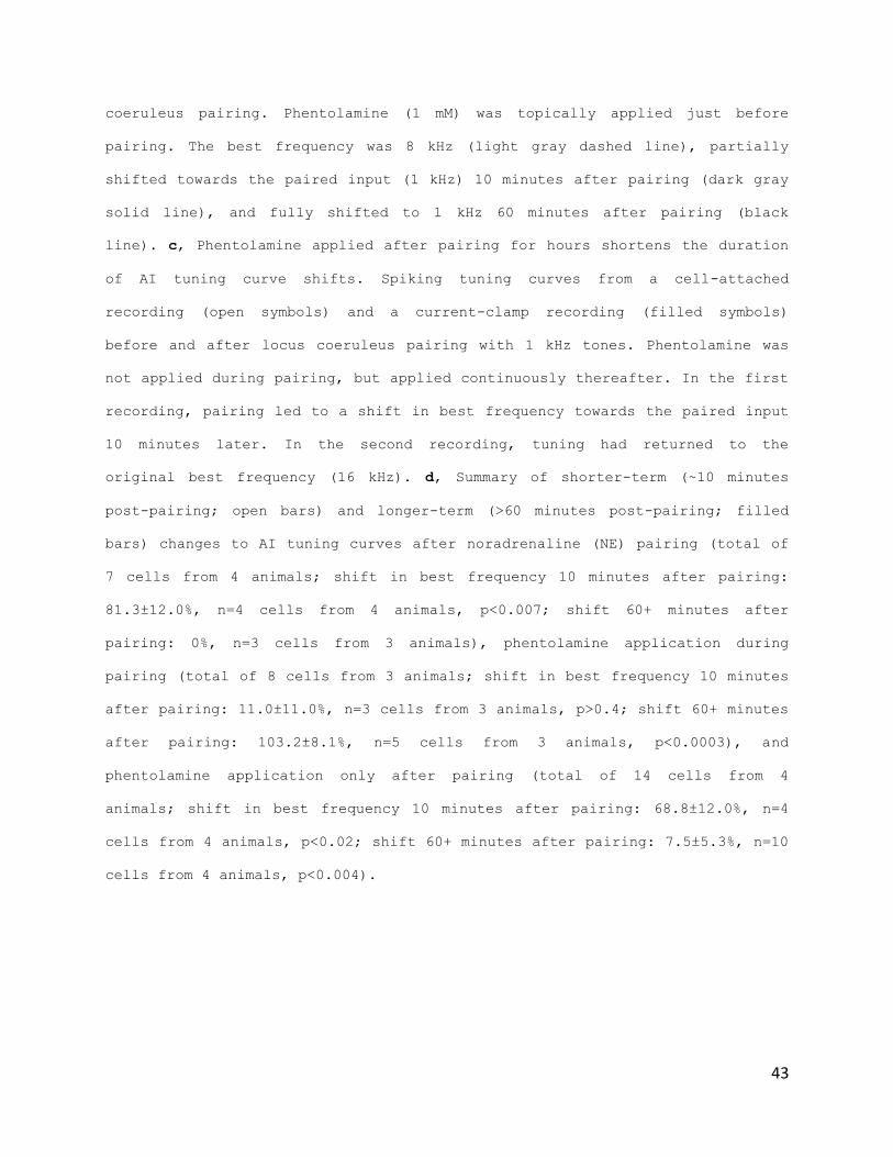
43
coeruleus pairing. Phentolamine (1 mM) was topically applied just before
pairing. The best frequency was 8 kHz (light gray dashed line), partially
shifted towards the paired input (1 kHz) 10 minutes after pairing (dark gray
solid line), and fully shifted to 1 kHz 60 minutes after pairing (black
line). c, Phentolamine applied after pairing for hours shortens the duration
of AI tuning curve shifts. Spiking tuning curves from a cell-attached
recording (open symbols) and a current-clamp recording (filled symbols)
before and after locus coeruleus pairing with 1 kHz tones. Phentolamine was
not applied during pairing, but applied continuously thereafter. In the first
recording, pairing led to a shift in best frequency towards the paired input
10 minutes later. In the second recording, tuning had returned to the
original best frequency (16 kHz). d, Summary of shorter-term (~10 minutes
post-pairing; open bars) and longer-term (>60 minutes post-pairing; filled
bars) changes to AI tuning curves after noradrenaline (NE) pairing (total of
7 cells from 4 animals; shift in best frequency 10 minutes after pairing:
81.3±12.0%, n=4 cells from 4 animals, p<0.007; shift 60+ minutes after
pairing: 0%, n=3 cells from 3 animals), phentolamine application during
pairing (total of 8 cells from 3 animals; shift in best frequency 10 minutes
after pairing: 11.0±11.0%, n=3 cells from 3 animals, p>0.4; shift 60+ minutes
after pairing: 103.2±8.1%, n=5 cells from 3 animals, p<0.0003), and
phentolamine application only after pairing (total of 14 cells from 4
animals; shift in best frequency 10 minutes after pairing: 68.8±12.0%, n=4
cells from 4 animals, p<0.02; shift 60+ minutes after pairing: 7.5±5.3%, n=10
cells from 4 animals, p<0.004).

44
Conclusion
The findings in this chapter demonstrate that noradrenergic
Locus coeruleus stimulation changes receptive fields in a
similar but also very different way than the cholinergic Nucleus
basalis. The first detectable effect of Locus coeruleus pairing
on cortical responses is to first greatly increase responses to
all sensory stimuli, regardless of context. Afterwards
eventually tuning is re-gained, but the tuning preference has
changed- the peak is now the paired frequency. This is
comparable to the clean shift in the tuning curve peak towards
the paired frequency seen with Nucleus basalis pairing. However,
looking at the duration of the specificity for the paired input,
there is a notable difference: while Nucleus basalis stimulation
allowed new specificity maintenance for only a few hours (~5h),
Locus coeruleus pairing allows the new preference to be
maintained for the duration of our recordings, at least twice
the time (~11h+).
Furthermore, the pharmacology data presented here suggests that
Locus coeruleus effects on cortex are mostly driven by the
effects of noradrenaline on cortical alpha-1 noradrenergic
receptors in primary auditory cortex. This data further exposes
a possible two phase process for Locus coeruleus modulation. The
45
initial release of noradrenaline in cortex from the stimulation,
represented by iontophoretic pairing experiment, is enough for
the initial stage of increased non-specific responses across all
frequencies, but not sufficient for the longer-term maintenance
of paired frequency preference. Moreover the noradrenergic
receptor blocker experiments show that there is a need for a
Locus coeruleus near-continual release of noradrenalin for
maintenance of cortical effect, since near-continual blocking of
noradrenergic cortical receptors is needed for muting the long-
term cortical changes.
These findings sparked an interest regarding differences that
might occur at the level of the neuromodulatory nuclei
themselves after activation, that might account for the
interesting observations of the two-phase long-duration of
cortical effects in the case of Locus Coeruleus, but not Nucleus
basalis pairing.
46
Chapter III. Locus coeruleus
plasticity
Preface
Cortical networks are embedded within complex neural circuits
including neuromodulatory systems such as the noradrenergic
locus coeruleus, providing context to environmental stimuli.
While neuromodulators are important for cortical plasticity and
adaptation, it is unknown how subcortical neuromodulatory
neurons themselves might respond and adapt to changes in sensory
input.
In this chapter I will explore this question, by using the same
experimental procedure and protocol as before, but instead of
recording from primary auditory cortical neurons, I will
directly record from Locus coeruleus neurons, while under the
same experimental conditions and pairing protocol, in an effort
to tie observations in cortical networks with subcortical
findings.
Most of this chapter is in resubmission for publication in
Nature Neuroscience.
47
Introduction
In the last chapter, the results showed an unusual and
unexpected observation: a need for a possible near-continual
release of noradrenaline for maintenance of longer-term cortical
effects.
Locus coeruleus neurons are activated by noxious, surprising
stimuli, and also respond directly to previously-innocuous
stimuli that have been linked to behaviorally-significant
episodes in the past ( Aston-Jones G., et al, 1994; Usher J.D.
et al., 1999; Yu A.J. and Dayan P., 2005; Sugyama D. et al.,
2012). However, it is unknown how locus coeruleus neurons are
affected by experience and come to respond to sensory inputs, or
how modifications to noradrenergic and cortical circuits
interact and are coordinated. In this chapter I set out to
explore this question, and attempt to further explain the
findings of the previous chapter, by recording directly from the
Locus coeruleus neurons in rat brainstem in vivo, under the same
experimental conditions recordings in primary auditory cortex
were made in the previous chapter, in order to tie the findings
in both areas, to the higher level changes in circuitry that
might be underlying the unusually long-term maintenance of
48
cortical effects. The goal was to understand the basic
mechanisms by which subcortical neuromodulatory centers might
acquire sensitivity to behaviorally-important stimuli, that
would contribute to the way future related (and unrelated)
stimuli would be contextualized and processed in the brain.
Electrophysiological Recording of Locus Coeruleus
Neurons in the Rat Brainstem
General surgical preparation of animals and patch clamping
internal solutions and recording equipment were the same as in
primary auditory cortex recordings.
Whole-cell recordings from locus coeruleus neurons were obtained
using two different methods, depending on the manner of
postsynaptic stimulation during pairing. For single cell
stimulation, recordings were obtained 5500-
surface. During pairing, cells were depolarized through the
patch pipette (20 Hz for 500 msec). For extracellular
stimulation, the cerebellum was aspirated with light suction and
recordings were obtained ~300 µm below the surface of the pons
(similar to Sugyama et al., 2012). For locus coeruleus pairing,
after recording baseline responses to the pseudo-random tone
sequence for each cell for 5-20 minutes, a non-preferred tone of
49
a given intensity level and frequency was repetitively presented
for 1-5 min, concurrent with locus coeruleus stimulation (500
msec, 20 Hz) starting at tone onset. Afterwards, locus coeruleus
stimulation was ceased and pseudo-random tone sequences were
resumed. Resting potential of locus coeruleus neurons: –
61.5±13.0 mV (s.d.); series resistance (Rs): 30.5±11.1 MΩ; input
resistance (Ri): 219.0±141.7 MΩ.
For analysis of tonal responses in locus coeruleus neurons
in Figure III-1f, we computed z-scores from mean peak EPSPs 20-
50 msec after tone onset post-pairing, compared to the mean and
standard deviation of responses during this same period before
pairing. For analysis of tuning curve shifts, best frequency
shift was computed as before for cortical tuning curves (see
Fig. II-3). For APV infusions in Figures 1 and 3, in some
experiments a custom hybrid cannula/stimulation electrode was
used, implanted on locus coeruleus, and APV was infused (1 mM in
saline, 1 µl total volume at 0.2µl/min). In other experiments in
which a hemi-cerebellectomy was performed, APV (1 mM) was
topically applied to locus coeruleus. For foot shock
experiments of Figure III-1a,b, a silver wire was connected to
the hindlimb footpad. Foot shock (20-100 Hz, 500 msec duration,

50
40-150 V) was applied for 2-5 minutes. Unless otherwise noted,
all statistics and error bars are reported as means±SEM.
Locus Coeruleus Plasticity
As a first approach to answer the question "can Locus coeruleus
exhibit adaptation to sensory stimuli after activation?",
extracellular recordings were made from Locus coeruleus neurons
from five anesthetized adult rats (Fig. III-1). Intense stimuli
such as foot shock was shown to be able to produce a phasic,
high-frequency spiking (Fig. III-1a), similar to the patterned
electrical stimulation of the nucleus used in the earlier
chapter. Innocuous stimuli (pure tones) did not evoke detectable
responses (Fig.II-1b, 'Pre'). However, after tones were
repetitively paired with foot shock for 1-5 minutes, paired
tones could evoke locus coeruleus spikes for 1+ hours (Fig. III-
1b, 'Post').

51
Figure III-1. Recording from locus coeruleus. a, Noxious stimuli evoke phasic
spike bursts in rat locus coeruleus. Top, brief tail pinch (100 msec
duration) evoked phasic spiking in locus coeruleus, as measured through the
stimulation electrode. Bottom, foot shock (100 Hz for 500 msec, 50 V) also
evoked high frequency bursts of activity as measured with unit recordings.
Traces shown are from separate animals. b, Pairing a pure tone with foot
shock leads to a long-lasting increase in tone-evoked spiking in locus
coeruleus. Top, example traces showing that before foot shock pairing
('Pre'), presentation of a pure tone (8 kHz, 80 dB SPL, 50 msec duration) did
not evoke significant spiking in locus coeruleus. Ten minutes after pairing
foot shock (as in a) with 8 kHz tones, the paired tone by itself evoked
spiking responses in locus coeruleus. Bottom, post-stimulus time histogram of
multiunit recording from locus coeruleus showing change in firing rate after
tonal presentation just before (gray) and one hour after (red) pairing.
Pairing induced a large (~1 Hz) increase in firing rate with ~30 msec latency
We then examined if pairing auditory stimuli with locus
coeruleus activity ('locus coeruleus pairing') was sufficient to
modify neuronal responses. Pairing was performed either by
depolarization through the recording electrode or extracellular
stimulation. For single-cell depolarization, we made current-
clamp recordings from locus coeruleus neurons, and measured
responses to pure tones 5-20 minutes before and 5+ minutes after
pairing a specific tone with postsynaptic spiking. After the

52
baseline period, neurons were phasically depolarized at 20 Hz
for 500 msec, repeated at 0.5-1 Hz for 1-5 minutes (Fig. III-
1b), similar to firing patterns observed in locus coeruleus
neurons during foot shock (Fig. III-1a). This procedure mimics
what occurs when sounds are linked to arousing situations
(Aston-Jones G., et al, 1994; Usher J.D. et al., 1999; Yu A.J.
and Dayan P., 2005; Sugiyama D. et al., 2012).
Pairing tones with postsynaptic depolarization induced
long-lasting responses to auditory stimuli in previously-
unresponsive locus coeruleus neurons. An example in vivo whole-
cell recording is shown in Figure III-2c. Initially, this cell
did not respond to sounds. After pairing, paired 16 kHz tones
evoked sizable EPSPs at ~30 msec latency for the recording
duration. This locus coeruleus plasticity required NMDA
receptors, as local infusion of APV (1 mM) prevented emergence
of tone-evoked responses after pairing (Fig. III-2d). These
results are reminiscent of 'silent synapses' activated after
induction of long-term potentiation, although here it appeared
that auditory responses had developed in formerly 'silent
cells'.
We tested how long these auditory responses would last
after pairing. To simultaneously induce plasticity in multiple
locus coeruleus neurons, we paired tones with extracellular
53
locus coeruleus stimulation (20 Hz, 500 msec at 0.5-1 Hz, 1-5
min II-1f).
When the first recording ended, we recorded from up to four more
neurons in the same animals to assess changes to other neurons
throughout locus coeruleus after a single pairing episode.
Three recordings from the same animal are shown in Figure
III-2e, a current-clamp recording before and after pairing (Fig.
III-2e, left; gray, red), a cell-attached recording 5+ hours
after pairing (Fig. III-2e, right), and a final current-clamp
recording 6+ hours afterward (Fig. III-2e, left; black). Locus
coeruleus neurons responded to tones for hours after pairing.
Additionally, locus coeruleus plasticity seemed specific to
paired tones, as responses to unpaired tones were sometimes
initially enhanced but not persistently modified after pairing
(Fig. III-2e, left).
The duration of locus coeruleus plasticity is shown in
Figure III-2f for 27 neurons from nine animals (filled symbols),
and 18 neurons from five animals after APV infusion (open
symbols). Significant increases in synaptic strength and spike
generation (as measured by z-scored responses relative to
baseline noise) lasted for at least several hours after pairing,
but were prevented by APV infusion. These findings demonstrate
that locus coeruleus neurons can become activated by previously-
54
innocuous sounds (i.e., stimuli that did not evoke any
detectable subthreshold response).

55
-1.0
0.0
1.0
2.0
0-10 2010
Martins_fig1
dc
Time (min)
LC whole-cell recording
EP
SP
(mV
)
LC synaptic responses
EP
SP
(mV
)
LC recording + LC APV
Frequency (kHz)
Sp
ike
s/t
on
e
LC whole-cell recordingPairing
Frequency (kHz)
Time (min)
EP
SP
(mV
)
Pairing
Syn
ap
tic r
esp
on
se
(z)
Time (hours)
0.5 1 2 4 8 16 320.0
0.1
0.2
25 ms0.3 mV
b 1. Pre(5-20 min)
3. Post(5+ min)
2. LC pairing(1-5 min)
Tone
LC stim
LC rec
...
500 ms, 20 Hz
a
LC
Tone
Whole-cellrecording
Time (hours)
Sp
ikin
g r
esp
on
se
(z)
f
e
0.0
2.0
-10 0 10 20
+ APV in LC
LC pairing (synaptic)
0.5 1 2 4 8 16 32-1.0
-0.5
0.0
0.5
1.0
1.5 -10 min+10 min (z: 1.1)+420 min (z: 1.8)
+360 min (z: 3.4)
LC spiking responses
LC pairing (spiking)
+ APV in LC
-4.0
0.0
4.0
8.0
0 2 4 6 8 10-4.0
0.0
4.0
8.0
0 2 4 6 8 10
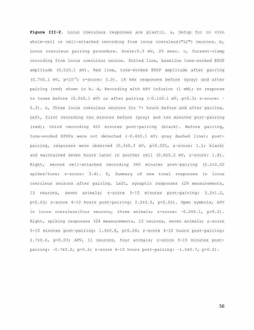
56
Figure III-2. Locus coeruleus responses are plastic. a, Setup for in vivo
whole-cell or cell-attached recording from locus coeruleus("LC") neurons. b,
Locus coeruleus pairing procedure. Scale:0.3 mV, 25 msec. c, Current-clamp
recording from locus coeruleus neuron. Dotted line, baseline tone-evoked EPSP
amplitude (0.0±0.1 mV). Red line, tone-evoked EPSP amplitude after pairing
(0.7±0.1 mV, p<10-8; z-score: 3.0). 16 kHz responses before (gray) and after
pairing (red) shown in b. d, Recording with APV infusion (1 mM); no response
to tones before (0.0±0.1 mV) or after pairing (–0.1±0.1 mV, p>0.3; z-score: –
0.3). e, Three locus coeruleus neurons for 7+ hours before and after pairing.
Left, first recording ten minutes before (gray) and ten minutes post-pairing
(red); third recording 420 minutes post-pairing (black). Before pairing,
tone-evoked EPSPs were not detected (–0.4±0.1 mV; gray dashed line); post-
pairing, responses were observed (0.3±0.3 mV, p<0.005, z-score: 1.1; black)
and maintained seven hours later in another cell (0.6±0.2 mV, z-score: 1.8).
Right, second cell-attached recording 360 minutes post-pairing (0.2±0.02
spikes/tone; z-score: 3.4). f, Summary of new tonal responses in locus
coeruleus neurons after pairing. Left, synaptic responses (29 measurements,
13 neurons, seven animals; z-score 5-15 minutes post-pairing: 3.2±1.2,
p<0.03; z-score 4-10 hours post-pairing: 2.2±0.5, p<0.02). Open symbols, APV
in locus coeruleus(four neurons, three animals; z-score: –0.2±0.1, p>0.2).
Right, spiking responses (24 measurements, 15 neurons, seven animals; z-score
5-15 minutes post-pairing: 1.9±0.8, p<0.04; z-score 4-10 hours post-pairing:
2.7±0.6, p<0.03; APV, 11 neurons, four animals; z-score 5-15 minutes post-
pairing: –0.7±0.2, p>0.2; z-score 4-10 hours post-pairing: –1.0±0.7, p>0.2).
57
This might lead to a different more prolonged noradrenergic
modulation of target projection areas in response to tonal
presentation alone Therefore, in our last physiological
experiment, we asked whether modifications to locus coeruleus
neurons were required for cortical plasticity observed. As APV
prevented locus coeruleus plasticity, we used this manipulation
to selectively block subcortical changes, measuring the effects
of pairing on AI in absence of locus coeruleus plasticity.
Infusing APV into locus coeruleus during pairing greatly reduced
the duration of AI tuning curve shifts. Three cells recorded
over eight hours from the 4 kHz region of AI are shown in Figure
III-3a. The first recording showed that 4 kHz was the best
frequency (Fig. III-3a, left, gray). 1 kHz was paired with locus
coeruleus stimulation, and three hours after pairing, a second
AI neuron had new preference for 1 kHz (Fig. III-3a, right). But
eight hours after pairing, tuning reverted to 4 kHz (Fig. III-
3a, left, black). We conclude that locus coeruleus plasticity is
required for long-term maintenance of AI plasticity. These
effects are summarized in Figure III-3b for nine whole-cell and
nine cell-attached recordings, showing that APV infused into
locus coeruleus dramatically reduced duration of cortical
modifications.

58
Figure III-3. Locus coeruleus plasticity controls the duration of cortical
plasticity. a, Three AI recordings for 7+ hours pre/post-pairing with APV in
locus coeruleus. Left, first current-clamp recording ten minutes before
(gray) pairing; third current-clamp recording 460 minutes post-pairing
(black). 4 kHz was original best frequency (arrowhead); 1 kHz was paired
frequency (arrow). Right, second cell-attached recording 200 minutes post-
pairing. b, Summary of AI best frequency shift of synaptic (left) and spiking
tuning curves (right) with APV in locus coeruleus. Shaded area, mean±SEM of
shifts from figure II-3, e .
Martins_fig6
a AI tuning curvesE
PS
P(m
V)
Frequency (kHz)
Sp
ike
s/t
on
e
Frequency (kHz)
Be
st
fre
qu
en
cy s
hift (%
)
Time (hours)
b
Be
st
fre
qu
en
cy s
hift (%
)
+ APV in LC
LC pairing (synaptic)
Time (hours)0 2 4 6 8 10 12
-100
0
100
200
-10 min
+460 min +200 min
0.5 1 2 4 8 16 320.0
1.0
2.0
3.0
0.5 1 2 4 8 16 320.0
0.1
0.2
0.3
+ APV in LC
LC pairing (spiking)
-100
0
100
200
0 2 4 6 8 10 12
59
Conclusion
These findings demonstrate that after pairing, Locus coeruleus
neurons became part of the overall circuit activated by once-
innocuous stimuli. Importantly, these data suggest that after
Locus coeruleus sensitization, noradrenergic modulation in
target projection areas might happen in response to tonal
presentation alone. This might be a causal mechanism supporting
the long duration of cortical effects: once sensitized, the
Locus will release a certain amount of noradrenaline in response
to passive presentation of the paired tone, maintaining the
cortical noradrenergic receptors active in the presence of this
tone, thereby keeping relevance of the new frequency preference
for long periods of time. Although it will be important in
future studies to examine which specific inputs to locus
coeruleus are activated and modified by pairing, we focused our
next experiments on the functional consequences of
neuromodulatory plasticity on cortical responses and perceptual
learning, described in the next chapter.

60
Chapter IV. Perceptual consequences of
Locus Coeruleus Pairing
Preface
Until now this thesis has explored how noradrenergic locus
coeruleus stimulation paired with tonal presentation, re-
organizes primary auditory cortical receptive fields, and how
these changes relate to plasticity in neurons in Locus coeruleus
itself.
In this chapter I examine how locus coeruleus neurons
experience, and the consequences of this plasticity on cortical
representations and sensory perception in behaving animals.
Most of this chapter is in review at Nature Neuroscience.
Introduction
Synapses and receptive fields of the cerebral cortex are
plastic. However, changes to specific inputs must be coordinated
within neural networks to ensure that excitability and feature
selectivity are appropriately configured for perception of the
sensory environment.
61
Locus coeruleus neurons are activated by noxious and surprising
stimuli, and also respond directly to previously-innocuous
stimuli that have been linked to behaviorally-significant
episodes in the past ( Aston-Jones G., et al, 1994; Usher J.D.
et al., 1999; Yu A.J. and Dayan P., 2005; Sugyama D. et al.,
2012). It is hypothesized that locus coeruleus plays a major
role in adjusting gains of cortical synapses in a task-dependent
manner in particular, higher-frequency phasic activity of
noradrenergic neurons may facilitate the formation of task-
specific behavioral patterns, to optimize perceptual abilities
and motor outputs (Aston-Jones G et al., 1994; Usher M., et al.,
1999, Yu A.J. and Dayan P., 2005, Kuo, S.P. and Trussel L.O.,
2011).
Previous work done in Froemke R.C. and Carcea I. et al., 2013,
has studied the perceptual consequences of activation of Nucleus
basalis. That work shows that Nucleus basalis stimulation and
pairing with a tone can both increase the recognition of a
paired frequency, and increase detection of a paired low decibel
level (see Fig I-4).
While in previous chapters, recordings were made directly from
brain areas in anesthetized animals, for the mechanical
stability required by the technique, in this chapter I set out
to explore the perceptual consequences of the network changes

62
measured. To this point and given the interesting functional
similarities and disparities between the cholinergic Nucleus
basalis and the noradrenergic Locus Coeruleus, the question was
asked: what will we see using the a similar operant task as with
Nucleus basalis, but stimulating Locus coeruleus instead.
In this chapter the question is posed: what do the interesting
cortical and subcortical changes recorded, by activating Locus
coeruleus in this high frequency phasic manner, mean for
auditory perception.
Behavior Training and Chronic Implantation of Adult
Rats
Rats were lightly food-deprived and pre-trained for 2-6 weeks on
a frequency recognition go/no-go task. Animals were rewarded
with food for nose-poking within three seconds of presentation
of a target tone (4 kHz, any intensity), and given a short (~5
s) time out if they incorrectly responded to non-target tones.
After learning to nosepoke to 4 kHz tones, spectrally-wideband
foils were also presented (0.5, 1, 2, 8, 16, 32 kHz). Animals
that achieved hit rates >80% for targets were then anesthetized
with ketamine/dexmedetomidine, had stimulation electrodes or

63
hybrid cannula/stimulation electrodes chronically implanted in
right locus coeruleus, and were allowed to recover for a week.
Each implanted animal was first tested on the 'wideband'
recognition task or the detection task for at least 1-2 days. On
the wideband and narrowband recognition tasks, tones (wideband
target: 4 kHz, foils: 0.5, 1, 2, 8, 16, 32 kHz; narrowband
target: 4 kHz, foils: 2.8, 3.2, 3.6, 4.5, 5.1, 5.7 kHz) were
presented at 70 dB SPL. For the detection task, tones were
presented at 20-90 dB SPL. On the first day at each task, tones
were presented for 30-60 minutes to assess performance at
baseline; 4 kHz tones (at 70 dB SPL for the wideband recognition
task; 30-45 dB SPL for the detection task; hits binned over 20-
40 dB SPL) were then paired with locus coeruleus stimulation in
the training box for two to three minutes, and behavior
performance assessed and quantified one, three, twelve and/or
twenty four hours after one single episode of stimulation. In
some cases, animal performance was monitored over the span of
weeks, once a day, to examine the duration of behavioral changes
induced by locus coeruleus pairing. For reversal learning, 17
animals had the rewarded tone switched from 4 kHz to 16 kHz on
the wideband task after achieving d'>2.0. Six of those animals
had stimulation electrodes implanted in locus coeruleus, and
locus coeruleus stimulation was paired with 16 kHz tones for
64
five minutes just prior to testing behavioral performance on the
first day that 16 kHz was rewarded. Performance was measured
daily thereafter. Cannulated animals had APV (1 mM in saline,
0.4-1 µl at 0.2µl/min) or saline infused into the locus
coeruleus immediately prior to stimulation for either task. d'
values were computed as the difference in z-scores between hits
and false positives: d' = z(hit rate) – z(false positive rate),
using the responses between 20-40 dB SPL for detection and
responses to 3.6-4.5 kHz for narrowband recognition. Power
analysis was performed to determine the number of animals
required for statistical significance.
Perceptual Consequences of Locus Coeruleus Pairing
To assess the functional consequences of cortical and
neuromodulatory plasticity findings reported, I set out to
determine how these changes might affect sensory perception.
Here a behavioral task is used, sensitive to plasticity of AI
tuning curves (Froemke R.C. et al., 2013). Three predictions
suggested by our physiological results were examined: 1)
behaviorally-important stimuli should be easier to detect and
recognize after locus coeruleus pairing, as primary auditory
cortical synaptic and spiking responses to tones of all
65
intensities are greatly enhanced; 2) recognition of specific
stimuli may initially be impaired, as primary auditory cortical
tuning curves first widen and responses to distinct stimuli
become more similar for a few hours after locus coeruleus
pairing; and 3) improvements to perceptual abilities should
persist for hours or perhaps indefinitely even after a single
brief pairing episode, given the prolonged maintenance of AI
plasticity by locus coeruleus plasticity.
A total of 33 adult rats were operantly conditioned to
nose-poke for a food reward in response to 4 kHz target stimuli,
withholding responses to six foil tones of other frequencies
irrespective of intensity (Fig. IV-1a). Animals had stimulation
electrodes and drug delivery cannulas implanted into locus
coeruleus. Psychophysical detection performance of a
representative animal is shown in Figure IV-1b. At ≥50 dB sound
pressure level (SPL), this animal had close-to-perfect
performance detecting target 4 kHz tones ('Hits', circles) and a
low false alarm rate ('F+', triangles). However, this animal had
a low response rate for tones of 20-40 dB SPL, resulting in a
poor d' of 0.5. After measuring behavior for 30-60 minutes, we
paired 4 kHz, 30 dB SPL tones with locus coeruleus stimulation
for 3 minutes in the training chamber. We then re-tested
detection abilities 12 hours later and found that detection at

66
20-40 dB SPL was greatly enhanced (Fig. IV-1b, red). Similar
improvements in detection were observed for 16 animals (Fig. IV-
1c). Remarkably, enhanced detection abilities lasted up to 20
days post-pairing (Fig. IV-1d). Behavioral changes were
prevented when APV (1 mM) was infused into locus coeruleus
before pairing (Fig. IV-1c, open), indicating that locus
coeruleus plasticity is required for behavioral improvement.
Thus locus coeruleus stimulation facilitates detection of
previously-imperceptible stimuli, and brief episodes of pairing
optimize signal processing and formation of sensorimotor
associations.
We then asked how pairing affected abilities to distinguish
targets from foils. Initially, foil stimuli were spectrally
dissimilar from the target, separated at one octave intervals.
Animals performed well on this 'wideband' task before pairing.
We then compressed the spectral range of foils to resemble the
target. Before pairing, behavioral performance on this
'narrowband' task was low (Fig. IV-2a ), and pairing initially
reduced recognition even further for a few hours (Fig. IV-2a,b).
However, performance improved above baseline levels 12 hours
post-pairing (Fig. IV-2c). These changes in auditory perception
seem similar to AI modifications: tuning profiles first
broadened (leading to similar neural responses for different
sensory stimuli) before tuning width recovered and many more AI
67
neurons responded strongly to paired stimuli (facilitating
detection and recognition of target tones). Changes in
performance required locus coeruleus plasticity, and were
prevented by infusion of APV into locus coeruleus(Fig. IV-2b,c,
open).
To document the time course of behavioral enhancement more
thoroughly, in our last experiment we asked whether locus
coeruleus pairing might also accelerate perceptual learning.
After initial training, the rewarded tone was switched to 16
kHz, 4 kHz became a foil, and one cohort of animals received
locus coeruleus stimulation paired with 16 kHz tones. We
monitored animals for several weeks afterward and found that
while unpaired animals required >20 days to recover initial
performance levels (Fig. IV-1e,g, black), paired animals learned
the new association in almost half the time (Fig. IV-1f,g, red).
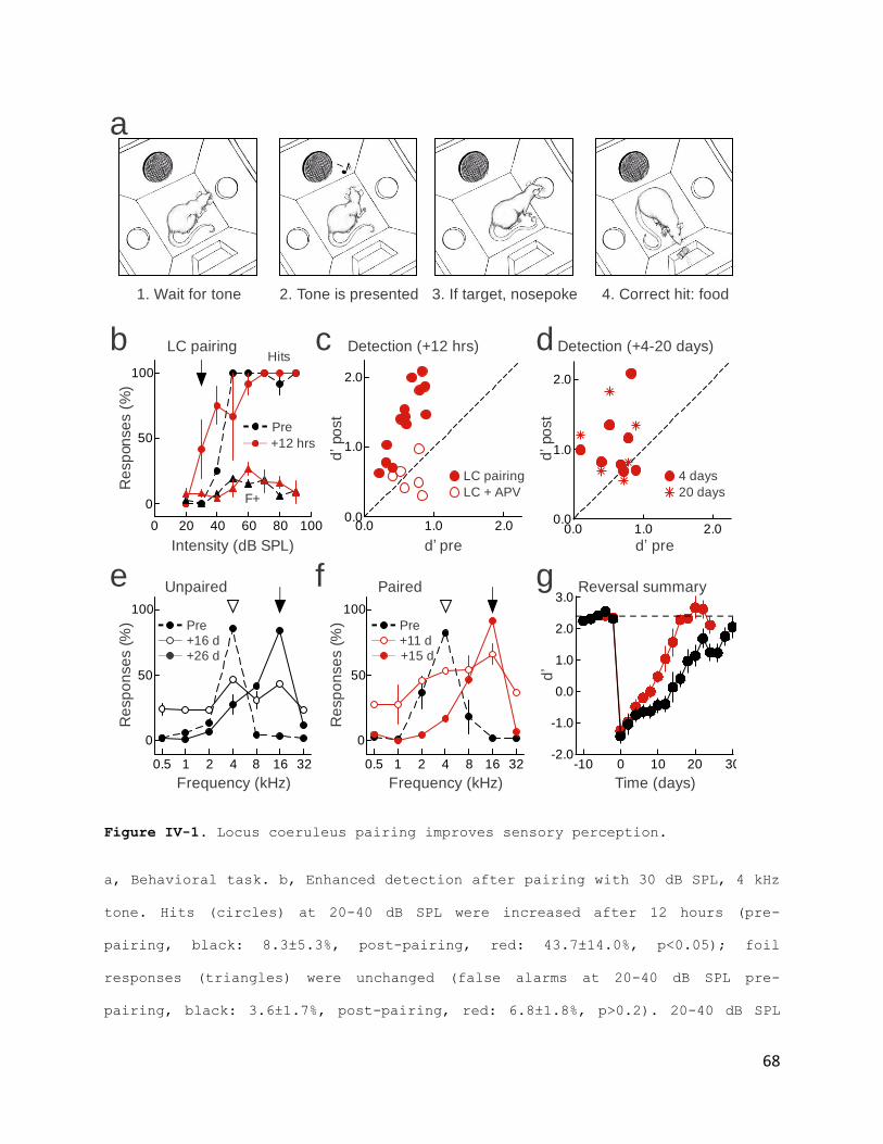
68
Figure IV-1. Locus coeruleus pairing improves sensory perception.
a, Behavioral task. b, Enhanced detection after pairing with 30 dB SPL, 4 kHz
tone. Hits (circles) at 20-40 dB SPL were increased after 12 hours (pre-
pairing, black: 8.3±5.3%, post-pairing, red: 43.7±14.0%, p<0.05); foil
responses (triangles) were unchanged (false alarms at 20-40 dB SPL pre-
pairing, black: 3.6±1.7%, post-pairing, red: 6.8±1.8%, p>0.2). 20-40 dB SPL
Martins_fig7
b c
Re
sp
on
se
s (
%)
Intensity (dB SPL)
LC pairing d
d’ pred’po
st
Hits
F+
Pre
+12 hrs
Detection (+4-20 days)
e f
Re
sp
on
se
s (
%)
Frequency (kHz)
g
Frequency (kHz) Time (days)
d’
Re
sp
on
se
s (
%)
Paired Reversal summary
d’ pre
d’po
st
LC pairing
LC + APV
Detection (+12 hrs)
0
50
100
0.5 1 2 4 8 16 32
Unpaired
Pre
+26 d+16 d
Pre
+15 d+11 d
0
50
100
0.5 1 2 4 8 16 32
0 20 40 60 80 100
0
50
100
0.0 1.0 2.00.0
1.0
2.0
4 days
20 days
a
1. Wait for tone 2. Tone is presented 3. If target, nosepoke 4. Correct hit: food
-10 0 10 20 30-2.0
-1.0
0.0
1.0
2.0
3.0
0.0 1.0 2.00.0
1.0
2.0

69
d' increased (0.5 to 1.4). c, d' values before and 12 hours after pairing (d'
before: 0.57±0.06, after: 1.25±0.14, n=16 animals, p<10-4). APV in locus
coeruleus prevented improvement (open circles; d' before: 0.65±0.07, after:
0.57±0.10, n=6, p>0.5). d, Detection was enhanced four (circles; d' before:
0.61±0.09, after: 1.07±0.17, n=8, p<0.04) and 20 days after pairing (stars;
d' before: 0.57±0.11, after: 1.04±0.19, n=6, p<0.05). e, Example of reversal
learning without pairing. After learning to respond to 70 dB SPL, 4 kHz tones
('Pre', d': 2.67), rewarded frequency was changed to 16 kHz. After 16 days,
hits increased, although false alarms were high (d': 0.39). After 26 days,
performance recovered (d': 2.03). f, One pairing episode accelerated reversal
learning. Before switching rewarded tone to 16 kHz, pairing with 16 kHz tones
was performed for 3-5 minutes. After initial training ('Pre', d': 2.17),
performance improved by day 11 (d': 0.64), and after 15 days, performance
recovered (d': 2.50). g, Accelerated reversal learning after pairing. A
sliding t-test (width: two days) was used to determine when performance
recovered to baseline (black, control: 22 days, n=11; red, paired animals: 13
days, n=6).
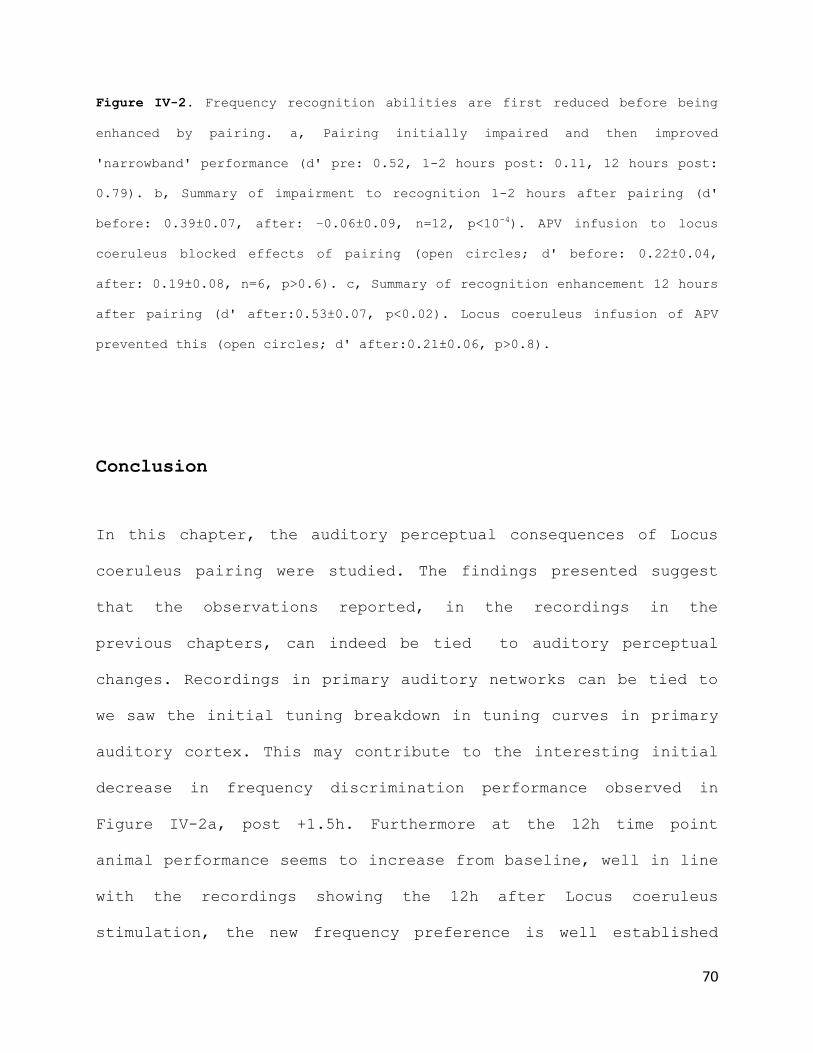
70
Figure IV-2. Frequency recognition abilities are first reduced before being
enhanced by pairing. a, Pairing initially impaired and then improved
'narrowband' performance (d' pre: 0.52, 1-2 hours post: 0.11, 12 hours post:
0.79). b, Summary of impairment to recognition 1-2 hours after pairing (d'
before: 0.39±0.07, after: –0.06±0.09, n=12, p<10-4). APV infusion to locus
coeruleus blocked effects of pairing (open circles; d' before: 0.22±0.04,
after: 0.19±0.08, n=6, p>0.6). c, Summary of recognition enhancement 12 hours
after pairing (d' after:0.53±0.07, p<0.02). Locus coeruleus infusion of APV
prevented this (open circles; d' after:0.21±0.06, p>0.8).
Conclusion
In this chapter, the auditory perceptual consequences of Locus
coeruleus pairing were studied. The findings presented suggest
that the observations reported, in the recordings in the
previous chapters, can indeed be tied to auditory perceptual
changes. Recordings in primary auditory networks can be tied to
we saw the initial tuning breakdown in tuning curves in primary
auditory cortex. This may contribute to the interesting initial
decrease in frequency discrimination performance observed in
Figure IV-2a, post +1.5h. Furthermore at the 12h time point
animal performance seems to increase from baseline, well in line
with the recordings showing the 12h after Locus coeruleus
stimulation, the new frequency preference is well established

71
and maintained both in cortex. and in the Locus Coeruleus.
Furthermore the Locus coeruleus exhibits sensitization to the
paired stimulus that remains for the duration of our recordings
(~10h). This along with the fact that locus coeruleus plays may
play a major role in adjusting gains of cortical synapses in a
task-dependent manner in particular, higher-frequency phasic
activity of noradrenegic neurons may facilitate the formation of
task-specific behavioral patterns, to optimize perceptual
abilities and motor outputs (Aston-Jones G et al., 1994; Usher
M., et al., 1999, Yu A.J. and Dayan P., 2005, Kuo, S.P. and
Trussel L.O., 2011), could explain the duration of the detection
changes in Figure IV-1b. Furthermore, the powerful involvement
of noradrenaline in learning and plasticity of cortical
networks, allowing re-organization around behaviorally salient
stimuli, could well explain the accelerated perceptual learning,
in Figure IV-1g.
Interestingly comparing, again, these findings to the perceptual
consequences of cholinergic Nucleus basalis stimulation,
provides us with a nice multilevel relationship between
synaptic, physiological, and functional roles of these two
similar but very different neuromodulator systems.
In conclusion, the data presented in this chapter demonstrates
that the Locus coeruleus improves perceptual abilities for at
72
least hours after pairing, and even longer-lasting gains in
performance can emerge over days to weeks even after just a
single pairing episode. These abilities can be enhanced beyond
the gains induced by reward-based training alone, accelerated by
neuromodulation and cortical plasticity.

73
Chapter V. Conclusion: Locus coeruleus
Plasticity Controls Duration of
Auditory Cortical Plasticity
Discussion
In the work presented in this thesis, I investigated the rules
by which activation of neuromodulator systems, can interact with
and change cortical networks. Furthermore I asked and explored
the question of sensory sensitization of these nuclei themselves
as an active part of a circuit engaged by a behaviorally
relevant stimulus. Specifically I studied the noradrenergic
Locus Coeruleus, and the cortical and subcortical synaptic
effects of its stimulation at a high frequency, behaviorally-
relevant phasic pattern, as well as the possible perceptual
consequences.
The locus coeruleus is the principal source of noradrenergic
modulation for the central nervous system, providing a
fundamental mechanism for rapidly adapting cortical circuits to
task demands. Here we showed that enduring contextual
associations can be rapidly formed within the locus coeruleus,
and neuromodulatory cells that were previously unresponsive to
sounds can become sensitized and tonally responsive. The rapid
74
induction and prolonged duration of these changes to the central
arousal system is reminiscent of changes to brain state and
behavior in cases of one-trial learning or post-traumatic stress
disorder, suggesting that locus coeruleus plasticity might be a
fundamental determinant of these memory processes.
This plasticity of noradrenalin release exerts a profound effect
on downstream neuromodulatory targets, controlling dynamics of
cortical plasticity and auditory perceptual learning from
induction to long-term maintenance. Noradrenergic modulation
seems to provide a specific disinhibitory signal to cortical
circuits, transiently reducing spontaneous inhibition in a
manner distinct from that provided by cholinergic modulation
from nucleus basalis, suggesting that separate neuromodulatory
systems can differentially control neural circuits.
Noradrenaline in particular seems to control overall gain of
cortical neurons, transiently increasing the effective salience
of incoming signals before more selective adjustments of
cortical representations become manifest. Although these changes
can initially impair some aspects of sensory perception, lasting
behavioral enhancements can emerge over a period of hours to
weeks, even after one pairing episode. While the full neural
circuit representing learned associations between external
stimuli and internal state is likely to be distributed

75
throughout much of the central nervous system, modifications to
neuromodulator systems such as locus coeruleus provide a
powerful mechanism for storing and restoring the most
behaviorally-important memories.

76
Future Directions
While the work presented here was an extensive study of the
question initially proposed, as with any interesting scientific
finding, from these results many more avenues of exploration
present themselves.
One major source variability might be the parameter space in
which stimulation was performed. While the parameters used where
robust, physiological and maintained constant throughout the
work, one might wonder what would happen if parameters where
changed: what if a different frequency of stimulation was used?
How about changing the tone presentation latency relative to the
stimulation?
The interesting question also comes about, of how it is that
auditory responses emerge in the Locus Coeruleus? This nucleus
has a notoriously expansive axonal network. What other auditory
areas engaged by the behaviorally relevant stimulus, and
receiving noradrenergic inputs from the Locus can change also?
What are the inputs into the Locus that are changed and allow
for auditory input to now reach and originate a response in
these noradrenergic cells. Furthermore what is the role of the
local inhibitory interneurons in this matter?
77
Moreover one can wonder, what is so special about the Locus
Coeruleus? One quaint characteristic that comes to mind is the
existence of gap junctions. What is their role in Locus
coeruleus sensitization to sensory stimuli? Conversely, will we
find any such type of sensitization if we record in other
neuromodulatory nuclei.
Interestingly, Locus coeruleus axons synapse directly on
astrocytic endfeet. What might be the contribution of astrocytes
to the intensity or duration of the cortical effects observed?
Finally, behavior is a very rich field that can be expansively
explored . In this work we used an operant task similar to the
previously used with Nucleus basalis stimulation studies, in
order to be able to compare the two systems. What if we tested
our parameters with a different task. Or what if we tested
different parameters with our task.
All these questions, important as they are, remain unanswered
and will be the subject of future studies.

78
References
Aston-Jones G, Bloom FE. Activity of norepinephrine-containing
locus coeruleus neurons in behaving rats anticipates
fluctuations in the sleep-waking cycle. J Neurosci 1: 876 –
886, (1981a).
Aston-Jones G, Bloom FE. Nonrepinephrine-containing locus
coeruleus neurons in behaving rats exhibit pronounced
responses to non-noxious environmental stimuli J Neurosci 1:
887–900, (1981b).
Aston-Jones, G. & Cohen, J.D. An integrative theory of locus
coeruleus-norepinephrine
function: adaptive gain and optimal performance. Annu. Rev.
Neurosci. 28,403-450 (2005).
Aston-Jones, G., Rajkowski, J., Kubiak, P. & Alexinsky, T.
Locus coeruleus neurons are selectively activated by attended
cues in a vigilance task. J. Neurosci. 14,4467-4480 (1994).
Bakin, J.S. & Weinberger, N.M. Induction of a physiological
memory in the cerebral cortex by stimulation of the nucleus
basalis. Proc. Natl. Acad. Sci. U S A 93,11219-11224 (1996).
79
Bear, M.F. & Singer, W. Modulation of visual cortical
plasticity by acetylcholine and noradrenaline. Nature 320,172-
176 (1986).
Berridge, C.W. Noradrenergic modulation of arousal. Brain Res.
Rev. 58,1-17 (2008).
Buonomano, D.V. & Merzenich, M.M. Cortical plasticity:
from synapses to maps Annu. Rev. Neurosci. 21, 149–186
(1998).
Bush, D.E., Casparosa, E.M., Gekker, A. & LeDoux, J. Beta-
adrenergic receptors in the lateral nucleus of the amygdala
contribute to the acquisition but not the consolidation of
auditory fear conditioning. Front. Behav. Neurosci. 4,154
(2010).
Carter, M.E. et al. Tuning arousal with optogenetic modulation
of locus coeruleus neurons. Nat. Neurosci. 13,1526-1533
(2010).
Chang, E.F., Bao, S., Imaizumi, K., Schreiner, C.E.,
Merzenich, M.M., Development of spectral and temporal response
selectivity in the auditory cortex. Proceedings of the
National Academy of Science U S A 102, 16460-65 (2005).
combinations produces spike tuning. Neuron 37, 663e680

80
Constantinople, C.M. & Bruno, R.M. Effects and mechanisms of
wakefulness on local cortical networks. Neuron 69,1061-1068
(2011).
Dahmen, J.C., Hartley, D.E. & King, A.J. Stimulus-
timing-dependent plasticity of cortical frequency
representation. J. Neurosci. 28, 13629–13639 (2008).
Dan, Y. & Poo, M.M. Spike timing-dependent plasticity: from
synapse to perception. Physiol. Rev. 86, 1033–1048 (2006).
de Villers-Sidani, E., Chang, E.F., Bao, S. & Merzenich, M.M.
Critical period window for spectral tuning defined in the
primary auditory cortex (A1) of the rat. J. Neurosci.27, 180–
189 (2007).
de Villers-Sidani, E., Chang, E.F., Bao, S., Merzenich, M.M.,
Critical period window for spectral tuning defined in the
primary auditory cortex (A1) in the rat. J. Neurosci 27, 180-
9 (2007).
Devilbiss D.M and Waterhousse B.D., Phasic and Tonic Patterns
of Locus Coeruleus Output Differentially Modulate Sensory
Network Function in the Awake Rat, J Neurophysiol 105, 69 –
87, (2011).
direction selectivity of synaptic inputs in visual cortical
neurons: a diversity of
81
Dorrn, A.L., Yuan, K., Barker, A.J., Schreiner, C.E. &
Froemke, R.C. Developmental sensory experience balances
cortical excitation and inhibition. Nature 465, 932–936
(2010).
Edeline, J.M., Manunta Y. & Hennevin, E. Induction of
selective plasticity in the frequency tuning of auditory
cortex and auditory thalamus neurons by locus coeruleus
stimulation. Hear. Res. 274,75-84 (2011).
Feldman, D.E. & Brecht, M. Map plasticity in somatosensory
cortex. Science 310,810-815 (2005).
Feldman, D.E.,. Synaptic mechanisms for plasticity in
neocortex. Annual Review in Neuroscience 32, 33-55 (2009).
Foote et al., Nucleus Locus Ceruleus: New Evidence of
Anatomical and Physiological Specificity Physiol Rev 63
(1983b)
Foote et al., Physiological properties of ascending locus
coeruleus axons in the squirrel monkey, Brain Research,
274: 381-387 (1983a)
Frégnac, Y., Shulz, D., Thorpe, S. & Bienenstock, E.
A cellular analogue of visual cortical plasticity. Nature
333, 367–370 (1988).

82
Fritz, J., Shamma, S., Elhilali, M. & Klein, D. Rapid task-
related plasticity of spectrotemporal receptive fields in
primary auditory cortex. Nat. Neurosci. 6,1216-1223 (2003).
Froemke, R.C. & Martins A.R.O. Spectrotemporal dynamics of
auditory cortical synaptic receptive field plasticity. Hear.
Res. 279,149-161 (2011).
Froemke, R.C. et al. Long-term modification of cortical
synapses improves sensory perception. Nat. Neurosci. 16,79-88
(2013).
Froemke, R.C., Merzenich, M.M. & Schreiner, C.E. A synaptic
memory trace for cortical receptive field plasticity. Nature
450,425-429 (2007).
Gilbert, C.D., Li, W. & Piech V. Perceptual learning and adult
cortical plasticity. J. Physiol. 587,2743-2751 (2009).
Gu, Q. Neuromodulatory transmitter systems in the cortex and
their role in cortical plasticity. Neuroscience 111,815-835
(2002).
Häusser, M., Mel, B., Dendrites: bug or feature? Current
Opinion in Neurobiology 3, 372-83 (2003).
Hensch, T.K. Critical period regulation. Annu. Rev. Neurosci.
27,549-579 (2004).

83
Hirsch, J.A. & Martinez, L.M. Circuits that build
visual cortical receptive fields, Trends Neurosci. 29,
30–39 (2006).
Hubel, D.H. & Wiesel, T.N. Receptive fields, binocular
interaction and functional architecture in the cat’s
visual cortex.. J. Physiol. 160, 106–154 (1962).
Huberman, A.D., Feller, M.B. & Chapman, B. Mechanisms
underlying development of visual maps and receptive
fields. Annu. Rev. Neurosci. 31, 479–509 (2008).
Insanally, M.N., Köver, H., Kim, H., Bao, S., Feature-
dependent sensitive periods in the development of complex
sound representation. J. Neurosci 29, 5456-62 (2009).
Jacob, V., Brasier, D.J., Erchova, I., Feldman, D. &
Shulz, D.E. Spike timingdependent synaptic depression in
the in vivo barrel cortex of the rat. J. Neurosci.27,
1271–1284 (2007).
Johansen, J.P., Cain, C.K., Octroff, L.E. & LeDoux, J.E.
Molecular mechanisms of fear learning and memory. Cell
147,509-524 (2011).
Katz, L.C. & Shatz, C.J. Synaptic activity and the
construction of cortical circuits. Science 274,1133-1138
(1996).
84
Kilgard, M.P., Merzenich, M.M., Cortical map reorganization
enabled by nucleus basalis activity. Science 279, 1714-18
(1998).
Kruglikov, I. & Rudy, B. Perisomatic GABA release and
thalamocortical integration onto neocortical excitatory cells
are regulated by neuromodulators. Neuron 58,911-924 (2008).
Kuhlman, S.J. et al., A disinhibitory microcircuit initiates
critical-period plasticity in the visual cortex. Nature 501,
543-46 (2013)
Kuo, S.P. & Trussell, L.O. Spontaneous spiking and synaptic
depression underlie noradrenergic control of feed-forward
inhibition. Neuron 71,306-318 (2011).
Letzkus, J.J. et al. A disinhibitory microcircuit for
associative fear learning in the auditory cortex. Nature
480,331-335 (2011).
Li, Y., Van Hooser, S.D., Mazurek, M., White, L.E. &
Fitzpatrick, D. Experience with moving visual stimuli
drives the early development of cortical direction
selectivity. Nature 456, 952–956 (2008)

85
Liao, D., Hessler, N.A. & Malinow, R. Activation of
postsynaptically silent synapses during pairing-induced LTP in
CA1 region of hippocampal slice. Nature 375,400-404 (1995).
Losonczy, A., Makara, J.K., Magee, J.C., Compartmentalized
dendritic plasticity and input feature storage in neurons.
Nature 452, 436-41 (2008).
Loughlin S.E., Foote S.L. and Grzanna R, Efferent projections
of nucleus locus coeruleus: morphologic subpopulations have
different efferent targets. Neuroscience , 18(2): 307-19
(1986).
Marder, E. From biophysics to models of network function.
Annu. Rev. Neurosci. 21,25-45 (1998).
Meliza, C.D. & Dan, Y. Receptive-field modification in
rat visual cortex induced by paired visual stimulation
and single-cell spiking. Neuron 49, 183–189 (2006).
Metherate, R., Kaur, S., Kawai, H., Lazar, R., Liang, K.,
Rose, H.J., Spectral integration in auditory cortex:
mechanisms and modulation. Hearing Research 206, 146-58
(2005).
Monier, C., Chavane, F., Baudot, P., Graham, L.J., Frégnac,
Y., 2003. Orientation and

86
Morrison J.H. and Foote S.L. , Noradrenergic and
serotoninergic innervation of cortical, thalamic, and tectal
visual structures in Old and New World monkeys. J Comp Neurol,
243: 117-138 (1986).
Popescu, M.V., Polley, D.B., Monaural deprivation disrupts
development of binaural selectivity in auditory midbrain and
cortex. Neuron 65, 718-31 (2010).
Rasmusson, D.D., Dykes, R.W., Long-term enhancement of evoked
potentials in cat somatosensory cortex produced by co-
activation of the basal forebrain and cutaneous receptors.
Experimental Brain Research 70, 27686 (1998).
Razak, K.A., Fuzessery, Z.M., Development of inhibitory
mechanisms underlying selectivity for the rate and direction
of frequency-modulated sweeps in the auditory cortex. J.
Neurosci 14, 1769-81 (2007).
Sally, S.L., Kelly, J.B., Organization of auditory cortex in
the albino rat: sound frequency. Journal of Neurophysiol 59,
1627-38 (1988).
Sanes, D.H. & Woolley, S.M. A behavioral framework to guide
research on central auditory development and plasticity.
Neuron 72,912-929 (2011).

87
Sanes, D.H., Bao, S., Tuning up the developing auditory CNS.
Current Opinion in Neurobiology 19, 188-199 (2009).
Sara, S.J. The locus coeruleus and noradrenergic modulation of
cognition. Nat. Rev. Neurosci. 10,211-223 (2009).
Simpson K.L. et al., Lateralization and functional
organization of the locus coeruleus projection to the
trigeminal somatosensory pathway in rat. J Comp Neurol, ,
385: 135-147 (1997).
Smith, G.B., Heynen, A.J. & Bear, M.F. Bidirectional synaptic
mechanisms of ocular dominance plasticity in visual
cortex. Phil. Trans. R. Soc. Lond. B 364, 357–367
(2009).
Sugiyama, D. et al. In vivo patch-clamp recording from locus
coeruleus neurones in the rat brainstem. J. Physiol. 590,2225-
2231 (2012).
Talwar, S.K. & Gerstein, G.L. Reorganization in awake
rat auditory cortex by local microstimulation and its
effect on frequency-discrimination behavior. J. Neurophysiol
86, 1555–1572 (2001).
Usher, M., Cohen, J.D., Servan-Schreiber, D., Rajkowski, J. &
Aston-Jones, G. The role of locus coeruleus in the regulation
of cognitive performance. Science 283,549-554 (1999).
88
Waterhouse B.D. and Berridge C.W. , The locus coeruleus–
noradrenergic system: modulation of behavioral state and
state-dependent cognitive processes, Brain Research Reviews
42, 33–84, (2003).
Wehr, M., Zador, A.M., Balanced inhibition underlies tuning
and sharpens spike timing in auditory cortex. Nature 426, 442-
46 (2003).
Weinberger, N.M., Auditory associative memory and
representational plasticity in the primary auditory cortex.
Hearing Research 229, 54-68 (2007).
Ye, C.Q., Poo, M.M., Dan, Y. & Zhang, X.H. Synaptic
mechanisms of direction selectivity in primary auditory
cortex. J. Neurosci. 30, 1861–1868 (2010)
Yu, A.J. & Dayan P. Uncertainty, neuromodulation, and
attention. Neuron 46,681-692 (2005).
Zhang, L.I., Tan, A.Y., Schreiner, C.E., Merzenich, M.M.,
2003. Topography and synaptic shaping of direction selectivity
in primary auditory cortex. Nature 424, 201-05 (2003).
Znamenskiy, P. & Zador, A.M. Corticostriatal neurons in
auditory cortex drive decisions during auditory
discrimination. Nature 497,482-485 (2013).