contents lists available at sciencedirect - sleepcity · 222 h. khazaie et al. / neuroscience and...
TRANSCRIPT

Neuroscience and Biobehavioral Reviews 77 (2017) 219–231
Contents lists available at ScienceDirect
Neuroscience and Biobehavioral Reviews
jou rn al h om epage: www.elsev ier .com/ locate /neubiorev
Review article
Functional reorganization in obstructive sleep apnoea and insomnia:A systematic review of the resting-state fMRI
Habibolah Khazaie a,1, Mattia Veronese b,1, Khadijeh Noori a, Farnoosh Emamian a,c,Mojtaba Zarei d,e, Keyoumars Ashkan b,f, Guy D. Leschziner b,g, Claudia R. Eickhoff h,i,Simon B. Eickhoff h,j, Mary J. Morrell k, Ricardo S. Osorio l, Kai Spiegelhalder m,Masoud Tahmasian a,d,e,∗, Ivana Rosenzweig b,g,1
a Sleep Disorders Research Center, Kermanshah University of Medical Sciences (KUMS), Kermanshah, Iranb Sleep and Brain Plasticity Centre, Department of Neuroimaging, IoPPN, King’s College, London, UKc Department of Psychiatry, University of Social Welfare and Rehabilitation Sciences, Tehran, Irand Institute of Medical Sciences and Technology, Shahid Beheshti University, Tehran, Irane School of Cognitive Sciences, Institute for Research in Fundamental Sciences (IPM), Tehran, Iranf Department of Neurosurgery, King’s College Hospital, London, UKg Sleep Disorders Centre, Guy’s and St Thomas’ Hospital, London, UKh Institute of Neuroscience and Medicine (INM-1), Research Center Jülich, Jülich, Germanyi Department of Psychiatry, Psychotherapy, and Psychosomatics, RWTH Aachen University, Aachen, Germanyj Institute of Clinical Neuroscience & Medical Psychology, Heinrich Heine University Düsseldorf, Düsseldorf, Germanyk Academic Unit of Sleep and Breathing, National Heart and Lung Institute, Imperial College London, UK and NIHR Respiratory Disease Biomedical ResearchUnit at the Royal Brompton and Harefield NHS Foundation Trust,Sydney Street, London, SW3 6NP, UKl Center for Brain Health, NYU School of Medicine, New York, NY, United Statesm Department of Clinical Psychology and Psychophysiology/Sleep Medicine, Center for Mental Disorders, University of Freiburg Medical Center, Freiburg,Germany
a r t i c l e i n f o
Article history:Received 13 November 2016Received in revised form 24 February 2017Accepted 21 March 2017Available online 23 March 2017
Keywords:Resting-state fMRIObstructive sleep apneaInsomnia disorderSleep disordersMajor depressive disorder
a b s t r a c t
Functional neuroimaging techniques have accelerated progress in the study of sleep disorders. Consider-ing the striking prevalence of these disorders in the general population, however, as well as their strongbidirectional relationship with major neuropsychiatric disorders, including major depressive disorder,their numbers are still surprisingly low. This review examines the contribution of resting state functionalMRI to current understanding of two major sleep disorders, insomnia disorder and obstructive sleepapnoea. An attempt is made to learn from parallels of previous resting state functional neuroimagingfindings in major depressive disorder. Moreover, shared connectivity biomarkers are suggested for eachof the sleep disorders. Taken together, despite some inconsistencies, the synthesis of findings to datehighlights the importance of the salience network in hyperarousal and affective symptoms in insom-nia. Conversely, dysfunctional connectivity of the posterior default mode network appears to underliecognitive and depressive symptoms of obstructive sleep apnoea.
© 2017 The Authors. Published by Elsevier Ltd. This is an open access article under the CC BY license(http://creativecommons.org/licenses/by/4.0/).
Contents
1. Introduction . . . . . . . . . . . . . . . . . . . . . . . . . . . . . . . . . . . . . . . . . . . . . . . . . . . . . . . . . . . . . . . . . . . . . . . . . . . . . . . . . . . . . . . . . . . . . . . . . . . . . . . . . . . . . . . . . . . . . . . . . . . . . . . . . . . . . . . . . . . 2202. Data source and study selection. . . . . . . . . . . . . . . . . . . . . . . . . . . . . . . . . . . . . . . . . . . . . . . . . . . . . . . . . . . . . . . . . . . . . . . . . . . . . . . . . . . . . . . . . . . . . . . . . . . . . . . . . . . . . . . . . . . . . . .2203. Applications of rs-fMRI in OSA . . . . . . . . . . . . . . . . . . . . . . . . . . . . . . . . . . . . . . . . . . . . . . . . . . . . . . . . . . . . . . . . . . . . . . . . . . . . . . . . . . . . . . . . . . . . . . . . . . . . . . . . . . . . . . . . . . . . . . . . 220
3.1. Abnormalities of functional networks in OSA . . . . . . . . . . . . . . . . . . . . . . . . . . . . . . . . . . . . . . . . . . . . . . . . . . . . . . . . . . . . . . . . . . . . . . . . . . . . . . . . . . . . . . . . . . . . . . . . . 224
∗ Corresponding author at: Institute of Medical Sciences and Technology, ShahidBeheshti University, Tehran, Iran.
E-mail address: [email protected] (M. Tahmasian).1 First and senior joint authors.
http://dx.doi.org/10.1016/j.neubiorev.2017.03.0130149-7634/© 2017 The Authors. Published by Elsevier Ltd. This is an open access article under the CC BY license (http://creativecommons.org/licenses/by/4.0/).

220 H. Khazaie et al. / Neuroscience and Biobehavioral Reviews 77 (2017) 219–231
3.2. Regional abnormalities in OSA. . . . . . . . . . . . . . . . . . . . . . . . . . . . . . . . . . . . . . . . . . . . . . . . . . . . . . . . . . . . . . . . . . . . . . . . . . . . . . . . . . . . . . . . . . . . . . . . . . . . . . . . . . . . . . . . .2264. Applications of rs-fMRI in insomnia disorder . . . . . . . . . . . . . . . . . . . . . . . . . . . . . . . . . . . . . . . . . . . . . . . . . . . . . . . . . . . . . . . . . . . . . . . . . . . . . . . . . . . . . . . . . . . . . . . . . . . . . . . . . 226
4.1. Abnormalities of functional networks in insomnia disorder . . . . . . . . . . . . . . . . . . . . . . . . . . . . . . . . . . . . . . . . . . . . . . . . . . . . . . . . . . . . . . . . . . . . . . . . . . . . . . . . . . 2264.2. Regional abnormalities in insomnia disorder . . . . . . . . . . . . . . . . . . . . . . . . . . . . . . . . . . . . . . . . . . . . . . . . . . . . . . . . . . . . . . . . . . . . . . . . . . . . . . . . . . . . . . . . . . . . . . . . . 226
5. Major intrinsic networks in sleep disorders, similarities and possible differential clinical biomarkers with MDD . . . . . . . . . . . . . . . . . . . . . . . . . . . . . . . . . 2276. Strengths and limitations of rs-fMRI . . . . . . . . . . . . . . . . . . . . . . . . . . . . . . . . . . . . . . . . . . . . . . . . . . . . . . . . . . . . . . . . . . . . . . . . . . . . . . . . . . . . . . . . . . . . . . . . . . . . . . . . . . . . 229
7. Conclusions and future directions. . . . . . . . . . . . . . . . . . . . . . . . . . . . . . . . . . . . . . . . . . . . . . . . . . . . . . . . . . . . . . . . . . . . . . . . . . . . . . . . . . . . . . . . . . . . . . . . . . . . . . . . . . . . . . . . . . . . .229Contributors . . . . . . . . . . . . . . . . . . . . . . . . . . . . . . . . . . . . . . . . . . . . . . . . . . . . . . . . . . . . . . . . . . . . . . . . . . . . . . . . . . . . . . . . . . . . . . . . . . . . . . . . . . . . . . . . . . . . . . . . . . . . . . . . . . . . . . . . . . . . . 230
Acknowledgements . . . . . . . . . . . . . . . . . . . . . . . . . . . . . . . . . . . . . . . . . . . . . . . . . . . . . . . . . . . . . . . . . . . . . . . . . . . . . . . . . . . . . . . . . . . . . . . . . . . . . . . . . . . . . . . . . . . . . . . . . . . . . . . . . . . 230References . . . . . . . . . . . . . . . . . . . . . . . . . . . . . . . . . . . . . . . . . . . . . . . . . . . . . . . . . . . . . . . . . . . . . . . . . . . . . . . . . . . . . . . . . . . . . . . . . . . . . . . . . . . . . . . . . . . . . . . . . . . . . . . . . . . . . . . . . . . . . 230
1. Introduction
It is known that sleep serves a restorative function for thebrain (Mander et al., 2016), emotions and cognition (Goldstein andWalker, 2014), and that it involves dramatic changes to our per-ception and consciousness (Deco et al., 2014). Even a transientperturbation during sleep can have a lasting impact on intrinsicactivity and responsivity during wake periods (Buzsaki and Watson,2012). The prevalence of sleep-wake cycle disturbances in psychi-atric and neurological diseases, such as major depressive disorder(MDD), is widely recognized (Morin et al., 2015b; Rosenzweig et al.,2017), and a possible role for sleep modulation as a therapeutic toolin several debilitating brain disorders has been reported (Landsnesset al., 2011; Lustenberger et al., 2016; Mander et al., 2016). Today,the worldwide prevalence of the two most common sleep disor-ders, namely insomnia disorder and obstructive sleep apnea (OSA),is thought to be on the rise due to an aging population, and dueto ever increasing demands and stressors of the “24/7” rhythmof the modern world (Lévy et al., 2015; Morin et al., 2015b). Thehidden economical costs of those two sleep disorders to society,patients and their families, as well as impact on patients’ qualityof life, increased propensity to workplace and traffic accidents, andincrease in number of co-morbidities, have all been increasinglyrecognized (Morin et al., 2015; Rosenzweig et al., 2015).
Over the last decade, different functional neuroimaging tech-niques (fMRI), including resting-state fMRI (rs-fMRI), have beenwidely applied in sleep disorders to enhance the understandingof the pathophysiology and potential compensatory mechanismsat play (also see reviews (Desseilles et al., 2008; Spiegelhalderet al., 2015; Tahmasian et al., 2015a; Tahmasian et al., 2016a)).This review examines the contribution of rs-fMRI to our currentunderstanding of their dysfunctional circuitry and explores neu-rocognitive similarities with synthesis of the blueprint findingsin studies of their shared co-morbid psychiatric disorder, MDD(Baglioni et al., 2011; Gupta and Simpson, 2015).
2. Data source and study selection
New network-based techniques allow us to identify large-scalebrain networks aberrations in a variety of disorders by lookingat changes in blood-oxygen-level-dependent (BOLD) signal usingfMRI (Mulders et al., 2015; Tagliazucchi and Laufs, 2014). Animportant methodological development was their consistent iden-tification during the “resting-state” condition, i.e. when a subjectis not engaged in any external task (Biswal et al., 1995; Snyderand Raichle, 2012). This independence of task-based paradigmsin rs-fMRI offers the important advantage of studying the intrin-sic functional organization of the brain and is reproducible acrossdifferent populations and study settings (Tagliazucchi and Laufs,2014). Short descriptions of different approaches to rs-fMRI anal-ysis that are referred to in this review are presented in Fig. 1.In this review, several recent studies that evaluated functionaldisturbances in sleep disorders using different rs-fMRI analysis
methods are summarised, and their putative role in investigationsis described further (Table 1). Based on the Preferred ReportingItems for Systematic Reviews and Meta-Analyses (PRISMA) state-ment (Moher et al., 2009), we conducted our search in the PubMeddatabase in April 2016 to systematically explore studies usingrs-fMRI in patients suffering from the two most prevalent sleepdisorders, insomnia and OSA. Key words were “(resting-state func-tional magnetic resonance imaging OR resting-state fMRI) AND(sleep disorders OR sleep-related breathing disorders OR sleepapnea OR OSA OR insomnia)”. We screened the original Englishliterature that was retrieved by the search string. A total number of64 studies emerged from the literature search. Subsequently; weexcluded studies that included only healthy controls; reviews; casereports; letters-to-editors; and studies that were not related to themain topic of the present review. Finally; we included 18 studies(9 for OSA; 9 for insomnia). Abnormalities of functional networksof the two sleep disorders were inferred from the findings of theindependent component analysis (ICA) and seed-based functionalconnectivity (FC) analysis studies. Conversely; regional abnormali-ties were demonstrated from the regional homogeneity (ReHo) andamplitude of low-frequency fluctuation (ALFF) analyses of rs-fMRIof the reported studies. Based on the rs-fMRI approach; 8 studiesapplied seed-based FC; 3 conducted an ICA; 3 used ALFF; and 4performed ReHo (Table 1; Fig. 2).
3. Applications of rs-fMRI in OSA
OSA arises from recurrent partial or complete pharyngeal seces-sion during sleep (Jordan et al., 2014; Lévy et al., 2015), and it maylead to cognitive decline, deficits in attention, working and episodicmemory, executive functioning, and quality of life (Rosenzweiget al., 2017; Rosenzweig et al., 2016). Patients with OSA are also twoto 13 times more likely to experience traffic accidents (Rosenzweiget al., 2017). It is considered as one of the rare modifiable risk fac-tors for dementia(Osorio et al., 2015; Rosenzweig et al., 2015; Yaffeet al., 2014), with some studies also suggesting that its prevalenceis higher in people with Alzheimer’s dementia (Emamian et al.,2016; Rosenzweig et al., 2017). Moreover, patients with OSA arereported to have higher rates of excessive daytime somnolence,lower work and school efficiency, dysfunctional interpersonal rela-tionships, and a higher rate of work accidents (Rosenzweig et al.,2017). In addition, high comorbidity of OSA with several psychiatricdisorders such as major depressive disorder, anxiety, and posttrau-matic stress disorder has been reported (Gupta and Simpson, 2015;Sharafkhaneh et al., 2005).
Recent rs-fMRI studies in OSA have demonstrated that long-term exposure to oxidative stress, intermittent hypoxia, hypo-and hypercapnia and sleep fragmentation, some of the major cul-prits behind OSA brain injury, may lead to significant global andregional connectivity deficits, especially in the default-mode net-work (DMN) and regions involved in the arousal and sensorimotorsystems (Fig. 3). Taken together, rs-fMRI studies to date suggest

H. Khazaie et al. / Neuroscience and Biobehavioral Reviews 77 (2017) 219–231 221
Fig 1. Short description of the most important analyses method for investigation of resting state functional MRI (rs-fMRI) findings (adapted from Tahmasian et al. (2015a)).

222
H.
Khazaie
et al.
/ N
euroscience and
Biobehavioral Review
s 77
(2017) 219–231
Table 1Studies entered into the meta-analysis are listed based on the year of publication and further alphabetically for each year. BMI = Body Mass Index; FC = Functional connectivity; fMRI = Functional magnetic resonance imaging;OSA = Obstructive sleep apnea; rs-fMRI = Resting-state functional magnetic resonance imaging; VBM = Voxel-based morphometry.
Author, year Number ofsubjects(patients/controls)
Number ofmalesubjects(patients/controls)
Age ofpatients/controls(Mean ± SD)
Type ofdisorder
Imagingmodality
Covariates global signalregression
motion correction method
1 Li et al.(2016a,b)
40/40 40/40 38.6 ± 8.1/39.3 ± 7.5
OSA seed-based FC BMI and age + six head motionparameters, all participantsshowed a maximumdisplacement of <1.5 mm)and a maximum rotation of<1.5◦ .
2 Park et al.(2016a)
69/82 52/58 48.3 ± 9.2/47.6± 9.1
OSA whole-brain FC age and gender +/− six head motionparameters, adding thefirst derivatives of themotion parameters ascovariates to minimizesignal changesdue to motion.
3 Park et al.(2016b)
67/75 51/56 48.0 ± 9.2/47.1 ± 9.3
OSA seed-based FC age and gender + six head motionparameters.
4 Li et al. (2014) 25/25 25/25 39.4 ± 1.7/39.5 ± 1.6
OSA ALFF age and yearsof education
– six head motionparameters, all subjectswith >1.5 mm maximumdisplacement andmaximum rotation>1.5◦were excluded.
5 Zhang et al.(2015)
24/21 24/21 44.6 ± 7.4/40.6 ± 11.4
OSA seed-based FC Age, framewisedisplace-men,BMI
+ six head motionparameters, all participantshad a maximumdisplacementof <2 mm andmaximum rotation of <2◦ .
6 Taylor et al.(2016)
19/17 15/11 58 ± 4/57 ± 4
OSA ICA meanframe-wisedisplacement
N/S Standard motioncorrection using FMRIB’sLinear ImagingRegistration Tool (FLIRT),dual-regression of meanframe-wise displacement.
7 Peng et al.(2014)
25/25 25/25 39.4 ± 1.7/39.5 ± 1.6
OSA ReHo Age N/S six head motionparameters, participantswith >1.5 mm maximumdisplacement andmaximum rotation >1.5◦
were excluded.8 Santarnecchi
et al. (2013)19/19 16/14 43.2 ± 8/
41 ± 6OSA ReHo age, gender,
total brainvolume, andBMI
N/S N/S
9 Zhang et al.(2013)
24/21 24/21 44.6 ± 7.4/40.6 ± 11.4
OSA ICA, VBM age N/S six head motionparameters, all participantshad a maximumdisplacementof <2 mm andmaximum rotation of < 2◦ .

H.
Khazaie
et al.
/ N
euroscience and
Biobehavioral Review
s 77
(2017) 219–231
223
Table 1 (Continued)
Author, year Number ofsubjects(patients/controls)
Number ofmalesubjects(patients/controls)
Age ofpatients/controls(Mean ± SD)
Type ofdisorder
Imagingmodality
Covariates global signalregression
motion correction method
10 Li et al.(2016a,b)
55/44 24/11 39.18 ± 10.34/39.91 ± 9.43
insomnia ALFF Sex, age,education level
head motion <1.5 mm or1.5◦ were included.
11 Regen et al.(2016)
20/20 8/8 42.7 ± 13.4/44.1 ± 10.6
insomnia Seed-based FC age, gender,BeckDepressionInventory andState-TraitAnxietyInventory
– six head motionparameters, additionalmovement correction wasperformed by censoringimages with a framewisedisplacement sum of morethan0.5 mm.
12 Nie et al. (2015) 42/42 15/18 49.24 ± 12.26/49.14 ±10.2
Insomnia Seed-based FC age, gender,and education
– six head motionparameters, subjects with>1.5 mm maximumdisplacement andmaximum rotation >1.5◦
were excluded.13 Dai et al. (2014) 42/42 15/18 49.21 ± 10.96/
49.14 ± 10.2Insomnia ALFF age, gender,
and years ofeducation
– six head motionparameters, subjects with>1.5 mm maximumdisplacement andmaximum rotation >1.5◦
were excluded.14 Wang et al.
(2015)59/47 21/14 39.3 ± 10.7/
40.0 ± 9.1Insomnia ReHo head motions N/S six head motion
parameters, none ofsubjects had maximumdisplacementof >1.5 mm and rotation of>◦1.5.
15 Dai et al. (2014) 24/24 7/12 54.8 ± 9.8/52.5 ± 6.6
Insomnia ReHo age, gender,years ofeducation
– six head motionparameters, subjects with>1.5 mm maximumdisplacement andmaximum rotation >1.5◦
were excluded.16 Li et al. (2014) 15/15 7/7 41.3 ± 8.9/
39.8 ± 11.2Insomnia Seed-based FC – N/S six head motion
parameters, none of thesubjects had >1.5 mmmaximum displacementand maximum rotation>1.5.◦
17 Chen et al.(2014)
17/17 0/0 27.16/27.56
Insomnia ICA – – six head motionparameters, motion fileswere used to censor TRs inwhich the derivative valueof any of 6 motionparameters exceededEuclidean norm of 1.2.
18 Huang et al.(2012)
10/10 5/5 37.5/35.5
Insomnia Seed-based FC – + six head motionparameters, none ofsubjects had >1.5 mmmaximum displacement or>1.5◦ .

224 H. Khazaie et al. / Neuroscience and Biobehavioral Reviews 77 (2017) 219–231
Fig. 2. Paper selection strategy flow chart.
several dysfunctional networks as a possible fingerprint of OSA(Fig. 3).
3.1. Abnormalities of functional networks in OSA
Alterations of intrinsic neural networks in patients with OSAhave been investigated in six studies listed in Table 1 (Li et al.,2016b; Park et al., 2016a,b; Taylor et al., 2016; Zhang et al., 2015;Zhang et al., 2013). In the study by Zhang et al. (2013), seven net-works of interest were identified and it has been reported thatpatients with OSA showed significant reduced FC within the ante-rior DMN, bilateral fronto-parietal network, sensorimotor networkand increased FC within the posterior DMN compared to healthycontrols (Zhang et al., 2013). A smaller grey matter volume in themedial prefrontal cortex (PFC) and left dorsolateral PFC was alsoreported (Zhang et al., 2013). Moreover, they were able to show asignificant association between the altered FC of the right fronto-parietal network and the severity of OSA (Zhang et al., 2013). Inanother study, FC between each pair of DMNs’ sub-regions was
explored in more details (Li et al., 2016b). Here, lower connectivitybetween the right hippocampus and the posterior cingulate cor-tex (PCC), medial PFC, and left medial temporal lobe (MTL) wasshown (Li et al., 2016b). Of note, FC disruption between these areashas been in the past associated with cognitive impairment (Kluppet al., 2015; Pasquini et al., 2015; Tahmasian et al., 2015c). In addi-tion, Li et al. (2016a,b) demonstrated a positive link between FCof the right hippocampus-left MTL and rapid eye movement (REM)sleep and a negative link between FC of the PCC-right hippocampusand delayed memory in OSA (Li et al., 2016b).
Park et al. (2016a,b) have assessed voxel-wise FC of the insularcortices in drug-naïve OSA patients (Park et al., 2016b). Here, FCdisruption between the insula and many other regions includingfrontal, parietal, temporal, cingulate, limbic, basal ganglia, thala-mus, occipital, cerebellar, and brainstem regions was found (Parket al., 2016b). Such findings are consistent with previous litera-ture of other imaging modalities (Tahmasian et al., 2016a; Wenget al., 2014). For example, an ALE meta-analysis on voxel-basedmorphometry studies revealed significant atrophy in the bilateral

H. Khazaie et al. / Neuroscience and Biobehavioral Reviews 77 (2017) 219–231 225
Fig. 3. The schematic presentation of symptoms observed in patients with obstructive sleep apnoea (OSA) and insomnia disorder, and their correlations with the aberrantfunctional connectivity in hubs of three major intrinsic networks, as suggested by resting state functional MRI (rs-fMRI). Three major intrinsic brain networks are shown(from right to left): The aberrant connectivity of the frontoparietal network (including central executive network (CEN); green) has been linked to severity of OSA. It haskey nodes in the dorsolateral prefrontal cortex (DLPFC) and the posterior parietal cortex (PPC). Its major task is in attentional selection of the relevant stimuli, and anydisturbance in this network is likely to have a domino effect on other three major intrinsic networks. Its malfunctioning could lead to executive deficits in some patients withOSA. On the other hand, salience signals are integrated in the salience network (blue), which has been affected in both OSA and insomnia. This network has a central role inthe detection of behaviorally relevant stimuli and the coordination of neural resources. It includes the insular cortices (IC) and the anterior cingulate cortex (ACC). In OSA,links between this network and increased sympathetic outflow have been reported. Similarly, in insomnia, correlation with hyperarousal and affective symptoms has beensuggested. The salience network via IC mediates the ‘switching’ between activation of the CEN and final major intrinsic the default-mode network (DMN; yellow) to guideappropriate responses to salient stimuli (adapted from Uddin (2015)). The DMN has key nodes in the posterior cingulate cortex (PCC) and the ventromedial prefrontal cortex(VMPFC). The DMN and CEN support self-related (or internally directed) and goal-oriented (or externally directed) cognition, respectively. In OSA, link between affectiveand cognitive symptoms (e.g. deficits in working and declarative memory) and aberrant connectivity of the posterior DMN has been shown. In insomnia, in contrast, theconnectivity within this network is more closely correlated with objective sleep disturbance parameters. (For interpretation of the references to color in this figure legend,the reader is referred to the web version of this article.).
parahippocampal and frontotemporal regions in patients with OSA(Weng et al., 2014). Using a similar approach in assessing con-vergent findings of structural and functional studies of OSA, ourgroup demonstrated deficits in functioning and hypotrophy in theright basolateral amygdala and hippocampus, and the right cen-tral insula in patients with OSA (Tahmasian et al., 2016a). It hasbeen further argued that FC alterations of insular cortices maysingularly, or in synchrony with other CNS deficits, give rise tonumerous autonomic, affective, sensorimotor, and cognitive dys-functions that have been reported in patients with OSA (Tahmasianet al., 2016a). Accordingly, FC disruptions of insular cortices havebeen reported correlated to several sleep-related, cognitive andpsychological parameters (Park et al., 2016b).
The insular cortex (Fig. 3) is a main node of the salience net-work (SN) that has a critical role in the detection and screeningof emotional stimuli (Uddin, 2015) and its localized atrophy hasbeen reported in OSA previously (Joo et al., 2013). The ante-rior insula in particular plays an important role in mediatingdynamic interactions between large-scale brain networks involvedin internal-oriented tasks (i.e. DMN) and external-oriented tasks(i.e. central executive network (CEN)) (Fig. 3) (Menon and Uddin,
2010). In keeping, Zhang et al. (2015) performed a seed-based FCanalysis between the anterior insula as a seed, and the main hubs ofthe DMN and the CEN (Zhang et al., 2015). The findings suggestedthat in OSA patients, FC between the right insula and the DMN isdisrupted and that this abnormal FC is associated with the severityof OSA (Zhang et al., 2015). In addition, the functional disconnectionbetween the right insula and the PCC, as a main hub of the DMN, waslinked with depressive scores and poorer working memory perfor-mance of OSA patients (Zhang et al., 2015). The group comparisonpointed to no significant FC disruption between the right insula andthe CEN (Zhang et al., 2015).
In view of these findings disruption of FC in the DMN may rep-resent an important potential biomarker of the rs-fMRI findingsin OSA. This also has biological plausibility, given that the DMNpresents an important network for episodic memory and over-all cognitive functioning (Greicius et al., 2003; McCormick et al.,2014), both of which have been also frequently reported as vari-ably impaired in patients with OSA (Osorio et al., 2015; Rosenzweiget al., 2017).
Topological characteristics of functional brain networks havealso been investigated using graph analysis (Park et al., 2016a).

226 H. Khazaie et al. / Neuroscience and Biobehavioral Reviews 77 (2017) 219–231
For example, Park et al. (2016a,b) demonstrated abnormal FCin patients with OSA in several regions, including the cerebel-lar, frontal, parietal, temporal, occipital, limbic, and basal ganglia(Park et al., 2016a). In particular, aberrant cerebro-cerebellar FCwas observed (Park et al., 2016a). Patients with OSA compared toheathy subjects, showed overall less efficient integration across thewhole-brain areas and reduced regional topological characteris-tics of functional integration and specialization characteristics inregions showing disrupted FC (Park et al., 2016a). In addition, thelink between the SN and sympathetic outflow in OSA has beenexplored by using ICA and resting post-ganglionic muscle sympa-thetic nerve activity (MSNA) (Taylor et al., 2016). A specific positivecorrelation between burst incidence and FC of the SN was reported(Taylor et al., 2016). This finding was not linked to the severity ofOSA and the sleep state of OSA patients. Similarly, there was noassociation between burst incidence and FC of the DMN or senso-rimotor networks (Taylor et al., 2016).
3.2. Regional abnormalities in OSA
Three studies demonstrated local functional differences inpatients with OSA compared to healthy subjects (Dai et al., 2015;Peng et al., 2014; Santarnecchi et al., 2013). Using ReHo analy-ses, a significant lower coherence in the right temporal, parietaland frontal areas and significant higher coherence in the bilateralthalamic,somatosensory and motor regions has been observed inpatients with severe OSA (Santarnecchi et al., 2013). These find-ings suggested a homeostatic reorganization of brain regions due toOSA with possible adaptive and maladaptive functional outcomes(Santarnecchi et al., 2013). Another OSA study, found a decrease ofReHo in the main hubs of DMN (Peng et al., 2014). However, higherReHo was reported in the right posterior lobe of the cerebellum,right cingulate gyrus, and bilateral cluster covering the lentiformnucleus, putamen, and insula. The lower mean ReHo value in theright cluster of the precuneus and angular gyrus had a negative cor-relation with sleep time, and higher ReHo in the right posterior lobeof the cerebellum showed a positive link with slow wave sleep andin the right cingulate gyrus showed a positive correlation with theREM sleep (Peng et al., 2014). Overall, patients with OSA showedsignificant regional spontaneous activity deficits in the DMN sub-regions, leading authors to suggest that ReHo method might provean useful noninvasive imaging tool for detection of early changes incerebral ReHo in patients with OSA (Peng et al., 2014). Similarly, Liet al. (2016a,b) reported deficits and a decrease of BOLD fluctuationin the main nodes of the DMN using ALFF analysis and correlatedthem to cognitive dysfunction and changes in sleep parameters (Liet al., 2016b).
4. Applications of rs-fMRI in insomnia disorder
Insomnia is the most common sleep disorder, characterizedby nocturnal and diurnal symptoms with a principal complaintof dissatisfaction with sleep quality or duration (Morin et al.,2015). Patients commonly complain of difficulties in initiating sleepat bedtime, frequent or prolonged awakenings, or early-morningawakening with an inability to return to sleep (Morin et al., 2015).Diagnosis is usually made when sleep complaints are present formore than three nights per week and last for more than threemonths (Morin et al., 2015). The bidirectional relationship withdepression, anxiety and pain is recognized (Morin et al., 2015). Thestate of research in insomnia is still in its infancy and it is hamperedby the heterogeneity of the disorder, which might reflect differentunderlying causal mechanisms (Morin et al., 2015). Nonetheless,rs-fMRI studies in insomnia disorder point to several intriguing
functional abnormalities, as demonstrated in the studies reportedhere (Table 1).
4.1. Abnormalities of functional networks in insomnia disorder
Using independent component analysis (ICA) and a dual regres-sion approach, Chen et al. (2014) have evaluated findings ofsimultaneous rs-fMRI and electroencephalography (EEG) (Chenet al., 2014). Their findings pointed out that patients with insom-nia compared with controls had increased synchronicity of bilateralanterior insula with SN (Chen et al., 2014). Moreover, FC betweenthe insula and SN was positively associated with self-reportedalertness and negative affect (Chen et al., 2014). In addition, thetime-course of the BOLD signal in the anterior insula was corre-lated with EEG gamma frequency power during rest in the patientsgroup. These results highlight a potential role of the SN and insulain insomnia (Chen et al., 2014). The insular cortex integrates emo-tional and bodily states, and its dysfunctional connectivity withother brain areas may underlie the vigilance, subjective distress,and poor sleep continuity of patients (Chen et al., 2014; Uddin,2015). Previous studies have suggested that the insula and leftmedial PFC are critical regions in maintaining sleep (Chuah et al.,2006; Koenigs et al., 2010). In another study, the lower connec-tivity between the amygdala and insula, striatum and thalamuswas reported, suggestive of dysfunctionality of emotional circuits inpatients with insomnia (Huang et al., 2012). Moreover, the observedhigher FC between the amygdala and premotor and sensorimo-tor cortex has been suggested as a compensatory mechanism. Theauthors also found a positive correlation between the PittsburghSleep Quality Index (PSQI), which measures subjective sleep qual-ity, and FC of the amygdala with the premotor cortex in the patientsgroup (Huang et al., 2012). In addition, increased amygdala activ-ity in response to the presentation of sleep-related stimuli has beenreported in patients with insomnia, suggesting an important role ofthe amygdala in insomnia-related emotional disruption (Baglioniet al., 2014).
In a recent study of 42 insomnia patients and 42 healthy controlsby Nie et al. (2015), eight regions within the DMN were definedand region-to-region FC analysis was applied (Nie et al., 2015).The patients showed significant decreased FC between the medialPFC and the right MTL, and also between the left MTL and the leftinferior parietal cortices (Nie et al., 2015). Of note is that reducedsynchronicity between frontal and posterior hubs of the DMN haspreviously been shown during sleep (Horovitz et al., 2009). It hasbeen argued that DMN might play a critical role in conscious aware-ness (Horovitz et al., 2009). Moreover, it has been shown that sleepdeprivation, such as might occur in insomnia leads to aberrant sta-bility and function of the DMN (De Havas et al., 2012; Gujar et al.,2010). The findings of another study suggested that greater wakingconnectivity between the retrosplenial cortex and hippocampusand various nodes of the DMN is associated with lower sleep effi-ciency, lower amounts of REM sleep and greater sleep-onset latency(Regen et al., 2016). Another study explored FC of the superior pari-etal lobe as an important region in spatial working memory, whichis often impaired in insomnia patients (Li et al., 2014). Group com-parison demonstrated increased FC between the bilateral SPL andseveral DMN areas including the right anterior cingulate cortex(ACC), left PCC, right splenium of the corpus callosum, pars triangu-laris, insular lobe and also decreased FC between the SPL and rightsuperior frontal gyrus in patients (Li et al., 2014).
4.2. Regional abnormalities in insomnia disorder
Regional homogeneity (ReHo) analysis has also been success-fully applied to assess functional alterations in insomnia disorder(Dai et al., 2015; Dai et al., 2014; Wang et al., 2015). For example,
H. Khazaie et al. / Neuroscience and Biobehavioral Reviews 77 (2017) 219–231 227
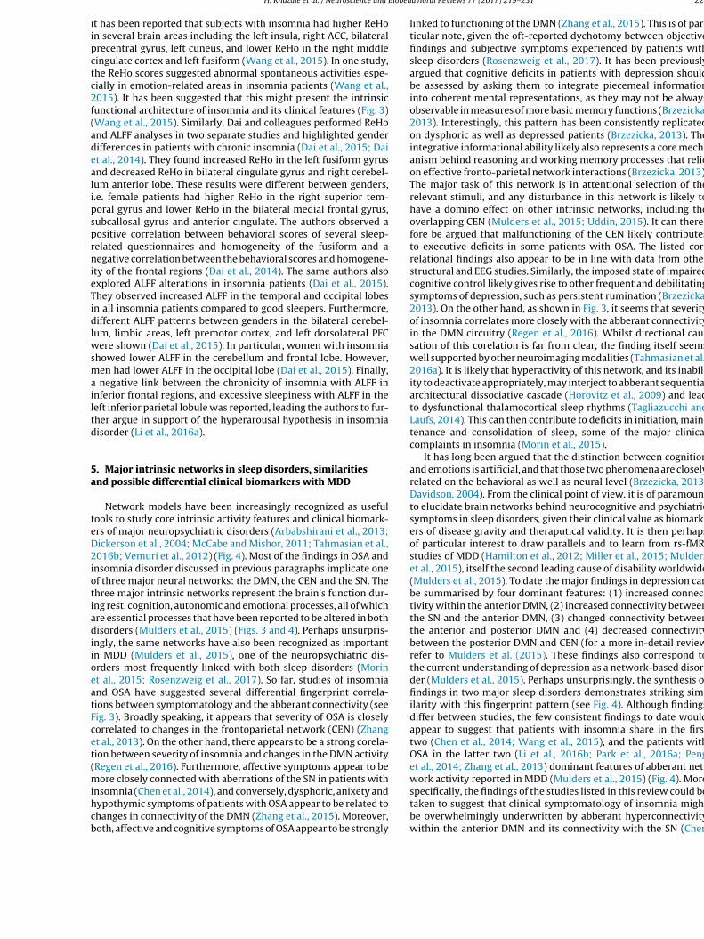
it has been reported that subjects with insomnia had higher ReHoin several brain areas including the left insula, right ACC, bilateralprecentral gyrus, left cuneus, and lower ReHo in the right middlecingulate cortex and left fusiform (Wang et al., 2015). In one study,the ReHo scores suggested abnormal spontaneous activities espe-cially in emotion-related areas in insomnia patients (Wang et al.,2015). It has been suggested that this might present the intrinsicfunctional architecture of insomnia and its clinical features (Fig. 3)(Wang et al., 2015). Similarly, Dai and colleagues performed ReHoand ALFF analyses in two separate studies and highlighted genderdifferences in patients with chronic insomnia (Dai et al., 2015; Daiet al., 2014). They found increased ReHo in the left fusiform gyrusand decreased ReHo in bilateral cingulate gyrus and right cerebel-lum anterior lobe. These results were different between genders,i.e. female patients had higher ReHo in the right superior tem-poral gyrus and lower ReHo in the bilateral medial frontal gyrus,subcallosal gyrus and anterior cingulate. The authors observed apositive correlation between behavioral scores of several sleep-related questionnaires and homogeneity of the fusiform and anegative correlation between the behavioral scores and homogene-ity of the frontal regions (Dai et al., 2014). The same authors alsoexplored ALFF alterations in insomnia patients (Dai et al., 2015).They observed increased ALFF in the temporal and occipital lobesin all insomnia patients compared to good sleepers. Furthermore,different ALFF patterns between genders in the bilateral cerebel-lum, limbic areas, left premotor cortex, and left dorsolateral PFCwere shown (Dai et al., 2015). In particular, women with insomniashowed lower ALFF in the cerebellum and frontal lobe. However,men had lower ALFF in the occipital lobe (Dai et al., 2015). Finally,a negative link between the chronicity of insomnia with ALFF ininferior frontal regions, and excessive sleepiness with ALFF in theleft inferior parietal lobule was reported, leading the authors to fur-ther argue in support of the hyperarousal hypothesis in insomniadisorder (Li et al., 2016a).
5. Major intrinsic networks in sleep disorders, similaritiesand possible differential clinical biomarkers with MDD
Network models have been increasingly recognized as usefultools to study core intrinsic activity features and clinical biomark-ers of major neuropsychiatric disorders (Arbabshirani et al., 2013;Dickerson et al., 2004; McCabe and Mishor, 2011; Tahmasian et al.,2016b; Vemuri et al., 2012) (Fig. 4). Most of the findings in OSA andinsomnia disorder discussed in previous paragraphs implicate oneof three major neural networks: the DMN, the CEN and the SN. Thethree major intrinsic networks represent the brain’s function dur-ing rest, cognition, autonomic and emotional processes, all of whichare essential processes that have been reported to be altered in bothdisorders (Mulders et al., 2015) (Figs. 3 and 4). Perhaps unsurpris-ingly, the same networks have also been recognized as importantin MDD (Mulders et al., 2015), one of the neuropsychiatric dis-orders most frequently linked with both sleep disorders (Morinet al., 2015; Rosenzweig et al., 2017). So far, studies of insomniaand OSA have suggested several differential fingerprint correla-tions between symptomatology and the abberant connectivity (seeFig. 3). Broadly speaking, it appears that severity of OSA is closelycorrelated to changes in the frontoparietal network (CEN) (Zhanget al., 2013). On the other hand, there appears to be a strong corela-tion between severity of insomnia and changes in the DMN activity(Regen et al., 2016). Furthermore, affective symptoms appear to bemore closely connected with aberrations of the SN in patients withinsomnia (Chen et al., 2014), and conversely, dysphoric, anixety andhypothymic symptoms of patients with OSA appear to be related tochanges in connectivity of the DMN (Zhang et al., 2015). Moreover,both, affective and cognitive symptoms of OSA appear to be strongly
linked to functioning of the DMN (Zhang et al., 2015). This is of par-ticular note, given the oft-reported dychotomy between objectivefindings and subjective symptoms experienced by patients withsleep disorders (Rosenzweig et al., 2017). It has been previouslyargued that cognitive deficits in patients with depression shouldbe assessed by asking them to integrate piecemeal informationinto coherent mental representations, as they may not be alwaysobservable in measures of more basic memory functions (Brzezicka,2013). Interestingly, this pattern has been consistently replicatedon dysphoric as well as depressed patients (Brzezicka, 2013). Theintegrative informational ability likely also represents a core mech-anism behind reasoning and working memory processes that relieon effective fronto-parietal network interactions (Brzezicka, 2013).The major task of this network is in attentional selection of therelevant stimuli, and any disturbance in this network is likely tohave a domino effect on other intrinsic networks, including theoverlapping CEN (Mulders et al., 2015; Uddin, 2015). It can there-fore be argued that malfunctioning of the CEN likely contributesto executive deficits in some patients with OSA. The listed cor-relational findings also appear to be in line with data from otherstructural and EEG studies. Similarly, the imposed state of impairedcognitive control likely gives rise to other frequent and debilitatingsymptoms of depression, such as persistent rumination (Brzezicka,2013). On the other hand, as shown in Fig. 3, it seems that severityof insomnia correlates more closely with the abberant connectivityin the DMN circuitry (Regen et al., 2016). Whilst directional cau-sation of this corelation is far from clear, the finding itself seemswell supported by other neuroimaging modalities (Tahmasian et al.,2016a). It is likely that hyperactivity of this network, and its inabil-ity to deactivate appropriately, may interject to abberant sequentialarchitectural dissociative cascade (Horovitz et al., 2009) and leadto dysfunctional thalamocortical sleep rhythms (Tagliazucchi andLaufs, 2014). This can then contribute to deficits in initiation, main-tenance and consolidation of sleep, some of the major clinicalcomplaints in insomnia (Morin et al., 2015).
It has long been argued that the distinction between cognitionand emotions is artificial, and that those two phenomena are closelyrelated on the behavioral as well as neural level (Brzezicka, 2013;Davidson, 2004). From the clinical point of view, it is of paramountto elucidate brain networks behind neurocognitive and psychiatricsymptoms in sleep disorders, given their clinical value as biomark-ers of disease gravity and theraputical validity. It is then perhapsof particular interest to draw parallels and to learn from rs-fMRIstudies of MDD (Hamilton et al., 2012; Miller et al., 2015; Mulderset al., 2015), itself the second leading cause of disability worldwide(Mulders et al., 2015). To date the major findings in depression canbe summarised by four dominant features: (1) increased connec-tivity within the anterior DMN, (2) increased connectivity betweenthe SN and the anterior DMN, (3) changed connectivity betweenthe anterior and posterior DMN and (4) decreased connectivitybetween the posterior DMN and CEN (for a more in-detail reviewrefer to Mulders et al. (2015). These findings also correspond tothe current understanding of depression as a network-based disor-der (Mulders et al., 2015). Perhaps unsurprisingly, the synthesis offindings in two major sleep disorders demonstrates striking sim-ilarity with this fingerprint pattern (see Fig. 4). Although findingsdiffer between studies, the few consistent findings to date wouldappear to suggest that patients with insomnia share in the firsttwo (Chen et al., 2014; Wang et al., 2015), and the patients withOSA in the latter two (Li et al., 2016b; Park et al., 2016a; Penget al., 2014; Zhang et al., 2013) dominant features of abberant net-work activity reported in MDD (Mulders et al., 2015) (Fig. 4). Morespecifically, the findings of the studies listed in this review could betaken to suggest that clinical symptomatology of insomnia mightbe overwhelmingly underwritten by abberant hyperconnectivitywithin the anterior DMN and its connectivity with the SN (Chen

228 H. Khazaie et al. / Neuroscience and Biobehavioral Reviews 77 (2017) 219–231
Fig. 4. Comparison of the blueprint fingerprint of within- and between-network connectivity changes in sleep disorders, obstructive sleep apnoea and insomnia disorder, withtheir major neuropsychiatric comorbidity, major depressive disorder (adapted from (Mulders et al. (2015)). A within-network increase/decrease in connectivity is presentedwith pink/blue outlines; lines (pink/blue) between networks represent a between-network increase/decrease in connectivity. Grey lines correspond to inconsistent or unclearfindings. Black ellipses represent key nodes related to connectivity. Numbers represent main findings previously reported for the major depressive disorder: (1): increasein the anterior DMN connectivity and inclusion of sgACC within the anterior DMN, (2): increased connectivity between the anterior DMN and SN, (3): changed connectivitybetween the anterior and posterior DMN, (4): decreased connectivity between the posterior DMN and CEN (Mulders et al., 2015). Abbreviations: CEN: central executivenetwork; DMN: default-mode network; MTL: medial temporal lobe; PMC: premotor cortex; SN: salience network; sgACC: subgenual anterior cingulate cortex. (For interpretation ofthe references to color in this figure legend, the reader is referred to the web version of this article.).
et al., 2014). This emphasis on the anterior DMN is noteworthy aswhilst both the anterior and posterior parts of the DMN are relatedto spontaneous or self-generated cognition, the anterior DMN ismore related to self-referential processing and emotion-regulation,partly through its strong connections with limbic areas such as theamygdala (Mulders et al., 2015). Hyperactivity of the amygdala,especially in response to sleep related stimuli, has been demon-strated in patients with insomnia (Morin et al., 2015) and couldunderlie the negative bias experienced by patients in relation tosleep-related stimuli. Similarly, changes in connectivity betweenthe anterior DMN and SN may constitute an increase in top-downmodulation of limbic hyperactivity, bottom-up interference of self-processing regions, or both, as has been previously argued to occurin depression (Mulders et al., 2015). Surprisingly, connectivity ofthe amygdala with striatum, including head of caudate, and otherbrain regions such as insula and thalamus, appears to be decreasedin insomnia (Huang et al., 2012). This could be of clinical importanceas it might indicate an inability to control and entrain amygdalaresponsivity by other limbic regions (Goldstein and Walker, 2014).It may also suggest an increased coupling and inteference by otherregions, such as the fight/flight adrenergic brainstem centre of thelocus coeruleus (Goldstein and Walker, 2014). Similar abberantconnectivity with locus coeruleus has been previously shown tooccur in studies of forced sleep restriction (Motomura et al., 2013).Taken together, this pattern of activity might also explain why in
insomnia affective symptomatology and hyperarousal are linkedwith abberations in the SN, and not with the DMN.
In contrast, the findings of OSA studies appear to highlight therole for the decreased synchronicity and connectivity between theanterior and posterior DMN, and between the posterior DMN andhippocampus and the rest of frontoparietal network (Fig. 4). Theposterior DMN has been implicated in both consciousness andmemory processing through its relation to the hippocampal for-mation (Mulders et al., 2015), and the abberant functioning of thispart of the network is in line with other structural studies of OSA(Tahmasian et al., 2016a). In line with the role of the posteriorDMN in awareness and directed attention (Leech and Sharp, 2014),and the role of the CEN in higher cognitive functioning (Corbettaand Shulman, 2002), the change in their interaction could underliedifficulty in switching from an internally directed state in whichthe DMN is dominant, to an externally directed state in which theCEN is dominant and attention is directed toward outward stimuli(Uddin, 2015). Several authors have suggested that the insular cor-tex might be crucial for this shift in network-dominance (Uddin,2015), which is supported by the increased connectivity of theinsula with the anterior DMN and decreased connectivity withother networks (Uddin, 2015) (Figs. 3 and 4). In support of thisview, in a recent meta-analysis of structural and funtional studies,significant deficits in this region have been highlighted (Tahmasianet al., 2016a). Apart from cognitive deficits that can arise from this

H. Khazaie et al. / Neuroscience and Biobehavioral Reviews 77 (2017) 219–231 229
abberation, this feature implies problems in sustained and dividedattention that arguably then contribute to well recognized drivingdifficulties in patients with OSA, even without excessive daytimesomnolence (Rosenzweig et al., 2017).
6. Strengths and limitations of rs-fMRI
It is widely accepted that rs-fMRI represents state of the artin the assessment of intrinsic neuronal activity (Tagliazucchi andLaufs, 2014). During rs-fMRI, in the absence of any externallyinduced task, distributed brain regions exhibit coherent low-frequency fluctuations binding them together into identifiablefunctional networks (Fig. 1) (Fox and Raichle, 2007). Similarly, it hasbeen generally accepted that rs-fMRI presents a promising tool inthe search for functional biomarkers of a variety of neurodegener-ative, neuropsychiatric, pain and sleep disorders (Tagliazucchi andLaufs, 2014; Tahmasian et al., 2015a). However, there are severalimportant limitations of this technique that need to be accountedfor. Firstly, resting-state is an uncontrolled, insufficiently under-stood condition that arguably differs in any one person duringvarious degrees of wakefulness, during different times of the day,and during differential baseline “consciousness” thought processes(Muto et al., 2016; Tagliazucchi and Laufs, 2014). It follows thatin sleep disorders, where it is specifically difficult to control fordegree of somnolence and ‘micro-sleeps’ even with co-registrationwith EEG, it might be particularly difficult to account for the exactdegree of wakefulness (Tagliazucchi and Laufs, 2014). By a wayof example, using self-report questionnaire (Amsterdam Resting-State questionnaire), it has been shown that sleepiness level isassociated with FC scores within the DMN, visual, and sensorimo-tor network (Deco et al., 2014; Stoffers et al., 2015). Also, it hasbeen demonstrated that even in those without known predisposingdisorders, 30% of individuals could not stay awake after three min-utes and 50% of them had at least one epoch of sleep (Tagliazucchiand Laufs, 2014). Hence, FC alteration due to falling sleep is animportant cofounding issue in patients with sleep disorders as theyhave higher daytime sleepiness (Morin et al., 2015; Rosenzweiget al., 2017) and might fall in sleep easier than healthy subjectsin the scanner. Secondly, the majority of neuropsychiatric condi-tions, including the two major sleep disorders reviewed here, aremultifactorial disorders, with predisposing genetic, promoting epi-genetic and environmental, and precipitating acute factors (Morinet al., 2015; Rosenzweig et al., 2017). Hence, the issue of direc-tional causation, the classical chicken or egg problem, might beparticularly difficult to decipher here. Namely, the chronicity ofsleep issues, homeostatic adaptive and maladaptive processes todisrupted sleep and idiosyncratic susceptibility are all contributingto an endogenous neuronal activity fingerprint at any point in time(Rosenzweig et al., 2017).
Despite all these issues, technically, a paradigm-less experi-mental setting of rs-fMRI appears especially suitable for buildingmulticenter databases whilst minimizing any confounding influ-ence of local clinical settings (Mulders et al., 2015; Tagliazucchiand Laufs, 2014). However, it is crucial to acknowledge all techno-logical limitations, some commonly shared with other functionalimaging methods (Tagliazucchi and Laufs, 2014). For example, it hasbeen recognized that the non-neuronal physiological signals suchas respiration and cardiovascular pulsatile rhythms may introducenoise and lead to misinterpretations of resting-state BOLD findings(Birn et al., 2006; Liu et al., 2006). Similarly, white matter and cere-brospinal fluid signals are other potential sources of noise, and theyshould be removed in the preprocessing of images (Fox et al., 2005).In that respect there is an ongoing discussion in the field regardingthe importance of the global signal regression approach, which canremove respiratory and pulsatile noise from the resting-state BOLD
signal but it can also introduce anti-correlations between regions,and increase the number of false negatives (Murphy et al., 2009).
Another critical unsolved technical issue in rs-fMRI is the issueof artefacts induced by head motion, which can introduce system-atic noise in the BOLD signal. Satterthwaite et al. (2013) highlightedthe fact that improved preprocessing and motion correction pro-vides better results (Satterthwaite et al., 2013). They suggested thathead motion of subjects should be explicitly reported as an impor-tant outcome measure in all rs-MRI studies (Satterthwaite et al.,2013). Hence, the onus is on all the future studies to employ carefulpreprocessing and verifiable statistical analyses as critical steps toavoid misleading results. Moreover, in order to increase both sen-sitivity and specificity of rs-fMRI studies, the brain state should bedetermined and accounted for in the related analysis strategies andresults should be critically reviewed for false positives originatingfrom unstable vigilance levels, especially if the state of wakeful-ness remains obscure (Tagliazucchi and Laufs, 2014). Finally, in arecent study, significant circadian rhythmicity of brain responsesin all brain regions except in DPFC, including the thalamus, head ofthe caudate, and putamen, was shown (Muto et al., 2016). Hence,it follows that apart from homeostatic influences, it is also the cir-cadian control over intrinsic activity that should be accounted forin any future studies (Muto et al., 2016).
Moreover, it should also be noted that rs-fMRI is measuringoxygen consumption and therefore is an indirect measurementof neural activity (Tagliazucchi and Laufs, 2014). Thus, the com-bination of fMRI with other brain mapping modalities (e.g. EEG)may provide more comprehensive information to explore thepathophysiology of sleep disorders (Tagliazucchi and Laufs, 2014).Beyond these applications, rs-fMRI is a potent, non-invasive tool forclinical purposes, e.g. to separate patients from healthy individualsor subtypes of particular sleep disorder or to observe the effects ofdifferent medication in functional networks (Mulders et al., 2015;Tagliazucchi and Laufs, 2014).
7. Conclusions and future directions
The studies listed above highlight the important role of rs-fMRIas a promising non-invasive method for better understanding of thepathophysiology of the two most prevalent sleep disorders, insom-nia disorder and OSA. The underlying core neural mechanisms andneurocircuitry of both disorders are still matter of some debate inthe field. It is increasingly recognized that, due to the wide rangeof idiosyncratic vulnerabilities and multiple epigenetic and geneticadaptive and maladaptive mechanisms (Baril et al., 2017) at play,it is very difficult to predict specific future risk for any individualpatient. In addition, an important issue of gender differences, andthe potential impact of sex hormones on pathophysiology of bothdisorders during reproductive age in women, whilst recognized inthe field (O’Halloran et al., 2016), is far from being fully explainedand documented. Similarly, the diversity and the diffuse natureof reported neurocognitive deficits, make the panacea “cure-all”treatment approach in these disorders less likely. Elucidating theindividual resting-state neurocircuitry connectivity and presenceof any neural network dysfunction might be the most appropri-ate first step for any personalized medicine approach in treatmentof any sleep and neuropsychiatric disorder. Notwithstanding this,the emerging blueprints of the specific patterns of dysfunctionalconnectivity for both of these sleep disorders are emerging (seeFig. 4), and they appear to bear striking similarity with specific, ifdiverse, aspects of aberrant activity in MDD. As such, they may pro-vide a potential clinical biomarker of functional deficits frequentlyreported by patients, as well as guide future therapeutic efforts.Future studies using standard preprocessing and statistical analy-sis are warranted in this field (Tagliazucchi and Laufs, 2014). Such

230 H. Khazaie et al. / Neuroscience and Biobehavioral Reviews 77 (2017) 219–231
studies should focus on the progression and trajectory of disor-ders and should aim to evaluate potential treatment effects. It hasbeen argued that adopting within-group repeated measures andrandomized controlled cross-over designs in future studies of sleepdisorders may allow for more precise phenotyping and assessmentof clinically useful neural markers that underpin functional daytimeimpairments (Vakulin and Stevens, 2016). Moreover, the recentadvent of hybrid positron emission tomography (PET)/MR scannersmay enrich our ability to investigate connectivity by introducingthe concept of metabolic connectivity whilst enabling insights onthe physiological and molecular bases underlying high-level neuralorganization (Aiello et al., 2016; Otte et al., 2016; Riedl et al., 2014;Tahmasian et al., 2015b). This multimodal imaging approach mightbe of particular clinical value in future studies of glymphatic system(Xie et al., 2013). The glympathic system has recently been impli-cated in the clearance of toxic metabolites in rodents during sleep(Xie et al., 2013), and an indirect clinical evidence could be arguablytaken to suggest its contributory role in pathophysiology of thetwo major sleep disorders, insomnia disorder and OSA (Cedernaeset al., 2017; Ju et al., 2016; Mander et al., 2016). In conclusion,an increasing recognition of the complexity of pathophysiologicalprocesses in sleep medicine is driving the move towards more per-sonalized diagnosis and treatment approaches where the rs-fMRImay present as a pivotal future clinical tool.
Contributors
Authors HK, MV, MT and IR wrote and planned the review. Allauthors contributed equally to the revision of this manuscript. Theauthors apologize to all the colleagues whose outstanding workcould not be cited due to space limitations.
Acknowledgements
The authors declare that the research was conducted in theabsence of any commercial or financial relationships that could beconstrued as a potential conflict of interest. This work was sup-ported by the Wellcome Trust [103952/Z/14/Z].
References
Aiello, M., Cavaliere, C., Salvatore, M., 2016. Hybrid PET/MR imaging and brainconnectivity. Front. Neurosci. 10, 64.
Arbabshirani, M.R., Kiehl, K.A., Pearlson, G.D., Calhoun, V.D., 2013. Classification ofschizophrenia patients based on resting-state functional network connectivity.Front. Neurosci. 7, 133.
Baglioni, C., Battagliese, G., Feige, B., Spiegelhalder, K., Nissen, C., Voderholzer, U.,Lombardo, C., Riemann, D., 2011. Insomnia as a predictor of depression: ameta-analytic evaluation of longitudinal epidemiological studies. J. Affect.Disord. 135, 10–19.
Baglioni, C., Spiegelhalder, K., Regen, W., Feige, B., Nissen, C., Lombardo, C., Violani,C., Hennig, J., Riemann, D., 2014. Insomnia disorder is associated with increasedamygdala reactivity to insomnia-related stimuli. Sleep 37, 1907–1917.
Baril, A.A., Gagnon, K., Brayet, P., Montplaisir, J., De Beaumont, L., Carrier, J., Lafond,C., L’Heureux, F., Gagnon, J.F., Gosselin, N., 2017. Gray matter hypertrophy andthickening with obstructive sleep apnea in middle-aged and older adults. Am.J. Respir. Crit. Care Med.
Birn, R.M., Diamond, J.B., Smith, M.A., Bandettini, P.A., 2006. Separatingrespiratory-variation-related fluctuations from neuronal-activity-relatedfluctuations in fMRI. Neuroimage 31, 1536–1548.
Biswal, B., Yetkin, F.Z., Haughton, V.M., Hyde, J.S., 1995. Functional connectivity inthe motor cortex of resting human brain using echo-planar MRI. Magn. Reson.Med. 34, 537–541.
Brzezicka, A., 2013. Integrative deficits in depression and in negative mood statesas a result of fronto-parietal network dysfunctions. Acta Neurobiol. Exp.(Wars) 73, 313–325.
Buzsaki, G., Watson, B.O., 2012. Brain rhythms and neural syntax: implications forefficient coding of cognitive content and neuropsychiatric disease. DialoguesClin. Neurosci. 14, 345–367.
Cedernaes, J., Osorio, R.S., Varga, A.W., Kam, K., Schioth, H.B., Benedict, C., 2017.Candidate mechanisms underlying the association between sleep-wakedisruptions and Alzheimer’s disease. Sleep Med. Rev. 31, 102–111.
Chen, M.C., Chang, C., Glover, G.H., Gotlib, I.H., 2014. Increased insula coactivationwith salience networks in insomnia. Biol. Psychol. 97, 1–8.
Chuah, Y.M., Venkatraman, V., Dinges, D.F., Chee, M.W., 2006. The neural basis ofinterindividual variability in inhibitory efficiency after sleep deprivation. J.Neurosci. 26, 7156–7162.
Corbetta, M., Shulman, G.L., 2002. Control of goal-directed and stimulus-drivenattention in the brain. Nat. Rev. Neurosci. 3, 201–215.
Dai, X.J., Nie, X., Liu, X., Pei, L., Jiang, J., Peng, D.C., Gong, H.H., Zeng, X.J., Wang, Y.J.,Zhan, Y., 2015. 2015. gender differences in regional brain activity in patientswith chronic primary insomnia: evidence from a resting-State fMRI study. J.Clin. Sleep Med.: JCSM.
Dai, X.J., Peng, D.C., Gong, H.H., Wan, A.L., Nie, X., Li, H.J., Wang, Y.X., 2014. Alteredintrinsic regional brain spontaneous activity and subjective sleep quality inpatients with chronic primary insomnia: a resting-state fMRI study.Neuropsychiatr. Dis. Treat. 10, 2163–2175.
Davidson, R.J., 2004. What does the prefrontal cortex do in affect: perspectives onfrontal EEG asymmetry research. Biol. Psychol. 67, 219–233.
De Havas, J.A., Parimal, S., Soon, C.S., Chee, M.W., 2012. Sleep deprivation reducesdefault mode network connectivity and anti-correlation during rest and taskperformance. Neuroimage 59, 1745–1751.
Deco, G., Hagmann, P., Hudetz, A.G., Tononi, G., 2014. Modeling resting-statefunctional networks when the cortex falls asleep: local and global changes.Cereb. Cortex 24, 3180–3194.
Desseilles, M., Dang-Vu, T., Schabus, M., Sterpenich, V., Maquet, P., Schwartz, S.,2008. Neuroimaging insights into the pathophysiology of sleep disorders.Sleep 31, 777–794.
Dickerson, B.C., Salat, D.H., Bates, J.F., Atiya, M., Killiany, R.J., Greve, D.N., Dale, A.M.,Stern, C.E., Blacker, D., Albert, M.S., Sperling, R.A., 2004. Medial temporal lobefunction and structure in mild cognitive impairment. Ann. Neurol. 56, 27–35.
Emamian, F., Khazaie, H., Tahmasian, M., Leschziner, G.D., Morrell, M.J., Hsiung,G.Y., Rosenzweig, I., Sepehry, A.A., 2016. The association between obstructivesleep apnea and Alzheimer’s disease: a meta-analysis perspective. Front. AgingNeurosci. 8, 78.
Fox, M.D., Raichle, M.E., 2007. Spontaneous fluctuations in brain activity observedwith functional magnetic resonance imaging. Nat. Rev. Neurosci. 8, 700–711.
Fox, M.D., Snyder, A.Z., Vincent, J.L., Corbetta, M., Van Essen, D.C., Raichle, M.E.,2005. The human brain is intrinsically organized into dynamic, anticorrelatedfunctional networks. Proc. Natl. Acad. Sci. U. S. A. 102, 9673–9678.
Goldstein, A.N., Walker, M.P., 2014. The role of sleep in emotional brain function.Annu. Rev. Clin. Psychol. 10, 679–708.
Greicius, M.D., Krasnow, B., Reiss, A.L., Menon, V., 2003. Functional connectivity inthe resting brain: a network analysis of the default mode hypothesis. Proc.Natl. Acad. Sci. U. S. A. 100, 253–258.
Gujar, N., Yoo, S.S., Hu, P., Walker, M.P., 2010. The unrested resting brain: sleepdeprivation alters activity within the default-mode network. J. Cogn. Neurosci.22, 1637–1648.
Gupta, M.A., Simpson, F.C., 2015. Obstructive sleep apnea and psychiatricdisorders: a systematic review. J. Clin. Sleep Med.: JCSM 11, 165–175.
Hamilton, J.P., Etkin, A., Furman, D.J., Lemus, M.G., Johnson, R.F., Gotlib, I.H., 2012.Functional neuroimaging of major depressive disorder: a meta-analysis andnew integration of base line activation and neural response data. Am. J.Psychiatry 169, 693–703.
Horovitz, S.G., Braun, A.R., Carr, W.S., Picchioni, D., Balkin, T.J., Fukunaga, M., Duyn,J.H., 2009. Decoupling of the brain’s default mode network during deep sleep.Proc. Natl. Acad. Sci. U. S. A. 106, 11376–11381.
Huang, Z., Liang, P., Jia, X., Zhan, S., Li, N., Ding, Y., Lu, J., Wang, Y., Li, K., 2012.Abnormal amygdala connectivity in patients with primary insomnia: evidencefrom resting state fMRI. Eur. J. Radiol. 81, 1288–1295.
Joo, E.Y., Jeon, S., Kim, S.T., Lee, J.M., Hong, S.B., 2013. Localized cortical thinning inpatients with obstructive sleep apnea syndrome. Sleep 36, 1153–1162.
Jordan, A.S., McSharry, D.G., Malhotra, A., 2014. Adult obstructive sleep apnoea.Lancet 383, 736–747.
Ju, Y.E., Finn, M.B., Sutphen, C.L., Herries, E.M., Jerome, G.M., Ladenson, J.H.,Crimmins, D.L., Fagan, A.M., Holtzman, D.M., 2016. Obstructive sleep apneadecreases central nervous system-derived proteins in the cerebrospinal fluid.Ann. Neurol. 80, 154–159.
Klupp, E., Grimmer, T., Tahmasian, M., Sorg, C., Yakushev, I., Yousefi, B.H., Drzezga,A., Forster, S., 2015. Prefrontal hypometabolism in alzheimer disease is relatedto longitudinal amyloid accumulation in remote brain regions. J. Nucl. Med. 56,399–404.
Koenigs, M., Holliday, J., Solomon, J., Grafman, J., 2010. Left dorsomedial frontalbrain damage is associated with insomnia. J. Neurosci. 30, 16041–16043.
Landsness, E.C., Goldstein, M.R., Peterson, M.J., Tononi, G., Benca, R.M., 2011.Antidepressant effects of selective slow wave sleep deprivation in majordepression: a high-density EEG investigation. J. Psychiatr. Res. 45, 1019–1026.
Leech, R., Sharp, D.J., 2014. The role of the posterior cingulate cortex in cognitionand disease. Brain 137, 12–32.
Lévy, P., Kohler, M., McNicholas, W.T., Barbé, F., McEvoy, R.D., Somers, V.K., Lavie,L., Pépin, J.-L., 2015. Obstructive sleep apnoea syndrome. Nat. Rev. Dis. Primers,15015.
Li, Y., Wang, E., Zhang, H., Dou, S., Liu, L., Tong, L., Lei, Y., Wang, M., Xu, J., Shi, D.,Zhang, Q., 2014. Functional connectivity changes between parietal andprefrontal cortices in primary insomnia patients: evidence from resting-statefMRI. Eur. J. Med. Res. 19, 32.
Li, C., Ma, X., Dong, M., Yin, Y., Hua, K., Li, M., Li, C., Zhan, W., Li, C., Jiang, G., 2016a.Abnormal spontaneous regional brain activity in primary insomnia: a

H. Khazaie et al. / Neuroscience and Biobehavioral Reviews 77 (2017) 219–231 231
resting-state functional magnetic resonance imaging study. Neuropsychiatr.Dis. Treat. 12, 1371–1378.
Li, H.J., Nie, X., Gong, H.H., Zhang, W., Nie, S., Peng, D.C., 2016b. Abnormalresting-state functional connectivity within the default mode networksubregions in male patients with obstructive sleep apnea. Neuropsychiatr. Dis.Treat. 12, 203–212.
Liu, C.S., Miki, A., Hulvershorn, J., Bloy, L., Gualtieri, E.E., Liu, G.T., Leigh, J.S.,Haselgrove, J.C., Elliott, M.A., 2006. Spatial and temporal characteristics ofphysiological noise in fMRI at 3T. Acad. Radiol. 13, 313–323.
Lustenberger, C., Boyle, M.R., Alagapan, S., Mellin, J.M., Vaughn, B.V., Frohlich, F.,2016. Feedback-controlled transcranial alternating current stimulation revealsa functional role of sleep spindles in motor memory consolidation. Curr. Biol.
Mander, B.A., Winer, J.R., Jagust, W.J., Walker, M.P., 2016. Sleep: a novelmechanistic pathway, biomarker, and treatment target in the pathology ofAlzheimer’s disease? Trends Neurosci. 39, 552–566.
McCabe, C., Mishor, Z., 2011. Antidepressant medications reducesubcortical-cortical resting-state functional connectivity in healthy volunteers.Neuroimage 57, 1317–1323.
McCormick, C., Protzner, A.B., Barnett, A.J., Cohn, M., Valiante, T.A., McAndrews,M.P., 2014. Linking DMN connectivity to episodic memory capacity: what canwe learn from patients with medial temporal lobe damage? NeuroImage. Clin.5, 188–196.
Menon, V., Uddin, L.Q., 2010. Saliency, switching, attention and control: a networkmodel of insula function. Brain Struct. Funct. 214, 655–667.
Miller, C.H., Hamilton, J.P., Sacchet, M.D., Gotlib, I.H., 2015. Meta-analysis offunctional neuroimaging of major depressive disorder in youth. JAMAPsychiatry 72, 1045–1053.
Moher, D., Liberati, A., Tetzlaff, J., Altman, D.G., Group, P., 2009. Preferred reportingitems for systematic reviews and meta-analyses: the PRISMA statement. PLoSMed. 6, e1000097.
Morin, C.M., Drake, C.L., Harvey, A.G., Krystal, A.D., Manber, R., Riemann, D.,Spiegelhalder, K., 2015. Insomnia disorder. Nat. Rev. Dis. Primers, 15026.
Motomura, Y., Kitamura, S., Oba, K., Terasawa, Y., Enomoto, M., Katayose, Y., Hida,A., Moriguchi, Y., Higuchi, S., Mishima, K., 2013. Sleep debt elicits negativeemotional reaction through diminished amygdala-anterior cingulatefunctional connectivity. PLoS One 8, e56578.
Mulders, P.C., van Eijndhoven, P.F., Schene, A.H., Beckmann, C.F., Tendolkar, I.,2015. Resting-state functional connectivity in major depressive disorder: areview. Neurosci. Biobehav. Rev. 56, 330–344.
Murphy, K., Birn, R.M., Handwerker, D.A., Jones, T.B., Bandettini, P.A., 2009. Theimpact of global signal regression on resting state correlations: areanti-correlated networks introduced? Neuroimage 44, 893–905.
Muto, V., Jaspar, M., Meyer, C., Kusse, C., Chellappa, S.L., Degueldre, C., Balteau, E.,Shaffii-Le Bourdiec, A., Luxen, A., Middleton, B., Archer, S.N., Phillips, C.,Collette, F., Vandewalle, G., Dijk, D.J., Maquet, P., 2016. Local modulation ofhuman brain responses by circadian rhythmicity and sleep debt. Science 353,687–690.
Nie, X., Shao, Y., Liu, S.Y., Li, H.J., Wan, A.L., Nie, S., Peng, D.C., Dai, X.J., 2015.Functional connectivity of paired default mode network subregions in primaryinsomnia. Neuropsychiatr. Dis. Treat. 11, 3085–3093.
O’Halloran, K.D., Lewis, P., McDonald, F., 2016. Sex, stress and sleep apnoea:decreased susceptibility to upper airway muscle dysfunction followingintermittent hypoxia in females. Respir. Physiol. Neurobiol.
Osorio, R.S., Gumb, T., Pirraglia, E., Varga, A.W., Lu, S.E., Lim, J., Wohlleber, M.E.,Ducca, E.L., Koushyk, V., Glodzik, L., Mosconi, L., Ayappa, I., Rapoport, D.M., deLeon, M.J., Disease, Alzheimer’s, Neuroimaging, I., 2015. Sleep-disorderedbreathing advances cognitive decline in the elderly. Neurology 84, 1964–1971.
Otte, A., Turkheimer, F., Rosenzweig, I., 2016. All you need is sleep. EBioMedicine12, 2–3.
Park, B., Palomares, J.A., Woo, M.A., Kang, D.W., Macey, P.M., Yan-Go, F.L., Harper,R.M., Kumar, R., 2016a. Disrupted functional brain network organization inpatients with obstructive sleep apnea. Brain Behav. 6, e00441.
Park, B., Palomares, J.A., Woo, M.A., Kang, D.W., Macey, P.M., Yan-Go, F.L., Harper,R.M., Kumar, R., 2016b. Aberrant insular functional network integrity inpatients with obstructive sleep apnea. Sleep.
Pasquini, L., Scherr, M., Tahmasian, M., Meng, C., Myers, N.E., Ortner, M., Muhlau,M., Kurz, A., Forstl, H., Zimmer, C., Grimmer, T., Wohlschlager, A.M., Riedl, V.,Sorg, C., 2015. Link between hippocampus’ raised local and eased globalintrinsic connectivity in AD. Alzheimers Dement 11, 475–484.
Peng, D.C., Dai, X.J., Gong, H.H., Li, H.J., Nie, X., Zhang, W., 2014. Altered intrinsicregional brain activity in male patients with severe obstructive sleep apnea: aresting-state functional magnetic resonance imaging study. Neuropsych Dis.Treat 10, 1819–1826.
Regen, W., Kyle, S.D., Nissen, C., Feige, B., Baglioni, C., Hennig, J., Riemann, D.,Spiegelhalder, K., 2016. Objective sleep disturbances are associated withgreater waking resting-state connectivity between the retrosplenialcortex/hippocampus and various nodes of the default mode network. J.Psychiatry Neurosci. 41, 140290.
Riedl, V., Bienkowska, K., Strobel, C., Tahmasian, M., Grimmer, T., Forster, S.,Friston, K.J., Sorg, C., Drzezga, A., 2014. Local activity determines functionalconnectivity in the resting human brain: a simultaneous FDG-PET/fMRI study.J. Neurosci. 34, 6260–6266.
Rosenzweig, I., Glasser, M., Crum, W.R., Kempton, M.J., Milosevic, M., McMillan, A.,Leschziner, G.D., Kumari, V., Goadsby, P., Simonds, A.K., Williams, S.C., Morrell,M.J., 2016. Changes in neurocognitive architecture in patients with obstructive
sleep apnea treated with continuous positive airway pressure. EBioMedicine 7,221–229.
Rosenzweig, I., Glasser, M., Polsek, D., Leschziner, G.D., Williams, S.C., Morrell, M.J.,2015. Sleep apnoea and the brain: a complex relationship. Lancet. Respir. Med.3, 404–414.
Rosenzweig, I., Weaver, T., Morrell, M.J., 2017. Obstructive sleep apnea and thecentral nervous system: neural adaptive processes, cognition andperformance. In: Kryger, M.H., Roth, T. (Eds.), Principles and Practice of SleepMedicine. , sixth edition. Elsevier, Philadelphia, PA.
Santarnecchi, E., Sicilia, I., Richiardi, J., Vatti, G., Polizzotto, N.R., Marino, D., Rocchi,R., Van De Ville, D., Rossi, A., 2013. Altered cortical and subcortical localcoherence in obstructive sleep apnea: a functional magnetic resonanceimaging study. J. Sleep Res. 22, 337–347.
Satterthwaite, T.D., Elliott, M.A., Gerraty, R.T., Ruparel, K., Loughead, J., Calkins,M.E., Eickhoff, S.B., Hakonarson, H., Gur, R.C., Gur, R.E., Wolf, D.H., 2013. Animproved framework for confound regression and filtering for control ofmotion artifact in the preprocessing of resting-state functional connectivitydata. Neuroimage 64, 240–256.
Sharafkhaneh, A., Giray, N., Richardson, P., Young, T., Hirshkowitz, M., 2005.Association of psychiatric disorders and sleep apnea in a large cohort. Sleep 28,1405–1411.
Snyder, A.Z., Raichle, M.E., 2012. A brief history of the resting state: theWashington University perspective. Neuroimage 62, 902–910.
Spiegelhalder, K., Regen, W., Baglioni, C., Nissen, C., Riemann, D., Kyle, S.D., 2015.Neuroimaging insights into insomnia. Curr. Neurol. Neurosci. Rep. 15, 9.
Stoffers, D., Diaz, B.A., Chen, G., den Braber, A., van ‘t Ent, D., Boomsma, D.I.,Mansvelder, H.D., de Geus, E., Van Someren, E.J., Linkenkaer-Hansen, K., 2015.Resting-state fMRI functional connectivity is associated with sleepiness,imagery, and discontinuity of mind. PLoS One 10, e0142014.
Tagliazucchi, E., Laufs, H., 2014. Decoding wakefulness levels from typical fMRIresting-state data reveals reliable drifts between wakefulness and sleep.Neuron 82, 695–708.
Tahmasian, M., Bettray, L.M., van Eimeren, T., Drzezga, A., Timmermann, L.,Eickhoff, C.R., Eickhoff, S.B., Eggers, C., 2015a. A systematic review on theapplications of resting-state fMRI in Parkinson’s disease: does dopaminereplacement therapy play a role? Cortex 73, 80–105.
Tahmasian, M., Eggers, C., Riedl, V., Sorg, C., Drzezga, A., 2015b. Utilization ofhybrid PET/MR in neuroimaging. Basic Clin. Neurosci. 6, 143–146.
Tahmasian, M., Pasquini, L., Scherr, M., Meng, C., Forster, S., Mulej Bratec, S., Shi, K.,Yakushev, I., Schwaiger, M., Grimmer, T., Diehl-Schmid, J., Riedl, V., Sorg, C.,Drzezga, A., 2015c. The lower hippocampus global connectivity, the higher itslocal metabolism in Alzheimer disease. Neurology 84, 1956–1963.
Tahmasian, M., Rosenzweig, I., Eickhoff, S.B., Sepehry, A.A., Laird, A.R., Fox, P.T.,Morrell, M.J., Khazaie, H., Eickhoff, C.R., 2016a. Structural and functional neuraladaptations in obstructive sleep apnea: an activation likelihood estimationmeta-analysis. Neurosci. Biobehav. Rev. 65, 142–156.
Tahmasian, M., Shao, J., Meng, C., Grimmer, T., Diehl-Schmid, J., Yousefi, B.H.,Forster, S., Riedl, V., Drzezga, A., Sorg, C., 2016b. Based on the networkdegeneration hypothesis: separating individual patients with differentneurodegenerative syndromes in a preliminary hybrid PET/MR study. J. Nucl.Med. 57, 410–415.
Taylor, K.S., Kucyi, A., Millar, P.J., Murai, H., Kimmerly, D.S., Morris, B.L., Bradley,T.D., Floras, J.S., 2016. Association between resting-state brain functionalconnectivity and muscle sympathetic burst incidence. J. Neurophysiol. 115,662–673.
Uddin, L.Q., 2015. Salience processing and insular cortical function anddysfunction. Nat. Rev. Neurosci. 16, 55–61.
Vakulin, A., Stevens, D., 2016. Early signs of neurobehavioral improvement aftershort-term continuous positive airway pressure in obstructive sleep apnea.EBioMedicine 7, 23–24.
Vemuri, P., Jones, D.T., Jack Jr., C.R., 2012. Resting state functional MRI inalzheimer’s disease. Alzheimers Res Ther 4, 2.
Wang, T., Li, S., Jiang, G., Lin, C., Li, M., Ma, X., Zhan, W., Fang, J., Li, L., Li, C., Tian, J.,2015. Regional Homogeneity Changes in Patients with Primary Insomnia. EurRadiol.
Weng, H.H., Tsai, Y.H., Chen, C.F., Lin, Y.C., Yang, C.T., Tsai, Y.H., Yang, C.Y., 2014.Mapping gray matter reductions in obstructive sleep apnea: an activationlikelihood estimation meta-analysis. Sleep 37, 167–175.
Xie, L., Kang, H., Xu, Q., Chen, M.J., Liao, Y., Thiyagarajan, M., O’Donnell, J.,Christensen, D.J., Nicholson, C., Iliff, J.J., Takano, T., Deane, R., Nedergaard, M.,2013. Sleep drives metabolite clearance from the adult brain. Science 342,373–377.
Yaffe, K., Falvey, C.M., Hoang, T., 2014. Connections between sleep and cognition inolder adults. Lancet Neurol. 13, 1017–1028.
Zhang, Q., Qin, W., He, X., Li, Q., Chen, B., Zhang, Y., Yu, C., 2015. Functionaldisconnection of the right anterior insula in obstructive sleep apnea. SleepMed. 16, 1062–1070.
Zhang, Q., Wang, D., Qin, W., Li, Q., Chen, B., Zhang, Y., Yu, C., 2013. Alteredresting-state brain activity in obstructive sleep apnea. Sleep 36, 651–659B.