contemporary reviews in cardiovascular...
TRANSCRIPT
Diagnosis and Management of the Cardiac AmyloidosesRodney H. Falk, MD
Cardiac amyloidosis is a manifestation of one of severalsystemic diseases known as the amyloidoses.1,2 This
uncommon disease is probably underdiagnosed, and evenwhen a diagnosis of amyloidosis of the heart is made, the factthat there are several types of amyloid, each with its uniquefeatures and treatment, is often unrecognized. This can lead toerrors in management and in the information conveyed to thepatient. The purpose of this review is to familiarize the readerwith the clinical features of amyloidosis and to address theapproach to the patient with this disease, focusing on thevarious types of amyloidosis, their prognosis and treatment.
The common feature of this group of diseases is theextracellular deposition of a proteinaceous material that,when stained with Congo red, demonstrates apple-greenbirefringence under polarized light and that has a distinctcolor when stained with sulfated Alcian blue (Figure 1).Viewed with electron microscopy, the amyloid deposits areseen to be composed of a �-sheet fibrillar material (Figure 2).These nonbranching fibrils have a diameter3 of 7.5 to 10 nmand are the result of protein misfolding.4,5 Cardiac involve-ment in amyloidosis may be the predominant feature or maybe found on investigation of a patient presenting with anothermajor organ involvement. The presence of cardiac amyloid-osis and its relative predominance varies with the type ofamyloidosis. Thus, senile systemic amyloidosis and someforms of transthyretin amyloidosis invariably affect the heart,whereas cardiac involvement ranges from absent to severe inamyloidosis derived from a light-chain precursor (AL amy-loidosis). Secondary amyloidosis almost never affects theheart in any clinically significant manner.6 The specificcomposition of the fibrils differs in the different types ofamyloid7 and are outlined in the Table. Both on the basis ofcommon usage and for the sake of simplicity, “cardiacamyloidosis” is used here to describe involvement of theheart by amyloid deposition, whether as part of systemicamyloidosis (as is most commonly the case) or as a localizedphenomenon.
Regardless of the underlying pathogenesis of amyloidproduction, cardiac amyloidosis is a myocardial diseasecharacterized by extracellular amyloid infiltration throughoutthe heart.8 Amyloid deposits occur in the ventricles and atria,as well as perivascularly (particularly in the small vessels)and in the valves. The conduction system may also beinvolved. The infiltrative process results in biventricular wall
thickening with nondilated ventricles. The ensuing elevationof pressure in the thin-walled atria is associated with atrialdilation, despite thickening of the atrial walls by amyloiddeposition.
Because cardiac involvement very frequently coexists withsignificant dysfunction of other major organs, the initialsuspicion of cardiac amyloidosis is often triggered by therecognition that the heart disease is part of a multiorgandisorder. Conversely, if other organ dysfunction such asnephrotic syndrome predominates, recognition of a cardiacproblem may be delayed because of the focus on these organsystems. Because the clinical manifestations and progressionof the disease may vary considerably on the basis of theamyloid fibril precursor, the various types of amyloid heartdisease are dealt with individually in this review.
AL AmyloidosisThe commonest form of amyloidosis is that associated with aplasma cell dyscrasia. Amyloid is produced from clonal lightchains, so the disease is referred to as AL amyloidosis. Thecommonest plasma cell dyscrasia is multiple myeloma, andAL amyloidosis overlaps with it. However, only a minority ofmyeloma patients develop amyloidosis, and most patientswith AL amyloidosis do not have multiple myeloma. Al-though AL amyloidosis is considered an uncommon disease,it has an incidence similar to better-known diseases such asHodgkin disease or chronic myelocytic leukemia,9 with anestimated 2000 to 2500 new cases annually in the UnitedStates. The heart in AL amyloidosis is affected in close to50% of cases (Figure 3), and congestive heart failure is thepresenting clinical manifestation in about half of these pa-tients.10 Even among patients in whom another organ systemdysfunction predominates, the presence of cardiac amyloid-osis is frequently the worst prognostic factor.11 Once conges-tive heart failure occurs, the median survival is �6 months inuntreated patients10,11; therefore, early recognition of thedisease and prompt initiation of therapy is critical.
Clinical FeaturesThe typical patient with heart failure resulting from ALamyloidosis frequently presents with rapidly progressivesigns and symptoms. Progressive dyspnea is common, almostalways associated with evidence of elevated right-sidedfilling pressure. Peripheral edema may be profound, and in
From the Department of Cardiology, Harvard Vanguard Medical Associates, and Cardiovascular Genetics Center, Brigham and Women’s Hospital,Boston, Mass.
Correspondence to Rodney H. Falk, MD, Department of Cardiology, Harvard Vanguard Medical Associates, 133 Brookline Ave, Boston, MA 02215.E-mail [email protected]
(Circulation. 2005;112:2047-2060.)© 2005 American Heart Association, Inc.
Circulation is available at http://www.circulationaha.org DOI: 10.1161/CIRCULATIONAHA.104.489187
2047
Contemporary Reviews in Cardiovascular Medicine
by guest on July 12, 2018http://circ.ahajournals.org/
Dow
nloaded from
late-stage disease, ascites is not uncommon. Weight loss,which is common, may represent the effects of the systemicdisease or may be a manifestation of cardiac cachexia.Patients with cardiac amyloidosis may present with chestdiscomfort. Most commonly, this is not typical of angina andis associated with congestive heart failure, but typical anginacan occur because of involvement of the small vessels of theheart.12 Imaging studies may be positive, leading to cardiaccatheterization with apparently normal epicardial coronaryarteries on coronary angiography. Myocardial flow reserve insuch patients is impaired13 because of the small vesselinvolvement, and a small but persistent elevation in serumtroponin may be present, leading to a misdiagnosis ofnon–Q-wave infarction.14–18 Presumably, the troponin eleva-tion represents ongoing myocyte necrosis, and it has beenshown be a negative prognostic factor.15–17 Small vesselcardiac amyloid may occur in the absence of wall thickeningon the echocardiogram, although there is often a mildelevation of left ventricular filling pressure, suggesting dia-stolic abnormalities of the ventricle. This presentation ofamyloidosis is rare; it is seen in only 1% to 2% of patientswith cardiac involvement.
Although sudden death is common in AL amyloidosis,ventricular arrhythmias are an uncommon presenting fea-
ture.19 Monitored sudden death in severe cardiac amyloid isoften found to have been due to electromechanical dissocia-tion rather than ventricular arrhythmia; in this, amyloidosis issimilar to other forms of very severe heart disease.20 Themanagement of syncope is discussed below, but a carefulhistory may help distinguish arrhythmia-induced syncopefrom other sources such as autonomic neuropathy. Sustainedventricular tachycardia or resuscitation from ventricular fi-brillation is a rare presenting manifestation that occurs inpatients with less severe heart failure, presumably becausepatients with more advanced disease do not survive an initialepisode.
Fewer than 5% of patients with AL amyloidosis involvingthe heart have clinically isolated cardiac disease.10 Com-plaints of noncardiac symptoms should be sought becausetheir presence is a clue to the systemic nature of the disease.The patient should be carefully questioned about dizzinessand syncope with emphasis on the positional nature of anysuch symptoms because there are several potential mecha-nisms of syncope in amyloidosis.20 Dermatological manifes-tations such as easy bruising and periorbital purpura mayoccur21,22; the latter is virtually pathognomonic of the disease.Macroglossia, characterized by a stiffening and enlargementof the tongue, often with tooth indentation, is seen in 10% to20% of patients and sometimes produces dysphonia ordysgeusia. It may occasionally be profound enough to inter-fere with eating, swallowing, or breathing. A subtle change inthe voice (particularly hoarseness toward the end of the day),a quite common complaint, probably represents vocal cordinvolvement.23 Neurological symptoms include carpal tunnelsyndrome and peripheral and autonomic neuropathy. Rightupper quadrant discomfort may be due to hepatic congestionor with amyloid hepatic infiltration.24 Carpal tunnel syn-drome often precedes other organ involvement by a few
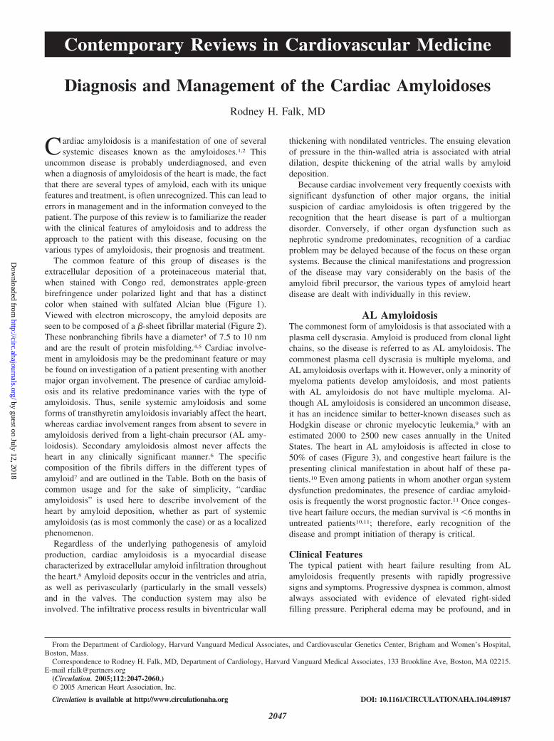
Figure 1. A, Endomyocardial biopsy specimen, stained withhematoxylin and eosin, from a patient with cardiac amyloidosis.The amyloid stains light pinkish red and is seen as an amor-phous material that separates the darker staining myocytes. B,Staining of the tissue from the same patient using sulfatedAlcian blue. The amyloid stains turquoise green and the myo-cytes stain yellow, characteristic of amyloid. Courtesy of DrGayle Winters, Brigham and Women’s Hospital. Boston, Mass.
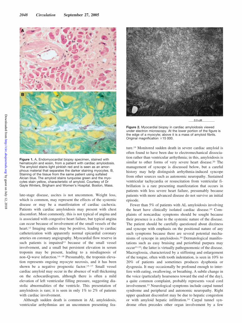
Figure 2. Myocardial biopsy in cardiac amyloidosis viewedunder electron microscopy. At the lower portion of the figure isthe edge of a myocyte; above it is a mass of amyloid fibrils.Original magnification �15 000.
2048 Circulation September 27, 2005
by guest on July 12, 2018http://circ.ahajournals.org/
Dow
nloaded from

years, and a history of surgical carpal tunnel release is notuncommon. Although widespread lymphadenopathy is pres-ent in only a small minority of patients, submandibularswelling caused by lymph node and salivary gland infiltrationis not uncommon and often is accompanied by macroglossia.Nail dystrophy (brittle and slow-growing nails) is sometimesseen, particular in the hands, and when present is a clue to thesystemic nature of the cardiac disease.25
The cardiovascular physical examination in a patient withheart failure resulting from amyloidosis usually reveals sinusrhythm with a normal to low radial pulse volume, althoughatrial arrhythmias (most commonly atrial fibrillation) occur in10% to 15% of patients. When present, atrial fibrillation isassociated with a very high incidence of thromboembolism.The jugular venous pressure is often markedly elevated, andthe waveform is generally unrevealing, but occasionally, aprominent X and Y descent is noted.26,27 In contrast toconstrictive pericarditis, with which it may be confused,Kussmaul’s sign is very rarely present. The apex beat isfrequently impalpable and, when it can be felt, is generallynot displaced. The first and second heart sounds are usuallynormal in character. A left ventricular third heart sound israrely heard, but in advanced cases, a right ventricular S3,which often is associated with right ventricular dilation anddysfunction on the echocardiogram, may be heard best at theleft parasternal edge. Despite the increased stiffness of the leftventricle, a fourth heart sound is almost never present,possibly because of atrial dysfunction resulting from amyloidinfiltration.28,29 Blood pressure is often low, even in theabsence of postural hypotension; this may represent de-creased cardiac output in conjunction with early autonomicdysfunction. Blood pressure may fall further on standing,particularly if autonomic neuropathy is present,30 and shouldbe measured in the supine, seated, and standing positions both
immediately after standing and after at least 2 minutesbecause the systolic pressure may continue to drift down inthe presence of autonomic dysfunction. Hypertension isunusual, and in patients with a history of hypertension,“spontaneous” resolution of hypertension over the precedingfew months is common. Examination of the chest may revealbilateral pleural effusions, but rales are rarely present, even inassociation with advanced heart failure. The pleural effusionsin a patient with AL amyloidosis may simply represent heartfailure, but patients with cardiac amyloid may also havepleural infiltration with amyloid, resulting in disproportion-ately large effusions that are diuretic resistant and rapidlyrecur after a pleural tap.31
Splenomegaly is rare, whereas hepatomegaly is commonand is due either to congestion from right heart failure or toamyloid infiltration.32,33 When extensive amyloid infiltrationof the liver is present, the organ is rock-hard and not tender,often extending several centimeters below the costal marginand crossing the midline. This contrasts with the firm,sometimes tender, liver of heart failure. Peripheral edemamay be profound, and if it appears disproportionate to thedegree of heart failure, the possibility of associated nephroticsyndrome should be considered. In addition to autonomicdysfunction, amyloidosis may cause a sensory neuropathy,and the patient may complain of numbness or painful extrem-ities.11 A history of weight loss is common, and proteinuria,frequently reaching nephrotic range (�3 g/24 h), coexistswith cardiac disease in 30% to 50% of cases.
Low voltage on the ECG (defined as all limb leads �5 mmin height) is found in a high proportion of patients and is oftenassociated with extreme left- or right-axis deviation (Figure4). Although voltage criteria for left ventricular hypertrophyhave been described in the precordial leads of some patientswith AL amyloidosis, increased limb lead voltage is ex-
Summary of the Main Forms of Amyloidosis That Affect the Heart
Nomenclature Precursor of Amyloid Fibril Organ Involvement Treatment Comment
AL Immunoglobulin light chain HeartKidneyLiver
Chemotherapy Plasma cell dyscrasia related to (butusually not associated with) multiple
myeloma
Peripheral/autonomic nervesSoft tissue
Gastrointestinal system
Heart disease occurs in 1/3 to 1/2 of ALpatients; heart failure tends to progressrapidly and has a very poor prognosis
ATTR (familial) Mutant transthyretin Peripheral/autonomic nerveHeart
Liver transplantation? New pharmacological
strategies to stabilize the TTR
Autosomal dominant; amyloid derivedfrom a mixture of mutant and wild-typeTTR; if present before, cardiac amyloid
may progress despite liver transplantation
AApoA1 Mutant apolipoprotein KidneyHeart
? Liver transplantation Kidney disease is the commonestpresentation; heart involvement rare
Senile systemic amyloid Wild-type transthyretin Heart Supportive? New pharmacological
strategies to stabilize the TTR.
Almost exclusively found in elderly men;slowly progressive symptoms
AA Serum amyloid A KidneyHeart (rarely)
Treat underlying inflammatoryprocess
Heart disease rare and, if present, rarelyclinically significant
AANP Atrial natriuretic peptide Localized to the atrium None required Very common; may increase risk of atrialfibrillation and/or be deposited in greater
amounts in the fibrillating atrium
Falk Cardiac Amyloidosis 2049
by guest on July 12, 2018http://circ.ahajournals.org/
Dow
nloaded from

tremely uncommon.34 When increased limb or precordial leadvoltage is present, it is frequently a result of an unrelatedcoexistent condition such as hypertension. Interestingly, rightbundle-branch block is uncommon, and left bundle-branchblock is very unusual unless it is a preexisting condition.10
The reason for the virtual absence of left bundle-branch blockin patients with AL amyloidosis is unclear. However, becauseamyloid deposition affects the heart uniformly, the morevulnerable right bundle is anticipated to be involved beforethe left bundle, so sparing of the right bundle with completeleft bundle-branch block would be very unlikely.
The echocardiographic features of advanced cardiac ALamyloidosis are distinctive. The initial descriptions concen-trated on patients with severe cardiac disease and depictednondilated ventricles with concentric left ventricular thicken-ing, right ventricular thickening, prominent valves, and infil-tration of the atrial septum. The myocardial texture wasabnormal and described as “granular sparkling.”26,35–37 Sub-sequent changes in image processing produced a myocardialappearance that is less “granular” in appearance, but ad-vanced amyloid heart disease still demonstrates an increasedechogenicity of the myocardium and often of the valves(Figure 5). The classic appearance of a restrictive pattern byDoppler echocardiography and associated increased echoge-nicity, biventricular thickening, and valvular infiltration is
limited to patients in the end stage of the disease. Morecommonly, the ventricle appears thickened to a degree that isdisproportionate to the degree of current or prior hyperten-sion, and the Doppler features depend on the stage of thedisease, with serial studies demonstrating a progression ofdiastolic dysfunction as myocardial infiltration progresses.36
The left ventricular ejection fraction is normal or nearlynormal until late in the disease, and because the left ventricledoes not dilate, a reduced ejection fraction is associated witha substantially reduced stroke volume. Because the thicken-ing of the ventricle in amyloidosis is due to myocardialinfiltration rather than hypertrophy, the ECG limb leadvoltage tends to decrease as the ventricle thickens. Thisresults in a decreased ratio of voltage to left ventricular mass,a finding that strongly suggests an infiltrative cardiomyopa-thy, of which amyloidosis is the commonest cause.38 In �5%of patients with cardiac amyloidosis, left ventricular infiltra-tion may mimic hypertrophic cardiomyopathy on the echo-cardiogram.10,39,40 These patients often have normal or evenmildly hyperdynamic left ventricular function with normalvoltage on the ECG. Associated postural hypotension iscommon in these patients, and low afterload may in partaccount for the normal to increased ejection fraction. Unliketrue hypertrophic cardiomyopathy, ventricular hypertrophyon the ECG limb leads is almost never seen and systolicanterior motion of the mitral valve is uncommon, althoughchordal anterior motion may be present with an associatedoutflow tract murmur.
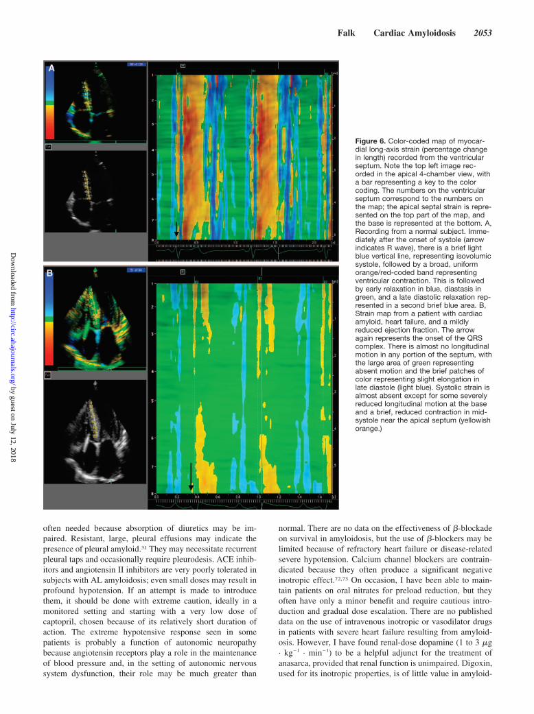
Doppler echocardiography is also useful in cardiac amy-loidosis. In advanced disease, there is a restrictive transmitralflow pattern characterized by a short deceleration time of theE wave and a low-velocity A wave, with associated abnor-malities in pulmonary venous flow.26,41,42 The decreasedtransmitral A wave in AL amyloidosis is related not only tolate-stage restrictive pathophysiology but also to atrial amy-loid infiltration, which results in intrinsic atrial dysfunc-tion28,29,43–46; thus, a normal deceleration time can be seen inassociation with a diminutive A wave. Further insights intocardiac function in AL amyloidosis can be gained by pulsedtissue Doppler imaging, which can demonstrate the presenceof diastolic dysfunction more accurately than transmitral andpulmonary flow and can provide evidence of longitudinalsystolic impairment before the ejection fraction becomesabnormal.47,48 Strain and strain rate imaging are even moresensitive than tissue Doppler, demonstrating long-axis dys-function in early cardiac amyloidosis and often showingdisproportionate impairment of longitudinal contraction de-spite apparently preserved fractional shortening (Figure 6). Inaddition to giving sensitive information about myocardialfunction, tissue Doppler and strain and strain rate imagingmay have potential for evaluating the prognosis in ALamyloidosis.47–49
Other imaging modalities such as cardiac magnetic reso-nance show promise for diagnosing cardiac amyloidosis ifechocardiographic features are suspicious.50 Recent descrip-tions of cardiac MRI in advanced cardiac amyloidosis showan unusual pattern characterized by global subendocardiallate gadolinium enhancement and associated abnormal myo-cardial and blood-pool gadolinium kinetics.50 However the
Figure 3. Autopsy specimen of a heart with extensive amyloidinfiltration. Note the nondilated ventricles with biventricularthickening and the biatrial enlargement with thickening of theatrial septum. Atrial infiltration leads to atrial failure and can beassociated with atrial thrombi.
2050 Circulation September 27, 2005
by guest on July 12, 2018http://circ.ahajournals.org/
Dow
nloaded from
sensitivity of this technique for detecting early disease is notknown, and the specificity of the described abnormalities islikely to be low in an unselected population of patients.51
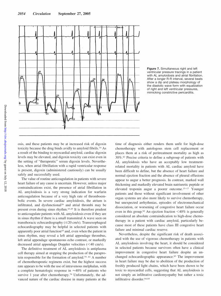
Cardiac CatheterizationThe noninvasive imaging features of amyloidosis describedabove are usually sufficient to strongly suspect the correctdiagnosis. Thus, cardiac catheterization, other than to obtainan endomyocardial biopsy, to better assess hemodynamics, orto evaluate coronary anatomy, currently is of limited value inthe routine evaluation of a patient with suspected amyloid-osis. Nevertheless, many patients with an eventual diagnosisof cardiac amyloidosis undergo cardiac catheterization duringthe workup, and if a full hemodynamic study is done, carefulexamination of the pressure tracing may provide clues to thediagnosis. Impaired ventricular filling in advanced cardiacamyloidosis is associated with an elevated left ventricularend-diastolic pressure, and the pressure tracings may reveal adip-and-plateau waveform52 (Figure 7). It has been suggestedthat, unlike constrictive pericarditis, amyloidosis is associatedwith a left ventricular end-diastolic pressure that exceedsright ventricular end-diastolic pressure by at least 7 mm Hg.52
However, this is not always the case, and both disorders maymanifest a dip-and-plateau diastolic pressure tracing withpressure equalization.53,54 A pulmonary artery systolic pres-sure �50 mm Hg is rarely seen in “uncomplicated” constric-tive pericarditis but may occur in cardiac amyloidosis,55 andthe finding of an inspiratory rise in right ventricular pressurewith an associated fall in left ventricular pressure, represent-ing ventricular interdependence, has been proposed as aspecific sign of constrictive pericarditis that distinguishes itfrom restrictive cardiomyopathy.56 However, although certainhemodynamic clues suggest one diagnosis or the other,
overlap remains, and the diagnosis should not be made onhemodynamic data alone. In suspected cases of amyloidosis,clinical examination and review of the echocardiogram aregenerally extremely valuable in favoring a diagnosis ofcardiac amyloidosis if present and should never be omitted.
Tissue DiagnosisThe diagnosis of amyloidosis requires a tissue biopsy thatdemonstrates apple-green birefringence when stained withCongo red and viewed under a polarizing microscope. Sul-fated Alcian blue is an alternative stain with a high specificityfor amyloid57 (Figure 1). It is not necessary to biopsy theheart if the echocardiographic appearances are typical forcardiac amyloidosis, providing that a histological diagnosishas been made from another tissue. Fine-needle aspiration ofthe abdominal fat is a simple procedure that is positive foramyloid deposits in �70% of patients with AL amyloid-osis.58,59 If the diagnosis is not confirmed by biopsy ofanother tissue, endomyocardial biopsy is a safe and relativelysimple procedure in skilled hands; it is virtually 100%sensitive because the amyloid is widely deposited throughoutthe heart.60,61 In patients with known amyloid deposits inother organs and a history of poorly controlled hypertension,there may be uncertainty as to whether ventricular thickeningrepresents amyloid infiltration or hypertensive heart disease.In such cases, endomyocardial biopsy may be helpful todetermine whether the heart is infiltrated with amyloid.
Once a tissue diagnosis of amyloid has been established,the confirmation that this is AL amyloid requires a search forthe presence of a plasma cell dyscrasia. Serum and urineimmunofixation should be performed rather than serum andurine electrophoresis because the amount of serum or urineparaprotein may be small and immunofixation is a much
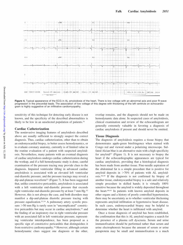
Figure 4. Typical appearance of the ECG in AL amyloidosis of the heart. There is low voltage with an abnormal axis and poor R-waveprogression in the precordial leads. The association of low voltage of this degree with thickening of the left ventricle on echocardio-gram is highly suggestive of an infiltrative cardiomyopathy.
Falk Cardiac Amyloidosis 2051
by guest on July 12, 2018http://circ.ahajournals.org/
Dow
nloaded from

more sensitive test. Even more sensitive is the recentlyintroduced serum free-light-chain assay, which can detectcirculating free light chains with �10-fold sensitivity thanimmunofixation62,63 This is a quantitative test. In AL amy-loidosis, free lambda or (less commonly) free kappa levels areelevated. The normal serum range of kappa free light chainsis 3.3 to 19.4 mg/dL; for lambda, 5.7 to 26.3 mg/dL with akappa-to-lambda ratio of 0.26 to 1.65.62,63 It is important toassess the ratio of kappa to lambda free light chains becausethey are renally excreted and renal impairment elevates kappaand lambda levels without changing the ratio. In AL amy-loidosis with renal impairment, elevated levels of both freelambda and free kappa will be seen because renal impairmentreduces light-chain excretion. However, the kappa-to-lambdaratio remains abnormal and should always be calculated inaddition to the absolute values. A kappa-to-lambda ratio�0.26 strongly suggests the presence of a population ofplasma cells producing clonal lambda free light chains,whereas a ratio �1.65 suggests production of clonal kappafree light chains. In 110 patients with AL amyloidosis, serumimmunofixation was positive in 69%, urine immunofixationwas positive in 83%, and the kappa-to-lambda ratio wasabnormal in 91%. The combination of an abnormal kappalambda ratio and a positive serum immunofixation identified99% of patients with AL amyloidosis.64 A bone marrow
biopsy is mandatory to assess the percentage of plasma cells,and immunoperoxidase staining will determine whether theabnormal plasma cells are producing kappa or lambda lightchains.65 Bone marrow biopsy is also required to excludemyeloma and other less common disorders that can beassociated with AL amyloidosis such as Waldenstrom’smacroglobulinemia. It is important to recognize that a mono-clonal band present on serum immunofixation may be seen asan apparently incidental finding in 5% to 10% of patients�70 years of age (“monoclonal gammopathy of uncertainsignificance”).66 The serum free-light-chain assay is oftennormal in such cases,67 but if any doubt exists about theclinical picture, further testing must be done to excludefamilial or senile forms of amyloid. Such testing includeseither special staining techniques of the amyloid such asimmunogold electron microscopy68–70 or genetic testing torule out familial forms of amyloid.71
ManagementManagement of cardiac amyloidosis requires a 2-fold ap-proach: management of the cardiac-related symptoms andtreatment of the underlying disease. The mainstay of thetreatment of heart failure in AL amyloidosis is the use ofdiuretics; higher doses than anticipated may be required if thealbumin level is low as a result of concomitant nephroticsyndrome. In a patient with anasarca, intravenous diuresis is
Figure 5. Selection of echocardiographic images from a patient with severe AL cardiac amyloidosis. Top left, Parasternal long-axisview showing concentric left ventricular thickening with a pericardial effusion. The echogenicity of the myocardium is increased, and thevalves are seen unusually clearly suggestive of infiltration. Top right, Apical 4-chamber view showing normal biventricular dimensionsand biatrial enlargement. The atrial septum is thickened, and a pacemaker/ICD lead is seen in the right ventricle. Bottom left, Transmi-tral Doppler flow showing a restrictive pattern with a short deceleration time and reduced A-wave velocity. Bottom right, Tissue Dopplerrecorded from the lateral mitral annulus. There is reduced myocardial velocity throughout systole and both phases of diastole, compati-ble with a restrictive pathophysiology.
2052 Circulation September 27, 2005
by guest on July 12, 2018http://circ.ahajournals.org/
Dow
nloaded from
often needed because absorption of diuretics may be im-paired. Resistant, large, pleural effusions may indicate thepresence of pleural amyloid.31 They may necessitate recurrentpleural taps and occasionally require pleurodesis. ACE inhib-itors and angiotensin II inhibitors are very poorly tolerated insubjects with AL amyloidosis; even small doses may result inprofound hypotension. If an attempt is made to introducethem, it should be done with extreme caution, ideally in amonitored setting and starting with a very low dose ofcaptopril, chosen because of its relatively short duration ofaction. The extreme hypotensive response seen in somepatients is probably a function of autonomic neuropathybecause angiotensin receptors play a role in the maintenanceof blood pressure and, in the setting of autonomic nervoussystem dysfunction, their role may be much greater than
normal. There are no data on the effectiveness of �-blockadeon survival in amyloidosis, but the use of �-blockers may belimited because of refractory heart failure or disease-relatedsevere hypotension. Calcium channel blockers are contrain-dicated because they often produce a significant negativeinotropic effect.72,73 On occasion, I have been able to main-tain patients on oral nitrates for preload reduction, but theyoften have only a minor benefit and require cautious intro-duction and gradual dose escalation. There are no publisheddata on the use of intravenous inotropic or vasodilator drugsin patients with severe heart failure resulting from amyloid-osis. However, I have found renal-dose dopamine (1 to 3 �g· kg�1 · min�1) to be a helpful adjunct for the treatment ofanasarca, provided that renal function is unimpaired. Digoxin,used for its inotropic properties, is of little value in amyloid-
Figure 6. Color-coded map of myocar-dial long-axis strain (percentage changein length) recorded from the ventricularseptum. Note the top left image rec-orded in the apical 4-chamber view, witha bar representing a key to the colorcoding. The numbers on the ventricularseptum correspond to the numbers onthe map; the apical septal strain is repre-sented on the top part of the map, andthe base is represented at the bottom. A,Recording from a normal subject. Imme-diately after the onset of systole (arrowindicates R wave), there is a brief lightblue vertical line, representing isovolumicsystole, followed by a broad, uniformorange/red-coded band representingventricular contraction. This is followedby early relaxation in blue, diastasis ingreen, and a late diastolic relaxation rep-resented in a second brief blue area. B,Strain map from a patient with cardiacamyloid, heart failure, and a mildlyreduced ejection fraction. The arrowagain represents the onset of the QRScomplex. There is almost no longitudinalmotion in any portion of the septum, withthe large area of green representingabsent motion and the brief patches ofcolor representing slight elongation inlate diastole (light blue). Systolic strain isalmost absent except for some severelyreduced longitudinal motion at the baseand a brief, reduced contraction in mid-systole near the apical septum (yellowishorange.)
Falk Cardiac Amyloidosis 2053
by guest on July 12, 2018http://circ.ahajournals.org/
Dow
nloaded from
osis, and these patients may be at increased risk of digoxintoxicity because the drug binds avidly to amyloid fibrils.74 Asa result of the binding to myocardial amyloid, cardiac digoxinlevels may be elevated, and digoxin toxicity can exist even inthe setting of “therapeutic” serum digoxin levels. Neverthe-less, when atrial fibrillation with a rapid ventricular responseis present, digoxin (administered cautiously) can be usuallysafely and successfully used.
The value of routine anticoagulation in patients with severeheart failure of any cause is uncertain. However, unless majorcontraindications exist, the presence of atrial fibrillation inAL amyloidosis is a very strong indication for warfarinanticoagulation because of a very high rate of thromboem-bolic events. In severe cardiac amyloidosis, the atrium isinfiltrated, and dysfunctional29 and atrial thrombi may bepresent even during sinus rhythm.43,45 It is therefore prudentto anticoagulate patients with AL amyloidosis even if they arein sinus rhythm if there is a small transmitral A wave seen ontransthoracic echocardiography (�20 cm/s). Transesophagealechocardiography may be helpful in selected patients withapparently poor atrial function45 and, even when the patient insinus rhythm, may reveal a left atrial appendage thrombus,left atrial appendage spontaneous echo contrast, or markedlydecreased atrial appendage Doppler velocities (�40 cm/s).
The definitive treatment of AL amyloidosis is antiplasmacell therapy aimed at stopping the production of the parapro-tein responsible for the formation of amyloid.75–81 A numberof chemotherapeutic regimens exist, but the highest successrate appears to be with the use of intravenous melphalan, witha complete hematologic response in �40% of patients whosurvive 1 year after chemotherapy.79 Unfortunately, the ad-vanced nature of the cardiac disease in many patients at the
time of diagnosis either renders them unfit for high-dosechemotherapy with autologous stem cell replacement orplaces them at a risk of peritreatment mortality as high as30%.82 Precise criteria to define a subgroup of patients withAL amyloidosis who have an acceptably low treatment-related mortality in patients with AL cardiac amyloid havebeen difficult to define, but the absence of heart failure andnormal ejection fraction and the absence of pleural effusionsappear to augur a better prognosis. In contrast, marked wallthickening and markedly elevated brain natriuretic peptide orelevated troponin augur a poorer outcome.15–17 Youngerpatients and those without significant involvement of otherorgan systems are also more likely to survive chemotherapy,but unexpected arrhythmias, episodes of electromechanicaldissociation, or worsening of congestive heart failure occureven in this group.82 An ejection fraction �40% is generallyconsidered an absolute contraindication to high-dose chemo-therapy in a patient with cardiac amyloid, particularly be-cause most of these patients have class III congestive heartfailure and minimal cardiac reserve.
Nevertheless, despite the significant risk of death associ-ated with the use of vigorous chemotherapy in patients withAL amyloidosis involving the heart, it should be consideredin selected patients because survivors often have a clinicalimprovement in congestive heart failure despite an un-changed echocardiographic appearance.83 The improvementin heart failure may be due to abolition of the production offreshly produced light chains, which have been shown to betoxic to myocardial cells, suggesting that AL amyloidosis isnot simply an infiltrative cardiomyopathy but rather a toxicinfiltrative disorder.84,85
Figure 7. Simultaneous right and leftventricular pressure tracings in a patientwith AL amyloidosis and atrial fibrillation.After a longer R-R interval, several beatsshow a dip and plateau morphology ofthe diastolic wave form with equalizationof right and left ventricular pressures,mimicking constrictive pericarditis.
2054 Circulation September 27, 2005
by guest on July 12, 2018http://circ.ahajournals.org/
Dow
nloaded from
For patients who cannot tolerate high-dose intravenousmelphalan, preliminary data from the UK amyloid groupsuggest that a modified intravenous regimen of melphalan,given monthly, may be better tolerated with a similar re-sponse rate, but no direct comparative study has been per-formed.86 The “standard regimen” of melphalan and pred-nisone given as a “pulsed” dose for 3 to 5 days every 6 weeksseems to have little benefit in patients with cardiac amyloid-osis, probably because several months are required to see aneffect.87 In addition, the steroid regimen may worsen conges-tive heart failure. Recently, we have used a low-dose “con-tinuous” melphalan regimen in patients with severe cardiacamyloid with evidence of hematologic response in 7 of 13patients.81 Unfortunately, the cardiac disease was often toosevere at the start of treatment to determine whether such aregimen has any impact on long-term survival. Regimens thatinclude the use of high-dose dexamethasone such as vincris-tine, adriamycin, and dexamethasone88 are generally nottolerated in cardiac amyloidosis because adriamycin, al-though used in relatively small doses, can produce cardiactoxicity and dexamethasone may aggravate heart failure.
In highly selected cases, cardiac transplantation may beconsidered. Early experience with cardiac transplantation inAL amyloidosis suggested that short- and medium-termmortality did not differ from that in other disorders,89 but alater report of a small series of patients treated at multipletransplantation centers demonstrated an apparently greaterlong-term mortality than expected, usually because of diseaseprogression in the heart or noncardiac organs.90,91 As a resultof these observations, many transplantation centers considerAL amyloidosis a contraindication to transplantation. How-ever, with the advent of high-dose chemotherapy and stemcell transplantation, it is possible to transplant the heart and toperform chemotherapy 6 to 12 months later to abolishamyloid production. Potential candidates for this combinedprocedure are uncommon because noncardiac organ involve-ment is a contraindication and cardiac disease is limitedclinically to the heart in �5% of cases. Nevertheless, anumber of patients have been treated successfully with thiscombined approach; several have obtained a long-term remis-sion from the disease without evidence of recurrence after 3to 5 years of follow-up.
Light-Chain CardiomyopathyRenal light-chain deposition disease is a well-recognizedentity in which renal failure may occur as a result of thedeposition of light chains either related to multiple myelomaor as a manifestation of a plasma cell dyscrasia.92 Less wellknown, and probably less common, is the cardiac manifesta-tion of light-chain deposition disease. Although not actually aform of amyloidosis, the rare condition of light-chain cardio-myopathy deserves mention because it may mimic ALamyloidosis. In this condition, nonfibrillar deposits of lightchains are found in the myocardium in association with eithermultiple myeloma or plasma cell dyscrasia.93 The echocar-diographic appearance is similar to cardiac amyloidosis, andheart failure and arrhythmias may occur, but Congo redstaining of the myocardium is negative.94 Kappa light-chaindeposition tends to be more common than lambda. The
importance of recognition of this entity relates to the occa-sional patient with evidence of a plasma cell dyscrasia and anechocardiogram suspicious of amyloidosis in whom no amy-loid is seen on endomyocardial biopsy. In such cases, electronmicroscopy with antikappa or antilambda immunogold label-ing may reveal granular deposits typical of light-chain dep-osition, thereby confirming the diagnosis.93 Chemotherapytargeted to the underlying plasma cell dyscrasia may lead toreversal of the cardiomyopathy.95
Hereditary AmyloidosisHereditary amyloidosis exists in a number of forms, but mostcases are due to the production of amyloid from a mutanttransthyretin protein.5 Transthyretin contains 125 pairs ofamino acids, and �70 mutations have been described, mostof which are amyloidogenic. The specific site of an aminoacid substitution determines the phenotype of the disease,which is transmitted as an autosomal dominant with highpenetrance. The onset occurs from the third decade on, mostcommonly after the age of 40. In some forms, peripheralneuropathy may predominate, with cardiac amyloid beingeither absent or limited to the conduction system, mostfrequently manifesting as sinus node dysfunction. Othermutations such as Thr-60-Ala (the substitution of alanine forthreonine at position 60) present with a predominant cardio-myopathy characterized by heart failure and conductionsystem disturbances with minimal neuropathy. Renal involve-ment is generally not a feature of transthyretin-associatedcardiac amyloidosis, and myocardial infiltration may be quitesevere before the onset of heart failure. This results in anechocardiographic appearance that is very similar to ad-vanced AL cardiac amyloidosis but is associated with lessheart failure and a much better long-term survival.96 Althoughstrain and strain rate imaging demonstrate subtle differencesin ventricular long-axis function between AL and familialamyloidosis,97 the difference in survival between these 2diseases is probably related to the toxic effect of light-chaindeposition on the myocardium in AL amyloidosis,84,85 whichis absent in transthyretin-related amyloidosis.
Among the familial TTR amyloidoses, the mutation char-acterized by a substitution of isoleucine for valine at position122 of the transthyretin molecule deserves special men-tion.98,99 Approximately 4% of the black population in theUnited States is heterozygous for this mutation,100 which mayresult in a late-onset cardiomyopathy in either sex, manifest-ing as progressive congestive heart failure. In our initialexperience of 12 cases101 and subsequent personal experienceof a similar number of cases, the disease is found to havefeatures that are remarkably consistent among patients. Theechocardiogram is similar to that seen in other variants ofTTR amyloidosis, with features of an infiltrative/restrictivecardiomyopathy. Signs of right-sided heart failure predomi-nate, and peripheral edema and ascites may be profound.Involvement of the cardiac valves may result in tricuspidregurgitation, which can further aggravate the right heartfailure. Because of the high prevalence of hypertensive heartdisease in blacks, left ventricular thickening seen on theechocardiogram may be mistakenly attributed tohypertension-induced hypertrophy, and the diagnosis of an
Falk Cardiac Amyloidosis 2055
by guest on July 12, 2018http://circ.ahajournals.org/
Dow
nloaded from

infiltrative cardiomyopathy can be overlooked. The presenceof right ventricular thickening, the absence of left ventricularhypertrophy on the ECG, and the clinical finding of rightheart failure in a black patient (particularly if a history ofcarpal tunnel syndrome is elicited) should strongly suggestthe diagnosis. Unlike AL amyloidosis, the abdominal fataspirate frequently stains negative for amyloid, and endomyo-cardial biopsy may be necessary unless tissue is available forstaining from prior carpal tunnel syndrome surgery. Thepenetrance of this disorder is unknown, but many other TTRmutations have a high penetrance, suggesting that the Ile 122variant is probably frequently overlooked. Regardless of thepenetrance, the high prevalence of the mutation probablymakes it the commonest familial amyloid cardiomyopathyand possibly the commonest type of amyloid heart disease.Unfortunately, the late age of onset and the universal mani-festation of heart failure preclude liver transplantation as atherapy (see below) in the vast majority of patients with thisdisorder, although we have treated 1 patient by cardiactransplantation who had excellent results over the succeedingseveral years.
Treatments for ATTR AmyloidosisAlthough transthyretin is produced by the liver, it has littleeffect on the liver function as liver deposition of amyloid isminimal or absent.102 Currently, the definitive treatment ofATTR is liver transplantation, which removes the source oftransthyretin and hence the precursor of amyloid deposi-tion.103 Optimally, liver transplantation should be performedin a patient with a known mutant transthyretin as soon asthere is clinical evidence of the disease documented by eitherdeposition of amyloid in fat pad aspirate or clinical evidenceof disease activity.104 Despite significant myocardial infiltra-tion in some patients, the clinical experience has been thatthey tolerate the surgical aspect of liver transplantation well.Because the liver is functionally normal, it has, on occasion,been removed from an amyloid patient and transplanted intoanother patient who requires an urgent liver transplant (dom-ino transplantation).105 To date, only a small number ofdomino operations have been done, and it is not known if, orwhen, the recipient will develop amyloidosis.
Initial enthusiasm for transplantation as a technique forarresting progressive cardiomyopathy has been tempered bythe observation that wall thickening progresses in somepatients who have amyloid cardiomyopathy at the time ofliver transplantation.106,107 This is most probably due to thecontinued deposition of wild-type transthyretin in the myo-cardium, a process akin to senile cardiac amyloidosis (SCA).Occasionally, combined liver and heart transplantation orheart transplantation alone has been performed for ATTRamyloid with significant cardiomyopathy.90,108
There is ongoing investigation into the development ofdrugs that will stabilize transthyretin and prevent the forma-tion of amyloid.109 In vitro evidence suggests that certainnonsteroidal agents such as diflunisal can stabilize transthy-retin.110 Although currently there is no clinical evidence thatthese agents can prevent the progression of TTR amyloidosis,clinical trials are in the planning stage. However, even ifnonsteroidal agents have some effect on disease progression,
a significant limitation in patients with cardiomyopathy is thepotential for precipitation or aggravation of congestive heartfailure. Thus, other agents without the potential for fluidretention are actively being sought.5,111
There are other, very rare, causes of familial nontransthy-retin amyloid cardiomyopathy. Mutations of the genes encod-ing apolipoprotein A may be amyloidogenic and can result anisolated cardiomyopathy that has been successfully treatedwith cardiac transplantation.112 Mutations of fibrinogen A�-chain and lysozyme can also cause amyloidosis, but dep-osition is predominantly in organs other than the heart.71
SCASCA is the predominant clinical manifestation of senilesystemic amyloidosis. It results from the cardiac deposition ofamyloid derived from wild-type transthyretin (ie, transthyre-tin with a normal amino acid constitution)113 and invariablypresents as congestive heart failure. The diagnosis requiresthe finding of amyloid deposits in the myocardium, inconjunction with evidence of an infiltrative cardiomyopathyon echocardiogram. The echocardiographic appearance isindistinguishable from that found in patients with AL amy-loidosis, although the degree of wall thickening may be verymarked despite relatively mild, or easily controllable, heartfailure.114 Once the diagnosis is suspected, confirmationusually requires an endomyocardial biopsy because noncar-diac involvement is rare. However, caution should be exer-cised in labeling an elderly patient as having senile amyloid-osis on the basis of an endomyocardial biopsy if the amyloiddeposits are sparse and the echocardiographic appearance isnot consistent with amyloidosis because small amounts ofamyloid derived from wild-type transthyretin are not uncom-mon in the very elderly.115,116 Exclusion of a plasma celldyscrasia is mandatory, and screening should be performed toexclude a mutant transthyretin. Although evaluation for aplasma cell dyscrasia is usually negative, an unrelated benignmonoclonal gammopathy of unknown significance will beexpected to be present by chance in 3% to 5% of patients ofthis age when sought by serum protein electrophoresis66,117
and even more commonly if the more sensitive immunofix-ation is used. If immunofixation is positive, but the clinicalpicture is most consistent with SCA other tests to exclude ALamyloidosis are mandatory. Immunochemistry or immuno-gold electron microscopy of biopsy tissue staining unequiv-ocally positive for TTR and negative for kappa and lambdaare confirmatory of the transthyretin origin of the amyloid.70
For unclear reasons, SCA is almost exclusively a disorderof men.114 The disease is rare individuals �70 years of age,and the median survival from the onset of heart failure is 7.5years compared with 15 months in patients with AL amyloid-osis and a similar degree of LV thickening.114 The clinicalmanifestations of senile cardiac amyloidosis are quite similaramong patients.114,118 Progression of heart failure in senileamyloidosis is insidious but inexorable, and the diagnosisshould be suspected in an elderly man with unexplainedright-sided or biventricular failure and an echocardiogramshowing left ventricular thickening with normal ventricularcavity size. The disease is not associated with any other majorclinical organ involvement, although carpal tunnel syndrome,
2056 Circulation September 27, 2005
by guest on July 12, 2018http://circ.ahajournals.org/
Dow
nloaded from

often preceding the cardiac disease by a few years, iscommon. Bifascicular block on the ECG is common, andprogression to complete AV block occurs not infrequently,necessitating permanent pacemaker implantation. Implanta-tion of a permanent pacemaker for conduction system diseasemay be followed by a worsening of heart failure; this may bedue to the dysynergy produced by right ventricular pacing inthe nondilated, infiltrated ventricle with its small cavity andreduced ejection fraction. Thus, if conduction system diseasewarrants a pacemaker, strong consideration should be givento biventricular pacing to maximize ventricular stroke vol-ume. Atrial fibrillation is a common arrhythmia in SCA,presumably because of the combination of atrial infiltrationwith amyloid, increased left atrial pressure, and the advancedage of the patient. Once atrial fibrillation occurs, thrombo-embolism is common, and warfarin anticoagulation should beprescribed unless a major contraindication exists. Restorationand maintenance of sinus rhythm by cardioversion andantiarrhythmic drugs such as amiodarone may provide somebenefit, but atrial function is usually impaired as a result ofamyloid infiltration. In addition, widespread conduction sys-tem disease may worsen in the presence of antiarrhythmicdrugs. Unlike AL amyloidosis, patients with SCA oftentolerate ACE inhibitors, although the mainstay of therapy isstill the judicious use of diuretics. There is no specifictreatment for SCA, but as in familial amyloidosis, drugs thatstabilize the transthyretin molecule hold some promise109 andare on the verge of clinical trials.
Secondary AmyloidosisSecondary amyloidosis is increasingly uncommon in thedeveloped world owing to the eradication of chronic infec-tions. However, it is still seen occasionally in association withjuvenile or adult rheumatoid arthritis and other rheumaticdisorders such as ankylosing spondylitis, as well as withinflammatory bowel disease. Hepatic and renal amyloiddeposition dominates the clinical picture, and clinical heartdisease related to cardiac amyloid is very rare.6 In the fewcases in which there is echocardiographic evidence of cardiacamyloidosis resulting from secondary amyloidosis, cardiacsymptoms are usually absent, although we have seen veryoccasional cases of mild heart failure with extensive leftventricular thickening, which was associated with suddendeath in 1 case.
Isolated Atrial AmyloidosisAtrial amyloid deposition is a common finding at autopsy,particularly in elderly patients.115,119 Immunohistochemicalevaluation demonstrates its origin from atrial natriureticpeptide.120 Unlike the other forms of amyloid discussed, atrialamyloid is a nonsystemic deposition, limited to the atrium.Until recently, it was believed to be a clinically insignificantfinding that increased in prevalence with increasing age andwith the presence of organic heart disease. Recent data basedon atrial biopsies taken at the time of cardiac surgery suggestthat isolated atrial amyloidosis may be commoner in womenand is more likely to occur in the presence of atrial fibrilla-
Figure 8. Flow diagram outlining the evaluation of a patient with suspected cardiac amyloidosis. Clinical evaluation may reveal clues thatstrengthen the likelihood of amyloidosis, but a tissue diagnosis is mandatory. Although special staining of the biopsy may confirm the type ofamyloid, further workup of AL amyloid is required to exclude myeloma and to quantify free light chains. If the biopsy stains positive for transt-hyretin, further testing is needed to determine whether this is a wild-type or mutant transthyretin. ApoA1 indicates apolipoprotein A1; IFE,immunofixation electrophoresis; FLC, free-light-chain assay; SSA, senile systemic amyloidosis; and TTR, transthyretin.
Falk Cardiac Amyloidosis 2057
by guest on July 12, 2018http://circ.ahajournals.org/
Dow
nloaded from
tion.121,122 Interestingly, an inverse relationship with thepresence of atrial fibrosis has been suggested.121,123 The roleof isolated atrial amyloid in the pathogenesis and mainte-nance of atrial fibrillation remains to be fully elucidated, butit may be a precipitating factor of atrial fibrillation in somepatients and may be produced as part of the remodelingassociated with this arrhythmia.121
ConclusionsIn summary, cardiac amyloidosis, although uncommon, ischaracterized by a typical appearance on echocardiography,the recognition of which should alert the astute clinician tothe probable diagnosis. It is critical to recognize that severalforms of amyloidosis may cause cardiomyopathy and thattreatment and prognosis of these individual cardiomyopathiesdiffer greatly from each other. A flow diagram to aid in thediagnosis of the type of amyloid is illustrated in Figure 8. InAL amyloidosis, chemotherapy may arrest or possibly reversethe disease, with resultant stabilization or improvement ofsymptoms. Thus, early diagnosis is critical because patientswith advanced disease are usually too ill for intensivechemotherapy. Recognition of non-AL cardiac amyloidosis isimportant to avoid unnecessary chemotherapy, to screenfamily members, and in the near future, to provide newmedications that will stabilize the amyloidogenic substance inthe blood and prevent the onset of progression of thisimportant and underdiagnosed condition.
References1. Merlini G, Bellotti V. Molecular mechanisms of amyloidosis. N Engl
J Med. 2003;349:583–596.2. Falk RH, Comenzo RL, Skinner M. The systemic amyloidoses. N Engl
J Med. 1997;337:898–909.3. Sipe JD, Cohen AS. History of the amyloid fibril. J Struct Biol. 2000;
130:88–98.4. Selkoe DJ. Folding proteins in fatal ways. Nature. 2003;426:900–904.5. Merlini G, Westermark P. The systemic amyloidoses: clearer under-
standing of the molecular mechanisms offers hope for more effectivetherapies. J Intern Med. 2004;255:159–178.
6. Dubrey SW, Cha K, Simms RW, Skinner M, Falk RH. Electrocardiog-raphy and Doppler echocardiography in secondary (AA) amyloidosis.Am J Cardiol. 1996;77:313–315.
7. Westermark P, Benson MD, Buxbaum JM, Cohen AS, Frangione B,Ikeda S, Masters CL, Merlini G, Saraiva MJ, Sipe J. Amyloid proteinfibril nomenclature–2002. Amyloid. 2002;9:197–200.
8. Smith TJ, Kyle RA, Lie JT. Clinical significance of histopathologicpatterns of cardiac amyloidosis. Mayo Clin Proc. 1984;59:547–555.
9. Gertz MA, Lacy MQ, Dispenzieri A. Amyloidosis hematology. OncolClin North Am. 1999;13:1211–1220.
10. Dubrey SW, Cha K, Anderson J, Chamarthi B, Reisinger J, Skinner M,Falk RH. The clinical features of immunoglobulin light-chain (AL)amyloidosis with heart involvement. QJM. 1998;91:141–157.
11. Kyle RA, Gertz MA. Primary systemic amyloidosis: clinical and labo-ratory features in 474 cases. Semin Hematol. 1995;32:45–59.
12. Mueller PS, Edwards WD, Gertz MA. Symptomatic ischemic heartdisease resulting from obstructive intramural coronary amyloidosis.Am J Med. 2000;109:181–188.
13. Miani D, Rocco M, Alberti E, Spedicato L, Fioretti PM. Amyloidosis ofepicardial and intramural coronary arteries as an unusual cause of myocardialinfarction and refractory angina pectoris. Ital Heart J. 2002;3:479–482.
14. Zabernigg A, Schranzhofer R, Kreczy A, Gattringer K. Continuouslyelevated cardiac troponin I in two patients with multiple myeloma andfatal cardiac amyloidosis. Ann Oncol. 2003;14:1791.
15. Dispenzieri A, Kyle RA, Gertz MA, Therneau TM, Miller WL,Chandrasekaran K, McConnell JP, Burritt MF, Jaffe AS. Survival inpatients with primary systemic amyloidosis and raised serum cardiactroponins. Lancet. 2003;361:1787–1789.
16. Dispenzieri A, Gertz MA, Kyle RA, Lacy MQ, Burritt MF, TherneauTM, McConnell JP, Litzow MR, Gastineau DA, Tefferi A, Inwards DJ,Micallef IN, Ansell SM, Porrata LF, Elliott MA, Hogan WJ, RajkumarSV, Fonseca R, Greipp PR, Witzig TE, Lust JA, Zeldenrust SR, SnowDS, Hayman SR, McGregor CG, Jaffe AS. Prognostication of survivalusing cardiac troponins and N-terminal pro-brain natriuretic peptide inpatients with primary systemic amyloidosis undergoing peripheral bloodstem cell transplantation. Blood. 2004;104:1881–1887.
17. Dispenzieri A, Gertz MA, Kyle RA, Lacy MQ, Burritt MF, TherneauTM, Greipp PR, Witzig TE, Lust JA, Rajkumar SV, Fonseca R, ZeldenrustSR, McGregor CG, Jaffe AS. Serum cardiac troponins and N-terminalpro-brain natriuretic peptide: a staging system for primary systemic amy-loidosis. J Clin Oncol. 2004;22:3751–3757.
18. Cantwell RV, Aviles RJ, Bjornsson J, Wright RS, Freeman WK, Oh JK, HoyerJD, Markovic S, Jaffe AS. Cardiac amyloidosis presenting with elevations ofcardiac troponin I and angina pectoris. Clin Cardiol. 2002;25:33–37.
19. Falk RH, Rubinow A, Cohen AS. Cardiac arrhythmias in systemicamyloidosis: correlation with echocardiographic abnormalities. J AmColl Cardiol. 1984;3:107–113.
20. Chamarthi B, Dubrey SW, Cha K, Skinner M, Falk RH. Features andprognosis of exertional syncope in light-chain associated AL cardiacamyloidosis. Am J Cardiol. 1997;80:1242–1245.
21. Rubinow A, Cohen AS. Skin involvement in generalized amyloidosis: astudy of clinically involved and uninvolved skin in 50 patients withprimary and secondary amyloidosis. Ann Intern Med. 1978;88:781–785.
22. Daoud MS, Lust JA, Kyle RA, Pittelkow MR. Monoclonal gam-mopathies and associated skin disorders. J Am Acad Dermatol. 1999;40:507–535; quiz 536–538.
23. Burroughs EI, Aronson AE, Duffy JR, Kyle RA. Speech disorders insystemic amyloidosis. Br J Disord Commun. 1991;26:201–206.
24. Park MA, Mueller PS, Kyle RA, Larson DR, Plevak MF, Gertz MA.Primary (AL) hepatic amyloidosis: clinical features and natural historyin 98 patients. Medicine. 2003;82:291–298.
25. Derrick EK, Price ML. Primary systemic amyloid with nail dystrophy.J Royal Soc Med. 1995;88:290P–291P.
26. Klein AL, Cohen GI. Doppler echocardiographic assessment of con-strictive pericarditis, cardiac amyloidosis, and cardiac tamponade.Cleveland Clin J Med. 1992;59:278–290.
27. Tyberg TI, Goodyer AV, Hurst VWD, Alexander J, Langou RA. Leftventricular filling in differentiating restrictive amyloid cardiomyopathyand constrictive pericarditis. Am J Cardiol. 1981;47:791–796.
28. Murphy L, Falk RH. Left atrial kinetic energy in AL amyloidosis: can itdetect early dysfunction? Am J Cardiol. 2000;86:244–246.
29. Modesto KM, Dispenzieri A, Cauduro AS, Lacy MQ, Khandheria BK,Pellika PA, Belohlavek M, Seward JB, Kyle RA, Tajik AJ, Gertz MA,Abraham TP. Left atrial myopathy in cardiac amyloidosis: implicationsof novel echocardiographic techniques. Eur Heart J. 2005;26:173–179.
30. Bernardi L, Passino C, Porta C, Anesi E, Palladini G, Merlini G.Widespread cardiovascular autonomic dysfunction in primary amy-loidosis: does spontaneous hyperventilation have a compensatory roleagainst postural hypotension? Heart. 2002;88:615–621.
31. Berk JL, Keane J, Seldin DC, Sanchorawala V, Koyama J, Dember LM,Falk RH. Persistent pleural effusions in primary systemic amyloidosis:etiology and prognosis. Chest. 2003;124:969–977.
32. Gillmore JD, Lovat LB, Hawkins PN. Amyloidosis and the liver.J Hepatol. 1999;30(suppl 1):17–33.
33. Chopra S, Rubinow A, Koff RS, Cohen AS. Hepatic amyloidosis: ahistopathologic analysis of primary (AL) and secondary (AA) forms.Am J Pathol. 1984;115:186–193.
34. Murtagh B, Hammill SC, Gertz MA, Kyle RA, Tajik AJ, Grogan M.Electrocardiographic findings in primary systemic amyloidosis andbiopsy-proven cardiac involvement. Am J Cardiol. 2005;95:535–537.
35. Siqueira-Filho AG, Cunha CL, Tajik AJ, Seward JB, Schattenberg TT,Giuliani ER. M-mode and two-dimensional echocardiographic featuresin cardiac amyloidosis. Circulation. 1981;63:188–196.
36. Klein AL, Hatle LK, Taliercio CP, Taylor CL, Kyle RA, Bailey KR,Seward JB, Tajik AJ. Serial Doppler echocardiographic follow-up of leftventricular diastolic function in cardiac amyloidosis. J Am Coll Cardiol.1990;16:1135–1141.
37. Child JS, Levisman JA, Abbasi AS, MacAlpin RN. Echocardiographicmanifestations of infiltrative cardiomyopathy: a report of seven casesdue to amyloid. Chest. 1976;70:726–731.
38. Carroll JD, Gaasch WH, McAdam KP. Amyloid cardiomyopathy: character-ization by a distinctive voltage/mass relation. Am J Cardiol. 1982;49:9–13.
2058 Circulation September 27, 2005
by guest on July 12, 2018http://circ.ahajournals.org/
Dow
nloaded from
39. Sedlis SP, Saffitz JE, Schwob VS, Jaffe AS. Cardiac amyloidosis sim-ulating hypertrophic cardiomyopathy. Am J Cardiol. 1984;53:969–970.
40. Hemmingson LO, Eriksson P. Cardiac amyloidosis mimicking hyper-trophic cardiomyopathy. Acta Med Scand. 1986;219:421–423.
41. Klein AL, Hatle LK, Burstow DJ, Seward JB, Kyle RA, Bailey KR,Luscher TF, Gertz MA, Tajik AJ. Doppler characterization of leftventricular diastolic function in cardiac amyloidosis. J Am Coll Cardiol.1989;13:1017–1026.
42. Abdalla I, Murray RD, Lee JC, Stewart WJ, Tajik AJ, Klein AL.Duration of pulmonary venous atrial reversal flow velocity and mitralinflow a wave: new measure of severity of cardiac amyloidosis. J AmSoc Echocardiogr. 1998;11:1125–1133.
43. Dubrey S, Pollak A, Skinner M, Falk RH. Atrial thrombi occurringduring sinus rhythm in cardiac amyloidosis: evidence for atrial electro-mechanical dissociation. Br Heart J. 1995;74:541–544.
44. Plehn JF, Southworth J, Cornwell GG. Brief report: atrial systolic failurein primary amyloidosis. N Engl J Med. 1992;327:1570–1573.
45. Santarone M, Corrado G, Tagliagambe LM, Manzillo GF, Tadeo G,Spata M, Longhi M. Atrial thrombosis in cardiac amyloidosis: diagnos-tic contribution of transesophageal echocardiography. J Am Soc Echo-cardiogr. 1999;12:533–536.
46. Moyssakis I, Triposkiadis F, Pantazopoulos NJ, Kyriakidis M,Nihoyannopoulos P. Left atrial systolic function in primary and familialamyloidosis: assessment from left atrial volume change. Clin Cardiol.2004;27:528–532.
47. Koyama J, Ray-Sequin PA, Davidoff R, Falk RH. Usefulness of pulsedtissue Doppler imaging for evaluating systolic and diastolic left ventric-ular function in patients with AL (primary) amyloidosis. Am J Cardiol.2002;89:1067–1071.
48. Koyama J, Davidoff R, Falk RH. Longitudinal myocardial velocitygradient derived from pulsed Doppler tissue imaging in AL amyloidosis:a sensitive indicator of systolic and diastolic dysfunction. J Am SocEchocardiogr. 2004;17:36–44.
49. Koyama J, Ray-Sequin PA, Falk RH. Longitudinal myocardial functionassessed by tissue velocity, strain, and strain rate tissue Doppler echo-cardiography in patients with AL (primary) cardiac amyloidosis. Circu-lation. 2003;107:2446–2452.
50. Maceira AM, Joshi J, Prasad SK, Moon JC, Perugini E, Harding I, SheppardMN, Poole-Wilson PA, Hawkins PN, Pennell DJ. Cardiovascular magneticresonance in cardiac amyloidosis. Circulation. 2005;111:186–193.
51. Kwong RY, Falk RH. Cardiovascular magnetic resonance in cardiacamyloidosis. Circulation. 2005;111:122–124.
52. Swanton RH, Brooksby IA, Davies MJ, Coltart DJ, Jenkins BS, Webb-Peploe MM. Systolic and diastolic ventricular function in cardiac amy-loidosis: studies in six cases diagnosed with endomyocardial biopsy.Am J Cardiol. 1977;39:658–664.
53. Kern MJ, Lorell BH, Grossman W. Cardiac amyloidosis masqueradingas constrictive pericarditis. Cathet Cardiovasc Diagn. 1982;8:629–635.
54. Robbins MA, Pizzarello RA, Stechel RP, Chiaramida SA, Gulotta SJ.Resting and exercise hemodynamics in constrictive pericarditis and acase of cardiac amyloidosis mimicking constriction. Cathet CardiovascDiagn. 1983;9:463–471.
55. Lorell BH, Grossman W. Profiles in constrictive pericarditis, restrictive cardio-myopathy and cardiac tamponade. In: Baim DS, Grossman W, eds. Grossman’sCardiac Catheterization, Angiography and Intervention. 6th ed. Philadelphia,Pa: Lippincott, Williams & Wilkins; 2000:829–850.
56. Hurrell DG, Nishimura RA, Higano ST, Appleton CP, Danielson GK,Holmes DR Jr, Tajik AJ. Value of dynamic respiratory changes in leftand right ventricular pressures for the diagnosis of constrictive peri-carditis. Circulation. 1996;93:2007–2013.
57. Pomerance A, Slavin G, McWatt J. Experience with the sodiumsulphate-Alcian blue stain for amyloid in cardiac pathology. J ClinPathol. 1976;29:22–26.
58. Libbey CA, Skinner M, Cohen AS. Use of abdominal fat tissue aspirate in thediagnosis of systemic amyloidosis. Arch Intern Med. 1983;143:1549–1552.
59. Ansari-Lari MA, Ali SZ. Fine-needle aspiration of abdominal fat pad foramyloid detection: a clinically useful test? Diagn Cytopathol. 2004;30:178–181.
60. Ardehali H, Qasim A, Cappola T, Howard D, Hruban R, Hare JM,Baughman KL, Kasper EK. Endomyocardial biopsy plays a role indiagnosing patients with unexplained cardiomyopathy. Am Heart J.2004;147:919–923.
61. Gertz MA, Grogan M, Kyle RA, Tajik AJ. Endomyocardial biopsy-proven light chain amyloidosis (AL) without echocardiographic featuresof infiltrative cardiomyopathy. Am J Cardiol. 1997;80:93–95.
62. Abraham RS, Katzmann JA, Clark RJ, Bradwell AR, Kyle RA, GertzMA. Quantitative analysis of serum free light chains: a new marker forthe diagnostic evaluation of primary systemic amyloidosis. Am J ClinPathol. 2003;119:274–278.
63. Katzmann JA, Clark RJ, Roshini S, Abraham RS, Bryant S, Lymp JF,Bradwell AR, Kyle RA. Serum reference intervals and diagnostic rangesfor free and free immunoglobulin light chains: relative sensitivity fordetection of monoclonal light chains. Clin Chem. 2002;48:1437–1444.
64. Katzmann JA, Abraham RS, Dispenzieri A, Lust JA, Kyle RA. Diag-nostic performance of quantitative kappa and lambda free light chainassays in clinical practice. Clin Chem. 2005;51:878–881.
65. Swan N, Skinner M, O’Hara CJ. Bone marrow core biopsy specimens inAL (primary) amyloidosis: a morphologic and immunohistochemicalstudy of 100 cases. Am J Clin Pathol. 2003;120:610–616.
66. Kyle RA, Therneau TM, Rajkumar SV, Larson DR, Plevak MF, MeltonLJ. Long-term follow-up of 241 patients with monoclonal gammopathyof undetermined significance: the original Mayo Clinic series 25 yearslater. Mayo Clin Proc. 2004;79:859–866.
67. Rajkumar SV, Kyle RA, Therneau TM, Clark RJ, Bradwell AR, Melton LJ,Larson DR, Plevak MF, Katzmann JA. Presence of monoclonal free lightchains in the serum predicts risk of progression in monoclonal gammopathy ofundetermined significance. Br J Haematol. 2004;127:308–310.
68. Anesi E, Palladini G, Perfetti V, Arbustini E, Obici L, Merlini G.Therapeutic advances demand accurate typing of amyloid deposits.Am J Med. 2001;111:243–244.
69. Arbustini E, Verga L, Concardi M, Palladini G, Obici L, Merlini G.Electron and immuno-electron microscopy of abdominal fat identifiesand characterizes amyloid fibrils in suspected cardiac amyloidosis.Amyloid. 2002;9:108–114.
70. O’Hara CJ, Falk RH. The diagnosis and typing of cardiac amyloidosis.Amyloid. 2003;10:127–129.
71. Lachmann HJ, Booth DR, Booth SE, Bybee A, Gilbertson JA, GillmoreJD, Pepys MB, Hawkins PN. Misdiagnosis of hereditary amyloidosis asAL (primary) amyloidosis. N Engl J Med. 2002;346:1786–1791.
72. Gertz MA, Falk RH, Skinner M, Cohen AS, Kyle RA. Worsening ofcongestive heart failure in amyloid heart disease treated by calciumchannel–blocking agents. Am J Cardiol. 1985;55:1645.
73. Pollak A, Falk RH. Left ventricular systolic dysfunction precipitated byverapamil in cardiac amyloidosis. Chest. 1993;104:618–620.
74. Rubinow A, Skinner M, Cohen AS. Digoxin sensitivity in amyloidcardiomyopathy. Circulation. 1981;63:1285–1288.
75. De Lorenzi E, Giorgetti S, Grossi S, Merlini G, Caccialanza G, Bellotti V.Pharmaceutical strategies against amyloidosis: old and new drugs in targeting a“protein misfolding disease.” Curr Med Chem. 2004;11:1065–1084.
76. Sanchorawala V, Wright DG, Seldin DC, Falk RH, Finn KT, DemberLM, Berk JL, Quillen K, Anderson JJ, Comenzo RL, Skinner M.High-dose intravenous melphalan and autologous stem cell transplan-tation as initial therapy or following two cycles of oral chemotherapy forthe treatment of AL amyloidosis: results of a prospective randomizedtrial. Bone Marrow Transplant. 2004;33:381–388.
77. Gertz MA, Lacy MQ, Dispenzieri A. Therapy for immunoglobulin lightchain amyloidosis: the new and the old. Blood Reviews. 2004;18:17–37.
78. Palladini G, Perfetti V, Obici L, Caccialanza R, Semino A, Adami F,Cavallero G, Rustichelli R, Virga G, Merlini G. Association of mel-phalan and high-dose dexamethasone is effective and well tolerated inpatients with AL (primary) amyloidosis who are ineligible for stem celltransplantation. Blood. 2004;103:2936–2938.
79. Skinner M, Sanchorawala V, Seldin DC, Dember LM, Falk RH, BerkJL, Anderson JJ, O’Hara C, Finn KT, Libbey CA, Wiesman J, QuillenK, Swan N, Wright DG. High-dose melphalan and autologous stem-celltransplantation in patients with AL amyloidosis: an 8-year study. AnnIntern Med. 2004;140:85–93.
80. Seldin DC, Choufani EB, Dember LM, Wiesman JF, Berk JL, Falk RH,O’Hara C, Fennessey S, Finn KT, Wright DG, Skinner M, Sanchorawala V.Tolerability and efficacy of thalidomide for the treatment of patients withlight chain-associated (AL) amyloidosis. Clin Lymphoma. 2003;3:241–246.
81. Sanchorawala V, Wright DG, Seldin DC, Falk RH, Berk JL, DemberLM, Finn KT, Skinner M. Low-dose continuous oral melphalan for thetreatment of primary systemic (AL) amyloidosis. Br J Haematol. 2002;117:886–889.
82. Falk RH, Reisinger J, Dubrey SW, Mendes LA, Sanchorawala V, EkeryD, Comenzo R, Vosburgh E, Skinner M. The effect of cardiacinvolvement on the outcome of intravenous melphalan therapy andautologous stem cell rescue for AL amyloidosis. In: Kyle RA, Gertz
Falk Cardiac Amyloidosis 2059
by guest on July 12, 2018http://circ.ahajournals.org/
Dow
nloaded from

MA, eds. Amyloid and the Amyloidoses: VIIIIth International Sym-posium on Amyloidosis. Rochester, Minn: Parthenon; 1998:181–183.
83. Dubrey S, Mendes L, Skinner M, Falk RH. Resolution of heart failure inpatients with AL amyloidosis. Ann Intern Med. 1996;125:481–484.
84. Liao R, Jain M, Teller P, Connors LH, Ngoy S, Skinner M, Falk RH,Apstein CS. Infusion of light chains from patients with cardiac amy-loidosis causes diastolic dysfunction in isolated mouse hearts. Circu-lation. 2001;104:1594–1597.
85. Brenner DA, Jain M, Pimentel DR, Wang B, Connors LH, Skinner M,Apstein CS, Liao R. Human amyloidogenic light chains directly impaircardiomyocyte function through an increase in cellular oxidant stress.Circ Res. 2004;94:1008–1010.
86. Lachmann HJ, Gallimore R, Gillmore JD, Carr-Smith HD, BradwellAR, Pepys MB, Hawkins PN. Outcome in systemic AL amyloidosis inrelation to changes in concentration of circulating free immunoglobulinlight chains following chemotherapy. Br J Haematol. 2003;122:78–84.
87. Skinner M, Anderson J, Simms R, Falk R, Wang M, Libbey C, JonesLA, Cohen AS. Treatment of 100 patients with primary amyloidosis: arandomized trial of melphalan, prednisone, and colchicine versus col-chicine only. Am J Med. 1996;100:290–298.
88. Wardley AM, Jayson GC, Goldsmith DJ, Venning MC, Ackrill P,Scarffe JH. The treatment of nephrotic syndrome caused by primary(light chain) amyloid with vincristine, doxorubicin and dexamethasone.Br J Cancer. 1998;78:774–776.
89. Hosenpud JD, Uretsky BF, Griffith BP, O’Connell JB, Olivari MT,Valantine HA. Successful intermediate-term outcome for patients withcardiac amyloidosis undergoing heart transplantation: results of a mul-ticenter survey. J Heart Transplant. 1990;9:346–350.
90. Dubrey SW, Burke MM, Hawkins PN, Banner NR. Cardiac transplan-tation for amyloid heart disease: the United Kingdom experience.J Heart Lung Transplant. 2004;23:1142–1153.
91. Hosenpud JD, DeMarco T, Frazier OH, Griffith BP, Uretsky BF, MenkisAH, O’Connell JB, Olivari MT, Valantine HA. Progression of systemicdisease and reduced long-term survival in patients with cardiac amy-loidosis undergoing heart transplantation: follow-up results of a multi-center survey. Circulation. 1991;84:III-338–II-43.
92. Pozzi C, D’Amico M, Fogazzi GB, Curioni S, Ferrario F, Pasquali S,Quattrocchio G, Rollino C, Segagni S, Locatelli F. Light chain depo-sition disease with renal involvement: clinical characteristics and prog-nostic factors. Am J Kidney Dis. 2003;42:1154–1163.
93. Gallo G, Goni F, Boctor F, Vidal R, Kumar A, Stevens FJ, Frangione B,Ghiso J. Light chain cardiomyopathy: structural analysis of the lightchain tissue deposits. Am J Pathol. 1996;148:1397–1406.
94. Buxbaum JN, Genega EN, Lazowski P, Kumar A, Tunick PA, KronzonI, Gallo GA. Infiltrative nonamyloidotic monoclonal immunoglobulinlight chain cardiomyopathy: an underappreciated manifestation ofplasma cell dyscrasias. Cardiology. 2000;93:220–228.
95. Nakamura M, Satoh M, Kowada S, Satoh H., Tashiro A, Sato F,Masuda, Hiramori K. Reversible restrictive cardiomyopathy due tolight-chain deposition disease. Mayo Clin Proc. 2002;77:193–196.
96. Dubrey SW, Cha K, Skinner M, LaValley M, Falk RH. Familial andprimary (AL) cardiac amyloidosis: echocardiographically similar diseaseswith distinctly different clinical outcomes. Heart. 1997;78:74–82.
97. Ogiwara F, Koyama J, Ikeda S, Kinoshita O, Falk RH. Comparison of thestrain Doppler echocardiographic features of familial amyloid polyneuro-pathy (FAP) and light-chain amyloidosis. Am J Cardiol. 2005;95:538–540.
98. Falk RH. The neglected entity of familial cardiac amyloidosis in AfricanAmericans. Ethnicity Dis. 2002;12:141–143.
99. Jacobson DR, Pastore RD, Yaghoubian R, Kane I, Gallo G, Buck FS,Buxbaum JN. Variant-sequence transthyretin (isoleucine 122) inlate-onset cardiac amyloidosis in black Americans. N Engl J Med.1997;336:466–473.
100. Jacobson DR, Pastore R, Pool S, Malendowicz S, Kane I, Shivji A,Embury SH, Ballas SK, Buxbaum JN. Revised transthyretin Ile 122allele frequency in African-Americans. Hum Genet. 1996;98:236–238.
101. Berg A, Falk RH, Connors LH, Theberge R, Skare J, Murakami A,Skinner M. Transthyretin Ile-122 in a series of black patients withamyloidosis. In: Kyle RA, Gertz MA, eds. Amyloid and the Amy-
loidoses: VIIIIth International Symposium on Amyloidosis. Rochester,Minn: Parthenon Publishing Group; 1998.
102. Hamilton JA, Benson MD. Transthyretin: a review from a structuralperspective. Cell Mol Life Sci. 2001;58:1491–1521.
103. Suhr OB, Herlenius G, Friman S, Ericzon BG. Liver transplantation forhereditary transthyretin amyloidosis. Liver Transplant. 2000;6:263–276.
104. Jonsen E, Suhr OB, Tashima K, Athlin E. Early liver transplantation isessential for familial amyloidotic polyneuropathy patients’ quality oflife. Amyloid. 2001;8:52–57.
105. Monteiro E, Perdigoto R, Furtado AL. Liver transplantation for familialamyloid polyneuropathy. Hepato-Gastroenterol. 1998;45:1375–1380.
106. Dubrey SW, Davidoff R, Skinner M, Bergethon P, Lewis D, Falk RH.Progression of ventricular wall thickening after liver transplantation forfamilial amyloidosis. Transplantation. 1997;64:74–80.
107. Stangou AJ, Hawkins PN, Heaton ND, Rela M, Monaghan M,Nihoyannopoulos P, O’Grady J, Pepys MB, Williams R. Progressivecardiac amyloidosis following liver transplantation for familial amyloidpolyneuropathy: implications for amyloid fibrillogenesis. Transplan-tation. 1998;66:229–233.
108. Ruygrok PN, Gane EJ, McCall JL, Chen XZ, Haydock DA, Munn SR.Combined heart and liver transplantation for familial amyloidosis. InternMed J. 2001;31:66–67.
109. Kelly JW. Attacking amyloid. N Engl J Med. 2005;352:722–723.110. Miller SR, Sekijima Y, Kelly JW. Native state stabilization by NSAIDs
inhibits transthyretin amyloidogenesis from the most common familialdisease variants. Lab Invest. 2004;84:545–552.
111. Lachmann HJ, Hawkins PN. Novel pharmacological strategies in amy-loidosis. Nephron Clin Pract. 2003;94:c85–c88.
112. Obici L, Bellotti V, Mangione P, Stoppini M, Arbustini E, Verga L,Zorzoli I, Anesi E, Zanotti G, Campana C, Vigano M, Merlini G. Thenew apolipoprotein A-I variant leu(174)3Ser causes hereditary cardiacamyloidosis, and the amyloid fibrils are constituted by the 93-residueN-terminal polypeptide. Am J Pathol. 1999;155:695–702.
113. Westermark P, Sletten K, Johansson B, Cornwell GG. Fibril in senilesystemic amyloidosis is derived from normal transthyretin. Proc NatlAcad Sci U S A. 1990;87:2843–2845.
114. Ng B, Connors LH, Davidoff R, Skinner M, Falk RH. Senile systemicamyloidosis presenting with heart failure: a comparison with light chain–associated (AL) amyloidosis. Arch Intern Med. 2005;165:1425–1429.
115. Cornwell GG, Murdoch WL, Kyle RA, Westermark P, Pitkanen P.Frequency and distribution of senile cardiovascular amyloid: a clinico-pathologic correlation. Am J Med. 1983;75:618–623.
116. Cornwell GGd, Westermark P. Senile amyloidosis: a protean manifes-tation of the aging process. J Clin Pathol. 1980;33:1146–1152.
117. Anagnostopoulos A, Evangelopoulou A, Sotou D, Gika D, MitsibounasD, Dimopoulos MA. Incidence and evolution of monoclonal gam-mopathy of undetermined significance (MGUS) in Greece. AnnHematol. 2002;81:357–361.
118. Kyle RA, Spittell PC, Gertz MA, Li CY, Edwards WD, Olson LJ,Thibodeau SN. The premortem recognition of systemic senile amy-loidosis with cardiac involvement. Am J Med. 1996;101:395–400.
119. Wright JR, Calkins E. Amyloid in the aged heart: frequency and clinicalsignificance. J Am Geriatr Socy. 1975;23:97–103.
120. Pucci A, Wharton J, Arbustini E, Grasso M, Diegoli M, Needleman P,Vigano M, Polak JM. Atrial amyloid deposits in the failing human heartdisplay both atrial and brain natriuretic peptide-like immunoreactivity.J Pathol. 1991;165:235–241.
121. Goette A, Rocken C. Atrial amyloidosis and atrial fibrillation: a gender-dependent “arrhythmogenic substrate”? Eur Heart J. 2004;25:1185–1186.
122. Leone O, Boriani G, Chiappini B, Pacini D, Cenacchi G, Martin SuarezS, Rapezzi C, Bacchi Reggiani ML, Marinelli G. Amyloid deposition asa cause of atrial remodelling in persistent valvular atrial fibrillation. EurHeart J. 2004;25:1237–1241.
123. Rocken C, Peters B, Juenemann G, Saeger W, Klein HU, Huth C,Roessner A, Goette A. Atrial amyloidosis: an arrhythmogenic substratefor persistent atrial fibrillation. Circulation. 2002;106:2091–2097.
KEY WORDS: amyloid � cardiomyopathy � echocardiography � heartdiseases � heart failure
2060 Circulation September 27, 2005
by guest on July 12, 2018http://circ.ahajournals.org/
Dow
nloaded from
Rodney H. FalkDiagnosis and Management of the Cardiac Amyloidoses
Print ISSN: 0009-7322. Online ISSN: 1524-4539 Copyright © 2005 American Heart Association, Inc. All rights reserved.
is published by the American Heart Association, 7272 Greenville Avenue, Dallas, TX 75231Circulation doi: 10.1161/CIRCULATIONAHA.104.489187
2005;112:2047-2060Circulation.
http://circ.ahajournals.org/content/112/13/2047World Wide Web at:
The online version of this article, along with updated information and services, is located on the
http://circ.ahajournals.org//subscriptions/
is online at: Circulation Information about subscribing to Subscriptions:
http://www.lww.com/reprints Information about reprints can be found online at: Reprints:
document. Permissions and Rights Question and Answer this process is available in the
click Request Permissions in the middle column of the Web page under Services. Further information aboutOffice. Once the online version of the published article for which permission is being requested is located,
can be obtained via RightsLink, a service of the Copyright Clearance Center, not the EditorialCirculationin Requests for permissions to reproduce figures, tables, or portions of articles originally publishedPermissions:
by guest on July 12, 2018http://circ.ahajournals.org/
Dow
nloaded from