conséquences de la dérégulation de met sur le phénotype
TRANSCRIPT

HAL Id: tel-01862741https://tel.archives-ouvertes.fr/tel-01862741
Submitted on 27 Aug 2018
HAL is a multi-disciplinary open accessarchive for the deposit and dissemination of sci-entific research documents, whether they are pub-lished or not. The documents may come fromteaching and research institutions in France orabroad, or from public or private research centers.
L’archive ouverte pluridisciplinaire HAL, estdestinée au dépôt et à la diffusion de documentsscientifiques de niveau recherche, publiés ou non,émanant des établissements d’enseignement et derecherche français ou étrangers, des laboratoirespublics ou privés.
Conséquences de la dérégulation de MET sur lephénotype des cancers bronchiques non à petites cellules
EGFR mutés devenus résistant aux inhibiteurs detyrosine kinase d’EGFR
Simon Baldacci
To cite this version:Simon Baldacci. Conséquences de la dérégulation de MET sur le phénotype des cancers bronchiquesnon à petites cellules EGFR mutés devenus résistant aux inhibiteurs de tyrosine kinase d’EGFR.Médecine humaine et pathologie. Université du Droit et de la Santé - Lille II, 2017. Français. �NNT :2017LIL2S043�. �tel-01862741�

1
THÈSE
En vue de l’obtention du grade de Docteur de l’Université Lille 2 Droit et Santé
École doctorale biologie – santé
Discipline : physiologie, physiopathologie, biologie systémique médicale Spécialité : Biologie
Conséquences de la dérégulation de MET sur le phénotype des cancers bronchiques non à petites
cellules EGFR mutés devenus résistants aux inhibiteurs de tyrosine kinase d’EGFR
Simon Baldacci
Soutenue le 12/12/2017
Sous la direction de : M. le Pr Alexis Cortot Co-encadrante : Mme le Dr Zoulika Kherrouche
Jury : M. le Pr Gérard Zalcman Mme. le Pr Marie Wislez M. le Dr Yvan De Launoit Mme. le Pr Michèle Beau-Faller (rapporteur) M. le Dr Luca Grumolato (rapporteur)

2

3
Conséquences de la dérégulation de MET sur le phénotype des cancers bronchiques non à petites cellules EGFR mutés devenus résistants aux inhibiteurs de tyrosine kinase d’EGFR
Résumé :
Introduction : Le traitement des cancers bronchiques non à petites cellules (CBNPC) EGFR mutés repose sur les inhibiteurs de tyrosine kinase (ITK) du récepteur de l’Epidermal Growth Factor (EGFR). Cependant tous les patients traités par ITK EGFR finissent par présenter une progression tumorale, du fait de mécanismes de résistance comme l’amplification du gène codant pour le récepteur tyrosine kinase MET. Il n’existe actuellement aucune donnée sur les modifications phénotypiques induites par l’activation de MET dans ce contexte. L’objectif de cette thèse est de déterminer si l’amplification de MET, lors de la résistance aux ITK EGFR dans les CBNPC EGFR mutés, confère aux cellules tumorales un phénotype plus agressif et modifie l’histoire naturelle de la maladie. Méthodes : Les capacités de prolifération, de croissance sans ancrage, de formation de sphéroïdes, de résistance à l’anoïkis et de migration ont été étudiées in vitro dans la lignée HCC827, dérivée d’un CBNPC EGFR muté, et dans sa lignée fille HCC827-GR6 (GR6) devenue résistante aux ITK EGFR via une amplification du gène MET. L’expression de la vimentine, de ZEB1, et de la E-cadherine a également été étudiée dans les deux lignées cellulaires afin d’évaluer l’impact de l’amplification de MET sur la transition épithélio-mésenchymateuse (TEM). In vivo la croissance tumorale et le potentiel métastatique ont respectivement été analysés dans des modèles murins de xénogreffe ectopique et d’injection intracardiaque. Enfin les données cliniques de patients issus de 15 centres avec un CBNPC EGFR muté métastatique, présentant une forte surexpression de MET en immunohistochimie (score 3+) ou une amplification de MET en FISH sur une re-biopsie réalisée après la progression sous ITK EGFR ont été analysées rétrospectivement. Résultats : In vitro, l’amplification de MET induisait une augmentation significative de la prolifération, de la croissance sans ancrage, de la formation de sphéroïdes, de la résistance à l’anoïkis et de la migration. En présence d’un inhibiteur de MET, le PHA-665752, ces différentes propriétés biologiques étaient réduites de façon significative dans les cellules GR6 porteuses de l’amplification de MET. Il était également mis en évidence dans les cellules GR6 une augmentation de l’expression de la vimentine et de ZEB1. In vivo, l’amplification de MET augmentait significativement la croissance tumorale et le potentiel métastatique. Un traitement par crizotinib, ITK ciblant MET, diminuait de façon significative le potentiel métastatique des cellules porteuses de l’amplification de MET. Enfin les patients atteints d’un CBNPC EGFR muté, porteur d’une amplification de MET à la résistance à l’ITK EGFR, présentaient une durée jusqu’à apparition de nouvelles métastases plus courte après progression sous ITK EGFR que les patients avec une forte surexpression de MET sans amplification génique. Conclusion L’amplification de MET dans un contexte de résistance aux ITK EGFR est associée à un phénotype tumoral plus agressif. Ces résultats plaident en faveur d’une utilisation précoce d’inhibiteurs de MET en association avec les ITK EGFR afin d’éviter l’émergence d’un clone tumoral résistant plus agressif.
Mots clefs : EGFR, MET, inhibiteur de tyrosine kinase, cancer bronchique non à petites cellules

4
Consequences of MET dysregulation during EGFR Tyrosine Kinase Inhibitor resistance in
EGFR mutated Non-Small Cell Lung Cancers
Abstract :
Introduction: Treatment of Epidermal Growth Factor Receptor (EGFR) mutated non-small cell lung cancers (NSCLC) relies on EGFR tyrosine kinase inhibitors (TKI). However, all patients treated with EGFR TKI eventually present tumor progression, due to mechanisms of resistance such as the MET amplification. There is currently no data on phenotypic changes induced by MET activation in this context. The objective of this thesis is to determine whether the MET amplification during EGFR TKI resistance in the EGFR mutated NSCLC induces a more aggressive phenotype in tumor cells and alters the natural history of the disease.
Methods: Proliferation, anchorage independent growth, spheroid formation, anoïkis resistance and migration were studied in vitro in the HCC827 cell line, derived from an EGFR mutated NSCLC, and in its daughter cell line HCC827-GR6 (GR6) which became resistant to EGFR TKI through MET amplification. The expression of vimentin, ZEB1, and E-cadherin was evaluated in these cellular models in order to investigate an epithelial to mesenchymal transition (EMT) process induced by the MET amplification. In vivo, the tumor growth and the metastatic potential were analyzed by subcutaneous xenograft and intracardiac injection in mouse models. Finally, the clinical data of patients from 15 centers with a metastatic EGFR mutated NSCLC, exhibiting high MET overexpression in immunohistochemistry (score 3+) or MET amplification assessed by FISH on a re-biopsy performed after TKI EGFR progression were analyzed retrospectively.
Results: In vitro, the MET amplification induced a significant increase in proliferation, anchorage independent growth, spheroid formation, anoïkis resistance and migration. Treatment with PHA-665752, a MET TKI, significantly reduced these biological properties in the GR6 cells harboring the MET amplification. An increase in the expression of vimentin and ZEB1 was also observed in the GR6 cells. In vivo, the MET amplification significantly increased the tumor growth and the metastatic potential. Treatment with crizotinib, another MET TKI, significantly decreased the metastatic potential of cells carrying MET amplification. Finally, patients with an EGFR mutated NSCLC, displayed a time to new metastases after TKI EGFR progression shorter than patients with high MET overexpression without MET amplification.
Conclusion: The MET amplification during EGFR TKI resistance is associated in EGFR muted NSCLC with a more aggressive tumor phenotype. These results argue for the early use of MET inhibitors in combination with EGFR TKIs to avoid the emergence of a more aggressive resistant tumor clone.
Key Words : EGFR, MET, tyrosine kinase inhibitor, non small cell lung cancer

5
REMERCIEMENTS
Au Pr Gérard Zalcman et au Pr Marie Wislez, pour avoir accepté d’examiner et d’évaluer
mes travaux en participant à mon jury de thèse. Je remercie sincèrement le Pr Michèle Beau-
Faller et le Dr Luca Grumolato d’avoir accepté d’être mes rapporteurs.
Au Dr Yvan de Launoit, pour son accueil bienveillant au sein de l’UMR8161 et sa
participation à mon jury de thèse.
Au Pr Alexis Cortot, pour m’avoir fait confiance, pour m’avoir donné la chance de
participer à cette aventure, et pour m’avoir dirigé avec finesse, rigueur et humanité.
Au Dr Zoulika Kherrouche, pour m’avoir accueilli et formé, pour la sincérité de son
engagement scientifique, et pour son obstination contagieuse.
Alexis et Zou, nous sommes maintenant quelques-uns à avoir profité de votre
enseignement. J’espère que nombreux seront ceux qui continueront à bénéficier de ce
compagnonnage. Ces années ont été pour moi un tournant fondamental tant sur le plan
personnel que professionnel, je veux vous témoigner toute ma gratitude et toute mon amitié.
Au Dr David Tulasne pour ses conseils avisés et cette équipe dont l’ambiance m’a donné
envie de retrouver mes cellules chaque matin.
Au Pr Marie-Christine Copin pour sa patience et sa pédagogie. J’ai apprécié ces précieux
échanges.
Aux membres, anciens et actuels, de l’équipe signal pour avoir partagé, entre joie et
galère, les montagnes russes de la recherche : Catherine, Alessandro, Priscilla, Samira, Fabrice,
Marie, Leslie, Hana, Jie Shuang, Elisabeth, Audrey.
A Eric Wasielewski pour son humour et son aide précieuse face aux méandres
administratifs et méthodologiques.
A Maéva Kyheng pour ses remarques statistiques aussi judicieuses que réactives.
Au Pr Benoît WALLAERT qui a su gérer avec tact mon retour à la clinique.

6
A mes co-internes de l’hôpital de jour Vincent Durand, Diane Pelletier de Chambure et
Margot Badelon pour leur gentillesse et leur indulgence.
A Nathalie Marchand pour toutes ces histoires vécues à l’animalerie et ses environs. J’ai
toujours beaucoup de plaisir à repenser à ces souvenirs plus ou moins racontables.
A Vincent Cockenpot et Luc Stoven, pour leur importante participation à ce travail et à
ses « à côté », j’espère vraiment que l’on retravaillera ensemble.
A Philippe Jamme qui a brillamment pris la relève, et à qui je souhaite le meilleur pour la
suite. Je suis très fier de t’avoir eu comme kohai.
A Nadim et Manu, alors et vous ?
A Anne et à Marc-Antoine.
A mes parents, mon frère et ma soeur.
A Marie-Flore, Léonard, et maintenant Adèle.

7
PUBLICATIONS EN LIEN AVEC LES TRAVAUX DE THÈSE
Publications :
Outcome of EGFR-mutated NSCLC patients with MET-driven resistance to EGFR tyrosine kinase inhibitors. Oncotarget (sous-presse). Baldacci S, Mazieres J, Tomasini P, Girard N, Guisier F, Audigier Valette C, Monnet I, Wislez M, Pérol M, Dô P, Dansin E, Leduc C, Giroux Leprieur E, Moro-Sibilot D, Tulasne D, Kherrouche Z, Labreuche J, Cortot AB. Resistance through MET amplification in EGFR mutated NSCLC promotes epithelium to mesenchyme transition and metastasis. JNCI (soumis). Baldacci S, Kherrouche Z, Cockenpot V, Stoven L, Copin MC, Werkmeister E, Marchand N, Kyheng M, Tulasne D, Cortot AB.
Les mutations des sites d’épissage de l’exon 14 de MET. La revue des maladies respiratoires,
(en révision).
Baldacci S, Kherrouche Z, Descarpentries C, Wislez M, Dansin E, Furlan A, Tulasne D, Cortot
AB.
Communications :
Poster : World Conference on Lung Cancer (Vienne 07/12/16) : P3.02b-051 Outcome of Advanced EGFR-Mutated NSCLC Patients with MET-Driven Acquired Resistance to EGFR TKI. Results of the METEORE Study. DOI:10.1016/j.jtho.2016.11.1718 Baldacci S, Mazieres J, Tomasini P, Girard N, Guisier F, Audigier Valette C, Monnet I, Wislez M, Pérol M, Dô P, Dansin E, Leduc C, Giroux Leprieur E, Moro-Sibilot D, Kherrouche Z, Labreuche J, Cortot A B
Communication Orale : European Respiratory Society (Amsterdam 30/09/15) : Met amplification induces an aggressive phenotype in EGFR tyrosine kinase inhibitors resistant non-small-cell lung cancer. European Respiratory Journal 09/2015; 46 (suppl 59). DOI:10.1183/13993003.congress-2015.OA4981 Baldacci S, Kherrouche Z, Stoven L, Werkmeister E, Marchand N, Tulasne D, Cortot A B (participation à pneumopulse 2015) Communication Orale : Société Française du Cancer (Paris 25/06/2015) : Amplification of MET is associated with a more aggressive phenotype in the resistance to EGFR inhibitors in lung cancer non-small cell. Bulletin du cancer 06/2015; 102(6). Baldacci S, Kherrouche Z, Stoven L, Werkmeister E, Marchand N, Tulasne D, Cortot A B

8
TABLE DES MATIERES
TABLE DES MATIERES ..................................................................................................................... 8
ABRÉVIATIONS .................................................................................................................................. 9
Liste des Figures et des Tableaux ............................................................................................ 11
INTRODUCTION .............................................................................................................................. 12 I. La voie MET ....................................................................................................................................... 13
1) Découverte et structure du récepteur MET .................................................................................... 13 2) Découverte et structure du ligand de MET : l’HGF ....................................................................... 16 3) La signalisation du récepteur MET ..................................................................................................... 17
a. L’activation de MET par l’HGF ............................................................................................................................ 17 b. Les voies de signalisation en aval du récepteur ......................................................................................... 17
4) Les réponses biologiques induites par MET ................................................................................... 21 a. La dispersion et la morphogenèse de branchement ................................................................................. 21 b. La prolifération et la résistance à l’apoptose ............................................................................................... 22
5) Rôle physiologique de MET ................................................................................................................... 23 6) La régulation de la signalisation de MET ......................................................................................... 24
a. Les relais de signalisation membranaires ..................................................................................................... 24 b. Internalisation, trafic et dégradation du récepteur MET ........................................................................ 28 c. Les phosphatases ...................................................................................................................................................... 29 d. La sérine 985 .............................................................................................................................................................. 30
7) Les dérégulations de MET dans le cancer ........................................................................................ 30 a. La surexpression de MET ...................................................................................................................................... 30 b. La sécrétion d’HGF ................................................................................................................................................... 33 c. L’amplification de MET .......................................................................................................................................... 33 d. Les mutations de MET ............................................................................................................................................ 35 e. Les fusions de MET .................................................................................................................................................. 37
II. MET et les cancers bronchiques non à petites cellules EGFR muté .............................. 38 1) Epidémiologie des cancers pulmonaires .......................................................................................... 38 2) L’EGFR et la découverte de ses mutations dans les cancers bronchiques non à petites cellules ...................................................................................................................................................................... 39 3) Caractéristiques cliniques des cancers bronchiques non à petites cellules EGFR mutés 41 4) Traitement des cancers bronchiques non à petites cellules EGFR mutés métastatiques 41 5) Résistance aux ITK EGFR : mécanismes et traitement ............................................................... 43 6) Le récepteur MET dans la résistance aux inhibiteurs d’EGFR ................................................. 45
III. Objectifs ......................................................................................................................................... 47
TRAVAUX PERSONNELS ............................................................................................................... 48
DISCUSSION ................................................................................................................................... 142

9
ABRÉVIATIONS
ADAM : A Disintegrin And Metalloproteinase
ADN : Acide DésoxyriboNucléique
AKT : AKT8 virus oncogene cellular homolog
ALK : Anaplasic Lymphoma Kinase
AMM : Autorisation de Mise sur le Marché
AP1 : Activated Protein 1
ARN : Acide RiboNucléique
ARNm : Acide RiboNucléique messager
ATP : adénosine triphosphate
BIM : BisIndolylMaleimide
BRAF : b-Rapidly Accelerated Fibrosarcoma
CBNPC : Cancer Bronchique Non à Petites Cellules
CBPC : Cancer Bronchique à Petites Cellules
CD44 : Cluster of Differenciation 44
CGH : Comparative Genomic Hybridization
CLIP2 : CAP-Gly domain Containing LInker Protein 2
COX-2 : CycloOXygenase 2
CRKL : CT regulator kinase like-proto oncogene
DISC : Death Inducing Signaling Complexe
EGFR : Epidermal Growth Factor Receptor
EGF : Epidermal Growth Factor
ELISA : enzyme-linked immunosorbent assay
ERK : Extracellular signal Regulated Kinase
ETS-1 : Avain Erythroblastosis Virus E26 homolog-1
FAK : Focal Adhesion kinase
FAS : Fibroblast ASsociated
FGFR : Fibroblast Growth Factor Receptor
FISH : Fluorescence In Situ Hybridization
GAB1 : GRB2-associated binder 1
GRB2 : Growth factor receptor –bound protein 2
HER : Human Epidermal growth factor Receptor
HGF : Hepatocyte Growth Factor
HIF : Hypoxia Inducible Factor
HRS : HGF Regulated tyrosine kinase Substrate
IHC : Immunohistochimie
IPT : Immonoglobulin-like region in Plexin and Transcription factors
ITK : Inhibiteur de Tyrosine Kinase

10
KO : Knock Out
KRAS : V-Ki-ras2 Kirsten Rat Sarcoma viral oncogene homolog
MAPK : Mitogen Activated Protein Kinase
MBD : MET binding domain
MDM2 : Murine Double Minute 2
mTOR : mammalian Target Of Rapamycin
Myc : Avian myelocytomatosis virus oncogene cellular homolog
NF-KB : Nuclear Factor κ B
PCR : Polymerase Chain Reaction
PDX : Patient Derived Xenograft
PH : Pleckstrin homology domain
PI3K : Phosphatidyl Inositol 3 Kinase
PIP2 (4, 5) : Phosphatidyl Inositol (4, 5) biPhosphate
PIP3 (3, 4, 5) : Phosphatidyl Inositol (3, 4, 5) triPhosphate
PKC : Protein kinase C
PLCy : Phospholipase C y
PPA2 : Protein phosphatase 2
PSI : Plexin Semaphorin Intergrin
PTB : PhosphoTyrosine Binding
PTEN : Phosphatase and TENsin homolog
RAC1 : RAs-related C3 botulinum toxin substrate 1
RAS : RAt Sarcoma
RECIST : Response Evaluation Criteria In Solid Tumors
RON : Récepteur d’Origine Nantais
RTK : Récepteur Tyrosine Kinase
SH2/3: Src Homology 2/3
SHC : SH2 domain containing transforming protein
SHP-2 : Src Homology region 2 domain containing phosphatase-2
ShRNA : Short Hairpin RiboNucleotic Acid
SOS : Son of Sevenless
SPH : Serine Proteinase Homology
SRC : SaRCome proto-oncogene
SSP : Survie sans progression
STAT 3 : Signal Transducer and Activator of Transcription 3
TEM : Transition épithélio-mésenchymateuse
TGF-α : Transforming Growth Factor α
TPR : Translocated Promoter Region
VEGFR : Vascular Endothelial Growth Factor

11
Liste des Figures et des Tableaux
Figure 1 : Représentation des 20 sous familles de récepteurs tyrosine kinase et de leurs principaux domaines fonctionnels (Lemmon and Schlessinger 2010).
Figure 2 : Représentation des principaux domaines fonctionnels du récepteur MET.
Figure 3 : Structure de l’Hepatocyte Growth Factor (HGF).
Figure 4 : Les voies de signalisation intracellulaires activées par MET (C. R. Graveel, Tolbert, and Vande Woude 2013).
Figure 5 : Score d’expression tumorale de MET en immunohistochimie avec l’anticorps monoclonal SP44 (Montagne et al. 2015).
Figure 6 : Représentation schématique des différents domaines fonctionnels de l’EGFR (adapté de (Lemmon, Schlessinger, and Ferguson 2014). Tableau 1 : Résultats des essais de phase 3 comparant la chimiothérapie aux ITK EGFR en première ligne (adapté de (Cortot and Jänne 2014)). Tableau 2 : Etudes évaluant la fréquence de l’amplification de MET lors la résistance aux ITK EGFR dans les CBNPC EGFR mutés. Tableau 3 : Liste des essais en cours testant un inhibiteur de MET en association à un inhibiteur d’EGFR dans les CBNPC EGFR mutés.

12
INTRODUCTION
La phosphorylation est une modification post traductionnelle réversible définie par le
transfert d’un groupement phosphate de l’adénosine triphosphate (ATP) vers un résidu
sérine, thréonine ou tyrosine. Ce processus contrôle l’activité biologique de multiples
protéines au sein de la cellule. La réaction de phosphorylation est catalysée par une classe
d’enzymes appelées kinases. Chez l’homme, plus de 520 kinases ont été identifiées et
environ 90 correspondent à des tyrosine kinases (Blume-Jensen and Hunter 2001; Robinson,
Wu, and Lin 2000). Les récepteurs tyrosine kinase (RTK) constituent une sous classe de 58
tyrosine kinases divisée en 20 sous familles impliquées dans la transmission de signaux
intercellulaires. Ces protéines partagent toutes une structure de base similaire comprenant :
un domaine extracellulaire de fixation au ligand, un domaine transmembranaire constitué
d’une seule hélice, et un domaine intracellulaire C-terminal porteur de l’activité kinase
(Lemmon and Schlessinger 2010). L’activation des RTK est un phénomène complexe
hautement régulé. En effet en l’absence de ligand, l’activité catalytique des RTK est inhibée
par des interactions intramoléculaires (Schlessinger 2000). La fixation spécifique du ligand
sur un RTK induit généralement sa dimérisation et engendre des modifications
conformationnelles responsables de la trans-autophoshoprylation de plusieurs de ses
résidus tyrosines. Ceci conduit à une activation de son domaine catalytique (Heldin 1995).
Plusieurs protéines de signalisation capables de reconnaître les tyrosines phosphorylées des
RTK sont alors recrutées. Elles sont à leur tour phosphorylées et activées par le RTK
conduisant ainsi à des cascades de signalisation intracellulaire impliquées dans divers
processus biologiques tels que la prolifération, la survie, la différenciation, l’apoptose,
l’autophagie ou la migration. La dérégulation de l’activité des RTK est à l’origine de
nombreuses pathologies humaines dont les cancers (McDonell et al. 2015). Les RTK
constituent en conséquence des cibles thérapeutiques majeures et de nombreuses
molécules ont été développées ou sont en cours de développement afin d’inhiber leur
activité.

13
Figure 1 : Représentation des 20 sous familles de récepteurs tyrosine kinase et de leurs
principaux domaines fonctionnels (Lemmon and Schlessinger 2010).
I. La voie MET
1) Découverte et structure du récepteur MET
MET est un récepteur tyrosine kinase dont le gène a été identifié il y a plus de 30 ans à
partir d’une lignée humaine d’ostéosarcome exposée à un carcinogène chimique le N-
méthyl-N’-nitroso-guaninidine (Cooper et al. 1984). Dans ce modèle, une translocation entre
le chromosome 1 et le chromosome 7 entraine la formation d’une protéine de fusion

14
oncogénique TPR-MET constituée du domaine de dimérisation de la nucléoporine TPR
(Tanslocated Promoter Region) et du domaine kinase de MET (M. Park et al. 1986). La
dimérisation induite par la partie TPR, provoque l’activation constitutive de la kinase de la
partie MET. L’identification de la séquence codante complète de MET a permis par la suite
de déterminer qu’il s’agissait d’un gène responsable de la synthèse d’un récepteur tyrosine
kinase (M. Park et al. 1987; S. Giordano et al. 1989). Ce gène, situé sur le bras long du
chromosome 7 (7q31), s’étend sur environ 126 kb et comprend 21 exons et 20 introns. Il
code pour un ARN messager de 8 kb dont la traduction permet la synthèse d’une protéine
immature de 170 kDa (Lin et al. 1998). Un épissage alternatif permet également la
production d’une forme minoritaire du récepteur comprenant 18 acides aminés
supplémentaires dans le domaine extracellulaire (G. A. Rodrigues, Naujokas, and Park 1991).
Le précurseur protéique majoritaire subit ensuite un clivage par la furine entre la lysine 307
et l’arginine 308 produisant ainsi une sous unité α de 50 kDa et une sous unité β de 145 kDa
(Komada et al. 1993). Les sous unités α et β, reliées entre elles par un pont disulfure,
constituent le récepteur MET mature (Tempest, Stratton, and Cooper 1988).
Le domaine extracellulaire de MET comprend un domaine SEMA N-terminal englobant
la sous unité α ainsi que les premiers acides aminés de la sous unité β, suivi d’un domaine
PSI (Plexin Semaphorin Intergin) riche en cystéines et de 4 domaines homologues aux
immunoglobulines IPT (Immonoglobulin-like region in Plexin and Transcription factors). La
structure tridimensionnelle du domaine extracellulaire a été caractérisée en 2003. Elle
montre que le domaine SEMA, qui partage une forte homologie avec d’autres protéines de
signalisation telles que les sémaphorines et les plexines, s’organise de façon globulaire en 7
feuillets beta. Le domaine SEMA est ensuite suivi d’une tige formée par les domaines PSI et
IPT (Gherardi et al. 2003). La partie extracellulaire du récepteur est ensuite reliée par une
unique hélice transmembranaire à son domaine intracellulaire. Ce dernier est formé d’une
région juxtamembranaire régulatrice, du domaine tyrosine kinase et d’une queue C-
terminale nécessaire au recrutement de protéines de signalisation d’aval (Schiering et al.
2003). L’ensemble de cette structure est également partagé par le récepteur tyrosine kinase
RON (Récepteur d’origine nantais)(Ronsin et al. 1993).

15
Figure 2 : Représentation des principaux domaines fonctionnels du récepteur MET : le
domaine SEMA , le domaine plexine-semaphorine-intergrine (PSI) , les quatre
domaines IPT (Immonoglobulin-like region in Plexin and Transcription factors) , le
domaine transmembranaire , le domaine juxta-membranaire , le domaine kinase
et le site d’ancrage multi-substrat . Les résidus phosphorylables sont indiqués en rouge.

16
2) Découverte et structure du ligand de MET : l’HGF
Le ligand de haute affinité spécifique de MET est l’HGF (Hepatocyte Growth Factor).
Cette protéine a été identifiée en 1984 à partir de séra de rats partiellement hépatectomisés,
comme un facteur de croissance des hépatocytes in vitro (T. Nakamura, Nawa, and Ichihara
1984). Trois ans plus tard, une protéine induisant la dispersion des cellules épithéliales en
culture est découverte et appelée Scatter Factor (Stoker et al. 1987). En 1991, Weider et al.
démontrent que l’HGF et le Scatter Factor correspondent en réalité à une seule et même
protéine codée par un seul et même gène (Weidner et al. 1991). La même année, plusieurs
publications permettent d’établir que l’HGF est le ligand de haute affinité capable de lier et
d’activer MET (Naldini, Vigna, et al. 1991; Naldini, Weidner, et al. 1991; Bottaro et al. 1991).
Le gène codant pour l’HGF est situé sur le bras long du chromosome 7 (7q21). Il s’étend sur
70 kb et comprend 18 exons et 17 introns (Fukuyama et al. 1991; Seki et al. 1991). L’HGF est
synthétisé sous forme d’un pro-HGF inactif structurellement apparenté à la famille des
sérines protéases et en particulier au plasminogen ainsi qu’au Macrophage Stimulating
Factor (MSP). La forme immature de l’HGF peut lier MET mais demeure incapable de
l’activer (Lokker et al. 1992). Le pro-HGF est activé par clivage protéolytique sur un site
tryptase like situé entre l’arginine 454 et la valine 495. Il est alors converti en un
hétérodimère actif (Gak et al. 1992; Naldini et al. 1992). Plusieurs protéases sont capables
d’activer l’HGF telles que l’urokinase type plasminogen activator (Mars, Zarnegar, and
Michalopoulos 1993), la matriptase (S.-L. Lee et al. 2010), l’hépsine (Kirchhofer et al. 2005),
l’hepatocyte growth factor activator (Shimomura et al. 1993), la plasma kallikrein (Peek et al.
2002) et le facteur de coagulation XIa (Shimomura et al. 1993). L’hétérodimère de 90 kDa
formant l’HGF mature est constitué d’une sous unité α de 69 kDa et d’une sous unité β de 34
kDa reliées par un pont disulfure (T. Nakamura et al. 1987; Naka et al. 1992). La sous unité α
inclut un domaine N-terminal, aussi appelé domaine HL pour hairpin loop, relié à 4 domaines
kringle (K1 à K4, correspondant à des boucles formées par des ponts disulfures intra-chaine)
tandis que la sous unité β est constituée d’un domaine SHP (serine protease homology)
dépourvu d’activité catalytique (Donate et al. 1994).

17
Figure 3 : Structure de l’Hepatocyte Growth Factor (HGF). Le domaine N terminal et les 4
domaines Kringle (K1 à K4) constituent la sous unité α de l’HGF mature, et le domaine serine
protease homology (SPH) la sous unité β.
3) La signalisation du récepteur MET
a. L’activation de MET par l’HGF
Aucune structure cristalline des chaines α et β de l’HGF mature lié au récepteur MET
n’a jusqu’à présent été réalisée. Cependant plusieurs sites de liaison entre l’HGF et MET ont
été identifiés. Ainsi le domaine SEMA de MET est capable de reconnaître le domaine SHP de
l’HGF mais cette seule interaction ne permet pas d’activer le récepteur (Stamos et al. 2004;
Kirchhofer et al. 2004). La région comprenant les domaines N et K1 de l’HGF peut également
fixer et activer MET via son domaine SEMA (Gherardi et al. 2006; Youles et al. 2008) ou via
ses domaines IPT3 et 4 (Basilico et al. 2008). La fixation de l’HGF sur MET induit la
dimérisation du récepteur et provoque l’autophosphorylation de deux résidus tyrosine
situés sur la boucle d’activation du domaine catalytique de MET : Y1234 et Y1235 (Longati et
al. 1994; Komada and Kitamura 1994). Ceci entraine un changement conformationnel
conduisant la phosphorylation d’autres résidus tyrosine situés en dehors du domaine
catalytique, notamment la Y1003 au niveau du domaine juxtamembranaire ainsi que la
Y1349 et la Y1356 au niveau C terminal (Ponzetto et al. 1994; Ferracini et al. 1991).
b. Les voies de signalisation en aval du récepteur
Le domaine C-terminal de MET correspond à un site d’ancrage multi-substrats
comprenant les résidus tyrosine Y1349 et Y1356. La phosphorylation de ces résidus est un
évènement fondamental, nécessaire in vitro et in vivo au fonctionnement de la signalisation
de MET (Maina et al. 1996; Ponzetto et al. 1994). En effet, l’autophosphorylation des

18
tyrosines Y1349 et Y1356 lors de l’activation de MET induit le recrutement de protéines
adaptatrices et de protéines de signalisation effectrices porteuses d’un domaine SH2 (Src
Homology 2) ou d’un domaine PTB (PhosphoTyrosine Binding) (Fournier et al. 1996; Royal
and Park 1995; Ponzetto et al. 1994; Zhu et al. 1994). Les protéines adaptatrices sont
dépourvues d’activité catalytique. Elles permettent le recrutement et l’activation de
nombreuses protéines de signalisation effectrices contribuant ainsi à diversifier et à
accentuer la signalisation induite par le récepteur (Brummer, Schmitz-Peiffer, and Daly 2010).
Les principales protéines adaptatrices recrutées par MET sont :
- GRB2 (Growth factor Receptor-Bound protein 2) : GRB2 est un adaptateur protéique
se fixant sur la tyrosine phosphorylée 1356 de MET via son domaine SH2 (Zhu et al.
1994; Ponzetto et al. 1994). Grâce à sa région d’homologie avec SRC3 (SH3), GRB2
est capable à son tour de recruter des protéines telles que SOS (Son of Sevenless),
inducteur de la voie Ras/MAPK et GAB1 une autre molécule adaptatrice (Bardelli et al.
1997; L. S. Lock et al. 2000).
- SHC : SHC est un adaptateur protéique se fixant sur les tyrosines phoshorylées du site
d’ancrage multisubstrat de MET via un domaine PTB (M. M. Zhou et al. 1995). SHC
est ensuite phosphorylé sur l’une de ses tyrosines par MET générant un site de haute
affinité pour GRB2 (Pelicci et al. 1995)
- GAB1 (GRB2-associated-binding protein 1) : GAB1 est une protéine adaptatrice qui
peut être recrutée soit de manière indirecte par MET via GRB2, soit de manière
directe via son domaine MDB (MET Binding Domain), capable de reconnaitre la
tyrosine 1349 phosphorylée de MET (Weidner et al. 1996; Schaeper et al. 2000; Lisa S.
Lock et al. 2003). GAB1 possède de multiples résidus tyrosine dont la
phosphorylation par MET, permet le recrutement de protéines porteuses d’un
domaine SH2 ou d’un domaine PTB. En particulier, GAB1 contribue au recrutement
de la PI3K via sa sous-unité p85 (Maroun et al. 1999), de la PLCγ (Gual et al. 2000), de
SHP2 (Maroun et al. 2000), et de protéines adaptatrices comme NCK (Kochhar and
Iyer 1996) et CRKL (Sakkab et al. 2000).

19
En recrutant soit directement, soit grâce à des adaptateurs protéiques, de nombreuses
protéines de signalisation d’aval, MET est capable d’activer de nombreuses voies telles que :
- La voie PI3K/AKT. La PI3K peut s’associer à MET via sa sous unité p85 de manière
directe au niveau de la tyrosine 1356 du site d’ancrage multi-substrats ou de manière
indirecte via GAB1 (Ponzetto et al. 1994). Après son recrutement la PI3K est
phosphorylée et activée (Graziani et al. 1991; Bardelli et al. 1992; Ponzetto et al.
1993). Elle catalyse alors la phosphorylation du PIP2 (phosphatidyl inositol (4,5)
phosphate) en PIP3 (phosphatidyl inositol (3, 4, 5) tri-phosphate). Ceci conduit à
l’activation de protéines présentant un domaine PH (Pleckstrin Homology) telle que
la sérine thréonine kinase AKT. L’activation d’AKT entraine la phosphorylation et
l’inhibition de nombreuses protéines principalement pro-apoptotiques favorisant
ainsi la survie cellulaire (Xiao et al. 2001). Plusieurs autres fonctions biologiques ont
été associées avec la voie PI3K comme la mobilité (Royal and Park 1995), l’invasion
(Trusolino et al. 2000), et la réorganisation du cytosquelette (Wells, Abo, and Ridley
2002). L’activité de la PI3K est cruciale pour le chimiotactisme, la tubulogenèse ainsi
que pour la prolifération (Derman et al. 1995; Skouteris and Georgakopoulos 1996).
- La voie Ras/MAPK. Cette voie de signalisation est principalement activée par la
fixation de GRB2 sur MET ou sur l’adaptateur SHC. En effet le recrutement de GRB2
provoque l’activation de l’échangeur guanidique SOS, qui, à son tour, active la
protéine G membranaire Ras. Le recrutement de SHP2 par GAB1 permet également
de favoriser l’activation de Ras (Maroun et al. 2000). L’activation de Ras déclenche
alors la cascade des MAP kinases : Ras active RAF, qui phosphoryle MEK1 (MAP/ERK
kinase 1), qui phosphoryle à son tour ERK (Extracellular Signal-Regulated kinase 1)
(Ponzetto et al. 1994; N. Li et al. 1993). ERK active enfin différents facteurs
transcriptionnels dont c-Fos, ETS-1. Lors de la réponse à l’HGF, la voie des MAPK est
principalement impliquée dans la prolifération cellulaire et la morphogenèse
(Tulasne et al. 2002; Schaeper et al. 2000; Maroun et al. 2000).
- La voie STAT3. STAT3 est recruté au niveau des tyrosines du site d’ancrage multi-
substrats de MET via son domaine SH2. Il est à son tour phosphorylé sur ses tyrosines

20
et transloqué dans le noyau. Il permet d’induire un programme transcriptionnel
nécessaire à la tubulogenèse (Boccaccio et al. 1998; Schaper et al. 1997). L’activation
de STAT3 est par ailleurs requise pour la croissance sans ancrage et la tumorigenèse
(Zhang et al. 2002).
- La voie de la PLCγ (Phospholipase C γ). La PLCγ est une phospholipase capable de
s’associer directement à la tyrosine 1356 phosphorylée du site d’ancrage multi-
substrats du récepteur MET activé. Elle peut également être recrutée indirectement
par GAB1. Une fois phosphorylée et activée par MET, la PLCγ induit alors la synthèse
d’IP3 (1, 4, 5, triphosphate) et de DG (1,2 diacylglycérol) (Bardelli et al. 1992). Ceci
entraine la libération de calcium, médiée par l’IP3, depuis les stocks intracellulaires
(Kaneko et al. 1992). L’accumulation dans le cytosol de calcium et de DG conduit
enfin à l’activation de la PKC (Protein Kinase C) qui participe à la prolifération en
activant à son tour les MAP kinases p38 et ERK (Awasthi and King 2000).
- La voie des protéines de la famille SRC. Les kinases SRC (Rahimi et al. 1998) et FYN
(Bardelli et al. 1992) sont capables de se lier directement sur les tyrosines
phosphorylées du site d’ancrage multi-substrat de MET et d’être activées à la suite
d’une stimulation du récepteur par l’HGF. La kinase SRC contribue à la
phosphorylation de GAB1 et est essentielle à la mobilité (Chan et al. 2003). Elle
phosphoryle et active également FAK (focal adhesion kinase) (T.-H. Chen et al. 2011).
Une fois activée FAK peut recruter GRB2 et induire la voie des MAPK (H. C. Chen et al.
1998).
- Les protéines adaptatrices de la famille CRK. Les protéines adaptatrices CRK-II et
CRKL peuvent se fixer sur GAB1 phosphorylé et induire l’activation des Rho GTPases
Rac1 et Rap1 impliquées dans la mobilité et la dispersion cellulaire (Lamorte et al.
2002; Sakkab et al. 2000; S. P. Rodrigues et al. 2005).
- Les phosphatases SHIP-1 et 2 (SH2 domain containing inositol 5 phosphatase 1 et 2).
Ces protéines fixent MET sur la Y1356 et stimulent la formation de lamellipodes ainsi
que la tubulogenèse (Stefan et al. 2001; Koch et al. 2005).

21
Figure 4 : Les voies de signalisation intracellulaires activées par MET (C. R. Graveel, Tolbert, and Vande Woude 2013). Après son activation à la membrane plasmique par son ligand l’HGF, le récepteur MET est capable d’induire diverses voies de signalisation intracellulaire dont les voies PI3K/AKT, RAS/MAPK, ou CRKL/RAC1 impliquées dans plusieurs réponses biologiques comme la prolifération, la survie cellulaire ou la migration. Les adaptateurs protéiques GRB2 et GAB1 jouent un rôle crucial dans le recrutement et l’activation des différents acteurs de ces voies de signalisation.
4) Les réponses biologiques induites par MET
L’activation de MET induit un programme de croissance dite « invasive », associant des
réponses biologiques au niveau cellulaire telles que la prolifération, la résistance à
l’apoptose, la dispersion, la mobilité cellulaire et la morphogenèse de branchement
(Comoglio and Trusolino 2002).
a. La dispersion et la morphogenèse de branchement
Les cellules d’origine épithéliale comme les MDCK (Madin-Darby canine kidney
epithelial cells) croissent en culture bidimensionnelle sous forme d’une monocouche de
cellules cubiques jointives. L’activation de MET par l’HGF induit dans ces cellules une

22
réorganisation du cytosquelette d’actine entrainant la formation de multiples
bourgeonnements membranaires (Stoker and Perryman 1985; Stoker et al. 1987).
Rapidement les complexes jonctionnels assurant l’adhésion intercellulaire sont perdus et les
cellules se dispersent en prenant une morphologie allongée proche de celle des fibroblastes
(Hay and Zuk 1995; Grisendi, Arpin, and Crepaldi 1998). Ce phénomène est dépendant de
l’activation par MET, via les voies de signalisation Ras MAPK et PI3K, des Rho GTPase Rac1,
RhoA, et Cdc42 (Royal and Park 1995; Potempa and Ridley 1998; Royal et al. 2000). La
stimulation par l’HGF permet enfin de phosphoryler la paxilline et FAK augmentant ainsi les
capacités migratoires des cellules (Z.-X. Liu et al. 2002; Matsumoto et al. 1994).
Lors d’une culture en 3 dimensions dans un gel riche en collagène, les cellules
d’origine épithéliale s’organisent en structures kystiques. Une stimulation par l’HGF conduit
ces kystes à former des réseaux tubulaires interconnectés (Brinkmann et al. 1995;
Montesano et al. 1998). Ce processus, appelé morphogenèse de branchement, est
dépendant de l’activation par MET des voies PI3K et MAPK ainsi que de l’activité des
métalloprotéases responsables d’une dégradation de la matrice extracellulaire (O’Brien et al.
2004; W. Yu et al. 2003).
b. La prolifération et la résistance à l’apoptose
L’activation de MET est responsable d’une augmentation de la résistance à l’apoptose
grâce à l’activation de la voie PI3K et dans une moindre mesure de la voie MAPK (Xiao et al.
2001; Zeng et al. 2002). En effet la voie PI3K/AKT entraine la phosphorylation et
l’inactivation de la protéine pro-apoptotique Bad et permet l’expression de molécules anti-
apoptotiques comme Bcl-xl et Mcl-1 (Y. Liu 1999; Schulze-Bergkamen et al. 2004). De plus la
voie PI3K/AKT, via la protéine kinase mTOR, stimule la synthèse et le transport nucléaire de
la protéine MDM2 responsable de l’inhibition de TP53 (Moumen et al. 2007).
Grâce à l’activation des voies PI3K et MAPK, MET favorise par ailleurs la prolifération
cellulaire en induisant la synthèse de facteurs transcriptionnels tels que JUN, FOS, Myc ou
NF-κB qui permettent la progression dans le cycle cellulaire (de Juan et al. 1994; Gómez-
Lechón et al. 1996; Takeuchi et al. 2001; Müller, Morotti, and Ponzetto 2002).

23
5) Rôle physiologique de MET
Durant les stades précoces du développement embryonnaire, l’HGF et MET sont
exprimés de manière concomitante dans le mésoderme et l‘ectoderme suggérant une
activation de la voie HGF/MET sur un mode autocrine. A partir du début de l’organogenèse,
l’expression de MET se restreint aux cellules épithéliales, aux myoblastes et aux précurseurs
neuronaux tandis que l’HGF est principalement exprimé par le tissu mésenchymateux
adjacent (Andermarcher, Surani, and Gherardi 1996; Sonnenberg et al. 1993). L’inactivation
de MET ou de l’HGF chez la souris produit des phénotypes identiques se traduisant par un
décès in utero au 15e jour de développement du fait d’un défaut d’organisation du
trophoblaste placentaire responsable d’une ischémie foetale (Uehara et al. 1995). Les souris
MET-/- et HGF-/- présentent de plus un foie atrophique, ainsi qu’un défaut de formation des
muscles squelettiques au niveau de la langue, du diaphragme et des membres en raison d’un
blocage de la migration des précurseurs musculaires dans les somites (C. Schmidt et al. 1995;
Bladt et al. 1995). L’inactivation conditionnelle de MET a par la suite permis de démontrer
l’implication du récepteur dans la formation de différentes structures comme des alvéoles
pulmonaires (Calvi et al. 2013), des néphrons (Ishibe et al. 2009), ou de certains muscles
squelettiques (Prunotto et al. 2004). MET intervient également dans le développement du
cortex, du cervelet ainsi que dans la prolifération et la différenciation des progéniteurs des
oligodendrocytes (Smith, Xu, and Powell 2012; Ohya et al. 2007; Powell et al. 2003; Ieraci,
Forni, and Ponzetto 2002). Cette implication de MET dans l’organisation du système nerveux
central pourrait notamment expliquer l’association observée entre certaines mutations de
MET et les troubles du spectre autistique (Campbell et al. 2008; Sousa et al. 2009).
Chez l’adulte, MET est principalement exprimé par les cellules épithéliales (Prat et al.
1991; Di Renzo et al. 1991), les cellules endothéliales (Bussolino et al. 1992), les progéniteurs
hématopoiétiques (Nishino et al. 1995), les lymphocytes B (Taher et al. 2002) et les cellules
cérébrales (Di Renzo et al. 1991). L’HGF quand à lui est essentiellement produit par les
cellules mésenchymateuses (Stoker et al. 1987). La voie HGF/MET joue un rôle majeur dans
la protection, la régénération, la réparation et l’homéostasie de nombreux tissus. Une
augmentation de la synthèse d’HGF est ainsi mise en évidence à la suite de lésions
hépatiques (Hamanoue et al. 1992), rénales (Igawa et al. 1993), pulmonaires (Yanagita et al.
1993), ou spinales (Shimamura et al. 2007). Il a de plus été montré que l’inactivation

24
conditionnelle de MET dans les hépatocytes de souris augmentait la sensibilité de ces
cellules au stimuli pro-apoptotiques, induisait un retard dans la régénération hépatique et
favorisait l’apparition de lésions de fibrose lors de la réparation tissulaire (Huh et al. 2004;
Factor et al. 2010; Marquardt et al. 2012). De manière similaire, l’inactivation de MET dans
les kératinocytes a permis de mettre en évidence que la voie HGF MET était nécessaire au
processus de cicatrisation cutanée (Chmielowiec et al. 2007). Enfin plusieurs travaux ont
également démontré le rôle de MET dans la régénération et la réparation rénale (Dai et al.
2010; D. Zhou et al. 2013; H. Ma et al. 2009).
6) La régulation de la signalisation de MET
a. Les relais de signalisation membranaires
MET possède la propriété remarquable de pouvoir s’associer de manière directe ou
indirecte avec de multiples molécules de signalisation membranaires. Les complexes
protéiques ainsi formés, permettent de bâtir de véritables plateformes de signalisation
modulant et diversifiant les réponses biologiques engendrées par MET.
i. Les Plexines
Les plexines sont les récepteurs transmembranaires des protéines de signalisation
extracellulaire, les semaphorines (Tamagnone et al. 1999). Elles permettent d’activer des
voies de signalisation impliquées dans le remodelage du cytosquelette, la migration et
l’invasion cellulaire notamment via le contrôle des Rho GTPases (Y. Tong et al. 2007;
Driessens et al. 2001). Les plexines et les sémaphorines présentent comme MET un domaine
SEMA. Cette homologie de structure permet à MET de s’associer avec la Plexine B1,
récepteur de la sémophorine SEMA 4D (Gherardi et al. 2004; Neufeld et al. 2012). Les effets
de la signalisation de la plexine B1 sur MET varient selon le modèle cellulaire étudié. Ainsi, il
a été rapporté dans des cellules épithéliales que la fixation de SEMA 4D sur la plexine B1
favorisait l’activation et la signalisation induite par MET augmentant ainsi les capacités
migratoires des cellules étudiées (Conrotto et al. 2005; Silvia Giordano et al. 2002). A
l’inverse, il a également été observé dans plusieurs lignées cancéreuses que la fixation
directe de la plexine B1 sur MET induisait une diminution de la migration cellulaire (Soong
and Scott 2013; Stevens et al. 2010; T. Sun, Krishnan, and Swiercz 2012). MET est par ailleurs

25
capable d’interagir avec la plexine B3 qui augmente l’activation du récepteur et favorise la
mobilité des cellules HUVEC (Artigiani et al. 2004).
ii. CD44
Les glycoprotéines CD44 sont une famille de protéines transmembranaires
comprenant plusieurs isoformes issues de l’épissage alternatif d’un même ARN. Leurs
principaux ligands sont l’acide hyaluronique et l’ostéospontine. Elles sont impliquées dans la
croissance tumorale et le chimiotactisme (Weber et al. 1996). En 1999, Van der Voort et al.
ont montré que l’isoforme CD44v3 peut également interagir avec l’HGF favorisant ainsi la
signalisation de MET (van der Voort et al. 1999). Par la suite il a été constaté dans plusieurs
lignées cellulaires que la dispersion et l’invasion induite par l’HGF dépend de l’interaction de
MET avec l’isoforme CD44v6 (Orian-Rousseau et al. 2002). Cette isoforme est en effet
requise pour l’autophoshorylation de MET et sa signalisation d’aval via une interaction avec
les protéines Ezrine, Radixine et Moézine (ERM) (Orian-Rousseau et al. 2002, 2007; Crepaldi
et al. 1997; Damm et al. 2010). D’autres isoformes de CD44, peuvent renforcer la
signalisation induite par MET. Ainsi dans des lignées de cancer de prostate, la liaison de
l’acide hyaluronique sur CD44v9 est nécessaire à l’activation de MET par l’HGF et favorise les
capacités d’invasion des cellules cancéreuses (Ghatak et al. 2010). Enfin, il a été établi que
CD44v10 entraine le recrutement de MET dans des radeaux lipidiques à la surface de cellules
endothéliales assurant ainsi le maintien d’une barrière vasculaire efficace (Singleton et al.
2007). Cette interaction entre les protéines CD44 et la voie HGF/ MET a été également
constatée in vivo. En effet, les souris KO pour CD44 présentent une haploinsuffisance pour
MET et HGF (Matzke et al. 2007).
iii. Les intégrines
Les intégrines sont des récepteurs membranaires hétéro-dimériques, formés d’une
sous unité α et d’une sous unité β, responsables de l’adhésion à la matrice extracellulaire ou
à d’autres cellules. Elles exercent une fonction de protéine échafaudage en recrutant un
réseau de protéines qui connecte le cytosquelette d’actine à l’environnement extracellulaire
(Winograd-Katz et al. 2014). Il s’agit également d’importantes molécules de signalisation qui
permettent la transduction et l’intégration de multiples signaux. Le dialogue entre MET et
les intégrines est complexe et implique des régulations réciproques pouvant être ligand

26
dépendante ou indépendante, conduisant le plus souvent à une augmentation de la
signalisation d’aval de MET (Chan et al. 2006). L’intégrine dont l’interaction avec MET est la
mieux étudiée est la α6β4, récepteur de la laminine. Bien qu’individuellement les deux
récepteurs favorisent l’invasion (J. Chung et al. 2004), plusieurs publications suggèrent que
leur association amplifie leur signalisation (Franco et al. 2010; Trusolino, Bertotti, and
Comoglio 2001; Bertotti, Comoglio, and Trusolino 2005; Yoshioka et al. 2013; Ephstein et al.
2013). En effet, l’interaction entre MET et α6β4 favorise la phosphorylation de la sous unité
β4 entrainant l’activation de SHC et de la PI3K, indépendamment de la liaison de l’intégrine à
son ligand (Trusolino, Bertotti, and Comoglio 2001). Les intégrines β1 peuvent également
s’associer à MET et induire un programme de croissance invasive ou participer à la
morphogenèse rénale (Rahman et al. 2005; Mitra et al. 2011; Yingjie Liu et al. 2009; Ju and
Zhou 2013; Barrow-McGee et al. 2016). Mitra et ses collaborateurs ont notamment rapporté
que, dans des cellules de cancer de l’ovaire, la fibronectine favorisait l’association de 51
et de MET, conduisant à la phosphorylation indépendante de l’HGF du RTK et à l’activation
de SRC et FAK (Mitra et al. 2011).
iv. Les récepteurs tyrosine kinase (RTK)
MET joue un rôle important dans l’activation et la potentialisation d’autres RTK et
inversement plusieurs RTK peuvent stimuler la voie MET. MET peut ainsi s’associer avec le
récepteur RON dont il partage une homologie structurale importante. Les deux récepteurs
sont en effet capables de se trans-phosphoryler en réponse à leur ligand respectif et de
s’activer mutuellement en formant un hétéro-dimère, ce qui conduit à une augmentation de
leur signalisation (Follenzi et al. 2000). Plusieurs publications ont également décrit les
activations croisées de MET et d’EGFR. Par exemple, Jo et al. ont montré que MET dans des
cellules cancéreuses épithéliales est phosphorylé et activé après une exposition à l’EGF ou
au TGFα via une liaison directe à l’EGFR (Jo et al. 2000). Inversement, l’HGF est capable
d’induire une phosphorylation de l’EGFR via l’activation de MET (Mueller et al. 2008; Reznik
et al. 2008). De plus, l’utilisation d’inhibiteurs d’EGFR peut bloquer la prolifération et la
mobilité induite par l’HGF (Bonine-Summers et al. 2007; Y.-W. Zhang et al. 2010). Bien que
MET soit capable de se lier directement à l’EGFR, plusieurs autres protéines participent à son
activation dans ce contexte. Il s’agit principalement des protéines SRC, des MAPK, et des
intrégrines β (Ju and Zhou 2013; Yamamoto et al. 2006; Breindel et al. 2013; Dulak et al.
27
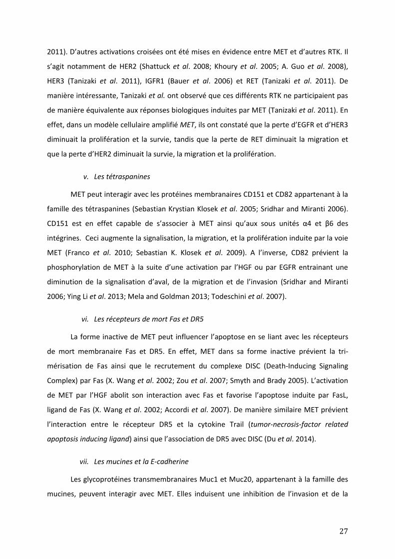
2011). D’autres activations croisées ont été mises en évidence entre MET et d’autres RTK. Il
s’agit notamment de HER2 (Shattuck et al. 2008; Khoury et al. 2005; A. Guo et al. 2008),
HER3 (Tanizaki et al. 2011), IGFR1 (Bauer et al. 2006) et RET (Tanizaki et al. 2011). De
manière intéressante, Tanizaki et al. ont observé que ces différents RTK ne participaient pas
de manière équivalente aux réponses biologiques induites par MET (Tanizaki et al. 2011). En
effet, dans un modèle cellulaire amplifié MET, ils ont constaté que la perte d’EGFR et d’HER3
diminuait la prolifération et la survie, tandis que la perte de RET diminuait la migration et
que la perte d’HER2 diminuait la survie, la migration et la prolifération.
v. Les tétraspanines
MET peut interagir avec les protéines membranaires CD151 et CD82 appartenant à la
famille des tétraspanines (Sebastian Krystian Klosek et al. 2005; Sridhar and Miranti 2006).
CD151 est en effet capable de s’associer à MET ainsi qu’aux sous unités α4 et β6 des
intégrines. Ceci augmente la signalisation, la migration, et la prolifération induite par la voie
MET (Franco et al. 2010; Sebastian K. Klosek et al. 2009). A l’inverse, CD82 prévient la
phosphorylation de MET à la suite d’une activation par l’HGF ou par EGFR entrainant une
diminution de la signalisation d’aval, de la migration et de l’invasion (Sridhar and Miranti
2006; Ying Li et al. 2013; Mela and Goldman 2013; Todeschini et al. 2007).
vi. Les récepteurs de mort Fas et DR5
La forme inactive de MET peut influencer l’apoptose en se liant avec les récepteurs
de mort membranaire Fas et DR5. En effet, MET dans sa forme inactive prévient la tri-
mérisation de Fas ainsi que le recrutement du complexe DISC (Death-Inducing Signaling
Complex) par Fas (X. Wang et al. 2002; Zou et al. 2007; Smyth and Brady 2005). L’activation
de MET par l’HGF abolit son interaction avec Fas et favorise l’apoptose induite par FasL,
ligand de Fas (X. Wang et al. 2002; Accordi et al. 2007). De manière similaire MET prévient
l’interaction entre le récepteur DR5 et la cytokine Trail (tumor-necrosis-factor related
apoptosis inducing ligand) ainsi que l’association de DR5 avec DISC (Du et al. 2014).
vii. Les mucines et la E-cadherine
Les glycoprotéines transmembranaires Muc1 et Muc20, appartenant à la famille des
mucines, peuvent interagir avec MET. Elles induisent une inhibition de l’invasion et de la

28
migration médiée par MET (Horm et al. 2012; Singh et al. 2008; Higuchi et al. 2004). La E-
cadhérine est une protéine transmembranaire impliquée dans la formation de jonctions
entre les cellules. Lors d’une stimulation par l’HGF, MET interagit directement avec la E-
cadhérine, et induit son endocytose. Ceci diminue l’adhésion inter-cellulaire, augmente les
capacités de migration et favorise la dispersion cellulaire (Hiscox and Jiang 1999; Kamei et al.
1999; Reshetnikova, Troyanovsky, and Rimm 2007).
b. Internalisation, trafic et dégradation du récepteur MET
Après son activation par son ligand l’HGF, MET, comme d’autres récepteurs tyrosine
kinase, est internalisé via le processus d’endocytose (Naka et al. 1993; Kamei et al. 1999).
L’internalisation de MET implique les molécules de clathrine et les dynamines mais
également le recrutement de l’E3 ubiquitine ligase Cbl (Casitas B-Cell Lineage) via le domaine
SH3 de Grb2 (Kermorgant and Parker 2008a; Joffre et al. 2011; Ning Li et al. 2007). En effet,
un complexe comprenant Cbl, l’adaptateur CIN85 et l’endophiline permet le transfert de
MET depuis les puits de clathrines jusqu’aux endosomes (Garcia-Guzman, Larsen, and Vuori
2000; Petrelli et al. 2002; Pascal Peschard et al. 2004). Au cours de son transfert vers les
endosomes, le récepteur internalisé reste activé et demeure capable d’induire une
signalisation. Ainsi l’activation complète après une stimulation par l’HGF, de ERK1/2, ou de la
GTPase Rac1 impliquée dans la migration cellulaire, requiert l’internalisation de MET et son
transfert vers les endosomes (Kermorgant, Zicha, and Parker 2004; Palamidessi et al. 2008;
Joffre et al. 2011). L’acheminement de MET jusqu’aux endosomes périnucléaires est de plus
nécessaire à l’activation de STAT3 et à son accumulation dans le noyau (Kermorgant and
Parker 2008b).
Une fois internalisé et transféré dans les endosomes, le récepteur MET est ensuite
soit dégradé, soit recyclé à la membrane. L’E3 ubiquitine ligase Cbl contient un domaine
reconnaissant de la tyrosine Y1003 phosphorylée de MET située dans le domaine
juxtamembranaire du récepteur (P. Peschard et al. 2001). Cette tyrosine phosphorylée lors
de l’activation de MET permet le recrutement direct de Cbl qui induit alors l’ubiquitination
du récepteur. MET ubiquitinylé est ensuite transféré via le recrutement et la
phosphorylation de la protéine HRS (hepatocyte growth factor-regulated tyrosine kinase
substrate) dans les endosomes tardifs, et est dégradé dans les lysosomes (D. E. Hammond et
29
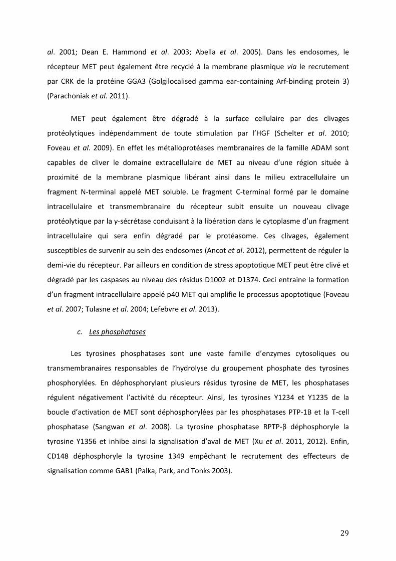
al. 2001; Dean E. Hammond et al. 2003; Abella et al. 2005). Dans les endosomes, le
récepteur MET peut également être recyclé à la membrane plasmique via le recrutement
par CRK de la protéine GGA3 (Golgilocalised gamma ear-containing Arf-binding protein 3)
(Parachoniak et al. 2011).
MET peut également être dégradé à la surface cellulaire par des clivages
protéolytiques indépendamment de toute stimulation par l’HGF (Schelter et al. 2010;
Foveau et al. 2009). En effet les métalloprotéases membranaires de la famille ADAM sont
capables de cliver le domaine extracellulaire de MET au niveau d’une région située à
proximité de la membrane plasmique libérant ainsi dans le milieu extracellulaire un
fragment N-terminal appelé MET soluble. Le fragment C-terminal formé par le domaine
intracellulaire et transmembranaire du récepteur subit ensuite un nouveau clivage
protéolytique par la γ-sécrétase conduisant à la libération dans le cytoplasme d’un fragment
intracellulaire qui sera enfin dégradé par le protéasome. Ces clivages, également
susceptibles de survenir au sein des endosomes (Ancot et al. 2012), permettent de réguler la
demi-vie du récepteur. Par ailleurs en condition de stress apoptotique MET peut être clivé et
dégradé par les caspases au niveau des résidus D1002 et D1374. Ceci entraine la formation
d’un fragment intracellulaire appelé p40 MET qui amplifie le processus apoptotique (Foveau
et al. 2007; Tulasne et al. 2004; Lefebvre et al. 2013).
c. Les phosphatases
Les tyrosines phosphatases sont une vaste famille d’enzymes cytosoliques ou
transmembranaires responsables de l’hydrolyse du groupement phosphate des tyrosines
phosphorylées. En déphosphorylant plusieurs résidus tyrosine de MET, les phosphatases
régulent négativement l’activité du récepteur. Ainsi, les tyrosines Y1234 et Y1235 de la
boucle d’activation de MET sont déphosphorylées par les phosphatases PTP-1B et la T-cell
phosphatase (Sangwan et al. 2008). La tyrosine phosphatase RPTP-β déphosphoryle la
tyrosine Y1356 et inhibe ainsi la signalisation d’aval de MET (Xu et al. 2011, 2012). Enfin,
CD148 déphosphoryle la tyrosine 1349 empêchant le recrutement des effecteurs de
signalisation comme GAB1 (Palka, Park, and Tonks 2003).

30
d. La sérine 985
La sérine 985 située dans le domaine juxtamembranaire de MET est phosphorylée
par les PKC δ et ε, et déphosphorylée par la phosphatase PPA2 (Protein phosphatase A2)
(Hashigasako et al. 2004). Elle participe à la régulation négative du récepteur en inhibant la
phosphorylation des tyrosines au sein de son domaine kinase inhibant ainsi son activité
catalytique (Gandino et al. 1994). In vivo, la régénération hépatique nécessitant l’activation
de la voie HGF/MET s’accompagne d’une diminution de la phosphorylation de la sérine 985
suggérant l’implication de ce résidu dans la régulation physiologique de l’activité de MET
(Nakayama et al. 2013).
7) Les dérégulations de MET dans le cancer
La voie HGF/MET est capable d’induire un programme de croissance invasive associant
prolifération cellulaire, mobilité et résistance à l’apoptose. A l’état physiologique l’activation
de cet axe de signalisation est hautement régulée. En pathologie humaine, l’activation
aberrante de la voie HGF/MET est impliquée dans le développement de nombreux cancers.
Plusieurs mécanismes de dérégulation ont été mis en évidence comme la surexpression de
MET, la sécrétion autocrine ou paracrine de l’HGF, l’amplification du gène MET, la survenue
de mutations de MET, ou l’apparition de fusion génique impliquant MET.
a. La surexpression de MET
L’implication de la surexpression de MET dans la tumorigenèse a été mise en
évidence dans plusieurs modèles murins. En effet, il a été démontré in vivo que la seule
surexpression de MET était capable d’induire des tumeurs mammaires ou hépatiques (Ponzo
et al. 2009; R. Wang et al. 2001). Différents mécanismes peuvent concourir à la
surexpression de MET dans les cancers. Tout d’abord, certaines altérations génétiques,
modifiant la synthèse ou la dégradation du récepteur, comme l’amplification de MET ou les
mutations des sites d’épissage de l’exon 14 de MET, sont associées à une augmentation de
l’expression de MET (Schildhaus et al. 2015; J. H. Tong et al. 2016). Ces anomalies sont
respectivement détaillées dans les paragraphes ci-dessous. Ensuite, des modifications
épigénétiques peuvent modifier l’expression de MET. Ainsi, l’hypométhylation de MET est
corrélée à une expression élevée du récepteur dans les adénocarcinomes canalaires

31
pancréatiques (Nones et al. 2014). Une augmentation de l’expression de MET peut aussi être
secondaire à une activation de la transcription soit en réponse à l’hypoxie via le facteur
HIF1α (Pennacchietti et al. 2003), soit à la suite d’une dérégulation des facteurs de
transcription Ets1 ou AP1 (Seol, Chen, and Zarnegar 2000; Furlan et al. 2008). La
surexpression de MET peut également être induite par l’inhibition de plusieurs micro-ARN
bloquant la traduction de l’ARNm de MET comme miR-1 (Migliore et al. 2012), miR-31
(Mitamura et al. 2013), miR-34a et b (Ying Zhang et al. 2014; Migliore et al. 2008), miR-139-
5p (C. Sun et al. 2015), ou miR-144-3p (Lan et al. 2015). Actuellement on estime que plus
d’une trentaine de micro-ARN sont impliqués dans la régulation de MET (Karagonlar, Korhan,
and Atabey 2015). Par ailleurs, la protéine p53 régule négativement l’expression de MET en
favorisant l’expression de miR-34 et en inhibant la fixation de SP-1 sur le promoteur de MET.
Par conséquent, l’inactivation de p53 est à même d’induire une surexpression de MET dans
les cellules cancéreuses (Hwang et al. 2011). Enfin, il a été montré que la phosphorylation de
la tyrosine Y1313 de MET permet le recrutement de la Tensine 4 qui prévient la dégradation
du récepteur et est associée à la surexpression de MET dans les cancers du colon et de
l’ovaire (Muharram et al. 2014).
L’expression du récepteur MET a été étudiée dans de nombreux cancers
principalement par immunohistochimie (IHC), par western blot, ou par RT-qPCR (Yanni
Zhang, Du, and Zhang 2016). Il s’agit d’un évènement fréquent mais dont la prévalence varie
de manière importante selon le type de cancer, la technique utilisée, et le seuil retenu pour
définir la surexpression. Ainsi en immunohistochimie une surexpression de MET a été mise
en évidence dans 9.6 à 71% des cancers gastriques (An et al. 2014; Huang et al. 2001), 13.7 à
70% des cancers bronchiques non à petites cellules (Sanghui Park et al. 2012; Tsao et al.
2001), 20 à 87.5% des carcinomes hépatocellulaires (Kiss et al. 1997; Chau et al. 2008), et 25
à 60% des cancers du sein (Ghoussoub et al. 1998; G. V. Scagliotti, Novello, and von Pawel
2013). En utilisant, en IHC, l’anticorps monoclonal SP44 dirigé contre MET ainsi que le
système d’évaluation qui lui est associé (Figure 5), la prévalence de surexpression de MET
dans les cancers bronchiques non à petites cellules métastatiques est estimée à environ 50%
(Spigel et al. 2013). La surexpression de MET a été associée à un mauvais pronostic dans les
cancers bronchiques non à petites cellules (B. Guo et al. 2014), les carcinomes mammaires
(Yan et al. 2015), et les cancers gastriques (Peng et al. 2014). Cependant le lien entre

32
surexpression et activation du récepteur reste controversé. En effet si certaines études ont
observé une association entre expression de MET et phosphorylation des résidus tyrosine
témoins de l’activation du récepteur (Y. Nakamura et al. 2007; Copin et al. 2016), d’autres
travaux ne retrouvent pas cette corrélation (Watermann et al. 2015; Tsuta et al. 2012). De
plus plusieurs essais cliniques de grande ampleur n’ont pas mis en évidence d’efficacité des
inhibiteurs de MET dans les cancers bronchiques non à petites cellules présentant une
surexpression MET (Spigel et al. 2017; G. Scagliotti et al. 2015).
Figure 5 : Score d’expression tumorale de MET en Immunohistochimie avec
l’anticorps monoclonal SP44 (Montagne et al. 2015). Le score 0 correspond à une absence
de marquage ou à un marquage présent dans moins de 50% de cellules tumorales quelque
soit son intensité. Le score 1 correspond à une intensité de marquage faible d’au moins 50%
des cellules tumorales ou à un marquage modéré de moins de 50% des cellules tumorales.
Le score 2 correspond à une intensité modérée d’au moins 50% des cellules tumorales ou à
un marquage fort de moins de 50% des cellules tumorales, et le score 3 à une intensité forte
d’au moins 50% des cellules tumorales.

33
b. La sécrétion d’HGF
L’HGF peut être sécrété de manière paracrine par les cellules du stroma tumoral ou
de manière autocrine par les cellules tumorales elles-mêmes (Ferracini et al. 1995). Plusieurs
lignées cellulaires humaines comme la lignée SNU-484, dérivée d’un cancer gastrique, ou la
lignée U-87, dérivée d’un gliome, présentent une sécrétion autocrine d’HGF (Minseon Park
et al. 2005; Ying Zhang et al. 2013). Dans ces modèles cellulaires, l’inhibition de la voie
HGF/MET par un inhibiteur de tyrosine kinase (ITK) ou par un anticorps bloquant l’HGF induit
une inhibition de la prolifération cellulaire. Le rôle oncogénique de l’HGF a été confirmé in
vivo dans des modèles de souris surexprimant ce facteur de croissance. En effet, les souris
qui surexpriment l’HGF dans l’ensemble de leur organisme, développent spontanément des
mélanomes, des sarcomes ou des carcinomes (Takayama et al. 1997). Lorsque la
surexpression est restreinte au niveau des glandes mammaires, les souris développent des
tumeurs mammaires pouvant métastaser au poumon (Gallego, Bierie, and Hennighausen
2003). Chez l’homme, une augmentation de la sécrétion d’HGF, mesurée au niveau sérique
par ELISA ou au niveau tissulaire par IHC, a été observée et associée à un mauvais pronostic
dans les cancers bronchiques à petites cellules (Canadas et al. 2013), les cancers
bronchiques non à petites cellules (Siegfried et al. 1997), les cancers du sein (H. Yang, Zhang,
and Cui 2015; Toi et al. 1998), les cancers de la prostate (Gupta et al. 2008), les cancers
rénaux à cellules claires (Tanimoto et al. 2008), les mélanomes (Hügel et al. 2016) et les
gliomes (X. Zhang et al. 2013). Enfin, il a été montré qu’un taux élevés d’HGF dans les lavages
broncho-alvéolaires de patients atteints de carcinomes bronchioloalvéolaires était un
facteur de risque indépendant de mortalité (Wislez et al. 2003).
c. L’amplification de MET
Le rôle oncogénique de l’amplification du gène MET a été initialement identifié dans
des fibroblastes de souris NIH3T3, qui présentaient une transformation cancéreuse
spontanée, lors d’expériences de transfections utilisant de l'ADN provenant de cellules
néoplasiques ou non néoplasiques (Cooper et al. 1986). En effet, dans ces cellules, il était
constaté par Southern blot une augmentation du nombre de copies du gène MET endogène,
conduisant à une augmentation majeure de l'ARNm de MET. L'amplification MET a par la
suite été détectée dans les cellules GTL16, une lignée cellulaire de tumeur gastrique

34
humaine dérivée d'un sous-clone de la lignée MKN45 (S. Giordano et al. 1989). Dans ce
modèle, l'amplification de MET conduit à une synthèse massive du récepteur et à son
activation constitutive, indépendamment de la stimulation par le ligand. Plus tard, plusieurs
lignées de cellules cancéreuses MET amplifiées ont été caractérisées et utilisées dans des
études précliniques d'inhibition de MET. Il s'agit notamment de lignées cellulaires dérivées
de cancers du poumon (EBC-1, H1993) (Zhao et al. 2005) ou de cancers gastriques (MKN45,
GTL-16, SNU5, KatoII) (Ponzetto et al. 1991; Rege-Cambrin et al. 1992; Smolen et al. 2006).
Ces lignées cellulaires ont permis de mettre en évidence un phénomène d’addiction
oncogénique secondaire à l’amplification de MET. En effet, il est possible d'induire, dans ces
modèles, un arrêt de la croissance cellulaire et/ou la mort cellulaire par traitement avec un
ITK de MET tel que PHA-665752 (Smolen et al. 2006) ou par inhibition de l’expression de
MET via un shRNA (Lutterbach et al. 2007). Jusqu'à présent, il n’existe pas de modèle in vivo
reproduisant l’amplification de MET. Cependant l’efficacité des thérapies ciblant MET in vivo
a pu être évaluée grâce à des modèles de xénogreffes de lignées cellulaires présentant une
amplification MET ou de xénogreffes de cellules tumorales dérivées du patients (PDX). Une
étude a ainsi montré, dans deux modèles PDX de cancers rénaux présentant une
amplification de MET, une inhibition de la croissance tumorale en réponse à AZD6094, un
inhibiteur sélectif de MET (Schuller et al. 2015).
La fréquence de l’amplification de MET varie selon le type de cancer étudié, la
technique utilisée (Fluorescence In Situ Hybridization (FISH), quantitative Polymerase Chain
Reaction (qPCR), Comparative Genomic Hybridization array (CGH array)), et les critères de
mesure retenus pour sa définition. En retenant une définition fondée sur un ratio
MET/CEP7≥2 (nombre de copie de MET/nombre de copie du centromère du chromosome 7)
en FISH, l’amplification de MET est mise en évidence chez 3 à 5% des patients atteints de
CBNPC, de cancers gastriques, de carcinomes hépatocellulaires, de carcinomes
nasopharyngés ou de tumeurs gliales. Dans la plupart des cas, l'amplification MET est
corrélée à un fort niveau d’expression protéique du récepteur ainsi qu’à un mauvais
pronostic (Seongyeol Park et al. 2015; Pyo, Kang, and Cho 2016; Yingqin Li et al. 2015; Burel-
Vandenbos et al. 2017; Kondo et al. 2013; K. Wang et al. 2013). Dans le cancer gastrique, par
exemple, les tumeurs MET amplifiées présentent une survie globale plus courte que celles
ne présentant pas cette altération (Catenacci et al. 2017). Dans les carcinomes du sein,

35
l'amplification du gène MET est associée à un risque accru de récidive à distance chez les
patients traités par chimiothérapie (Veenstra et al. 2016). Enfin dans les gliomes,
l'amplification du gène MET est associée à l'agressivité du gliome, et ne se retrouve que dans
les tumeurs de grade IV (Kwak et al. 2015).
d. Les mutations de MET
i. Mutations du domaine kinase de MET
Les mutations activatrices du domaine kinase de MET ont pour la première fois été
décrites dans les carcinomes papillaires rénaux sporadiques et héréditaires (L. Schmidt et al.
1997). Il s’agit de mutations faux sens du domaine kinase (M1149T, V1206L, V1238L,
D1246N, D1246H, Y1228C et M1268T) situées à proximité des résidus tyrosine Y1234 et
Y1235 (L. Schmidt et al. 1997, 1998; Durinck et al. 2015). Ce type de mutations a également
été rapporté dans les carcinomes hépatocellulaires infantiles (M1268I) (W. S. Park et al.
1999) et dans les cancers avancés de la tête et du cou (Y1253D) (Aebersold et al. 2003;
Ghadjar et al. 2009). In vitro, ces altérations entrainent généralement une activation
constitutive de MET et favorisent la transformation des cellules fibroblastiques en cellules
cancéreuses (Jeffers et al. 1997). Cependant, il semble que pour certaines de ces mutations
une stimulation par l’HGF soit nécessaire au processus de transformation cancéreuse
(Michieli et al. 1999). Par ailleurs, certaines mutations (M1268T et D1246N) affectent non
seulement l’activité du récepteur mais aussi son trafic intracellulaire en favorisant son
recyclage à la membrane plasmique (Joffre et al. 2011). L'implication de ces mutations dans
la tumorigenèse a été confirmée par des modèles murins de souris knock-in exprimant une
version mutée du récepteur MET (D1226N, Y1228C, M1248T, M1248T/L1193V) (C. Graveel
et al. 2004a; C. R. Graveel, London, and Vande Woude 2005). En effet, des processus
néoplasiques ont été observés dans ces différentes lignées murines. Néanmoins le type de
tumeur obtenue n’était pas le même pour toutes les mutations. Par exemple, les souris
présentant la mutation MET 1248T ont développé des tumeurs associant des carcinomes et
des lymphomes, tandis que les souris présentant les mutations D1226N, Y1228C et
M1248T/L1193V ont développé principalement des sarcomes et des lymphomes (C. Graveel
et al. 2004b). Chez l’homme, l’efficacité des inhibiteurs de MET sur les tumeurs porteuses de
ce type d’altération est encore incertaine. Un essai clinique de phase II évaluant l'efficacité
36
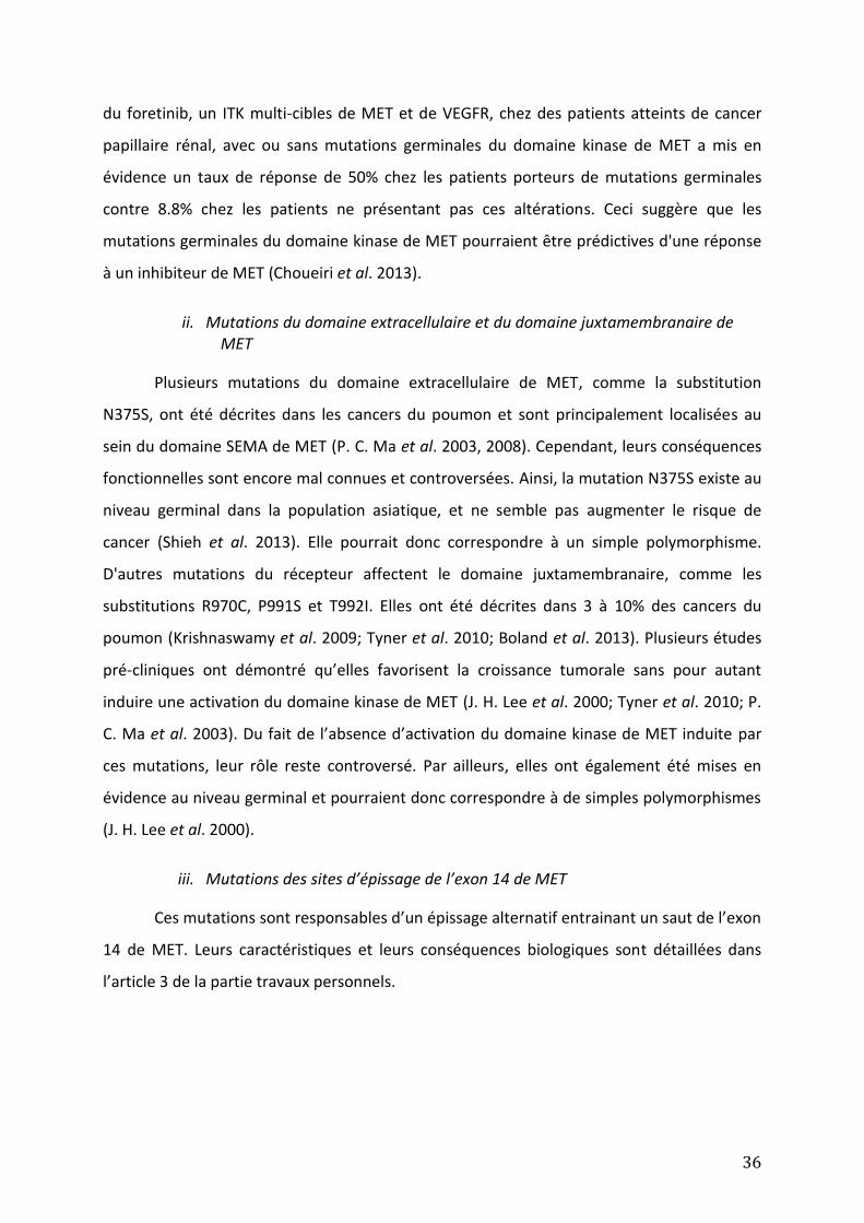
du foretinib, un ITK multi-cibles de MET et de VEGFR, chez des patients atteints de cancer
papillaire rénal, avec ou sans mutations germinales du domaine kinase de MET a mis en
évidence un taux de réponse de 50% chez les patients porteurs de mutations germinales
contre 8.8% chez les patients ne présentant pas ces altérations. Ceci suggère que les
mutations germinales du domaine kinase de MET pourraient être prédictives d'une réponse
à un inhibiteur de MET (Choueiri et al. 2013).
ii. Mutations du domaine extracellulaire et du domaine juxtamembranaire de MET
Plusieurs mutations du domaine extracellulaire de MET, comme la substitution
N375S, ont été décrites dans les cancers du poumon et sont principalement localisées au
sein du domaine SEMA de MET (P. C. Ma et al. 2003, 2008). Cependant, leurs conséquences
fonctionnelles sont encore mal connues et controversées. Ainsi, la mutation N375S existe au
niveau germinal dans la population asiatique, et ne semble pas augmenter le risque de
cancer (Shieh et al. 2013). Elle pourrait donc correspondre à un simple polymorphisme.
D'autres mutations du récepteur affectent le domaine juxtamembranaire, comme les
substitutions R970C, P991S et T992I. Elles ont été décrites dans 3 à 10% des cancers du
poumon (Krishnaswamy et al. 2009; Tyner et al. 2010; Boland et al. 2013). Plusieurs études
pré-cliniques ont démontré qu’elles favorisent la croissance tumorale sans pour autant
induire une activation du domaine kinase de MET (J. H. Lee et al. 2000; Tyner et al. 2010; P.
C. Ma et al. 2003). Du fait de l’absence d’activation du domaine kinase de MET induite par
ces mutations, leur rôle reste controversé. Par ailleurs, elles ont également été mises en
évidence au niveau germinal et pourraient donc correspondre à de simples polymorphismes
(J. H. Lee et al. 2000).
iii. Mutations des sites d’épissage de l’exon 14 de MET
Ces mutations sont responsables d’un épissage alternatif entrainant un saut de l’exon
14 de MET. Leurs caractéristiques et leurs conséquences biologiques sont détaillées dans
l’article 3 de la partie travaux personnels.
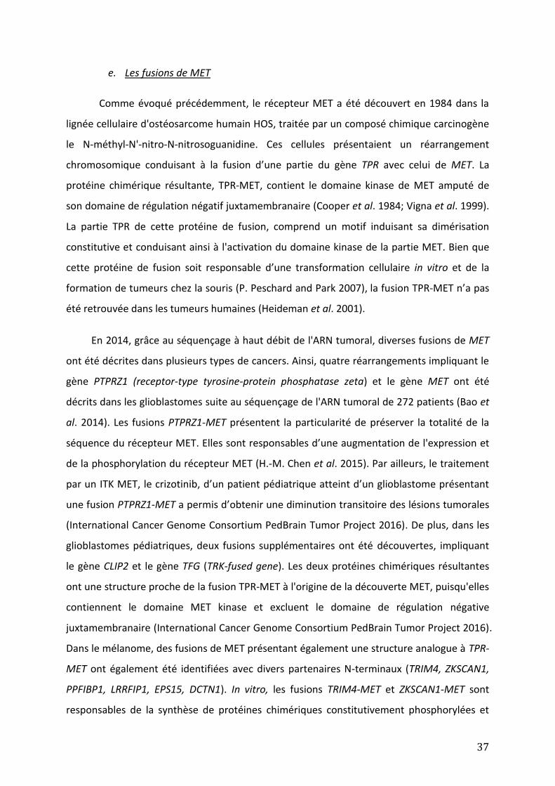
37
e. Les fusions de MET
Comme évoqué précédemment, le récepteur MET a été découvert en 1984 dans la
lignée cellulaire d'ostéosarcome humain HOS, traitée par un composé chimique carcinogène
le N-méthyl-N'-nitro-N-nitrosoguanidine. Ces cellules présentaient un réarrangement
chromosomique conduisant à la fusion d’une partie du gène TPR avec celui de MET. La
protéine chimérique résultante, TPR-MET, contient le domaine kinase de MET amputé de
son domaine de régulation négatif juxtamembranaire (Cooper et al. 1984; Vigna et al. 1999).
La partie TPR de cette protéine de fusion, comprend un motif induisant sa dimérisation
constitutive et conduisant ainsi à l'activation du domaine kinase de la partie MET. Bien que
cette protéine de fusion soit responsable d’une transformation cellulaire in vitro et de la
formation de tumeurs chez la souris (P. Peschard and Park 2007), la fusion TPR-MET n’a pas
été retrouvée dans les tumeurs humaines (Heideman et al. 2001).
En 2014, grâce au séquençage à haut débit de l'ARN tumoral, diverses fusions de MET
ont été décrites dans plusieurs types de cancers. Ainsi, quatre réarrangements impliquant le
gène PTPRZ1 (receptor-type tyrosine-protein phosphatase zeta) et le gène MET ont été
décrits dans les glioblastomes suite au séquençage de l'ARN tumoral de 272 patients (Bao et
al. 2014). Les fusions PTPRZ1-MET présentent la particularité de préserver la totalité de la
séquence du récepteur MET. Elles sont responsables d’une augmentation de l'expression et
de la phosphorylation du récepteur MET (H.-M. Chen et al. 2015). Par ailleurs, le traitement
par un ITK MET, le crizotinib, d’un patient pédiatrique atteint d’un glioblastome présentant
une fusion PTPRZ1-MET a permis d’obtenir une diminution transitoire des lésions tumorales
(International Cancer Genome Consortium PedBrain Tumor Project 2016). De plus, dans les
glioblastomes pédiatriques, deux fusions supplémentaires ont été découvertes, impliquant
le gène CLIP2 et le gène TFG (TRK-fused gene). Les deux protéines chimériques résultantes
ont une structure proche de la fusion TPR-MET à l'origine de la découverte MET, puisqu'elles
contiennent le domaine MET kinase et excluent le domaine de régulation négative
juxtamembranaire (International Cancer Genome Consortium PedBrain Tumor Project 2016).
Dans le mélanome, des fusions de MET présentant également une structure analogue à TPR-
MET ont également été identifiées avec divers partenaires N-terminaux (TRIM4, ZKSCAN1,
PPFIBP1, LRRFIP1, EPS15, DCTN1). In vitro, les fusions TRIM4-MET et ZKSCAN1-MET sont
responsables de la synthèse de protéines chimériques constitutivement phosphorylées et

38
induisent la formation de tumeurs dans des modèles animaux de xénogreffe (Yeh et al.
2015). Enfin, le cas clinique d’un patient atteint d’un adénocarcinome pulmonaire
présentant une fusion HLA-DRB1-MET a récemment été rapporté. A nouveau de manière
similaire à la fusion TPR-MET, la protéine chimérique HLA-DRB1-MET inclut le domaine
kinase de MET sans la séquence régulatrice juxtamembranaire. Le traitement de ce patient
par crizotinib a conduit à une réponse tumorale majeure sur 8 mois (Davies et al. 2017). Au
total, diverses fusions MET ont été décrites dans au moins trois types de cancers et deux cas
de réponse à un inhibiteur de MET chez des patients présentant des fusions MET ont été
rapportés. Ces résultats suggèrent que de telles fusions peuvent créer une situation
d’addiction oncogénique accessible à un traitement inhibiteur de MET.
II. MET et les cancers bronchiques non à petites cellules EGFR muté
1) Epidémiologie des cancers pulmonaires
Le cancer pulmonaire est la première cause de mortalité par cancer en France et dans
le monde. Avec 1.8 million de nouveaux cas par an, il s’agit du premier cancer en terme
d’incidence au niveau mondial (Torre et al. 2015). Les principaux facteurs de risque à
l'origine de cette pathologie sont l'exposition au tabac, à l'amiante, et dans une moindre
mesure, aux métaux lourds, aux radiations ionisantes et aux polluants atmosphériques
(Alberg et al. 2013). 87% des cancers pulmonaires sont des cancers bronchiques non à
petites cellules (CBNPC) (DeSantis et al. 2014). A un stade métastatique le pronostic de ces
cancers reste sombre avec seulement 4% de patients en vie à 5 ans (Siegel et al. 2012).
Néanmoins, l’identification d’altérations moléculaires à l’origine de la survenue du processus
tumoral a conduit au développement de thérapies ciblées qui ont démontré leur efficacité et
augmenté l’espérance de vie de certains patients. Le meilleur exemple de cette approche
thérapeutique est représenté par les ITK du récepteur de l’Epidermal Growth Factor (EGFR)
dans le traitement des CBNPC porteurs d’une mutation EGFR.

39
2) L’EGFR et la découverte de ses mutations dans les cancers bronchiques non à petites cellules
L’EGFR (aussi appelé HER1) est un RTK appartenant à la famille ERBB, elle-même
composée de 4 membres (HER ou ERBB 1 à 4) (Yosef Yarden and Pines 2012). Le gène de
l’EGFR est situé sur le bras court du chromosome 7 (7p11) et code pour une protéine de
1186, constituée d’un domaine extracellulaire subdivisé en 4 sous domaines, d’une hélice
transmembranaire et d’un domaine intracellulaire porteur de l’activité kinase (Figure 6).
Figure 6 : Représentation schématique des différents domaines fonctionnels de l’EGFR
(adapté de (Lemmon, Schlessinger, and Ferguson 2014).

40
L’EGFR est capable de lier 7 ligands différents qui partagent tous un domaine EGF-
like permettant leur fixation au récepteur (Harris, Chung, and Coffey 2003; Schneider and
Wolf 2009). Il s’agit de l’EGF, du TGF-α, de la bétacelluline (BTC), de l’HB-EGF, de
l’amphiréguline (aussi appelé ARG), de l’épiréguline (aussi appelée EPR), et de l’EGN. La
fixation de l’un des ligands sur les sous domaines extracellulaires I et III d’EGFR induit son
homo-dimérisation ou son hétéro-dimérisation avec ERBB2, ERBB3 ou ERBB4 (Y. Yarden and
Sliwkowski 2001; Ferguson et al. 2000). Ceci entraine l’activation du récepteur et la
phosphorylation des résidus tyrosine de la queue C-terminale. Les tyrosines phosphorylées
servent alors de site d’ancrage multi-substrats pour des protéines de signalisation porteuses
d’un domaine de reconnaissance SH2 (Lemmon, Schlessinger, and Ferguson 2014). Plusieurs
voies de signalisation d’aval sont enfin activées dont la voie PI3K/AKT et RAS/MAPK
induisant ainsi des réponses de prolifération et de survie cellulaire. L’activité d’EGFR est
nécessaire au développement embryonnaire de différents organes dont la peau, les yeux, le
cœur, le système nerveux, et les os (Sibilia et al. 2007).
Dans les cancers pulmonaires, les mutations activatrices d’EGFR ont été découvertes
simultanément par deux équipes en 2004 (Lynch et al. 2004; Paez et al. 2004). Il s’agit de
mutations survenant exclusivement dans le domaine tyrosine kinase d’EGFR, codé par les
exons 18 à 21 (Johnson and Jänne 2005). Deux de ces altérations représentent 90% de
l’ensemble des mutations d’EGFR : les délétions dans l’exon 19, et la substitution L858R dans
l’exon 21. Les autres altérations décrites sont des mutations dans l’exon 18 (substitution
G719X, 3%), l’exon 20 (substitution T790M, 2%), l’exon 21 (substitution L861Q, 2%) et des
insertions dans l’exon 20 (5%). Les mutations d’EGFR sont en règle générale mutuellement
exclusives de la présence d’altérations dans d’autres oncogènes comme ALK, KRAS, ERBB2,
ou BRAF (Cancer Genome Atlas Research Network 2014; Gainor et al. 2013). Elles ont pour
conséquence une activation constitutive de la kinase d’EGFR, indépendante de la liaison de
son ligand. Ceci induit une activation des voies de signalisation sous-jacentes, PI3K-AKT et
RAS-MAPK, aboutissant à un signal excessif et incontrôlé de survie et de prolifération
cellulaire, à l’origine du phénotype transformant et de la croissance tumorale. Parallèlement,
la présence d’une mutation d’EGFR est généralement associée à un phénomène de talon
d’Achille : la perte d’expression ou l’inhibition de l’activité kinase d’EGFR entrainent une
inhibition des voies sous-jacentes, un arrêt de la prolifération cellulaire et une fonte
tumorale.

41
3) Caractéristiques cliniques des cancers bronchiques non à petites cellules EGFR mutés
En France, les mutations d’EGFR sont détectées chez 11% des patients atteints de
CBNPC (Barlesi et al. 2016). Elles sont retrouvées presque exclusivement dans les tumeurs
de type non épidermoïdes et observées plus fréquemment chez les non-fumeurs, les
femmes, et les patients d’origine asiatique. Cependant, ces critères cliniques ne sont pas
suffisamment prédictifs de la présence d’une mutation d’EGFR. Par exemple, écarter les
hommes et les fumeurs d’une recherche systématique de mutation d’EGFR conduirait à
manquer plus de 57% des patients porteurs d’une mutation du récepteur (D’Angelo et al.
2011). C’est pourquoi une recherche systématique de mutation d’EGFR est recommandée
dans les CBNPC non épidermoïdes avancés quel que soit le statut tabagique, le sexe ou
l’origine ethnique du patient (Novello et al. 2016).
4) Traitement des cancers bronchiques non à petites cellules EGFR mutés métastatiques
Quatre inhibiteurs d’EGFR, le géfitinib, l’erlotinib, l’afatinib et l’osimertinib possèdent
une autorisation de mise sur le marché (AMM) dans le traitement des CBNPC EGFR mutés
métastatiques. Les deux premiers sont des inhibiteurs spécifiques d’EGFR, dits de première
génération, qui se fixent de façon réversible à l’EGFR muté. Ils disposent d’une AMM dans
les CBNPC EGFR mutés quelle que soit la ligne de traitement. L’afatinib est un ITK EGFR dit
de deuxième génération, qui se distingue du géfitinib ou de l’erlotinib de 2 manières : d’une
part, il s’agit d’un inhibiteur « pan-HER », c’est-à-dire capable d’inhiber l’activité kinase de
tous les membres de la famille HER. D’autre part, il s’agit d’un ITK irréversible capable de se
lier au récepteur EGFR de façon définitive par une liaison covalente (Riely 2008). Il possède
une AMM dans le traitement des patients atteints d’un CBNPC EGFR muté métastatique naïf
de traitement par un ITK EGFR. Enfin, l’osimertinib est un ITK EGFR dit de 3ème génération,
irréversible, qui se distingue par sa capacité à inhiber le récepteur EGFR y compris en
présence d’une mutation T790M, altération sur laquelle les inhibiteurs de première et
deuxième génération sont inefficaces (W. Zhou et al. 2009). L’osimertinib possède une AMM
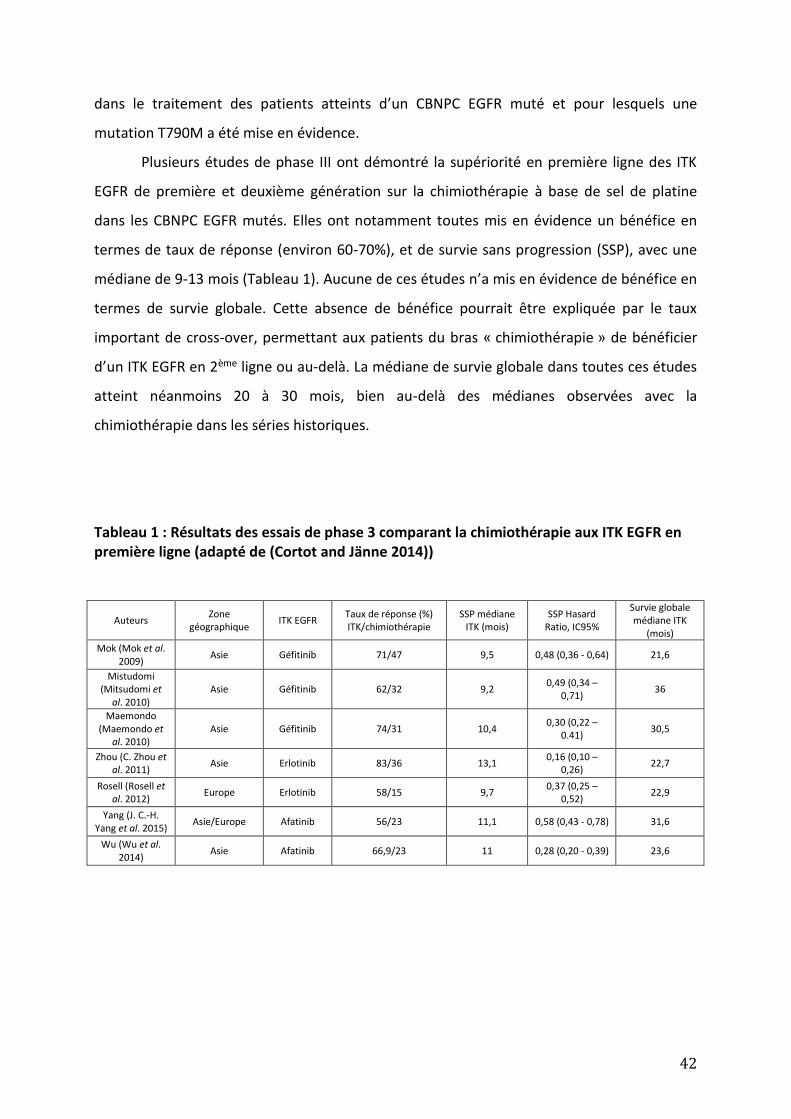
42
dans le traitement des patients atteints d’un CBNPC EGFR muté et pour lesquels une
mutation T790M a été mise en évidence.
Plusieurs études de phase III ont démontré la supériorité en première ligne des ITK
EGFR de première et deuxième génération sur la chimiothérapie à base de sel de platine
dans les CBNPC EGFR mutés. Elles ont notamment toutes mis en évidence un bénéfice en
termes de taux de réponse (environ 60-70%), et de survie sans progression (SSP), avec une
médiane de 9-13 mois (Tableau 1). Aucune de ces études n’a mis en évidence de bénéfice en
termes de survie globale. Cette absence de bénéfice pourrait être expliquée par le taux
important de cross-over, permettant aux patients du bras « chimiothérapie » de bénéficier
d’un ITK EGFR en 2ème ligne ou au-delà. La médiane de survie globale dans toutes ces études
atteint néanmoins 20 à 30 mois, bien au-delà des médianes observées avec la
chimiothérapie dans les séries historiques.
Tableau 1 : Résultats des essais de phase 3 comparant la chimiothérapie aux ITK EGFR en première ligne (adapté de (Cortot and Jänne 2014))
Auteurs Zone
géographique ITK EGFR
Taux de réponse (%) ITK/chimiothérapie
SSP médiane ITK (mois)
SSP Hasard Ratio, IC95%
Survie globale médiane ITK
(mois)
Mok (Mok et al. 2009)
Asie Géfitinib 71/47 9,5 0,48 (0,36 - 0,64) 21,6
Mistudomi (Mitsudomi et
al. 2010) Asie Géfitinib 62/32 9,2
0,49 (0,34 – 0,71)
36
Maemondo (Maemondo et
al. 2010) Asie Géfitinib 74/31 10,4
0,30 (0,22 – 0.41)
30,5
Zhou (C. Zhou et al. 2011)
Asie Erlotinib 83/36 13,1 0,16 (0,10 –
0,26) 22,7
Rosell (Rosell et al. 2012)
Europe Erlotinib 58/15 9,7 0,37 (0,25 –
0,52) 22,9
Yang (J. C.-H. Yang et al. 2015)
Asie/Europe Afatinib 56/23 11,1 0,58 (0,43 - 0,78) 31,6
Wu (Wu et al. 2014)
Asie Afatinib 66,9/23 11 0,28 (0,20 - 0,39) 23,6

43
5) Résistance aux ITK EGFR : mécanismes et traitement
Tous les patients répondeurs ou contrôlés par ITK EGFR présenteront une
progression tumorale, qui survient généralement 9 à 12 mois après l’instauration du
traitement. On distingue la résistance primaire survenant avant le 3ème mois de traitement
par ITK EGFR, de la résistance secondaire ou acquise qui apparait plus tardivement et qui
représente 90% des cas de résistance. Plusieurs facteurs peuvent expliquer la résistance
primaire : la non-observance du traitement, l’altération du métabolisme de l’ITK EGFR
notamment du fait d’un tabagisme actif ou d’un traitement interférant avec le cytochrome
P450 (y compris la phytothérapie), la présence d’une mutation de résistance aux ITK-EGFR
(insertion exon 20, mutation T790M), ou l’existence d’autres altérations oncogéniques
(polymorphisme de BIM, perte de PTEN, mutation PIK3CA) (Chong and Jänne 2013; Eck and
Yun 2010; De Pas et al. 2011; Costa et al. 2014). Il est à noter que les lésions osseuses
ostéocondensantes hypermétaboliques apparaissant sous ITK-EGFR sont le reflet d’une
réaction ostéoblastique et ne doivent pas être considérées comme une progression mais
comme une réponse aux ITK-EGFR (Pluquet et al. 2010).
La résistance acquise a été définie par Jackman selon les critères suivants (Jackman
et al. 2010) :
- Traitement par ITK EGFR en monothérapie pendant au moins 1 mois
- Présence d’une mutation activatrice d’EGFR ou bénéfice clinique objectif (>6
mois) sous ITK EGFR
- Progression tumorale (définie selon les critères RECIST (Eisenhauer et al.
2009)), apparaissant sous traitement ou dans un délai de 30 jours après l’arrêt du traitement
par ITK EGFR.
Les mécanismes à l’origine d’une résistance secondaire sont bien identifiés, et
doivent être recherchés sur une nouvelle biopsie tumorale pratiquée lors de la progression
sous ITK EGFR. Ils sont divisés en 3 grandes catégories (Cortot and Jänne 2014; Chong and
Jänne 2013) :
- l’acquisition de la mutation d’EGFR T790M survenant en cis de la mutation EGFR initiale
dans 50-60% des cas de résistance et diminuant l’affinité du récepteur pour les ITK EGFR de
première et deuxième génération (Oxnard et al. 2011; W. Zhou et al. 2009; Cross et al. 2014).

44
- l’activation d’autres récepteurs tyrosine kinase comme MET, HER2, IGF1R, ou AXL, dans 20
à 40% des cas de résistance, ce qui permet un maintien de l’activation des voies de
signalisation PI3K/AKT et RAS/MAPK malgré l’ITK EGFR (Engelman et al. 2007; Takezawa et al.
2012; Peled et al. 2013; Z. Zhang et al. 2012)
- la transformation histologique dans 2-10% des cas de résistance qui inclut les phénomènes
de transition épithélio-mésenchymateuse et de transdifférenciation (transformation d’une
histologie de type adénocarcinome en carcinome bronchique à petites cellules) (Sequist et al.
2011).
Des innovations techniques récentes ont permis de montrer la pré-existence de
certains mécanismes de résistance acquise au sein d’une population tumorale naïve de tout
traitement. Ainsi, Turke et al ont constaté en analysant, par FISH à haut débit, plus de 4200
cellules de la lignée HCC827 dérivée d’un CBNPC EGFR muté, la pré-existence de 0.14 % de
cellules porteuses d’une amplification de MET avant le traitement par ITK EGFR (Turke et al.
2010). Cette hétérogénéité tumorale a récemment été modélisée à l’aide de la technique du
Crispr-Cas9 barcoding (Guernet et al. 2016). Cette technique permet l’introduction, dans le
génome d’une partie des cellules, d’une mutation spécifique d’intérêt (par exemple la
mutation T790M) associée à un barcode, constitué d’une mutation silencieuse, permettant
le suivi de la sous-population mutée. Les auteurs ont ainsi pu mettre en évidence la pression
de sélection exercée par un traitement par ITK EGFR à l’origine de l’émergence d’une sous-
population résistante au traitement.
La survenue de la mutation T790M, et la transformation histologique en cancer
bronchique à petites cellules sont les seuls mécanismes qui conduisent actuellement à des
prises en charge spécifiques hors essais cliniques. En effet, en cas de mise en évidence d’une
mutation T790M, il est possible de proposer un traitement par osimertinib, ITK EGFR de
troisième génération actif sur cette altération. Le taux de réponse tumorale attendu dans
cette indication est alors d’environ 60% (Jänne et al. 2015). La transformation en cancer
bronchique à petites cellules, nécessite quant à elle, l’initiation d’une chimiothérapie
adaptée à ce type histologique (Sequist et al. 2011). La recherche du mécanisme de
résistance est donc un élément fondamental dans la prise en charge de la progression sous
ITK EGFR. La recherche de la mutation T790M sur l’ADN tumoral circulant dans le plasma de
patients progressant sous ITK EGFR, est ainsi recommandée en première intention. En cas de
négativité, une nouvelle biopsie tumorale doit être systématiquement envisagée, lorsque

45
cela est possible (Novello et al. 2016). En l’absence de mécanisme de résistance accessible à
un traitement spécifique, la seule stratégie validée actuellement est la chimiothérapie à base
de sels de platine.
6) Le récepteur MET dans la résistance aux inhibiteurs d’EGFR
Le récepteur MET est impliqué dans la résistance aux ITK EGFR dans les CBNPC EGFR
mutés à travers deux mécanismes de dérégulation : la sécrétion de son ligand l’HGF d’une
part et son amplification génique d’autre part. Concernant la sécrétion d’HGF, il a été
montré dans des modèles pré-cliniques in vitro et in vivo que l’activation de MET par l’HGF
permettait d’induire une résistance aux ITK EGFR en maintenant actives les voies de
signalisation MAPK/AKT et PI3K/AKT via la phosphorylation de GAB1 (Yano et al. 2008; Turke
et al. 2010). Par ailleurs, il a été mis en évidence une forte expression d’HGF chez 60% des
patients présentant une résistance acquise à l’ITK EGFR et 24% des patients présentant une
résistance primaire (Yano et al. 2011).
Concernant l’amplification de MET, il a été montré que cette altération engendre un
maintien de l’activation de la voie PI3K/AKT via la phosphorylation de ERBB3 malgré le
traitement par ITK EGFR et conduit à la progression tumorale (Engelman et al. 2007).
Plusieurs études portant sur des cohortes de patients rebiopsiés ont permis d’estimer que
l’amplification de MET (mise en évidence par FISH, PCR, ou CGH) est impliquée dans 5 à 22%
des cas de résistance aux ITK EGFR de première génération (Tableau 2). Cependant, compte
tenu du faible nombre de patients présentant une amplification de MET dans ces études, les
caractéristiques cliniques et le pronostic de ces patients restent encore mal connus. Par
ailleurs, si des données précliniques montrent qu’un traitement associant un inhibiteur de
MET à l’ITK EGFR permet de surmonter la résistance in vitro et in vivo, l’attitude
thérapeutique à adopter chez les patients avec une amplification de MET à la résistance au
ITK EGFR n’est pas encore définie. En effet, seuls quelques cas isolés de patients progressant
sous ITK EGFR avec une amplification de MET traités par inhibiteur de MET ont jusqu’à
maintenant été rapportés (Yoshimura et al. 2017; Ou, Agarwal, and Ali 2016; Y. Q. Li et al.
2017). Les conséquences de l’amplification de MET sur le phénotype tumoral et le pronostic
des patients atteints d’un CBNPC EGFR muté lors de la résistance au ITK EGFR sont encore
inconnues.

46
Tableau 2 : Etudes évaluant la fréquence de l’amplification de MET lors la résistance aux ITK EGFR dans les CBNPC EGFR mutés.
Auteurs Nombre de
patients rebiopsiés Méthodologie
Nombre de patients chez
qui l’amplification de MET a
été recherchée
Nombre de patients EGFR
mutés MET amplifiés à la
résistance
Fréquence
(Bean et al.
2007) 43 rétrospective 43 9 21%
(Engelman et
al. 2007) 18 rétrospective 18 4 22%
(Arcila et al.
2011) 121 prospective 37 4 11%
(Sequist et al.
2011) 37 rétrospective 37 2 5%
(Oxnard et al.
2013) 93 prospective 38 4 11%
(H. A. Yu et al.
2013) 155 prospective 75 4 5%
(Ji et al. 2013) 26 rétrospective 26 3 12%
(Noro et al.
2015) 9 Non précisée 9 1 10%

47
III. Objectifs
La dérégulation de la voie HGF/MET représente une cible thérapeutique majeure dans la
prise en charge des CBNPC.
Dans le contexte des CBNPC avec mutation EGFR, l’activation de MET est actuellement
considérée comme un simple moyen de maintenir la prolifération cellulaire en présence de
l’ITK EGFR. Les conséquences de la survenue d’une amplification de MET, comme
mécanisme de résistance à l’ITK EGFR, tant sur le phénotype tumoral que sur le pronostic
des patients sont inconnues. Or le récepteur MET est impliqué dans de nombreux processus
biologiques pouvant favoriser l’agressivité tumorale. Les objectifs de ce travail sont :
- de décrire dans une étude clinique de cohorte rétrospective les caractéristiques
cliniques et le pronostic de patients présentant un CBNPC EGFR muté résistant aux
ITK EGFR avec une amplification de MET ou une forte surexpression de MET (Article
1 : Outcome of EGFR-mutated NSCLC patients with MET-driven resistance to EGFR
tyrosine kinase inhibitors).
- de déterminer par une étude pré-clinique si la survenue d’une amplification de MET
dans une lignée de CBNPC EGFR-muté, induit au niveau cellulaire un phénotype
tumoral plus agressif (Article 2 : Resistance through MET amplification in EGFR
mutated NSCLC promotes epithelium to mesenchyme transition and metastasis).
Dans le contexte des CBNPC avec mutation des sites d’épissage de l’exon 14 de MET
récemment décrits, le ciblage thérapeutique de MET semble particulièrement encourageant.
Plusieurs cas de réponse aux inhibiteurs de MET, chez des patients atteints de CBNPC et
présentant de telles altérations ont de plus été décrits. La physiopathologie et les
conséquences fonctionnelles de ces mutations sont toutefois très différentes des mutations
survenant dans le domaine kinase des RTK. Une synthèse de la littérature concernant ces
altérations a donc été réalisée au cours de ce travail de thèse (Article 3 : Les mutations des
sites d’épissage de l’exon 14 de MET).

48
TRAVAUX PERSONNELS

49
Article 1 : Outcome of EGFR-mutated NSCLC patients with MET-driven resistance to EGFR tyrosine kinase inhibitors
Simon Baldacci1,15, Julien Mazieres2, Pascale Tomasini3, Nicolas Girard4, Florian Guisier5, Clarisse Audigier-Valette6, Isabelle Monnet7, Marie Wislez8, Maurice Pérol9, Pascal Dô10, Eric Dansin11, Charlotte Leduc12, Etienne Giroux Leprieur13, Denis Moro-Sibilot14, David Tulasne15, Zoulika Kherrouche1,15, Julien Labreuche16, Alexis B. Cortot1,15
1CHU Lille, Thoracic Oncology Department, Univ. Lille, Siric ONCOLille, F-59000, Lille, France. 2Toulouse University Hospital, Université Paul Sabatier, Toulouse, France. 3 Aix-Marseille University, Assistance Publique Hôpitaux de Marseille, Multidisciplinary Oncology & Therapeutic Innovations dpt. Marseille, France. 4Louis Pradel Hospital, Hospices Civils de Lyon, Lyon, France. 5Rouen University Hospital, Thoracic oncology unit & Normandy University, IRIB, LITIS Lab, EA 4103 QuantIF team, Rouen, France. 6Service de pneumologie, Centre Hospitalier Sainte Musse, Toulon, France. 7Centre Hospitalier Intercommunal de Créteil, Créteil, France. 8Pulmonary Medicine Unit, AP-HP, GH HUEP, Hôpital Tenon, GRC n°04, Theranoscan, Sorbonne Universités, UPMC Univ Paris 06 Paris, France. 9Department of Medical Oncology, Centre Léon Bérard, Lyon, France. 10Centre Régional de Lutte Contre le Cancer François Baclesse, Caen, France. 11Centre Oscar Lambret, Lille, France. 12CHU Strasbourg, Strasbourg, France. 13APHP – Hôpital Ambroise Paré, Boulogne-Billancourt, France. 14Unité d’oncologie thoracique, Service de Pneumologie, CHU Grenoble-Alpes, France. 15 Univ. Lille, CNRS, Institut Pasteur de Lille, UMR 8161 - M3T - Mechanisms of Tumorigenesis and Targeted Therapies, F-59000 Lille, France. 16EA 2694 University of Lille, Lille, France.
Corresponding author : Alexis B. Cortot CHU Lille, Thoracic Oncology Department, Boulevard du Professeur Leclercq, 59037, Lille, France. Tel number : +33 3 20 44 49 98 Fax number : +33 3 20 44 56 11 Email address : [email protected] Key words : Non small cell lung cancer, EGFR, tyrosine kinase inhibitors, resistance, MET Number of figures : 3 Number of tables : 5

50
Etude des caractéristiques cliniques et du pronostic des patients atteints d’un cancer bronchique
non à petites cellules EGFR muté résistant aux inhibiteurs de tyrosine kinase d’EGFR avec une
surexpression ou une amplification de MET
Contexte :
Plusieurs mécanismes de résistance aux inhibiteurs de tyrosine kinase (ITK) de l’EGFR ont été décrits,
comme la mutation T790M ou l’amplification de MET. Alors que la mutation T790M est associée à
une meilleure survie et une sensibilité aux ITK EGFR de troisième génération, peu de données sont
disponibles sur les caractéristiques et le pronostic des patients présentant une résistance ITK EGFR
secondaire à la surexpression ou à l’amplification de MET.
Méthodes
Les patients issus de 15 centres avec un CBNPC EGFR muté métastatique, présentant une forte
surexpression de MET en immunohistochimie (score 3+) ou une amplification de MET en FISH sur
une re-biopsie réalisée après la progression sous TKI EGFR ont été inclus dans cette étude. Les
données cliniques et moléculaires ont été analysées rétrospectivement.
Résultats
Quarante deux patients ont été inclus. L’âge médian était de 65 ans. Une majorité de patients était
des femmes (67%) et des non fumeurs (71%). Plus de 90% des mutations EGFR détectées sur la
biopsie initiale étaient des délétions de l’exon 19 ou des substitutions L858R. Les médianes de survie
globale et de survie post-progression sous TKI EGFR étaient respectivement de 36,2 mois [IC95%
27,3-66,5] et de 18,5 mois [IC95% 10,6-27,4] pour l’ensemble des patients inclus. 19 des 36 patients
ayant eu une FISH sur la rebiopsie réalisée après progression sous TKI EGFR présentaient une
amplification de MET. Une mutation T790M a été retrouvée chez 11 patients après l’initiation du TKI
EGFR. Un traitement par inhibiteur de MET a été initié chez 19 patients. Une seule réponse objective
a été observée parmi les 12 patients évaluables traités par inhibiteur de MET en monothérapie, et
une autre parmi les 2 patients évaluables traités par un inhibiteur de MET associé à un inhibiteur
d’EGFR.
Conclusion
Les caractéristiques cliniques et le pronostic des patients atteints d’un CBNPC EGFR muté résistant
aux ITK EGFR avec une surexpression ou une amplification de MET sont comparables à celles de
l’ensemble des CBNPC EGFR mutés. Le traitement de ces patients par inhibiteur de MET en
monothérapie est peu efficace avec un faible taux de réponse tumorale. La détection d’une
surexpression de MET ou d’une amplification de MET n’est pas exclusive de la mutation T790M.

51
Abstract
Background
Several mechanisms of acquired resistance to EGFR tyrosine kinase inhibitors (TKIs) in EGFR-mutated
NSCLC have been described including the T790M mutation and MET amplification. Whereas T790M
mutation confers prolonged survival and sensitivity to 3rd generation TKIs, data are lacking on clinical
features and outcome of MET-driven resistant EGFR-mutated NSCLC patients.
Methods
Patients with metastatic EGFR-mutated NSCLC displaying high MET overexpression or MET
amplification, detected on a biopsy performed after progression on EGFR TKI, were identified in 15
centers. Clinical and molecular data were retrospectively collected.
Results
Forty two patients were included. The median overall survival (OS), and the median post EGFR TKI
progression overall survival (PPOS) were 36.2 months [95%CI 27.3-66.5] and 18.5 months [95%CI
10.6-27.4] respectively. Nineteen out of 36 tumors tested for MET FISH had MET amplification. A
T790M mutation was found in 11/41 (26.8%) patients. T790M-positive patients had a better OS than
T790M-negative patients (p=0.0224). Nineteen patients received a MET TKI. Objective response was
reported in 1 out of 12 evaluable patients treated with a MET inhibitor as a single agent and in 1 of 2
patients treated with a combination of MET and EGFR TKIs.
Conclusion
MET-driven resistance to EGFR TKI defines a specific pattern of resistance characterized by low
objective response rate to MET inhibitors given alone and overlapping with T790M mutations.
Further studies are warranted to define adequate therapeutic strategies for MET-driven resistance to
EGFR TKI.
52
Introduction
EGFR mutations are found in 10% of non small cell lung cancer (NSCLC) in Caucasians and
40% in Asians [1]. Treatment of advanced EGFR-mutated NSCLC patients relies on EGFR tyrosine
kinase inhibitors (TKIs), which demonstrated superiority over chemotherapy as first-line therapy [2–
5]. However, despite initial efficacy, all the patients will eventually develop resistance to EGFR TKIs
resulting in tumor progression [6]. The most frequent mechanism of resistance is the T790M
mutation, a second EGFR mutation that can be successfully targeted with third generation EGFR TKIs
which have been specifically designed to overcome T790M-driven resistance [7,8]
Besides T790M mutation, bypass activation of other tyrosine kinase receptors including MET
or HER2 is the second most common mechanism of resistance to EGFR TKI. MET amplification leads
to overexpression and constitutive activation of the receptor, thus activating the PI3K pathway and
bypassing EGFR [9]. MET amplification has been detected in 5 to 22% of patients with an acquired
resistance to EGFR TKI [9–14]. MET amplification is highly associated with high overexpression of
MET in NSCLC (p<0.001) [15,16]. Moreover, high MET overexpression with a 3+ immunoscore (IHC3+)
by immunohistochemistry was recently found in 27% of EGFR mutated NSCLC with acquired
resistance to EGFR TKI [17].
Little data is available about clinical characteristics and outcome of EGFR-mutated NSCLC
patients with MET-driven resistance to EGFR TKIs. Moreover, optimal treatment of these patients is
still unknown. Preclinical data and case reports suggest that MET-amplified EGFR-mutated NSCLC are
addicted to both MET and EGFR and that combination of MET and EGFR TKIs is required to overcome
this mechanism of resistance [9,18,19].
Whereas available data and specific treatments emerge for T790M-driven resistance in EGFR-
mutated NSCLC patients, MET-driven resistance is still an unexplored field [20–22]. In the present

53
study, we report clinical features, outcome and treatment in a series of EGFR-mutated NSCLC
patients with MET-driven resistance to EGFR TKI.
Results
Clinico-pathological and molecular characteristics
Forty six patients with metastatic NSCLC displaying both EGFR mutation and MET
overexpression or MET gene amplification were retrospectively identified in 15 centers. Four patients
were excluded : 3 had only a biopsy performed before EGFR TKI initiation, and 1 had no data
available on the treatments received. Forty two patients were included. Re-biopsies of these patients
had been performed from May 2011 to May 2016.
Patient clinical characteristics are summarized in Table 1. All 42 patients were diagnosed with
metastatic lung adenocarcinoma. Median age was 65.1 years (range 30-82.7). The majority of
patients were women (66.7%) and never smokers (70.7%). Most of the EGFR mutations detected on
the initial biopsy were exon 19 deletions or exon 21 L858R point mutations.
The most frequent site of rebiopsy was the lung and re-biopsies were performed, in 90.5% of
the cases, after the Response Evaluation Criteria in Solid Tumor (RECIST) progression on EGFR TKI,
which was given as first or second line treatment (supplementary Table 1). The median time between
EGFR TKI initiation and re-biopsy was 15.6 months (range 2.1-61.3).
Among the 42 patients included in the study, 36 tumor re-biopsy samples were tested for
MET FISH and 19 (52.8%) were found MET amplified (Figure 1). MET IHC was performed on the re-
biopsy of 36 patients and all displayed a high level of MET expression (IHC3+). Six patients had a MET
FISH but no MET IHC on their re-biopsy and conversely 6 other patients had MET IHC and no
interpretable MET FISH. No difference was found regarding the MET amplified status between
patients with an EGFR exon 19 deletion or an EGFR exon 21 mutation on the initial biopsy
(supplementary table 2). Re-biopsies of 34 patients were tested for the BRAF mutations and none

54
harbored a mutation of this oncogene. Otherwise no histological transformation in small cell lung
cancer was reported among the 42 patients of the study.
Table 1 : Patient characteristics. * : range ; ** : 1 exon 20 S768I mutation, 1 association of exon 21 L858R and K860I
mutations, 1 association of exon 19 R761Y and exon 18 G719A mutations ; † : one missing data in total population ; TKI :
tyrosine kinase inhibitor ; p : p value
Overall
population MET
amplification MET
overexpression
no MET
amplification
p T790M+ T790M- p
n = 42 n = 19 n = 17 n = 11 n = 30 Median age
(years) 65,1 (30-
82,7)* 64,6 (30-
74,6)* 65,6 (38,4-
82,7)* 0,73 56,9
(45,2-
70,3)* 67,4
(30-
82,7)* 0,038
Gender 0,35 0,28 Men 14 (33,3%) 5 (26,3%) 7 (41,2%) 2
(18,2%) 12
(40%)
Women 28 (66,7%) 14 (73,7%) 10 (58,8%) 9
(81,8%) 18
(60%)
Smoking status† 0,45 1 Never smoker 29 (70,7%) 12 (63,2%) 12 (75%) 8
(72,7%) 20
(69%)
Former and
current smoker 12 (29,3%) 7 (36,8%) 4 (25%) 3
(27,3%) 9 (31%)
Stade IV 42 (100%) 19 (100%) 17 (100%) 11
(100%) 30
(100%)
Adenocarcinoma 42 (100%) 19 (100%) 17 (100%) 11
(100%) 30
(100%)
Initial EGFR
mutation 0,59 0,55 Exon 19 deletion or Exon 21 L858R mutation
39 (92,9%) 18 (94,7%) 15 (88,2%) 11 (100%) 27
(90%) Other** 3 (7,1%) 1 (5,3%) 2 (11,8%) 0 (0%) 3 (10%)

55
Figure 1 : Flow chart of MET overexpression and MET amplification status on post EGFR TKI initiation
sample. IHC : Immunohistochemistry ; FISH : Fluorescence In Situ Hybridization ; TKI : tyrosine kinase inhibitor
Clinical outcome and EGFR TKI treatment characteristics
Characteristics of initial EGFR TKI therapy are shown in Table 2. All patients received a first or
second generation EGFR TKI. The overall response rate (ORR) was 82.1%, and the median progression
free survival (PFS) was 11.1 months [95%CI 7.6-14.1]. In 73.2% of the cases, tumor progression
involved a new lesion. The main site of new metastasis was the lung. 27 patients (66%) developed
more than one progressive lesion at EGFR TKI resistance. The median post-progression overall
survival (PPOS) and the median overall survival (OS) were respectively 18.5 months [95%CI 10.6-27.4]
and 36.2 months [95%CI 27.3-66.5].
The characteristics of the EGFR TKI therapy were not significantly different according to the
MET FISH status. No significant difference was found between patients with MET amplification and
those with MET overexpression and no MET amplification in terms of OS (median OS : 42.8 vs. 36.2
months p = 0.69 ; Figure 2A), PPOS (median PPOS : 13.7 vs. 23.8 months p =0.77) and PFS on EGFR
TKI (median PFS : 10.5 vs. 10.1 months p = 0.08). There were also no significant differences between
the patients with EGFR exon 19 deletion on the initial biopsy and the patients with EGFR exon 21

56
mutation in terms of OS (median OS : 36.2 vs. 27.3 months p = 0.33), PPOS (median PPOS 18.5 vs.
12.6 months p = 0.91), and PFS on EGFR TKI (median PFS : 11.7 vs. 9.2 months p = 0.56). Of note only
one patient with an EGFR exon 19 deletion received afatinib, and this drug was not used in patients
with EGFR exon 21 mutation (supplementary table 2).
Table 2 : EGFR TKI treatment characteristics. * : range ; TKI :tyrosine kinase inhibitor ; p : p value ; † : 3 missing
data ; ‡ : one missing data
Overall
population
MET
amplification
MET
overexpression
no MET
amplification
p T790M+ T790M- p
n = 42 n = 19 n = 17 n = 11 n = 30
First EGFR TKI
received
0,49 1
Erlotinib or
Gefinitib
40 (95,2%) 17 (89,5%) 17 (100%) 11
(100%)
28
(93,3%)
Afatinib 2 (4,76%) 2 (10,5%) 0 (0%) 0 (0%) 2 (6,7%)
Line of the first EGFR TKI therapy
0,81 1
1 29 (69%) 13 (68,4%) 11 (64,7%) 8 (72,7%) 20
(66,7%)
2 13 (31%) 6 (31,6%) 6 (35,3%) 3 (27,3%) 10
(33,3%)
Response to
the EGFR TKI †
1 0,17
Objective response
32 (82,1%) 14 (77,8%) 12 (80%) 10 (100%)
22 (78,6%)
Stable disease
and
Progression
7 (17,9%) 4 (22,2%) 3 (20%) 0 (0%) 6
(21,4%)
Median duration of TKI EGFR therapy (months)‡
13,1 (1,4-52,5)*
13,8 (4,1-21,6)*
10,2 (1,4-45,4)* 0,61 14,0 (7,7-25)*
10,7 (1,4-
52,5)*
0,51
EGFR TKI progression involving a new metastasis ‡
30 (73,2%) 14 (77,8%) 12 (70,6%) 0,71 8 (80%) 22 (73,3%)
1
EGFR TKI progression involving more than one progressive lesion ‡
27 (65.8%) 12 (63.2%) 11 (68.8%) 1 10 (90,9%)
16 (55.2%)
0.065

57
Figure 2 : Overall survival according to T790M status and MET amplification status. A : Kaplan Meier
estimates of overall survival in MET non amplified (blue) and MET amplified (red) patients. B : Kaplan
Meier estimates of overall survival in T790M-negative (blue) and T790M-positive (red) patients.

58
Impact of the T790M mutational status
Eleven among the 41 patients (26.8%) tested for the T790M mutation were T790M positive.
In 9 patients, the T790M mutation was detected in the tumor re-biopsy displaying MET
overexpression or MET amplification. In the remaining 2 patients, the T790M mutation was detected
in circulating free DNA in one case and in a tumor biopsy obtained before the re-biopsy displaying
MET overexpression and MET amplification in another case. Three patients had both MET
amplification and T790M mutation in the same sample. The T790M-positive patients were
significantly younger than T790M-negative patients and the time between EGFR TKI initiation and re-
biopsy was significantly longer in T790M positive patients (Table 1 and Supplementary Table 1).
T790M-positive patients had a better OS (median OS 43.1 vs. 32.2 months, p=0.0224) than
T790M-negative patients (Figure 2B). There was also a trend to a better PPOS in T790M-positive
patients compared to the T790M-negative patients although it did not reach statistical significance
(median 23.8 vs. 11.0 months, p=0.075, supplementary table 3).
Treatment with MET inhibitors and third generation EGFR TKI therapy
Nineteen patients received a MET inhibitor, mostly crizotinib, including 13 MET-amplified
patients (Table 3). The MET inhibitor was given as a monotherapy in 15 patients and in combination
with an EGFR TKI in 4 patients. The MET inhibitor was used as 2nd or 3rd line in most of the cases.
Objective response was reported in 1 out of 12 evaluable patients treated with a MET inhibitor as
single agent and in 1 out of 2 evaluable patients treated with a combination of MET and EGFR
inhibitors (Figure 3A and 3B). The MET inhibitor was stopped because of elevated liver enzymes in 2
patients and diarrhea in one patient. Two of these patients received the MET inhibitor in
combination with an EGFR TKI. The median times between re-biopsy and MET inhibitor initiation and
first EGFR TKI withdrawal and MET inhibitor initiation were respectively 2.3 months and 3.1 months.

59
Ten patients received a 3rd generation EGFR TKI (Table 4) mostly osimertinib. Two partial
responses were reported among the 5 T790M-positive evaluable patients who were treated
following detection of MET overexpression or MET amplification. 3 patients were treated with 3rd
generation EGFR TKI before they received a MET inhibitor.
Table 3 : MET inhibitor tumor response. ID : identification number ; IHC : Immunochemistry ; PD : progressive
disease ; SD : stable disease ; PR : partial response ; TKI : tyrosine kinase inhibitor ; RECIST : response evaluation criteria in
solid tumor
ID MET amplification
IHC MET 3+ T790M MET inhibitor EGFR TKI in combination
therapy
Line RECIST Response
MET inhibitor
status Duration of
MET inhibitor (days)
1 - + - crizotinib gefitinib 2 - stopped (toxicity) 10
2 + + - crizotinib -
5 PD stopped (PD) 37
4 + - crizotinib - 4 - stopped (patient’s decision)
2
5 + + crizotinib - 5 SD ongoing 107 6 + + - crizotinib - 2 SD ongoing 15 7 + + - crizotinib - 3 PD stopped
(PD) 20 9 + + - crizotinib - 2 - stopped
(toxicity) 9 10 + + - crizotinib - 3 PD stopped
(PD) 59 12 - + - other other 2 SD ongoing 49 14 + - crizotinib - 2 SD ongoing 42 18 + + crizotinib - 4 SD stopped
(PD) 111 19 + + crizotinib - 4 PD stopped
(PD) 55 20 - + - other - 6 SD stopped
(PD) 119 25 + - crizotinib - 2 PR ongoing 41 32 - + - other - 3 PD stopped
(PD) 22 37 + + - other other 3 PR stopped
(PD) 145 42 - + - crizotinib - 6 SD stopped
(PD) 60 43 + - crizotinib geftinib 4 - stopped
(toxicity) 28 45 + + + crizotinib - 4 - ongoing 15

60
Figure 3 Duration of treatment with MET inhibitors. A : Patients treated with a MET inhibitor as a
single agent ; B : Patients treated with a MET inhibitor in combination with an EGFR TKI. ID :
identification number, Red: progressive disease as best response according to RECIST criteria ; Orange : stable disease as
best response according to RECIST criteria ; Green : partial response as best response according to RECIST criteria, White : no
tumor response evaluation available ; Arrow indicates that the MET inhibitor therapy is still ongoing.

61
Table 4 : Third generation EGFR TKI tumor response. ID : identification number ; IHC : Immunochemistry ; TKI :
tyrosine kinase inhibitor ; RECIST : response evaluation criteria in solid tumor ; 3rd G EGFR TKI : Third Generation EGFRTKI
ID MET amplification
IHC MET 3+
T790M Re-biopsy performed
before 3rd
G EFGR TKI therapy
3rd
G EFGR TKI
Line RECIST Response 3
rd G
EFGR TKI status
Duration
of 3rd
G EFGR TKI
(days) 5 + + + osimertinib 3 PD stopped
(PD) 48
16 - + + + other 2 PR stopped
(toxicity) 56
18 + + + osimertinib 5 PD stopped
(PD) 80
20 - + - + osimertinib 5 PR stopped
(PD) 157
22 + + + osimertinib 2 PR ongoing 315 26 - + + + osimertinib 3 PD stopped
(PD) 84
27 + + + osimertinib 4 - ongoing 15 28 + + + + osimertinib 2 - ongoing 18 45 + + + - osimertinib 3 SD stopped
(PD) 288
46 + + + - osimertinib 4 PR ongoing 277
Discussion
Bypass activation of tyrosine kinase receptors is a well described mechanism of resistance to
EGFR TKIs in EGFR-mutated NSCLC. MET-driven resistance has been reported in up to 22% of patients
with acquired resistance to EGFR TKIs [9]. Still, although the biological basis of this mechanism of
resistance has been extensively studied, there is very little data on the clinical characteristics and
outcome of patients with MET-driven acquired resistance to EGFR TKIs. In this multicenter
retrospective study, we report for the first time clinical features, response to MET inhibitors and
outcome of 42 metastatic EGFR-mutated NSCLC patients with MET amplification or MET
overexpression, as assessed on a post-progression re-biopsy. Because of the very low number of
patients with MET-driven resistance to EGFR TKIs, we performed a multicentric retrospective study.

62
Therefore, we could not perform a central analysis of RECIST responses, MET FISH, and MET IHC
analysis. The absence of MET FISH and MET IHC analysis on the initial biopsy is also challenging.
Indeed, we cannot exclude that some patients might have a MET overexpression or a MET
amplification before EGFR TKI therapy. Most patients with EGFR exon 19 deletions in our study
received a first generation EGFR TKI and were not treated with afatinib which is now therapeutic
standard for these patients. There were no specific clinical characteristics of the patients included in
our study but we observed a low rate of objective response to MET inhibitors when used as a
monotherapy and a substantial rate of concomitant T790M mutations, which were still partially
associated with efficacy of 3rd generation EGFR TKIs and favorable prognosis, even in association with
MET overexpression or MET amplification.
In our study, MET amplification was defined using the criteria provided by Schildhaus et al. to
define high-level MET amplification, i.e. as an average MET gene copy number (GCN) per cell ≥ 6 or a
ratio MET/CEP7 ≥ 2 or the presence of MET clusters [15]. Indeed these criteria were already widely
used across our pathological centers to define positive MET FISH. However, many other definitions of
MET amplification based on FISH have been proposed based on various thresholds for MET/CEP7
ratio or mean MET GNC [21,23–27]. Unlike the mean MET GNC, the ratio MET/CEP7 is thought to
discriminate real amplification from polysomia. Recent data showed that a MET/CEP7 ratio >5 was
able to discriminate lung adenocarcinoma with no other driver mutations and was associated with
high objective response rate to crizotinib [27,28]. However, the right definition of a positive FISH that
would allow identification of MET gene amplification remains to be determined. The ability to detect
MET amplification through next generation sequencing may favor routine screening and
harmonization of the definition of MET amplification.
In our study, EGFR mutated NSCLC patients with either MET amplification or MET
overexpression (MET IHC3+) were included. In lung adenocarcinoma, MET amplification is
significantly associated with IHC3+ MET overexpression [16]. In our study, all the patients with a MET
amplification who also underwent a MET IHC were scored 3+. MET overexpression, regardless MET

63
amplification status, has been found to induce addiction to the MET pathway, and was recently
found in 27.1% of EGFR mutated NSCLC with acquired resistance to EGFR TKI [29] [17]. Moreover, an
ongoing clinical trial evaluating the efficacy of combining MET and EGFR TKIs in EGFR-mutated NSCLC
patients with MET-driven resistance to EGFR TKIs includes patients with both MET amplification or
MET overexpression (NCT01610336). In patients without MET amplification, the cause of MET
overexpression at the time of re-biopsy may involve other molecular alterations including MET exon
14 splicing sites mutations which have been reported to be associated with MET overexpression [30]
and may be involved in resistance to EGFR TKI [31]. Because of the unavailability of most of the
tumor samples, we could not test for other genomic alterations.
In our cohort, metastatic EGFR mutated NSCLC with a MET-driven resistance to EGFR TKI did
not display specific clinical features compared to those observed in previous studies. Median PPOS
(18.5 months) was also in the range of what has been reported in previous studies focusing on
patients with acquired resistance to EGFR TKIs (14.3-20 months) [13,32,33]. Moreover, although MET
GCN alterations and MET overexpression have been associated with poor prognosis in resected
NSCLC [34,35], we did not find poor outcome for the patients included in our study. These findings
suggest that the prognostic impact of MET activation might depend on the stage of the disease and
on the oncogenic environment. We observed only one objective response out of 12 patients treated
with MET inhibitor monotherapy. In preclinical models, a double inhibition of EGFR and MET
pathways was required to overcome MET driven resistance to EGFR TKI [9,36]. Cases of EGFR
mutated MET amplified NSCLC responding to combined MET and EGFR TKIs have been reported
[18,19]. This dual inhibition approach is currently evaluated in several phase I/II clinical trials in this
setting using various EGFR (gefitinib, erlotinib, EGF816, osimertinib) and MET (capmatinib, volitinib,
tepotinib) TKIs (NCT02468661, NCT01610336, NCT02335944, NCT02374645, NCT02143466,
NCT01982955). Preliminary results from a phase II study, evaluating the combination of capmatinib
and gefitinib in EGFR mutated NSCLC patients who progressed on EGFR TKI, reported an ORR of 19%

64
in patients with MET IHC3+ or MET IHC≥2+ and MET GCN≥5 and raised to 30% in the subgroup of
patients with MET GCN≥6 [37].
In our study, 11 patients were T790M positive. Three had both a MET amplification and a
T790M mutation on the same sample. The coexistence of these mechanisms of resistance has been
previously described [10]. Compared to the T790M negative, the T790M positive patients in our
study had a better OS and a trend to a better PPOS. Several studies reported that, amongst patients
with EGFR TKI acquired resistance,T790M-positive patients had a better PPOS than T790M-negative
patients [13,32,38]. However, none of these studies evaluated the impact of concomitant MET
amplification and overexpression. Of note, Gou et al. found that patients with a T790M mutation and
MET overexpression had a worse PPOS than patients with only MET overexpression or the T790M
mutation alone. This discrepancy with our results might be due to the small size of both cohorts, and
variations in the characteristics of the study population (higher rate of never smokers and caucasians
in our study). We cannot also exclude the influence of confounding factors such as the younger age
and the use of 3rd generation EGFR TKI in T790M positive patients. Indeed in our study, 9 T790M-
positive patients received a 3rd generation EGFR TKI and 3 partial responses were achieved. Very few
data are available regarding the efficacy of 3rd generation TKIs in the context of multiple resistance
mechanisms. Our results suggest that 3rd generation TKIs may retain activity against T790M-positive
tumors in some patients, even in the presence of MET activation, which may be due to the spatial
heterogeneity of resistance mechanisms rather than co-existence of 2 resistance mechanisms in the
same tumor cells.
Finally, MET-driven resistance to EGFR TKI defines a specific pattern of resistance
characterized by low objective response rate to MET TKIs given alone and overlapping with T790M
mutations. Even when associated with MET dysregulation, the T790M mutation was still associated
with relative efficacy of 3rd generation EGFR TKIs and prolonged survival. Further studies are
warranted to define adequate therapeutic strategies for MET-driven resistance to EGFR TKI.

65
Materials and Methods
Patients
We constituted a multicenter retrospective observational cohort of patients identified
among 15 French centers. Inclusion criteria were documented diagnosis of metastatic NSCLC,
detection of an EGFR mutation on tumor sample at diagnosis, treatment with at least one EGFR TKI
and detection of MET overexpression or MET amplification after the time of clinical or radiological
progression on EGFR TKI. Post-progression re-biopsy and MET status assessment were routinely
performed in participating centers during the study period. Clinical and pathological data were
retrospectively collected in each center for all included patients. Best overall response, defined as
the best response from the start of treatment until disease progression, was assessed by
investigators from available follow-up exams in each center using Response Evaluation Criteria in
Solid Tumor (RECIST) v1.1. Crizotinib, gefinitib, and osimertinib were respectively prescribed with the
following doses and schedules : 250mg bid, 250mg daily, 80mg daily. The study was approved by a
national ethic committee (CEPRO 2016-001).
Histological and molecular analyses
Histological and molecular analyses of formalin-fixed paraffin-embedded (FFPE) samples
were prospectively assessed by local pathologists in accredited and quality controlled laboratories, as
part of the routine procedure. MET immunochemistry and MET FISH analyses were realized
according to locally certified and nationally approved procedures. MET overexpression was defined
as a 3+ MET Immunoscore (≥ 50% of tumor cells showing high-intensity staining) on MET
immunochemistry using MET monoclonal antibody (clone SP44 Ventana). MET amplification was
defined by Fluorescence In Situ Hybridization (FISH) as a mean MET GNC per cell ≥ 6, or a Ratio
MET/CEP7 ≥ 2, or the presence of MET clusters [15]. Patients were considered “T790M positive” if an
EGFR T790M mutation was detected in post-progression circulating tumor DNA, on the re-biopsy on

66
which MET overexpression or MET amplification was found, or on a tumor sample collected before
the re-biopsy.
Statistical analyses
Categorical variables are expressed as frequencies and percentages. Quantitative variables
are expressed as medians (range). Normality of distribution was assessed graphically and by using
the Shapiro–Wilk test. Bivariate analyses were realized to assess the sub-group comparability (MET
amplified vs. MET non-amplified and T790M+ vs. T790M-). Chi-Squared tests or Fisher’s exact test
(when expected cell frequency <5) were used to study the association between categorical variables
and different groups. A Mann-Withney U test was used to compare age, time between EGFR TKI
initiation and rebiopsy, and duration of TKI EGFR therapy between groups. We estimated and
compared overall survival (OS), post progression overall survival (PPOS) and progression free survival
(PFS) between the study groups (MET amplified vs. MET non amplified and T790M+ vs. T790M-)
using the Kaplan-Meier Method and log-rank test. OS was measured from the date of metastatic
NSCLC diagnosis to the date of death from any cause or last follow-up. The PPOS was measured as
the time from EGFR TKI RECIST progression to death from any cause. The PFS was defined as the time
from treatment start to disease progression or death from any cause. Objective response rate (ORR)
was defined as the percentage of patients with partial or complete response to the indicated
treatment. Statistical testing was conducted at the 2-tailed α level of 0.05. Data were analyzed with
SAS software version 9.3 (SAS Institute,Cary, NC).
Abbreviations
Non small cell lung cancer (NSCLC), Tyrosine kinase inhibitor (TKI), overall survival (OS), post
progression overall survival (PPOS), progression free survival (PFS), objective response rate (ORR),
response evaluation criteria in solid tumor (RECIST), gene copy number (GNC),

67
immunohistochemistry (IHC), formalin-fixed paraffin-embedded (FFPE), Fluorescence In Situ
Hybridization (FISH).
Acknowledgement
This work was supported by grants from the SIRIC ONCOlille, the Fondation du Souffle, and the Fonds
de Dotation Recherche en Santé Respiratoire. The authors are grateful to Ms Maeva Kyheng of the
biostatistics unit of Lille University Hospital for her assistance regarding the statistical analysis, and
Mr Eric Wasielewski for his support regarding the administrative procedure of the protocol approval.
Funding
This work was supported by grants from the SIRIC ONCOlille, the Fondation du Souffle, and the Fond
de Dotation Recherche en Santé Respiratoire.

68
REFERENCES
1. Barlesi F, Mazieres J, Merlio JP, Debieuvre D, Mosser J, Lena H, Ouafik L, Besse B, Rouquette I, Westeel V, Escande F, Monnet I, Lemoine A, et al. Routine molecular profiling of patients with advanced non-small-cell lung cancer: results of a 1-year nationwide programme of the French Cooperative Thoracic Intergroup (IFCT). Lancet Lond Engl. 2016; 387: 1415–26. doi: 10.1016/S0140-6736(16)00004-0.
2. Mok TS, Wu YL, Thongprasert S, Yang CH, Chu DT, Saijo N, Sunpaweravong P, Han B, Margono B, Ichinose Y, Nishiwaki Y, Ohe Y, Yang JJ, et al. Gefitinib or carboplatin-paclitaxel in pulmonary adenocarcinoma. N Engl J Med. 2009; 361: 947–57. doi: 10.1056/NEJMoa0810699.
3. Maemondo M, Inoue A, Kobayashi K, Sugawara S, Oizumi S, Isobe H, Gemma A, Harada M, Yoshizawa H, Kinoshita I, Fujita Y, Okinaga S, Hirano H, et al. Gefitinib or chemotherapy for non-small-cell lung cancer with mutated EGFR. N Engl J Med. 2010; 362: 2380–8. doi: 10.1056/NEJMoa0909530.
4. Rosell R, Carcereny E, Gervais R, Vergnenegre A, Massuti B, Felip E, Palmero R, Garcia Gomez R, Pallares C, Sanchez JM, Porta R, Cobo M, Garrido P, et al. Erlotinib versus standard chemotherapy as first-line treatment for European patients with advanced EGFR mutation-positive non-small-cell lung cancer (EURTAC): a multicentre, open-label, randomised phase 3 trial. Lancet Oncol. 2012; 13: 239–46. doi: 10.1016/S1470-2045(11)70393-X.
5. Sequist LV, Yang JCH, Yamamoto N, O’Byrne K, Hirsh V, Mok T, Geater SL, Orlov S, Tsai CM, Boyer M, Su WC, Bennouna J, Kato T, et al. Phase III study of afatinib or cisplatin plus pemetrexed in patients with metastatic lung adenocarcinoma with EGFR mutations. J Clin Oncol Off J Am Soc Clin Oncol. 2013; 31: 3327–34. doi: 10.1200/JCO.2012.44.2806.
6. Cortot AB, Jänne PA. Molecular mechanisms of resistance in epidermal growth factor receptor-mutant lung adenocarcinomas. Eur Respir Rev Off J Eur Respir Soc. 2014; 23: 356–66. doi: 10.1183/09059180.00004614.
7. Zhou W, Ercan D, Chen L, Yun CH, Li D, Capelletti M, Cortot AB, Chirieac L, Iacob RE, Padera R, Engen JR, Wong KK, Eck MJ, et al. Novel mutant-selective EGFR kinase inhibitors against EGFR T790M. Nature. 2009; 462: 1070–4. doi: 10.1038/nature08622.
8. Jänne PA, Yang JCH, Kim DW, Planchard D, Ohe Y, Ramalingam SS, Ahn MJ, Kim SW, Su WC, Horn L, Haggstrom D, Felip E, Kim JH, et al. AZD9291 in EGFR inhibitor-resistant non-small-cell lung cancer. N Engl J Med. 2015; 372: 1689–99. doi: 10.1056/NEJMoa1411817.
9. Engelman JA, Zejnullahu K, Mitsudomi T, Song Y, Hyland C, Park JO, Lindeman N, Gale CM, Zhao X, Christensen J, Kosaka T, Holmes AJ, Rogers AM, et al. MET amplification leads to gefitinib resistance in lung cancer by activating ERBB3 signaling. Science. 2007; 316: 1039–43. doi: 10.1126/science.1141478.
10. Bean J, Brennan C, Shih JY, Riely G, Viale A, Wang L, Chitale D, Motoi N, Szoke J, Broderick S, Balak M, Chang WC, Yu CJ, et al. MET amplification occurs with or without T790M mutations in EGFR mutant lung tumors with acquired resistance to gefitinib or erlotinib. Proc Natl Acad Sci U S A. 2007; 104: 20932–7. doi: 10.1073/pnas.0710370104.

69
11. Arcila ME, Oxnard GR, Nafa K, Riely GJ, Solomon SB, Zakowski MF, Kris MG, Pao W, Miller VA, Ladanyi M. Rebiopsy of lung cancer patients with acquired resistance to EGFR inhibitors and enhanced detection of the T790M mutation using a locked nucleic acid-based assay. Clin Cancer Res Off J Am Assoc Cancer Res. 2011; 17: 1169–80. doi: 10.1158/1078-0432.CCR-10-2277.
12. Sequist LV, Waltman BA, Dias Santagata D, Digumarthy S, Turke AB, Fidias P, Bergethon K, Shaw AT, Gettinger S, Cosper AK, Akhavanfard S, Heist RS, Temel J, et al. Genotypic and histological evolution of lung cancers acquiring resistance to EGFR inhibitors. Sci Transl Med. 2011; 3: 75ra26. doi: 10.1126/scitranslmed.3002003.
13. Yu HA, Arcila ME, Rekhtman N, Sima CS, Zakowski MF, Pao W, Kris MG, Miller VA, Ladanyi M, Riely GJ. Analysis of tumor specimens at the time of acquired resistance to EGFR-TKI therapy in 155 patients with EGFR-mutant lung cancers. Clin Cancer Res Off J Am Assoc Cancer Res. 2013; 19: 2240–7. doi: 10.1158/1078-0432.CCR-12-2246.
14. Noro R, Seike M, Zou F, Soeno C, Matsuda K, Sugano T, Nishijima N, Matsumoto M, Kitamura K, Kosaihira S, Minegishi Y, Yoshimura A, Kubota K, et al. MET FISH-positive status predicts short progression-free survival and overall survival after gefitinib treatment in lung adenocarcinoma with EGFR mutation. BMC Cancer. 2015; 15: 31. doi: 10.1186/s12885-015-1019-1.
15. Schildhaus HU, Schultheis AM, Rüschoff J, Binot E, Merkelbach Bruse S, Fassunke J, Schulte W, Ko YD, Schlesinger A, Bos M, Gardizi M, Engel Riedel W, Brockmann M, et al. MET amplification status in therapy-naïve adeno- and squamous cell carcinomas of the lung. Clin Cancer Res Off J Am Assoc Cancer Res. 2015; 21: 907–15. doi: 10.1158/1078-0432.CCR-14-0450.
16. Park S, Koh J, Kim DW, Kim M, Keam B, Kim TM, Jeon YK, Chung DH, Heo DS. MET amplification, protein expression, and mutations in pulmonary adenocarcinoma. Lung Cancer Amst Neth. 2015; 90: 381–7. doi: 10.1016/j.lungcan.2015.10.022.
17. Gou LY, Li AN, Yang JJ, Zhang XC, Su J, Yan HH, Xie Z, Lou NN, Liu SY, Dong ZY, Gao HF, Zhou Q, Zhong WZ, et al. The coexistence of MET over-expression and an EGFR T790M mutation is related to acquired resistance to EGFR tyrosine kinase inhibitors in advanced non-small cell lung cancer. Oncotarget. 2016; . doi: 10.18632/oncotarget.9697.
18. Gainor JF, Niederst MJ, Lennerz JK, Dagogo-Jack I, Stevens S, Shaw AT, Sequist LV, Engelman JA. Dramatic Response to Combination Erlotinib and Crizotinib in a Patient with Advanced, EGFR-Mutant Lung Cancer Harboring De Novo MET Amplification. J Thorac Oncol Off Publ Int Assoc Study Lung Cancer. 2016; 11: e83-85. doi: 10.1016/j.jtho.2016.02.021.
19. Bahcall M, Sim T, Paweletz CP, Patel JD, Alden RS, Kuang Y, Sacher AG, Kim ND, Lydon C, Awad MM, Jaklitsch MT, Sholl LM, Jänne PA, et al. Acquired MET D1228V Mutation and Resistance to MET Inhibition in Lung Cancer. Cancer Discov. 2016; . doi: 10.1158/2159-8290.CD-16-0686.
20. Tanaka A, Sueoka-Aragane N, Nakamura T, Takeda Y, Mitsuoka M, Yamasaki F, Hayashi S, Sueoka E, Kimura S. Co-existence of positive MET FISH status with EGFR mutations signifies poor prognosis in lung adenocarcinoma patients. Lung Cancer Amst Neth. 2012; 75: 89–94. doi: 10.1016/j.lungcan.2011.06.004.
21. Cappuzzo F, Marchetti A, Skokan M, Rossi E, Gajapathy S, Felicioni L, Del Grammastro M, Sciarrotta MG, Buttitta F, Incarbone M, Toschi L, Finocchiaro G, Destro A, et al. Increased MET gene copy number negatively affects survival of surgically resected non-small-cell lung cancer

70
patients. J Clin Oncol Off J Am Soc Clin Oncol. 2009; 27: 1667–74. doi: 10.1200/JCO.2008.19.1635.
22. Cappuzzo F, Jänne PA, Skokan M, Finocchiaro G, Rossi E, Ligorio C, Zucali PA, Terracciano L, Toschi L, Roncalli M, Destro A, Incarbone M, Alloisio M, et al. MET increased gene copy number and primary resistance to gefitinib therapy in non-small-cell lung cancer patients. Ann Oncol Off J Eur Soc Med Oncol. 2009; 20: 298–304. doi: 10.1093/annonc/mdn635.
23. Kris MG, Johnson BE, Berry LD, Kwiatkowski DJ, Iafrate AJ, Wistuba II, Varella-Garcia M, Franklin WA, Aronson SL, Su PF, Shyr Y, Camidge DR, Sequist LV, et al. Using multiplexed assays of oncogenic drivers in lung cancers to select targeted drugs. JAMA. 2014; 311: 1998–2006. doi: 10.1001/jama.2014.3741.
24. Casadevall D, Gimeno J, Clavé S, Taus Á, Pijuan L, Arumí M, Lorenzo M, Menéndez S, Cañadas I, Albanell J, Serrano S, Espinet B, Salido M, et al. MET expression and copy number heterogeneity in nonsquamous non-small cell lung cancer (nsNSCLC). Oncotarget. 2015; 6: 16215–26. doi: 10.18632/oncotarget.3976.
25. Watermann I, Schmitt B, Stellmacher F, Müller J, Gaber R, Kugler C, Reinmuth N, Huber RM, Thomas M, Zabel P, Rabe KF, Jonigk D, Warth A, et al. Improved diagnostics targeting c-MET in non-small cell lung cancer: expression, amplification and activation? Diagn Pathol. 2015; 10: 130. doi: 10.1186/s13000-015-0362-5.
26. Beau-Faller M, Ruppert AM, Voegeli AC, Neuville A, Meyer N, Guerin E, Legrain M, Mennecier B, Wihlm JM, Massard G, Quoix E, Oudet P, Gaub MP. MET gene copy number in non-small cell lung cancer: molecular analysis in a targeted tyrosine kinase inhibitor naïve cohort. J Thorac Oncol Off Publ Int Assoc Study Lung Cancer. 2008; 3: 331–9. doi: 10.1097/JTO.0b013e318168d9d4.
27. Camidge DR, Ou SHI, Shapiro G, Otterson GA, Villaruz LC, Villalona-Calero MA, Iafrate AJ, Varella Garcia M, Dacic S, Cardarella S, Zhao W, Tye L, Stephenson P, et al. Efficacy and safety of crizotinib in patients with advanced c-MET-amplified non-small cell lung cancer (NSCLC). J Clin Oncol [Internet]. 2014 [cited 2016 Nov 13]; 32:5s. Available from http://meetinglibrary.asco.org/content/132030-144
28. Noonan SA, Berry L, Lu X, Gao D, Barón AE, Chesnut P, Sheren J, Aisner DL, Merrick D, Doebele RC, Varella-Garcia M, Camidge DR. Identifying the Appropriate FISH Criteria for Defining MET Copy Number-Driven Lung Adenocarcinoma through Oncogene Overlap Analysis. J Thorac Oncol Off Publ Int Assoc Study Lung Cancer. 2016; 11: 1293–304. doi: 10.1016/j.jtho.2016.04.033.
29. Kryeziu K, Pirker C, Englinger B, van Schoonhoven S, Spitzwieser M, Mohr T, Körner W, Weinmüllner R, Tav K, Grillari J, others. Chronic arsenic trioxide exposure leads to enhanced aggressiveness via Met oncogene addiction in cancer cells. Oncotarget. 2016; 7: 27379.
30. Tong JH, Yeung SF, Chan AWH, Chung LY, Chau SL, Lung RWM, Tong CY, Chow C, Tin EKY, Yu YH, Li H, Pan Y, Chak WP, et al. MET Amplification and Exon 14 Splice Site Mutation Define Unique Molecular Subgroups of Non-Small Cell Lung Carcinoma with Poor Prognosis. Clin Cancer Res. 2016; 22: 3048–56. doi: 10.1158/1078-0432.CCR-15-2061.
31. Lee J, Ou SHI, Lee JM, Kim HC, Hong M, Kim SY, Jang J, Ahn S, Kang SY, Lee S, Kim ST, Kim B, Choi J, et al. Gastrointestinal malignancies harbor actionable MET exon 14 deletions. Oncotarget. 2015; 6: 28211–22. doi: 10.18632/oncotarget.4721.

71
32. Oxnard GR, Arcila ME, Sima CS, Riely GJ, Chmielecki J, Kris MG, Pao W, Ladanyi M, Miller VA. Acquired resistance to EGFR tyrosine kinase inhibitors in EGFR-mutant lung cancer: distinct natural history of patients with tumors harboring the T790M mutation. Clin Cancer Res Off J Am Assoc Cancer Res. 2011; 17: 1616–22. doi: 10.1158/1078-0432.CCR-10-2692.
33. Sun JM, Ahn MJ, Choi YL, Ahn JS, Park K. Clinical implications of T790M mutation in patients with acquired resistance to EGFR tyrosine kinase inhibitors. Lung Cancer Amst Neth. 2013; 82: 294–8. doi: 10.1016/j.lungcan.2013.08.023.
34. Dimou A, Non L, Chae YK, Tester WJ, Syrigos KN. MET gene copy number predicts worse overall survival in patients with non-small cell lung cancer (NSCLC); a systematic review and meta-analysis. PloS One. 2014; 9: e107677. doi: 10.1371/journal.pone.0107677.
35. Guo B, Cen H, Tan X, Liu W, Ke Q. Prognostic value of MET gene copy number and protein expression in patients with surgically resected non-small cell lung cancer: a meta-analysis of published literatures. PloS One. 2014; 9: e99399. doi: 10.1371/journal.pone.0099399.
36. Turke AB, Zejnullahu K, Wu YL, Song Y, Dias-Santagata D, Lifshits E, Toschi L, Rogers A, Mok T, Sequist L, Lindeman NI, Murphy C, Akhavanfard S, et al. Preexistence and clonal selection of MET amplification in EGFR mutant NSCLC. Cancer Cell. 2010; 17: 77–88. doi: 10.1016/j.ccr.2009.11.022.
37. Wu YL, Kim DW, Felip E, Zhang L, Liu X, Zhou CC, Lee DH, Han JY, Krohn A, Lebouteiller R, Xu C, Squires M, Akimov M, et al. Phase (Ph) II safety and efficacy results of a single-arm ph ib/II study of capmatinib (INC280) + gefitinib in patients (pts) with EGFR-mutated (mut), cMET-positive (cMET+) non-small cell lung cancer (NSCLC). J Clin Oncol [Internet]. 2016 [cited 2016 Nov 16]; 34. Available from http://meetinglibrary.asco.org/content/165978-176
38. Hata A, Katakami N, Yoshioka H, Takeshita J, Tanaka K, Nanjo S, Fujita S, Kaji R, Imai Y, Monden K, Matsumoto T, Nagata K, Otsuka K, et al. Rebiopsy of non-small cell lung cancer patients with acquired resistance to epidermal growth factor receptor-tyrosine kinase inhibitor: Comparison between T790M mutation-positive and mutation-negative populations. Cancer. 2013; 119: 4325–32. doi: 10.1002/cncr.28364.

72
Supplementary table 1 : Re-biopsy characteristics. * : range ; † : one missing data in total population ; TKI :
tyrosine kinase inhibitor ; p : p value
Overall
population MET
amplification MET
overexpression
no MET
amplification
p T790M+ T790M- p
n = 42 n = 19 n = 17 n = 11 n = 30 Re-biopsy obtained after EGFR TKI RECIST progression
38 (90,5%) 15 (78,9%) 17 (100%) 0,11 10 (90,9%) 27 (90%) 1
Number of lines ongoing or over at the time of re-biopsy
1 1
≤2 37 (88,1%) 17 (89,5%) 15 (88,2%) 10
(90,9%) 26
(86,7%)
≥3 5 (11,9%) 2 (10,5%) 2 (11,8%) 1 (9,1%) 4 (13,3%) Median time between re-biopsy and EGFR TKI initiation (months)
15,6 (2,1-61,3)* 14,1 (3,6-
31,8)* 20,7 (2,1-31,3)*
0,39 22,1 (9,1-36)* 12,1 (2,1-
61,3)* 0,050
Re-biopsy obtained while TKI EGFR is still ongoing†
28 (68,3%) 15 (79,0%) 9 (56,3%) 0,15 5 (50%) 23 (76,7%)
0,13
Re-biopsy site
Lung 23(54,8%) 12(63,2%) 8(47,1%) 6(54,5%) 16(53,3%)
Pleura 3(7,1%) 2(10,5%) 1(5,9%) 0(0%) 3(10%)
Lymph node metastasis
4(9,5%) 1(5,3%) 1(5,9%) 1(9,1%) 3(10%)
Brain 3(7,1%) 0(0%) 3(17,6%) 1(9,1%) 2(6,7%)
Liver 4(9,5%) 2(10,5%) 1(5,9%) 2(18,2%) 2(6,7%)
Skin 1(2,4%) 1(5,3%) 0(0%) 0(0%) 1(3,3%)
Adrenal glands
2(4,8%) 0(0%) 2(11,8%) 0(0%) 2(6,7%)
Bone 1(2,4%) 0(0%) 1(5,9%) 1(9,1%) 0(0%)
Muscle 1(2,4%) 1(5,3%) 0(0%) 0(0%) 1(3,3%)

73
Supplementary table 2 : First EGFR TKI therapy and MET alterations of patients with an EGFR exon 19
deletion or an EGFR exon 21 mutation on the initial biopsy. *: % calculated with the number of patients with a
MET FISH ; ** : % calculated with the number of patients with a MET IHC ; TKI : tyrosine kinase inhibitor ; IHC :
Immunochemistry ; FISH : Fluorescence In Situ Hybridization ; p : p value
Patients with EGFR exon 19 deletion
Patients with EGFR exon 21 L858R mutation
p
N = 23 N =16
First EGFR TKI 1
gefintib or erlotinib 22 (95,7%) 16 (100%)
afatinib 1 (4,3%) 0 (0%)
MET FISH on re-biopsy 21 (91,3%) 12 (75%) 0,21
MET amplified on re-biopsy 12 (57,1%)* 6 (50%)* 0,73
MET IHC on re-biopsy 20 (87%) 13 (81,25%) 0,67
MET IHC 3+ on re-biopsy 20 (100%)** 13 (100%)** 1
Supplementary table 3 : Molecular alterations and prognosis of T790M positive patients. * lost of follow
up immediately after EGFR TKI progression. * patient 45 : the T790M mutation was not detected on the re-biopsy displaying
MET amplification. However, very few tumor cells were found in this sample. The T790M mutation was detected in a tumor
biopsy obtained before the re-biopsy displaying MET amplification in this patient. † patient 46 : the research of T790M
mutation was not performed on the re-biopsy displaying MET amplification. The T790M mutation was detected in
circulating free DNA in this patient. OS : overall survival. PPOS : post progression overall survival. OS and PPOS are expressed
in months.
ID MET amplification Primary EGFR mutation OS PPOS
5 + Exon 19 deletion 121,3 8,7
16 - Exon 21 L858R mutation 26,7 16,0
18 + Exon 21 L858R mutation 43,1 36,9
22 Exon 19 deletion 36,6 14,6
26 - Exon 19 deletion 36,2 23,8
27 Exon 21 L858R mutation 25,0 17,0
28 + Exon 21 L858R mutation 21,8 1,6
34* - Exon 19 deletion 37,1 0
35 Exon 19 deletion 27,7 13,7
45** + Exon 19 deletion 33,8 18,6
46† + Exon 19 deletion 44,3 12,9

74

75
Article 2 (soumis, JNCI) : Resistance through MET amplification in EGFR mutated NSCLC
promotes epithelial to mesenchymal transition and metastasis
S Baldacci1,2,3*, Z Kherrouche3,4*, V Cockenpot2,3,5, L Stoven1,2,3, MC Copin2,3,5, E Werkmeister6, N Marchand3, M Kyheng7, D Tulasne3, A.B. Cortot1,2,3
(1) Thoracic Oncology Department, CHU Lille, Siric OncoLille, F-59000, Lille, France; (2) Univ Lille, Lille,
France ; (3) Univ. Lille, CNRS, Institut Pasteur de Lille, UMR 8161 - M3T - Mechanisms of
Tumorigenesis and Targeted Therapies, F-59000 Lille, France ; (4) Institut Pasteur de Lille ; (5)
Pathology department CHRU Lille ; (6) BioImaging Center Lille Nord de France ; (7) EA 2694
Université de Lille
* These authors contributed equally to this work.
Corresponding Authors : Pr Alexis Cortot, [email protected]

76
Dans les CBNPC EGFR muté l'amplification de MET lors de la résistance à l’ITK EGFR induit un phénotype métastatique et une transition épithélio-mésenchymateuse. Contexte : Le traitement des cancers bronchiques non à petites cellules (CBNPC) EGFR mutés repose sur les inhibiteurs de tyrosine kinase (ITK) du récepteur de l’Epidermal Growth Factor (EGFR). Cependant tous les patients traités par ITK EGFR finissent par présenter une progression tumorale, du fait de mécanismes de résistance comme l’amplification du gène codant pour le récepteur tyrosine kinase MET. Il n’existe actuellement aucune donnée sur les modifications phénotypiques induites par l’activation de MET dans ce contexte. L’objectif de cette étude est de déterminer si l’amplification de MET, lors de la résistance aux ITK EGFR dans les CBNPC EGFR mutés, confère aux cellules tumorales un phénotype plus agressif. Méthodes : Les capacités de prolifération, de croissance sans ancrage, de formation de sphéroïdes, de résistance à l’anoïkis et de migration ont été étudiées in vitro dans la lignée HCC827, dérivée d’un CBNPC EGFR muté, et dans sa lignée fille HCC827-GR6 (GR6) devenue résistante aux ITK EGFR via une amplification du gène MET. L’expression de la vimentine, de ZEB1, et de la E-cadherine a également été étudiée dans les deux lignées cellulaires afin d’évaluer l’impact de l’amplification de MET sur la transition épithélio-mésenchymateuse (TEM). In vivo la croissance tumorale et le potentiel métastatique ont respectivement été analysés dans des modèles murins de xénogreffe ectopique et d’injection intracardiaque. Enfin la durée jusqu’à apparition de nouvelles métastases, après la progression sous ITK EGFFR, a été évaluée dans une cohorte rétrospective multicentrique de patients avec un CBNPC EGFR muté métastatique, présentant une forte surexpression de MET en immunohistochimie (score 3+) ou une amplification de MET en FISH sur une re-biopsie réalisée après la progression sous ITK EGFR. Résultats : In vitro, l’amplification de MET induisait une augmentation significative de la prolifération, de la croissance sans ancrage, de la formation de sphéroïdes, de la résistance à l’anoïkis et de la migration. En présence d’un inhibiteur de MET, le PHA-665752, ces différentes propriétés biologiques étaient réduites de façon significative dans les cellules GR6 porteuses de l’amplification de MET. Il était également mis en évidence dans les cellules GR6 une augmentation de l’expression de la vimentine et de ZEB1. In vivo, l’amplification de MET augmentait significativement la croissance tumorale et le potentiel métastatique. Un traitement par crizotinib, ITK ciblant MET, diminuait de façon significative le potentiel métastatique des cellules porteuses de l’amplification de MET. Enfin les patients atteints d’un CBNPC EGFR muté, porteur d’une amplification de MET à la résistance à l’ITK EGFR, présentaient une durée jusqu’à apparition de nouvelles métastases plus courte après progression sous ITK EGFR que les patients avec une forte surexpression de MET sans amplification génique. Conclusion L’amplification de MET dans un contexte de résistance aux ITK EGFR est associée à un phénotype tumoral plus agressif. Ces résultats plaident en faveur d’une utilisation précoce d’inhibiteurs de MET en association avec les ITK EGFR afin d’éviter l’émergence d’un clone tumoral résistant plus agressif.

77
Abstract
Background: Five to 20% of metastatic EGFR mutated non-small cell lung cancers (NSCLC) displayed
EGFR Tyrosine Kinase Inhibitors (TKI) acquired resistance through MET amplification. The effects of
the MET amplification on the tumor cell phenotype in this context remain unknown. This study
aimed to determine whether acquisition of MET amplification in EGFR mutated NSCLC affects
biological properties of cancer cells beyond resistance to EGFR TKIs.
Methods: biological properties of the HCC827 cell line, derived from an EGFR mutated NSCLC, were
compared in vitro and in vivo to its daughter cell line HCC827-GR6 which became resistant to EGFR
TKI through MET amplification. The effect of MET TKI in these models was also analysed. We
evaluated the time to new metastasis after EGFR TKI progression in EGFR mutated NSCLC, exhibiting
MET amplification or high MET overexpression on a re-biopsy performed after TKI EGFR progression.
Results: In vitro, the MET amplification significantly enhanced proliferation, anchorage independent
growth, spheroid formation, anoïkis resistance, migration, and the expression of Vimentin and ZEB1.
Treatment with a MET TKI, the PHA-665752, significantly reduced these characteristics. In vivo, the
MET amplification significantly increased the tumor growth and the metastatic potential. Finally,
patients with an MET amplification displayed a time to new metastases after EGFR TKI progression
significantly shorter than patients with only high MET overexpression without MET amplification.
Conclusion: Resistance through MET amplification in EGFR mutated NSCLC promotes an aggressive
phenotype characterized by an increase of the metastatic spread and by an epithelial to
mesenchymal transition.
78
Introduction
EGFR mutated NSCLC accounts for 10 to 40% of all NSCLC, depending on the geographic
area1. At a metastatic stage, the treatment of these cancers relies on EGFR tyrosine kinase
inhibitors (TKIs)2,3. Despite the initial efficacy of TKIs, all patients eventually develop tumor
progression due to various mechanisms of resistance4. The most frequent is the emergence
of a T790M mutation which is a second EGFR mutation. In 5 to 22% of the cases, the
resistance is due to the amplification of a gene coding for the MET tyrosine kinase receptor5–
10. This amplification leads to overexpression and constitutive activation of the MET
receptor. Thereby MET maintains activation of proliferative signals in the cell, such as the
PI3K or the MAPK pathways despite EGFR TKI5. Whether the type of mechanisms of
resistance impacts tumor behavior is still poorly known. It has been shown that the T790M
mutation induces a less aggressive tumor phenotype during EGFR TKI resistance. Indeed it
decreases cell proliferation and patients that present this resistance mechanism have a
better prognosis than the others11,12. Regarding the MET amplification, the effects on the
tumor cell phenotype and prognosis of patients remain unknown. In the present study, we
aimed to determine whether acquisition of MET amplification in EGFR mutated NSCLC
affects biological properties of cancer cells beyond resistance to EGFR TKIs and induces an
aggressive tumor phenotype.
Results
MET amplification enhances proliferation and tumor growth in EGFR mutated non small
cell lung cancer
The impact of MET amplification was investigated in vitro in the HCC827 cell line which
harbors an activating EGFR mutation, and in its derived cell line HCC827GR6, later called
GR6, which has acquired resistance to EGFR TKI through a MET amplification5. As previously
published, treatment with the EGFR TKI gefitinib was sufficient to inhibit the
phosphorylation of AKT and ERK in the HCC827 cell line (Figure 1A). Conversely, the
activation of the PI3K and MAPK pathways persisted in the GR6 cells despite the presence of
gefitinib. A cotreatment with the MET TKI PHA665752 (PHA) and gefitinib was required to

79
inhibit activation of these pathways in GR6 cells. As expected, gefitinib completely inhibited
HCC827 cell proliferation whereas PHA had no impact (Figure 1B). By contrast, in GR6 cells
inhibition of EGFR did not stop the GR6 growth and PHA alone partly reduced proliferation
(Figure 1C). Combination of gefitinib and PHA was necessary to stop proliferation of GR6
cells. We then compared directly the proliferation of HCC827 and GR6 cells and we observed
a higher proliferation of GR6 cells (Figure 1D). These results suggest that MET amplification
is not only a way to maintain proliferation under EGFR inhibition, but it also enhances
proliferation of GR6 cells whereas MET activity is not involved in the proliferation of HCC827
cells.
In order to determine whether enhanced proliferation of GR6 cells translated into
increased tumor growth, HCC827 and GR6 cells were subcutaneously injected in
immunocompromised SCID mice and tumor volume was monitored every 2 to 3 days during
one month. Mice injected with GR6 cells showed significantly faster tumor growth and
increased median tumor volume (7.6 fold increase by day 25), compared with mice injected
with HCC827 cells (Figure 1E).

80
Figure 1 : MET amplification enhances proliferation and tumor growth of EGFR mutated non small cell lung cancer cells. A) HCC827 and GR6 cells were treated for 24h with DMSO, or with a MET tyrosine kinase inhibitor (TKI), the PH665752 (PHA ; 1 μM) alone, or with an EGFR TKI, the gefitinib (GEFI ; 1 μM) alone, or in combination. Western blot analysis with the indicated antibodies was performed on cell lysates of each condition. GAPDH was used as a loading control. Results are representative of three experiments. B) and C) Proliferation of HCC827 and GR6 cells treated with DMSO, or with PHA665752 (PHA ; 1 μM), or gefitinib (GEFI ; 1 μM), or in combination (PHA GEFI ; 1 μM) was measured at the indicated time point by thiazolyl blue tetrazolium bromide (MTT) assays. Results are presented as mean ± SEM of three independent experiments. Statistical significance was calculated by two-tailed Mann-Withney test (*P < .05, ***P < .001).D) Proliferation of HCC827 and GR6 cells were assessed at the indicated time point by MTT assays without any treatment. Statistical significance was calculated by two-tailed Mann-Withney test (**P < .01). E) SCID mice were subcutaneously injected with HCC827or GR6 cells (n=10 mice per group). Tumor size was measured at the indicated time point. Data are mean ± SEM. Statistical significance was calculated by two-way analysis of variance (ANOVA) (***P<.001).
80

81
MET amplification induces an aggressive phenotype in EGFR mutated NSCLC cells
Subsequently, we aimed to determine whether MET amplification confers other
properties than resistance to EGFR TKIs and higher proliferation. When cultured in
suspension in non-adherent tubes, GR6 cells showed a dramatically enhanced ability to grow
unanchored as spheroid compared to parental cells (Figure 2A). Exposure of HCC827 cells to
gefitinib significantly reduced the spheroid formation, whereas PHA had no effect (Figure
2B). By contrast, we observed a significant decrease in GR6 cells spheroid formation after
treatment with PHA or gefitinib alone. The spheroid formation was almost abolished when
the two drugs were combined.
The effect of the MET amplification on the anchorage independent growth was also
assessed by soft agar colony assays. In absence of treatment, the number of clones obtained
with the GR6 cells was almost 2 fold higher compared to the HCC827 cells (Figure 2C). In
HCC827 cells, gefitinib significantly reduced colony formation of HCC827 whereas PHA had
no effect (Figure 2D). On the contrary, gefitinib and PHA alone significantly reduced the
number of clones obtained with the GR6 cells and a greater decrease of anchorage
independent growth abilities was observed with both drugs given together. Moreover we
observed a significant decrease in GR6 cells by silencing MET with a siRNA (supplementary
figure 1)
Migration was studied by time lapse microscopy to allow quantitative analysis
regardless of any potential effect of cell proliferation and/or survival. Cell tracking revealed a
migration velocity of GR6 cells higher than HCC827 cells with a median speed of 37.7 µm and
14.5 µm per hour respectively (Figure 2E). While inhibition of EGFR activity is sufficient to
reduce the migration ability of HCC827 cells, it did not significantly impact the MET amplified
cell line velocity. By contrast, PHA treatment of GR6 cells significantly reduced their
migration capacities, demonstrating the involvement of MET in this gain of mobility further
inhibited by co-treatment.
In order to evaluate the impact of MET amplification on anoikis, a programmed cell
death induced by the loss of attachment to the appropriate matrix, HCC827 and GR6 cells
were cultured in non-adherent tubes before annexin V staining. After 5 days in suspension,
the proportion of apoptotic cells was significantly lower with GR6 than HCC827 cells (11.2 %
versus 34.5 %) (Figure 2F). Proportion of HCC827 dead cells was not affected by PHA but
82
increased to 66.5% following gefitinib. Inversely, proportion of GR6 dead cells was not
significantly affected by gefitinib but increased significantly to 22.8% following PHA
treatment. Combined inhibition of EGFR and MET receptors raised the level of GR6 dead
cells to 74.1%.
All together these results show that MET amplification provides new capacities to
EGFR-mutated cells through an increase of spheroid formation, anchorage independent
growth, cell migration and anoikis resistance. Interestingly, in the MET amplified cell line
these biological responses are dependent on the MET activity but remain also partly
dependent on EGFR activity.

83
Figure 2 : MET amplification increases spheroid formation, anchorage independent growth, cell
migration and inhibits anoikis in EGFR mutated non small cell lung cancer cells.

84
Figure 2 : MET amplification increases spheroid formation, anchorage independent growth, cell migration and inhibits anoikis in EGFR mutated non small cell lung cancer cells. A) Spheroid formation of HCC827 and GR6 cells was evaluated by MTT assays after 7 days of culture in non adherent tubes. Results are presented as mean ± SEM of three independent experiments. Statistical significance was calculated by t-test (***P < .001). B) The effect of PHA665752 (PHA ; 1 μM) and/or gefitinib (GEFI ; 1 μM) on spheroid formation was evaluated in both cell lines. Results are presented as mean ± SEM of three independent experiments. Statistical significance was calculated by t-test (*P < .05, **P < .01, ***P < .001, ns non significative). C) Anchorage independent growth of HCC827 and GR6 cells was measured by counting clone formation in soft agar after 14 days of culture. Statistical significance was calculated by two-tailed Mann-Withney (***P < .001). D) The effect of PHA665752 (PHA ; 1 μM) and/or gefitinib (GEFI ; 1 μM) on anchorage independent growth was evaluated in both cell lines. Results are presented as mean ± SEM of three independent experiments. Statistical significance was calculated by two-tailed Mann-Withney (*P < .05, **P < .01, ***P < .001, ns non significative). E) Migration speed of isolated HCC827 and GR6 cells, with or without treatment with PHA665752 (PHA ; 1 μM) and/or gefitnib (GEFI ; 1 μM) was assessed by time lapse microscopy. Tracks of 50 randomly selected cells per condition were represented and placed on the same origin (1 pixel = 0.645µm). The pooled results of three independent experiments are presented as box plot of three independent experiments. Statistical significance was calculated by two-tailed Mann-Withney test (***P < .001). F) Proportion of apoptotic cells in HCC827 and GR6 cell lines, with or without treatment with PHA665752 (PHA ; 1 μM) and/or gefitinib (GEFI ; 1 μM), was assessed by flow cytometry after 4 days of culture in non adherent tubes and after labelling with annexing V-APC. Results are presented as mean ± SEM of three independent experiments. Statistical significance was calculated by two-tailed Mann-Withney test (*P < .05, **P < .01, ***P < .001, ns non significative).

85
MET amplification promotes metastasis formation in vivo
Because we found that MET amplification increases tumor migration, resistance to
anoikis and anchorage-independent growth, we hypothesized that it could favor formation
of metastasis. We established and characterized HCC827 and GR6 cell lines stably
transfected with a luciferase-expressing vector. The levels of bioluminescence produced in
presence of luciferin by these cells, named HCC827-luc and GR6-luc, were similar
(supplementary Figure 2). The metastatic potential of both cell lines was investigated in SCID
mice by measuring bioluminescence 35 days after intracardiac injection in the left ventricle,
a model of cell dissemination via the bloodstream allowing for the monitoring of metastasis
formation. Mice injected with GR6-luc cells showed a significantly higher overall
bioluminescence than those injected with HCC827-luc cells, suggesting an increase of the
metastatic burden induced by the MET amplification (Figure 3A and 3B, images of all mice
are available in supplementary Figures S3A and S3B). This result was further confirmed by
the histological analysis of 5 organs systematically collected after the mice sacrifice. Indeed,
in mice injected with the HCC827-luc cells, a mean of 0.7 organ with at least one metastasis
per mice was observed versus 1.8 in mice injected with the GR6-luc cells (Figure 3C).
Moreover, the number of metastases found inside each collected organ was higher in mice
injected with GR6-luc cells compared to those injected with the HCC827-luc cells (Figure 3D).
Furthermore, because the lung is a classical site of metastasis for NSCLC cells, lung
metastasis formation was analyzed also in a model of SCID mice injected in the right
ventricle, rather than the left ventricle. By histological analysis, we observed a mean of 3.7
metastases per lung of HCC827-luc injected mice and 7.2 metastases per lung of GR6-luc
injected mice (supplementary Figure S4A).
In order to confirm the role of the MET pathway in the metastatic advantage
observed in MET-amplified cells, SCID mice injected with GR6-luc cells in the left ventricle
were treated either with a MET TKI, crizotinib, or vehicle. We found that crizotinib
significantly decreased the overall bioluminescence of the mice as well as the number of
metastatic organs detected by histological analysis ex vivo (Figure 3E, 3F and 3G, images of
all mice are available in supplementary Figures S3C and S3D). The number of metastases
identified by histological analysis inside each collected organ was also reduced by crizotinib
(Figure 3H). In a similar manner, crizotinib treatment reduced the number of lung

86
metastases in mice injected in the right ventricle with GR6-luc cells (supplementary Figure
S4B).
Taken together, these results demonstrate that MET amplification favors formation
of metastases in EGFR mutated NCSLC tumors. Inhibition of MET activity is sufficient to
reduce the metastatic advantage conferred by MET amplification.

87
Figure 3 : MET amplification favors metastasis development of EGFR mutated non small cell lung cancer cells.

88
Figure 3 : MET amplification favors metastasis development of EGFR mutated non small cell lung cancer cells. A) Representative bioluminescence imaging of SCID mice 35 days after injection in the left ventricle with HCC827-luc or GR6-luc cells (images of all mice are available in supplementary Figures S2A and S2B). B) The metastatic potential was assessed by measuring average radiance at luminescent peak of SCID mice 35 days after injection in the left ventricle with HCC827-luc or GR6-luc cells. Results are presented as mean ± SEM and each data point represents average radiance of one mouse. Statistical significance was calculated by two-tailed Mann-Withney test (**P < .01). C) Number of metastatic organs with a least one metastasis identified by histological analysis among 5 organs (lungs, liver, spleen, brain, and heart) systematically collected at the sacrifice of SCID mice 37 days after the injection in the left ventricle with HCC827-luc or GR6-luc. Results are presented as mean ± SEM and each data point represents the number of metastatic organs of one mouse. Statistical significance was calculated by two-tailed Mann-Withney test (**P < .01). D) Mean number of metastases identified by histological analysis inside each collected organs at the sacrifice of SCID mice 37 days after the injection in the left ventricle with HCC827-luc or GR6-luc. Results are presented as mean ± SEM. E) Representative bioluminescence imaging of SCID mice, orally treated with crizotinib (50mg/kg daily) or with a vehicle, 35 days after injection in the left ventricle with GR6-luc cells (images of all mice are available in supplementary Figures S2C and S2D). F) The metastatic potential was assessed by measuring average radiance at luminescent peak of SCID mice, orally treated with crizotinib or with a vehicle, 35 days later after injection in the left ventricle GR6-luc cells. Results are presented as mean ± SEM and each data point represents average radiance of one mouse. Statistical significance was calculated by two-tailed Mann-Withney test (*P < .05). G) Number of metastatic organs identified by histological analysis among the 5 organs systematically collected at the sacrifice of SCID mice, 37 days after injection in the left ventricle with GR6-luc and initiation of a daily treatment with crizotinib or a vehicle. Results are presented as mean ± SEM and each data point represents the number of metastatic organs for one mouse. Statistical significance was calculated by two-tailed Mann-Withney test (*P < .05). H) Mean number of metastases identified by histological analysis inside each collected organs at the sacrifice of SCID mice 37 days after injection in the left ventricle with GR6-luc and initiation of a daily treatment with crizotinib or a vehicle. Results are presented as mean ± SEM.

89
MET amplification causes Epithelial to Mesenchymal Transition in EGFR-mutated NSCLC
Given the involvement of Epithelial to Mesenchymal Transition (EMT) in formation of
metastasis, we hypothesized that MET amplification could induce EMT in EGFR mutated
NSCLC. We analyzed in the HCC827 and GR6 cell lines the gene expression of an epithelial
marker, the E-cadherin, and two mesenchymal markers vimentin and ZEB1 (Figure 4A, 4B,
and 4C). The E-cadherin mRNA level was not significantly different between the two cell
lines. Yet, we observed a mean mRNA level of 9.2 fold higher for the vimentin, and 3.9
higher for ZEB1 in the GR6 cells compared to the HCC827. PHA significantly reduced
expression of vimentin and ZEB1 in GR6 cells whereas it did not modify the mRNA level of
these markers in HCC827 cells (Figure 4D and 4E).
To evaluate EMT in a context of tumor progression, immunohistochemistry (IHC)
were performed on HCC827 and GR6 xenografted tumors and the stainings were quantified
using an H-score analysis. As expected, higher expression of the MET receptor and increased
MET phosphorylation were obtained in GR6 compared to HCC827 subcutaneous xenograft
tumors in SCID mice (Figure 4F). Expression of the mesenchymal markers, vimentin and
ZEB1, were also increased in the GR6 tumors compared to the HCC827 tumors, whereas the
epithelial marker E-cadherin was strongly reduced (Figure 4F).
In pulmonary GR6-luc metastases of mice treated with crizotinib we observed a
decrease of MET phosphorylation along with a decrease of vimentin expression and ZEB1
nuclear location and an increase of E-cadherin compared to metastases of mice treated with
a vehicle (Figure 4G). Taken together, our results evidence that MET amplification induces an
EMT phenotype which is indeed dependent of MET tyrosine kinase activity.

90
Figure 4 : MET amplification causes Epithelial to Mesenchymal Transition in EGFR mutated non small cell lung cancer cells.

91
Figure 4 : MET amplification causes Epithelial to Mesenchymal Transition in EGFR mutated non small cell lung cancer cells.
A) B) and C) mRNA expression of E-cadherin, Vimentin and ZEB1 were measured by quantitative RT-PCR (RT-qPCR). Results are presented as mean ± SEM of three independent experiments. Statistical significance was calculated by two-tailed t-test (***P < .001, ns non significative). D) and E)
HCC827 and GR6 cells were treated 72h with 1 M of PHA-665752. and mRNA expression of
E-cadherin, vimentin and ZEB1 were measured by quantitative RT-PCR (RT-qPCR). Results are presented as mean ± SEM of three independent experiments. Statistical significance was calculated by two-tailed t-test (***P < .001, ns non significative). F) Representative images at 200X magnification and H-scores of MET, phospho-MET (Tyr1234/Tyr1235), E-cadherin, Vimentin, and ZEB1 immuno-histochemistry in HCC827 and GR6 subcutaneous tumors (scale bar = 50μm). G) Representative images at 200X magnification and H-scores of MET, phospho-MET (Tyr1234/Tyr1235), E-cadherin, Vimentin, and ZEB1 immuno-histochemistry in GR6-luc lung metastases with or without crizotinib treatment (scale bar = 50μm).

92
MET amplification promotes metastases in EGFR mutated NSCLC patients following
progression on EGFR TKI
Finally, we addressed the question whether MET amplification could promote
metastases in patients. For that purpose, we took advantage of the clinical data from a
previous study of EGFR mutated NSCLC patients with either MET amplification or MET high
overexpression following progression on EGFR TKI (Article 1). In this nationwide study, the
time to new metastasis after EGFR TKI progression was available for 17 patients harboring
MET amplification and for 14 patients displaying a MET overexpression without MET
amplification. No significant difference was observed between patients with MET
amplification and those with MET overexpression without MET amplification regarding
clinical characteristics and EGFR TKI treatment. We found that patients harboring MET
amplification had a significantly shorter time to new metastasis compared to patients with
only MET overexpression without MET amplification (Hazard Ratio = 3.44, CI95% (1.20 –
9.83) ; median 7.6 months vs 9.5 months p = 0.02) (Figure 5). This result suggests that MET
amplification following EGFR TKI progression promotes new metastasis in EGFR mutated
NSCLC patients.
Figure 5 : Compared to MET overexpression alone, MET amplification favors the metastatic progression in EGFR mutated NSCLC after EGFR TKI resistance. The median time to new metastasis after EGFR TKI progression is estimated by Kaplan-Meier curves in a previously published cohort of EGFR mutated NSCLC harboring a MET amplification (17 patients) or a MET overexpression (14 patients) without MET amplification after EGFR TKI progression. Statistical significance was calculated by log-rank test.

93
Discussion
While the MET amplification is a well-characterized EGFR TKI mechanism of
resistance in EGFR mutated NSCLC, the consequences of this alteration on the tumor
phenotype have not been studied. In this report, we show that MET amplification is not only
a way to by-pass EGFR signaling during EGFR TKI resistance but it also enhances cell
proliferation, anchorage independent growth, anoikis resistance and migration. In vivo, MET
amplification increases tumor growth and metastases formation, and is associated with an
EMT phenotype. Finally, we found that patients harboring MET amplification as a
mechanism of resistance to EGFR TKI display higher risk of developing new metastases.
MET receptor has already been shown to promote invasive growth. In epithelial cells,
MET activation induces scattering, migration, escape from apoptosis and proliferation13.
These properties are essential to the development of epithelial organs, placenta, muscles,
and neurons during embryogenesis in which MET is highly implicated 14–18. Dysregulation of
the MET activity through autocrine secretion of HGF or overexpression of the receptor leads
to an increase of motility, invasion, and resistance to anoikis in cancer cells 19–22. In vivo,
Navab et al. demonstrated that overexpression of MET and HGF in the H460 NSCLC cell line
promotes tumor growth and systemic metastasis23. Yet, the additional impact of MET
amplification on the phenotype of EGFR mutated NSCLC cells in the particular context of
resistance to EGFR TKI had never been addressed so far. Our results show that MET
amplification induces an aggressive phenotype and metastasis in this particular context. This
was not anticipated as in EGFR mutated NSCLC cells, EGFR is already known to stabilize and
crossactivate MET and this crosstalk facilitates EGF-induced migration and invasion and
promotes brain metastases24. Still, we found that MET amplification provides supplementary
deleterious biological properties to cancer cells.
Epithelial to mesenchymal transition (EMT) may be responsible for the aggressive
phenotype observed in MET-amplified EGFR-mutated tumors. EMT is a biological process
characterized by the loss of epithelial hallmarks and the acquisition of mesenchymal
features. It has been involved in promotion of local invasion and metastasis dissemination 25.
HGF is known to be involved in EMT trough MET activation26,27. Furthermore, HGF-induced

94
EMT enhances tumorigenicity and chemoresistance and is associated with poor prognosis in
small cell lung cancer28. Here, we show that MET amplification can also induce an EMT
phenotype independently of HGF stimulation. EMT has been evidenced on tumor samples of
patients with acquired resistance to EGFR TKI in EGFR mutated NSCLC8,29,30. Of note, one
patient displaying both MET amplification and EMT after progression on EGFR TKI has been
reported31. Other mechanisms leading to EMT have been proposed including TGFβ1
autocrine secretion and FGFR1 transcriptional up regulation32,33. Importantly, we show that
treatment with MET TKI can reverse EMT induced by MET amplification, which may have
clinical relevance for these patients.
Identifying mechanisms of resistance at progression on EGFR TKI in EGFR mutated
NSCLC patients is commonly performed, with the objective of adapting next treatment to
the identified mechanism, as for osimertinib in case of T790M mutation. Here, we show that
the type of mechanism of resistance is not only important for choosing the following
treatment, but it also determinates subsequent tumor behavior. Change of the natural
history of EGFR-mutated NSCLC according to the mechanism of resistance has been poorly
studied so far. Chmielecki et al. have shown that the T790M is associated with a lower
proliferation rate than EGFR mutated cells without T790M mutation12. Accordingly, patients
with a T790M mutation may have a favorable prognosis compared to those with another
type of mechanism of resistance, independently of subsequent 3rd generation TKI11.
Inversely, we found that MET amplification is associated with more aggressive cell and
tumor phenotypes, and this translates into a propensity for developing new metastases. This
is of tremendous relevance since resistance to EGFR TKI through MET amplification may
become more common as 3rd generation EGFR TKI, which are able to prevent emergence of
T790M clones, will be given as first-line therapy34. Indeed, MET amplification has been found
in 30% of patients at progression on osimertinib35.
Despite association with poor prognosis, only few clinical data are available on the
impact of MET dysregulation on the metastatic spread of NSCLC. Benedettini et al. reported
an increase of MET expression and MET copy number gain in cerebral metastasis compared
to primary tumors36 of 17 patients. Another study performed in surgically resected NSCLC
patients showed that MET expression and MET gene copy number are more frequent in
lymph node metastasis than in primary tumors 37. In the present work, we observed a

95
shorter time to new metastasis after EGFR TKI progression in patients with a metatastic
EGFR mutated NSCLC and a MET amplification on rebiopsy compared to patients with only
high MET overexpression (IHC 3+). This result suggests a specific effect of MET amplification
on metastasis compared to the MET overexpression alone and might reflect an increase of
MET signaling activity in the MET amplified tumor.
Taken together, our results suggest that allowing emergence of MET amplification
expose to the risk of a more aggressive disease characterized by an increase of the
metastatic potential of EGFR-mutated NSCLC. This finding raises the question of combining
EGFR and MET inhibitors before acquired resistance to EGFR TKI in order to prevent
emergence of MET amplification and guide the tumor evolution to a less deleterious
mechanism of resistance such as the T790M. Indeed combining EGFR and MET inhibitors in
vitro is effective for preventing MET amplification as a mechanism of resistance38. However
this strategy might be challenged by the tumoral heterogeneity and by the risk of emergence
of other aggressive clones such as EMT independently from MET signaling. Early and non
invasive detection of MET amplification in circulating tumor DNA could represent an
attractive tool for selecting patients who may benefit from the combination. Most clinical
trials evaluating combination of MET and EGFR inhibitors are proposed once MET
amplification has emerged. Our results suggest that this combination should also be
evaluated before emergence of MET amplification, as first-line therapy, in order to prevent
development of an aggressive tumor phenotype. Our findings also support evaluation of this
combination in adjuvant setting for patients with localized EGFR-mutated NSCLC. Given the
role of MET activation in acquired resistance of other oncogene-addicted cancers to various
targeted therapies, including HER2 breast cancers or BRAF-mutated melanoma, our findings
support further evaluation of the role of MET in these cancers, and evaluation of the benefit
of MET inhibition39.
Materials and methods
Drugs, and cell cultures
DMSO, PHA-665752 and crizotinib were purchased from Sigma-Aldrich (Saint-Louis,
Missouri). Gefitinib were obtained from Santa Cruz (Dallas, Texas), and neomycin from Life
Technologies (Carlsbad, California). DilC12 fluorescent dye was purchased from BD

96
Biosciences (Franklin Lakes, New Jersey), luciferin from Perkin Elmer (Waltham,
Massachusetts), and isoflurane from Baxter (Deerfield, Illinois).
The lung adenocarcinoma HCC827 and its MET amplified counterpart HCC827-GR6
cell lines were obtained from Professor P.A. Jänne and cultured as previously described5 .
Immunoblotting and Quantitative real-time RT-PCR
Immunoblotting and Quantitative real-time RT-PCR are detailed in the
Supplementary Materials.
Cell proliferation, spheroid growth and Anoikis assay
For proliferation analysis, cells were plated in 96-well plates at 2500 per well and
treated in complete medium the following day with the relevant agents. At each time point,
proliferation was assessed by MTT assay as explained in the Supplementary Materials.
For spheroids growth, 10000 cells per condition were seeded in non-adherent
polystyrene tubes (Falcon) in 2ml of complete medium with the corresponding treatment.
Fresh medium with the relevant agent were added every 48h. MTT assays were performed
after seven days of culture.
For anoikis assay, 200000 cells per condition were seeded in non-adherent tubes in 4ml of
complete medium with the corresponding treatment. Fresh medium with the relevant agent
were added 48h later. After 4 days of culture, cells were centrifuged, dissociated by
trypsinization, and labelled with APC-conjugated Annexin V (Molecular Probe, Eugene,
Oregon). Cells were analysed on a FACS CantoII (BD biosciences, Franklin Lakes, New Jersey).
Anchorage Independent Growth Assays
Cells (8000/well in 12-well plates) were seeded in a 0.4 % agarose in medium mixture
and grown for 14 days. One ml of complete medium with or without treatment was added on the
top and changed every 48h. The method used to count the colonies is further described in the
Supplementary Materials.
Live cell microscopy

97
Mobility of isolated cells was measured with time-lapse microscopy by acquiring 1
image every 10 minutes on 10 fields per sample during 6 hours in an incubation unit heated
at 37°C with 5% CO2. Further details are provided in the Supplementary Materials.
Mice
Six to eight-week-old SCID mice were maintained in the animal house facility of the
Pasteur Institute of Lille, France (Agreement B59-350009). The project (2016071316421826)
received ethical approval by French Committee on Animal Experimentation and the Ministry
of Education and Research. All experiments were performed in accordance with relevant
guidelines and regulations.
Subcutaneous xenograft model
A solution of 50μl of growth factor reduced Matrigel Matrix (Corning, Corning, New
York) and 50uL of PBS (Life Technologies, Carlsbad, California) with 1 million of cells were
injected sub-cutaneously into the flank of SCID mice. Tumor size was assessed twice a week
by caliper measurements of tumor length (L) and width (W). The tumor volume (V) was
calculated with the formula V=0.5 x (LxW2). Mice were sacrificed after 4 weeks of follow up
and subcutaneous tumors were fixed in formalin 4% and embedded in paraffin.
Metastasis Assays and in vivo treatment
The creation of the HCC827-luc and GR6-luc cell lines and the method of evaluation
of their bioluminescence in vitro are available in the Supplementary Materials. Cells
suspended in 100ul of PBS were injected into the heart of anesthetized mice. Left or right
ventricle injections were controlled 40 minutes after injection by bioluminescence imaging
with an IVIS Lumina XR (Perkin Elmer, Waltham, Massachusetts). Average radiance of each
mouse at luminescent peak was measured 35 days after intracardiac injection as explained
in the Supplementary Materials. Mice were sacrificed 37 days after injection. Lungs, spleen,
liver, brain and heart were systematically collected for mice injected in the left ventricle and
lungs were collected for mice injected in the right ventricle. Organs were fixed in formalin
4% and embedded in paraffin. For in vivo treatment experiment mice were randomized after
intracardiac injection either in a group orally treated with crizotinib 50 mg/kg/d or a group
that received a vehicle daily.

98
Histopathology and immunohistochemistry
Formalin fixed and paraffin embedded tissues were sliced at 6 different levels per
block. The 5μm thick sections were mounted on glass slides and stained with hematoxylin,
erythrosine and saffron (HES) using standard procedures. Evaluation of metastasis count was
performed blindly by a senior pathologist. IHC was performed using BenchMark XT (Ventana,
Oro Valley, Arizona) automated IHC stainer using 3,3’-diaminobenzidine (DAB) revelation
and hematoxylin counter-staining (Bluing Reagent, Ventana, Oro Valley, Arizona). The
method used for immunochemistry was further described in the Supplementary Materials.
Statistical analysis
All results are expressed as mean +/- s.e.m. of at least three independent
measurements. According to their distribution quantitative variables were compared with a
Student’s T-test or Mann Withney test. Two-way ANOVA was performed for tumor growth
analysis in subcutaneous xenograft model. All statistical testing was conducted at the 2-
tailed α level of 0.05. Data were analyzed with GraphPad Prism software version 7
(GraphPad software, San Diego California). Patients and clinical data analysis was further
described in the Supplementary Materials.
References
1. Barlesi F, Mazieres J, Merlio J-P, et al. Routine molecular profiling of patients with advanced
non-small-cell lung cancer: results of a 1-year nationwide programme of the French Cooperative Thoracic Intergroup (IFCT). Lancet Lond Engl. 2016;387(10026):1415-1426. doi:10.1016/S0140-6736(16)00004-0.
2. Maemondo M, Inoue A, Kobayashi K, et al. Gefitinib or chemotherapy for non-small-cell lung cancer with mutated EGFR. N Engl J Med. 2010;362(25):2380-2388. doi:10.1056/NEJMoa0909530.
3. Mok TS, Wu Y-L, Thongprasert S, et al. Gefitinib or carboplatin-paclitaxel in pulmonary adenocarcinoma. N Engl J Med. 2009;361(10):947-957. doi:10.1056/NEJMoa0810699.
4. Cortot AB, Jänne PA. Molecular mechanisms of resistance in epidermal growth factor receptor-mutant lung adenocarcinomas. Eur Respir Rev Off J Eur Respir Soc. 2014;23(133):356-366. doi:10.1183/09059180.00004614.

99
5. Engelman JA, Zejnullahu K, Mitsudomi T, et al. MET amplification leads to gefitinib resistance in lung cancer by activating ERBB3 signaling. Science. 2007;316(5827):1039-1043. doi:10.1126/science.1141478.
6. Bean J, Brennan C, Shih J-Y, et al. MET amplification occurs with or without T790M mutations in EGFR mutant lung tumors with acquired resistance to gefitinib or erlotinib. Proc Natl Acad Sci U S A. 2007;104(52):20932-20937. doi:10.1073/pnas.0710370104.
7. Arcila ME, Oxnard GR, Nafa K, et al. Rebiopsy of lung cancer patients with acquired resistance to EGFR inhibitors and enhanced detection of the T790M mutation using a locked nucleic acid-based assay. Clin Cancer Res Off J Am Assoc Cancer Res. 2011;17(5):1169-1180. doi:10.1158/1078-0432.CCR-10-2277.
8. Sequist LV, Waltman BA, Dias-Santagata D, et al. Genotypic and histological evolution of lung cancers acquiring resistance to EGFR inhibitors. Sci Transl Med. 2011;3(75):75ra26. doi:10.1126/scitranslmed.3002003.
9. Yu HA, Arcila ME, Rekhtman N, et al. Analysis of tumor specimens at the time of acquired resistance to EGFR-TKI therapy in 155 patients with EGFR-mutant lung cancers. Clin Cancer Res Off J Am Assoc Cancer Res. 2013;19(8):2240-2247. doi:10.1158/1078-0432.CCR-12-2246.
10. Noro R, Seike M, Zou F, et al. MET FISH-positive status predicts short progression-free survival and overall survival after gefitinib treatment in lung adenocarcinoma with EGFR mutation. BMC Cancer. 2015;15:31. doi:10.1186/s12885-015-1019-1.
11. Oxnard GR, Lo PC, Nishino M, et al. Natural history and molecular characteristics of lung cancers harboring EGFR exon 20 insertions. J Thorac Oncol Off Publ Int Assoc Study Lung Cancer. 2013;8(2):179-184. doi:10.1097/JTO.0b013e3182779d18.
12. Chmielecki J, Foo J, Oxnard GR, et al. Optimization of dosing for EGFR-mutant non-small cell lung cancer with evolutionary cancer modeling. Sci Transl Med. 2011;3(90):90ra59. doi:10.1126/scitranslmed.3002356.
13. Gentile A, Trusolino L, Comoglio PM. The Met tyrosine kinase receptor in development and cancer. Cancer Metastasis Rev. 2008;27(1):85-94. doi:10.1007/s10555-007-9107-6.
14. Bladt F, Riethmacher D, Isenmann S, Aguzzi A, Birchmeier C. Essential role for the c-met receptor in the migration of myogenic precursor cells into the limb bud. Nature. 1995;376(6543):768-771. doi:10.1038/376768a0.
15. Schmidt C, Bladt F, Goedecke S, et al. Scatter factor/hepatocyte growth factor is essential for liver development. Nature. 1995;373(6516):699-702. doi:10.1038/373699a0.
16. Uehara Y, Minowa O, Mori C, et al. Placental defect and embryonic lethality in mice lacking hepatocyte growth factor/scatter factor. Nature. 1995;373(6516):702-705. doi:10.1038/373702a0.
17. Maina F, Hilton MC, Ponzetto C, Davies AM, Klein R. Met receptor signaling is required for sensory nerve development and HGF promotes axonal growth and survival of sensory neurons. Genes Dev. 1997;11(24):3341-3350.

100
18. Calvi C, Podowski M, Lopez-Mercado A, et al. Hepatocyte growth factor, a determinant of airspace homeostasis in the murine lung. PLoS Genet. 2013;9(2):e1003228. doi:10.1371/journal.pgen.1003228.
19. Vadnais J, Nault G, Daher Z, et al. Autocrine activation of the hepatocyte growth factor receptor/met tyrosine kinase induces tumor cell motility by regulating pseudopodial protrusion. J Biol Chem. 2002;277(50):48342-48350. doi:10.1074/jbc.M209481200.
20. Park M, Park H, Kim W-H, Cho H, Lee J-H. Presence of autocrine hepatocyte growth factor-Met signaling and its role in proliferation and migration of SNU-484 gastric cancer cell line. Exp Mol Med. 2005;37(3):213-219. doi:10.1038/emm.2005.29.
21. Laser-Azogui A, Diamant-Levi T, Israeli S, Roytman Y, Tsarfaty I. Met-induced membrane blebbing leads to amoeboid cell motility and invasion. Oncogene. 2014;33(14):1788-1798. doi:10.1038/onc.2013.138.
22. Zeng Q, Chen S, You Z, et al. Hepatocyte growth factor inhibits anoikis in head and neck squamous cell carcinoma cells by activation of ERK and Akt signaling independent of NFkappa B. J Biol Chem. 2002;277(28):25203-25208. doi:10.1074/jbc.M201598200.
23. Navab R, Liu J, Seiden-Long I, et al. Co-overexpression of Met and hepatocyte growth factor promotes systemic metastasis in NCI-H460 non-small cell lung carcinoma cells. Neoplasia N Y N. 2009;11(12):1292-1300.
24. Breindel JL, Haskins JW, Cowell EP, Zhao M, Nguyen DX, Stern DF. EGF receptor activates MET through MAPK to enhance non-small cell lung carcinoma invasion and brain metastasis. Cancer Res. 2013;73(16):5053-5065. doi:10.1158/0008-5472.CAN-12-3775.
25. De Craene B, Berx G. Regulatory networks defining EMT during cancer initiation and progression. Nat Rev Cancer. 2013;13(2):97-110. doi:10.1038/nrc3447.
26. Ogunwobi OO, Liu C. Hepatocyte growth factor upregulation promotes carcinogenesis and epithelial-mesenchymal transition in hepatocellular carcinoma via Akt and COX-2 pathways. Clin Exp Metastasis. 2011;28(8):721-731. doi:10.1007/s10585-011-9404-x.
27. Grotegut S, von Schweinitz D, Christofori G, Lehembre F. Hepatocyte growth factor induces cell scattering through MAPK/Egr-1-mediated upregulation of Snail. EMBO J. 2006;25(15):3534-3545. doi:10.1038/sj.emboj.7601213.
28. Canadas I, Rojo F, Taus A, et al. Targeting epithelial to mesenchymal transition with Met inhibitors reverts chemoresistance in small cell lung cancer. Clin Cancer Res Off J Am Assoc Cancer Res. November 2013. doi:10.1158/1078-0432.CCR-13-1330.
29. Chung J-H, Rho JK, Xu X, et al. Clinical and molecular evidences of epithelial to mesenchymal transition in acquired resistance to EGFR-TKIs. Lung Cancer Amst Neth. 2011;73(2):176-182. doi:10.1016/j.lungcan.2010.11.011.
30. Uramoto H, Iwata T, Onitsuka T, Shimokawa H, Hanagiri T, Oyama T. Epithelial-mesenchymal transition in EGFR-TKI acquired resistant lung adenocarcinoma. Anticancer Res. 2010;30(7):2513-2517.

101
31. Miyoshi S, Kato T, Katayama H, et al. A case of EGFR mutant lung adenocarcinoma that acquired resistance to EGFR-tyrosine kinase inhibitors with MET amplification and epithelial-to-mesenchymal transition. OncoTargets Ther. 2015;8:783-787. doi:10.2147/OTT.S78911.
32. Soucheray M, Capelletti M, Pulido I, et al. Intratumoral Heterogeneity in EGFR-Mutant NSCLC Results in Divergent Resistance Mechanisms in Response to EGFR Tyrosine Kinase Inhibition. Cancer Res. 2015;75(20):4372-4383. doi:10.1158/0008-5472.CAN-15-0377.
33. Jakobsen KR, Demuth C, Madsen AT, et al. MET amplification and epithelial-to-mesenchymal transition exist as parallel resistance mechanisms in erlotinib-resistant, EGFR-mutated, NSCLC HCC827 cells. Oncogenesis. 2017;6(4):e307. doi:10.1038/oncsis.2017.17.
34. Ramalingam SS, Yang JC-H, Lee CK, et al. Osimertinib As First-Line Treatment of EGFR Mutation-Positive Advanced Non-Small-Cell Lung Cancer. J Clin Oncol Off J Am Soc Clin Oncol. August 2017:JCO2017747576. doi:10.1200/JCO.2017.74.7576.
35. Piotrowska Z, Thress KS, Mooradian M, et al. MET amplification (amp) as a resistance mechanism to osimertinib. J Clin Oncol. 2017;35(15_suppl):9020-9020. doi:10.1200/JCO.2017.35.15_suppl.9020.
36. Benedettini E, Sholl LM, Peyton M, et al. Met activation in non-small cell lung cancer is associated with de novo resistance to EGFR inhibitors and the development of brain metastasis. Am J Pathol. 2010;177(1):415-423. doi:10.2353/ajpath.2010.090863.
37. Tran TN, Selinger CI, Kohonen-Corish MRJ, et al. Alterations of MET Gene Copy Number and Protein Expression in Primary Non-Small-Cell Lung Cancer and Corresponding Nodal Metastases. Clin Lung Cancer. 2016;17(1):30-38.e1. doi:10.1016/j.cllc.2015.08.002.
38. Suda K, Murakami I, Katayama T, et al. Reciprocal and complementary role of MET amplification and EGFR T790M mutation in acquired resistance to kinase inhibitors in lung cancer. Clin Cancer Res Off J Am Assoc Cancer Res. 2010;16(22):5489-5498. doi:10.1158/1078-0432.CCR-10-1371.
39. Wilson TR, Fridlyand J, Yan Y, et al. Widespread potential for growth-factor-driven resistance to anticancer kinase inhibitors. Nature. 2012;487(7408):505-509. doi:10.1038/nature11249.

102
Supplementary Figures
Supplementary Figure S1 : Effect of MET inhibition with a siRNA on anchorage independent growth of HCC827 and GR6 cells. Anchorage independent growth of HCC827 and GR6 cells transfected with the indicated siRNA was measured by counting clone formation in soft agar after 14 days of culture. Results are presented as mean ± SEM of three independent experiments. Statistical significance was calculated by two-tailed Mann-Withney (***P < .001, ns non significative).
Supplementary Figure S2 : Bioluminescence of HCC827-luc and GR6-luc cells in vitro. Bioluminescence of HCC827-luc and GR6-luc cells were assessed in vitro by measuring average radiance at the bioluminescent peak in presence of luciferin for the indicating concentration of cells.

103
Supplementary Figure S3 : Bioluminescence imaging of metastases. A) and B) Bioluminescence imaging of all SCID mice 35 days after injection in the left ventricle of HCC827-luc or GR6-luc cells. C) and D) Bioluminescence imaging of all SCID mice, daily treated with crizotinib (50 mg/kg) or vehicle, 35 days after injection in the left ventricle of GR6-luc cells.
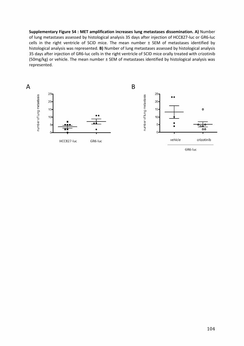
104
Supplementary Figure S4 : MET amplification increases lung metastases dissemination. A) Number of lung metastases assessed by histological analysis 35 days after injection of HCC827-luc or GR6-luc cells in the right ventricle of SCID mice. The mean number ± SEM of metastases identified by histological analysis was represented. B) Number of lung metastases assessed by histological analysis 35 days after injection of GR6-luc cells in the right ventricle of SCID mice orally treated with crizotinib (50mg/kg) or vehicle. The mean number ± SEM of metastases identified by histological analysis was represented.

105
Supplementary Table S1 : RT-qPCR primer sequences.
Target gene Forward primer (5’-3’) Reverse primer (5’-3’) Amplicon size (bp)
beta2 microglobulin
(B2M) GCTGGCGGGCATTCCTGAAG TGCTGGATGACGTGAGTAAACCTG 144
MET TTTCAAATGGCCACGGGACAACACA TGGGCTGGGGTATAACATTCAAGA 222 E-Cadherin
(CDH1) TTTGACGCCGAGAGCTACAC GAATCGGGTGTCGAGGGAAA 131
Vimentin (VIM) AGAGAGGAAGCCGAAAACACC CTGGATTTCCTCTTCGTGGAGT 143
ZEB1 ATGACCTGCCAACAGACCAG GCTTCAGGCCCCAGGATTT 117
Supplementary Materials
Western blot analysis and antibodies
For western blot analysis, 800000 cells were plated on 10 cm dishes in complete
medium. In treatment experiments, drugs were added 24h later. The cells were lysed 48h
after seeding in M-PER lysis buffer (Thermo Scientific, Waltham, Massachusetts) with 1%
HALT protease and phosphatase inhibitor cocktail (Thermo Scientific, Waltham,
Massachusetts). Protein concentrations were determined with the BCA Protein Assay Kit
(Thermo Scientific, Waltham, Massachusetts). Equal amount of total proteins were
separated on SDS-polyacrylamide gel electrophoresis (10%) at 120 V for 2h, and proteins
were transferred at 100 V for 1.5 hours on Immobilon-P polyvinylidene fluoride microporous
transfer membranes (Millipore, Billerica, Massachusetts). Prestained broad-range molecular
mass markers (Thermo Scientific, Waltham, Massachusetts) were used as standards in each
gel. Protein detection was performed using the ECL Western blotting detection Reagent (GE-
Healthcare, Little Chalfont, United Kingdom) or SuperSignal West Dura (Thermo Scientific,
Waltham, Massachusetts) when required. The following antibodies were used at a 1:1000
dilution unless otherwise indicated : MET (Cell Signaling, Danvers, Massachusetts, 3148),
phospho-MET (Tyr1234/Tyr1235) (Cell Signaling, Danvers, Massachusetts, 3126), EGFR (Cell
Signaling, Danvers, Massachusetts, 2646), phospho-EGFR (Tyr1068) (Cell Signaling, Danvers,
Massachusetts, 2236), AKT (Santa Cruz, sc-8312), phospho-AKT (Ser473) (Cell Signaling,
Danvers, Massachusetts, 4060), ERK (Santa Cruz, Dallas, Texas, sc-154), phospho-ERK
(Thr202/Tyr204) (Cell Signaling, Danvers, Massachusetts, 9106), GAPDH (Santa Cruz, Dallas,
Texas, sc-32233). Anti-mouse (Jackson ImmunoResearch, West Grove, Pennsylvania, 115-
035-146) and anti-rabbit (Jackson ImmunoResearch, West Grove, Pennsylvania, 711-035-

106
152) secondary antibodies were diluted at 1:10000. Each experiment was repeated at least
three times.
Quantitative real-time RT-PCR
Total RNA was extracted using the Nucleospin RNA/Protein Kit (Macherey-Nagel,
Düren, Germany) according to the manufacturer’s protocol and reverse-transcribed using
the High Capacity cDNA Reverse Transcriptase Kit (Applied Biosystems, Foster City,
California). Analysis of transcript relative fold copy number was carried out by quantitative
real-time PCR using Fast SYBR Green mix (Applied Biosystems, Foster City, California) in an
MX3005P instrument (Stratagene, San Diego, California) as described by the supplier, with
specific primers listed in supplementary table S3. The relative expression was calculated
using the 2-Ct method. The mRNA levels of each target gene were normalized to the
levels of the housekeeping B2M gene and presented as fold induction relative to the control
DMSO or the untreated HCC827 cell line depending on the experiment. Results were
represented as means of at least three independent experiments.
RNA interference
For silencing, 300000 cells were incubated with 4.5 µL Lipofectamine 2000
(Invitrogen, Carlsbad, California) mixed with 120 nM of a siRNA (Invitrogen, Carlsbad,
California) targeting the genes of interest. The cells were then plated in a 6-well plate in a
final volume of 1.5 mL of complete medium. Transfected cells were trypsinised after 24h soft
agar assays.
The following siRNA were purchased from Life technology : Control Stealth siRNA negative
control, MET [5′-CCGAGGGAAUCAUCAUGAAAGAUUU-3′]. Each experiment was repeated at
least three times.
MTT proliferation assays
At the indicated time point, cells were incubated for 2h with MTT (Invitrogen,
Carlsbad, California) diluted at 0.5mg/ml in 200 µl of medium at 37°C. The resultant
formazan crystals were dissolved in isopropanolol. The absorbance resulting of the dye
uptake in viable cells was detected at 540 nm with a Multiskan RC Microplate Reader
(ThermoLabsystems, Waltham, Massachusetts) with a reference wavelength of 620 nm.

107
Each assay consisted of three replicate wells. Results were represented as means optic
density of at least three independent experiments.
Agar clones count
For each anchorage independent growth assay, 4 fields per well at 2 different z stacks
were randomly photographed with Zeiss Axio Vert.A1 microscope (Zeiss, Oberkochen,
Germany) equipped with a Zeiss N-Acroplan 5x objective (numerical aperture [NA]0.15,
Zeiss, Oberkochen, Germany). Colonies of each field were counted using Fiji software (NIH,
Bethesda, Maryland). Results were illustrated with representative fields of clones and the
means number of clones per field of three independent experiments.
Time lapse microscopy
Time-lapse microscopy was performed on a Zeiss AxioObserverZ1 fluorescence
microscope (Zeiss, Oberkochen, Germany) equipped with a 10x ECPLAN (numerical aperture
[NA] 0.3), an Axio mRm camera, a Lambda DG4 excitation source (Sutter Instrument
Company, Novato, California), a DsRed Filter (Zeiss Filter Set 43HE, Zeiss, Oberkochen,
Germany) and an incubation Unit (XL Unit, Pecon, Erbach, Germany). The interior of the
incubator surrounding the entire microscope system was heated to 37°C and the CO2 level
was regulated to 5%. Cells were labeled with DilC12 (17µg/ml) and seeded at 30000 per well
in 15µ slide 8 wells (Ibidi, Martinsried, Germany) with complete medium 24h before
microscopy. Images were acquired with the Zen software (Zeiss, Oberkochen, Germany) at a
rate of 1 image per time interval of 10 minutes during 6h. The Plugin MTrack2 implemented
in the Fiji software (NIH, Bethesda, Maryland) enabled to obtain the trajectory (xy-path) of
each cell. Cell migration was assessed as the total path length in micrometers
Stable transfection procedure
HCC827 or HCC827GR6 cells were plated on 6-well plates (800000 per well) and
transfected the next day with pGL4.51 vector and FuGENE HD tranfection reagent (Promega,
Madison, Wisconsin ; 2 μg DNA / 8 μl FuGENE reagent in 2 mL complete medium). After 24h
of incubation, cells were cultivated in complete medium with neomycin (600 µg/ml) for 36
days. At the end of the selection step, the stably transfected cell lines were named

108
respectively HCC827-luc and HCC827-GR6-luc and maintained in complete medium without
neomycin.
In vitro bioluminescence imaging
Bioluminescence of HCC827-luc and GR6-luc cell lines were evaluated in 96-wells
plate (BD bioscience, Franklin Lakes, New Jersey) at the indicated concentration in 25 ul of
complete medium per well. Luciferin at 0.3 mg/ml in 25ul of PBS was added in each well. A
bioluminescence imaging was immediately performed with an IVIS Lumina XR apparatus
(Perkin Elmer, Waltham, Massachusetts) at an automatically determined exposure time and
repeated every 5 minutes during 30 minutes. Average radiance (photons-1sec-1cm-2sr-1) of
each well at luminescent peak was quantified using IVIS Living Image version 4.3.1 (Perkin
Elmer, Waltham, Massachusetts) to describe the bioluminescence of both cell lines.
In vivo bioluminescence imaging
Mice were anesthetized with isoflurane 2% and 250000 HCC827-luc or GR6-luc cells
suspended in 100ul of PBS were injected into their heart. Left or right ventricle injection
were controlled by bioluminescence imaging with an IVIS Lumina XR spectrum 40 minutes
after intracardiac injection and 10 minutes after subcutaneous administration of luciferin
150 mg/kg. 35 days after the intracardiac injection, anesthetized mice were injected
subcutaneously with luciferin 150 mg/kg, and bioluminescence imaging was immediately
performed every 5 minutes during 30 minutes with automatically determined time
exposure. Average radiance of each mouse at luminescent peak was calculated for all
experiments using IVIS Live imaging.
Immunohistochemistry
IHC was performed using BenchMark XT (Ventana, Oro Valley, Arizona) automated
IHC stainer using 3,3’-diaminobenzidine (DAB) revelation and hematoxylin counter-staining
(Bluing Reagent, Ventana, Oro Valley, Arizona). MET antibody (Ventana, Oro Valley, Arizona,
790-4430, SP44 clone), prediluted by the manufacturer, was incubated 16 seconds after
basic CC1 pre-treatment. Phospho-MET (Tyr1234/Tyr1235) antibody (Cell Signaling, Danvers,
Massachusetts 3077, D26 clone) was incubated 32 seconds at a 1/100 dilution with basic
CC1 (Ventana, Oro Valley, Arizona) and protease 3 (Ventana, Oro Valley, Arizona)

109
endopeptidase pre-treatment. E-cadherin antibody (Invitrogen, Carlsbad, California, 180223,
4A2C7 clone) is diluted at 1/25 with CC1, and ZEB1 antibody (Novusbio, Littleton, Colorado,
23484, 2A8A6 clone) at 1/500. Vimentin (Ventana, Oro Valley, Arizona, 790-2917, V9 clone)
antibody is pre-diluted by the manufacturer. Immunohistochemistry was evaluated using the
H-score method, corresponding to the percentage of stained tumoral cells at each intensity.
The staining intensity was evaluated within a scale ranging from 0–3 and divided into 4
categories, no staining : 0, weak : 1, intermediate : 2 and strong staining : 3. H-score was
defined as a continuous variable ranging from 0 to 300 and was determined using the
formula : 1 × (percentage of intensity score 1 cells) + 2 × (percentage of intensity score 2
cells) + 3 × (percentage of intensity score 3 cells).
Patients and clinical data analysis
We previoulsy published a multicentric retrospective observational cohort of patients
with a metastatic EGFR mutated NSCLC, exhibiting MET amplification or high MET
overexpression on a re-biopsy performed after TKI EGFR progression identified among 15
French centers (Article 1). Inclusion criteria in this study were documented diagnosis of
metastatic NSCLC, detection of an EGFR mutation on tumor sample at diagnosis, treatment
with at least one EGFR TKI and detection of MET overexpression or MET amplification after
the time of clinical or radiological progression on EGFR TKI. The study was approved by a
national ethic committee (CEPRO 2016-001). MET overexpression was defined as a 3+ MET
Immunoscore (≥ 50% of tumor cells showing high-intensity staining) on MET
immunochemistry using MET monoclonal antibody (clone SP44 Ventana). MET amplification
was defined by Fluorescence In Situ Hybridization (FISH) as a mean MET GNC per cell ≥ 6, or
a Ratio MET/CEP7 ≥ 2, or the presence of MET clusters. In the present work, the time to new
metastasis after EGFR TKI tumoral progression was measured from the date of EGFR TKI
RECISTv1.1 progression to the date of first progression with a new lesion after EGFR TKI
progression. The time to new metastasis after EGFR TKI tumoral progression were described
with Kaplan-Meier Method and compared between the study groups with log-rank test.
Hazard ratios were obtained with Cox model. Bivariate comparisons between the two study
groups (amplified vs non amplified) were made using Student t test for Gaussian continuous
variables, Mann-Whitney U test for non-Gaussian continuous, Chi-Square test (or Fisher’s

110
exact test when the expected cell frequency was <5) for categorical variables, as
appropriate. All statistical testing was conducted at the 2-tailed α level of 0.05. Data were
analyzed with GraphPad Prism software version 7 (GraphPad software, San Diego California).

111

112
Article 3 (en révision, La revue des maladies respiratoires) : Les mutations des sites
d’épissage de l’exon 14 de MET
Sous-titre : Une nouvelle opportunité thérapeutique dans le cancer du poumon
Simon BALDACCI1, 2, Zoulika KHERROUCHE1, Clotilde DESCARPENTRIES3, Marie
WISLEZ4, Eric DANSIN5, Alessandro FURLAN1, 6, David TULASNE1, Alexis B.
CORTOT*1,2.
1 Univ. Lille, CNRS, Institut Pasteur de Lille, UMR 8161 - M3T – Mécanismes de
tumorigenèse et thérapies ciblées
2 Univ. Lille, CHU Lille, Service de Pneumologie et Oncologie Thoracique, Siric OncoLille
3 CHU Lille, Plateforme de Biologie Moléculaire des cancers, Service de Biochimie et
Biologie Moléculaire
4 AP-HP, GH HUEP, Hôpital Tenon, Service de Pneumologie Paris, France. GRC n°04,
Theranoscan, Sorbonne Universités, UPMC Univ Paris 06, Paris, France.
5 CLCC Oscar Lambret, Lille, Département de Cancérologie Cervico-Faciale & Thoracique
6 Univ. Lille, CNRS UMR 8523 PhLAM, Equipe de Biophotonique Cellulaire Fonctionnelle,
Titre court : Les mutations de MET dans le cancer du poumon
*Auteur-correspondant : Alexis Cortot,. Service de Pneumologie et Oncologie Thoracique,
Hôpital Calmette, CHRU de Lille, 59000, Lille, France. [email protected]

113
S.B. a reçu des financements permettant une participation à des formations, des
communications, des congrès et des réunions de groupe d'expert de la part de Pfizer, GSK,
Lilly, et de Astrazeneca
A.B.C. a reçu des honoraires de Novartis, Pfizer, Astra-Zeneca, Boehringer-Ingelheim, MSD,
BMS, Roche
E.D. a reçu des honoraires pour la participation aux comités scientifiques de Novartis, BMS,
Lilly et de Pfizer et un financement de Roche pour une étude Clinique (2014)
Les autres auteurs ne déclarent aucun conflit d’intérêt.

114
Résumé
(Nombre de mots : 124)
Les mutations conduisant à un saut de l’exon 14 du gène codant pour le récepteur tyrosine
kinase (RTK) MET constituent une nouvelle classe d’altérations moléculaires décrites dans
différentes pathologies néoplasiques dont les cancers bronchiques non à petites cellules
(CBNPC). Ces mutations, appelées MET14 semblent associées à une efficacité des
inhibiteurs de tyrosine kinase (ITK) de MET. Cette synthèse revient sur les mécanismes
moléculaires responsables du saut de l'exon 14 de MET ainsi que sur les conséquences de la
perte de cet exon sur l’activité du récepteur. Nous décrivons ensuite les caractéristiques
cliniques des patients porteurs d’une mutation MET14. Enfin, nous abordons les
problématiques liées à la détection de ces mutations dans les cancers pulmonaires, et la
nécessité d’anticiper la résistance aux inhibiteurs de MET.
Mots clés : Cancer du poumon, MET, Récepteur tyrosine kinase, Thérapies ciblées

115
Titre : Les mutations des sites d’épissage de l’exon 14 de MET
Title : MET exon 14 splicing sites mutations : a new therapeutic opportunity in lung
cancer
Abstract
(107 words)
The mutations leading to MET exon 14 skipping represent a new class of molecular
alterations described in various cancers including non-small cell lung cancer (NSCLC).
Several cases of NSCLC carrying such alterations and displaying objective response to MET
tyrosine kinase inhibitors (TKIs) were recently published. This review summarizes the
molecular mechanisms responsible for MET exon 14 skipping as well as the consequences of
the loss of this exon on the receptor activity. We also describe the clinical characteristics of
patients with MET14 mutations. Finally, we address the issues related to the detection of
these mutations in lung cancers, and the need to anticipate resistance to MET inhibitors.
Keywords : Lung cancer, MET, Tyrosine kinase receptor, Targeted-therapies

116
Le récepteur MET
MET est un récepteur tyrosine kinase (RTK) principalement exprimé dans les cellules
d’origine épithéliale. Il est activé par son ligand l’HGF (Hepatocyte Growth Factor également
appelé Scatter Factor) [1]. MET est constitué de deux sous-unités α et β reliées par un pont
disulfure. Le domaine extracellulaire du récepteur comprend un domaine SEMA en N-
terminal (formé de la sous-unité α ainsi que des premiers acides aminés de la sous-unité β), un
domaine plexine-semaphorine-integrine (PSI) et 4 domaines immunoglobulin-like (IPT). La
région intracellulaire contient un domaine juxtamembranaire, un domaine tyrosine kinase et
une queue C-terminale (figure 1) [2,3]. L’interaction de MET avec son ligand provoque sa
dimérisation et son auto-phosporylation sur deux résidus tyrosine du domaine catalytique
(Y1234 et Y1235). D’autres résidus tyrosine, situés en dehors du domaine kinase, comme la
tyrosine 1003 dans le domaine juxtamembranaire et les tyrosines 1349 et 1356 de la queue C-
terminale, sont à leur tour phosphorylés (figure 1) [4,5]. La phosphorylation des résidus situés
en C-terminal permet le recrutement de plusieurs effecteurs de signalisation importants
comme GAB1, GRB2, SHC, PI3K, SRC, STAT3 et la PLC. La voie HGF/MET est
essentielle au développement embryonnaire. Elle est en particulier nécessaire à la formation
des organes épithéliaux, du foie, du placenta, des muscles, des neurones et des alvéoles
pulmonaires [6–10]. Chez l’adulte, elle joue par ailleurs un rôle important dans plusieurs
processus physiologiques comme la cicatrisation cutanée ou la régénération hépatique [11–
13].

117
Figure 1 : Représentation des principaux domaines fonctionnels du récepteur MET. Les résidus phosphorylables sont indiqués en rouge.
Les dérégulations de MET dans les cancers
En pathologie humaine, une activation de la voie HGF/MET est observée dans de
nombreux cancers. Plusieurs mécanismes de dérégulation de cette voie peuvent être impliqués
tels que la surexpression de MET ou de son ligand, l’amplification du gène codant MET ou
encore les mutations activatrices du domaine kinase du récepteur (figure 2). La surexpression
de l’HGF ou de MET est constatée dans de nombreux cancers et se trouve fréquemment
associée à un mauvais pronostic [14–20]. La sécrétion d’HGF par la tumeur est de plus
associée à une résistance aux inhibiteurs de tyrosine kinase (ITK) d’EGFR dans les cancers
bronchiques non à petites cellules (CBNPC) EGFR mutés [21,22]. La valeur prédictive de la

118
surexpression de MET d’une efficacité des inhibiteurs de MET n’a pas été démontrée [23]
L’amplification du gène MET est, quant à elle, retrouvée dans 1 à 5% des cancers gastriques
et pulmonaires [24–26]. Cette altération est responsable d’une synthèse massive du récepteur,
ce qui provoque son activation, indépendamment de la présence de son ligand. Plusieurs
études préliminaires suggèrent que l’amplification de MET est associée à une efficacité des
inhibiteurs de MET dans les CBNPC [27–29] Par ailleurs, l’amplification de MET est un
mécanisme de résistance aux inhibiteurs d’EGFR dans les CBNPC EGFR-mutés [30]. Enfin,
les mutations du domaine kinase de MET ont d’abord été découvertes au niveau germinal
chez des patients atteints de cancers rénaux héréditaires [31]. Par la suite, des mutations
somatiques du domaine kinase de MET ont été identifiées dans des cancers rénaux
sporadiques, et plus rarement dans des cancers pulmonaires et ORL. Ces mutations, comme la
D1228N, la Y1235D et la M1250T, sont capables d’induire une activation constitutive de
l’activité kinase de MET et semblent associées à une efficacité des inhibiteurs de MET
[32,33].
Le développement du screening moléculaire massif en cancérologie a récemment
permis la mise en évidence, dans les CBNPC, d’une nouvelle classe de mutations
dérégulatrices de MET. Ces mutations, appelées MET14, sont situées à proximité des
jonctions intron-exon de part et d’autre de l’exon 14. Elles touchent des sites critiques de
l’ARN pré-messager de MET, normalement reconnus par des ribonucléoprotéines nucléaires
lors du processus d’épissage (point de branchement, séquence polypyrimidique, sites
d’épissage donneurs et accepteurs). Une altération de ces sites aboutit à un saut de l’exon 14
au cours de son épissage. Plusieurs cas de réponses tumorales aux ITK de MET ont de plus
été décrits chez des patients présentant une mutation MET14.

119
Figure 2 : Les différents modes de dérégulation de la voie HGF/MET dans les cancers.
La découverte des mutations responsables d’un saut de l’exon 14 de MET dans les CBNPC
En 2003, Ma et al. rapportent pour la première fois l’existence de mutations
introniques en amont de l’exon 14 de MET susceptibles d’induire un épissage alternatif de
l’ARN messager, responsable d’un saut de l’exon 14 [34]. L’analyse de l’ADN
complémentaire d’une cohorte de 127 CBNPC a ensuite permis de mettre en évidence cet
épissage et la perte de l’exon 14 chez 1 patient [35]. Le lien entre mutations somatiques
affectant les sites d’épissage encadrant l’exon 14 et saut d’exon a finalement été formellement
démontré en 2006 grâce à la comparaison des séquences d’ADN génomique et d’ADN

120
complémentaire de 2 biopsies d’adénocarcinome bronchique et de la lignée cellulaire issue
d’un CBNPC, NCI-H596 (ATCC HTB-178) [36]. Par ailleurs, d’autres mutations ont été
décrites, affectant cette fois directement l’exon 14 et non les sites d’épissage. Toutefois ces
mutations dont la prévalence reste mal connue ont des conséquences fonctionnelles très
limitées, ne provoquent pas d’activation de MET, et ne semblent pas associées à une efficacité
des inhibiteurs de MET. Nous ne les aborderons pas dans cette revue [37–39].
En 2014, la caractérisation de 230 adénocarcinomes pulmonaires par séquençage de
l’exome combiné au séquençage du transcriptome a révélé un taux relativement élevé
(environ 3%) de mutations somatiques survenant sur les sites d’épissage de l’exon 14 et
conduisant à un saut de l'exon 14 [40]. Un an plus tard, l’analyse de l’ADN génomique de
38028 tumeurs par séquençage de nouvelle génération a confirmé la présence de ces
mutations dans 3% des adénocarcinomes pulmonaires, mais également dans 2,3 % des
tumeurs pulmonaires non adénocarcinomateuses, et dans 0,4% des gliomes. Cette étude a
aussi mis en évidence une grande diversité au sein de ces mutations avec l’identification de
126 variants [41]. Plus de 160 altérations différentes, touchant des séquences consensus
nécessaires à un épissage normal, ont depuis été répertoriées (figure 3A) [42]. A l’extrémité 5’
de l’exon 14, ces anomalies correspondent en majorité à des délétions, à des insertions, ou à
des modifications complexes combinant à la fois des délétions et insertions. Elles touchent
principalement le site d’épissage accepteur, le point de branchement ou la séquence
polypyrimidique. Les altérations situées à l’extrémité 3’ de l’exon 14 sont principalement des
mutations ponctuelles affectant le site donneur d’épissage. Toutes ces altérations sont
susceptibles d’entrainer le saut de l’exon 14 lors de l’épissage.

121
Figure 3 : Implication de l’exon 14 dans la régulation de MET (A) Schéma de l’exon 14 situé entre les positions génomiques g.116411902 et g.1164122042 (Homo sapiens chromosome 7, GRCh37, GenBank: CM000669.1) ou entre les positions c.2888 et c.3088 (transcript ID ENST00000397752) de l’ADN complémentaire. Le point de branchement, la séquence polypyrimidique et les sites accepteur et donneur d’épissage sont représentés. Entre parenthèse : prévalence de chaque type de mutation MET14 dans Frampton et al. parmi l’ensemble de patients
muté MET14. Sur la séquence d’acides aminés codée par l’exon 14, les principaux sites de régulation négative sont respectivement représentés en rouge.

122
Conséquences fonctionnelles du saut de l’exon 14 sur la régulation de MET
Bien que l’étude des mutations induisant la perte de l’exon 14 soit relativement
récente en cancérologie, l’implication du domaine juxtamembranaire codé par cet exon dans
le contrôle de l’activité de MET est connue depuis les années 1990. En effet plusieurs sites de
régulation négative de MET ont été caractérisés dans la région du domaine juxtamembranaire
codée par l’exon 14.
Le premier mécanisme de régulation négative identifié dans le domaine
juxtamembranaire est la phosphorylation de la sérine 985 (figure 3A). Cette phosphorylation
inhibe la phosphorylation des résidus tyrosine du domaine kinase et C-terminal de MET, et
permet ainsi de contrôler la réponse cellulaire au ligand, l’HGF [43]. La sérine 985 est
phosphorylée par la protéine kinase C (PKC) et déphosphorylée par la phosphatase 2A
(PP2A) [44]. Ainsi, après une lésion hépatique, l’activation de MET par l’HGF, requise pour
la régénération tissulaire, est associée à une diminution de la phosphorylation de la sérine 985
[45]. Ceci suggère l’implication de la sérine 985 dans la régulation physiologique de l’activité
de MET. La délétion de l’exon 14 pourrait donc conférer une résistance aux signaux
inhibiteurs transmis par la PKC et ainsi favoriser l’activation de MET (figure 3B).

123
Figure 3 : Implication de l’exon 14 dans la régulation de MET (B) Schéma du récepteur MET, activé par son ligand l’HGF. Le domaine juxta-membranaire est le site de plusieurs mécanismes de régulation négative telles que la phosphorylation PKC-dépendante de la sérine 985 qui conduit à l’inhibition de l’activité tyrosine kinase du récepteur, ou la phosphorylation ligand dépendante de la tyrosine 1003 qui entraine le recrutement de l’E3-ubiqitine ligase sur le site DpYR et provoque ainsi l’ubiquitiniation suivie de la dégradation lysosomale du récepteur. La
délétion du domaine juxta-membranaire codé par l’exon 14 pourrait conduire à une augmentation de la
stabilité du récepteur et de son activité tyrosine kinase. Le ciblage de MET par des inhibiteurs de tyrosine kinase tel que le crizotinib, le capmatinib et le cabozantinib pourrait constituer une stratégie thérapeutique efficace chez les patients atteints d’une tumeur porteuse d’une mutation perte de l’exon
14 de MET.
Le deuxième résidu du domaine juxtamembranaire impliqué dans l’inhibition de MET
est la tyrosine 1003 (figure 3A). Après l’activation de MET, la Tyr1003 est phosphorylée
permettant le recrutement de l’E3 ubiquitine ligase CBL (Casitas B-lineage Lymphoma) via
notamment son domaine TKB (Tyrosine Kinase Binding domain). Ceci entraine une
ubiquitination de MET et induit la dégradation du récepteur (figure 3B) [46]. Une mutation de
la Tyr1003 est suffisante pour bloquer l’interaction de MET avec CBL et son ubiquitination.
124
Ceci conduit à une stabilisation du récepteur et à un potentiel oncogénique plus important de
la forme MET mutée Tyr1003 comparée à la forme sauvage [47].
Le troisième site de régulation négative présent dans l’exon 14 est un site impliqué
dans les clivages de MET par les caspases durant l’apoptose. Sous l’effet d’un stress et en
l’absence de ligand, MET est d’abord clivé sur un site consensus caspase au niveau de sa
queue C-terminale. Ce clivage initial est suivi d’un second clivage caspase au niveau de
l’acide aspartique 1002 situé dans un motif ESVD codé par l’exon 14 (figure 3A). Ceci
permet la formation d’un fragment cytoplasmique C-terminal de 40 kDa (p40-MET)
contenant le domaine tyrosine kinase [48,49]. Ces clivages caspases diminuent fortement la
quantité de récepteur MET pleine longueur fonctionnel pendant l’apoptose. Ils permettent
également de former un fragment de MET capable d’amplifier l’apoptose en favorisant la
perméabilisation mitochondriale [50]. Les clivages caspases de MET constituent ainsi un
mécanisme de régulation négative supplémentaire, situé dans le domaine juxtamembranaire et
perdu par épissage de l’exon 14.
La partie du domaine juxtamembranaire de MET codé par l’exon 14 comprend donc
plusieurs sites qui permettent de prévenir l’activation de MET ou de convertir MET en un
facteur pro-apoptotique. L’épissage de cet exon pourrait donner aux cellules porteuses d’une
mutation MET14 un avantage en termes de prolifération et de survie.
Caractéristiques histologiques, cliniques, et moléculaires des cancers présentant des
mutations induisant un saut de l’exon 14 de MET
Les mutations MET14 ont été observées dans différents types de tumeurs dont les
cancers pulmonaires (2.6% à 22%), les glioblastomes (0.4%) et plus rarement dans d’autres
pathologies néoplasiques (sarcomes des tissus mous, carcinomes urothéliaux, carcinomes à
cellules de Merkel, cortico-surrénalomes, cancers du rein, carcinomes hépatocellulaires)

125
[41,42,51]. Parmi les cancers pulmonaires, le taux de mutations des sites d’épissage de l’exon
14 semble particulièrement élevé dans les adénocarcinomes (2.6 à 3.2%) [41,52,53], les
tumeurs sarcomatoides (2.6% à 31.8%) [39,42,52–54], et les carcinomes adénosquameux (4 à
8,2%) [42,52]. Les mutations MET14 ont également été observées à des fréquences plus
faibles dans d’autres tumeurs pulmonaires : les cancers épidermoïdes (2,1%), les carcinomes à
grandes cellules (0.8%), les CBNPC sans autre spécificité, et les cancers bronchiques à petites
cellules [41,42,52,53,55].
Schrock et al. ont décrit les caractéristiques cliniques d’une série de 298 patients
atteints d’un cancer pulmonaire avec une mutation des sites d’épissage de l’exon 14 de MET.
Il s’agit de patients âgés (âge médian : 73 ans) en majorité de sexe féminin (60%). Dans cette
étude, les données concernant le statut tabagique n’étaient disponibles que pour 36 patients
dont 25 étaient non-fumeurs. La faible charge mutationnelle observée dans les tumeurs
MET14, suggère effectivement qu’une proportion importante de ces patients pourraient être
non-fumeurs [42]. D’autres études retrouvent des caractéristiques cliniques similaires à celles
rapportées dans l’étude de Schrock, se traduisant principalement par un âge plus élevé que
dans les autres CBNPC, un sex-ratio proche de 1, et une proportion de non-fumeurs de 30 à
50%. L’impact des mutations MET14 sur la survie n’a pas encore été étudié de façon
exhaustive. Tong et al. ont cependant réalisé une analyse de survie sur 687 patients atteints de
CBNPC de différents stades. Dans cette étude, l’analyse multivariée montre que les mutations
des sites d’épissage de l’exon 14 de MET, l’amplification de MET et le stade avancé sont des
facteurs indépendants de mauvais pronostic [53]. Enfin, dans les populations asiatiques
atteintes d’adénocarcinome bronchique, la fréquence de mutations MET14 semble plus
faible que dans la population caucasienne (0.9 à 1.8%) [56,57].
Les mutations entrainant la perte d’exon 14 ont été détectées principalement par des
techniques recherchant de façon multiplexée de nombreuses autres altérations génétiques

126
[36,37,41,42,51,54,58–62]. Dans la plupart des cas, ces mutations ne sont pas associées à
d’autres altérations de drivers oncogéniques comme les mutations EGFR, KRAS, HER2, ou
les réarrangements impliquant les gènes ALK, ROS1, et RET. Plusieurs publications ont
néanmoins observé une association fréquente avec certaines altérations génétiques touchant la
voie p53. Par exemple, dans l’étude de Schrock et al., 34.6% des tumeurs avec une mutation
des sites d’épissage de l’exon 14 présentaient une amplification de MDM2 [42]. Plus rarement
des mutations de BRAF, de KRAS ou de la PI3KCA ont été détectées en association avec des
mutations MET14 [52,53].
Plusieurs travaux ont décrit une association entre les mutations des sites d’épissage de
l’exon 14 de MET et l’amplification de MET. Grâce à une analyse NGS, Awad et al. ont par
exemple évalué le nombre de copies de MET dans des CBNPC de différents stades. Dans
cette étude, un ratio MET/chromosome 7 (CEP7) supérieur à 3 définissait un gain de copie de
MET considéré comme élevé, tandis qu’un ratio compris entre 1 et 3 représentait un gain de
copie considéré comme bas. Avec ces critères, un gain élevé de copie de MET a été observé
chez 21% des patients avec une mutation des sites d’épissage de l’exon 14 et un gain faible
chez 29% [52]. De façon intéressante, le ratio MET/CEP7 était significativement plus élevé
dans les CBNPC porteurs d’une mutation MET14 de stade IV comparés aux stades I-III,
suggérant que l’amplification de MET pourrait favoriser la progression des tumeurs MET14.
Tong et al. ont également mis en évidence une association significative entre les mutations
des sites d’épissage de l’exon 14 de MET et l’augmentation du nombre de copies de MET
définie en FISH par un nombre de copies de MET ≥5, un ratio MET/CEP7 ≥2 ou la présence
de cluster de MET [53]. Enfin Schrock a observé par NGS une prévalence relativement élevée
de l’amplification de MET en cas de mutation de perte de l’exon 14 de MET (14.8%) [42].
Comme la perte de l’exon 14 de MET empêche la dégradation du récepteur, une
augmentation de l’expression de MET sur les prélèvements tumoraux était attendue. Tong et

127
al. ont ainsi montré une association significative entre les mutations des sites d’épissage de
l’exon 14 de MET et l’intensité du marquage en IHC de MET [53]. Cependant, il est important
de noter qu’au moins une mutation des sites d’épissage de l’exon 14 de MET a été détectée
dans un prélèvement tumoral avec un faible niveau d’expression de MET en IHC [52].
Les mutations induisant un saut de l’exon 14 de MET, un biomarqueur prédictif de
l’efficacité des inhibiteurs de MET ?
MET est perçu comme une cible potentielle pour le traitement de plusieurs cancers.
Plus de 300 essais cliniques sont actuellement en cours pour évaluer l’efficacité et la tolérance
de thérapies dirigées contre MET, ou contre son ligand l’HGF. Bien que plusieurs études
évaluant des approches thérapeutiques ciblant MET dans des populations non sélectionnées
ont rapporté des résultats décevants, des données récentes suggèrent que des inhibiteurs de
MET comme le crizotinib (ITK ciblant à la fois MET, ALK et ROS1, et disposant d’une
AMM dans le traitement des cancers pulmonaires réarrangés ALK ou ROS1) pourraient être
actifs contre certaines tumeurs présentant une amplification de MET [29]. La découverte des
mutations des sites d’épissage de l’exon 14 de MET a rapidement conduit à évaluer
l’efficacité des inhibiteurs de MET dans ces tumeurs, d’abord de façon sporadique puis dans
le cadre d’essais cliniques évaluant différents ITK MET comme le crizotinib, le capmatinib,
le tepotinib ou le savolitinib (tableau 1).

128
Tableau 1 : essais cliniques ciblant les CBNPC avec des mutations des sites d'épissage de l'exon 14 de MET (ClinicalTrial.gov juillet 2017)

129
Plusieurs cas de patients avec une mutation MET14, présentant une réponse
objective après un traitement par un ITK MET, ont été rapportés (tableau supplémentaire 1)
[41,42,52,54–56,63,64]. Ces patients étaient tous porteurs d’un cancer du poumon (le plus
souvent un adénocarcinome), à l’exception d’un cas de sarcome histiocytique [41]. Les
inhibiteurs de MET utilisés étaient le crizotinib, le capmatinib, et le cabozantinib. Les
résultats préliminaires d’une étude de phase 1, l’essai PROFILE 1001, qui évalue un
traitement par crizotinib chez les patients atteints d’un CBNPC de stade avancé porteur d’une
mutation des sites d’épissage de l’exon 14 de MET, ont rapporté 11 réponses partielles sur 28
patients évaluables, soit un taux de réponse de 39% (IC95% 22-59), avec une survie sans
progression médiane de 8 mois (IC95% 6,9-10,8) [65].
Bien que l’interprétation de l’ensemble de ces données soit limitée par le petit nombre de
patients, et par des biais de sélection, le taux et la durée des réponses tumorales rapportées
sont compatibles avec celles obtenues avec les thérapies ciblées dans d’autres modèles
d’addiction oncogénique. Il est important de noter que les réponses obtenues sous ITK MET
concernaient à la fois des patients fumeurs et non-fumeurs, des patients avec ou sans gain de
copie de MET, et des patients avec ou sans atteinte de la voie p53. De façon surprenante, un
patient atteint d’un CBNPC à la fois porteur d’une mutation des sites d’épissage de l’exon 14
de MET et d’une mutation activatrice de PIK3CA a présenté une réponse partielle au
crizotinib [59]. Cette observation va à l’encontre des données de réponse au crizotinib
obtenues in vitro avec la lignée NCI-H596 également porteuse d’une mutation MET14 et
d’une mutation PIK3CA [56]. Plusieurs essais cliniques, de phase I ou II, évaluant différents
ITK MET sont actuellement en cours dans le traitement des cancers mutés MET. Ils évaluent
notamment le crizotinib (NCT02034981, NCT00585195, NCT02465060 (subprotocol C2), le
capmatinib (NCT02414139, NCT02750215), le tepotinib (NCT02864992), le merestinib
130
(NCT02920996), le glesatinib (NCT02544633), le sitravatinib (NCT02219711) et le
savolitinib (NCT02897479) (tableau 1).
Perspectives
Un nouveau type de mutations en oncologie thoracique
Jusqu’à présent, les mutations des récepteurs tyrosine kinase connues pour induire une
addiction oncogénique dans le cancer pulmonaire étaient principalement des mutations
activatrices du domaine tyrosine kinase. Les mutations des sites d’épissage de l’exon 14 sont
d’un autre ordre. Elles entrainent la perte d’une partie du récepteur MET, ce qui a comme
conséquence de supprimer plusieurs sites de régulation négative. Du fait de ce mécanisme
inédit, toutes les conséquences fonctionnelles de ces mutations n’ont pas encore été explorées.
Il est ainsi difficile de savoir si une stimulation par l’HGF est nécessaire à l’activation du
récepteur MET amputé de l’exon 14.
Il est par ailleurs important de noter qu’à la différence des mutations EGFR et des
réarrangements ALK, les mutations perte d’exon 14 de MET surviennent régulièrement chez
les patients fumeurs, ce qui pourrait avoir un impact important puisque que ce tabagisme est
associé à une plus haute charge mutationnelle [42] et une sensibilité moindre aux ITKs . De
plus, l’existence de mutations dans d’autres oncogènes pourrait affecter la sensibilité aux ITK
des tumeurs mutées MET, comme cela a été constaté avec le crizotinib dans la lignée NCI-
H596 porteuse également d’une mutation PIK3CA. Les altérations fréquentes de la voie p53
pourraient refléter la forte proportion de fumeurs parmi les patients porteurs d’une mutation
des sites d’épissage de l’exon 14 de MET et pourraient entraver la réponse aux ITK MET.
Cependant des cas de réponses objectives ont été rapportés malgré la présence d’une
amplification de MDM2 ou d’une mutation TP53 [41,61,63].
131
La sensibilité des tumeurs porteuses de mutations MET14 à d’autres traitements que
les thérapies ciblées a été très peu étudiée. Sabari et al. ont rapporté un faible taux de réponse
(6,7%) parmi 15 patients traités par immunothérapie pour un CBNPC [66]. Dans une analyse
translationnelle associée à cette étude, on observait un niveau d’expression de PDL1 plutôt
élevé (46% de tumeurs avec expression de PDL1 dans plus de 50% des cellules tumorales)
mais une charge mutationnelle moins élevée que dans des tumeurs sans mutation MET14.
La recherche des mutations des sites d’épissage de l’exon 14 de MET : un défi en
pratique clinique
- Comme les mutations des sites d’épissage de l’exon 14 de MET sont présentes
chez un nombre restreint de patients, il est nécessaire de mettre en place une stratégie efficace
pour identifier ces altérations afin de sélectionner les patients susceptibles de bénéficier des
thérapies ciblant MET. Le NGS est une approche attractive dans ce contexte, du fait de sa
large implantation dans les laboratoires de biologie moléculaire et de sa capacité à détecter
simultanément un grand nombre d’altérations génétiques. Cependant, les régions introniques
sont rarement incluses dans les panels ciblés de NGS ainsi que dans les analyses
bioinformatiques. Le premier défi est donc d’adapter ces techniques de séquençage pour
détecter toutes les mutations capables d’induire une perte de l’exon 14 de MET. Une
optimisation de l’analyse bioinformatique est également requise pour détecter ces mutations
de tailles et de séquences variables comme l’illustre l’étude de Frampton et al. dans laquelle
les 120 altérations génétiques différentes aboutissant à une perte de l’exon 14 de MET ont été
identifiées à l’aide d’une analyse bioinformatique appropriée [41]. La quantité et la qualité de
l’ADN ainsi que le nombre limité de cellules tumorales des prélèvements fixés au
formaldéhyde et inclus en paraffine (FFPE) sont également des paramètres importants
pouvant affecter l’efficacité de détection de ces mutations. Une autre approche serait de

132
détecter la perte de l’exon 14 directement sur l’ARN. Cependant, le séquençage de l’ARN se
heurte à des problèmes de coût, de quantité de matériel nécessaire et à une plus délicate
utilisation sur les prélèvements fixés que congelés, rendant son utilisation encore peu
compatible avec la pratique clinique.
L’émergence prévisible de mécanismes de résistance aux inhibiteurs de MET
Comme dans les CBNPC avec mutation EGFR ou réarrangement ALK, il est anticipé
que le traitement des CBNPC avec mutation MET par thérapies ciblées sera systématiquement
suivi d’une progression tumorale due à l’émergence de mécanismes de résistance. Les
mécanismes de résistances aux thérapies ciblées impliquent généralement des mutations du
domaine kinase qui restaurent l’affinité pour l’ATP et/ou altèrent l’affinité aux ITK [67]. Il a
ainsi été montré dans des modèles pré-cliniques d’addiction oncogénique à MET que les
mutations des résidus D1228 et Y1230 du domaine kinase de MET sont responsables d’une
résistance aux inhibiteurs de MET [68,69]. Plusieurs cas cliniques de CBNPC MET14
présentant une résistance au crizotinib avec des mutations touchant ces résidus ont d’ailleurs
été récemment publiés [70–74]. D’autres mécanismes de résistance au ITK MET ont
également été décrits dans des tumeurs non mutées MET présentant une addiction
oncogénique à MET [69,75–79]. Il s’agit notamment d’altérations de MYC, de surexpression
de ABCB1, de mutations de KRAS, d’amplifications de HER2, d’activation d’EGFR, ou de
fusions de BRAF. Il est possible que de telles altérations participent à la résistance aux ITK
MET dans les tumeurs présentant une mutation MET14. La construction de lignées
cellulaires porteuses d’une mutation des sites d’épissage de l’exon 14 de MET et devenues
résistantes aux ITK MET, ainsi que la constitution de séries de prélèvements tumoraux pré- et
post- résistance aux ITK MET, pourraient permettre d’identifier et d’étudier les mécanismes à
l’origine de résistance.

133
Inversement les mutations des sites d’épissage de l’exon 14 de MET pourraient
conférer une résistance aux ITK ciblant d’autres récepteurs tyrosine kinase. De façon
intéressante un adénocarcinome pulmonaire initialement porteur d’une mutation activatrice de
l’EGFR et d’une mutation T790M a présenté au cours de sa prise en charge une mutation
MET14 [51]. Bien que cela ne soit pas décrit dans cette étude, il est raisonnable de suspecter
que la mutation induisant un saut de l’exon 14 de MET puisse conférer une résistance au ITK
EGFR dans les cancers EGFR mutés de la même façon que l’amplification de MET, ou la
surexpression d’HGF [22,30]. La comparaison de prélèvements tumoraux de patients avec un
CBNPC EGFR muté avant et après traitement par ITK EGFR pourrait permettre d’identifier
des cas impliquant les mutations MET14 dans la résistance.
Conclusion
Au total, les données compilées ici suggèrent que les patients atteints de CBNPC avec
mutation des sites d’épissage de l’exon 14 de MET peuvent tirer bénéfice d’un traitement par
inhibiteurs de MET. Bien que l’existence de processus biologiques conduisant à un saut de
l’exon 14 de MET soit connue depuis plus d’une décennie, ces mutations n’ont été mises en
évidence dans un nombre important de tumeurs humaines que depuis 3 ans. L’amélioration
des techniques de séquençage et d’analyses bioinformatiques permettra probablement de
mettre en évidence l’implication de sauts d’exon dans le développement d’autres pathologies
humaines. Ainsi, il a récemment été montré que des mutations germinales induisant un saut de
l’exon 14 de MET étaient responsables d’une forme autosomique dominante de dysplasie
ostéo-fibreuse (DOF) [80]. Il parait plutôt déroutant de constater que ces mutations germinales
n’induisent pas un plus haut risque de pathologies néoplasiques. Une explication pourrait être
que les mutations MET14 nécessiteraient la survenue d’autres altérations génétiques pour

134
induire un processus cancéreux, comme cela est suggéré par leur fréquente association avec
les amplifications de MDM2.
De façon plus générale, l’identification d’un type d’altération moléculaire inédit
affectant l’épissage ouvre des perspectives nouvelles. Par exemple, l’épissage alternatif du
récepteur tyrosine kinase RON, apparenté à MET, a été découvert dans des xénogreffes
d’adénocarcinomes pancréatiques dérivés de patients. Cette altération était associée in vitro à
des capacités oncogéniques [81]. Comme dans le cas des mutations des sites d’épissage de
l’exon 14 de MET, de nouvelles études sont nécessaires pour comprendre les conséquences
fonctionnelles et l’implication clinique de telles altérations.
Points forts généraux :
- Les mutations des sites d’épissage de l’exon 14 du récepteur MET sont observées dans
2 à 3% des cancers bronchiques non à petites cellules.
- Il s’agit d’altérations majoritairement introniques, responsables d’un défaut de
maturation de l’ARN messager de MET induisant un saut de l’exon 14 du récepteur.
- L’exon 14 de MET code pour un domaine de régulation négative du récepteur ; sa
perte entraine une dérégulation de la signalisation de MET.
- Du fait de leur grande diversité, les mutations de sites d’épissage de MET nécessitent
une optimisation des techniques de biologie moléculaire.
- Plusieurs cas de réponse aux inhibiteurs de MET chez des patients atteints de cancer
bronchique non à petites cellules présentant des mutations des sites d’épissage de
l’exon 14 de MET ont été publiés.
- Des mécanismes de résistance aux inhibiteurs de MET ont déjà été identifiés,
consistant en l’acquisition d’une mutation du domaine kinase de MET.

135
References
[1] Birchmeier C, Birchmeier W, Gherardi E, Vande Woude GF. Met, metastasis, motility and more. Nat Rev Mol Cell Biol 2003;4:915–25. doi:10.1038/nrm1261.
[2] Schiering N, Knapp S, Marconi M, Flocco MM, Cui J, Perego R, et al. Crystal structure of the tyrosine kinase domain of the hepatocyte growth factor receptor c-Met and its complex with the microbial alkaloid K-252a. Proc Natl Acad Sci U S A 2003;100:12654–9. doi:10.1073/pnas.1734128100.
[3] Furlan A, Kherrouche Z, Montagne R, Copin M-C, Tulasne D. Thirty years of research on met receptor to move a biomarker from bench to bedside. Cancer Res 2014;74:6737–44. doi:10.1158/0008-5472.CAN-14-1932.
[4] Longati P, Bardelli A, Ponzetto C, Naldini L, Comoglio PM. Tyrosines1234-1235 are critical for activation of the tyrosine kinase encoded by the MET proto-oncogene (HGF receptor). Oncogene 1994;9:49–57.
[5] Ponzetto C, Bardelli A, Zhen Z, Maina F, dalla Zonca P, Giordano S, et al. A multifunctional docking site mediates signaling and transformation by the hepatocyte growth factor/scatter factor receptor family. Cell 1994;77:261–71.
[6] Bladt F, Riethmacher D, Isenmann S, Aguzzi A, Birchmeier C. Essential role for the c-met receptor in the migration of myogenic precursor cells into the limb bud. Nature 1995;376:768–71. doi:10.1038/376768a0.
[7] Schmidt C, Bladt F, Goedecke S, Brinkmann V, Zschiesche W, Sharpe M, et al. Scatter factor/hepatocyte growth factor is essential for liver development. Nature 1995;373:699–702. doi:10.1038/373699a0.
[8] Uehara Y, Minowa O, Mori C, Shiota K, Kuno J, Noda T, et al. Placental defect and embryonic lethality in mice lacking hepatocyte growth factor/scatter factor. Nature 1995;373:702–5. doi:10.1038/373702a0.
[9] Maina F, Hilton MC, Ponzetto C, Davies AM, Klein R. Met receptor signaling is required for sensory nerve development and HGF promotes axonal growth and survival of sensory neurons. Genes Dev 1997;11:3341–50.
[10] Calvi C, Podowski M, Lopez-Mercado A, Metzger S, Misono K, Malinina A, et al. Hepatocyte growth factor, a determinant of airspace homeostasis in the murine lung. PLoS Genet 2013;9:e1003228. doi:10.1371/journal.pgen.1003228.
[11] Chmielowiec J, Borowiak M, Morkel M, Stradal T, Munz B, Werner S, et al. c-Met is essential for wound healing in the skin. J Cell Biol 2007;177:151–62. doi:10.1083/jcb.200701086.
[12] Borowiak M, Garratt AN, Wüstefeld T, Strehle M, Trautwein C, Birchmeier C. Met provides essential signals for liver regeneration. Proc Natl Acad Sci U S A 2004;101:10608–13. doi:10.1073/pnas.0403412101.
[13] Huh C-G, Factor VM, Sánchez A, Uchida K, Conner EA, Thorgeirsson SS. Hepatocyte growth factor/c-met signaling pathway is required for efficient liver regeneration and repair. Proc Natl Acad Sci U S A 2004;101:4477–82. doi:10.1073/pnas.0306068101.
[14] Olivero M, Rizzo M, Madeddu R, Casadio C, Pennacchietti S, Nicotra MR, et al. Overexpression and activation of hepatocyte growth factor/scatter factor in human non-small-cell lung carcinomas. Br J Cancer 1996;74:1862–8.
[15] Ichimura E, Maeshima A, Nakajima T, Nakamura T. Expression of c-met/HGF receptor in human non-small cell lung carcinomas in vitro and in vivo and its prognostic significance. Jpn J Cancer Res Gann 1996;87:1063–9.
[16] Wislez M, Rabbe N, Marchal J, Milleron B, Crestani B, Mayaud C, et al. Hepatocyte growth factor production by neutrophils infiltrating bronchioloalveolar subtype pulmonary adenocarcinoma: role in tumor progression and death. Cancer Res 2003;63:1405–12.

136
[17] Park S, Choi Y-L, Sung CO, An J, Seo J, Ahn M-J, et al. High MET copy number and MET overexpression: poor outcome in non-small cell lung cancer patients. Histol Histopathol 2012;27:197–207.
[18] Koochekpour S, Jeffers M, Rulong S, Taylor G, Klineberg E, Hudson EA, et al. Met and hepatocyte growth factor/scatter factor expression in human gliomas. Cancer Res 1997;57:5391–8.
[19] Ferracini R, Di Renzo MF, Scotlandi K, Baldini N, Olivero M, Lollini P, et al. The Met/HGF receptor is over-expressed in human osteosarcomas and is activated by either a paracrine or an autocrine circuit. Oncogene 1995;10:739–49.
[20] Tuck AB, Park M, Sterns EE, Boag A, Elliott BE. Coexpression of hepatocyte growth factor and receptor (Met) in human breast carcinoma. Am J Pathol 1996;148:225–32.
[21] Turke AB, Zejnullahu K, Wu Y-L, Song Y, Dias-Santagata D, Lifshits E, et al. Preexistence and clonal selection of MET amplification in EGFR mutant NSCLC. Cancer Cell 2010;17:77–88. doi:10.1016/j.ccr.2009.11.022.
[22] Yano S, Yamada T, Takeuchi S, Tachibana K, Minami Y, Yatabe Y, et al. Hepatocyte growth factor expression in EGFR mutant lung cancer with intrinsic and acquired resistance to tyrosine kinase inhibitors in a Japanese cohort. J Thorac Oncol Off Publ Int Assoc Study Lung Cancer 2011;6:2011–7. doi:10.1097/JTO.0b013e31823ab0dd.
[23] Spigel DR, Edelman MJ, O’Byrne K, Paz-Ares L, Mocci S, Phan S, et al. Results From the Phase III Randomized Trial of Onartuzumab Plus Erlotinib Versus Erlotinib in Previously Treated Stage IIIB or IV Non-Small-Cell Lung Cancer: METLung. J Clin Oncol Off J Am Soc Clin Oncol 2017;35:412–20. doi:10.1200/JCO.2016.69.2160.
[24] Ponzetto C, Giordano S, Peverali F, Della Valle G, Abate ML, Vaula G, et al. c-met is amplified but not mutated in a cell line with an activated met tyrosine kinase. Oncogene 1991;6:553–9.
[25] Watermann I, Schmitt B, Stellmacher F, Müller J, Gaber R, Kugler C, et al. Improved diagnostics targeting c-MET in non-small cell lung cancer: expression, amplification and activation? Diagn Pathol 2015;10:130. doi:10.1186/s13000-015-0362-5.
[26] Yang Y, Wu N, Shen J, Teixido C, Sun X, Lin Z, et al. MET overexpression and amplification define a distinct molecular subgroup for targeted therapies in gastric cancer. Gastric Cancer Off J Int Gastric Cancer Assoc Jpn Gastric Cancer Assoc 2016;19:778–88. doi:10.1007/s10120-015-0545-5.
[27] Vassal G, Ledeley M, Tournigand C, Aparicio T, Ray-Coquard I, Taillandier L, et al. Activity of crizotinib in relapsed MET amplified malignancies: Results of the French AcSé Program. J Clin Oncol 2015;33:2595–2595. doi:10.1200/jco.2015.33.15_suppl.2595.
[28] Bauer TM, Schuler M, Berardi R, Lim W-T, Van Geel R, De Jonge M, et al. MINI01.03: Phase (Ph) I Study of the Safety and Efficacy of the cMET Inhibitor Capmatinib (INC280) in Patients with Advanced cMET+ NSCLC: Topic: Medical Oncology. J Thorac Oncol Off Publ Int Assoc Study Lung Cancer 2016;11:S257–8. doi:10.1016/j.jtho.2016.09.018.
[29] Camidge DR, Ou S-HI, Shapiro G, Otterson GA, Villaruz LC, Villalona-Calero MA, et al. Efficacy and safety of crizotinib in patients with advanced c-MET-amplified non-small cell lung cancer (NSCLC). J Clin Oncol 2014;32:5s.
[30] Engelman JA, Zejnullahu K, Mitsudomi T, Song Y, Hyland C, Park JO, et al. MET amplification leads to gefitinib resistance in lung cancer by activating ERBB3 signaling. Science 2007;316:1039–43. doi:10.1126/science.1141478.
[31] Schmidt L, Duh FM, Chen F, Kishida T, Glenn G, Choyke P, et al. Germline and somatic mutations in the tyrosine kinase domain of the MET proto-oncogene in papillary renal carcinomas. Nat Genet 1997;16:68–73. doi:10.1038/ng0597-68.
[32] Jeffers M, Schmidt L, Nakaigawa N, Webb CP, Weirich G, Kishida T, et al. Activating mutations for the met tyrosine kinase receptor in human cancer. Proc Natl Acad Sci U S A 1997;94:11445–50.
[33] Joffre C, Barrow R, Ménard L, Calleja V, Hart IR, Kermorgant S. A direct role for Met endocytosis in tumorigenesis. Nat Cell Biol 2011;13:827–37. doi:10.1038/ncb2257.

137
[34] Ma PC, Kijima T, Maulik G, Fox EA, Sattler M, Griffin JD, et al. c-MET mutational analysis in small cell lung cancer: novel juxtamembrane domain mutations regulating cytoskeletal functions. Cancer Res 2003;63:6272–81.
[35] Ma PC, Jagadeeswaran R, Jagadeesh S, Tretiakova MS, Nallasura V, Fox EA, et al. Functional expression and mutations of c-Met and its therapeutic inhibition with SU11274 and small interfering RNA in non-small cell lung cancer. Cancer Res 2005;65:1479–88. doi:10.1158/0008-5472.CAN-04-2650.
[36] Kong-Beltran M, Seshagiri S, Zha J, Zhu W, Bhawe K, Mendoza N, et al. Somatic mutations lead to an oncogenic deletion of met in lung cancer. Cancer Res 2006;66:283–9. doi:10.1158/0008-5472.CAN-05-2749.
[37] Onozato R, Kosaka T, Kuwano H, Sekido Y, Yatabe Y, Mitsudomi T. Activation of MET by gene amplification or by splice mutations deleting the juxtamembrane domain in primary resected lung cancers. J Thorac Oncol Off Publ Int Assoc Study Lung Cancer 2009;4:5–11. doi:10.1097/JTO.0b013e3181913e0e.
[38] Seo J-S, Ju YS, Lee W-C, Shin J-Y, Lee JK, Bleazard T, et al. The transcriptional landscape and mutational profile of lung adenocarcinoma. Genome Res 2012;22:2109–19. doi:10.1101/gr.145144.112.
[39] Vieira T, Antoine M, Ruppert A-M, Fallet V, Duruisseaux M, Giroux Leprieur E, et al. Blood vessel invasion is a major feature and a factor of poor prognosis in sarcomatoid carcinoma of the lung. Lung Cancer Amst Neth 2014;85:276–81. doi:10.1016/j.lungcan.2014.06.004.
[40] Cancer Genome Atlas Research Network. Comprehensive molecular profiling of lung adenocarcinoma. Nature 2014;511:543–50. doi:10.1038/nature13385.
[41] Frampton GM, Ali SM, Rosenzweig M, Chmielecki J, Lu X, Bauer TM, et al. Activation of MET via Diverse Exon 14 Splicing Alterations Occurs in Multiple Tumor Types and Confers Clinical Sensitivity to MET Inhibitors. Cancer Discov 2015;5:850–9. doi:10.1158/2159-8290.CD-15-0285.
[42] Schrock AB, Frampton GM, Suh J, Chalmers ZR, Rosenzweig M, Erlich RL, et al. Characterization of 298 Patients with Lung Cancer Harboring MET Exon 14 Skipping Alterations. J Thorac Oncol Off Publ Int Assoc Study Lung Cancer 2016;11:1493–502. doi:10.1016/j.jtho.2016.06.004.
[43] Gandino L, Longati P, Medico E, Prat M, Comoglio PM. Phosphorylation of serine 985 negatively regulates the hepatocyte growth factor receptor kinase. J Biol Chem 1994;269:1815–20.
[44] Hashigasako A, Machide M, Nakamura T, Matsumoto K, Nakamura T. Bi-directional regulation of Ser-985 phosphorylation of c-met via protein kinase C and protein phosphatase 2A involves c-Met activation and cellular responsiveness to hepatocyte growth factor. J Biol Chem 2004;279:26445–52. doi:10.1074/jbc.M314254200.
[45] Nakayama M, Sakai K, Yamashita A, Nakamura T, Suzuki Y, Matsumoto K. Met/HGF receptor activation is regulated by juxtamembrane Ser985 phosphorylation in hepatocytes. Cytokine 2013;62:446–52. doi:10.1016/j.cyto.2013.04.006.
[46] Peschard P, Fournier TM, Lamorte L, Naujokas MA, Band H, Langdon WY, et al. Mutation of the c-Cbl TKB domain binding site on the Met receptor tyrosine kinase converts it into a transforming protein. Mol Cell 2001;8:995–1004.
[47] Weidner KM, Sachs M, Riethmacher D, Birchmeier W. Mutation of juxtamembrane tyrosine residue 1001 suppresses loss-of-function mutations of the met receptor in epithelial cells. Proc Natl Acad Sci U S A 1995;92:2597–601.
[48] Tulasne D, Deheuninck J, Lourenco FC, Lamballe F, Ji Z, Leroy C, et al. Proapoptotic function of the MET tyrosine kinase receptor through caspase cleavage. Mol Cell Biol 2004;24:10328–39. doi:10.1128/MCB.24.23.10328-10339.2004.
[49] Foveau B, Leroy C, Ancot F, Deheuninck J, Ji Z, Fafeur V, et al. Amplification of apoptosis through sequential caspase cleavage of the MET tyrosine kinase receptor. Cell Death Differ 2007;14:752–64. doi:10.1038/sj.cdd.4402080.
[50] Lefebvre J, Muharram G, Leroy C, Kherrouche Z, Montagne R, Ichim G, et al. Caspase-generated fragment of the Met receptor favors apoptosis via the intrinsic pathway independently of its tyrosine kinase activity. Cell Death Dis 2013;4:e871. doi:10.1038/cddis.2013.377.

138
[51] Lee J, Ou S-HI, Lee JM, Kim HC, Hong M, Kim SY, et al. Gastrointestinal malignancies harbor actionable MET exon 14 deletions. Oncotarget 2015;6:28211–22. doi:10.18632/oncotarget.4721.
[52] Awad MM, Oxnard GR, Jackman DM, Savukoski DO, Hall D, Shivdasani P, et al. MET Exon 14 Mutations in Non-Small-Cell Lung Cancer Are Associated With Advanced Age and Stage-Dependent MET Genomic Amplification and c-Met Overexpression. J Clin Oncol Off J Am Soc Clin Oncol 2016;34:721–30. doi:10.1200/JCO.2015.63.4600.
[53] Tong JH, Yeung SF, Chan AWH, Chung LY, Chau SL, Lung RWM, et al. MET Amplification and Exon 14 Splice Site Mutation Define Unique Molecular Subgroups of Non-Small Cell Lung Carcinoma with Poor Prognosis. Clin Cancer Res 2016;22:3048–56. doi:10.1158/1078-0432.CCR-15-2061.
[54] Liu X, Jia Y, Stoopler MB, Shen Y, Cheng H, Chen J, et al. Next-Generation Sequencing of Pulmonary Sarcomatoid Carcinoma Reveals High Frequency of Actionable MET Gene Mutations. J Clin Oncol Off J Am Soc Clin Oncol 2015. doi:10.1200/JCO.2015.62.0674.
[55] Heist RS, Shim HS, Gingipally S, Mino-Kenudson M, Le L, Gainor JF, et al. MET Exon 14 Skipping in Non-Small Cell Lung Cancer. The Oncologist 2016;21:481–6. doi:10.1634/theoncologist.2015-0510.
[56] Liu S-Y, Gou L-Y, Li A-N, Lou N-N, Gao H-F, Su J, et al. The Unique Characteristics of MET Exon 14 Mutation in Chinese Patients with NSCLC. J Thorac Oncol Off Publ Int Assoc Study Lung Cancer 2016;11:1503–10. doi:10.1016/j.jtho.2016.05.016.
[57] Li S, Choi Y-L, Gong Z, Liu X, Lira M, Kan Z, et al. Comprehensive Characterization of Oncogenic Drivers in Asian Lung Adenocarcinoma. J Thorac Oncol Off Publ Int Assoc Study Lung Cancer 2016;11:2129–40. doi:10.1016/j.jtho.2016.08.142.
[58] Jenkins RW, Oxnard GR, Elkin S, Sullivan EK, Carter JL, Barbie DA. Response to Crizotinib in a Patient With Lung Adenocarcinoma Harboring a MET Splice Site Mutation. Clin Lung Cancer 2015;16:e101-104. doi:10.1016/j.cllc.2015.01.009.
[59] Jorge SE, Schulman S, Freed JA, VanderLaan PA, Rangachari D, Kobayashi SS, et al. Responses to the multitargeted MET/ALK/ROS1 inhibitor crizotinib and co-occurring mutations in lung adenocarcinomas with MET amplification or MET exon 14 skipping mutation. Lung Cancer Amst Neth 2015;90:369–74. doi:10.1016/j.lungcan.2015.10.028.
[60] Mendenhall MA, Goldman JW. MET-Mutated NSCLC with Major Response to Crizotinib. J Thorac Oncol Off Publ Int Assoc Study Lung Cancer 2015;10:e33-34. doi:10.1097/JTO.0000000000000491.
[61] Paik PK, Drilon A, Fan P-D, Yu H, Rekhtman N, Ginsberg MS, et al. Response to MET Inhibitors in Patients with Stage IV Lung Adenocarcinomas Harboring MET Mutations Causing Exon 14 Skipping. Cancer Discov 2015;5:842–9. doi:10.1158/2159-8290.CD-14-1467.
[62] Waqar SN, Morgensztern D, Sehn J. MET Mutation Associated with Responsiveness to Crizotinib. J Thorac Oncol Off Publ Int Assoc Study Lung Cancer 2015;10:e29-31. doi:10.1097/JTO.0000000000000478.
[63] Mahjoubi L, Gazzah A, Besse B, Lacroix L, Soria J-C. A never-smoker lung adenocarcinoma patient with a MET exon 14 mutation (D1028N) and a rapid partial response after crizotinib. Invest New Drugs 2016;34:397–8. doi:10.1007/s10637-016-0332-0.
[64] Shea M, Huberman MS, Costa DB. Lazarus-Type Response to Crizotinib in a Patient with Poor Performance Status and Advanced MET Exon 14 Skipping Mutation-Positive Lung Adenocarcinoma. J Thorac Oncol Off Publ Int Assoc Study Lung Cancer 2016;11:e81-82. doi:10.1016/j.jtho.2016.01.017.
[65] Drilon AE, Camidge DR, Ou S-HI, Clark JW, Socinski MA, Weiss J, et al. Efficacy and safety of crizotinib in patients (pts) with advanced MET exon 14-altered non-small cell lung cancer (NSCLC). J Clin Oncol 2016;34.
[66] Sabari JK, Montecalvo J, Chen R, Dienstag JA, Mrad C, Bergagnini I, et al. PD-L1 expression and response to immunotherapy in patients with MET exon 14-altered non-small cell lung cancers (NSCLC). J Clin Oncol 2017;35:8512–8512. doi:10.1200/JCO.2017.35.15_suppl.8512.

139
[67] Camidge DR, Pao W, Sequist LV. Acquired resistance to TKIs in solid tumours: learning from lung cancer. Nat Rev Clin Oncol 2014;11:473–81. doi:10.1038/nrclinonc.2014.104.
[68] Tiedt R, Degenkolbe E, Furet P, Appleton BA, Wagner S, Schoepfer J, et al. A drug resistance screen using a selective MET inhibitor reveals a spectrum of mutations that partially overlap with activating mutations found in cancer patients. Cancer Res 2011;71:5255–64. doi:10.1158/0008-5472.CAN-10-4433.
[69] Qi J, McTigue MA, Rogers A, Lifshits E, Christensen JG, Jänne PA, et al. Multiple mutations and bypass mechanisms can contribute to development of acquired resistance to MET inhibitors. Cancer Res 2011;71:1081–91. doi:10.1158/0008-5472.CAN-10-1623.
[70] Heist RS, Sequist LV, Borger D, Gainor JF, Arellano RS, Le LP, et al. Acquired Resistance to Crizotinib in NSCLC with MET Exon 14 Skipping. J Thorac Oncol Off Publ Int Assoc Study Lung Cancer 2016;11:1242–5. doi:10.1016/j.jtho.2016.06.013.
[71] Ou S-HI, Young L, Schrock AB, Johnson A, Klempner SJ, Zhu VW, et al. Emergence of Preexisting MET Y1230C Mutation as a Resistance Mechanism to Crizotinib in NSCLC with MET Exon 14 Skipping. J Thorac Oncol Off Publ Int Assoc Study Lung Cancer 2017;12:137–40. doi:10.1016/j.jtho.2016.09.119.
[72] Dong H-J, Li P, Wu C-L, Zhou X-Y, Lu H-J, Zhou T. Response and acquired resistance to crizotinib in Chinese patients with lung adenocarcinomas harboring MET Exon 14 splicing alternations. Lung Cancer Amst Neth 2016;102:118–21. doi:10.1016/j.lungcan.2016.11.006.
[73] Schrock AB, Lai A, Ali SM, Miller VA, Raez LE. Mutation of MET Y1230 as an Acquired Mechanism of Crizotinib Resistance in NSCLC with MET Exon 14 Skipping. J Thorac Oncol Off Publ Int Assoc Study Lung Cancer 2017;12:e89–90. doi:10.1016/j.jtho.2017.02.017.
[74] Lu X, Peled N, Greer J, Wu W, Choi P, Berger AH, et al. MET exon 14 mutation encodes an actionable therapeutic target in lung adenocarcinoma. Cancer Res 2017. doi:10.1158/0008-5472.CAN-16-1944.
[75] Shen A, Wang L, Huang M, Sun J, Chen Y, Shen Y-Y, et al. c-Myc alterations confer therapeutic response and acquired resistance to c-Met inhibitors in MET-addicted cancers. Cancer Res 2015;75:4548–59. doi:10.1158/0008-5472.CAN-14-2743.
[76] Sugano T, Seike M, Noro R, Soeno C, Chiba M, Zou F, et al. Inhibition of ABCB1 Overcomes Cancer Stem Cell-like Properties and Acquired Resistance to MET Inhibitors in Non-Small Cell Lung Cancer. Mol Cancer Ther 2015;14:2433–40. doi:10.1158/1535-7163.MCT-15-0050.
[77] Kwak EL, Ahronian LG, Siravegna G, Mussolin B, Godfrey JT, Clark JW, et al. Molecular Heterogeneity and Receptor Coamplification Drive Resistance to Targeted Therapy in MET-Amplified Esophagogastric Cancer. Cancer Discov 2015;5:1271–81. doi:10.1158/2159-8290.CD-15-0748.
[78] Lee NV, Lira ME, Pavlicek A, Ye J, Buckman D, Bagrodia S, et al. A novel SND1-BRAF fusion confers resistance to c-Met inhibitor PF-04217903 in GTL16 cells through [corrected] MAPK activation. PloS One 2012;7:e39653. doi:10.1371/journal.pone.0039653.
[79] McDermott U, Pusapati RV, Christensen JG, Gray NS, Settleman J. Acquired resistance of non-small cell lung cancer cells to MET kinase inhibition is mediated by a switch to epidermal growth factor receptor dependency. Cancer Res 2010;70:1625–34. doi:10.1158/0008-5472.CAN-09-3620.
[80] Gray MJ, Kannu P, Sharma S, Neyt C, Zhang D, Paria N, et al. Mutations Preventing Regulated Exon Skipping in MET Cause Osteofibrous Dysplasia. Am J Hum Genet 2015;97:837–47. doi:10.1016/j.ajhg.2015.11.001.
[81] Chakedis J, French R, Babicky M, Jaquish D, Howard H, Mose E, et al. A novel protein isoform of the RON tyrosine kinase receptor transforms human pancreatic duct epithelial cells. Oncogene 2016;35:3249–59. doi:10.1038/onc.2015.384.

140
Tableau supplémentaire 1 : Caractéristiques des patients présentant une tumeur porteuse d'une
mutation des sites d'épissage de l'exon 14 de MET traités par un inhibiteur de MET. Ce tableau
résume les données disponibles concernant les cas publiés de patients présentant une tumeur porteuse
d’une mutation des sites d'épissage de l’exon 14 de MET traités par un inhibiteur de MET. (M/F-
Masculin/Féminin ; NA-Non Applicable ; GCN-Gene Copy number ; ITK-inhibiteur de tyrosine
kinase ; IHC-immunohistochimie, FISH-Fluorescence In Situ Hyrbidization)

141

142
DISCUSSION
L’amplification de MET est considérée comme l’une des voies de résistance aux ITK
EGFR les mieux caractérisées dans les CBNPC EGFR mutés. Pourtant il n’existe actuellement
que très peu de données sur les caractéristiques cliniques, et le pronostic des patients
présentant une résistance médiée par MET. De plus, les conséquences de l’amplification de
MET sur le phénotype des cellules tumorales n’ont jusqu’à présent pas été étudiées.
Dans ce travail de thèse, une première publication (article 1) a permis de décrire les
caractéristiques cliniques et le pronostic d’une cohorte rétrospective de 42 patients
présentant un CBNPC EGFR muté avec une amplification ou une forte surexpression de MET
après la progression sous ITK EGFR. Bien que limitée par son caractère rétrospectif et
l’absence de centralisation des analyses moléculaires, cette étude constitue la plus grande
cohorte de patients atteints d’un CBNPC EGFR muté avec une amplification de MET. De plus,
il s’agit de la première série rapportant l’utilisation d’inhibiteurs de MET chez ces patients.
Un deuxième travail en cours de publication (article 2) a permis d’analyser les
conséquences de l’amplification de MET sur plusieurs réponses biologiques impliquées dans
l’agressivité tumorale d’une lignée de CBNPC EGFR muté. Cette étude montre que
l’amplification de MET lors de la résistance aux ITK EGFR induit un phénotype cellulaire
agressif caractérisé entre autre par une transition épithélio-mésenchymateuse et une
augmentation de la diffusion métastatique.
Enfin un travail de synthèse (article 3) a été réalisé sur les mutations des sites
d’épissage de l’exon 14 de MET. En effet, ces altérations, récemment caractérisées dans les
CBNPC, constituent un possible biomarqueur prédictif de réponse aux inhibiteurs de MET.
La bonne définition de l’amplification de MET
A la différence d’une mutation qui est une variable binaire caractérisée par sa simple
présence ou absence, éventuellement pondérée par sa fréquence allélique, l’amplification
génique pose le problème du seuil retenu pour sa définition. Diverses techniques telles que
la CGH, la qPCR, ou la FISH ont été utilisées pour détecter les amplifications de MET mais

143
toutes ont été confrontées à cette difficulté (Bean et al. 2007; Michèle Beau-Faller et al.
2008; Cappuzzo et al. 2009; Schildhaus et al. 2015). Actuellement, si la FISH est considérée
comme la technique de référence, il n’existe pas de consensus sur son seuil de positivité.
Certaines études se fondent sur la mesure du nombre moyen de copies de MET, d’autres sur
le ratio du nombre de copies de MET rapporté au nombre de copies du centromère
chromosome 7 (ratio MET/CEP7) avec différents seuils proposés pour chacune de ces
mesures (nombre moyen de copies de MET ≥ 5 (Cappuzzo et al. 2009; Casadevall et al.
2015), ou ≥6 (Schildhaus et al. 2015) ; ratio MET/CEP7 ≥ 1.8 (Camidge et al. 2014) ou ≥ 2
(Arcila et al. 2011; Noro et al. 2015) ou ≥ 2.2 (Kris et al. 2014; Watermann et al. 2015)). Le
principal intérêt du ratio MET/CEP7 par rapport au simple nombre moyen de copies de MET
est de séparer les « vraies » amplifications des polysomies. Cette distinction est importante
d’un point de vue thérapeutique car les polysomies entrainent une augmentation du
nombre de copies de l’ensemble des gènes portés par le chromosome 7. Elles pourraient
donc ne pas s’avérer prédictives d’une situation d’addiction oncogénique à MET. Ainsi,
Noonan et al. ont constaté dans une cohorte de 1164 adénocarcinomes bronchiques que
seul un ratio MET/CEP7 ≥ 5 permettait de définir un groupe de patients ne présentant pas
d’association avec d’autres drivers oncogéniques, ce qui pourrait être considéré comme un
critère pertinent d’addiction oncogénique (Noonan et al. 2016). Un autre critère pourrait
être le caractère prédictif de la réponse au traitement. Les résultats préliminaires d’une
étude de phase 1 chez les patients présentant une amplification de MET ont montré une
variation du taux de réponse au crizotinib selon le seuil du ratio MET/CEP7 (ratio MEP/CEP7
≥ 1.8 and ≤ 2.2 : taux de réponse global (TRG) = 0%, ratio MET/CEP7 ≥ 2.2 and < 5 : TRG
=20%, et ratio MET/CEP7 ≥ 5 : TRG =50%) (Camidge et al. 2014). Là encore, le seuil de 5 pour
le ratio MET/CEP7 semble être le plus discriminant.
Néanmoins, il est important de noter qu’un cas de réponse au crizotinib chez un
patient présentant un nombre de copies de MET à 6.7 et un ratio MET/CEP7 à 1.2 a été
rapporté (Yan Zhang et al. 2016). Par ailleurs, le ratio MET/CEP7 peut être pris en défaut car
la région centromérique CEP7 peut elle-même être amplifiée sous estimant ainsi le rapport
(Schildhaus et al. 2015; Schuller et al. 2015). Dans l’article 1 nous avons retenu la définition
proposée par Schildhaus et al. qui retient pour la positivité de la FISH un ratio MET/CEP7 ≥ 2
ou un nombre de copies de MET ≥ 6 ou la présence de clusters de MET (Schildhaus et al.
2015). Cette définition relativement large correspond aux pratiques de l’ensemble des

144
centres de l’étude. De manière intéressante dans notre étude 2 patients présentaient une
réponse à un ITK MET. L’un présentait un ratio MET/CEP7 à 5.1 et un nombre moyen de
copies de MET à 12 et l’autre un ratio MET/CEP7 1.76 et un nombre moyen de copies de
MET à 6.85. Les résultats des essais cliniques en cours chez les patients présentant une
amplification de MET apporteront probablement un éclairage décisif sur la manière de
définir l’amplification, ou du moins le groupe de patients présentant une anomalie du
nombre de copies de MET et une sensibilité à l’inhibition du récepteur. Enfin, l’utilisation
croissante du séquençage de nouvelle génération pour évaluer les variations de nombre de
copies des gènes pourrait à nouveau modifier les critères utilisés pour définir l’amplification
de MET, notamment en apportant des informations sur la taille exacte de la zone amplifiée.
Un profil classique de patient EGFR muté ?
Les caractéristiques cliniques des patients inclus dans l’article 1 étaient semblables à
celles habituellement décrites pour les patients atteints d’un CBNPC EGFR muté (Barlesi et al.
2016). En effet, il a été observé dans notre cohorte un âge médian de 65 ans, ainsi qu’une
majorité de femmes et de non-fumeurs. En outre, la prévalence des mutations EGFR, avec
plus de 90% de délétions de l’exon 19 et de substitutions L858R de l’exon 21, était
comparable à celle classiquement observée dans l’ensemble des CBNPC EGFR mutés
(Johnson and Jänne 2005; M. Beau-Faller et al. 2014). Dans notre étude, le taux de réponse
aux ITK EGFR était de 82%, la survie globale médiane de 36.2 mois IC95%[27.3-66.5] et la
survie sans progression sous ITK EGFR médiane de 11.1 mois IC95%[7.6-14.1]. Le pronostic
des patients inclus apparait donc similaire à celui des CBNPC EGFR mutés traités en première
ligne par ITK EGFR de première et deuxième génération dans les essais historiques de phase
III (Tableau 1 de l’introduction). De plus la médiane de survie post progression sous ITK EGFR
de 18.5 mois IC95%[10.6-27.4] dans l’étude, correspond à celles rapportées dans les séries
de CBNPC EGFR mutés rebiopsiés à progression sous ITK EGFR (14.3 à 20 mois) (Oxnard et al.
2011; H. A. Yu et al. 2013; J.-M. Sun et al. 2013). Enfin, il n’a pas été observé de différence
significative entre les patients présentant une amplification de MET et ceux présentant une
simple surexpression de MET sans amplification que ce soit en termes de caractéristiques
cliniques initiales, de survie globale, de survie sans progression sous ITK EGFR, de survie post
progression sous ITK EGFR. L’effet pronostic négatif d’une augmentation du nombre de

145
copies de MET, décrit au diagnostic dans les CBNPC (Dimou et al. 2014; B. Guo et al. 2014),
ne semble donc pas se retrouver dans les CBNPC EGFR mutés lors de la résistance aux ITK
EGFR. Néanmoins, il est possible que le groupe de patients porteurs d’une surexpression de
MET sans amplification soit lui-même différent du reste des patients EGFR-mutés en
progression, et limite ainsi l’interprétation de ces résultats. Par ailleurs, il est mis en
évidence dans l’article 2 que les patients présentant une amplification de MET ont la
particularité de présenter une survie jusqu’à apparition de nouvelles métastases après la
progression sous ITK EGFR, significativement plus courte que celle des patients ne
présentant pas cette altération. Ceci suggère donc un rôle spécifique dans la diffusion
métastatique, joué par l’amplification de MET, lors de la résistance aux inhibiteurs d’EGFR.
MET, Métastases et TEM
Dans l’article 2, nous montrons que l’amplification de MET permet non seulement
d’induire une résistance aux ITK EGFR dans les cellules de CBNPC EGFR mutés, mais qu’elle
entraine également in vitro une augmentation de la prolifération, de la croissance sans
ancrage, de la formation de sphéroïdes de la résistance à l’anoikis, et de la migration. Ceci se
traduit in vivo par une augmentation de la croissance tumorale et de la formation de
métastases. Comme expliqué dans l’introduction de ce travail, l’induction par la voie
HGF/MET de réponses biologiques comme la prolifération, la croissance sans ancrage, la
résistance à l’apoptose, et la migration, constitue un programme de croissance invasive
nécessaire au développement embryonnaire (Gentile and Comoglio 2004; Gentile, Trusolino,
and Comoglio 2008). Dans les cancers, le détournement de ce programme lié à une
dérégulation de la voie HGF/MET engendre un phénotype tumoral plus agressif. Ainsi, dans
plusieurs modèles de cellules cancéreuses, il a ainsi été montré que la dérégulation de
l’activité de MET via une sécrétion autocrine d’HGF permet de stimuler l’invasion, la mobilité
cellulaire et la résistance à l’anoïkis (Vadnais et al. 2002; Minseon Park et al. 2005; Laser-
Azogui et al. 2014; Zeng et al. 2002). In vivo, Navab et al. ont observé que la surexpression
de MET et d’HGF favorise la croissance tumorale de la lignée cellulaire de CBNPC H460 ainsi
que la formation de métastases (Navab et al. 2009). Par ailleurs, l’inhibition de MET par un
shRNA dans la lignée GTL16, dérivée d’un cancer gastrique et présentant une amplification

146
de MET, est responsable d’une inhibition de la prolifération, de la croissance sans ancrage,
de l’invasion, de la croissance tumorale et de la formation de métastases (Corso et al. 2008).
Dans les cellules de CBNPC EGFR muté, il a été montré qu’EGFR stabilise et
transactive le récepteur MET. Dans ces cellules, MET est donc activé même en l’absence de
ligand, ce qui favorise la migration induite par l’EGF et la formation de métastases cérébrales
(Breindel et al. 2013). Cependant un effet additionnel, lié à l’amplification de MET sur le
phénotype de cellules de CBNPC EGFR muté n’avait pas jusqu’à présent été étudié. De plus,
dans notre étude, l’augmentation du potentiel métastatique constaté dans notre modèle
cellulaire, semble trouver une traduction clinique puisque les patients porteur d’une
amplification de MET présentent une vitesse de progression métastatique plus élevée que
ceux ayant une simple surexpression de MET sans amplification. De manière étonnante
relativement peu de données cliniques sont disponibles sur les conséquences de
l’amplification de MET sur la diffusion métastatique dans les CBNPC. Benedettini et al. ont
notamment rapporté dans une série de 17 patients une augmentation de l’expression et du
nombre de copies de MET dans les métastases cérébrales comparées à la tumeur primaire
(Benedettini et al. 2010). Une autre étude réalisée chez des patients opérés d’un CBNPC
montre que les augmentations de nombre de copies de MET sont plus fréquentes dans les
métastases ganglionnaires que dans la tumeur primaire (Tran et al. 2016). Ces données
suggèrent que l’amplification de MET pourrait être un évènement relativement tardif dans la
cancérogenèse mais qui favoriserait la diffusion métastatique des cellules tumorales.
Dans l’article 2, nos résultats suggèrent que le phénotype agressif observé dans les
cellules amplifiées MET peut être expliqué par un phénomène de transition épithélio-
mésenchymateuse (TEM). En effet, la TEM est un processus biologique qui conjugue, à des
degrés variables, la perte des caractéristiques d’une cellule épithéliale comme la polarité
apico-basale, la présence de jonctions serrées, ou l’expression de la E-cadhérine, à
l’acquisition d’attributs mésenchymateux tels que l’expression de la vimentine, de la N-
Cadhérine, ou du facteur de transcription ZEB1 (Kalluri and Weinberg 2009). Ce processus,
régulé par plusieurs facteurs de transcription comme SNAIL, SLUG ou ZEB1, favorise
l’invasion des cellules cancéreuses et la progression métastatique (De Craene and Berx 2013).
L’analyse de l’expression en IHC de la E-cadhérine et de la vimentine a permis de mettre en
évidence l’apparition d’une TEM chez certains les patients EGFR mutés, lors de la
progression sous ITK EGFR (Sequist et al. 2011). Nous montrons dans nos travaux que

147
l’amplification de MET engendre dans les cellules de CBNPC EGFR muté, une diminution de
la E-cadherine au niveau protéique et une augmentation de l’expression des marqueurs
mésenchymateux étudiés que sont la vimentine et ZEB1, à la fois au niveau transcriptionnel
et protéique. Ce processus est de plus réversible puisque l’inhibition de l’activité de MET par
un ITK permet de restaurer un phénotype épithélial. Ces résultats sont compatibles avec les
données de la littérature sur MET et la TEM dans un contexte différent de celui de la
résistance aux thérapies ciblées. En effet, il a été montré dans plusieurs lignées cellulaires
que l’HGF comme d’autres facteurs de croissance est capable induire une TEM via
l’activation de MET (De Craene and Berx 2013; Ogunwobi and Liu 2011). Par exemple dans la
lignée BNL dérivée d’hépatocytes, l’HGF inhibe via l’activation d’AKT et de la voie COX-2
l’expression de la E-cadherine et augmente celle de marqueurs mésenchymateux comme la
vimentine, SNAIL, SLUG ou la fibronectine (Ogunwobi and Liu 2011). Dans d’autre lignées
cancéreuses hépatiques, il a été montré que TEM induite par la voie HGF/MET implique la
voie MAPK et le facteur de transcription Egr1 (Grotegut et al. 2006). Dans la lignée H69 de
cancers bronchiques à petites cellules (CBPC), il a été rapporté une diminution de la E-
cadherine et une augmentation de l’expression de SNAIL en réponse à l’activation de MET
l’HGF (Canadas et al. 2013). Ce phénotype était de plus caractérisé par une agressivité
tumorale accrue ainsi qu’une résistance à l’étoposide (Canadas et al. 2013). Dans cette
même étude, il a été observé une association entre la phosphorylation de MET et des
marqueurs mésenchymateux comme vimentine, SPARC et SNAIL sur les prélèvements
tumoraux de patients atteints de CBPC. L’expression de ces marqueurs était de plus corrélée
à un mauvais pronostic. Dans les CBNPC EGFR mutés, un phénomène de TEM a été mis en
évidence sur certains prélèvements tumoraux réalisés à la progression sous ITK EGFR
(Sequist et al. 2011; J.-H. Chung et al. 2011; Uramoto et al. 2010). Les mécanismes
moléculaires à l’origine de ce processus ne sont pour l’instant pas encore complètement
élucidés. Plusieurs mécanismes ont néanmoins été proposés par des études précliniques.
Ainsi, il a été mis en évidence dans des modèles cellulaires incluant la lignée HCC827 que la
sécrétion autocrine de TGFβ1 et la dérégulation transcriptionnelle de FGFR1 induisait à la
fois une résistance aux ITK EGFR et une transition épithélio-mésenchymateuse (Soucheray et
al. 2015; Jakobsen et al. 2017). Par ailleurs, un phénomène de TEM a été constaté dans des
cellules EGFR muté MET amplifié après un arrêt du traitement par ITK EGFR (La Monica et al.
2013). Un cas de CBNPC EGFR muté présentant une TEM associée à une amplification de

148
MET a été rapportée (Miyoshi et al. 2015). Ce patient présentait en effet un prélèvement
initial positif en IHC pour la E-cadherine et négatif pour la vimentine, alors que le
prélèvement post mortem réalisé après progression sous ITK EGFR était positif pour la
vimentine et négatif pour la E-cadherine. Néanmoins la proportion de patients présentant
une TEM secondaire à une amplification de MET lors de la résistance aux ITK EGFR dans les
CBNPC EGFR muté demeure jusqu’à présent inconnue. L’étude de l’expression des
marqueurs de TEM sur les prélèvements tumoraux des patients de l’article 1 réalisés au
diagnostic et après la résistance au ITK EGFR pourrait permettre de préciser l’implication de
MET dans l’induction d’un phénotype mésenchymateux.
Association des mécanismes de résistance aux ITK EGFR
Onze patients inclus dans l’article 1 présentaient une mutation EGFR T790M en plus
de la surexpression ou de l’amplification de MET après la progression sous ITK EGFR. La
coexistence de la mutation T790M avec l’amplification de MET ou la surexpression de MET a
été rapportée dans plusieurs travaux (Bean et al. 2007; Ji et al. 2013; Gou et al. 2016). Dans
notre étude, la survie globale était significativement plus élevée chez les patients avec une
mutation T790M. Un meilleur pronostic a également été observé en cas de mutation T790M
lors de la résistance à l’ITK EGFR dans plusieurs études mais indépendamment de la
présence d’une surexpression ou d’une amplification de MET (H. A. Yu et al. 2013; Oxnard et
al. 2011; J.-M. Sun et al. 2013). A l’inverse, un effet péjoratif sur le pronostic de l’association
d’une mutation T790M et d’une surexpression de MET lors de la résistance aux ITK EGFR a
été constaté par Gou et al. (Gou et al. 2016). Cette différence par rapport à nos résultats
peut s’expliquer par les faibles effectifs des cohortes, par des différences de population
(proportion de non fumeurs plus faible dans l’étude de Gou et al.) et par d’éventuels
facteurs confondants tel que l’âge dans notre étude. Par ailleurs, 9 patients présentant une
mutation T790M dans l’article 1 ont reçu un ITK EGFR de 3e génération et 3 réponses
partielles sur 7 patients évaluables ont été observées. Ceci pourrait également expliquer le
pronostic plus favorable des patients avec une mutation T90M dans l’article 1. De manière
intéressante, 5 patients avec une mutation T790M ont reçu un ITK EGFR de troisième
génération après la mise en évidence de la surexpression ou de l’amplification de MET et
deux de ces patients ont présenté une réponse partielle. Ceci suggère une possible efficacité

149
de ce type de traitement sur les tumeurs présentant une mutation T790M malgré une
dérégulation de MET. Cependant ces réponses pourraient également être le reflet d’une
hétérogénéité tumorale avec un contingent de clones muté T790M prédominant sur le
contingent présentant une dérégulation de MET.
Vers une association ITK EGFR et ITK MET ?
Dix neuf patients ont reçu un inhibiteur de MET dans l’article 1. Dans la majorité des
cas, il s’agissait d’un traitement par crizotinib en monothérapie, IKT ciblant MET, ALK et
ROS1 et disposant d’une AMM dans les CBNPC métastatiques avec réarrangement de ALK ou
de ROS1. L’utilisation du crizotinib en monothérapie dans notre étude s’explique par
l’absence de données sur la tolérance d’une association du crizotinib avec un inhibiteur
d’EGFR au moment de la prise en charge des patients. Récemment, une étude de phase 1 a
montré qu’un traitement associant le crizotinib à l’erlotinib chez des patients atteints d’un
CBNPC métastatique était faisable et généralement bien toléré, mais nécessitait une
réduction de la dose de l’ITK EGFR en raison d’une interaction du crizotinib avec le CYP3A4
(Ou et al. 2017). Dans notre étude, parmi les 12 patients évaluables et traités par un
inhibiteur de MET en monothérapie, une seule réponse tumorale a été observée. Ce faible
taux de réponse est en accord avec des données précliniques publiées montrant qu’un
simple traitement par inhibiteur de MET est insuffisant pour empêcher la prolifération de
cellules cancéreuses EGFR mutées MET amplifiées (Engelman et al. 2007). Ceci renforce le
rationnel et l’intérêt des études en cours de phase I/II testant différents inhibiteurs d’EGFR,
en association avec un inhibiteur de MET (Tableau 3). Cependant le choix de l’ITK EGFR
associé à l’inhibiteur de MET devra probablement tenir compte de l’association possible
entre mutation T790M et amplification de MET mentionnée précédemment.

150
Tableau 3 : Liste des essais en cours testant un inhibiteur de MET en association à un
inhibiteur d’EGFR dans les CBNPC EGFR mutés.
L’observation, rapportée dans l’article 1, d’une réponse sous monothérapie par ITK
MET, chez un patient EGFR muté MET amplifié, est toutefois intéressante. En effet, un cas
similaire a déjà été rapporté (Yoshimura et al. 2017). De plus, un travail préclinique suggère
qu’une cellule peut changer d’addiction, passant d’une addiction à EGFR à une addiction à
MET au moment de la survenue de l’amplification de MET (Mizuuchi et al. 2016). Dans les
travaux de l’article 2, nous n’avons jamais observé d’addiction des cellules GR6 aux
inhibiteurs de MET seuls, même s’il existait un effet partiel de l’inhibition de MET sur
certaines propriétés biologiques. La survenue d’un tel phénomène semble par ailleurs très
rare (une seule réponse sur 12 patients évaluables dans l’article 1), ce qui n’incite pas à
proposer une telle stratégie thérapeutique en cas de progression tumorale liée à
l’amplification de MET, ce d’autant plus que les premiers résultats préliminaires
Inhibiteur de MET/Inhibiteur d’EGFR
Phase Inclusion Numéro
d'identification
Capmatinib/erlotinib 1/2 CBNPC avancé EGFR muté MET amplifié avec
résistance acquise à l'ITK EGFR NCT02468661
Capmatinib/erlotinib 1/2 CBNPC avancé EGFR muté avec résistance
acquise à l'ITK EGFR et dérégulation de MET (IHC2+ ou copie de MET >5)
NCT01610336
Tepotinib/gefitinib 1/2 CBNPC avancé EGFR muté avec résistance
acquise à l'ITK EGFR NCT01982955
Capmatinib/EGF816 1/2 CBNPC avancé EGFR muté progressant sous ITK EGFR ou naïf de tout ITK EGFR mais avec T790M
de novo NCT02335944
Volitinib/osimertinib 1 CBNPC avancé EGFR muté progressant sous ITK
EGFR NCT02143466
Volitinib/gefitinib 1 CBNPC avancé EGFR muté progressant sous ITK
EGFR NCT02374645
LY2875358/erlotinib 2 CBNPC avancé EGFR muté naïf de tout
traitement NCT01897480
Onartuzumab/erlotinib 2 CBNPC avancé EGFR muté naïf de tout
traitement NCT01887886
Cabozantinib/erlotinib 2 CBNPC EGFR muté ayant progressé sous ITK
EGFR NCT01866410

151
d’association de TKI MET et EGFR dans ce contexte font état d’un taux de réponse objective
de 30% (Wu et al. 2016).
La séquence thérapeutique optimale
La recherche du mécanisme de résistance à l’origine de la progression sous ITK EGFR
est généralement réalisée dans le but d’adapter le traitement au mécanisme de résistance.
Ainsi, une transformation histologique en cancer bronchique à petites cellules orientera vers
une chimiothérapie adaptée et, surtout, la présence d’une mutation T790M conduit à un
traitement par osimertinib qui a démontré sa supériorité à la chimiothérapie dans cette
indication (Mok et al. 2017). L’impact du mécanisme de résistance sur l’histoire naturelle de
la tumeur a pour l’instant été peu étudié. Les principaux travaux concernent la mutation
T790M. En effet, Chmielecki et al. ont notamment montré que la mutation T790M est
responsable d’une diminution du taux de prolifération cellulaire (Chmielecki et al. 2011).
Ceci se traduit par un pronostic plus favorable des patients EGFR mutés indépendamment de
tout traitement par un ITK EGFR de troisième génération (Oxnard et al. 2011). Dans l’article
2, nous montrons également un effet du mécanisme de résistance sur l’histoire naturelle de
la pathologie néoplasique. En effet, l’amplification de MET induit dans notre étude une
augmentation des capacités de diffusion métastatique des cellules de CBNPC EGFR muté.
Ceci se traduit chez les patients progressant sous ITK EGFR avec une amplification de MET,
par une vitesse d’apparition de nouvelles métastases plus élevée. Le phénotype agressif,
induit par l’amplification de MET, plaide donc en faveur d’une utilisation précoce des
inhibiteurs MET en association avec les ITK EGFR afin de prévenir l’émergence de ce
mécanisme de résistance. Néanmoins, l’utilisation d’une association d’ITK MET et EGFR dès
la 1ère ligne de traitement pose d’autres questions, comme celle de la tolérance ou celle des
mécanismes de résistance induits par cette association. Dans cette optique, nous avons
constitué des clones issus des cellules HCC827, devenus résistants à une association d’ITK
EGFR et MET, afin de pouvoir étudier ultérieurement, dans le cadre d’un autre projet, leurs
mécanismes de résistance.
Les résultats récents de l’étude de phase III FLAURA ayant comparé l’osimertinib au
géfitinib ou à l’erlotinib en 1ère ligne des CBNPC EGFR-mutés ont démontré la supériorité de
l’osimertinib dans cette indication, en termes de survie sans progression (Ramalingam et al.

152
2017). Même si l’impact de cette stratégie sur la survie globale reste incertain, ces résultats
pourraient modifier la prise en charge actuelle et faire considérer l’osimertinib dès la 1ère
ligne de traitement. Toutefois, là encore, l’amplification de MET pourrait représenter un des
principaux mécanismes de résistance, puisque des résultats préliminaires ont rapporté la
survenue d’une amplification de MET dans 30% des progressions sous osimertinib
(Piotrowska et al. 2017). Ainsi, l’étude d’associations entre l’osimertinib et les ITK MET
semble particulièrement importante pour éviter la survenue de ce mécanisme de résistance
péjoratif. Ce type d’association est actuellement évalué dans le cadre de l’essai TATTON avec
le savolitinib (NCT02143466)(Oxnard et al. 2015).
De nouveaux modes de dérégulation de MET dans la résistance aux ITK EGFR ?
Dans l’article 1, dix-sept patients présentaient une forte surexpression de MET sans
amplification du gène codant pour ce récepteur. La question de savoir si d’autres altérations
génétiques de MET peuvent être à l’origine de la surexpression de MET dans les CBNPC EGFR
mutés lors de la progression sous ITK EGFR n’est jusqu’à présent pas résolue. Comme
expliqué dans l’article 3, les mutations des sites d’épissage de l’exon 14 de MET sont
responsables d’une dérégulation du récepteur via le blocage de sa dégradation. Ces
mutations sont associées à une surexpression de MET en IHC (J. H. Tong et al. 2016). De
plus, plusieurs cas de réponses aux inhibiteurs de MET ont été rapportés chez des patients
atteints de CBNPC avec des mutations des sites d’épissage de l’exon 14 (Awad et al. 2016;
Paik et al. 2015; S.-Y. Liu et al. 2016; Schrock et al. 2016). Par ailleurs, certaines fusions de
MET comme PTPRZ1-MET sont aussi responsables d’une surexpression et d’une activation
du récepteur (H.-M. Chen et al. 2015). Enfin, des mutations du domaine kinase de MET
pourraient également être à l’origine de résistances aux ITK EGFR, comme nous l’avons
observé chez un patients suivi au CHRU de Lille, ce qui fait actuellement l’objet d’un projet
de recherche indépendant. Cependant ces différentes altérations génétiques n’ont pour
l’instant pas été recherchées lors de la résistance aux inhibiteurs d’EGFR dans les CBNPC
EGFR mutés. Elles pourraient constituer des mécanismes de résistance originaux ainsi que
des biomarqueurs prédictifs de l’efficacité des inhibiteurs de MET dans le contexte de la
résistance aux inhibiteurs d’EGFR.

153
Conclusion
En conclusion, ce travail de thèse a permis de décrire les caractéristiques cliniques et
le pronostic des patients atteints d’un CBNPC EGFR muté avec une amplification ou une
forte surexpression de MET après la progression sous ITK EGFR. En particulier, il a pu être
montré que ces patients présentaient un faible taux de réponse aux inhibiteurs de MET en
monothérapie. De plus, il a été observé que l’amplification de MET lors de la résistance aux
ITK EGFR induit un phénotype cellulaire agressif caractérisé notamment par une transition
épithélio-mésenchymateuse et une augmentation de la diffusion métastatique. Ces résultats
semblent avoir une traduction clinique puisque les patients présentant une amplification de
MET après la progression sous ITK EGFR ont une vitesse de progression par apparition de
nouvelle métastase plus rapide que ceux présentant une surexpression de MET sans
amplification. L’ensemble de ces résultats plaide en faveur de l’utilisation précoce des
inhibiteurs de MET en association aux inhibiteurs d’EGFR afin d’éviter l’émergence d’un
clone tumoral agressif. La meilleure stratégie d’association de ces inhibiteurs et l’étude des
mécanismes de résistance induits par une telle association restent à déterminer.

154
BIBLIOGRAPHIE
Abella, Jasmine V., Pascal Peschard, Monica A. Naujokas, Tong Lin, Caroline Saucier, Sylvie Urbé, and Morag Park. 2005. “Met/Hepatocyte Growth Factor Receptor Ubiquitination Suppresses Transformation and Is Required for Hrs Phosphorylation.” Molecular and Cellular Biology 25 (21):9632–45. https://doi.org/10.1128/MCB.25.21.9632-9645.2005.
Accordi, Benedetta, Serena Pillozzi, Marta Campo Dell’Orto, Giovanni Cazzaniga, Annarosa Arcangeli, Geertruy Te Kronnie, and Giuseppe Basso. 2007. “Hepatocyte Growth Factor Receptor c-MET Is Associated with FAS and When Activated Enhances Drug-Induced Apoptosis in Pediatric B Acute Lymphoblastic Leukemia with TEL-AML1 Translocation.” The Journal of Biological Chemistry 282 (40):29384–93. https://doi.org/10.1074/jbc.M706314200.
Aebersold, Daniel M., Olfert Landt, Sylvie Berthou, Günther Gruber, Karl T. Beer, Richard H. Greiner, and Yitzhak Zimmer. 2003. “Prevalence and Clinical Impact of Met Y1253D-Activating Point Mutation in Radiotherapy-Treated Squamous Cell Cancer of the Oropharynx.” Oncogene 22 (52):8519–23. https://doi.org/10.1038/sj.onc.1206968.
Alberg, Anthony J, Malcolm V Brock, Jean G Ford, Jonathan M Samet, and Simon D Spivack. 2013. “Epidemiology of Lung Cancer: Diagnosis and Management of Lung Cancer, 3rd Ed: American College of Chest Physicians Evidence-Based Clinical Practice Guidelines.” Chest 143 (5 Suppl):e1S–29S. https://doi.org/10.1378/chest.12-2345.
An, Xin, Fang Wang, Qiong Shao, Feng-Hua Wang, Zhi-Qiang Wang, Zhi-Qiang Wang, Cui Chen, et al. 2014. “MET Amplification Is Not Rare and Predicts Unfavorable Clinical Outcomes in Patients with Recurrent/Metastatic Gastric Cancer after Chemotherapy.” Cancer 120 (5):675–82.
Ancot, Frédéric, Catherine Leroy, Ghaffar Muharram, Jonathan Lefebvre, Jérôme Vicogne, Arnaud Lemiere, Zoulika Kherrouche, et al. 2012. “Shedding-Generated Met Receptor Fragments Can Be Routed to Either the Proteasomal or the Lysosomal Degradation Pathway.” Traffic (Copenhagen, Denmark) 13 (9):1261–72. https://doi.org/10.1111/j.1600-0854.2012.01384.x.
Andermarcher, E., M. A. Surani, and E. Gherardi. 1996. “Co-Expression of the HGF/SF and c-Met Genes during Early Mouse Embryogenesis Precedes Reciprocal Expression in Adjacent Tissues during Organogenesis.” Developmental Genetics 18 (3):254–66. https://doi.org/10.1002/(SICI)1520-6408(1996)18:3<254::AID-DVG6>3.0.CO;2-8.
Arcila, Maria E., Geoffrey R. Oxnard, Khedoudja Nafa, Gregory J. Riely, Stephen B. Solomon, Maureen F. Zakowski, Mark G. Kris, William Pao, Vincent A. Miller, and Marc Ladanyi. 2011. “Rebiopsy of Lung Cancer Patients with Acquired Resistance to EGFR Inhibitors and Enhanced Detection of the T790M Mutation Using a Locked Nucleic Acid-Based Assay.” Clinical Cancer Research: An Official Journal of the American Association for Cancer Research 17 (5):1169–80. https://doi.org/10.1158/1078-0432.CCR-10-2277.
Artigiani, Stefania, Paolo Conrotto, Pietro Fazzari, Giorgio F. Gilestro, Davide Barberis, Silvia Giordano, Paolo M. Comoglio, and Luca Tamagnone. 2004. “Plexin-B3 Is a

155
Functional Receptor for Semaphorin 5A.” EMBO Reports 5 (7):710–14. https://doi.org/10.1038/sj.embor.7400189.
Awad, Mark M., Geoffrey R. Oxnard, David M. Jackman, Daniel O. Savukoski, Dimity Hall, Priyanka Shivdasani, Jennifer C. Heng, et al. 2016. “MET Exon 14 Mutations in Non-Small-Cell Lung Cancer Are Associated With Advanced Age and Stage-Dependent MET Genomic Amplification and c-Met Overexpression.” Journal of Clinical Oncology: Official Journal of the American Society of Clinical Oncology 34 (7):721–30. https://doi.org/10.1200/JCO.2015.63.4600.
Awasthi, V., and R. J. King. 2000. “PKC, p42/p44 MAPK, and p38 MAPK Are Required for HGF-Induced Proliferation of H441 Cells.” American Journal of Physiology. Lung Cellular and Molecular Physiology 279 (5):L942-949.
Bao, Zhao-Shi, Hui-Min Chen, Ming-Yu Yang, Chuan-Bao Zhang, Kai Yu, Wan-Lu Ye, Bo-Qiang Hu, et al. 2014. “RNA-Seq of 272 Gliomas Revealed a Novel, Recurrent PTPRZ1-MET Fusion Transcript in Secondary Glioblastomas.” Genome Research 24 (11):1765–73. https://doi.org/10.1101/gr.165126.113.
Bardelli, A., P. Longati, D. Gramaglia, M. C. Stella, and P. M. Comoglio. 1997. “Gab1 Coupling to the HGF/Met Receptor Multifunctional Docking Site Requires Binding of Grb2 and Correlates with the Transforming Potential.” Oncogene 15 (25):3103–11. https://doi.org/10.1038/sj.onc.1201561.
Bardelli, A., F. Maina, I. Gout, M. J. Fry, M. D. Waterfield, P. M. Comoglio, and C. Ponzetto. 1992. “Autophosphorylation Promotes Complex Formation of Recombinant Hepatocyte Growth Factor Receptor with Cytoplasmic Effectors Containing SH2 Domains.” Oncogene 7 (10):1973–78.
Barlesi, Fabrice, Julien Mazieres, Jean-Philippe Merlio, Didier Debieuvre, Jean Mosser, Hervé Lena, L’Houcine Ouafik, et al. 2016. “Routine Molecular Profiling of Patients with Advanced Non-Small-Cell Lung Cancer: Results of a 1-Year Nationwide Programme of the French Cooperative Thoracic Intergroup (IFCT).” Lancet (London, England) 387 (10026):1415–26. https://doi.org/10.1016/S0140-6736(16)00004-0.
Barrow-McGee, Rachel, Naoki Kishi, Carine Joffre, Ludovic Ménard, Alexia Hervieu, Bakhouche A. Bakhouche, Alejandro J. Noval, et al. 2016. “Beta 1-Integrin-c-Met Cooperation Reveals an inside-in Survival Signalling on Autophagy-Related Endomembranes.” Nature Communications 7 (June):11942. https://doi.org/10.1038/ncomms11942.
Basilico, Cristina, Addolorata Arnesano, Maria Galluzzo, Paolo M. Comoglio, and Paolo Michieli. 2008. “A High Affinity Hepatocyte Growth Factor-Binding Site in the Immunoglobulin-like Region of Met.” The Journal of Biological Chemistry 283 (30):21267–77. https://doi.org/10.1074/jbc.M800727200.
Bauer, Todd W., Ray J. Somcio, Fan Fan, Wenbiao Liu, Marjorie Johnson, Donald P. Lesslie, Douglas B. Evans, Gary E. Gallick, and Lee M. Ellis. 2006. “Regulatory Role of c-Met in Insulin-like Growth Factor-I Receptor-Mediated Migration and Invasion of Human Pancreatic Carcinoma Cells.” Molecular Cancer Therapeutics 5 (7):1676–82. https://doi.org/10.1158/1535-7163.MCT-05-0175.
Bean, James, Cameron Brennan, Jin-Yuan Shih, Gregory Riely, Agnes Viale, Lu Wang, Dhananjay Chitale, et al. 2007. “MET Amplification Occurs with or without T790M Mutations in EGFR Mutant Lung Tumors with Acquired Resistance to Gefitinib or

156
Erlotinib.” Proceedings of the National Academy of Sciences of the United States of America 104 (52):20932–37. https://doi.org/10.1073/pnas.0710370104.
Beau-Faller, M., N. Prim, A.-M. Ruppert, I. Nanni-Metéllus, R. Lacave, L. Lacroix, F. Escande, et al. 2014. “Rare EGFR Exon 18 and Exon 20 Mutations in Non-Small-Cell Lung Cancer on 10 117 Patients: A Multicentre Observational Study by the French ERMETIC-IFCT Network.” Annals of Oncology: Official Journal of the European Society for Medical Oncology / ESMO 25 (1):126–31. https://doi.org/10.1093/annonc/mdt418.
Beau-Faller, Michèle, Anne-Marie Ruppert, Anne-Claire Voegeli, Agnès Neuville, Nicolas Meyer, Eric Guerin, Michèle Legrain, et al. 2008. “MET Gene Copy Number in Non-Small Cell Lung Cancer: Molecular Analysis in a Targeted Tyrosine Kinase Inhibitor Naïve Cohort.” Journal of Thoracic Oncology: Official Publication of the International Association for the Study of Lung Cancer 3 (4):331–39. https://doi.org/10.1097/JTO.0b013e318168d9d4.
Benedettini, Elisa, Lynette M. Sholl, Michael Peyton, John Reilly, Christopher Ware, Lenora Davis, Natalie Vena, et al. 2010. “Met Activation in Non-Small Cell Lung Cancer Is Associated with de Novo Resistance to EGFR Inhibitors and the Development of Brain Metastasis.” The American Journal of Pathology 177 (1):415–23. https://doi.org/10.2353/ajpath.2010.090863.
Bertotti, Andrea, Paolo M. Comoglio, and Livio Trusolino. 2005. “Beta4 Integrin Is a Transforming Molecule That Unleashes Met Tyrosine Kinase Tumorigenesis.” Cancer Research 65 (23):10674–79. https://doi.org/10.1158/0008-5472.CAN-05-2827.
Bladt, F., D. Riethmacher, S. Isenmann, A. Aguzzi, and C. Birchmeier. 1995. “Essential Role for the c-Met Receptor in the Migration of Myogenic Precursor Cells into the Limb Bud.” Nature 376 (6543):768–71. https://doi.org/10.1038/376768a0.
Blume-Jensen, P., and T. Hunter. 2001. “Oncogenic Kinase Signalling.” Nature 411 (6835):355–65. https://doi.org/10.1038/35077225.
Boland, Jennifer M., Jin Sung Jang, Jun Li, Adam M. Lee, Jason A. Wampfler, Michele R. Erickson-Johnson, Ibere Soares, et al. 2013. “MET and EGFR Mutations Identified in ALK-Rearranged Pulmonary Adenocarcinoma: Molecular Analysis of 25 ALK-Positive Cases.” Journal of Thoracic Oncology: Official Publication of the International Association for the Study of Lung Cancer 8 (5):574–81. https://doi.org/10.1097/JTO.0b013e318287c395.
Bonine-Summers, Alyssa R., Mary E. Aakre, Kimberly A. Brown, Carlos L. Arteaga, Jennifer A. Pietenpol, Harold L. Moses, and Nikki Cheng. 2007. “Epidermal Growth Factor Receptor Plays a Significant Role in Hepatocyte Growth Factor Mediated Biological Responses in Mammary Epithelial Cells.” Cancer Biology & Therapy 6 (4):561–70.
Bottaro, D. P., J. S. Rubin, D. L. Faletto, A. M. Chan, T. E. Kmiecik, G. F. Vande Woude, and S. A. Aaronson. 1991. “Identification of the Hepatocyte Growth Factor Receptor as the c-Met Proto-Oncogene Product.” Science (New York, N.Y.) 251 (4995):802–4.
Breindel, Jerrica L., Jonathan W. Haskins, Elizabeth P. Cowell, Minghui Zhao, Don X. Nguyen, and David F. Stern. 2013. “EGF Receptor Activates MET through MAPK to Enhance Non-Small Cell Lung Carcinoma Invasion and Brain Metastasis.” Cancer Research 73 (16):5053–65. https://doi.org/10.1158/0008-5472.CAN-12-3775.
Brinkmann, V., H. Foroutan, M. Sachs, K. M. Weidner, and W. Birchmeier. 1995. “Hepatocyte Growth Factor/Scatter Factor Induces a Variety of Tissue-Specific

157
Morphogenic Programs in Epithelial Cells.” The Journal of Cell Biology 131 (6 Pt 1):1573–86.
Brummer, Tilman, Carsten Schmitz-Peiffer, and Roger J. Daly. 2010. “Docking Proteins.” The FEBS Journal 277 (21):4356–69. https://doi.org/10.1111/j.1742-4658.2010.07865.x.
Burel-Vandenbos, F., M. Ngo-Mai, B. Dadone, I. Di Mauro, S. Gimet, E. Saada-Bouzid, V. Bourg, et al. 2017. “MET Immunolabelling Is a Useful Predictive Tool for MET Gene Amplification in Glioblastoma.” Neuropathology and Applied Neurobiology 43 (3):252–66. https://doi.org/10.1111/nan.12320.
Bussolino, F., M. F. Di Renzo, M. Ziche, E. Bocchietto, M. Olivero, L. Naldini, G. Gaudino, L. Tamagnone, A. Coffer, and P. M. Comoglio. 1992. “Hepatocyte Growth Factor Is a Potent Angiogenic Factor Which Stimulates Endothelial Cell Motility and Growth.” The Journal of Cell Biology 119 (3):629–41.
Calvi, Carla, Megan Podowski, Armando Lopez-Mercado, Shana Metzger, Kaori Misono, Alla Malinina, Dustin Dikeman, et al. 2013. “Hepatocyte Growth Factor, a Determinant of Airspace Homeostasis in the Murine Lung.” PLoS Genetics 9 (2):e1003228. https://doi.org/10.1371/journal.pgen.1003228.
Camidge, D. Ross, Sai-Hong Ignatius Ou, Geoffrey Shapiro, Gregory Alan Otterson, Liza Cosca Villaruz, Miguel Angel Villalona-Calero, A. John Iafrate, et al. 2014. “Efficacy and Safety of Crizotinib in Patients with Advanced c-MET-Amplified Non-Small Cell Lung Cancer (NSCLC).” Journal of Clinical Oncology 32:5s (suppl; abstr 8001). http://meetinglibrary.asco.org/content/132030-144.
Campbell, Daniel B., Chun Li, James S. Sutcliffe, Antonio M. Persico, and Pat Levitt. 2008. “Genetic Evidence Implicating Multiple Genes in the MET Receptor Tyrosine Kinase Pathway in Autism Spectrum Disorder.” Autism Research: Official Journal of the International Society for Autism Research 1 (3):159–68. https://doi.org/10.1002/aur.27.
Canadas, Israel, Federico Rojo, Alvaro Taus, Oriol Arpi, Montserrat Arumi-Uria, Lara Pijuan, Silvia Menendez, et al. 2013. “Targeting Epithelial to Mesenchymal Transition with Met Inhibitors Reverts Chemoresistance in Small Cell Lung Cancer.” Clinical Cancer Research: An Official Journal of the American Association for Cancer Research, November. https://doi.org/10.1158/1078-0432.CCR-13-1330.
Cancer Genome Atlas Research Network. 2014. “Comprehensive Molecular Profiling of Lung Adenocarcinoma.” Nature 511 (7511):543–50. https://doi.org/10.1038/nature13385.
Cappuzzo, Federico, Antonio Marchetti, Margaret Skokan, Elisa Rossi, Sujatha Gajapathy, Lara Felicioni, Maela Del Grammastro, et al. 2009. “Increased MET Gene Copy Number Negatively Affects Survival of Surgically Resected Non-Small-Cell Lung Cancer Patients.” Journal of Clinical Oncology: Official Journal of the American Society of Clinical Oncology 27 (10):1667–74. https://doi.org/10.1200/JCO.2008.19.1635.
Casadevall, David, Javier Gimeno, Sergi Clavé, Álvaro Taus, Lara Pijuan, Miriam Arumí, Marta Lorenzo, et al. 2015. “MET Expression and Copy Number Heterogeneity in Nonsquamous Non-Small Cell Lung Cancer (nsNSCLC).” Oncotarget 6 (18):16215–26. https://doi.org/10.18632/oncotarget.3976.
Catenacci, Daniel V. T., Agnes Ang, Wei-Li Liao, Jing Shen, Emily O’Day, Robert D. Loberg, Fabiola Cecchi, Todd Hembrough, Annamaria Ruzzo, and Francesco Graziano. 2017.

158
“MET Tyrosine Kinase Receptor Expression and Amplification as Prognostic Biomarkers of Survival in Gastroesophageal Adenocarcinoma.” Cancer 123 (6):1061–70. https://doi.org/10.1002/cncr.30437.
Chan, Po-Chao, Shu-Yi Chen, Chun-Hwa Chen, and Hong-Chen Chen. 2006. “Crosstalk between Hepatocyte Growth Factor and Integrin Signaling Pathways.” Journal of Biomedical Science 13 (2):215–23. https://doi.org/10.1007/s11373-005-9061-7.
Chan, Po-Chao, Yen-Ling Chen, Chi-Hung Cheng, Kuo-Ching Yu, Leslie A. Cary, Kuo-Hsiung Shu, William L. Ho, and Hong-Chen Chen. 2003. “Src Phosphorylates Grb2-Associated Binder 1 upon Hepatocyte Growth Factor Stimulation.” The Journal of Biological Chemistry 278 (45):44075–82. https://doi.org/10.1074/jbc.M305745200.
Chau, G.-Y., W.-Y. Lui, C.-W. Chi, Y.-P. Chau, A.-F. Li, H.-L. Kao, and C.-W. Wu. 2008. “Significance of Serum Hepatocyte Growth Factor Levels in Patients with Hepatocellular Carcinoma Undergoing Hepatic Resection.” European Journal of Surgical Oncology: The Journal of the European Society of Surgical Oncology and the British Association of Surgical Oncology 34 (3):333–38. https://doi.org/10.1016/j.ejso.2006.12.007.
Chen, H. C., P. C. Chan, M. J. Tang, C. H. Cheng, and T. J. Chang. 1998. “Tyrosine Phosphorylation of Focal Adhesion Kinase Stimulated by Hepatocyte Growth Factor Leads to Mitogen-Activated Protein Kinase Activation.” The Journal of Biological Chemistry 273 (40):25777–82.
Chen, Hui-Min, Kai Yu, Xiao-yan Tang, Zhao-shi Bao, Tao Jiang, Xiao-Long Fan, Xiao-Wei Chen, and Xiao-Dong Su. 2015. “Enhanced Expression and Phosphorylation of the MET Oncoprotein by Glioma-Specific PTPRZ1-MET Fusions.” FEBS Letters 589 (13):1437–43. https://doi.org/10.1016/j.febslet.2015.04.032.
Chen, T.-H., P.-C. Chan, C.-L. Chen, and H.-C. Chen. 2011. “Phosphorylation of Focal Adhesion Kinase on Tyrosine 194 by Met Leads to Its Activation through Relief of Autoinhibition.” Oncogene 30 (2):153–66. https://doi.org/10.1038/onc.2010.398.
Chmielecki, Juliann, Jasmine Foo, Geoffrey R Oxnard, Katherine Hutchinson, Kadoaki Ohashi, Romel Somwar, Lu Wang, et al. 2011. “Optimization of Dosing for EGFR-Mutant Non-Small Cell Lung Cancer with Evolutionary Cancer Modeling.” Science Translational Medicine 3 (90):90ra59. https://doi.org/10.1126/scitranslmed.3002356.
Chmielowiec, Jolanta, Malgorzata Borowiak, Markus Morkel, Theresia Stradal, Barbara Munz, Sabine Werner, Jürgen Wehland, Carmen Birchmeier, and Walter Birchmeier. 2007. “C-Met Is Essential for Wound Healing in the Skin.” The Journal of Cell Biology 177 (1):151–62. https://doi.org/10.1083/jcb.200701086.
Chong, Curtis R., and Pasi A. Jänne. 2013. “The Quest to Overcome Resistance to EGFR-Targeted Therapies in Cancer.” Nature Medicine 19 (11):1389–1400. https://doi.org/10.1038/nm.3388.
Choueiri, Toni K., Ulka Vaishampayan, Jonathan E. Rosenberg, Theodore F. Logan, Andrea L. Harzstark, Ronald M. Bukowski, Brian I. Rini, et al. 2013. “Phase II and Biomarker Study of the Dual MET/VEGFR2 Inhibitor Foretinib in Patients with Papillary Renal Cell Carcinoma.” Journal of Clinical Oncology: Official Journal of the American Society of Clinical Oncology 31 (2):181–86. https://doi.org/10.1200/JCO.2012.43.3383.
Chung, Jin-Haeng, Jin Kyung Rho, Xianhua Xu, Jong Seok Lee, Ho Il Yoon, Choon Taek Lee, Yun Jung Choi, Hye-Ryoun Kim, Cheol Hyeon Kim, and Jae Cheol Lee. 2011. “Clinical and Molecular Evidences of Epithelial to Mesenchymal Transition in Acquired Resistance to

159
EGFR-TKIs.” Lung Cancer (Amsterdam, Netherlands) 73 (2):176–82. https://doi.org/10.1016/j.lungcan.2010.11.011.
Chung, Jun, Sang-Oh Yoon, Elizabeth A. Lipscomb, and Arthur M. Mercurio. 2004. “The Met Receptor and Alpha 6 Beta 4 Integrin Can Function Independently to Promote Carcinoma Invasion.” The Journal of Biological Chemistry 279 (31):32287–93. https://doi.org/10.1074/jbc.M403809200.
Comoglio, Paolo M., and Livio Trusolino. 2002. “Invasive Growth: From Development to Metastasis.” The Journal of Clinical Investigation 109 (7):857–62. https://doi.org/10.1172/JCI15392.
Conrotto, Paolo, Donatella Valdembri, Simona Corso, Guido Serini, Luca Tamagnone, Paolo Maria Comoglio, Federico Bussolino, and Silvia Giordano. 2005. “Sema4D Induces Angiogenesis through Met Recruitment by Plexin B1.” Blood 105 (11):4321–29. https://doi.org/10.1182/blood-2004-07-2885.
Cooper, C. S., M. Park, D. G. Blair, M. A. Tainsky, K. Huebner, C. M. Croce, and G. F. Vande Woude. 1984. “Molecular Cloning of a New Transforming Gene from a Chemically Transformed Human Cell Line.” Nature 311 (5981):29–33.
Cooper, C. S., P. R. Tempest, M. P. Beckman, C. H. Heldin, and P. Brookes. 1986. “Amplification and Overexpression of the Met Gene in Spontaneously Transformed NIH3T3 Mouse Fibroblasts.” The EMBO Journal 5 (10):2623–28.
Copin, Marie-Christine, Marie Lesaffre, Mélanie Berbon, Louis Doublet, Catherine Leroy, Emmanuelle Tresch, Henri Porte, et al. 2016. “High-MET Status in Non-Small Cell Lung Tumors Correlates with Receptor Phosphorylation but Not with the Serum Level of Soluble Form.” Lung Cancer (Amsterdam, Netherlands) 101 (November):59–67. https://doi.org/10.1016/j.lungcan.2016.09.009.
Corso, S., C. Migliore, E. Ghiso, G. De Rosa, P. M. Comoglio, and S. Giordano. 2008. “Silencing the MET Oncogene Leads to Regression of Experimental Tumors and Metastases.” Oncogene 27 (5):684–93. https://doi.org/10.1038/sj.onc.1210697.
Cortot, Alexis B., and Pasi A. Jänne. 2014. “Molecular Mechanisms of Resistance in Epidermal Growth Factor Receptor-Mutant Lung Adenocarcinomas.” European Respiratory Review: An Official Journal of the European Respiratory Society 23 (133):356–66. https://doi.org/10.1183/09059180.00004614.
Costa, Carlota, Miguel Angel Molina, Ana Drozdowskyj, Ana Giménez-Capitán, Jordi Bertran-Alamillo, Niki Karachaliou, Radj Gervais, et al. 2014. “The Impact of EGFR T790M Mutations and BIM mRNA Expression on Outcome in Patients with EGFR-Mutant NSCLC Treated with Erlotinib or Chemotherapy in the Randomized Phase III EURTAC Trial.” Clinical Cancer Research: An Official Journal of the American Association for Cancer Research 20 (7):2001–10. https://doi.org/10.1158/1078-0432.CCR-13-2233.
Crepaldi, T., A. Gautreau, P. M. Comoglio, D. Louvard, and M. Arpin. 1997. “Ezrin Is an Effector of Hepatocyte Growth Factor-Mediated Migration and Morphogenesis in Epithelial Cells.” The Journal of Cell Biology 138 (2):423–34.
Cross, Darren A. E., Susan E. Ashton, Serban Ghiorghiu, Cath Eberlein, Caroline A. Nebhan, Paula J. Spitzler, Jonathon P. Orme, et al. 2014. “AZD9291, an Irreversible EGFR TKI, Overcomes T790M-Mediated Resistance to EGFR Inhibitors in Lung Cancer.” Cancer Discovery 4 (9):1046–61. https://doi.org/10.1158/2159-8290.CD-14-0337.

160
Dai, Chunsun, Moin A. Saleem, Lawrence B. Holzman, Peter Mathieson, and Youhua Liu. 2010. “Hepatocyte Growth Factor Signaling Ameliorates Podocyte Injury and Proteinuria.” Kidney International 77 (11):962–73. https://doi.org/10.1038/ki.2010.40.
Damm, Sabine, Petra Koefinger, Martina Stefan, Christian Wels, Gabor Mehes, Erika Richtig, Helmut Kerl, Marcus Otte, and Helmut Schaider. 2010. “HGF-Promoted Motility in Primary Human Melanocytes Depends on CD44v6 Regulated via NF-Kappa B, Egr-1, and C/EBP-Beta.” The Journal of Investigative Dermatology 130 (7):1893–1903. https://doi.org/10.1038/jid.2010.45.
D’Angelo, Sandra P., M. Catherine Pietanza, Melissa L. Johnson, Gregory J. Riely, Vincent A. Miller, Camelia S. Sima, Maureen F. Zakowski, Valerie W. Rusch, Marc Ladanyi, and Mark G. Kris. 2011. “Incidence of EGFR Exon 19 Deletions and L858R in Tumor Specimens from Men and Cigarette Smokers with Lung Adenocarcinomas.” Journal of Clinical Oncology: Official Journal of the American Society of Clinical Oncology 29 (15):2066–70. https://doi.org/10.1200/JCO.2010.32.6181.
Davies, Kurtis D., Terry L. Ng, Adriana Estrada-Bernal, Anh T. Le, Peter R. Ennever, D. Ross Camidge, Robert C. Doebele, and Dara L. Aisner. 2017. “Dramatic Response to Crizotinib in a Patient With Lung Cancer Positive for an HLA-DRB1-MET Gene Fusion.” JCO Precision Oncology, no. 1 (August):1–6. https://doi.org/10.1200/PO.17.00117.
De Craene, Bram, and Geert Berx. 2013. “Regulatory Networks Defining EMT during Cancer Initiation and Progression.” Nature Reviews. Cancer 13 (2):97–110. https://doi.org/10.1038/nrc3447.
De Pas, Tommaso, Francesca Toffalorio, Michela Manzotti, Caterina Fumagalli, Gianluca Spitaleri, Chiara Catania, Angelo Delmonte, et al. 2011. “Activity of Epidermal Growth Factor Receptor-Tyrosine Kinase Inhibitors in Patients with Non-Small Cell Lung Cancer Harboring Rare Epidermal Growth Factor Receptor Mutations.” Journal of Thoracic Oncology: Official Publication of the International Association for the Study of Lung Cancer 6 (11):1895–1901. https://doi.org/10.1097/JTO.0b013e318227e8c6.
Derman, M. P., M. J. Cunha, E. J. Barros, S. K. Nigam, and L. G. Cantley. 1995. “HGF-Mediated Chemotaxis and Tubulogenesis Require Activation of the Phosphatidylinositol 3-Kinase.” The American Journal of Physiology 268 (6 Pt 2):F1211-1217.
DeSantis, Carol E., Chun Chieh Lin, Angela B. Mariotto, Rebecca L. Siegel, Kevin D. Stein, Joan L. Kramer, Rick Alteri, Anthony S. Robbins, and Ahmedin Jemal. 2014. “Cancer Treatment and Survivorship Statistics, 2014.” CA: A Cancer Journal for Clinicians 64 (4):252–71. https://doi.org/10.3322/caac.21235.
Di Renzo, M. F., R. P. Narsimhan, M. Olivero, S. Bretti, S. Giordano, E. Medico, P. Gaglia, P. Zara, and P. M. Comoglio. 1991. “Expression of the Met/HGF Receptor in Normal and Neoplastic Human Tissues.” Oncogene 6 (11):1997–2003.
Dimou, Anastasios, Lemuel Non, Young Kwang Chae, William J. Tester, and Konstantinos N. Syrigos. 2014. “MET Gene Copy Number Predicts Worse Overall Survival in Patients with Non-Small Cell Lung Cancer (NSCLC); a Systematic Review and Meta-Analysis.” PloS One 9 (9):e107677. https://doi.org/10.1371/journal.pone.0107677.
Donate, L. E., E. Gherardi, N. Srinivasan, R. Sowdhamini, S. Aparicio, and T. L. Blundell. 1994. “Molecular Evolution and Domain Structure of Plasminogen-Related Growth Factors (HGF/SF and HGF1/MSP).” Protein Science: A Publication of the Protein Society 3 (12):2378–94. https://doi.org/10.1002/pro.5560031222.

161
Driessens, M. H., H. Hu, C. D. Nobes, A. Self, I. Jordens, C. S. Goodman, and A. Hall. 2001. “Plexin-B Semaphorin Receptors Interact Directly with Active Rac and Regulate the Actin Cytoskeleton by Activating Rho.” Current Biology: CB 11 (5):339–44.
Du, Wanlu, Liubov Uslar, Sindhura Sevala, and Khalid Shah. 2014. “Targeting c-Met Receptor Overcomes TRAIL-Resistance in Brain Tumors.” PloS One 9 (4):e95490. https://doi.org/10.1371/journal.pone.0095490.
Dulak, A. M., C. T. Gubish, L. P. Stabile, C. Henry, and J. M. Siegfried. 2011. “HGF-Independent Potentiation of EGFR Action by c-Met.” Oncogene 30 (33):3625–35. https://doi.org/10.1038/onc.2011.84.
Durinck, Steffen, Eric W. Stawiski, Andrea Pavía-Jiménez, Zora Modrusan, Payal Kapur, Bijay S. Jaiswal, Na Zhang, et al. 2015. “Spectrum of Diverse Genomic Alterations Define Non-Clear Cell Renal Carcinoma Subtypes.” Nature Genetics 47 (1):13–21. https://doi.org/10.1038/ng.3146.
Eck, Michael J., and Cai-Hong Yun. 2010. “Structural and Mechanistic Underpinnings of the Differential Drug Sensitivity of EGFR Mutations in Non-Small Cell Lung Cancer.” Biochimica Et Biophysica Acta 1804 (3):559–66. https://doi.org/10.1016/j.bbapap.2009.12.010.
Eisenhauer, E A, P Therasse, J Bogaerts, L H Schwartz, D Sargent, R Ford, J Dancey, et al. 2009. “New Response Evaluation Criteria in Solid Tumours: Revised RECIST Guideline (Version 1.1).” European Journal of Cancer (Oxford, England: 1990) 45 (2):228–47. https://doi.org/10.1016/j.ejca.2008.10.026.
Engelman, Jeffrey A., Kreshnik Zejnullahu, Tetsuya Mitsudomi, Youngchul Song, Courtney Hyland, Joon Oh Park, Neal Lindeman, et al. 2007. “MET Amplification Leads to Gefitinib Resistance in Lung Cancer by Activating ERBB3 Signaling.” Science (New York, N.Y.) 316 (5827):1039–43. https://doi.org/10.1126/science.1141478.
Ephstein, Yulia, Patrick A. Singleton, Weiguo Chen, Lichun Wang, Ravi Salgia, Prasad Kanteti, Steven M. Dudek, Joe G. N. Garcia, and Jeffrey R. Jacobson. 2013. “Critical Role of S1PR1 and Integrin β4 in HGF/C-Met-Mediated Increases in Vascular Integrity.” The Journal of Biological Chemistry 288 (4):2191–2200. https://doi.org/10.1074/jbc.M112.404780.
Factor, Valentina M., Daekwan Seo, Tsuyoshi Ishikawa, Pal Kaposi-Novak, Jens U. Marquardt, Jesper B. Andersen, Elizabeth A. Conner, and Snorri S. Thorgeirsson. 2010. “Loss of c-Met Disrupts Gene Expression Program Required for G2/M Progression during Liver Regeneration in Mice.” PloS One 5 (9). https://doi.org/10.1371/journal.pone.0012739.
Ferguson, K. M., P. J. Darling, M. J. Mohan, T. L. Macatee, and M. A. Lemmon. 2000. “Extracellular Domains Drive Homo- but Not Hetero-Dimerization of erbB Receptors.” The EMBO Journal 19 (17):4632–43. https://doi.org/10.1093/emboj/19.17.4632.
Ferracini, R., M. F. Di Renzo, K. Scotlandi, N. Baldini, M. Olivero, P. Lollini, O. Cremona, M. Campanacci, and P. M. Comoglio. 1995. “The Met/HGF Receptor Is over-Expressed in Human Osteosarcomas and Is Activated by Either a Paracrine or an Autocrine Circuit.” Oncogene 10 (4):739–49.
Ferracini, R., P. Longati, L. Naldini, E. Vigna, and P. M. Comoglio. 1991. “Identification of the Major Autophosphorylation Site of the Met/Hepatocyte Growth Factor Receptor Tyrosine Kinase.” The Journal of Biological Chemistry 266 (29):19558–64.

162
Follenzi, A., S. Bakovic, P. Gual, M. C. Stella, P. Longati, and P. M. Comoglio. 2000. “Cross-Talk between the Proto-Oncogenes Met and Ron.” Oncogene 19 (27):3041–49. https://doi.org/10.1038/sj.onc.1203620.
Fournier, T. M., D. Kamikura, K. Teng, and M. Park. 1996. “Branching Tubulogenesis but Not Scatter of Madin-Darby Canine Kidney Cells Requires a Functional Grb2 Binding Site in the Met Receptor Tyrosine Kinase.” The Journal of Biological Chemistry 271 (36):22211–17.
Foveau, B., Frédéric Ancot, Catherine Leroy, Annalisa Petrelli, Karina Reiss, Valérie Vingtdeux, Silvia Giordano, Véronique Fafeur, and David Tulasne. 2009. “Down-Regulation of the Met Receptor Tyrosine Kinase by Presenilin-Dependent Regulated Intramembrane Proteolysis.” Molecular Biology of the Cell 20 (9):2495–2507. https://doi.org/10.1091/mbc.E08-09-0969.
Foveau, B., C. Leroy, F. Ancot, J. Deheuninck, Z. Ji, V. Fafeur, and D. Tulasne. 2007. “Amplification of Apoptosis through Sequential Caspase Cleavage of the MET Tyrosine Kinase Receptor.” Cell Death and Differentiation 14 (4):752–64. https://doi.org/10.1038/sj.cdd.4402080.
Franco, Mélanie, Claudia Muratori, Simona Corso, Enrico Tenaglia, Andrea Bertotti, Lorena Capparuccia, Livio Trusolino, Paolo M. Comoglio, and Luca Tamagnone. 2010. “The Tetraspanin CD151 Is Required for Met-Dependent Signaling and Tumor Cell Growth.” The Journal of Biological Chemistry 285 (50):38756–64. https://doi.org/10.1074/jbc.M110.145417.
Fukuyama, R., Y. Ichijoh, S. Minoshima, N. Kitamura, and N. Shimizu. 1991. “Regional Localization of the Hepatocyte Growth Factor (HGF) Gene to Human Chromosome 7 Band q21.1.” Genomics 11 (2):410–15.
Furlan, Alessandro, Chantal Vercamer, Xavier Desbiens, and Albin Pourtier. 2008. “Ets-1 Triggers and Orchestrates the Malignant Phenotype of Mammary Cancer Cells within Their Matrix Environment.” Journal of Cellular Physiology 215 (3):782–93. https://doi.org/10.1002/jcp.21360.
Gainor, Justin F., Anna M. Varghese, Sai-Hong Ignatius Ou, Sheheryar Kabraji, Mark M. Awad, Ryohei Katayama, Amanda Pawlak, et al. 2013. “ALK Rearrangements Are Mutually Exclusive with Mutations in EGFR or KRAS: An Analysis of 1,683 Patients with Non-Small Cell Lung Cancer.” Clinical Cancer Research: An Official Journal of the American Association for Cancer Research 19 (15):4273–81. https://doi.org/10.1158/1078-0432.CCR-13-0318.
Gak, E., W. G. Taylor, A. M. Chan, and J. S. Rubin. 1992. “Processing of Hepatocyte Growth Factor to the Heterodimeric Form Is Required for Biological Activity.” FEBS Letters 311 (1):17–21.
Gallego, Marta I, Brian Bierie, and Lothar Hennighausen. 2003. “Targeted Expression of HGF/SF in Mouse Mammary Epithelium Leads to Metastatic Adenosquamous Carcinomas through the Activation of Multiple Signal Transduction Pathways.” Oncogene 22 (52):8498–8508. https://doi.org/10.1038/sj.onc.1207063.
Gandino, L., P. Longati, E. Medico, M. Prat, and P. M. Comoglio. 1994. “Phosphorylation of Serine 985 Negatively Regulates the Hepatocyte Growth Factor Receptor Kinase.” The Journal of Biological Chemistry 269 (3):1815–20.

163
Garcia-Guzman, M., E. Larsen, and K. Vuori. 2000. “The Proto-Oncogene c-Cbl Is a Positive Regulator of Met-Induced MAP Kinase Activation: A Role for the Adaptor Protein Crk.” Oncogene 19 (35):4058–65. https://doi.org/10.1038/sj.onc.1203750.
Gentile, Alessandra, and Paolo M. Comoglio. 2004. “Invasive Growth: A Genetic Program.” The International Journal of Developmental Biology 48 (5–6):451–56. https://doi.org/10.1387/ijdb.041799ag.
Gentile, Alessandra, Livio Trusolino, and Paolo M. Comoglio. 2008. “The Met Tyrosine Kinase Receptor in Development and Cancer.” Cancer Metastasis Reviews 27 (1):85–94. https://doi.org/10.1007/s10555-007-9107-6.
Ghadjar, Pirus, Wieslawa Blank-Liss, Mathew Simcock, Ivan Hegyi, Karl T. Beer, Holger Moch, Daniel M. Aebersold, and Yitzhak Zimmer. 2009. “MET Y1253D-Activating Point Mutation and Development of Distant Metastasis in Advanced Head and Neck Cancers.” Clinical & Experimental Metastasis 26 (7):809–15. https://doi.org/10.1007/s10585-009-9280-9.
Ghatak, Shibnath, Vincent C. Hascall, Roger R. Markwald, and Suniti Misra. 2010. “Stromal Hyaluronan Interaction with Epithelial CD44 Variants Promotes Prostate Cancer Invasiveness by Augmenting Expression and Function of Hepatocyte Growth Factor and Androgen Receptor.” The Journal of Biological Chemistry 285 (26):19821–32. https://doi.org/10.1074/jbc.M110.104273.
Gherardi, Ermanno, Christopher A. Love, Robert M. Esnouf, and E. Yvonne Jones. 2004. “The Sema Domain.” Current Opinion in Structural Biology 14 (6):669–78. https://doi.org/10.1016/j.sbi.2004.10.010.
Gherardi, Ermanno, Sara Sandin, Maxim V. Petoukhov, John Finch, Mark E. Youles, Lars-Göran Ofverstedt, Ricardo N. Miguel, et al. 2006. “Structural Basis of Hepatocyte Growth Factor/Scatter Factor and MET Signalling.” Proceedings of the National Academy of Sciences of the United States of America 103 (11):4046–51. https://doi.org/10.1073/pnas.0509040103.
Gherardi, Ermanno, Mark E. Youles, Ricardo N. Miguel, Tom L. Blundell, Luisa Iamele, Julian Gough, Abhishek Bandyopadhyay, Guido Hartmann, and P. Jonathan G. Butler. 2003. “Functional Map and Domain Structure of MET, the Product of the c-Met Protooncogene and Receptor for Hepatocyte Growth Factor/Scatter Factor.” Proceedings of the National Academy of Sciences of the United States of America 100 (21):12039–44. https://doi.org/10.1073/pnas.2034936100.
Ghoussoub, R. A., D. A. Dillon, T. D’Aquila, E. B. Rimm, E. R. Fearon, and D. L. Rimm. 1998. “Expression of c-Met Is a Strong Independent Prognostic Factor in Breast Carcinoma.” Cancer 82 (8):1513–20.
Giordano, S., C. Ponzetto, M. F. Di Renzo, C. S. Cooper, and P. M. Comoglio. 1989. “Tyrosine Kinase Receptor Indistinguishable from the c-Met Protein.” Nature 339 (6220):155–56. https://doi.org/10.1038/339155a0.
Giordano, Silvia, Simona Corso, Paolo Conrotto, Stefania Artigiani, Giorgio Gilestro, Davide Barberis, Luca Tamagnone, and Paolo M. Comoglio. 2002. “The Semaphorin 4D Receptor Controls Invasive Growth by Coupling with Met.” Nature Cell Biology 4 (9):720–24. https://doi.org/10.1038/ncb843.
Gómez-Lechón, M. J., I. Guillén, X. Ponsoda, R. Fabra, R. Trullenque, T. Nakamura, and J. V. Castell. 1996. “Cell Cycle Progression Proteins (Cyclins), Oncogene Expression, and

164
Signal Transduction during the Proliferative Response of Human Hepatocytes to Hepatocyte Growth Factor.” Hepatology (Baltimore, Md.) 23 (5):1012–19. https://doi.org/10.1002/hep.510230511.
Gou, Lan-Ying, An-Na Li, Jin-Ji Yang, Xu-Chao Zhang, Jian Su, Hong-Hong Yan, Zhi Xie, et al. 2016. “The Coexistence of MET over-Expression and an EGFR T790M Mutation Is Related to Acquired Resistance to EGFR Tyrosine Kinase Inhibitors in Advanced Non-Small Cell Lung Cancer.” Oncotarget, May. https://doi.org/10.18632/oncotarget.9697.
Graveel, Carrie R, Cheryl A London, and George F Vande Woude. 2005. “A Mouse Model of Activating Met Mutations.” Cell Cycle (Georgetown, Tex.) 4 (4):518–20.
Graveel, Carrie R., David Tolbert, and George F. Vande Woude. 2013. “MET: A Critical Player in Tumorigenesis and Therapeutic Target.” Cold Spring Harbor Perspectives in Biology 5 (7). https://doi.org/10.1101/cshperspect.a009209.
Graveel, Carrie, Yanli Su, Julie Koeman, Ling-Mei Wang, Lino Tessarollo, Michele Fiscella, Carmen Birchmeier, Pamela Swiatek, Roderick Bronson, and George Vande Woude. 2004a. “Activating Met Mutations Produce Unique Tumor Profiles in Mice with Selective Duplication of the Mutant Allele.” Proceedings of the National Academy of Sciences of the United States of America 101 (49):17198–203. https://doi.org/10.1073/pnas.0407651101.
———. 2004b. “Activating Met Mutations Produce Unique Tumor Profiles in Mice with Selective Duplication of the Mutant Allele.” Proceedings of the National Academy of Sciences of the United States of America 101 (49):17198–203. https://doi.org/10.1073/pnas.0407651101.
Graziani, A., D. Gramaglia, L. C. Cantley, and P. M. Comoglio. 1991. “The Tyrosine-Phosphorylated Hepatocyte Growth Factor/Scatter Factor Receptor Associates with Phosphatidylinositol 3-Kinase.” The Journal of Biological Chemistry 266 (33):22087–90.
Grisendi, S., M. Arpin, and T. Crepaldi. 1998. “Effect of Hepatocyte Growth Factor on Assembly of Zonula Occludens-1 Protein at the Plasma Membrane.” Journal of Cellular Physiology 176 (3):465–71. https://doi.org/10.1002/(SICI)1097-4652(199809)176:3<465::AID-JCP3>3.0.CO;2-M.
Grotegut, Stefan, Dietrich von Schweinitz, Gerhard Christofori, and François Lehembre. 2006. “Hepatocyte Growth Factor Induces Cell Scattering through MAPK/Egr-1-Mediated Upregulation of Snail.” The EMBO Journal 25 (15):3534–45. https://doi.org/10.1038/sj.emboj.7601213.
Gual, P., S. Giordano, T. A. Williams, S. Rocchi, E. Van Obberghen, and P. M. Comoglio. 2000. “Sustained Recruitment of Phospholipase C-Gamma to Gab1 Is Required for HGF-Induced Branching Tubulogenesis.” Oncogene 19 (12):1509–18. https://doi.org/10.1038/sj.onc.1203514.
Guernet, Alexis, Sathish Kumar Mungamuri, Dorthe Cartier, Ravi Sachidanandam, Anitha Jayaprakash, Sahil Adriouch, Myriam Vezain, et al. 2016. “CRISPR-Barcoding for Intratumor Genetic Heterogeneity Modeling and Functional Analysis of Oncogenic Driver Mutations.” Molecular Cell 63 (3):526–38. https://doi.org/10.1016/j.molcel.2016.06.017.
Guo, Ailan, Judit Villén, Jon Kornhauser, Kimberly A. Lee, Matthew P. Stokes, Klarisa Rikova, Anthony Possemato, et al. 2008. “Signaling Networks Assembled by Oncogenic

165
EGFR and c-Met.” Proceedings of the National Academy of Sciences of the United States of America 105 (2):692–97. https://doi.org/10.1073/pnas.0707270105.
Guo, Baoping, Hong Cen, Xiaohong Tan, Wenjian Liu, and Qing Ke. 2014. “Prognostic Value of MET Gene Copy Number and Protein Expression in Patients with Surgically Resected Non-Small Cell Lung Cancer: A Meta-Analysis of Published Literatures.” PloS One 9 (6):e99399. https://doi.org/10.1371/journal.pone.0099399.
Gupta, Amit, Pierre I. Karakiewicz, Claus G. Roehrborn, Yair Lotan, Alexandre R. Zlotta, and Shahrokh F. Shariat. 2008. “Predictive Value of Plasma Hepatocyte Growth Factor/Scatter Factor Levels in Patients with Clinically Localized Prostate Cancer.” Clinical Cancer Research: An Official Journal of the American Association for Cancer Research 14 (22):7385–90. https://doi.org/10.1158/1078-0432.CCR-07-5110.
Hamanoue, M., K. Kawaida, S. Takao, H. Shimazu, S. Noji, K. Matsumoto, and T. Nakamura. 1992. “Rapid and Marked Induction of Hepatocyte Growth Factor during Liver Regeneration after Ischemic or Crush Injury.” Hepatology (Baltimore, Md.) 16 (6):1485–92.
Hammond, D. E., S. Urbé, G. F. Vande Woude, and M. J. Clague. 2001. “Down-Regulation of MET, the Receptor for Hepatocyte Growth Factor.” Oncogene 20 (22):2761–70. https://doi.org/10.1038/sj.onc.1204475.
Hammond, Dean E., Stephanie Carter, John McCullough, Sylvie Urbé, George Vande Woude, and Michael J. Clague. 2003. “Endosomal Dynamics of Met Determine Signaling Output.” Molecular Biology of the Cell 14 (4):1346–54. https://doi.org/10.1091/mbc.E02-09-0578.
Harris, Raymond C., Eunkyung Chung, and Robert J. Coffey. 2003. “EGF Receptor Ligands.” Experimental Cell Research 284 (1):2–13.
Hashigasako, Atsuko, Mitsuru Machide, Takahiro Nakamura, Kunio Matsumoto, and Toshikazu Nakamura. 2004. “Bi-Directional Regulation of Ser-985 Phosphorylation of c-Met via Protein Kinase C and Protein Phosphatase 2A Involves c-Met Activation and Cellular Responsiveness to Hepatocyte Growth Factor.” The Journal of Biological Chemistry 279 (25):26445–52. https://doi.org/10.1074/jbc.M314254200.
Hay, E. D., and A. Zuk. 1995. “Transformations between Epithelium and Mesenchyme: Normal, Pathological, and Experimentally Induced.” American Journal of Kidney Diseases: The Official Journal of the National Kidney Foundation 26 (4):678–90.
Heideman, D. A., P. J. Snijders, E. Bloemena, C. J. Meijer, G. J. Offerhaus, S. G. Meuwissen, W. R. Gerritsen, and M. E. Craanen. 2001. “Absence of Tpr-Met and Expression of c-Met in Human Gastric Mucosa and Carcinoma.” The Journal of Pathology 194 (4):428–35.
Heldin, C. H. 1995. “Dimerization of Cell Surface Receptors in Signal Transduction.” Cell 80 (2):213–23.
Higuchi, Toshio, Takuya Orita, Ken Katsuya, Yoshiki Yamasaki, Kiyotaka Akiyama, Huiping Li, Tadashi Yamamoto, Yutaka Saito, and Motonao Nakamura. 2004. “MUC20 Suppresses the Hepatocyte Growth Factor-Induced Grb2-Ras Pathway by Binding to a Multifunctional Docking Site of Met.” Molecular and Cellular Biology 24 (17):7456–68. https://doi.org/10.1128/MCB.24.17.7456-7468.2004.
Hiscox, S., and W. G. Jiang. 1999. “Association of the HGF/SF Receptor, c-Met, with the Cell-Surface Adhesion Molecule, E-Cadherin, and Catenins in Human Tumor Cells.”

166
Biochemical and Biophysical Research Communications 261 (2):406–11. https://doi.org/10.1006/bbrc.1999.1002.
Horm, Teresa M., Benjamin G. Bitler, Derrick M. Broka, Jeanne M. Louderbough, and Joyce A. Schroeder. 2012. “MUC1 Drives c-Met-Dependent Migration and Scattering.” Molecular Cancer Research: MCR 10 (12):1544–54. https://doi.org/10.1158/1541-7786.MCR-12-0296.
Huang, T. J., J. Y. Wang, S. R. Lin, S. T. Lian, and J. S. Hsieh. 2001. “Overexpression of the c-Met Protooncogene in Human Gastric Carcinoma--Correlation to Clinical Features.” Acta Oncologica (Stockholm, Sweden) 40 (5):638–43.
Hügel, Rainer, Axel Muendlein, Lennart Volbeding, Heinz Drexel, Erika Richtig, Ulrike Wehkamp, Clemes Painsi, Bernhard Lange-Asschenfeldt, Axel Hauschild, and Friederike Egberts. 2016. “Serum Levels of Hepatocyte Growth Factor as a Potential Tumor Marker in Patients with Malignant Melanoma.” Melanoma Research 26 (4):354–60. https://doi.org/10.1097/CMR.0000000000000269.
Huh, Chang-Goo, Valentina M. Factor, Aránzazu Sánchez, Koichi Uchida, Elizabeth A. Conner, and Snorri S. Thorgeirsson. 2004. “Hepatocyte Growth Factor/C-Met Signaling Pathway Is Required for Efficient Liver Regeneration and Repair.” Proceedings of the National Academy of Sciences of the United States of America 101 (13):4477–82. https://doi.org/10.1073/pnas.0306068101.
Hwang, Chang-Il, Andres Matoso, David C. Corney, Andrea Flesken-Nikitin, Stefanie Körner, Wei Wang, Carla Boccaccio, et al. 2011. “Wild-Type p53 Controls Cell Motility and Invasion by Dual Regulation of MET Expression.” Proceedings of the National Academy of Sciences of the United States of America 108 (34):14240–45. https://doi.org/10.1073/pnas.1017536108.
Ieraci, Alessandro, Paolo E. Forni, and Carola Ponzetto. 2002. “Viable Hypomorphic Signaling Mutant of the Met Receptor Reveals a Role for Hepatocyte Growth Factor in Postnatal Cerebellar Development.” Proceedings of the National Academy of Sciences of the United States of America 99 (23):15200–205. https://doi.org/10.1073/pnas.222362099.
Igawa, T., K. Matsumoto, S. Kanda, Y. Saito, and T. Nakamura. 1993. “Hepatocyte Growth Factor May Function as a Renotropic Factor for Regeneration in Rats with Acute Renal Injury.” The American Journal of Physiology 265 (1 Pt 2):F61-69.
International Cancer Genome Consortium PedBrain Tumor Project. 2016. “Recurrent MET Fusion Genes Represent a Drug Target in Pediatric Glioblastoma.” Nature Medicine 22 (11):1314–20. https://doi.org/10.1038/nm.4204.
Ishibe, Shuta, Anil Karihaloo, Hong Ma, Junhui Zhang, Arnaud Marlier, Mitchihiro Mitobe, Akashi Togawa, et al. 2009. “Met and the Epidermal Growth Factor Receptor Act Cooperatively to Regulate Final Nephron Number and Maintain Collecting Duct Morphology.” Development (Cambridge, England) 136 (2):337–45. https://doi.org/10.1242/dev.024463.
Jackman, David, William Pao, Gregory J. Riely, Jeffrey A. Engelman, Mark G. Kris, Pasi A. Jänne, Thomas Lynch, Bruce E. Johnson, and Vincent A. Miller. 2010. “Clinical Definition of Acquired Resistance to Epidermal Growth Factor Receptor Tyrosine Kinase Inhibitors in Non-Small-Cell Lung Cancer.” Journal of Clinical Oncology: Official Journal of the

167
American Society of Clinical Oncology 28 (2):357–60. https://doi.org/10.1200/JCO.2009.24.7049.
Jakobsen, K. R., C. Demuth, A. T. Madsen, D. Hussmann, J. Vad-Nielsen, A. L. Nielsen, and B. S. Sorensen. 2017. “MET Amplification and Epithelial-to-Mesenchymal Transition Exist as Parallel Resistance Mechanisms in Erlotinib-Resistant, EGFR-Mutated, NSCLC HCC827 Cells.” Oncogenesis 6 (4):e307. https://doi.org/10.1038/oncsis.2017.17.
Jänne, Pasi A., James Chih-Hsin Yang, Dong-Wan Kim, David Planchard, Yuichiro Ohe, Suresh S. Ramalingam, Myung-Ju Ahn, et al. 2015. “AZD9291 in EGFR Inhibitor-Resistant Non-Small-Cell Lung Cancer.” The New England Journal of Medicine 372 (18):1689–99. https://doi.org/10.1056/NEJMoa1411817.
Jeffers, M., L. Schmidt, N. Nakaigawa, C. P. Webb, G. Weirich, T. Kishida, B. Zbar, and G. F. Vande Woude. 1997. “Activating Mutations for the Met Tyrosine Kinase Receptor in Human Cancer.” Proceedings of the National Academy of Sciences of the United States of America 94 (21):11445–50.
Ji, Wonjun, Chang-Min Choi, Jin Kyung Rho, Se Jin Jang, Young Soo Park, Sung-Min Chun, Woo Sung Kim, et al. 2013. “Mechanisms of Acquired Resistance to EGFR-Tyrosine Kinase Inhibitor in Korean Patients with Lung Cancer.” BMC Cancer 13 (December):606. https://doi.org/10.1186/1471-2407-13-606.
Jo, M., D. B. Stolz, J. E. Esplen, K. Dorko, G. K. Michalopoulos, and S. C. Strom. 2000. “Cross-Talk between Epidermal Growth Factor Receptor and c-Met Signal Pathways in Transformed Cells.” The Journal of Biological Chemistry 275 (12):8806–11.
Joffre, Carine, Rachel Barrow, Ludovic Ménard, Véronique Calleja, Ian R. Hart, and Stéphanie Kermorgant. 2011. “A Direct Role for Met Endocytosis in Tumorigenesis.” Nature Cell Biology 13 (7):827–37. https://doi.org/10.1038/ncb2257.
Johnson, Bruce E., and Pasi A. Jänne. 2005. “Epidermal Growth Factor Receptor Mutations in Patients with Non-Small Cell Lung Cancer.” Cancer Research 65 (17):7525–29. https://doi.org/10.1158/0008-5472.CAN-05-1257.
Ju, Lixia, and Caicun Zhou. 2013. “Association of Integrin beta1 and c-MET in Mediating EGFR TKI Gefitinib Resistance in Non-Small Cell Lung Cancer.” Cancer Cell International 13 (1):15. https://doi.org/10.1186/1475-2867-13-15.
Juan, C. de, A. Sánchez, T. Nakamura, I. Fabregat, and M. Benito. 1994. “Hepatocyte Growth Factor up-Regulates Met Expression in Rat Fetal Hepatocytes in Primary Culture.” Biochemical and Biophysical Research Communications 204 (3):1364–70.
Kalluri, Raghu, and Robert A Weinberg. 2009. “The Basics of Epithelial-Mesenchymal Transition.” The Journal of Clinical Investigation 119 (6):1420–28. https://doi.org/10.1172/JCI39104.
Kamei, T., T. Matozaki, T. Sakisaka, A. Kodama, S. Yokoyama, Y. F. Peng, K. Nakano, K. Takaishi, and Y. Takai. 1999. “Coendocytosis of Cadherin and c-Met Coupled to Disruption of Cell-Cell Adhesion in MDCK Cells--Regulation by Rho, Rac and Rab Small G Proteins.” Oncogene 18 (48):6776–84. https://doi.org/10.1038/sj.onc.1203114.
Kaneko, A., N. Hayashi, H. Tsubouchi, Y. Tanaka, T. Ito, Y. Sasaki, H. Fusamoto, Y. Daikuhara, and T. Kamada. 1992. “Intracellular Calcium as a Second Messenger for Human Hepatocyte Growth Factor in Hepatocytes.” Hepatology (Baltimore, Md.) 15 (6):1173–78.

168
Karagonlar, Zeynep F., Peyda Korhan, and Neşe Atabey. 2015. “Targeting c-Met in Cancer by MicroRNAs: Potential Therapeutic Applications in Hepatocellular Carcinoma.” Drug Development Research 76 (7):357–67. https://doi.org/10.1002/ddr.21274.
Kermorgant, Stéphanie, and Peter J. Parker. 2008a. “Receptor Trafficking Controls Weak Signal Delivery: A Strategy Used by c-Met for STAT3 Nuclear Accumulation.” The Journal of Cell Biology 182 (5):855–63. https://doi.org/10.1083/jcb.200806076.
———. 2008b. “Receptor Trafficking Controls Weak Signal Delivery: A Strategy Used by c-Met for STAT3 Nuclear Accumulation.” The Journal of Cell Biology 182 (5):855–63. https://doi.org/10.1083/jcb.200806076.
Kermorgant, Stéphanie, Daniel Zicha, and Peter J. Parker. 2004. “PKC Controls HGF-Dependent c-Met Traffic, Signalling and Cell Migration.” The EMBO Journal 23 (19):3721–34. https://doi.org/10.1038/sj.emboj.7600396.
Khoury, Hanane, Monica A. Naujokas, Dongmei Zuo, Veena Sangwan, Melanie M. Frigault, Stephanie Petkiewicz, David L. Dankort, William J. Muller, and Morag Park. 2005. “HGF Converts ErbB2/Neu Epithelial Morphogenesis to Cell Invasion.” Molecular Biology of the Cell 16 (2):550–61. https://doi.org/10.1091/mbc.E04-07-0567.
Kirchhofer, Daniel, Mark Peek, Michael T. Lipari, Karen Billeci, Bin Fan, and Paul Moran. 2005. “Hepsin Activates pro-Hepatocyte Growth Factor and Is Inhibited by Hepatocyte Growth Factor Activator Inhibitor-1B (HAI-1B) and HAI-2.” FEBS Letters 579 (9):1945–50. https://doi.org/10.1016/j.febslet.2005.01.085.
Kirchhofer, Daniel, Xiaoyi Yao, Mark Peek, Charles Eigenbrot, Michael T. Lipari, Karen L. Billeci, Henry R. Maun, et al. 2004. “Structural and Functional Basis of the Serine Protease-like Hepatocyte Growth Factor Beta-Chain in Met Binding and Signaling.” The Journal of Biological Chemistry 279 (38):39915–24. https://doi.org/10.1074/jbc.M404795200.
Kiss, A., N. J. Wang, J. P. Xie, and S. S. Thorgeirsson. 1997. “Analysis of Transforming Growth Factor (TGF)-Alpha/Epidermal Growth Factor Receptor, Hepatocyte Growth Factor/C-met,TGF-Beta Receptor Type II, and p53 Expression in Human Hepatocellular Carcinomas.” Clinical Cancer Research: An Official Journal of the American Association for Cancer Research 3 (7):1059–66.
Klosek, Sebastian K., Koh-ichi Nakashiro, Shingo Hara, Hiroyuki Goda, Hitoshi Hasegawa, and Hiroyuki Hamakawa. 2009. “CD151 Regulates HGF-Stimulated Morphogenesis of Human Breast Cancer Cells.” Biochemical and Biophysical Research Communications 379 (4):1097–1100. https://doi.org/10.1016/j.bbrc.2009.01.023.
Klosek, Sebastian Krystian, Koh-Ichi Nakashiro, Shingo Hara, Satoru Shintani, Hitoshi Hasegawa, and Hiroyuki Hamakawa. 2005. “CD151 Forms a Functional Complex with c-Met in Human Salivary Gland Cancer Cells.” Biochemical and Biophysical Research Communications 336 (2):408–16. https://doi.org/10.1016/j.bbrc.2005.08.106.
Koch, Alexandra, Annalisa Mancini, Omar El Bounkari, and Teruko Tamura. 2005. “The SH2-Domian-Containing Inositol 5-Phosphatase (SHIP)-2 Binds to c-Met Directly via Tyrosine Residue 1356 and Involves Hepatocyte Growth Factor (HGF)-Induced Lamellipodium Formation, Cell Scattering and Cell Spreading.” Oncogene 24 (21):3436–47. https://doi.org/10.1038/sj.onc.1208558.
Kochhar, K. S., and A. P. Iyer. 1996. “Hepatocyte Growth Factor Induces Activation of Nck and Phospholipase C-Gamma in Lung Carcinoma Cells.” Cancer Letters 104 (2):163–69.

169
Komada, M., K. Hatsuzawa, S. Shibamoto, F. Ito, K. Nakayama, and N. Kitamura. 1993. “Proteolytic Processing of the Hepatocyte Growth Factor/Scatter Factor Receptor by Furin.” FEBS Letters 328 (1–2):25–29.
Komada, M., and N. Kitamura. 1994. “Regulatory Role of Major Tyrosine Autophosphorylation Site of Kinase Domain of c-Met Receptor (Scatter Factor/Hepatocyte Growth Factor Receptor).” The Journal of Biological Chemistry 269 (23):16131–36.
Kondo, Shunsuke, Hidenori Ojima, Hitoshi Tsuda, Jun Hashimoto, Chigusa Morizane, Masafumi Ikeda, Hideki Ueno, et al. 2013. “Clinical Impact of c-Met Expression and Its Gene Amplification in Hepatocellular Carcinoma.” International Journal of Clinical Oncology 18 (2):207–13. https://doi.org/10.1007/s10147-011-0361-9.
Kris, Mark G., Bruce E. Johnson, Lynne D. Berry, David J. Kwiatkowski, A. John Iafrate, Ignacio I. Wistuba, Marileila Varella-Garcia, et al. 2014. “Using Multiplexed Assays of Oncogenic Drivers in Lung Cancers to Select Targeted Drugs.” JAMA 311 (19):1998–2006. https://doi.org/10.1001/jama.2014.3741.
Krishnaswamy, Soundararajan, Rajani Kanteti, Jonathan S. Duke-Cohan, Sivakumar Loganathan, Wanqing Liu, Patrick C. Ma, Martin Sattler, et al. 2009. “Ethnic Differences and Functional Analysis of MET Mutations in Lung Cancer.” Clinical Cancer Research: An Official Journal of the American Association for Cancer Research 15 (18):5714–23. https://doi.org/10.1158/1078-0432.CCR-09-0070.
Kwak, Yoonjin, Seong-Ik Kim, Chul-Kee Park, Sun Ha Paek, Soon-Tae Lee, and Sung-Hye Park. 2015. “C-MET Overexpression and Amplification in Gliomas.” International Journal of Clinical and Experimental Pathology 8 (11):14932–38.
La Monica, Silvia, Cristina Caffarra, Francesca Saccani, Elena Galvani, Maricla Galetti, Claudia Fumarola, Mara Bonelli, et al. 2013. “Gefitinib Inhibits Invasive Phenotype and Epithelial-Mesenchymal Transition in Drug-Resistant NSCLC Cells with MET Amplification.” PloS One 8 (10):e78656. https://doi.org/10.1371/journal.pone.0078656.
Lamorte, Louie, Isabelle Royal, Monica Naujokas, and Morag Park. 2002. “Crk Adapter Proteins Promote an Epithelial-Mesenchymal-like Transition and Are Required for HGF-Mediated Cell Spreading and Breakdown of Epithelial Adherens Junctions.” Molecular Biology of the Cell 13 (5):1449–61. https://doi.org/10.1091/mbc.01-10-0477.
Lan, Fengming, Huiming Yu, Man Hu, Tingyi Xia, and Xiao Yue. 2015. “miR-144-3p Exerts Anti-Tumor Effects in Glioblastoma by Targeting c-Met.” Journal of Neurochemistry 135 (2):274–86. https://doi.org/10.1111/jnc.13272.
Laser-Azogui, A., T. Diamant-Levi, S. Israeli, Y. Roytman, and I. Tsarfaty. 2014. “Met-Induced Membrane Blebbing Leads to Amoeboid Cell Motility and Invasion.” Oncogene 33 (14):1788–98. https://doi.org/10.1038/onc.2013.138.
Lee, J. H., S. U. Han, H. Cho, B. Jennings, B. Gerrard, M. Dean, L. Schmidt, B. Zbar, and G. F. Vande Woude. 2000. “A Novel Germ Line Juxtamembrane Met Mutation in Human Gastric Cancer.” Oncogene 19 (43):4947–53. https://doi.org/10.1038/sj.onc.1203874.
Lee, Sheau-Ling, Pao-Yi Huang, Peter Roller, Eun-Gyung Cho, Dongeun Park, and Robert B. Dickson. 2010. “Matriptase/Epithin Participates in Mammary Epithelial Cell Growth and Morphogenesis through HGF Activation.” Mechanisms of Development 127 (1–2):82–95. https://doi.org/10.1016/j.mod.2009.10.004.

170
Lefebvre, J., G. Muharram, C. Leroy, Z. Kherrouche, R. Montagne, G. Ichim, S. Tauszig-Delamasure, et al. 2013. “Caspase-Generated Fragment of the Met Receptor Favors Apoptosis via the Intrinsic Pathway Independently of Its Tyrosine Kinase Activity.” Cell Death & Disease 4 (October):e871. https://doi.org/10.1038/cddis.2013.377.
Lemmon, Mark A., and Joseph Schlessinger. 2010. “Cell Signaling by Receptor Tyrosine Kinases.” Cell 141 (7):1117–34. https://doi.org/10.1016/j.cell.2010.06.011.
Lemmon, Mark A., Joseph Schlessinger, and Kathryn M. Ferguson. 2014. “The EGFR Family: Not so Prototypical Receptor Tyrosine Kinases.” Cold Spring Harbor Perspectives in Biology 6 (4):a020768. https://doi.org/10.1101/cshperspect.a020768.
Li, N., A. Batzer, R. Daly, V. Yajnik, E. Skolnik, P. Chardin, D. Bar-Sagi, B. Margolis, and J. Schlessinger. 1993. “Guanine-Nucleotide-Releasing Factor hSos1 Binds to Grb2 and Links Receptor Tyrosine Kinases to Ras Signalling.” Nature 363 (6424):85–88. https://doi.org/10.1038/363085a0.
Li, Ning, Marta Lorinczi, Keith Ireton, and Lisa A. Elferink. 2007. “Specific Grb2-Mediated Interactions Regulate Clathrin-Dependent Endocytosis of the cMet-Tyrosine Kinase.” The Journal of Biological Chemistry 282 (23):16764–75. https://doi.org/10.1074/jbc.M610835200.
Li, Y. Q., S. S. Song, S. H. Jiang, and X. Y. Zhang. 2017. “Combination Therapy of Erlotinib/Crizotinib in a Lung Adenocarcinoma Patient with Primary EGFR Mutation plus Secondary MET Amplification and a Novel Acquired Crizotinib-Resistant Mutation MET G1108C.” Annals of Oncology: Official Journal of the European Society for Medical Oncology 28 (10):2622–24. https://doi.org/10.1093/annonc/mdx324.
Li, Ying, Xiaohua Huang, Jianing Zhang, Yuzhong Li, and Keli Ma. 2013. “Synergistic Inhibition of Cell Migration by Tetraspanin CD82 and Gangliosides Occurs via the EGFR or cMet-Activated Pl3K/Akt Signalling Pathway.” The International Journal of Biochemistry & Cell Biology 45 (11):2349–58. https://doi.org/10.1016/j.biocel.2013.08.002.
Li, Yingqin, Wenfei Li, Qingmei He, Yafei Xu, Xianyue Ren, Xinran Tang, Xin Wen, et al. 2015. “Prognostic Value of MET Protein Overexpression and Gene Amplification in Locoregionally Advanced Nasopharyngeal Carcinoma.” Oncotarget 6 (15):13309–19. https://doi.org/10.18632/oncotarget.3751.
Lin, J. C., M. Naujokas, H. Zhu, S. Nolet, and M. Park. 1998. “Intron-Exon Structure of the MET Gene and Cloning of an Alternatively-Spliced Met Isoform Reveals Frequent Exon-Skipping of a Single Large Internal Exon.” Oncogene 16 (7):833–42. https://doi.org/10.1038/sj.onc.1201599.
Liu, Si-Yang, Lan-Ying Gou, An-Na Li, Na-Na Lou, Hong-Fei Gao, Jian Su, Jin-Ji Yang, et al. 2016. “The Unique Characteristics of MET Exon 14 Mutation in Chinese Patients with NSCLC.” Journal of Thoracic Oncology: Official Publication of the International Association for the Study of Lung Cancer 11 (9):1503–10. https://doi.org/10.1016/j.jtho.2016.05.016.
Liu, Y. 1999. “Hepatocyte Growth Factor Promotes Renal Epithelial Cell Survival by Dual Mechanisms.” The American Journal of Physiology 277 (4 Pt 2):F624-633.
Liu, Yingjie, Nibedita Chattopadhyay, Shan Qin, Charles Szekeres, Tetyana Vasylyeva, Zhen X. Mahoney, Mary Taglienti, et al. 2009. “Coordinate Integrin and c-Met Signaling

171
Regulate Wnt Gene Expression during Epithelial Morphogenesis.” Development (Cambridge, England) 136 (5):843–53. https://doi.org/10.1242/dev.027805.
Liu, Zhen-Xiang, Cheng Fang Yu, Christian Nickel, Sheila Thomas, and Lloyd G. Cantley. 2002. “Hepatocyte Growth Factor Induces ERK-Dependent Paxillin Phosphorylation and Regulates Paxillin-Focal Adhesion Kinase Association.” The Journal of Biological Chemistry 277 (12):10452–58. https://doi.org/10.1074/jbc.M107551200.
Lock, L. S., I. Royal, M. A. Naujokas, and M. Park. 2000. “Identification of an Atypical Grb2 Carboxyl-Terminal SH3 Domain Binding Site in Gab Docking Proteins Reveals Grb2-Dependent and -Independent Recruitment of Gab1 to Receptor Tyrosine Kinases.” The Journal of Biological Chemistry 275 (40):31536–45. https://doi.org/10.1074/jbc.M003597200.
Lock, Lisa S., Melanie M. Frigault, Caroline Saucier, and Morag Park. 2003. “Grb2-Independent Recruitment of Gab1 Requires the C-Terminal Lobe and Structural Integrity of the Met Receptor Kinase Domain.” The Journal of Biological Chemistry 278 (32):30083–90. https://doi.org/10.1074/jbc.M302675200.
Lokker, N. A., M. R. Mark, E. A. Luis, G. L. Bennett, K. A. Robbins, J. B. Baker, and P. J. Godowski. 1992. “Structure-Function Analysis of Hepatocyte Growth Factor: Identification of Variants That Lack Mitogenic Activity yet Retain High Affinity Receptor Binding.” The EMBO Journal 11 (7):2503–10.
Longati, P., A. Bardelli, C. Ponzetto, L. Naldini, and P. M. Comoglio. 1994. “Tyrosines1234-1235 Are Critical for Activation of the Tyrosine Kinase Encoded by the MET Proto-Oncogene (HGF Receptor).” Oncogene 9 (1):49–57.
Lutterbach, Bart, Qinwen Zeng, Lenora J. Davis, Harold Hatch, Gaozhen Hang, Nancy E. Kohl, Jackson B. Gibbs, and Bo-Sheng Pan. 2007. “Lung Cancer Cell Lines Harboring MET Gene Amplification Are Dependent on Met for Growth and Survival.” Cancer Research 67 (5):2081–88. https://doi.org/10.1158/0008-5472.CAN-06-3495.
Lynch, Thomas J, Daphne W Bell, Raffaella Sordella, Sarada Gurubhagavatula, Ross A Okimoto, Brian W Brannigan, Patricia L Harris, et al. 2004. “Activating Mutations in the Epidermal Growth Factor Receptor Underlying Responsiveness of Non-Small-Cell Lung Cancer to Gefitinib.” The New England Journal of Medicine 350 (21):2129–39. https://doi.org/10.1056/NEJMoa040938.
Ma, Hong, Maryanna Saenko, Anthony Opuko, Akashi Togawa, Keita Soda, Arnaud Marlier, Gilbert W. Moeckel, Lloyd G. Cantley, and Shuta Ishibe. 2009. “Deletion of the Met Receptor in the Collecting Duct Decreases Renal Repair Following Ureteral Obstruction.” Kidney International 76 (8):868–76. https://doi.org/10.1038/ki.2009.304.
Ma, Patrick C., Takashi Kijima, Gautam Maulik, Edward A. Fox, Martin Sattler, James D. Griffin, Bruce E. Johnson, and Ravi Salgia. 2003. “C-MET Mutational Analysis in Small Cell Lung Cancer: Novel Juxtamembrane Domain Mutations Regulating Cytoskeletal Functions.” Cancer Research 63 (19):6272–81.
Ma, Patrick C., Maria S. Tretiakova, Alexander C. MacKinnon, Nithya Ramnath, Candace Johnson, Sascha Dietrich, Tanguy Seiwert, et al. 2008. “Expression and Mutational Analysis of MET in Human Solid Cancers.” Genes, Chromosomes & Cancer 47 (12):1025–37. https://doi.org/10.1002/gcc.20604.
Maemondo, Makoto, Akira Inoue, Kunihiko Kobayashi, Shunichi Sugawara, Satoshi Oizumi, Hiroshi Isobe, Akihiko Gemma, et al. 2010. “Gefitinib or Chemotherapy for Non-

172
Small-Cell Lung Cancer with Mutated EGFR.” The New England Journal of Medicine 362 (25):2380–88. https://doi.org/10.1056/NEJMoa0909530.
Maina, F., F. Casagranda, E. Audero, A. Simeone, P. M. Comoglio, R. Klein, and C. Ponzetto. 1996. “Uncoupling of Grb2 from the Met Receptor in Vivo Reveals Complex Roles in Muscle Development.” Cell 87 (3):531–42.
Maroun, C. R., M. Holgado-Madruga, I. Royal, M. A. Naujokas, T. M. Fournier, A. J. Wong, and M. Park. 1999. “The Gab1 PH Domain Is Required for Localization of Gab1 at Sites of Cell-Cell Contact and Epithelial Morphogenesis Downstream from the Met Receptor Tyrosine Kinase.” Molecular and Cellular Biology 19 (3):1784–99.
Maroun, C. R., M. A. Naujokas, M. Holgado-Madruga, A. J. Wong, and M. Park. 2000. “The Tyrosine Phosphatase SHP-2 Is Required for Sustained Activation of Extracellular Signal-Regulated Kinase and Epithelial Morphogenesis Downstream from the Met Receptor Tyrosine Kinase.” Molecular and Cellular Biology 20 (22):8513–25.
Marquardt, Jens U., Daekwan Seo, Luis E. Gómez-Quiroz, Koichi Uchida, Matthew C. Gillen, Mitsuteru Kitade, Pal Kaposi-Novak, Elizabeth A. Conner, Valentina M. Factor, and Snorri S. Thorgeirsson. 2012. “Loss of c-Met Accelerates Development of Liver Fibrosis in Response to CCl(4) Exposure through Deregulation of Multiple Molecular Pathways.” Biochimica Et Biophysica Acta 1822 (6):942–51. https://doi.org/10.1016/j.bbadis.2012.02.012.
Mars, W. M., R. Zarnegar, and G. K. Michalopoulos. 1993. “Activation of Hepatocyte Growth Factor by the Plasminogen Activators uPA and tPA.” The American Journal of Pathology 143 (3):949–58.
Matsumoto, K., K. Matsumoto, T. Nakamura, and R. H. Kramer. 1994. “Hepatocyte Growth Factor/Scatter Factor Induces Tyrosine Phosphorylation of Focal Adhesion Kinase (p125FAK) and Promotes Migration and Invasion by Oral Squamous Cell Carcinoma Cells.” The Journal of Biological Chemistry 269 (50):31807–13.
Matzke, Alexandra, Vardanush Sargsyan, Bettina Holtmann, Gayane Aramuni, Esther Asan, Michael Sendtner, Giuseppina Pace, et al. 2007. “Haploinsufficiency of c-Met in cd44-/- Mice Identifies a Collaboration of CD44 and c-Met in Vivo.” Molecular and Cellular Biology 27 (24):8797–8806. https://doi.org/10.1128/MCB.01355-07.
McDonell, Laura M., Kristin D. Kernohan, Kym M. Boycott, and Sarah L. Sawyer. 2015. “Receptor Tyrosine Kinase Mutations in Developmental Syndromes and Cancer: Two Sides of the Same Coin.” Human Molecular Genetics 24 (R1):R60-66. https://doi.org/10.1093/hmg/ddv254.
Mela, Angeliki, and James E. Goldman. 2013. “CD82 Blocks cMet Activation and Overcomes Hepatocyte Growth Factor Effects on Oligodendrocyte Precursor Differentiation.” The Journal of Neuroscience: The Official Journal of the Society for Neuroscience 33 (18):7952–60. https://doi.org/10.1523/JNEUROSCI.5836-12.2013.
Michieli, P., C. Basilico, S. Pennacchietti, A. Maffè, L. Tamagnone, S. Giordano, A. Bardelli, and P. M. Comoglio. 1999. “Mutant Met-Mediated Transformation Is Ligand-Dependent and Can Be Inhibited by HGF Antagonists.” Oncogene 18 (37):5221–31. https://doi.org/10.1038/sj.onc.1202899.
Migliore, Cristina, Valentina Martin, Vera P. Leoni, Angelo Restivo, Luigi Atzori, Annalisa Petrelli, Claudio Isella, et al. 2012. “MiR-1 Downregulation Cooperates with MACC1 in Promoting MET Overexpression in Human Colon Cancer.” Clinical Cancer Research: An

173
Official Journal of the American Association for Cancer Research 18 (3):737–47. https://doi.org/10.1158/1078-0432.CCR-11-1699.
Migliore, Cristina, Annalisa Petrelli, Elena Ghiso, Simona Corso, Lorena Capparuccia, Adriana Eramo, Paolo M. Comoglio, and Silvia Giordano. 2008. “MicroRNAs Impair MET-Mediated Invasive Growth.” Cancer Research 68 (24):10128–36. https://doi.org/10.1158/0008-5472.CAN-08-2148.
Mitamura, T., H. Watari, L. Wang, H. Kanno, M. K. Hassan, M. Miyazaki, Y. Katoh, et al. 2013. “Downregulation of miRNA-31 Induces Taxane Resistance in Ovarian Cancer Cells through Increase of Receptor Tyrosine Kinase MET.” Oncogenesis 2 (March):e40. https://doi.org/10.1038/oncsis.2013.3.
Mitra, A. K., K. Sawada, P. Tiwari, K. Mui, K. Gwin, and E. Lengyel. 2011. “Ligand-Independent Activation of c-Met by Fibronectin and α(5)β(1)-Integrin Regulates Ovarian Cancer Invasion and Metastasis.” Oncogene 30 (13):1566–76. https://doi.org/10.1038/onc.2010.532.
Mitsudomi, Tetsuya, Satoshi Morita, Yasushi Yatabe, Shunichi Negoro, Isamu Okamoto, Junji Tsurutani, Takashi Seto, et al. 2010. “Gefitinib versus Cisplatin plus Docetaxel in Patients with Non-Small-Cell Lung Cancer Harbouring Mutations of the Epidermal Growth Factor Receptor (WJTOG3405): An Open Label, Randomised Phase 3 Trial.” The Lancet. Oncology 11 (2):121–28. https://doi.org/10.1016/S1470-2045(09)70364-X.
Miyoshi, Seigo, Takahide Kato, Hitoshi Katayama, Ryoji Ito, Yosuke Mizuno, Takafumi Okura, and Jitsuo Higaki. 2015. “A Case of EGFR Mutant Lung Adenocarcinoma That Acquired Resistance to EGFR-Tyrosine Kinase Inhibitors with MET Amplification and Epithelial-to-Mesenchymal Transition.” OncoTargets and Therapy 8:783–87. https://doi.org/10.2147/OTT.S78911.
Mizuuchi, Hiroshi, Kenichi Suda, Isao Murakami, Kazuko Sakai, Katsuaki Sato, Yoshihisa Kobayashi, Masaki Shimoji, et al. 2016. “Oncogene Swap as a Novel Mechanism of Acquired Resistance to Epidermal Growth Factor Receptor-Tyrosine Kinase Inhibitor in Lung Cancer.” Cancer Science 107 (4):461–68. https://doi.org/10.1111/cas.12905.
Mok, Tony S., Yi-Long Wu, Myung-Ju Ahn, Marina C. Garassino, Hye R. Kim, Suresh S. Ramalingam, Frances A. Shepherd, et al. 2017. “Osimertinib or Platinum-Pemetrexed in EGFR T790M-Positive Lung Cancer.” The New England Journal of Medicine 376 (7):629–40. https://doi.org/10.1056/NEJMoa1612674.
Mok, Tony S., Yi-Long Wu, Sumitra Thongprasert, Chih-Hsin Yang, Da-Tong Chu, Nagahiro Saijo, Patrapim Sunpaweravong, et al. 2009. “Gefitinib or Carboplatin-Paclitaxel in Pulmonary Adenocarcinoma.” The New England Journal of Medicine 361 (10):947–57. https://doi.org/10.1056/NEJMoa0810699.
Montagne, R., M. Berbon, L. Doublet, N. Debreuck, A. Baranzelli, H. Drobecq, C. Leroy, et al. 2015. “Necrosis- and Apoptosis-Related Met Cleavages Have Divergent Functional Consequences.” Cell Death & Disease 6 (May):e1769. https://doi.org/10.1038/cddis.2015.132.
Montesano, R., J. V. Soriano, K. M. Malinda, M. L. Ponce, A. Bafico, H. K. Kleinman, D. P. Bottaro, and S. A. Aaronson. 1998. “Differential Effects of Hepatocyte Growth Factor Isoforms on Epithelial and Endothelial Tubulogenesis.” Cell Growth & Differentiation: The Molecular Biology Journal of the American Association for Cancer Research 9 (5):355–65.

174
Moumen, Anice, Salvatore Patané, Almudena Porras, Rosanna Dono, and Flavio Maina. 2007. “Met Acts on Mdm2 via mTOR to Signal Cell Survival during Development.” Development (Cambridge, England) 134 (7):1443–51. https://doi.org/10.1242/dev.02820.
Mueller, Kelly L., Lauren A. Hunter, Stephen P. Ethier, and Julie L. Boerner. 2008. “Met and c-Src Cooperate to Compensate for Loss of Epidermal Growth Factor Receptor Kinase Activity in Breast Cancer Cells.” Cancer Research 68 (9):3314–22. https://doi.org/10.1158/0008-5472.CAN-08-0132.
Muharram, Ghaffar, Pranshu Sahgal, Taina Korpela, Nicola De Franceschi, Riina Kaukonen, Katherine Clark, David Tulasne, Olli Carpén, and Johanna Ivaska. 2014. “Tensin-4-Dependent MET Stabilization Is Essential for Survival and Proliferation in Carcinoma Cells.” Developmental Cell 29 (4):421–36. https://doi.org/10.1016/j.devcel.2014.03.024.
Müller, Markus, Alessandro Morotti, and Carola Ponzetto. 2002. “Activation of NF-kappaB Is Essential for Hepatocyte Growth Factor-Mediated Proliferation and Tubulogenesis.” Molecular and Cellular Biology 22 (4):1060–72.
Naka, D., T. Ishii, Y. Yoshiyama, K. Miyazawa, H. Hara, T. Hishida, and N. Kidamura. 1992. “Activation of Hepatocyte Growth Factor by Proteolytic Conversion of a Single Chain Form to a Heterodimer.” The Journal of Biological Chemistry 267 (28):20114–19.
Naka, D., T. Shimomura, Y. Yoshiyama, M. Sato, M. Sato, T. Ishii, and H. Hara. 1993. “Internalization and Degradation of Hepatocyte Growth Factor in Hepatocytes with down-Regulation of the Receptor/C-Met.” FEBS Letters 329 (1–2):147–52.
Nakamura, T., K. Nawa, and A. Ichihara. 1984. “Partial Purification and Characterization of Hepatocyte Growth Factor from Serum of Hepatectomized Rats.” Biochemical and Biophysical Research Communications 122 (3):1450–59.
Nakamura, T., K. Nawa, A. Ichihara, N. Kaise, and T. Nishino. 1987. “Purification and Subunit Structure of Hepatocyte Growth Factor from Rat Platelets.” FEBS Letters 224 (2):311–16.
Nakamura, Yu, Toshiro Niki, Akiteru Goto, Teppei Morikawa, Keiji Miyazawa, Jun Nakajima, and Masashi Fukayama. 2007. “C-Met Activation in Lung Adenocarcinoma Tissues: An Immunohistochemical Analysis.” Cancer Science 98 (7):1006–13. https://doi.org/10.1111/j.1349-7006.2007.00493.x.
Nakayama, Mizuho, Katsuya Sakai, Atsuko Yamashita, Takahiro Nakamura, Yoshinori Suzuki, and Kunio Matsumoto. 2013. “Met/HGF Receptor Activation Is Regulated by Juxtamembrane Ser985 Phosphorylation in Hepatocytes.” Cytokine 62 (3):446–52. https://doi.org/10.1016/j.cyto.2013.04.006.
Naldini, L., L. Tamagnone, E. Vigna, M. Sachs, G. Hartmann, W. Birchmeier, Y. Daikuhara, H. Tsubouchi, F. Blasi, and P. M. Comoglio. 1992. “Extracellular Proteolytic Cleavage by Urokinase Is Required for Activation of Hepatocyte Growth Factor/Scatter Factor.” The EMBO Journal 11 (13):4825–33.
Naldini, L., E. Vigna, R. P. Narsimhan, G. Gaudino, R. Zarnegar, G. K. Michalopoulos, and P. M. Comoglio. 1991. “Hepatocyte Growth Factor (HGF) Stimulates the Tyrosine Kinase Activity of the Receptor Encoded by the Proto-Oncogene c-MET.” Oncogene 6 (4):501–4.

175
Naldini, L., K. M. Weidner, E. Vigna, G. Gaudino, A. Bardelli, C. Ponzetto, R. P. Narsimhan, G. Hartmann, R. Zarnegar, and G. K. Michalopoulos. 1991. “Scatter Factor and Hepatocyte Growth Factor Are Indistinguishable Ligands for the MET Receptor.” The EMBO Journal 10 (10):2867–78.
Navab, Roya, Jiang Liu, Isolde Seiden-Long, Warren Shih, Ming Li, Bizhan Bandarchi, Yan Chen, et al. 2009. “Co-Overexpression of Met and Hepatocyte Growth Factor Promotes Systemic Metastasis in NCI-H460 Non-Small Cell Lung Carcinoma Cells.” Neoplasia (New York, N.Y.) 11 (12):1292–1300.
Neufeld, Gera, Adi D. Sabag, Noa Rabinovicz, and Ofra Kessler. 2012. “Semaphorins in Angiogenesis and Tumor Progression.” Cold Spring Harbor Perspectives in Medicine 2 (1):a006718. https://doi.org/10.1101/cshperspect.a006718.
Nishino, T., H. Hisha, N. Nishino, M. Adachi, and S. Ikehara. 1995. “Hepatocyte Growth Factor as a Hematopoietic Regulator.” Blood 85 (11):3093–3100.
Nones, Katia, Nic Waddell, Sarah Song, Ann-Marie Patch, David Miller, Amber Johns, Jianmin Wu, et al. 2014. “Genome-Wide DNA Methylation Patterns in Pancreatic Ductal Adenocarcinoma Reveal Epigenetic Deregulation of SLIT-ROBO, ITGA2 and MET Signaling.” International Journal of Cancer 135 (5):1110–18. https://doi.org/10.1002/ijc.28765.
Noonan, Sinéad A., Lynne Berry, Xian Lu, Dexiang Gao, Anna E. Barón, Patrick Chesnut, Jamie Sheren, et al. 2016. “Identifying the Appropriate FISH Criteria for Defining MET Copy Number-Driven Lung Adenocarcinoma through Oncogene Overlap Analysis.” Journal of Thoracic Oncology: Official Publication of the International Association for the Study of Lung Cancer 11 (8):1293–1304. https://doi.org/10.1016/j.jtho.2016.04.033.
Noro, Rintaro, Masahiro Seike, Fenfei Zou, Chie Soeno, Kuniko Matsuda, Teppei Sugano, Nobuhiko Nishijima, et al. 2015. “MET FISH-Positive Status Predicts Short Progression-Free Survival and Overall Survival after Gefitinib Treatment in Lung Adenocarcinoma with EGFR Mutation.” BMC Cancer 15 (February):31. https://doi.org/10.1186/s12885-015-1019-1.
Novello, S., F. Barlesi, R. Califano, T. Cufer, S. Ekman, M. Giaj Levra, K. Kerr, et al. 2016. “Metastatic Non-Small-Cell Lung Cancer: ESMO Clinical Practice Guidelines for Diagnosis, Treatment and Follow-Up.” Annals of Oncology: Official Journal of the European Society for Medical Oncology 27 (suppl 5):v1–27. https://doi.org/10.1093/annonc/mdw326.
O’Brien, Lucy Erin, Kitty Tang, Ellen S. Kats, Amy Schutz-Geschwender, Joshua H. Lipschutz, and Keith E. Mostov. 2004. “ERK and MMPs Sequentially Regulate Distinct Stages of Epithelial Tubule Development.” Developmental Cell 7 (1):21–32. https://doi.org/10.1016/j.devcel.2004.06.001.
Ogunwobi, Olorunseun O., and Chen Liu. 2011. “Hepatocyte Growth Factor Upregulation Promotes Carcinogenesis and Epithelial-Mesenchymal Transition in Hepatocellular Carcinoma via Akt and COX-2 Pathways.” Clinical & Experimental Metastasis 28 (8):721–31. https://doi.org/10.1007/s10585-011-9404-x.
Ohya, Wakana, Hiroshi Funakoshi, Tsutomu Kurosawa, and Toshikazu Nakamura. 2007. “Hepatocyte Growth Factor (HGF) Promotes Oligodendrocyte Progenitor Cell Proliferation and Inhibits Its Differentiation during Postnatal Development in the Rat.” Brain Research 1147 (May):51–65. https://doi.org/10.1016/j.brainres.2007.02.045.

176
Orian-Rousseau, Véronique, Linfeng Chen, Jonathan P. Sleeman, Peter Herrlich, and Helmut Ponta. 2002. “CD44 Is Required for Two Consecutive Steps in HGF/C-Met Signaling.” Genes & Development 16 (23):3074–86. https://doi.org/10.1101/gad.242602.
Orian-Rousseau, Véronique, Helen Morrison, Alexandra Matzke, Thor Kastilan, Giuseppina Pace, Peter Herrlich, and Helmut Ponta. 2007. “Hepatocyte Growth Factor-Induced Ras Activation Requires ERM Proteins Linked to Both CD44v6 and F-Actin.” Molecular Biology of the Cell 18 (1):76–83. https://doi.org/10.1091/mbc.E06-08-0674.
Ou, Sai-Hong Ignatius, Nikita Agarwal, and Siraj M. Ali. 2016. “High MET Amplification Level as a Resistance Mechanism to Osimertinib (AZD9291) in a Patient That Symptomatically Responded to Crizotinib Treatment Post-Osimertinib Progression.” Lung Cancer (Amsterdam, Netherlands) 98 (August):59–61. https://doi.org/10.1016/j.lungcan.2016.05.015.
Ou, Sai-Hong Ignatius, Ramaswamy Govindan, Keith D. Eaton, Gregory A. Otterson, Martin E. Gutierrez, Alain C. Mita, Athanassios Argiris, et al. 2017. “Phase I Results from a Study of Crizotinib in Combination with Erlotinib in Patients with Advanced Nonsquamous Non-Small Cell Lung Cancer.” Journal of Thoracic Oncology: Official Publication of the International Association for the Study of Lung Cancer 12 (1):145–51. https://doi.org/10.1016/j.jtho.2016.09.131.
Oxnard, Geoffrey R., Maria E. Arcila, Camelia S. Sima, Gregory J. Riely, Juliann Chmielecki, Mark G. Kris, William Pao, Marc Ladanyi, and Vincent A. Miller. 2011. “Acquired Resistance to EGFR Tyrosine Kinase Inhibitors in EGFR-Mutant Lung Cancer: Distinct Natural History of Patients with Tumors Harboring the T790M Mutation.” Clinical Cancer Research: An Official Journal of the American Association for Cancer Research 17 (6):1616–22. https://doi.org/10.1158/1078-0432.CCR-10-2692.
Oxnard, Geoffrey R., Peter C. Lo, Mizuki Nishino, Suzanne E. Dahlberg, Neal I. Lindeman, Mohit Butaney, David M. Jackman, Bruce E. Johnson, and Pasi A. Jänne. 2013. “Natural History and Molecular Characteristics of Lung Cancers Harboring EGFR Exon 20 Insertions.” Journal of Thoracic Oncology: Official Publication of the International Association for the Study of Lung Cancer 8 (2):179–84. https://doi.org/10.1097/JTO.0b013e3182779d18.
Oxnard, Geoffrey R., Suresh S. Ramalingam, Myung-Ju Ahn, Sang-We Kim, Helena Alexandra Yu, Hideo Saka, Leora Horn, et al. 2015. “Preliminary Results of TATTON, a Multi-Arm Phase Ib Trial of AZD9291 Combined with MEDI4736, AZD6094 or Selumetinib in EGFR-Mutant Lung Cancer.” Journal of Clinical Oncology 33 (15_suppl):2509–2509. https://doi.org/10.1200/jco.2015.33.15_suppl.2509.
Paez, J. Guillermo, Pasi A. Jänne, Jeffrey C. Lee, Sean Tracy, Heidi Greulich, Stacey Gabriel, Paula Herman, et al. 2004. “EGFR Mutations in Lung Cancer: Correlation with Clinical Response to Gefitinib Therapy.” Science (New York, N.Y.) 304 (5676):1497–1500. https://doi.org/10.1126/science.1099314.
Paik, Paul K., Alexander Drilon, Pang-Dian Fan, Helena Yu, Natasha Rekhtman, Michelle S. Ginsberg, Laetitia Borsu, et al. 2015. “Response to MET Inhibitors in Patients with Stage IV Lung Adenocarcinomas Harboring MET Mutations Causing Exon 14 Skipping.” Cancer Discovery 5 (8):842–49. https://doi.org/10.1158/2159-8290.CD-14-1467.
Palamidessi, Andrea, Emanuela Frittoli, Massimiliano Garré, Mario Faretta, Marina Mione, Ilaria Testa, Alberto Diaspro, Letizia Lanzetti, Giorgio Scita, and Pier Paolo Di

177
Fiore. 2008. “Endocytic Trafficking of Rac Is Required for the Spatial Restriction of Signaling in Cell Migration.” Cell 134 (1):135–47. https://doi.org/10.1016/j.cell.2008.05.034.
Palka, Helena L., Morag Park, and Nicholas K. Tonks. 2003. “Hepatocyte Growth Factor Receptor Tyrosine Kinase Met Is a Substrate of the Receptor Protein-Tyrosine Phosphatase DEP-1.” The Journal of Biological Chemistry 278 (8):5728–35. https://doi.org/10.1074/jbc.M210656200.
Parachoniak, Christine Anna, Yi Luo, Jasmine Vanessa Abella, James H. Keen, and Morag Park. 2011. “GGA3 Functions as a Switch to Promote Met Receptor Recycling, Essential for Sustained ERK and Cell Migration.” Developmental Cell 20 (6):751–63. https://doi.org/10.1016/j.devcel.2011.05.007.
Park, M., M. Dean, C. S. Cooper, M. Schmidt, S. J. O’Brien, D. G. Blair, and G. F. Vande Woude. 1986. “Mechanism of Met Oncogene Activation.” Cell 45 (6):895–904.
Park, M., M. Dean, K. Kaul, M. J. Braun, M. A. Gonda, and G. Vande Woude. 1987. “Sequence of MET Protooncogene cDNA Has Features Characteristic of the Tyrosine Kinase Family of Growth-Factor Receptors.” Proceedings of the National Academy of Sciences of the United States of America 84 (18):6379–83.
Park, Minseon, Hyelee Park, Wook-Hwan Kim, Hyeseong Cho, and Jae-Ho Lee. 2005. “Presence of Autocrine Hepatocyte Growth Factor-Met Signaling and Its Role in Proliferation and Migration of SNU-484 Gastric Cancer Cell Line.” Experimental & Molecular Medicine 37 (3):213–19. https://doi.org/10.1038/emm.2005.29.
Park, Sanghui, Yoon-La Choi, Chang Ok Sung, Jungsuk An, Jinwon Seo, Myung-Ju Ahn, Jin Seok Ahn, et al. 2012. “High MET Copy Number and MET Overexpression: Poor Outcome in Non-Small Cell Lung Cancer Patients.” Histology and Histopathology 27 (2):197–207.
Park, Seongyeol, Jaemoon Koh, Dong-Wan Kim, Miso Kim, Bhumsuk Keam, Tae Min Kim, Yoon Kyung Jeon, Doo Hyun Chung, and Dae Seog Heo. 2015. “MET Amplification, Protein Expression, and Mutations in Pulmonary Adenocarcinoma.” Lung Cancer (Amsterdam, Netherlands) 90 (3):381–87. https://doi.org/10.1016/j.lungcan.2015.10.022.
Park, W. S., S. M. Dong, S. Y. Kim, E. Y. Na, M. S. Shin, J. H. Pi, B. J. Kim, et al. 1999. “Somatic Mutations in the Kinase Domain of the Met/Hepatocyte Growth Factor Receptor Gene in Childhood Hepatocellular Carcinomas.” Cancer Research 59 (2):307–10.
Peek, Mark, Paul Moran, Nerissa Mendoza, Dineli Wickramasinghe, and Daniel Kirchhofer. 2002. “Unusual Proteolytic Activation of pro-Hepatocyte Growth Factor by Plasma Kallikrein and Coagulation Factor XIa.” The Journal of Biological Chemistry 277 (49):47804–9. https://doi.org/10.1074/jbc.M209778200.
Peled, Nir, Murry W. Wynes, Norihiko Ikeda, Tatsuo Ohira, Koichi Yoshida, Jin Qian, Maya Ilouze, et al. 2013. “Insulin-like Growth Factor-1 Receptor (IGF-1R) as a Biomarker for Resistance to the Tyrosine Kinase Inhibitor Gefitinib in Non-Small Cell Lung Cancer.” Cellular Oncology (Dordrecht) 36 (4):277–88. https://doi.org/10.1007/s13402-013-0133-9.
Pelicci, G., S. Giordano, Z. Zhen, A. E. Salcini, L. Lanfrancone, A. Bardelli, G. Panayotou, M. D. Waterfield, C. Ponzetto, and P. G. Pelicci. 1995. “The Motogenic and Mitogenic Responses to HGF Are Amplified by the Shc Adaptor Protein.” Oncogene 10 (8):1631–38.

178
Peng, Zhi, Yan Zhu, Qianqian Wang, Jing Gao, Yilin Li, Yanyan Li, Sai Ge, and Lin Shen. 2014. “Prognostic Significance of MET Amplification and Expression in Gastric Cancer: A Systematic Review with Meta-Analysis.” PloS One 9 (1):e84502. https://doi.org/10.1371/journal.pone.0084502.
Pennacchietti, Selma, Paolo Michieli, Maria Galluzzo, Massimiliano Mazzone, Silvia Giordano, and Paolo M. Comoglio. 2003. “Hypoxia Promotes Invasive Growth by Transcriptional Activation of the Met Protooncogene.” Cancer Cell 3 (4):347–61.
Peschard, P., T. M. Fournier, L. Lamorte, M. A. Naujokas, H. Band, W. Y. Langdon, and M. Park. 2001. “Mutation of the c-Cbl TKB Domain Binding Site on the Met Receptor Tyrosine Kinase Converts It into a Transforming Protein.” Molecular Cell 8 (5):995–1004.
Peschard, P., and M. Park. 2007. “From Tpr-Met to Met, Tumorigenesis and Tubes.” Oncogene 26 (9):1276–85. https://doi.org/10.1038/sj.onc.1210201.
Peschard, Pascal, Noboru Ishiyama, Tong Lin, Stan Lipkowitz, and Morag Park. 2004. “A Conserved DpYR Motif in the Juxtamembrane Domain of the Met Receptor Family Forms an Atypical c-Cbl/Cbl-B Tyrosine Kinase Binding Domain Binding Site Required for Suppression of Oncogenic Activation.” The Journal of Biological Chemistry 279 (28):29565–71. https://doi.org/10.1074/jbc.M403954200.
Petrelli, Annalisa, Giorgio F. Gilestro, Stefania Lanzardo, Paolo M. Comoglio, Nicola Migone, and Silvia Giordano. 2002. “The Endophilin-CIN85-Cbl Complex Mediates Ligand-Dependent Downregulation of c-Met.” Nature 416 (6877):187–90. https://doi.org/10.1038/416187a.
Piotrowska, Zofia, Kenneth Stephen Thress, Meghan Mooradian, Rebecca Suk Heist, Christopher G. Azzoli, Jennifer S. Temel, Coleen Rizzo, et al. 2017. “MET Amplification (Amp) as a Resistance Mechanism to Osimertinib.” Journal of Clinical Oncology 35 (15_suppl):9020–9020. https://doi.org/10.1200/JCO.2017.35.15_suppl.9020.
Pluquet, Emilie, Jacques Cadranel, Anne Legendre, Michelle Beau Faller, Pierre Jean Souquet, Gerard Zalcman, Maurice Perol, et al. 2010. “Osteoblastic Reaction in Non-Small Cell Lung Carcinoma and Its Association to Epidermal Growth Factor Receptor Tyrosine Kinase Inhibitors Response and Prolonged Survival.” Journal of Thoracic Oncology: Official Publication of the International Association for the Study of Lung Cancer 5 (4):491–96. https://doi.org/10.1097/JTO.0b013e3181cf0440.
Ponzetto, C., A. Bardelli, F. Maina, P. Longati, G. Panayotou, R. Dhand, M. D. Waterfield, and P. M. Comoglio. 1993. “A Novel Recognition Motif for Phosphatidylinositol 3-Kinase Binding Mediates Its Association with the Hepatocyte Growth Factor/Scatter Factor Receptor.” Molecular and Cellular Biology 13 (8):4600–4608.
Ponzetto, C., A. Bardelli, Z. Zhen, F. Maina, P. dalla Zonca, S. Giordano, A. Graziani, G. Panayotou, and P. M. Comoglio. 1994. “A Multifunctional Docking Site Mediates Signaling and Transformation by the Hepatocyte Growth Factor/Scatter Factor Receptor Family.” Cell 77 (2):261–71.
Ponzetto, C., S. Giordano, F. Peverali, G. Della Valle, M. L. Abate, G. Vaula, and P. M. Comoglio. 1991. “C-Met Is Amplified but Not Mutated in a Cell Line with an Activated Met Tyrosine Kinase.” Oncogene 6 (4):553–59.
Ponzo, Marisa G., Robert Lesurf, Stephanie Petkiewicz, Frances P. O’Malley, Dushanthi Pinnaduwage, Irene L. Andrulis, Shelley B. Bull, et al. 2009. “Met Induces Mammary Tumors with Diverse Histologies and Is Associated with Poor Outcome and Human Basal

179
Breast Cancer.” Proceedings of the National Academy of Sciences of the United States of America 106 (31):12903–8. https://doi.org/10.1073/pnas.0810402106.
Potempa, S., and A. J. Ridley. 1998. “Activation of Both MAP Kinase and Phosphatidylinositide 3-Kinase by Ras Is Required for Hepatocyte Growth Factor/Scatter Factor-Induced Adherens Junction Disassembly.” Molecular Biology of the Cell 9 (8):2185–2200.
Powell, Elizabeth M., Daniel B. Campbell, Gregg D. Stanwood, Caleb Davis, Jeffrey L. Noebels, and Pat Levitt. 2003. “Genetic Disruption of Cortical Interneuron Development Causes Region- and GABA Cell Type-Specific Deficits, Epilepsy, and Behavioral Dysfunction.” The Journal of Neuroscience: The Official Journal of the Society for Neuroscience 23 (2):622–31.
Prat, M., R. P. Narsimhan, T. Crepaldi, M. R. Nicotra, P. G. Natali, and P. M. Comoglio. 1991. “The Receptor Encoded by the Human c-MET Oncogene Is Expressed in Hepatocytes, Epithelial Cells and Solid Tumors.” International Journal of Cancer 49 (3):323–28.
Prunotto, Chiara, Tiziana Crepaldi, Paolo E. Forni, Alessandro Ieraci, Robert G. Kelly, Shahragim Tajbakhsh, Margaret Buckingham, and Carola Ponzetto. 2004. “Analysis of Mlc-lacZ Met Mutants Highlights the Essential Function of Met for Migratory Precursors of Hypaxial Muscles and Reveals a Role for Met in the Development of Hyoid Arch-Derived Facial Muscles.” Developmental Dynamics: An Official Publication of the American Association of Anatomists 231 (3):582–91. https://doi.org/10.1002/dvdy.20177.
Pyo, Jung-Soo, Guhyun Kang, and Hyunjin Cho. 2016. “Clinicopathological Significance and Diagnostic Accuracy of c-MET Expression by Immunohistochemistry in Gastric Cancer: A Meta-Analysis.” Journal of Gastric Cancer 16 (3):141–51. https://doi.org/10.5230/jgc.2016.16.3.141.
Rahimi, N., W. Hung, E. Tremblay, R. Saulnier, and B. Elliott. 1998. “C-Src Kinase Activity Is Required for Hepatocyte Growth Factor-Induced Motility and Anchorage-Independent Growth of Mammary Carcinoma Cells.” The Journal of Biological Chemistry 273 (50):33714–21.
Rahman, Salman, Yatin Patel, Jacqueline Murray, Kirti V. Patel, Rushika Sumathipala, Michael Sobel, and Errol S. Wijelath. 2005. “Novel Hepatocyte Growth Factor (HGF) Binding Domains on Fibronectin and Vitronectin Coordinate a Distinct and Amplified Met-Integrin Induced Signalling Pathway in Endothelial Cells.” BMC Cell Biology 6 (1):8. https://doi.org/10.1186/1471-2121-6-8.
Ramalingam, S., T. Reungwetwattana, B. Chewaskulyong, A. Dechaphunkul, K. H. Lee, F. Imamura, N. Nogami, et al. 2017. “LBA2_PROsimertinib vs Standard of Care (SoC) EGFR-TKI as First-Line Therapy in Patients (Pts) with EGFRm Advanced NSCLC: FLAURA.” Annals of Oncology 28 (suppl_5). https://doi.org/10.1093/annonc/mdx440.050.
Rege-Cambrin, G., P. Scaravaglio, F. Carozzi, S. Giordano, C. Ponzetto, P. M. Comoglio, and G. Saglio. 1992. “Karyotypic Analysis of Gastric Carcinoma Cell Lines Carrying an Amplified c-Met Oncogene.” Cancer Genetics and Cytogenetics 64 (2):170–73.
Reshetnikova, Galina, Sergei Troyanovsky, and David L. Rimm. 2007. “Definition of a Direct Extracellular Interaction between Met and E-Cadherin.” Cell Biology International 31 (4):366–73. https://doi.org/10.1016/j.cellbi.2007.01.022.
Reznik, Thomas E., Yingying Sang, Yongxian Ma, Roger Abounader, Eliot M. Rosen, Shuli Xia, and John Laterra. 2008. “Transcription-Dependent Epidermal Growth Factor

180
Receptor Activation by Hepatocyte Growth Factor.” Molecular Cancer Research: MCR 6 (1):139–50. https://doi.org/10.1158/1541-7786.MCR-07-0236.
Riely, Gregory J. 2008. “Second-Generation Epidermal Growth Factor Receptor Tyrosine Kinase Inhibitors in Non-Small Cell Lung Cancer.” Journal of Thoracic Oncology: Official Publication of the International Association for the Study of Lung Cancer 3 (6 Suppl 2):S146-149. https://doi.org/10.1097/JTO.0b013e318174e96e.
Robinson, D. R., Y. M. Wu, and S. F. Lin. 2000. “The Protein Tyrosine Kinase Family of the Human Genome.” Oncogene 19 (49):5548–57. https://doi.org/10.1038/sj.onc.1203957.
Rodrigues, G. A., M. A. Naujokas, and M. Park. 1991. “Alternative Splicing Generates Isoforms of the Met Receptor Tyrosine Kinase Which Undergo Differential Processing.” Molecular and Cellular Biology 11 (6):2962–70.
Rodrigues, Sonia P., Kelly E. Fathers, Gabriel Chan, Dongmei Zuo, Fawaz Halwani, Sarkis Meterissian, and Morag Park. 2005. “CrkI and CrkII Function as Key Signaling Integrators for Migration and Invasion of Cancer Cells.” Molecular Cancer Research: MCR 3 (4):183–94. https://doi.org/10.1158/1541-7786.MCR-04-0211.
Ronsin, C., F. Muscatelli, M. G. Mattei, and R. Breathnach. 1993. “A Novel Putative Receptor Protein Tyrosine Kinase of the Met Family.” Oncogene 8 (5):1195–1202.
Rosell, Rafael, Enric Carcereny, Radj Gervais, Alain Vergnenegre, Bartomeu Massuti, Enriqueta Felip, Ramon Palmero, et al. 2012. “Erlotinib versus Standard Chemotherapy as First-Line Treatment for European Patients with Advanced EGFR Mutation-Positive Non-Small-Cell Lung Cancer (EURTAC): A Multicentre, Open-Label, Randomised Phase 3 Trial.” The Lancet. Oncology 13 (3):239–46. https://doi.org/10.1016/S1470-2045(11)70393-X.
Royal, I., N. Lamarche-Vane, L. Lamorte, K. Kaibuchi, and M. Park. 2000. “Activation of cdc42, Rac, PAK, and Rho-Kinase in Response to Hepatocyte Growth Factor Differentially Regulates Epithelial Cell Colony Spreading and Dissociation.” Molecular Biology of the Cell 11 (5):1709–25.
Royal, I., and M. Park. 1995. “Hepatocyte Growth Factor-Induced Scatter of Madin-Darby Canine Kidney Cells Requires Phosphatidylinositol 3-Kinase.” The Journal of Biological Chemistry 270 (46):27780–87.
Sakkab, D., M. Lewitzky, G. Posern, U. Schaeper, M. Sachs, W. Birchmeier, and S. M. Feller. 2000. “Signaling of Hepatocyte Growth Factor/Scatter Factor (HGF) to the Small GTPase Rap1 via the Large Docking Protein Gab1 and the Adapter Protein CRKL.” The Journal of Biological Chemistry 275 (15):10772–78.
Sangwan, Veena, Grigorios N. Paliouras, Jasmine V. Abella, Nadia Dubé, Anie Monast, Michel L. Tremblay, and Morag Park. 2008. “Regulation of the Met Receptor-Tyrosine Kinase by the Protein-Tyrosine Phosphatase 1B and T-Cell Phosphatase.” The Journal of Biological Chemistry 283 (49):34374–83. https://doi.org/10.1074/jbc.M805916200.
Scagliotti, Giorgio, Joachim von Pawel, Silvia Novello, Rodryg Ramlau, Adolfo Favaretto, Fabrice Barlesi, Wallace Akerley, et al. 2015. “Phase III Multinational, Randomized, Double-Blind, Placebo-Controlled Study of Tivantinib (ARQ 197) Plus Erlotinib Versus Erlotinib Alone in Previously Treated Patients With Locally Advanced or Metastatic Nonsquamous Non-Small-Cell Lung Cancer.” Journal of Clinical Oncology: Official Journal of the American Society of Clinical Oncology 33 (24):2667–74. https://doi.org/10.1200/JCO.2014.60.7317.

181
Scagliotti, Giorgio V, Silvia Novello, and Joachim von Pawel. 2013. “The Emerging Role of MET/HGF Inhibitors in Oncology.” Cancer Treatment Reviews 39 (7):793–801. https://doi.org/10.1016/j.ctrv.2013.02.001.
Schaeper, U., N. H. Gehring, K. P. Fuchs, M. Sachs, B. Kempkes, and W. Birchmeier. 2000. “Coupling of Gab1 to c-Met, Grb2, and Shp2 Mediates Biological Responses.” The Journal of Cell Biology 149 (7):1419–32.
Schelter, Florian, Julia Kobuch, Marcia L. Moss, J. David Becherer, Paolo M. Comoglio, Carla Boccaccio, and Achim Krüger. 2010. “A Disintegrin and Metalloproteinase-10 (ADAM-10) Mediates DN30 Antibody-Induced Shedding of the Met Surface Receptor.” The Journal of Biological Chemistry 285 (34):26335–40. https://doi.org/10.1074/jbc.M110.106435.
Schiering, Nikolaus, Stefan Knapp, Marina Marconi, Maria M. Flocco, Jean Cui, Rita Perego, Luisa Rusconi, and Cinzia Cristiani. 2003. “Crystal Structure of the Tyrosine Kinase Domain of the Hepatocyte Growth Factor Receptor c-Met and Its Complex with the Microbial Alkaloid K-252a.” Proceedings of the National Academy of Sciences of the United States of America 100 (22):12654–59. https://doi.org/10.1073/pnas.1734128100.
Schildhaus, Hans-Ulrich, Anne M. Schultheis, Josef Rüschoff, Elke Binot, Sabine Merkelbach-Bruse, Jana Fassunke, Wolfgang Schulte, et al. 2015. “MET Amplification Status in Therapy-Naïve Adeno- and Squamous Cell Carcinomas of the Lung.” Clinical Cancer Research: An Official Journal of the American Association for Cancer Research 21 (4):907–15. https://doi.org/10.1158/1078-0432.CCR-14-0450.
Schlessinger, J. 2000. “Cell Signaling by Receptor Tyrosine Kinases.” Cell 103 (2):211–25.
Schmidt, C., F. Bladt, S. Goedecke, V. Brinkmann, W. Zschiesche, M. Sharpe, E. Gherardi, and C. Birchmeier. 1995. “Scatter Factor/Hepatocyte Growth Factor Is Essential for Liver Development.” Nature 373 (6516):699–702. https://doi.org/10.1038/373699a0.
Schmidt, L., F. M. Duh, F. Chen, T. Kishida, G. Glenn, P. Choyke, S. W. Scherer, et al. 1997. “Germline and Somatic Mutations in the Tyrosine Kinase Domain of the MET Proto-Oncogene in Papillary Renal Carcinomas.” Nature Genetics 16 (1):68–73. https://doi.org/10.1038/ng0597-68.
Schmidt, L., K. Junker, G. Weirich, G. Glenn, P. Choyke, I. Lubensky, Z. Zhuang, et al. 1998. “Two North American Families with Hereditary Papillary Renal Carcinoma and Identical Novel Mutations in the MET Proto-Oncogene.” Cancer Research 58 (8):1719–22.
Schneider, Marlon R., and Eckhard Wolf. 2009. “The Epidermal Growth Factor Receptor Ligands at a Glance.” Journal of Cellular Physiology 218 (3):460–66. https://doi.org/10.1002/jcp.21635.
Schrock, Alexa B., Garrett M. Frampton, James Suh, Zachary R. Chalmers, Mark Rosenzweig, Rachel L. Erlich, Balazs Halmos, et al. 2016. “Characterization of 298 Patients with Lung Cancer Harboring MET Exon 14 Skipping Alterations.” Journal of Thoracic Oncology: Official Publication of the International Association for the Study of Lung Cancer 11 (9):1493–1502. https://doi.org/10.1016/j.jtho.2016.06.004.
Schuller, Alwin G., Evan R. Barry, Rhys D. O. Jones, Ryan E. Henry, Melanie M. Frigault, Garry Beran, David Linsenmayer, et al. 2015. “The MET Inhibitor AZD6094 (Savolitinib, HMPL-504) Induces Regression in Papillary Renal Cell Carcinoma Patient-Derived Xenograft Models.” Clinical Cancer Research: An Official Journal of the American

182
Association for Cancer Research 21 (12):2811–19. https://doi.org/10.1158/1078-0432.CCR-14-2685.
Schulze-Bergkamen, Henning, Dirk Brenner, Andreas Krueger, Dorothee Suess, Stefanie C. Fas, Christian R. Frey, Andreas Dax, et al. 2004. “Hepatocyte Growth Factor Induces Mcl-1 in Primary Human Hepatocytes and Inhibits CD95-Mediated Apoptosis via Akt.” Hepatology (Baltimore, Md.) 39 (3):645–54. https://doi.org/10.1002/hep.20138.
Seki, T., M. Hagiya, M. Shimonishi, T. Nakamura, and S. Shimizu. 1991. “Organization of the Human Hepatocyte Growth Factor-Encoding Gene.” Gene 102 (2):213–19.
Seol, D. W., Q. Chen, and R. Zarnegar. 2000. “Transcriptional Activation of the Hepatocyte Growth Factor Receptor (c-Met) Gene by Its Ligand (Hepatocyte Growth Factor) Is Mediated through AP-1.” Oncogene 19 (9):1132–37. https://doi.org/10.1038/sj.onc.1203404.
Sequist, Lecia V, Belinda A Waltman, Dora Dias-Santagata, Subba Digumarthy, Alexa B Turke, Panos Fidias, Kristin Bergethon, et al. 2011. “Genotypic and Histological Evolution of Lung Cancers Acquiring Resistance to EGFR Inhibitors.” Science Translational Medicine 3 (75):75ra26. https://doi.org/10.1126/scitranslmed.3002003.
Shattuck, David L., Jamie K. Miller, Kermit L. Carraway, and Colleen Sweeney. 2008. “Met Receptor Contributes to Trastuzumab Resistance of Her2-Overexpressing Breast Cancer Cells.” Cancer Research 68 (5):1471–77. https://doi.org/10.1158/0008-5472.CAN-07-5962.
Shieh, Jiunn-Min, Yen-An Tang, Tsung-Han Yang, Chih-Yi Chen, Han-Shui Hsu, Yi-Hung Carol Tan, Ravi Salgia, and Yi-Ching Wang. 2013. “Lack of Association of C-Met-N375S Sequence Variant with Lung Cancer Susceptibility and Prognosis.” International Journal of Medical Sciences 10 (8):988–94. https://doi.org/10.7150/ijms.5944.
Shimamura, Munehisa, Naoyuki Sato, Masataka Sata, Kouji Wakayama, Toshio Ogihara, and Ryuichi Morishita. 2007. “Expression of Hepatocyte Growth Factor and c-Met after Spinal Cord Injury in Rats.” Brain Research 1151 (June):188–94. https://doi.org/10.1016/j.brainres.2007.03.022.
Shimomura, T., J. Kondo, M. Ochiai, D. Naka, K. Miyazawa, Y. Morimoto, and N. Kitamura. 1993. “Activation of the Zymogen of Hepatocyte Growth Factor Activator by Thrombin.” The Journal of Biological Chemistry 268 (30):22927–32.
Sibilia, Maria, Renate Kroismayr, Beate M. Lichtenberger, Anuradha Natarajan, Manfred Hecking, and Martin Holcmann. 2007. “The Epidermal Growth Factor Receptor: From Development to Tumorigenesis.” Differentiation; Research in Biological Diversity 75 (9):770–87. https://doi.org/10.1111/j.1432-0436.2007.00238.x.
Siegel, Rebecca, Carol DeSantis, Katherine Virgo, Kevin Stein, Angela Mariotto, Tenbroeck Smith, Dexter Cooper, et al. 2012. “Cancer Treatment and Survivorship Statistics, 2012.” CA: A Cancer Journal for Clinicians 62 (4):220–41. https://doi.org/10.3322/caac.21149.
Siegfried, J M, L A Weissfeld, P Singh-Kaw, R J Weyant, J R Testa, and R J Landreneau. 1997. “Association of Immunoreactive Hepatocyte Growth Factor with Poor Survival in Resectable Non-Small Cell Lung Cancer.” Cancer Research 57 (3):433–39.
Singh, Pankaj K., Michelle E. Behrens, John P. Eggers, Ronald L. Cerny, Jennifer M. Bailey, Kandavel Shanmugam, Sandra J. Gendler, Eric P. Bennett, and Michael A. Hollingsworth.

183
2008. “Phosphorylation of MUC1 by Met Modulates Interaction with p53 and MMP1 Expression.” The Journal of Biological Chemistry 283 (40):26985–95. https://doi.org/10.1074/jbc.M805036200.
Singleton, Patrick A., Ravi Salgia, Liliana Moreno-Vinasco, Jaideep Moitra, Saad Sammani, Tamara Mirzapoiazova, and Joe G. N. Garcia. 2007. “CD44 Regulates Hepatocyte Growth Factor-Mediated Vascular Integrity. Role of c-Met, Tiam1/Rac1, Dynamin 2, and Cortactin.” The Journal of Biological Chemistry 282 (42):30643–57. https://doi.org/10.1074/jbc.M702573200.
Skouteris, G. G., and E. Georgakopoulos. 1996. “Hepatocyte Growth Factor-Induced Proliferation of Primary Hepatocytes Is Mediated by Activation of Phosphatidylinositol 3-Kinase.” Biochemical and Biophysical Research Communications 218 (1):229–33. https://doi.org/10.1006/bbrc.1996.0040.
Smith, Jacob M., Jennifer Xu, and Elizabeth M. Powell. 2012. “Age Dependent Forebrain Structural Changes in Mice Deficient in the Autism Associated Gene Met Tyrosine Kinase.” NeuroImage. Clinical 1 (1):66–74. https://doi.org/10.1016/j.nicl.2012.09.002.
Smolen, Gromoslaw A., Raffaella Sordella, Beth Muir, Gayatry Mohapatra, Anne Barmettler, Heidi Archibald, Woo J. Kim, et al. 2006. “Amplification of MET May Identify a Subset of Cancers with Extreme Sensitivity to the Selective Tyrosine Kinase Inhibitor PHA-665752.” Proceedings of the National Academy of Sciences of the United States of America 103 (7):2316–21. https://doi.org/10.1073/pnas.0508776103.
Smyth, Lesley Ann, and Hugh J. M. Brady. 2005. “cMet and Fas Receptor Interaction Inhibits Death-Inducing Signaling Complex Formation in Endothelial Cells.” Hypertension (Dallas, Tex.: 1979) 46 (1):100–106. https://doi.org/10.1161/01.HYP.0000167991.82153.16.
Sonnenberg, E., D. Meyer, K. M. Weidner, and C. Birchmeier. 1993. “Scatter Factor/Hepatocyte Growth Factor and Its Receptor, the c-Met Tyrosine Kinase, Can Mediate a Signal Exchange between Mesenchyme and Epithelia during Mouse Development.” The Journal of Cell Biology 123 (1):223–35.
Soong, Joanne, and Glynis Scott. 2013. “Plexin B1 Inhibits MET through Direct Association and Regulates Shp2 Expression in Melanocytes.” Journal of Cell Science 126 (Pt 2):688–95. https://doi.org/10.1242/jcs.119487.
Soucheray, Margaret, Marzia Capelletti, Inés Pulido, Yanan Kuang, Cloud P. Paweletz, Jeffrey H. Becker, Eiki Kikuchi, et al. 2015. “Intratumoral Heterogeneity in EGFR-Mutant NSCLC Results in Divergent Resistance Mechanisms in Response to EGFR Tyrosine Kinase Inhibition.” Cancer Research 75 (20):4372–83. https://doi.org/10.1158/0008-5472.CAN-15-0377.
Sousa, Inês, Taane G. Clark, Claudio Toma, Kazuhiro Kobayashi, Maja Choma, Richard Holt, Nuala H. Sykes, et al. 2009. “MET and Autism Susceptibility: Family and Case-Control Studies.” European Journal of Human Genetics: EJHG 17 (6):749–58. https://doi.org/10.1038/ejhg.2008.215.
Spigel, David R., Martin J. Edelman, Kenneth O’Byrne, Luis Paz-Ares, Simonetta Mocci, See Phan, David S. Shames, et al. 2017. “Results From the Phase III Randomized Trial of Onartuzumab Plus Erlotinib Versus Erlotinib in Previously Treated Stage IIIB or IV Non-Small-Cell Lung Cancer: METLung.” Journal of Clinical Oncology: Official Journal of the

184
American Society of Clinical Oncology 35 (4):412–20. https://doi.org/10.1200/JCO.2016.69.2160.
Spigel, David R, Thomas J Ervin, Rodryg A Ramlau, Davey B Daniel, Jerome H Goldschmidt Jr, George R Blumenschein Jr, Maciej J Krzakowski, et al. 2013. “Randomized Phase II Trial of Onartuzumab in Combination with Erlotinib in Patients with Advanced Non-Small-Cell Lung Cancer.” Journal of Clinical Oncology: Official Journal of the American Society of Clinical Oncology 31 (32):4105–14. https://doi.org/10.1200/JCO.2012.47.4189.
Sridhar, S. C., and C. K. Miranti. 2006. “Tetraspanin KAI1/CD82 Suppresses Invasion by Inhibiting Integrin-Dependent Crosstalk with c-Met Receptor and Src Kinases.” Oncogene 25 (16):2367–78. https://doi.org/10.1038/sj.onc.1209269.
Stamos, Jennifer, Robert A. Lazarus, Xiaoyi Yao, Daniel Kirchhofer, and Christian Wiesmann. 2004. “Crystal Structure of the HGF Beta-Chain in Complex with the Sema Domain of the Met Receptor.” The EMBO Journal 23 (12):2325–35. https://doi.org/10.1038/sj.emboj.7600243.
Stefan, M., A. Koch, A. Mancini, A. Mohr, K. M. Weidner, H. Niemann, and T. Tamura. 2001. “Src Homology 2-Containing Inositol 5-Phosphatase 1 Binds to the Multifunctional Docking Site of c-Met and Potentiates Hepatocyte Growth Factor-Induced Branching Tubulogenesis.” The Journal of Biological Chemistry 276 (5):3017–23. https://doi.org/10.1074/jbc.M009333200.
Stevens, Laurel, Lindy McClelland, Alex Fricke, Magali Williamson, Ihsin Kuo, and Glynis Scott. 2010. “Plexin B1 Suppresses c-Met in Melanoma: A Role for Plexin B1 as a Tumor-Suppressor Protein through Regulation of c-Met.” The Journal of Investigative Dermatology 130 (6):1636–45. https://doi.org/10.1038/jid.2010.13.
Stoker, M., E. Gherardi, M. Perryman, and J. Gray. 1987. “Scatter Factor Is a Fibroblast-Derived Modulator of Epithelial Cell Mobility.” Nature 327 (6119):239–42. https://doi.org/10.1038/327239a0.
Stoker, M., and M. Perryman. 1985. “An Epithelial Scatter Factor Released by Embryo Fibroblasts.” Journal of Cell Science 77 (August):209–23.
Sun, Chengcao, Ming Sang, Shujun Li, Xiaodong Sun, Cuili Yang, Yongyong Xi, Liang Wang, et al. 2015. “Hsa-miR-139-5p Inhibits Proliferation and Causes Apoptosis Associated with down-Regulation of c-Met.” Oncotarget 6 (37):39756–92. https://doi.org/10.18632/oncotarget.5476.
Sun, Jong-Mu, Myung-Ju Ahn, Yoon-La Choi, Jin Seok Ahn, and Keunchil Park. 2013. “Clinical Implications of T790M Mutation in Patients with Acquired Resistance to EGFR Tyrosine Kinase Inhibitors.” Lung Cancer (Amsterdam, Netherlands) 82 (2):294–98. https://doi.org/10.1016/j.lungcan.2013.08.023.
Sun, Tianliang, Rameshkumar Krishnan, and Jakub M. Swiercz. 2012. “Grb2 Mediates Semaphorin-4D-Dependent RhoA Inactivation.” Journal of Cell Science 125 (Pt 15):3557–67. https://doi.org/10.1242/jcs.101063.
Taher, Taher E. I., Esther P. M. Tjin, Esther A. Beuling, Jannie Borst, Marcel Spaargaren, and Steven T. Pals. 2002. “C-Cbl Is Involved in Met Signaling in B Cells and Mediates Hepatocyte Growth Factor-Induced Receptor Ubiquitination.” Journal of Immunology (Baltimore, Md.: 1950) 169 (7):3793–3800.

185
Takayama, H., W. J. LaRochelle, R. Sharp, T. Otsuka, P. Kriebel, M. Anver, S. A. Aaronson, and G. Merlino. 1997. “Diverse Tumorigenesis Associated with Aberrant Development in Mice Overexpressing Hepatocyte Growth Factor/Scatter Factor.” Proceedings of the National Academy of Sciences of the United States of America 94 (2):701–6.
Takeuchi, K., S. Shibamoto, K. Nagamine, I. Shigemori, S. Omura, N. Kitamura, and F. Ito. 2001. “Signaling Pathways Leading to Transcription and Translation Cooperatively Regulate the Transient Increase in Expression of c-Fos Protein.” The Journal of Biological Chemistry 276 (28):26077–83. https://doi.org/10.1074/jbc.M102704200.
Takezawa, Ken, Valentina Pirazzoli, Maria E. Arcila, Caroline A. Nebhan, Xiaoling Song, Elisa de Stanchina, Kadoaki Ohashi, et al. 2012. “HER2 Amplification: A Potential Mechanism of Acquired Resistance to EGFR Inhibition in EGFR-Mutant Lung Cancers That Lack the Second-Site EGFRT790M Mutation.” Cancer Discovery 2 (10):922–33. https://doi.org/10.1158/2159-8290.CD-12-0108.
Tamagnone, L., S. Artigiani, H. Chen, Z. He, G. I. Ming, H. Song, A. Chedotal, et al. 1999. “Plexins Are a Large Family of Receptors for Transmembrane, Secreted, and GPI-Anchored Semaphorins in Vertebrates.” Cell 99 (1):71–80.
Tanimoto, Shuji, Tomoharu Fukumori, Gad El-Moula, Avirmed Shiirevnyamba, Shinichiro Kinouchi, Takahiro Koizumi, Ryoichi Nakanishi, et al. 2008. “Prognostic Significance of Serum Hepatocyte Growth Factor in Clear Cell Renal Cell Carcinoma: Comparison with Serum Vascular Endothelial Growth Factor.” The Journal of Medical Investigation: JMI 55 (1–2):106–11.
Tanizaki, J., I. Okamoto, K. Sakai, and K. Nakagawa. 2011. “Differential Roles of Trans-Phosphorylated EGFR, HER2, HER3, and RET as Heterodimerisation Partners of MET in Lung Cancer with MET Amplification.” British Journal of Cancer 105 (6):807–13. https://doi.org/10.1038/bjc.2011.322.
Tempest, P. R., M. R. Stratton, and C. S. Cooper. 1988. “Structure of the Met Protein and Variation of Met Protein Kinase Activity among Human Tumour Cell Lines.” British Journal of Cancer 58 (1):3–7.
Todeschini, Adriane Regina, Jose Nilson Dos Santos, Kazuko Handa, and Sen-itiroh Hakomori. 2007. “Ganglioside GM2-Tetraspanin CD82 Complex Inhibits Met and Its Cross-Talk with Integrins, Providing a Basis for Control of Cell Motility through Glycosynapse.” The Journal of Biological Chemistry 282 (11):8123–33. https://doi.org/10.1074/jbc.M611407200.
Toi, M., T. Taniguchi, T. Ueno, M. Asano, N. Funata, K. Sekiguchi, H. Iwanari, and T. Tominaga. 1998. “Significance of Circulating Hepatocyte Growth Factor Level as a Prognostic Indicator in Primary Breast Cancer.” Clinical Cancer Research: An Official Journal of the American Association for Cancer Research 4 (3):659–64.
Tong, J. H., S. F. Yeung, A. W. H. Chan, L. Y. Chung, S. L. Chau, R. W. M. Lung, C. Y. Tong, et al. 2016. “MET Amplification and Exon 14 Splice Site Mutation Define Unique Molecular Subgroups of Non-Small Cell Lung Carcinoma with Poor Prognosis.” Clinical Cancer Research 22 (12):3048–56. https://doi.org/10.1158/1078-0432.CCR-15-2061.
Tong, Yufeng, Preeti Chugha, Prasanta K. Hota, Rebecca S. Alviani, Mei Li, Wolfram Tempel, Limin Shen, Hee-Won Park, and Matthias Buck. 2007. “Binding of Rac1, Rnd1, and RhoD to a Novel Rho GTPase Interaction Motif Destabilizes Dimerization of the

186
Plexin-B1 Effector Domain.” The Journal of Biological Chemistry 282 (51):37215–24. https://doi.org/10.1074/jbc.M703800200.
Torre, Lindsey A., Freddie Bray, Rebecca L. Siegel, Jacques Ferlay, Joannie Lortet-Tieulent, and Ahmedin Jemal. 2015. “Global Cancer Statistics, 2012.” CA: A Cancer Journal for Clinicians 65 (2):87–108. https://doi.org/10.3322/caac.21262.
Tran, Thang N., Christina I. Selinger, Maija R. J. Kohonen-Corish, Brian McCaughan, Catherine Kennedy, Sandra A. O’Toole, and Wendy A. Cooper. 2016. “Alterations of MET Gene Copy Number and Protein Expression in Primary Non-Small-Cell Lung Cancer and Corresponding Nodal Metastases.” Clinical Lung Cancer 17 (1):30–38.e1. https://doi.org/10.1016/j.cllc.2015.08.002.
Trusolino, L., A. Bertotti, and P. M. Comoglio. 2001. “A Signaling Adapter Function for alpha6beta4 Integrin in the Control of HGF-Dependent Invasive Growth.” Cell 107 (5):643–54.
Trusolino, L., S. Cavassa, P. Angelini, M. Andó, A. Bertotti, P. M. Comoglio, and C. Boccaccio. 2000. “HGF/Scatter Factor Selectively Promotes Cell Invasion by Increasing Integrin Avidity.” FASEB Journal: Official Publication of the Federation of American Societies for Experimental Biology 14 (11):1629–40.
Tsao, M. S., Y. Yang, A. Marcus, N. Liu, and L. Mou. 2001. “Hepatocyte Growth Factor Is Predominantly Expressed by the Carcinoma Cells in Non-Small-Cell Lung Cancer.” Human Pathology 32 (1):57–65. https://doi.org/10.1053/hupa.2001.21133.
Tsuta, Koji, Yoshiki Kozu, Takahiro Mimae, Akihiko Yoshida, Takashi Kohno, Ikuo Sekine, Tomohide Tamura, Hisao Asamura, Koh Furuta, and Hitoshi Tsuda. 2012. “C-MET/Phospho-MET Protein Expression and MET Gene Copy Number in Non-Small Cell Lung Carcinomas.” Journal of Thoracic Oncology: Official Publication of the International Association for the Study of Lung Cancer 7 (2):331–39. https://doi.org/10.1097/JTO.0b013e318241655f.
Tulasne, David, Julien Deheuninck, Filipe Calheiros Lourenco, Fabienne Lamballe, Zongling Ji, Catherine Leroy, Emilie Puchois, et al. 2004. “Proapoptotic Function of the MET Tyrosine Kinase Receptor through Caspase Cleavage.” Molecular and Cellular Biology 24 (23):10328–39. https://doi.org/10.1128/MCB.24.23.10328-10339.2004.
Tulasne, David, Rejane Paumelle, Catherine Leroy, Sylvie Reveneau, Bernard Vandenbunder, and Veronique Fafeur. 2002. “Involvement of RAS-ERK Signaling in Multiple Biological Responses to HGF/SF.” Annals of the New York Academy of Sciences 973 (November):105–8.
Turke, Alexa B., Kreshnik Zejnullahu, Yi-Long Wu, Youngchul Song, Dora Dias-Santagata, Eugene Lifshits, Luca Toschi, et al. 2010. “Preexistence and Clonal Selection of MET Amplification in EGFR Mutant NSCLC.” Cancer Cell 17 (1):77–88. https://doi.org/10.1016/j.ccr.2009.11.022.
Tyner, Jeffrey W., Luke B. Fletcher, Ellen Q. Wang, Wayne F. Yang, Michael L. Rutenberg-Schoenberg, Carol Beadling, Motomi Mori, et al. 2010. “MET Receptor Sequence Variants R970C and T992I Lack Transforming Capacity.” Cancer Research 70 (15):6233–37. https://doi.org/10.1158/0008-5472.CAN-10-0429.
Uehara, Y., O. Minowa, C. Mori, K. Shiota, J. Kuno, T. Noda, and N. Kitamura. 1995. “Placental Defect and Embryonic Lethality in Mice Lacking Hepatocyte Growth Factor/Scatter Factor.” Nature 373 (6516):702–5. https://doi.org/10.1038/373702a0.

187
Uramoto, Hidetaka, Teruo Iwata, Takamitsu Onitsuka, Hidehiko Shimokawa, Takeshi Hanagiri, and Tsunehiro Oyama. 2010. “Epithelial-Mesenchymal Transition in EGFR-TKI Acquired Resistant Lung Adenocarcinoma.” Anticancer Research 30 (7):2513–17.
Vadnais, Julie, Geneviève Nault, Zeinab Daher, Mohammad Amraei, Yolaine Dodier, Ivan Robert Nabi, and Josette Noël. 2002. “Autocrine Activation of the Hepatocyte Growth Factor Receptor/Met Tyrosine Kinase Induces Tumor Cell Motility by Regulating Pseudopodial Protrusion.” The Journal of Biological Chemistry 277 (50):48342–50. https://doi.org/10.1074/jbc.M209481200.
Veenstra, Cynthia, Gizeh Pérez-Tenorio, Anna Stelling, Elin Karlsson, Sanam Mirwani Mirwani, Bo Nordensköljd, Tommy Fornander, and Olle Stål. 2016. “Met and Its Ligand HGF Are Associated with Clinical Outcome in Breast Cancer.” Oncotarget 7 (24):37145–59. https://doi.org/10.18632/oncotarget.9268.
Vigna, E., D. Gramaglia, P. Longati, A. Bardelli, and P. M. Comoglio. 1999. “Loss of the Exon Encoding the Juxtamembrane Domain Is Essential for the Oncogenic Activation of TPR-MET.” Oncogene 18 (29):4275–81. https://doi.org/10.1038/sj.onc.1202791.
Voort, R. van der, T. E. Taher, V. J. Wielenga, M. Spaargaren, R. Prevo, L. Smit, G. David, G. Hartmann, E. Gherardi, and S. T. Pals. 1999. “Heparan Sulfate-Modified CD44 Promotes Hepatocyte Growth Factor/Scatter Factor-Induced Signal Transduction through the Receptor Tyrosine Kinase c-Met.” The Journal of Biological Chemistry 274 (10):6499–6506.
Wang, Kai, Ho Yeong Lim, Stephanie Shi, Jeeyun Lee, Shibing Deng, Tao Xie, Zhou Zhu, et al. 2013. “Genomic Landscape of Copy Number Aberrations Enables the Identification of Oncogenic Drivers in Hepatocellular Carcinoma.” Hepatology (Baltimore, Md.) 58 (2):706–17. https://doi.org/10.1002/hep.26402.
Wang, R, L D Ferrell, S Faouzi, J J Maher, and J M Bishop. 2001. “Activation of the Met Receptor by Cell Attachment Induces and Sustains Hepatocellular Carcinomas in Transgenic Mice.” The Journal of Cell Biology 153 (5):1023–34.
Wang, Xue, Marie C. DeFrances, Yu Dai, Peter Pediaditakis, Carla Johnson, Aaron Bell, George K. Michalopoulos, and Reza Zarnegar. 2002. “A Mechanism of Cell Survival: Sequestration of Fas by the HGF Receptor Met.” Molecular Cell 9 (2):411–21.
Watermann, I., B. Schmitt, F. Stellmacher, J. Müller, R. Gaber, Ch Kugler, N. Reinmuth, et al. 2015. “Improved Diagnostics Targeting c-MET in Non-Small Cell Lung Cancer: Expression, Amplification and Activation?” Diagnostic Pathology 10 (July):130. https://doi.org/10.1186/s13000-015-0362-5.
Weber, G. F., S. Ashkar, M. J. Glimcher, and H. Cantor. 1996. “Receptor-Ligand Interaction between CD44 and Osteopontin (Eta-1).” Science (New York, N.Y.) 271 (5248):509–12.
Weidner, K. M., N. Arakaki, G. Hartmann, J. Vandekerckhove, S. Weingart, H. Rieder, C. Fonatsch, H. Tsubouchi, T. Hishida, and Y. Daikuhara. 1991. “Evidence for the Identity of Human Scatter Factor and Human Hepatocyte Growth Factor.” Proceedings of the National Academy of Sciences of the United States of America 88 (16):7001–5.
Weidner, K. M., S. Di Cesare, M. Sachs, V. Brinkmann, J. Behrens, and W. Birchmeier. 1996. “Interaction between Gab1 and the c-Met Receptor Tyrosine Kinase Is Responsible for Epithelial Morphogenesis.” Nature 384 (6605):173–76. https://doi.org/10.1038/384173a0.

188
Wells, Claire M., Arie Abo, and Anne J. Ridley. 2002. “PAK4 Is Activated via PI3K in HGF-Stimulated Epithelial Cells.” Journal of Cell Science 115 (Pt 20):3947–56.
Winograd-Katz, Sabina E., Reinhard Fässler, Benjamin Geiger, and Kyle R. Legate. 2014. “The Integrin Adhesome: From Genes and Proteins to Human Disease.” Nature Reviews. Molecular Cell Biology 15 (4):273–88. https://doi.org/10.1038/nrm3769.
Wislez, Marie, Nathalie Rabbe, Joëlle Marchal, Bernard Milleron, Bruno Crestani, Charles Mayaud, Martine Antoine, Paul Soler, and Jacques Cadranel. 2003. “Hepatocyte Growth Factor Production by Neutrophils Infiltrating Bronchioloalveolar Subtype Pulmonary Adenocarcinoma: Role in Tumor Progression and Death.” Cancer Research 63 (6):1405–12.
Wu, Yi-Long, Dong-Wan Kim, Enriqueta Felip, Li Zhang, Xiaoqing Liu, Cai Cun Zhou, Dae Ho Lee, et al. 2016. “Phase (Ph) II Safety and Efficacy Results of a Single-Arm Ph Ib/II Study of Capmatinib (INC280) + Gefitinib in Patients (Pts) with EGFR-Mutated (Mut), cMET-Positive (cMET+) Non-Small Cell Lung Cancer (NSCLC).” Journal of Clinical Oncology 34 (suppl; abstr 9020). http://meetinglibrary.asco.org/content/165978-176.
Wu, Yi-Long, Caicun Zhou, Cheng-Ping Hu, Jifeng Feng, Shun Lu, Yunchao Huang, Wei Li, et al. 2014. “Afatinib versus Cisplatin plus Gemcitabine for First-Line Treatment of Asian Patients with Advanced Non-Small-Cell Lung Cancer Harbouring EGFR Mutations (LUX-Lung 6): An Open-Label, Randomised Phase 3 Trial.” The Lancet. Oncology 15 (2):213–22. https://doi.org/10.1016/S1470-2045(13)70604-1.
Xiao, G. H., M. Jeffers, A. Bellacosa, Y. Mitsuuchi, G. F. Vande Woude, and J. R. Testa. 2001. “Anti-Apoptotic Signaling by Hepatocyte Growth Factor/Met via the Phosphatidylinositol 3-Kinase/Akt and Mitogen-Activated Protein Kinase Pathways.” Proceedings of the National Academy of Sciences of the United States of America 98 (1):247–52. https://doi.org/10.1073/pnas.011532898.
Xu, Yiru, Wei Xia, Dustin Baker, Jin Zhou, Hyuk Chol Cha, John J. Voorhees, and Gary J. Fisher. 2011. “Receptor-Type Protein Tyrosine Phosphatase Beta (RPTP-Beta) Directly Dephosphorylates and Regulates Hepatocyte Growth Factor Receptor (HGFR/Met) Function.” The Journal of Biological Chemistry 286 (18):15980–88. https://doi.org/10.1074/jbc.M110.212597.
Xu, Yiru, Jin Zhou, Thomas E. Carey, Jonathan B. McHugh, John J. Voorhees, and Gary J. Fisher. 2012. “Receptor-Type Protein Tyrosine Phosphatase β Regulates Met Phosphorylation and Function in Head and Neck Squamous Cell Carcinoma.” Neoplasia (New York, N.Y.) 14 (11):1015–22.
Yamamoto, Natsumi, Gunay Mammadova, Robert X.-D. Song, Yasuo Fukami, and Ken-ichi Sato. 2006. “Tyrosine Phosphorylation of p145met Mediated by EGFR and Src Is Required for Serum-Independent Survival of Human Bladder Carcinoma Cells.” Journal of Cell Science 119 (Pt 22):4623–33. https://doi.org/10.1242/jcs.03236.
Yan, Shunchao, Xin Jiao, Huawei Zou, and Kai Li. 2015. “Prognostic Significance of c-Met in Breast Cancer: A Meta-Analysis of 6010 Cases.” Diagnostic Pathology 10 (June):62. https://doi.org/10.1186/s13000-015-0296-y.
Yanagita, K., K. Matsumoto, K. Sekiguchi, H. Ishibashi, Y. Niho, and T. Nakamura. 1993. “Hepatocyte Growth Factor May Act as a Pulmotrophic Factor on Lung Regeneration after Acute Lung Injury.” The Journal of Biological Chemistry 268 (28):21212–17.

189
Yang, Hua, Chongjian Zhang, and Shude Cui. 2015. “Expression of Hepatocyte Growth Factor in Breast Cancer and Its Effect on Prognosis and Sensitivity to Chemotherapy.” Molecular Medicine Reports 11 (2):1037–42. https://doi.org/10.3892/mmr.2014.2808.
Yang, James Chih-Hsin, Yi-Long Wu, Martin Schuler, Martin Sebastian, Sanjay Popat, Nobuyuki Yamamoto, Caicun Zhou, et al. 2015. “Afatinib versus Cisplatin-Based Chemotherapy for EGFR Mutation-Positive Lung Adenocarcinoma (LUX-Lung 3 and LUX-Lung 6): Analysis of Overall Survival Data from Two Randomised, Phase 3 Trials.” The Lancet. Oncology 16 (2):141–51. https://doi.org/10.1016/S1470-2045(14)71173-8.
Yano, Seiji, Wei Wang, Qi Li, Kunio Matsumoto, Haruko Sakurama, Takahiro Nakamura, Hirokazu Ogino, et al. 2008. “Hepatocyte Growth Factor Induces Gefitinib Resistance of Lung Adenocarcinoma with Epidermal Growth Factor Receptor-Activating Mutations.” Cancer Research 68 (22):9479–87. https://doi.org/10.1158/0008-5472.CAN-08-1643.
Yano, Seiji, Tadaaki Yamada, Shinji Takeuchi, Keisei Tachibana, Yuko Minami, Yasushi Yatabe, Tetsuya Mitsudomi, et al. 2011. “Hepatocyte Growth Factor Expression in EGFR Mutant Lung Cancer with Intrinsic and Acquired Resistance to Tyrosine Kinase Inhibitors in a Japanese Cohort.” Journal of Thoracic Oncology: Official Publication of the International Association for the Study of Lung Cancer 6 (12):2011–17. https://doi.org/10.1097/JTO.0b013e31823ab0dd.
Yarden, Y., and M. X. Sliwkowski. 2001. “Untangling the ErbB Signalling Network.” Nature Reviews. Molecular Cell Biology 2 (2):127–37. https://doi.org/10.1038/35052073.
Yarden, Yosef, and Gur Pines. 2012. “The ERBB Network: At Last, Cancer Therapy Meets Systems Biology.” Nature Reviews. Cancer 12 (8):553–63. https://doi.org/10.1038/nrc3309.
Yeh, Iwei, Thomas Botton, Eric Talevich, A. Hunter Shain, Alyssa J. Sparatta, Arnaud de la Fouchardiere, Thaddeus W. Mully, et al. 2015. “Activating MET Kinase Rearrangements in Melanoma and Spitz Tumours.” Nature Communications 6 (May):7174. https://doi.org/10.1038/ncomms8174.
Yoshimura, Katsuhiro, Naoki Inui, Masato Karayama, Yusuke Inoue, Noriyuki Enomoto, Tomoyuki Fujisawa, Yutaro Nakamura, Kengo Takeuchi, Haruhiko Sugimura, and Takafumi Suda. 2017. “Successful Crizotinib Monotherapy in EGFR-Mutant Lung Adenocarcinoma with Acquired MET Amplification after Erlotinib Therapy.” Respiratory Medicine Case Reports 20:160–63. https://doi.org/10.1016/j.rmcr.2017.02.009.
Yoshioka, Toshiaki, Javier Otero, Yu Chen, Young-Mi Kim, Jason A. Koutcher, Jaya Satagopan, Victor Reuter, et al. 2013. “β4 Integrin Signaling Induces Expansion of Prostate Tumor Progenitors.” The Journal of Clinical Investigation 123 (2):682–99. https://doi.org/10.1172/JCI60720.
Youles, Mark, Oliver Holmes, Maxim V. Petoukhov, Merel A. Nessen, Simona Stivala, Dmitri I. Svergun, and Ermanno Gherardi. 2008. “Engineering the NK1 Fragment of Hepatocyte Growth Factor/Scatter Factor as a MET Receptor Antagonist.” Journal of Molecular Biology 377 (3):616–22. https://doi.org/10.1016/j.jmb.2008.01.034.
Yu, Helena A., Maria E. Arcila, Natasha Rekhtman, Camelia S. Sima, Maureen F. Zakowski, William Pao, Mark G. Kris, Vincent A. Miller, Marc Ladanyi, and Gregory J. Riely. 2013. “Analysis of Tumor Specimens at the Time of Acquired Resistance to EGFR-TKI Therapy in 155 Patients with EGFR-Mutant Lung Cancers.” Clinical Cancer Research: An Official

190
Journal of the American Association for Cancer Research 19 (8):2240–47. https://doi.org/10.1158/1078-0432.CCR-12-2246.
Yu, Wei, Lucy E. O’Brien, Fei Wang, Henry Bourne, Keith E. Mostov, and Mirjam M. P. Zegers. 2003. “Hepatocyte Growth Factor Switches Orientation of Polarity and Mode of Movement during Morphogenesis of Multicellular Epithelial Structures.” Molecular Biology of the Cell 14 (2):748–63. https://doi.org/10.1091/mbc.E02-06-0350.
Zeng, Qinghua, Shaoqiong Chen, Zongbing You, Fan Yang, Thomas E. Carey, Daniel Saims, and Cun-Yu Wang. 2002. “Hepatocyte Growth Factor Inhibits Anoikis in Head and Neck Squamous Cell Carcinoma Cells by Activation of ERK and Akt Signaling Independent of NFkappa B.” The Journal of Biological Chemistry 277 (28):25203–8. https://doi.org/10.1074/jbc.M201598200.
Zhang, Xu-chao, Jingchuan Zhang, Ming Li, Xiao-sui Huang, Xue-ning Yang, Wen-zhao Zhong, Liang Xie, et al. 2013. “Establishment of Patient-Derived Non-Small Cell Lung Cancer Xenograft Models with Genetic Aberrations within EGFR, KRAS and FGFR1: Useful Tools for Preclinical Studies of Targeted Therapies.” Journal of Translational Medicine 11:168. https://doi.org/10.1186/1479-5876-11-168.
Zhang, Yan, WeiYa Wang, Yan Wang, Yong Xu, Ye Tian, MeiJuan Huang, and You Lu. 2016. “Response to Crizotinib Observed in Lung Adenocarcinoma with MET Copy Number Gain but without a High-Level MET/CEP7 Ratio, MET Overexpression, or Exon 14 Splicing Mutations.” Journal of Thoracic Oncology: Official Publication of the International Association for the Study of Lung Cancer 11 (5):e59-62. https://doi.org/10.1016/j.jtho.2015.12.102.
Zhang, Yanni, Zhiqiang Du, and Mingqiang Zhang. 2016. “Biomarker Development in MET-Targeted Therapy.” Oncotarget 7 (24):37370–89. https://doi.org/10.18632/oncotarget.8276.
Zhang, Ying, Kaitlyn E. Farenholtz, Yanzhi Yang, Fadila Guessous, Charles G. Dipierro, Valerie S. Calvert, Jianghong Deng, et al. 2013. “Hepatocyte Growth Factor Sensitizes Brain Tumors to c-MET Kinase Inhibition.” Clinical Cancer Research: An Official Journal of the American Association for Cancer Research 19 (6):1433–44. https://doi.org/10.1158/1078-0432.CCR-12-2832.
Zhang, Ying, David Schiff, Deric Park, and Roger Abounader. 2014. “MicroRNA-608 and microRNA-34a Regulate Chordoma Malignancy by Targeting EGFR, Bcl-xL and MET.” PloS One 9 (3):e91546. https://doi.org/10.1371/journal.pone.0091546.
Zhang, Yu-Wen, Ben Staal, Curt Essenburg, Yanli Su, Liang Kang, Rich West, Dafna Kaufman, et al. 2010. “MET Kinase Inhibitor SGX523 Synergizes with Epidermal Growth Factor Receptor Inhibitor Erlotinib in a Hepatocyte Growth Factor-Dependent Fashion to Suppress Carcinoma Growth.” Cancer Research 70 (17):6880–90. https://doi.org/10.1158/0008-5472.CAN-10-0898.
Zhang, Zhenfeng, Jae Cheol Lee, Luping Lin, Victor Olivas, Valerie Au, Thomas LaFramboise, Mohamed Abdel-Rahman, et al. 2012. “Activation of the AXL Kinase Causes Resistance to EGFR-Targeted Therapy in Lung Cancer.” Nature Genetics 44 (8):852–60. https://doi.org/10.1038/ng.2330.
Zhao, Xiaojun, Barbara A. Weir, Thomas LaFramboise, Ming Lin, Rameen Beroukhim, Levi Garraway, Javad Beheshti, et al. 2005. “Homozygous Deletions and Chromosome Amplifications in Human Lung Carcinomas Revealed by Single Nucleotide Polymorphism

191
Array Analysis.” Cancer Research 65 (13):5561–70. https://doi.org/10.1158/0008-5472.CAN-04-4603.
Zhou, Caicun, Yi-Long Wu, Gongyan Chen, Jifeng Feng, Xiao-Qing Liu, Changli Wang, Shucai Zhang, et al. 2011. “Erlotinib versus Chemotherapy as First-Line Treatment for Patients with Advanced EGFR Mutation-Positive Non-Small-Cell Lung Cancer (OPTIMAL, CTONG-0802): A Multicentre, Open-Label, Randomised, Phase 3 Study.” The Lancet. Oncology 12 (8):735–42. https://doi.org/10.1016/S1470-2045(11)70184-X.
Zhou, Dong, Roderick J. Tan, Lin Lin, Lili Zhou, and Youhua Liu. 2013. “Activation of Hepatocyte Growth Factor Receptor, c-Met, in Renal Tubules Is Required for Renoprotection after Acute Kidney Injury.” Kidney International 84 (3):509–20. https://doi.org/10.1038/ki.2013.102.
Zhou, M. M., K. S. Ravichandran, E. F. Olejniczak, A. M. Petros, R. P. Meadows, M. Sattler, J. E. Harlan, W. S. Wade, S. J. Burakoff, and S. W. Fesik. 1995. “Structure and Ligand Recognition of the Phosphotyrosine Binding Domain of Shc.” Nature 378 (6557):584–92. https://doi.org/10.1038/378584a0.
Zhou, Wenjun, Dalia Ercan, Liang Chen, Cai-Hong Yun, Danan Li, Marzia Capelletti, Alexis B. Cortot, et al. 2009. “Novel Mutant-Selective EGFR Kinase Inhibitors against EGFR T790M.” Nature 462 (7276):1070–74. https://doi.org/10.1038/nature08622.
Zhu, H., M. A. Naujokas, E. D. Fixman, K. Torossian, and M. Park. 1994. “Tyrosine 1356 in the Carboxyl-Terminal Tail of the HGF/SF Receptor Is Essential for the Transduction of Signals for Cell Motility and Morphogenesis.” The Journal of Biological Chemistry 269 (47):29943–48.
Zou, Chunbin, Jihong Ma, Xue Wang, Lida Guo, Zhenqi Zhu, John Stoops, Amanda E. Eaker, et al. 2007. “Lack of Fas Antagonism by Met in Human Fatty Liver Disease.” Nature Medicine 13 (9):1078–85. https://doi.org/10.1038/nm1625.

192
Résumé
Titre : Conséquences de la dérégulation de MET sur le phénotype des cancers bronchiques non à petites cellules EGFR mutés devenus résistants aux inhibiteurs de tyrosine kinase d’EGFR
Introduction : Le traitement des cancers bronchiques non à petites cellules (CBNPC) EGFR mutés repose sur les inhibiteurs de tyrosine kinase (ITK) du récepteur de l’Epidermal Growth Factor (EGFR). Cependant tous les patients traités par ITK EGFR finissent par présenter une progression tumorale, du fait de mécanismes de résistance comme l’amplification du gène codant pour le récepteur tyrosine kinase MET. Il n’existe actuellement aucune donnée sur les modifications phénotypiques induites par l’activation de MET dans ce contexte. L’objectif de cette thèse est de déterminer si l’amplification de MET, lors de la résistance aux ITK EGFR dans les CBNPC EGFR mutés, confère aux cellules tumorales un phénotype plus agressif et modifie l’histoire naturelle de la maladie. Méthodes : Les capacités de prolifération, de croissance sans ancrage, de formation de sphéroïdes, de résistance à l’anoïkis et de migration ont été étudiées in vitro dans la lignée HCC827, dérivée d’un CBNPC EGFR muté, et dans sa lignée fille HCC827-GR6 (GR6) devenue résistante aux ITK EGFR via une amplification du gène MET. L’expression de la vimentine, de ZEB1, et de la E-cadherine a également été étudiée dans les deux lignées cellulaires afin d’évaluer l’impact de l’amplification de MET sur la transition épithélio-mésenchymateuse (TEM). In vivo la croissance tumorale et le potentiel métastatique ont respectivement été analysés dans des modèles murins de xénogreffe ectopique et d’injection intracardiaque. Enfin les données cliniques de patients issus de 15 centres avec un CBNPC EGFR muté métastatique, présentant une forte surexpression de MET en immunohistochimie (score 3+) ou une amplification de MET en FISH sur une re-biopsie réalisée après la progression sous ITK EGFR ont été analysées rétrospectivement. Résultats : In vitro, l’amplification de MET induisait une augmentation significative de la prolifération, de la croissance sans ancrage, de la formation de sphéroïdes, de la résistance à l’anoïkis et de la migration. En présence d’un inhibiteur de MET, le PHA-665752, ces différentes propriétés biologiques étaient réduites de façon significative dans les cellules GR6 porteuses de l’amplification de MET. Il était également mis en évidence dans les cellules GR6 une augmentation de l’expression de la vimentine et de ZEB1. In vivo, l’amplification de MET augmentait significativement la croissance tumorale et le potentiel métastatique. Un traitement par crizotinib, ITK ciblant MET, diminuait de façon significative le potentiel métastatique des cellules porteuses de l’amplification de MET. Enfin les patients atteints d’un CBNPC EGFR muté, porteur d’une amplification de MET à la résistance à l’ITK EGFR, présentaient une durée jusqu’à apparition de nouvelles métastases plus courte après progression sous ITK EGFR que les patients avec une forte surexpression de MET sans amplification génique. Conclusion : L’amplification de MET dans un contexte de résistance aux ITK EGFR est associée à un phénotype tumoral plus agressif. Ces résultats plaident en faveur d’une utilisation précoce d’inhibiteurs de MET en association avec les ITK EGFR afin d’éviter l’émergence d’un clone tumoral résistant plus agressif.
Mots clefs : EGFR, MET, inhibiteur de tyrosine kinase, cancer bronchique non à petites cellules