consensus recommendations for incident learning database
TRANSCRIPT

Consensus recommendations for incident learning database structures in radiationoncologyE. C. Ford, L. Fong de Los Santos, T. Pawlicki, S. Sutlief, and P. Dunscombe Citation: Medical Physics 39, 7272 (2012); doi: 10.1118/1.4764914 View online: http://dx.doi.org/10.1118/1.4764914 View Table of Contents: http://scitation.aip.org/content/aapm/journal/medphys/39/12?ver=pdfcov Published by the American Association of Physicists in Medicine Articles you may be interested in Decision support system for localizing prostate cancer based on multiparametric magnetic resonance imaging Med. Phys. 39, 4093 (2012); 10.1118/1.4722753 AAPM recommendations on dose prescription and reporting methods for permanent interstitial brachytherapy forprostate cancer: Report of Task Group 137 Med. Phys. 36, 5310 (2009); 10.1118/1.3246613 Combining multiple models to generate consensus: Application to radiation-induced pneumonitis prediction Med. Phys. 35, 5098 (2008); 10.1118/1.2996012 Comparison of dose calculation algorithms for colorectal cancer brachytherapy treatment with a shieldedapplicator Med. Phys. 35, 4824 (2008); 10.1118/1.2982149 An artificial neural network for predicting the incidence of radiation pneumonitis Med. Phys. 32, 318 (2005); 10.1118/1.1835611

Consensus recommendations for incident learning database structuresin radiation oncology
E. C. Forda)
Department of Radiation Oncology, University of Washington Medical Center, Box 356043,1959 Northeast Pacific Street, Seattle, Washington 98195
L. Fong de Los SantosDepartment of Radiation Oncology, Mayo Clinic, Rochester, Minnesota 55905
T. PawlickiDepartment of Radiation Medicine and Applied Sciences, University of California, San Diego,La Jolla, California 92093
S. SutliefVA Puget Sound Health Care System, 1660 South Columbian Way, Seattle, Washington, 98108
P. DunscombeDepartment of Oncology, University of Calgary, Calgary, Alberta T2N 1N4, Canada
(Received 29 June 2012; revised 16 August 2012; accepted for publication 15 October 2012;published 26 November 2012)
Purpose: Incident learning plays a key role in improving quality and safety in a wide range of indus-tries and medical disciplines. However, implementing an effective incident learning system is com-plex, especially in radiation oncology. One current barrier is the lack of technical standards to guideusers or developers. This report, the product of an initiative by the Work Group on Prevention of Er-rors in Radiation Oncology of the American Association of Physicists in Medicine, provides technicalrecommendations for the content and structure of incident learning databases in radiation oncology.Methods: A panel of experts was assembled and tasked with developing consensus recommendationsin five key areas: definitions, process maps, severity scales, causality taxonomy, and data elements.Experts included representatives from all major North American radiation oncology organizationsas well as users and developers of public and in-house reporting systems with over two decadesof collective experience. Recommendations were developed that take into account existing incidentlearning systems as well as the requirements of outside agencies.Results: Consensus recommendations are provided for the five major topic areas. In the process map-ping task, 91 common steps were identified for external beam radiation therapy and 88 in brachyther-apy. A novel feature of the process maps is the identification of “safety barriers,” also known ascritical control points, which are any process steps whose primary function is to prevent errors ormistakes from occurring or propagating through the radiotherapy workflow. Other recommendationsinclude a ten-level medical severity scale designed to reflect the observed or estimated harm to a pa-tient, a radiation oncology-specific root causes table to facilitate and regularize root-cause analyses,and recommendations for data elements and structures to aid in development of electronic databases.Also presented is a list of key functional requirements of any reporting system.Conclusions: Incident learning is recognized as an invaluable tool for improving the qualityand safety of treatments. The consensus recommendations in this report are intended to fa-cilitate the implementation of such systems within individual clinics as well as on broadernational and international scales. © 2012 American Association of Physicists in Medicine.[http://dx.doi.org/10.1118/1.4764914]
Key words: patient safety, radiation therapy, brachytherapy, incident learning
I. INTRODUCTION
A heightened public and professional awareness of the riskspatients unknowingly accept when entering medical systemshas developed over the last decade.1–3 The magnitude of thepatient safety issue in radiotherapy specifically is, however,unclear. On the one hand, Towards Safer Radiotherapy statesthat in the UK experience “around 3 per 100 000 courses of ra-diotherapy were likely to have a clinically significant adverse
outcome.”2 In contrast, Ford and Terezakis4 have estimatedthe rate of errors in radiotherapy at 1 in 600 per patient, whichappears to agree with other recent findings.5 Determining theactual error rate is challenging, given the paucity of data andalso disagreement on the relevant clinical endpoints and eventerminology. Whatever the actual error rate, it would appearthat safety performance in radiotherapy is worse than in someother areas of medicine such as modern anesthesiology.6 Amajor initiative is now underway in radiation oncology to
7272 Med. Phys. 39 (12), December 2012 © 2012 Am. Assoc. Phys. Med. 72720094-2405/2012/39(12)/7272/19/$30.00

7273 Ford et al.: Incident learning structure 7273
improve this situation, with several conferences and sessionsat professional meetings dedicated to this topic over the lastseveral years.7
Of the many approaches to improving patient safety andtreatmet quality, incident learning is recognized as playinga key role and is employed across a wide range of high-reliability industries. Incident learning refers to the entirefeedback loop of reporting an incident and then analyzing itfor salient detail and developing interventions to prevent itfrom happening again.8, 9 For example, the Commercial Avi-ation Safety Team (CAST) approach has reduced the risk offatal accidents by 73% in 10 years by carrying out systematicinvestigations of airline crashes and near misses.10, 11 Incidentlearning is also a basic feature of nuclear power operations,where both in-plant and international incident learning sys-tems are in wide use.12
It must be recognized that implementing incident report-ing and learning on a wide scale within radiation oncologyrepresents a considerable challenge. It will require additionalclinical resources as well as a change in mindset and culturewith an increased emphasis on incident learning to uncoverlatent error pathways. While the challenges are significant,the impact of this initiative cannot be overstated. For furtherdiscussion and motivation the reader is referred to a numberof recent articles in the radiation oncology literature.3, 9, 13–16
Lest the challenges be thought to be insurmountable, it mustbe remembered that incident learning systems have alreadybeen successfully used for a number of years in several radi-ation oncology clinics.9, 13–15, 17 On a broader scale, ASTROhas called for a national incident reporting/learning system aspart of its six-point “target safely” plan to improve patientsafety in radiation therapy.18 The American Association ofPhysicists in Medicine (AAPM) has supported this concept.7
Even more broadly, the Radiation Oncology Safety Informa-tion System, ROSIS, is a voluntary international reportingsystem which has been in online use for 8 years.19 A simi-lar effort is now underway under the auspices of the Interna-tional Atomic Energy Agency (IAEA) called Safety in Radi-ation Oncology, SAFRON.20
There is clearly an interest in pursuing incident learning asa means of improving patient safety and treatment quality. Inthe context of radiation oncology, however, two needs becomeimmediately apparent: (1) the reporting/learning system mustbe specifically designed for the discipline of radiation oncol-ogy and (2) standards must be established that describe thestructure and function of incident learning systems. Though itis possible to use a generic hospital reporting system whichmay also provide for comparative statistics, the complexityof the processes involved in radiation oncology and the needto guide the user in collecting relevant information call for adiscipline-specific system. Secondly, without harmonizationbased on commonly accepted standards it will be impossi-ble for systems, and indeed people, to effectively communi-cate with one another and hence maximize learning oppor-tunities by sharing information. Both of the above issues areimportant for incident learning systems employed within in-dividual clinics as well as for distributed systems, national orotherwise. The need for a well-organized system is particu-
larly important when a large number of incident reports areexpected. The volume of reports can become quite large ina clinic which is serious about quality improvement throughincident learning. Mutic et al. have observed an incident re-port rate of 1 per 1.6 patients treated13 which would translateinto approximately 26 reports per month for a clinic treating500 patients per year (this includes both incidents that reachthe patient and near-miss incidents that are intercepted be-fore reaching the patient). From the literature it is clear thatthe number of reports per patient varies hugely across insti-tutions, presumably reflecting local culture, reporting criteria(e.g., operational and/or patient safety), the ease of reporting,and a host of other factors. However, it is worth noting thatthe overall goal of any system should be to collect a largenumber of incidents including near misses with very limiteddirect clinical impact. Such an approach facilitates continu-ous proactive improvement which can lead to the correctionof small and/or latent system weaknesses before they result inmuch more severe events.
In summary, discipline-specific incident learning systemswould significantly improve the practice of radiation oncol-ogy and yet no consensus exists as yet on the structure anddesign of such systems. To fill this unmet need, the AAPMWork Group on the Prevention of Errors (WGPE) undertookan initiative in June 2010 to provide consensus recommen-dations for incident reporting systems. These recommenda-tions are technical in nature and also include a list of what arethought to be key functional features of a reporting system(Table I). This document summarizes this work, and shouldaid in the development of incident learning systems whosepurpose is to improve the safety and quality of care by sup-porting the systematic learning from errors. This documenthas been reviewed and approved by AAPM, the American So-ciety of Therapeutic Radiation and Oncology (ASTRO), andthe Society of Radiation Oncology Administrators (SROA).
II. MATERIALS AND METHODS
The overall process of implementing and using an inci-dent learning system consists of first developing the report-ing and analysis system. The central thrust of the presentpaper is to facilitate this implementation by providing recom-mendations for the necessary elements of an incident learningdatabase. Our recommendations are detailed in Secs. II andIII and the five appendices. Once the incident learning sys-tem is in place, clinical staff uses it to file reports of incidentsthat occur in the course of clinical operations (see Fig. 1 foran example report). These reports are then investigated andanalyzed. Though a detailed discussion of the operations ofincident learning systems is beyond the scope of this report,salient recommendations are provided in Sec. IV.
Terminology is further clarified in Appendix A, butthroughout this report we refer to “incidents” (vs “errors,”“mistakes,” or “adverse events”) mainly to highlight the factthat all deviations are potentially of interest even those thatdo not necessarily impact the patient. Other terms could beused equally well such as “variance,” “event,” or “condition.”Incidents also include deviations or variations to expected
Medical Physics, Vol. 39, No. 12, December 2012

7274 Ford et al.: Incident learning structure 7274
TABLE I. Important features of incident reporting systems in radiation oncology.
Requirement Notes
Electronic Ease of use; data mining; interconnectivityEase of use Especially for front-line reportersProvide feedback Feedback to both the clinic and to the person reportingCompliant with standard Supports extra-institutional data sharingValidated with test-case scenarios Test cases are also useful for training usersSupport statistical analysis and filtering Filtering by process map steps, causes, and other fieldsSupport for near-miss incidentsTools for incident investigation Examples: root-cause-analysis structures, severity taggingSupport semi-anonymous reporting See text for a discussion of this issueCorrective action tracking Management system for tracking incident follow-upMultisite support Support for analysis, etc.Workflow tools Examples include alerts to managers, pages, etc.Secure communication tools Tools for communicating between users (e.g., blogging)Clear definition of reporting threshold Ensure consistency in what is considered reportable
workflow, conditions that would impede the smooth comple-tion of a task without a workaround of the standard process.We also refer to “incident learning” to underscore the fact that“incident reporting” is not sufficient. That is, a rigorous sys-tem of learning, feedback, and action are required for this ap-proach to have a meaningful impact on patient care. “Learn-ing” also has the implication that the learning might be as-sessed in the way that student learning is assessed with tests.
Five focus areas were identified for this effort:
� Definitions: Common terminology for incident report-ing specific to radiation oncology.
� Process maps: Workflow maps representing the essentialgeneric steps for any radiation oncology practice. Inci-dents are codified to this map, greatly aiding analysisand triage.
� Severity metrics: A scale for harm that is specific to ra-diation oncology.
FIG. 1. An example incident report form, showing the interface throughwhich clinical staff input the initial report information. The incident learn-ing system consists of other levels beyond this, through which reports arefurther analyzed and followed up.
� Causal taxonomies: A structure to guide the user in iden-tifying the root causes and contributory factors of an in-cident.
� Data elements: Key data and data structures required forreporting and meaningful analysis.
It was recognized throughout the project that input from otherorganizations would be not only valuable but essential if con-sensus recommendations were to be generated and accepted.This input was obtained by inviting participation in the devel-opment of the recommendations by representatives from thefollowing groups: American Society for Radiation Oncology(ASTRO), American College of Radiology (ACR), NationalInstitutes of Health (NIH), Conference of Radiation ControlProgram Directors (CRCPD), American Association of Med-ical Dosimetrists (AAMD), American Society of Radiologi-cal Technologists (ASRT), Canadian Organization of Medi-cal Physicists (COMP), and Dr. Ola Holmberg, a core devel-oper of the ROSIS and SAFRON systems. While the WorkingGroup on the Prevention of Errors acknowledged the innova-tive nature of both ROSIS and SAFRON it felt that a databasestructure more closely reflective of North American practiceswould gain wider acceptance across the continent. However,the need for compatibility with international databases wasalso recognized and hence the participation in the group de-liberations of a key architect of both ROSIS and SAFRON.
The participants listed above attended a workshop on April14–15, 2011 in Washington DC to finalize the recommenda-tions. Draft recommendations were discussed extensively atthe workshop by preassigned smaller groups selected from allparticipants and then by the group as a whole. These discus-sions, based as they were on extensive preworkshop prepa-ration, led to recommended structural components whichgarnered broad consensus from the group. Throughout theentire process there was a concerted effort to make therecommended structures consistent with those being devel-oped for the National Radiation Oncology Registry (NROR)(Ref. 21) and those recommended by the Agency for Health-care Research and Quality (AHRQ).22 At the conclusion ofthe workshop all the key decisions had been made with only
Medical Physics, Vol. 39, No. 12, December 2012

7275 Ford et al.: Incident learning structure 7275
minor modifications remaining. The process was completedin June 2011.
The organizers of the workshop realized that testing of theconsensus structural recommendations developed would bean essential component of the overall project. Approximatelyone quarter of the workshop time was devoted to pilot test-ing. The pilot testing was focused on determining the usabilityof the proposed structure, for example the severity scales andthe database elements and structure. A prototype electronicdatabase was quickly implemented at the workshop based onthe discussions and recommendations. Three hypothetical er-ror scenarios were then logged into this database to confirmthat the proposed structure could accommodate a range of sit-uations. This exercise identified a few issues and several mi-nor adjustments were made to the recommendations based onthis experience. The three scenarios used in the pilot testingexercise were:
1. An incorrect lateral treatment due to image reversalcaused by feet-first MR scanning as described by theIAEA.23
2. A situation in which an incorrect plan is transferredfrom the treatment planning system to the record andverify system.
3. Radiation overdose due to a pattern of open MLCs dur-ing IMRT treatment, as described in detail in numer-ous recent forums.7
To describe the pilot testing exercise in more detail we con-sider the first example scenario listed above of a wrong lat-erality treatment which occurred on October 24, 2007. Thisexample was chosen because a good deal of information isavailable in the public realm through the IAEA report23 andthe description in NRC report #08-03. We started from theincident description outlined in the NRC report, briefly re-viewing the events in which a patient being treated with ra-diosurgery using the Gamma Knife (Elekta Inc., Stockholm,Sweden) was scanned in the MRI unit in “caudal” mode (feetfirst) rather than “cranial” mode resulting in a right-to-leftreversal of images in the treatment planning system and asubsequent treatment to the wrong location. The details ofthe incident were entered into the prototype database. Cer-tain information was not available, for example the number ofstaff present at the time of the incident (Appendix E, element2.32) or where the error was discovered (Appendix E, element1.10). However, it was found that all of the database elementsflagged as “required” (Appendix E) could be satisfactorilyanswered with the information available and approximatelyhalf of the other elements could be completed as well. It waseven possible to assign a medical severity grade, since theNRC report included an evaluation by an independent medi-cal consultant. The assignment of a root cause was somewhatmore challenging. Multiple options on the causality table (Ap-pendix D) could be appropriate but given the detail of the in-formation available, it was not possible to definitively assignroot causes. This pilot testing exercise underscored the needto have rapid and detailed follow-up of incidents by a personperforming an evaluation. It was felt that the recommendeddata elements for the evaluator (Sec. 2 of Appendix E)
would provide a good template to ensure that all of this in-formation is obtained at the time of review.
III. RESULTS
The resulting consensus recommendations are presented inAppendices A–E.
III.A. Appendix A—Definitions
Though relatively straightforward, the challenge with thedefinitions project was to generate the briefest list whichwould minimize ambiguity in communication. In reviewingrecent published studies reporting error rates, such as thosereferenced in the Introduction,2, 4 we encountered interpreta-tion difficulties arising from the use of some terms. Whilecomplete removal of ambiguity is probably an unachievableaspiration, Appendix A presents, as a consensus view, a min-imum set of useful definitions for use in incident learning.Many definitions have been drawn from the National PatientSafety Foundation website.24 This website provides multipledefinitions for many of the terms listed, which underscoresthe ambiguity issue.
III.B. Appendix B—Process maps
Process maps facilitate the development of learning sys-tems and provide a means of codifying each incident in termsof its origin along the radiotherapy workflow path. It is im-portant to distinguish the fact that a process map is useful foraddressing the point at which an incident originates or is de-tected, but it cannot answer the question of how or why the in-cident occurred. The latter question is more directly addressedwith the narrative descriptions of the incident and with thecausality table outlined in Appendix D.
Several radiation-oncology specific process maps have ap-peared in the literature.2, 3, 25, 26 Section 1 of Appendix Bshows our consensus recommendation for high level processmaps of external beam radiotherapy (left) and brachyther-apy (right). It was necessary to consider these two treatmentmodalities separately since they are different in their details,though certain aspects are the same. Sections 2 and 3 ofAppendix B list the detailed process steps for external beamradiotherapy (EBRT) and brachytherapy, respectively. Thereader is referred to the appendices for details, but broadlythe process steps are: patient assessment, imaging for treat-ment planning, treatment planning, pretreatment plan reviewand verification, patient setup verification, treatment deliv-ery, On-treatment quality management, posttreatment com-pletion, and equipment and software quality management. Forthe purposes of this work, we defined radiation therapy pro-cess as beginning at the time of the patient consult in theradiation oncology department and extending into posttreat-ment follow-up. There are 91 process steps in EBRT and 88in brachytherapy. The group felt that it was essential to de-scribe the radiotherapy workflow at this level of detail in or-der to support the accurate codification of incidents and tofacilitate root-cause analysis by pinpointing the processstep(s) that failed. Note that the “other” category listed in
Medical Physics, Vol. 39, No. 12, December 2012

7276 Ford et al.: Incident learning structure 7276
Secs. 2 and 3 of Appendix B is meant to support the use ofdrop-down lists by a reporting database. The process stepsoutlined in Appendix B serve as input to the following dataelements in Appendix E: 1.9 (where the incident was found)and 2.42 (where the incident originated).
One novel additional feature of the process maps devel-oped here is the identification of “safety barriers” (“SB” inSecs. 2 and 3 of Appendix B). We define a safety barrier,sometimes known as a critical control point, as any processstep whose primary function is to prevent an error or mistakefrom occurring or propagating through the radiotherapy work-flow. There are 35 identified possible safety barriers in theEBRT workflow (out of a total 91 steps) and 32 in brachyther-apy (of 88 steps). These same safety barriers are referenced inthe Causal Taxonomies, Appendix D, and Data Elements, Ap-pendix E (data element 3.3), providing links between three ofthe structural components of an incident learning database.
We note that a somewhat different process tree specificto IMRT is presented in AAPM Task Group 100 (TG100,“Application of Risk Analysis Methods to Radiation TherapyQuality Management” Huq et al., part 2). The two processoutlines cover very similar content, although specifics differ.The most notable difference is the inclusion of safety barri-ers in the present report. These were deliberately excludedfrom TG100 because in the failure mode and effects analysis(FMEA) formalism failure modes are assessed without refer-ence to the barriers that might prevent them. In FMEA theeffect of barriers is included in the use of an undetectabilityscore.
III.C. Appendix C—Severity metrics
Appendix C presents recommendations for two comple-mentary severity scales. We recommend assigning severitiesto both actual and near miss incidents as both provide richlearning opportunities. In item 1.5 of Sec. 1 of Appendix E,Data Elements, the reporter distinguishes between actual andnear miss incidents early on in the reporting process.
The reader is referred to the appendix for full details, butseverities are defined which span a range from prematuredeath (10), to permanent major disability (or grade 3/4 toxic-ity), to temporary side effect with intervention indicated (2).
The assignment of a severity to an actual or potential (nearmiss) incident is difficult and the issue has not been satisfac-torily resolved. While an attempt has been made to developobjective quantitative measures of at least the dosimetric im-pact of generic inappropriate treatments,27 it is clear that suchan approach may require additional physics resources whichmay not always be available. Alternatively severity metricscan be based on regulatory requirements as described in To-wards Safer Radiotherapy.2 The French nuclear regulatoryagency has developed a rating scale for radiotherapy28 andthe Cancer Therapy Evaluation Program of the National Can-cer Institute has proposed a disease specific scheme withina generic framework.29 The medical severity table in Sec. 1of Appendix C is a consensus recommendation drawing onthese various sources. The table is also compatible with theharm index scale advocated by AHRQ.22
The severity scale presented here differs from that pro-posed by AAPM Task Group 100 (TG100, “Application ofRisk Analysis Methods to Radiation Therapy Quality Man-agement” Huq et al., Table I). The differences are largely dueto the different intended uses. TG100’s aim is to demonstratethe use of FMEA in crafting a QM program and much of theTG100 terminology is more qualitative and descriptive. Theintention of the present document is to provide a standardfor intra- and interinstitutional database reporting purposesthat will permit subsequent data mining. We deliberately sep-arate dosimetric and clinical severities; for a reporting sys-tem which may be mined for in-depth information, we mustdistinguish between failures with large dosimetric but minorclinical consequences, failures with modest dosimetric but se-rious clinical consequences and failures where both dosimet-ric and clinical consequences are of commensurate severity.TG100 reasoned along generic lines, arguing that dosimet-ric errors with minor consequences for a particular incidentcould become serious in a future case. TG100 also specificallyrecognized failures which disrupt clinical workflow (e.g., lostimmobilization, tardy volume delineation) even though thesemay not impact patient safety directly. Finally, the TG100scores all start at 1 rather than zero to avoid zero for the cal-culated risk priority numbers.
The medical severity score assigned using Sec. 1 ofAppendix C will likely depend on whether one is consideringlate vs early toxicities. This issue is at least partly accommo-dated by the recommended data element 2.39 in Appendix E,namely the time point at which the severity is assessed or es-timated. It must be noted that the tracking of late toxicitiescan be challenging. Also in Appendix E is an accommoda-tion for the severity assessment type, i.e., based on an actualobservation vs estimation (element 2.38). The latter may beuseful for near-miss incidents. Rating severity for near-missincidents may be especially difficult since one has to estimatethe harm that would have reached the patient several stepsdown the chain of events.
The second, and complementary, scaling system is listedin Sec. 2 of Appendix C, namely the deviation of the deliv-ered dose from that intended. The reader is referred to theAppendix for full details, but the range spans from 100% dosedeviation from the intended dose to any structure (10), downto <5% deviation (level 1–2). Radiotherapy has the advantageof being quantitative as far as dose deviations are concerned,at least to a point in or around the target. However, a sim-ple metric of dose deviation clearly may not capture all ofthe information about an incident. A treatment may be inap-propriate for example due to a deviation in the other compo-nent of the physician’s directive, viz. the volume(s).30 In thespecific case of a geographic miss, we propose that the dosi-metric severity of an incident be rated in comparison with adeviation in dose in terms of isoeffect, recognizing the diffi-culties involved in such calculations. In many cases, includinggeographic misses or contouring errors, it is clear that ratingthe medical severity of the incident will require the exerciseof judgment based on clinical training and experience. Thatjudgment will have to include the likely severity of late reac-tions or compromised disease control. It is important to note
Medical Physics, Vol. 39, No. 12, December 2012

7277 Ford et al.: Incident learning structure 7277
that the two scales in Appendix C, one for medical severityand one for dosimetric deviation, are separate and should notbe intermixed. Clearly in some situations large dosimetric de-viations may have only a small medical impact, while the re-verse may be true in another case. It is also the case that someincidents will have no dosimetric component in which casethe correct choice for Sec. 2 of Appendix C would be “notapplicable.” Examples include contrast reaction, Tandem andOvoid HDR skin burn, and equipment failure not leading toradiation dose errors. In analyzing a radiation therapy incidentit is recommended that both a medical and dosimetric severityscore be assigned.
Related to the concept of severity of harm is a term thatwe refer to as “clinical action scale” rated on a scale of A-D(see Sec. 3 of Appendix E data element #3.1). The clinicalaction scale guides the follow-up to a reported incident andis established locally reflecting local organizational structureand administrative processes. This has been found to be use-ful in incident reporting because it is not always straightfor-ward to precisely judge the clinical significance or harm of anindividual incident, especially a near-miss incident, and be-cause some relatively low-harm incidents may deserve moreextensive preventative actions, such as in the case of recurrentproblems that may later manifest themselves as large errors.To take an example, consider an incident in which the wrongpatient medical record number is entered into the radiationoncology information system (OIS). Though this may receivea low potential-severity score by itself, it may be given a highclinical action score due to potential for future error (e.g.,import of the wrong pathology report into the OIS due to apatient identification number mismatch). Each clinic shoulddetermine the meaning of the scale (A vs B vs C) and thecorresponding course of action, but we recommend that A bestandardized as high-priority with priority decreasing in B, C,and D levels.
III.D. Appendix D—Causal taxonomies
The goal of the causal table is to facilitate the identificationof all root causes and contributory factors that underlie an in-cident and to improve consistency of interpretation amongstdifferent users. The reader is referred to the Appendix for thefull causal table, but the table includes listing such as “orga-nizational management,” “technical issues,” and “human be-havior involving staff.” These levels are further divided intomore specific causes. For example, under staff behavior, rel-evant causes include acting outside one’s scope of practice, aslip, or poor judgment.
The taxonomy outlined in Appendix D is designed to en-compass the conceivable basic causes of, and contributoryfactors to, errors that can occur in radiation oncology. Thelists are organized into a hierarchy so that similar causes aregrouped together. The utility of this taxonomy depends onseveral factors including:
1. Ease of use: so that the classifier can quickly find thecauses he or she is looking for.
2. Appropriateness: so that poorly conceived causes arenot listed.
3. Robustness: so that different classifiers looking at thesame situation are likely to select the same causes fromthe list.
4. Mappability: so that the taxonomy elements in a listcan be identified with the taxonomy elements in thelists used by other organizations.
When implementing a clinical reporting system, the designshould encourage the user to enter all root-cause/contributoryfactor choices that are applicable, since it is a rare situationin which only one single cause is at work. It is also desirablethat the causes be grouped into major contributing causes andminor contributing causes. In the implementation and use ofa clinical reporting system it must be established which caseswill require a full root-cause analysis (which is time consum-ing) and which can be more quickly categorized. For quickcategorization and trending some clinics have found it use-ful to employ a list of “apparent causes,” that is a relativelycompact list of commonly known error pathways. An exam-ple might be “unclear physician directive at the time of simu-lation.” A list of apparent causes is beyond the scope of theserecommendations.
Several considerations went into the creation of the causal-ity table in Appendix D. First, the causal list must be robustwhen used by individuals with different levels of expertise,cultural backgrounds, professional perspectives, and trainingin root-cause analysis. The goal is to have multiple users look-ing at the same incident identify the same causes. This willimprove the quality of statistical causal data and thus clarifytrends in the data. To promote this, the hierarchy was kept tothree levels with each node having from four to eight subn-odes. The descriptions were kept reasonably short and areconsistently stated in the negative. In spite of these efforts,some aspects of the causality table will likely require guid-ance or training in order to be used consistently. Section 6(Procedural Issues) is an example; it contains concepts suchas event detection, interpretation, rule selection, response ap-proach, and response execution which will likely require ex-planation for the frontline user.
A final consideration is the prevention of incomplete or in-appropriate root cause or apparent cause selections. There aremany excellent, though at times conflicting, methods for howto perform root-cause analysis, but it is worth pointing outsome of the wrong directions which one should avoid in root-cause analysis. A common inappropriate use of root-causeanalysis is to affix blame to an individual or group as the onlyroot cause, which may lead to corrective actions that fail to ad-dress an underlying cause.1, 8 This is not to say that personalaccountability is not relevant or important, but rather that anindividual’s actions must be viewed in the context in whichthey act when performing root-cause analysis. Another inap-propriate use is to focus primarily on staffing levels. Certainlystaffing issues may be key component of some incidents,but root-cause analysis should also examine inappropriatedivision of work and inadequate training, as well as insuffi-cient training, training materials and/or onsite training by ven-dors. A third inappropriate use is to focus too heavily on therole of policies and procedures, which in isolation have weak
Medical Physics, Vol. 39, No. 12, December 2012

7278 Ford et al.: Incident learning structure 7278
influence on behavior,7 though it should be noted that inci-dent reports may aid in correcting unclear or contradictorypolicies and procedures. The root-cause taxonomy, by itself,cannot address these issues and, as with other aspects of inci-dent learning, awareness and training will be required in orderfor the system to be used consistently.
We note a “human factors” table is also presented inAAPM Task Group 100 (TG100, “Application of Risk Analy-sis Methods to Radiation Therapy Quality Management” Huqet al.). The TG100 table is less detailed form of the table pre-sented here, and all of the information there is also includedhere.
III.E. Appendix E—Data elements
Appendix E presents a summary of the key data elementsfor a radiation oncology incident learning system and the as-sociated structures for each. This may be thought of as a com-mon standard for patient safety similar to the use of DICOM-RT as standard for software requirements in radiation therapy.Appendix E identifies and distinguishes between those dataelements that are considered “required” from those that are“recommended” or purely “optional.” These terms are meantto indicate which fields should be implemented in a clinicaldatabase, but may not necessarily represent which data arelogged in an actual incident report. Some “recommended”data elements are clearly irrelevant for some error scenarios(e.g., treatment unit and treatment planning system informa-tion are irrelevant to a situation where a patient has an allergicreaction to contrast at the time of CT simulation).
Data elements are structured in Appendix E to support theuse of a relational database design, i.e., linked tables for “pull-down menus” that will support fast and effective queries. Inan actual database implementation, the working group notedthat logic functions would be useful. For example, if the userselects electron treatment modality (1.14) then the treatmenttechniques (2.17) would not, for example, include modulatedarc therapy. We note that some elements outlined in AppendixE already exist in other parts of the department or hospital on-cology information system, for example, attending physician,diagnosis, prescription, etc. In implementation of a clinicalsystem, these data should be pulled from such systems or, ifthey are not, data integrity checks need to be in place.
Appendix E recommends that the system be structured intothree levels, a design which is intended to improve the ease ofuse of the system. The first level, a reporter’s form, is intendedto be used by the person initially logging the report, and in-cludes only the most essential information (see Fig. 1 for anexample). We note that it is essential to keep the reporter’sform as simple as possible to facilitate use in a busy clinicalenvironment. Further benchmarking is required to determinethe time required to enter an incident report. A reasonable de-sign goal is less than 1 min. If further streamlining is needed,some of the elements in Sec. 1 of Appendix E may need tobe moved up to the analyst’s level. As is standard in other in-dustries, the person initiating the report does not perform anyanalysis but simply enters a brief description of the incident.
The second level (Sec. 2 of Appendix E) is the analyst’sform which includes much more information and is intendedto be completed by a second person or persons investigatingthe incident in more detail. The analyst likely should checkback with the reporter to make sure the analysis is correct.The third level is for response to the incident and may be com-pleted by the person performing the analysis or by someonedifferent as dictated by the departmental quality managementprogram. This structure represents a consensus of people atthe workshop and of those who have experience using such re-porting systems. There was some debate as to how elementsshould be grouped among levels. Some participants, for ex-ample, advocated the inclusion of a causality table at both thereporter’s level and at the evaluator’s level. Appendix E wasconstructed to minimize the detail at the reporter’s level basedon the experience of people using reporting systems in clini-cal operation.
It remains to be determined which of the data elementsin Appendix E are needed for a distributed national or in-ternational system. We recommend that all of the elementsin Appendix E be supported in the design of a distributedsystem. We believe that the “required” elements listed inAppendix E will need to be present in every report, while the“recommended” and “optional” elements may be less strictlyenforced. Appendix E also lists the elements that likely wouldneed to be stripped or encrypted if data will be shared be-tween organizations in a distributed system. The database de-sign may facilitate this process by organizing all of these el-ements into a single table. Hiding or encrypting this singletable would then, in principle, make the data anonymous.
Several specific elements deserve further discussion. First,the facilities profile (element 2.2) is intended to be a set ofdata that describes the operations, workload and services ofthe clinic. The exact information included in the profile re-mains to be determined but would likely include such descrip-tors as geographic location, number of staff, number and typesof equipment, number of patients on-treatment for each ser-vice, and so on. Next, element number 2.4 (patient’s race) is afield that is required by AHRQ for the purposes of disparitiesresearch. The options listed in Appendix E are those outlinedby AHRQ as applicable for North America. Internationallyan expanded and altered list may be necessary. Finally of noteis the fact that data element 3.3 (safety barriers) refers to theprocess steps in Appendix B (process maps) which are iden-tified as functioning primarily as safety barriers. The relevantdata to be collected for element 3.3 are which safety barrier(s)prevented the error from reaching the patient and which safetybarrier(s) could have prevented the error from propagating asfar as it did.
IV. DISCUSSION
The data in Appendices A–E represent the consensus rec-ommendations from a wide range of experts on an incidentlearning database structure specific to radiation oncology.These recommendations are intended to be complete to theextent that a database programmer could use this documentin a straightforward manner to develop a system. This was
Medical Physics, Vol. 39, No. 12, December 2012

7279 Ford et al.: Incident learning structure 7279
demonstrated by the fact that during the April 2011 work-shop, a prototype database was developed in a matter of afew hours based on these recommendations. To the extentpossible the recommendations were made compatible withthe “common formats” for patient safety reporting promul-gated by the Agency on Healthcare Research and Quality(http://www.ahrq.gov), and a concerted effort was made tomake them compatible with other standards and systems inuse such as ROSIS and SAFRON. Within the constraints ofthis project validation of the recommendations has been per-formed. Both longer term application of the recommendedstructure and the changing radiotherapy clinic environmentwill almost certainly suggest further development of thisstructure. In implementing these recommendations within anelectronic database the prudent developer will incorporate thenecessary flexibility to permit future changes.
Several general features of an incident learning system willimpact its utility. Table I summarizes key functional, as op-posed to structural, requirements of a useful system as iden-tified at the WGPE April 2011 workshop. These are rankedroughly in order of priority. Two of the most important fea-tures are that the system be electronic and be easy to use.These are particularly crucial if a busy health care provideris expected to use the system to log reports.
Another series of features is related to the effective useof the system. These include: feedback, tools for incident in-vestigation, corrective action tracking, and workflow tools.Though detailed recommendations on these aspects are be-yond the scope of the present work, several points can bemade. First, the need for follow-up and feedback has beenfound to be an essential element of incident learning. That is,both individual reporters and participating institutions need toknow the resolution of their particular report and the improve-ments that come about because of reporting in general. Work-flow tools that support this are therefore considered essential.These might include alerts to the appropriate manager or per-son responsible for QA oversight and the ability to track andclose-out reports. Also if the follow-up to a particular incidentinvolves corrective, preventive or learning actions (see dataelements #3.4, 3.5, and 3.6) then there must be some mech-anism to assess whether these actions were completed8 andwhether they were effective. The need for action levels andworkflow tools becomes especially acute when large numbersof incident reports are expected.
Each clinic should also determine the course of follow-upaction for each incident. Some reports will require a compre-hensive root-cause analysis and intervention while others maybe best handled by tracking to assess for long-term trends orrecurrent issues. It must be recognized that the medical sever-ity scale may not be the most appropriate metric to use in es-tablishing the priority level for action. Some clinics with ex-perience using incident learning systems have found it moreuseful to directly assign each incident a score called a clinicalaction score (see data element #3.1). This score then dictates,by established policy, the follow-up that will occur.
We recommend that the overall structure of the incidentlearning system consist of three levels: (1) report, (2) analy-sis, and (3) follow-up. The report level may involve a simple
entry form to be used by front-line providers, an example ofwhich is shown in Fig. 1. Unanswered questions remain asto which staff should be responsible for levels 2 and 3, es-pecially in smaller clinics where the majority of patients aretreated. For workflow efficiency, it may be advisable to havea cross-disciplinary group of approximately three people in-vestigating incidents as part of a periodic review. The issue ofindependence and anonymity is important as well and lessonsmay be taken from the nuclear power industry where in-plantincident reporting is required in the US by the Nuclear Reg-ulatory Commission and is accomplished successfully within-house investigational teams.
In a distributed system on the national or international levela parallel three-level structure would exist wherein a separateanalyst investigates the incident and completes the data sec-tions outlined in Secs. 2 and 3 of Appendix E. Part of theresponse should include both validation and quality controlof the data, which are essential elements of a good database.
In the course of developing these recommendations sev-eral other important issues have been raised. One that gener-ated considerable discussion was whether or not anonymousreporting should be supported within an institution. Anony-mous reporting may address the issue of reprisal against theperson reporting. We therefore recommend that the systemprovide an option to register anonymous reports. However,the safety culture in any department should be such that theanonymous reporting option is rarely used. This is similar tothe practice in the nuclear power industry where anonymousevent reporting is allowed, but the number of such reports istracked as an indicator of safety climate. For a discussion ofsafety culture within radiation oncology we refer the readerto Marks et al.31 Not only is the necessity for anonymous re-porting possibly a reflection of a negative safety culture, butanonymity can also impede the ability to perform further in-vestigation and follow-up into root causes since it may notbe clear which staff to query for further information. Further-more, it must be acknowledged that in a smaller clinic trulyanonymous reporting may not be possible as it may be obvi-ous who the reporter is. We also recommend that reports bemade visible only to the person logging that report and to theperson responsible for oversight and follow-up. This approachwould protect confidentiality and promote the use of the sys-tem. Redacted reports may be made more widely available forlearning purposes.
Looking beyond a single institution, the need foranonymity and protection becomes more complex and willlikely need to be supported by the mechanism of a PatientSafety Organization (PSO). This mechanism established bythe US Congress as part of the Patient Safety and Qual-ity Improvement Act of 2005 (Public Law 109-41) providesmedical-legal protection to encourage confidential reporting.PSOs are widely used in other areas of medicine. An attempthas been made in Appendix E to outline which data elementsmight need to be either stripped or encrypted in order to pro-vide confidentiality within such a system.
Another important issue in the operation and evaluation ofan incident learning system is metrics to quantify effective-ness. Ongoing evaluation of the incident learning system and
Medical Physics, Vol. 39, No. 12, December 2012

7280 Ford et al.: Incident learning structure 7280
updating the system is recommended. The choice of evalua-tion metrics is a difficult challenge with little research to in-form it. The total number of reports will likely not be a veryuseful indicator; it is highly variable over time and subject toreporting bias. A somewhat better indicator may be the num-ber of a certain type of event (e.g., high-severity) divided bythe total number of reported events in the same time period(see, e.g., Clarke et al. 2006). Theoretically, one should seethe pattern of events shift to less severe as the system is used.It may also be possible to develop metrics related to follow-upof events, for example, the number of reports that resulted insome type of intervention and the time to completion of theseinterventions. Finally it may be useful to consider recurrencesas a measure of effectiveness. In other words, a better func-tioning learning environment will show fewer repeats of thesame type of event. During the development of the database,designers should consider the methods for collecting data tomeasure the impact of the system and should, when feasible,design into the database upfront, data elements that facilitatethis.
While it is hoped that the recommendations presented herewill facilitate the development and adoption of compatibleincident learning databases it has been the experience of usersof currently available systems that effective implementationis considerably more involved than just getting the systemonline. It is important to consider the specifics of clinicaloperation of an incident learning system. As a starting point,we can rely on recommendations from other industries suchas the nuclear power or airline industries. Specific recommen-dations for radiation oncology can be found in Clark et al.(2006). We recommend that clinical operations be establishedaccording to a written policy. One person should be identifiedwho is responsible for the initial review of reports. The strat-egy for response, investigation and follow-up should be pri-oritized based on the initial clinical action score. For the mostserious incidents senior management, supervisor, and thephysician should be notified immediately (as well as relevantauthorities), while for less serious incidents and near-missesonly the supervisor need be notified. For minor incidentsor near-misses an initial investigation should be completedwithin ten working days and should involve the individualinvolved and the supervisor. For more serious incidents aninitial investigation should be completed by the next businessday and should involve the individual involved, other domainmembers, the supervisor, and senior management. Furtherfollow-up and implementation of interventions should alsofollow an established policy. Ultimately, very specific recom-mendations will be developed over time as the system is usedand collective knowledge is shared via the professional so-cieties and Patient Safety Organization that deploys the eventreporting system. The way in which the system is rolled-outis also important in order to maximize the buy-in from allclinical staff.8 Careful consideration must be given to theseissues in order to maximize the improvement in patient care.
As with any other process change, training is essentialto maximize the benefit of an incident learning system.The resources needed for appropriate training in terms ofstaff time should not be underestimated. Just as inadequate
training has been recognized as contributing to accidentsin radiotherapy2, 3, 7 so also will inadequate training on theuse of incident learning systems limit the impact on qualityimprovement.
A final consideration is the staff time and resources nec-essary for implementing and managing an incident learningsystem. The technical realization of a computer interface maybe relatively straightforward as witnessed by the fact that a ba-sic relational database was constructed for pilot testing at theApril 2011 workshop in a matter of a few hours by a physi-cist with little specialized IT training. Ultimately commercialsolutions may become available, though to our knowledge nosuch systems exist yet and so it is not possible to estimatethe cost of such a system. Operational management resourcesmust be considered as well; the act of logging an incident re-port may take no more than a few minutes (cf. example inFig. 1), but the subsequent management and follow-up can beresource intensive. This must be recognized, and a clear insti-tutional commitment must be present if incident learning is topositively impact patient care.
Realistically, not every institution will adopt every recom-mended element of the structures developed and describedhere. By defining and implementing the required elements inthis consensus report, such customization need only have aminor impact on our ability to share information if we canmap from one system onto another. In the development ofthese recommendations we made a concerted effort to ensurecompatibility among the systems known to be in use.
V. CONCLUSIONS
Incident learning is recognized as an invaluable tool forimproving quality and safety across a wide range of industriesand medical disciplines. However, developing an incident re-porting system is time consuming and difficult. In addition,clinical or engineering groups working in isolation withoutany recommendations on the basic structure are likely to leaveout one or more essential elements. The difficulties are espe-cially acute in a field as complex and specialized as radiationoncology. It is the hope that the consensus recommendationsin this report will facilitate the design and implementation ofincident learning systems within individual clinics as well ason broader national and international scales.
ACKNOWLEDGMENTS
The American Association of Physicists in Medicine isacknowledged for funding the members and guests of theWGPE who attended the Workshop. The partnering organi-zations, mentioned above, are also acknowledged for theirsupport of this endeavor by sponsoring their representatives.Corbi Foster and Noel Crisman-Fillhart, from AAPM Head-quarters provided much of the logistical support of the Work-shop. Finally all the participants are recognized for theiractive involvement and contributions to this effort. WGPEmembers and guests, fully sponsored by the AAPM, were:Peter Dunscombe, Luis Fong de Los Santos, Eric Ford, AnneGreener, Jennifer Johnson, Ajay Kapur, Janaki Krishnamoor-thy, Sasa Mutic, Todd Pawlicki, Steven Sutlief, Stephanie
Medical Physics, Vol. 39, No. 12, December 2012

7281 Ford et al.: Incident learning structure 7281
Terezakis M.D., Bruce Thomadsen. Representatives of part-nering organizations were: Jean-Pierre Bissonnette (COMP),Jim Deye (NIH), Rachel Hackett (AAMD), Sandra Hayden(ASRT), James Hevezi (ACR), Ola Holmberg (IAEA), Jatin-der Palta (ASTRO/NROR), Emily Wilson (ASTRO). Dr. Bar-rett Caldwell served as a consultant to the Causal Taxonomiesgroup. The authors also acknowledge valuable discussionswith Peter Gabriel, M.D. and Edna Volz, MS of the Uni-versity of Pennsylvania, and Howard Bergendahl, M.S., J.D.(Bergendahl Institute, LLC). This report has been reviewedand approved by the AAPM. References to AAPM TaskGroup-100 have been reviewed and approved by the TG-100writing group and parent committees within AAPM.
APPENDIX A: DEFINITIONS
Term Definition
Adverse event An incident that occurs during the process of providinghealth care that results in suboptimal clinical outcomeincluding unintended injury or complication leading todisability, death, or prolonged hospital stay for the patient
Cause A situation, condition, action, or omission that leads to anincident
Calibration ofdose
The determination of the relationship between theabsorbed dose of radiation at a point under specifiedreference conditions in a medium and the user setparameter which limits the emission, such as monitorunits or time
Clinicaldosimetry
The process of measuring radiation dose in a clinicalsetting. Accomplished and/or supervised by a qualifiedmedical physicist
ClinicalInfrastructure
Hardware, software, protocols, beam, and equipment dataused in activities for all or a subset of patients
ClinicalProcesses
A series of activities which use the Clinical Infrastructureto prepare and deliver the prescribed treatment
Commissioning Characterization and preparation for clinical use of anycomponent of clinical infrastructure or clinical process,according to applicable published guidelines
Contributingfactor
A circumstance, action or influence which is thought tohave played a part in the origin or development of anincident or to increase the risk of an incident
Delivery All the activities carried out during a visit for treatmentand necessary for the safe and effective administration oftherapeutic radiation
Dosimetrictreatmentplanning
The process of translating a physician’s prescription into acorresponding plan-of-treatment and the concomitantdetermination of the user determined settings on thetreatment unit. Normally accomplished by a qualifieddosimetrist or qualified medical physicist. Also known astreatment planning
Equipment andsoftware QM
A comprehensive program that monitors, evaluates, andoptimizes the hardware and software used in radiationtreatment planning and delivery
Error Failure to complete a planned action as intended or theuse of an incorrect plan of action to achieve a given aim
Failure mode A state arrived at when a process and result deviate fromintent
Term Definition
Failure modesand effectsanalysis
A prospective assessment that identifies and analyzesstates arrived at when a process and result deviate fromintent. Thus facilitating development of control measuresto reduce the likelihood that the failure modes wouldoccur, or reduce the severity or improve thedetectability
Human factors The study of the interrelationships between individuals,the tools they use, and the environment in which they liveand work
Incident An unwanted or unexpected change from a normal systembehavior which causes or has the potential to cause anadverse effect to persons or equipment
Infrastructureprocesses
Activities to prepare and maintain the ClinicalInfrastructure at the level required for safe and effectivetreatment
Inter-treatmentreview andverification
Processes that confirm that the treatment is beingdelivered in accordance with the physician’s prescription
Latent condition Underlying inadequacies in the design organization,training, or maintenance that have the potential to lead tooperator errors
Medicaldosimetrist
A member of the radiation oncology team, who hasknowledge of the overall characteristics and clinicalrelevance of radiation oncology treatment and planningequipment, is cognizant of procedures commonly used inbrachytherapy and external beam radiotherapy, and hasthe education and expertise necessary to generateradiation dose distributions and dose calculations underthe direction of the qualified medicalphysicist
Medicalphysicist
A physical scientist who is responsible for themeasurement and calculation of radiation doses fromtreatment units and in the design of appropriate qualityassurance measures for clinical processes (see alsoqualified medical physicist)
Mistake Implementation of a plan unlikely to achieve its intendedoutcome even if executed correctly. Blunder, wrongjudgment, wrong action or statement from faultyjudgment; inadequate knowledge or inattention
Near miss An event or situation that could have resulted in anaccident, injury, or illness but did not either by chance orthrough timely intervention. Also known as a close call,good catch or near hit
Patientassessment
Acquisition and analysis of the patient’s medicalinformation that indicates and permits the initiation of theradiotherapy process
Preparation All the clinical processes carried out prior to andnecessary for the safe and effective treatment of a patientwith radiation
Prescription In Radiation Oncology, a radiation oncologist’s directivefor treatment including but not limited to volume (site) tobe treated, description of portals [anteroposterior (AP),posteroanterior (PA), lateral, etc.], radiation modality,energy, dose per fraction, number of fractions per day,number of fractions per week, total number of fractions,total tumor dose, and prescription point, or isodosevolume
Medical Physics, Vol. 39, No. 12, December 2012

7282 Ford et al.: Incident learning structure 7282
Term Definition
Pre-treatment reviewand verification
Confirmation that the instructions to the treatmentdelivery system will result in a treatment incompliance with the physician’s directive
Post-treatmentreview
Retrospective evaluation of the treatment course andassessment of the patient outcome
Qualified medicalphysicist
For the purpose of providing clinical professionalservices, a qualified medical physicist (QMP) is anindividual who is competent to independently provideclinical professional services in one or more of thesubfields (Ref. 1) of medical physics, specifically forthis report, therapeutic medical physics
Quality assurance All those planned and systematic actions necessary toprovide adequate confidence that a product or servicefulfills the requirement for quality
Quality control The operational techniques that are used to evaluateand correct conditions or settings to achieve thedesired quality
Quality of care Degree to which health services for individuals andpopulations increase the likelihood of desired healthoutcomes and are consistent with current professionalknowledge
Quality management Framework to guide an organization towardsimproved performance. Quality management includesquality planning, quality control, quality assuranceand quality improvement. Resources are acquired andadministered to achieve the desired level of quality
Radiation oncologist A medical doctor who has completed a residency inradiation oncology or has been certified by the ABRin radiation oncology
Radiation safety A set of policies and processes implemented to insurethat the public, staff and patient exposures to radiationare kept to within regulatory limits and further arekept As Low As Reasonably Achievable
Term Definition
Radiation therapistR.T.(T)/registeredtechnologist intherapy
An individual educated in the delivery of ionizingradiation for the treatment and control of disease,primarily cancer, and certified depending on localstandards
Random Having no discernable pattern or cause-and-effectrelationship with any known variable.
Risk The estimated probability of an event’s occurring andthe severity should it occur
Safety barrier Any process step whose primary function is to preventan error or mistake from occurring or propagatingthrough the radiotherapy workflow
Root-cause analysis A process for identifying the contributing factors thatunderlie variations in performance associated withincidents
Severity The extent to which an action causes harm
Sporadic Occurs in an unpredictable fashion
Systematic Occurs predictably under similar circumstances
Treatment delivery The process of administering radiation to the patientin accordance with a radiation oncologist’sprescription
Treatment planning In the context of radiation oncology, treatmentplanning refers to the process of translating thephysician’s prescription into instructions for thetreatment delivery device
Uncertainty The range within which the value of a measurablequantity is known to fall at a stated confidence level
APPENDIX B: PROCESS MAPS*Note that these process trees differ somewhat from those
specific to IMRT as presented in AAPM Task Group 100(TG100, “Application of Risk Analysis Methods to RadiationTherapy Quality Management” Huq et al., part 2) (see text forfull description of the differences).
Medical Physics, Vol. 39, No. 12, December 2012

7283 Ford et al.: Incident learning structure 7283
1. Process map, EBRT
“SB” indicates a process step that serves primarily as asafety barrier. The symbol ☺ indicates processes in which thepatient is physically present during at least some part of theprocess.
1. Patient assessment ☺SB 1.1 Verification of patient ID by two methods
1.2 Diagnosis definition including imaging and outsiderecords
SB 1.3 Review and verification of pathology report1.4 Physical exam1.5 Clinical staging1.6 Evaluation of patient medical conditions1.7 Evaluation of special needs for radiotherapy (e.g.,
pacemakers)1.8 Evaluation of previous radiotherapy treatments (including
treatment port images and planning records)1.9 Evaluation of other treatment modalities (i.e., chemo,
surgery)1.10 Decision to treat1.11 Entering patient information into radiation oncology
information system1.12 Selection of clinical protocol1.13 Selection of clinical trial (if any)1.14 Patient consent1.15 Patient education1.16 Insurance evaluation
SB 1.17 Peer review of treatment decision (e.g., tumor board)1.18 Fiducial placement
SB 1.19 Evaluation/ordering of workup for IV contrast1.20 Social work and nutritional assessment1.21 Other
2. Imaging for RT planning ☺SB 2.1 Verification of patient ID
2.2 Imaging decision (type and technique)2.3 Physician directive for imaging technique and
immobilization2.4 Patient positioning2.5 Construction of immobilization and ancillary devices2.6 Documentation of patient positioning and immobilization
and ancillary devices2.7 Contrast administration2.8 Primary image acquisition (CT)2.9 Marking reference point on patient and/or localization
device and in software2.10 Utilization of other imaging modalities (i.e., MRI, US,
PET)2.11 Transfer of images to treatment planning system2.12 Transfer of images to archiving system2.13 Other
3.Treatment planning3.1 Registration of image sets3.2 Delineation of target(s)3.3 Delineation of organs-at-risk3.4 Preliminary prescription parameters, constraints &
technique (i.e., physician intent)SB 3.5 Physics consult
3.6 Isocenter definition3.7 Dose distribution optimization3.8 Dose distribution calculation
SB 3.9 Preliminary evaluation of treatment plan by physicistSB 3.10 Preliminary evaluation of treatment plan by physician
3.11 Iteration of treatment plan3.12 Set up for image-guidance/motion management3.13 Final plan and prescription approval by physician3.14 Plan information transfer to radiation oncology
information system3.15 Scheduling treatment session(s)3.16 Archiving of the treatment plan (images, RT dose and RT
structures)3.17 Other
4. Pretreatment review and verificationSB 4.1 Physics plan reviewSB 4.2 Independent dose calculation
4.3 Plan data transfer to treatment unitSB 4.4 Verification of parameters at treatment unitSB 4.5 Pretreatment patient specific plan measurement (e.g.,
IMRT QA)SB 4.6 Physics verification/approvalSB 4.7 Physician plan peer review (e.g., chart rounds)SB 4.8 Therapists chart check
4.9 Other
5. Treatment delivery ☺SB 5.1 Verification of patient IDSB 5.2 Time-out (e.g., verification of clinical parameters,
treatment consent, etc.)5.3 Prepare patient for treatment (medications, IV, anesthesia,
sedation, etc.)5.4 Selection of intended course/session5.5 Plan information transfer to treatment unit5.6 Selection of intended field5.7 Patient positioning and immobilization5.8 Setting treatment accessories and treatment unit
parametersSB 5.9 Validation of treatment accessories and treatment unit
parametersSB 5.10 Image-guided verification
5.11 Utilization of motion management systemSB 5.12 Physician verification before treatmentSB 5.13 In vivo dosimetry
5.14 Treatment deliverySB 5.15 Intratreatment monitoring
5.16 Record of treatment delivery5.17 Monitor evaluation of special needs (e.g., pacemaker
protocol)5.18 Other
6. On-treatment quality management ☺SB 6.1 Initial physics checkSB 6.2 Review of portal imagesSB 6.3 Review of localization images (including CBCT)
6.4 Adaptive replanningSB 6.5 Weekly physics chart check,SB 6.6 Weekly physician management visit, social work,
nutrition and nursingSB 6.7 Weekly therapist chart check
6.8 Other7. Post-treatment completion ☺SB 7.1 Verification of patient IDSB 7.2 Final chart check
7.3 End of treatment summary to patient and referringproviders
Medical Physics, Vol. 39, No. 12, December 2012

7284 Ford et al.: Incident learning structure 7284
7.4 Follow up imaging for treatment evaluation7.5 Follow up lab work7.6 Follow up patient management visit7.7 Other
8. Equipment and software quality managementSB 8.1 Acceptance testingSB 8.2 Commissioning
8.3 Application/system trainingSB 8.4 Ongoing quality management (e.g., daily, monthly, annual
QA, etc.)SB 8.5 Preventive maintenance (PM)
8.6 Equipment repair and software changes/updatesSB 8.7 Post-repair/changes verification
8.8 Documentation of quality management8.9 Respond to medical device alerts
8.10 Other
2. Process map, Brachytherapy
“SB” indicates a process step that serves primarily as asafety barrier. The symbol ☺ indicates processes in which thepatient is physically present during at least some part of theprocess.
1. Patient Assessment ☺SB 1.1 Verification of patient ID
1.2 Diagnosis definition including imaging and outsiderecords
SB 1.3 Review and verification of pathology report1.4 Physical exam1.5 Clinical staging1.6 Evaluation of patient medical conditions1.7 Evaluation of special needs for radiotherapy (e.g.,
pacemakers)1.8 Evaluation of previous radiotherapy treatments (including
treatment port images and planning records)1.9 Evaluation of other treatment modalities (i.e., chemo,
surgery)1.10 Decision to treat1.11 Entering patient information into radiation oncology
information system1.12 Selection of clinical protocol1.13 Selection of clinical trial (if any)1.14 Patient consent1.15 Patient education1.16 Insurance evaluation
SB 1.17 Peer review of treatment decision (e.g., tumor board)1.18 Fiducial placement
SB 1.19 Evaluation/Ordering of workup for IV contrast1.20 Social work and nutritional assessment1.21 Other
2. Imaging for RT planning ☺SB 2.1 Verification of patient ID
2.2 Imaging decision (type and technique)2.3 Physician directive for imaging technique and
immobilization
2.4 Patient Positioning2.5 Construction of immobilization and ancillary devices2.6 Documentation of patient positioning and immobilization
and ancillary devices2.7 Contrast administration2.8 Primary image acquisition2.9 Utilization of other imaging modalities (i.e., MRI, US,
PET)2.10 Transfer of images to treatment planning system2.11 Transfer of images to archiving system2.12 Other
3. Treatment planning3.1 Registration of image sets3.2 Delineation of Target(s)3.3 Delineation of organs-at-risk3.4 Preliminary prescription parameters, constraints &
technique (i.e., physician intent)3.5 Selection of applicator3.6 Selection of source3.7 Selection of template or other auxiliary instruments3.8 Source ordering
SB 3.9 Physics consult3.10 Dose distribution optimization3.11 Dose distribution calculation
SB 3.12 Preliminary evaluation of treatment plan by physicistSB 3.13 Preliminary evaluation of treatment plan by physician
3.14 Iteration of treatment plan3.15 Final plan approval and prescription by physician3.16 Plan information transfer to radiation oncology
information system3.17 Other
4. Pre-treatment review and verificationSB 4.1 Physics plan reviewSB 4.2 Independent dose calculationSB 4.3 Independent source assaySB 4.4 Source and/or dwell verification
4.5 Plan data transfer to treatment unitSB 4.6 Verification of parameters at treatment unit (if
appropriate)SB 4.7 Physics verification/approvalSB 4.8 Physician verification and approval of plan/written
directiveSB 4.9 Physician plan peer review (e.g., chart rounds)
4.10 Other
5. Treatment delivery ☺SB 5.1 Verification of patient IDSB 5.2 Time-out (e.g., verification of clinical parameters,
treatment consent, etc.)5.3 Prepare patient for treatment (medications, IV, anesthesia,
sedation, etc.)5.4 Selection of intended course/session5.5 Patient positioning5.6 Source preparation
SB 5.7 Validation of proper applicator5.8 Applicator placement
SB 5.9 Validation of proper sourceSB 5.10 Image-guided verificationSB 5.11 In vivo dosimetry
5.12 Treatment delivery/Source placement5.13 Verification of source removal time5.14 Source removal
Medical Physics, Vol. 39, No. 12, December 2012

7285 Ford et al.: Incident learning structure 7285
SB 5.15 Postradiation survey/release criteria5.16 Management of non-utilized or return sources5.17 Record of treatment delivery5.18 Weekly on-treatment visit by attending, NP, etc.5.19 Other
6. Post-treatment completion ☺SB 6.1 Verification of patient ID
6.2 Follow up imaging for treatment evaluationSB 6.3 Post-treatment dosimetry and reviewSB 6.4 Review of written directiveSB 6.5 Final chart check
6.6 End of treatment summary to patient and referringproviders
6.7 Follow up lab work6.8 Follow up patient management visit6.9 Other
7. Equipment and software quality managementSB 7.1 Acceptance testingSB 7.2 Commissioning
7.3 Application/system trainingSB 7.4 Ongoing quality management (e.g., daily, monthly, annual
QA, etc.)SB 7.5 Preventive maintenance (PM)
7.6 Equipment repair and software changes/updatesSB 7.7 Post repair/changes verification.
7.8 Documentation of quality management7.9 Respond to medical device alerts7.10 Other
APPENDIX C: SEVERITY SCALES
1. Medical severity scale
Score Consequences (actual or predicted)
10 Premature death8/9 Life threatening—intervention essential. Possible
recurrence due to underdose.7 Permanent major disability (or grade 3/4 permanent
toxicity)5/6 Permanent minor disability (or grade 1/2 permanent
toxicity)3/4 Temporary side effects—major
treatment/hospitalization2 Temporary side effects—intervention indicated1 Temporary side effects-–intervention not indicated0 No harm. . . Unknown
Note: Near-miss incidents should be assigned the es-timated harm that would have occurred had the incidentreached the patient. Consequences may arise from under-dosing a target structure as well (with a recommended scoreof 9 in the above table). This recommendation may changeover time based on developing quantitative knowledge of tu-mor control probabilities. Some of the words and notions inthe table are taken from the common terminology criteria foradverse events (CTCAE) grading system.29 *Note that this
scale differs from the one presented in AAPM Task Group100 (TG100, “Application of Risk Analysis Methods to Radi-ation Therapy Quality Management” Huq et al.) (see text forfull description).
2. Dosimetric scale
Score Dose deviation per course
9/10 >100% absolute dose deviation from the total prescription for anystructure
7/8 >25%–100% absolute dose deviation from the total prescriptionfor any structure
5/6 >10%–25% absolute dose deviation from the total prescription forany structure
3/4 >5%–10% absolute dose deviation from the total prescription forany structure
1/2 <5% absolute dose deviation from the total prescription for anystructure
. . . Not applicable
Note: A geographic miss or treatment delay is given thescore that describes a similar expected dosimetric outcomeas an absolute dose deviation for a geometrically correctdelivery at the correct time. *Note that this severity scalediffers from the one presented in AAPM Task Group 100(TG100, “Application of Risk Analysis Methods to RadiationTherapy Quality Management” Huq et al.) (see text for fulldescription).
APPENDIX D: CAUSALITY
1. Organizational managementa. Planning for program maintenance or expansion
i. Inadequate human resources1. Staffing not consistent with professional clinicalrecommendations2. Staffing not consistent with vendor specs, or3. Staffing not consistent with regulations4. No provision for incremental increases in activities(reports, additional duties, committee participation)
ii. Inadequate capital resources1. Inadequate budget for equipment2. Inadequate support/service contracts3. Inadequate training support4. Insufficient educational budgets5. Insufficient IT infrastructure6. Inappropriate or inadequate equipment
iii. Admin/contractor negotiations for support or staffb. Policies, procedures, regulations
i. Relevant policy nonexistentii. Policy not implementediii. Policy inadequateiv. Policy not followedv. External regulation not followedvi. Conflicting policies
Medical Physics, Vol. 39, No. 12, December 2012

7286 Ford et al.: Incident learning structure 7286
c. Training; acquiring, and transmitting knowledge and skillsi. Appropriate skills not acquired from facility trainingii. Appropriate skills not acquired from vendor providedtrainingiii. Inadequate periodic assessment of staff competenciesiv. Lack of continuing education
d. Communicationi. Poor, incomplete, unclear or missing documentationii. Inadequate communication patterns designediii. Inappropriate or misdirected communicationiv. Failure to request needed informationv. Outside medical records old/incorrect/incomplete/absentvi. Lack of timelinessvii. External factorsviii. Verbal instructions not supported by writtendocumentation
e. Physical environmenti. Physical environment inadequate (poor lighting, excessivesound etc.)ii. Distracting environmentiii. Interruptions
f. Leadership and external issuesi. Inadequate safety cultureii. Failure to remedy past known shortcomingsiii. Environment not conducive to safetyiv. Hostile work environmentv. Inadequate supervisionvi. Lack of peer reviewvii. Leaders not fluent in the disciplineviii. Outdated practices
2. Technicala. Proper acceptance testing and commissioning of new equipment
i. Not following explicit referral to best-practice documents(AAPM TG reports, ASTRO, ACR, IPEM, COMP,etc)ii. Lack of independent reviewiii. Lack of review of preexisting reportsiv. Lack of effective documentation (vendor or self)
b. Equipment design and construction issuesi. Inadequate policies and procedures for quality assuranceand quality controlii. Poor human factors engineeringiii. Interoperability problemsiv. Networking problems (IT)v. Software operation failurevi. Hardware failurevii. Poor construction (physical)
c. Equipment maintenance issuesi. Failure to report problems to vendorii. Failure to follow vendor notices (field change orders)iii. Failure to provide adequate preventive maintenanceiv. Failure on the vendor’s part to share failure/safety issuesin a timely mannerv. Unavailability of local and field support as needed
d. Environment (within the facility)i. Ergonomics (room layout and equipment setup)ii. Machine collision issues (room specific)iii. Environment (water, HVAC, electrical, gas)iv. IT infrastructure and networking issues (includingcompliance to expected security and capacity standards)v. Delay in corrective actions for facility problems
3. Human behavior involving staffa. Acting outside one’s scope of practiceb. Slip causing physical error (failure in performance of highlydeveloped skills as intended or maintained)c. Poor judgment (e.g., failure to carry out quality control on apatient due to time limitation)d. Language and comprehension issuese. Intentional rules violations (sabotage/criminal acts, criminal intent,intentional violation)f. Negligence (risky behavior, poor judgment in failure to addressissues or extreme demands, lack of vigilance; recklessness)
4. Patient-related circumstancesa. Misleading representationb. Cognitive performance issuesc. Non-complianced. Language issues and comprehensione. Patient medical condition (inability to be positioned or remain still)
5. External factors (beyond facility control)a. Natural environmentb. Hazards
6. Procedural issuesa. Failure to detect a developing problem
i. Environmental maskingii. Distractioniii. Loss of attentioniv. Lack of information
b. Failure to interpret the nature of the developing problemi. Inadequate searchii. Missing informationiii. Incorrect informationiv. Expectation bias
c. Failure to select the correct rule to address problemi. Incomplete or faulty ruleii. Old or invalid ruleiii. Misapplication of a rule
1. Similarity bias/stereotype fixation2. Familiar pattern not recognized3. Familiar association short-cut
d. Failure to develop an effective plani. Information not seen or soughtii. Inappropriate assumptionsiii. Information misinterpretediv. Side effects not adequately consideredv. Mistaken options
e. Failure to execute the planned actioni. Stereotype take-over/faulty triggeringii. Plan forgotten in progressiii. Plan misinterpretediv. Plan too complicated (bounded reality)
7. Other
APPENDIX E: DATA ELEMENTS
Data elements are organized into a trilevel system:
(1) Report. To be completed by the person initially report-ing the incident.
(2) Analysis. To be completed by the person performingfollow-up analysis.
(3) Response. To be completed as part of the response tothe incident.
Medical Physics, Vol. 39, No. 12, December 2012

7287 Ford et al.: Incident learning structure 7287
1. Reporter’s form
# Data element Level Format Encrypt Definition
1.1 Date of incident(s) Required YYYY-MM-DD N The date that the incident occurred. If the incident occurred onmore than one date, this is the date of the first occurrence
1.2 Time of incident(s) Optional HH:mm 24-hrformat UTC
N The time of day that the incident occurred. If the incidentoccurred on more than one time this is the time of the firstoccurrence
1.3 Date of report Recommend YYYY-MM-DD N The date that the report is logged1.4 Date of discovery Optional YYYY-MM-DD N The date that the incident was first discovered1.5 Incident type Required Pulldown N Near miss or actual event
Options: 1: actual incident 2: near-miss; no-one affected
1.6 Person affected Required Pulldown N The person(s) affected in the incident
Options: 0: no one affected 1: one patient affected 2: several patients affected (give number) 3: staff 4: other (specify) 5: unknown
1.7 Number of fractionsdelivered incorrectly
Required Integer N The number of radiation treatment fractions delivered incorrectly
1.8 Incident description Required Text N Description of the incident provided by the person reporting. Thisshould be a detailed description that can be read and understoodby any radiation therapy professional in any clinic
1.9 Where found Recommend Pulldown N The point in the workflow where the incident was initiallydiscovered
Options: refer to detailed process maps (Secs. 2 and 3 of Appendix B, WGPE process maps)1.10 How discovered Recommend Text N A brief description of how the event was discovered1.11 Patient’s first name Recommend Text Y The first name of the patient affected. “Multiple” if multiple
patients were affected1.12 Patient’s last name Recommend Text Y The last name of the patient affected. “Multiple” if multiple
patients were affected1.13 Patient’s medical record
numberRecommend Text Y The medical record number of the patient affected. “Multiple” if
multiple patients were affected1.14 Treatment modality Recommend Pulldown N The radiation therapy modality used or planning to be used at the
time of the incidentOptions: 1: photons 2: electrons 3: particles 4: brachytherapy 5: radioisotope 6: other (specify) 7: not applicable
1.15 Person reporting firstname
Optional Text Strip The first name of the person filing the initial report
1.16 Person reporting lastname
Optional Text Strip The last name of the person filing the initial report
1.17 Person reporting ’s role Optional Pulldown Strip The departmental role of the person filing the initial report
Options: 1: attending radiation oncologist 2: resident radiation oncologist 3: other physician 4: radiation therapist 5: dosimetrist 6: physicist 7:
nurse, NP or PA 8: administrator 9: patient 10: other (specify)
1.18 Action taken by reporter Optional Text N The immediate response of the person reporting. This informationis provided so the evaluator has it up front for follow up
2. Analyst’s form
# Data Element Level Format Encrypt Definition
2.1 Location Recommend Text Y City and state/province where the incident occurred2.2 Facility profile Recommend TBD Y Information about the facility where the incident occurred
including number of linacs, patients on treatment, etc.2.3 Patient’s age at the time of
the incidentRecommend Integer Y The age of the patient when the incident occurred
2.4 Patient’s race Recommend Pulldown N The ethnic race of the patient.Options: 1: American Indian or Alaska Native 2: Asian 3: Black or African American 4: Hispanic 5: Native Hawaiian or Other Pacific Islander
6: White 7: Some other race 8: More than one race 9: Unknown 10: not applicable2.5 Gender Recommend M/F/unknown N The sex of the patient2.6 Patient notification Recommend Y/N/unknown N Were patient, patient’s family or guardian notified?
Medical Physics, Vol. 39, No. 12, December 2012
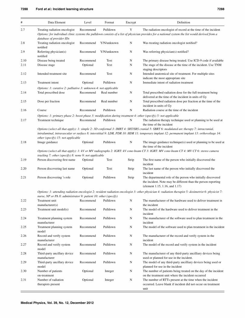
7288 Ford et al.: Incident learning structure 7288
# Data Element Level Format Encrypt Definition
2.7 Treating radiation oncologist Recommend Pulldown Y The radiation oncologist of record at the time of the incidentOptions: for individual clinic systems the pulldown consists of a list of physician provides for a national system the list would derived from adatabase of provider IDs
2.8 Treating radiation oncologistnotified
Recommend Y/N/unknown N Was treating radiation oncologist notified?
2.9 Referring physician(s)notified
Recommend Y/N/unknown N Was referring physician(s) notified?
2.10 Disease being treated Recommend Text N The primary disease being treated. Use ICD-9 code if available2.11 Disease stage Optional Text N The stage of the disease at the time of the incident. Use TNM
staging descriptors2.12 Intended treatment site Recommend Text N Intended anatomical site of treatment. For multiple sites
indicate the most appropriate site2.13 Treatment intent Optional Pulldown N Immediate intent of radiation treatment
Options: 1: curative 2: palliative 3: unknown 4: not applicable2.14 Total prescribed dose Recommend Real number N Total prescribed radiation dose for the full treatment being
delivered at the time of the incident in units of Gy2.15 Dose per fraction Recommend Real number N Total prescribed radiation dose per fraction at the time of the
incident in units of Gy2.16 Course Recommend Pulldown N Radiation course at the time of the incident
Options: 1: primary phase 2: boost phase 3: modification during treatment 4: other (specify) 5: not applicable2.17 Treatment technique Recommend Pulldown N The radiation therapy technique used or planning to be used at
the time of the incidentOptions (select all that apply): 1: simple 2: 3D conformal 3: IMRT 4: SRT/SRS cranial 5: SBRT 6: modulated arc therapy 7: intracranial,intraluminal, intravascular or surface 8: interstitial 9: LDR, PDR 10: HDR 11: temporary implant 12: permanent implant 13: orthovoltage 14:
other (specify) 15: not applicable
2.18 Image guidance Optional Pulldown N The image-guidance technique(s) used or planning to be used atthe time of the incident
Options (select all that apply): 1: kV or MV radiographs 2: IGRT: kV cone-beam CT 3: IGRT: MV cone-beam CT 4: MV CT 6: stereo camera
tracking 7: other (specify) 8: none 9: not applicable2.19 Person discovering first name Optional Text Strip The first name of the person who initially discovered the
incident2.20 Person discovering last name Optional Text Strip The last name of the person who initially discovered the
incident2.21 Person discovering ’s role Optional Pulldown Strip The departmental role of the person who initially discovered
the incident. Note may be different than the person reporting(element 1.15, 1.16, and 1.17)
Options: 1: attending radiation oncologist 2: resident radiation oncologist 3: other physician 4: radiation therapist 5: dosimetrist 6: physicist 7:
nurse, NP or PA 8: administrator 9: patient 10: other (specify)2.22 Treatment unit
manufacturer(s)Recommend Pulldown N The manufacturer of the hardware used to deliver treatment in
the incident2.23 Treatment unit model(s) Recommend Pulldown N The model of the hardware used to deliver treatment in the
incident2.24 Treatment planning system
manufacturerRecommend Pulldown N The manufacturer of the software used to plan treatment in the
incident2.25 Treatment planning system
modelRecommend Pulldown N The model of the software used to plan treatment in the incident
2.26 Record and verify systemmanufacturer
Recommend Pulldown N The manufacturer of the record and verify system in theincident
2.27 Record and verify systemmodel
Recommend Pulldown N The model of the record and verify system in the incident
2.28 Third-party ancillary devicemanufacturer
Recommend Pulldown N The manufacturer of any third-party ancilliary devices beingused or planned for use in the incident.
2.29 Third-party ancillary devicemodel
Recommend Pulldown N The model of any third-party ancilliary devices being used orplanned for use in the incident
2.30 Number of patientson-treatment
Optional Integer N The number of patients being treated on the day of the incidenton the treatment unit where the incident occurred
2.31 Number of radiationtherapists present
Optional Integer N The number of RTTs present at the time when the incidentoccurred. Leave blank if incident did not occur on treatmentunit
Medical Physics, Vol. 39, No. 12, December 2012

7289 Ford et al.: Incident learning structure 7289
# Data Element Level Format Encrypt Definition
2.32 Number of other staff present Optional Integer N The number of other staff present at the time when the incidentoccurred. Leave blank if incident did not occur on treatmentunit
2.33 Staff involved Optional Pulldown Y Names and roles of staff present when the incident occurred2.34 Error type Optional Pulldown N The type of error that occurred or potentially could occur
Options (select all that apply): 1: wrong patient or potentially wrong patient 2: wrong anatomical site or potentially wrong site 3: wrong laterality
or potentially wrong laterality 4: wrong dose to all or part of the tumor or normal tissue or potentially wrong dose 5: wrong modality or energy or
potentially wrong modality or energy 6: other (specify) 7: not applicable2.35 Dosimetric severity Optional Pulldown N See Appendix C
Options: refer to Appendix C, WGPE severity scales
2.36 Reportable event Optional Y/N/unknown N Was the event reportable to the relevant regulatory agency(state, NRC)?
2.37 Medical severity Required Pulldown N See Appendix C. For potential incidents (near-misses) assume aworst-case scenario if the incident were actually to occur
Options: refer to Appendix C, WGPE severity scales2.38 Severity assessment type Required Pulldown N The method used for assessing the severity of harm
Options: 1: estimated severity 2: actual observed severity 3: not applicable2.39 When severity assessed Recommend Pulldown N The time after the incident at which the severity of harm was
observed or at which it is estimated to occurOptions: 1: before 24 hours 2: after 24 hours but before 3 days 3: three days or later 4: unknown
2.40 Summary title Optional Text N A very brief title phrase describing the incident2.41 Incident analysis (from
evaluator)Optional Text N Description of the incident provided by the person evaluating
the incident in the clinic. This should provide very detailedinformation from which one can infer root causes
2.42 Where originated Recommend Pulldown N The point in the workflow where the incident originatedOptions: refer to detailed process maps (Secs. 2 and 3 of Appendix B, WGPE process maps)
2.43 Attached files Optional File N Files (e.g., screenshots, pdf reports, etc.) that may help clarifythe details of the incident
2.44 Causes Required Pulldown N List all the causes and contributing factors at work in theincident. For potential incidents assume a worst-case scenario ifthe incident had actually occurred. See Appendix D
Options (select all that apply): Refer to Appendix D, WGPE causality table
3. Responder’s form
# Data Element Level Format Encrypt Definition
3.1 Clinical actionscale
Optional Pulldown N Priority scale to guide the follow-up actions of the clinic
Options: A,B, C, D (exact meaning of each to be determined by the individual clinic)3.2 Intervention Recommend Pulldown N Was any intervention attempted in order to “rescue” the patient
(i.e., to prevent, minimize or reverse harm)?
Options (select all that apply): 1: treatment stopped 2: treatment modified 3: added additional treatment fractions 4: patient’s stay in hospitalextended 5: intervention pending 6: no intervention 7: unknown 8: other (specify) 9: not applicable
3.3 Safety barriers Recommend Pulldown N The safety barriers or quality control measures in place whichprevented or could prevent the incident
Options: (select all that apply). Refer to detailed process maps (Secs. 2 and 3 of Appendix B, WGPE process maps)
3.4 Corrective action Optional Text N Corrective action(s) taken to mitigate the harm for this particularpatient
3.5 Preventive action Optional Text N Preventive action taken to insure that a similar incident will nothappen to a patient in the future
3.6 Learning actions Optional Text N The learning activities that were undertaken in response to theincident, for example, presentation at rounds, etc.
3.7 Incident closed Optional Y/N N Has the incident been closed by the evaluator responsible forincident handling? This includes the implementation andevaluation of corrective, preventive and learning actions. Qualitycontrol of the data should also be performed
Medical Physics, Vol. 39, No. 12, December 2012

7290 Ford et al.: Incident learning structure 7290
a)Author to whom correspondence should be addressed. Electronic mail:[email protected]
1To Err is Human: Building a Safer Health System. Committee on Qualityof Health Care in America, 1st ed., edited by L. T. Kohn, J. M. Corrigan,and M. S. Donaldson (National Academies Press, Washington, DC, 2000).
2The Royal College of Radiologists, Society and College of Radiogra-phers, Institute of Physics and Engineering in Medicine, National PatientSafety Agency, British Institute of Radiology, “Towards Safer Radiother-apy,” London: The Royal College of Radiologists (2008).
3L. Donaldson, Radiotherapy Risk Profile: Technical Manual [World HealthOrganization (WHO) Press, Geneva 27, Switzerland, 2008].
4E. C. Ford and S. Terezakis, “How safe is safe?: Risk in radiotherapy,” Int.J. Radiat. Oncol. Biol. Phys. 78, 321 (2010).
5A. Mans, M. Wendling, L. N. McDermott, J.-J. Sonke, R. Tielenburg, R. Vi-jlbrief, B. Mijnheer, M. Van Herk, and J. C. Stroom, “Catching errors within vivo EPID dosimetry,” Med. Phys. 37, 2638–2644 (2010).
6R. Botney, “Improving patient safety in anesthesia: A success story?,” Int.J. Radiat. Oncol. Biol. Phys. 71, S182–S186 (2008).
7W. R. Hendee and M. G. Herman, “Improving patient safety in radiationoncology,” Med. Phys. 38, 78–82 (2011).
8D. L. Cooke, A Reference Guide for Learning from Incidents in RadiationTreatment, HTA Initiative, Vol. 22 (Alberta Heritage Foundation for Medi-cal Research, Edmonton, Alberta, Canada, 2006).
9M. V. Williams, “Improving patient safety in radiotherapy by learning fromnear misses, incidents and errors,” Br. J. Radiol. 80, 297–301 (2007).
10P. J. Pronovost, C. A. Goeschel, K. L. Olsen, J. C. Pham, M. R. Miller,S. M. Berenholtz, J. B. Sexton, J. A. Marsteller, L. L. Morlock, A. W. Wu,J. M. Loeb, and C. M. Clancy, “Reducing health care hazards: Lessonsfrom the Commercial Aviation Safety Team,” Health Aff. 28, w479–w489(2009).
11F. Fiorino, “CAST-ing a safety net,” Aviat. Week Space Technol. 163, 72–73 (2005).
12X. Bernard-Bruls, Nuclear Power Plant Operating Experience from theIAEA/NEA International Reporting System for Operating Experience2005–2008 (2010), p. 1453.
13S. Mutic, R. S. Brame, S. Oddiraju, P. Parikh, M. A. Westfall, M. L. Hop-kins, A. D. Medina, J. C. Danieley, J. M. Michalski, I. M. El Naqa,D. A. Low, and B. Wu, “Event (error and near-miss) reporting and learningsystem for process improvement in radiation oncology,” Med. Phys. 37,5027–5036 (2010).
14J.-P. Bissonnette and G. Medlam, “Trend analysis of radiation therapy in-cidents over seven years,” Radiother. Oncol. 96, 139–144 (2010).
15D. L. Cooke, P. B. Dunscombe, and R. C. Lee, “Using a survey of inci-dent reporting and learning practices to improve organisational learning ata cancer care centre,” Qual. Saf. Health Care 16, 342–348 (2007).
16Quality and Safety in Radiotherapy, 1st ed., edited by T. Pawlicki,P. B. Dunscombe, A. J. Mundt, and P. Scalliet (Taylor & Francis, BocaRaton, FL, 2010).
17B. G. Clark, R. J. Brown, J. L. Ploquin, A. L. Kind, and L. Grimard, “Themanagement of radiation treatment error through incident learning,” Ra-diother. Oncol. 95(3), 344–349 (2010).
18T. R. Williams, Target Safely: American Society for Radiation Oncology2010 Year in Review (American Society for Radiation Oncology (ASTRO),Fairfax, VA, 2010).
19J. Cunningham, M. Coffey, T. Knöös, and O. Holmberg, “Radiation Oncol-ogy Safety Information System (ROSIS) - Profiles of participants and thefirst 1074 incident reports,” Radiother. Oncol. 97, 601–607 (2010).
20O. Holmberg, J. Malone, M. Rehani, D. McLean, and R. Czarwinski, “Cur-rent issues and actions in radiation protection of patients,” Eur. J. Radiol.76, 15–19 (2010).
21J. R. Palta, J. A. Efstathiou, J. E. Bekelman, S. Mutic, C. R. Bogardus,T. R. McNutt, P. E. Gabriel, C. A. Lawton, A. L. Zietman, and C. M. Rose,“Developing a national radiation oncology registry: From acorns to oaks,”Pract. Radiat. Oncol. 2(1), 10–17 (2012).
22“Concepts underlying AHRQ’s Common Formats,” Agency forHealthcare Research and Quality, 2011, see http://www.pso.ahrq.gov/formats/eventdesc.htm.
23IAEA training materials: Module 2, prevention of accidental exposure inradiotherapy, International Atomic Energy Agency, Vienna, 2009.
24National Patient Safety Foundation, 2011, see http://npsf.org/rc/mp/definitions.php. The current location is here: http://www.npsf.org/for-healthcare-professionals/resource-center/definitions-and-hot-topics/.
25M. S. Huq, B. A. Fraass, P. B. Dunscombe, J. P. Gibbons, Jr., G. S. Ib-bott, P. M. Medin, A. Mundt, S. Mutic, J. R. Palta, B. R. Thomadsen,J. F. Williamson, and E. D. Yorke, “A method for evaluating quality as-surance needs in radiation therapy,” Int. J. Radiat. Oncol. Biol. Phys. 71,S170–S173 (2008).
26E. C. Ford, R. Gaudette, L. Myers, B. Vanderver, L. Engineer, R. Zellars,D. Y. Song, J. Wong, and T. L. DeWeese, “Evaluation of safety in a ra-diation oncology setting using failure mode and effects analysis,” Int. J.Radiat. Oncol. Biol. Phys. 74, 852–858 (2009).
27P. B. Dunscombe, S. Iftody, N. Ploquin, E. U. Ekaette, and R. C. Lee, “Theequivalent uniform dose as a severity metric for radiation treatment inci-dents,” Radiother. Oncol. 84, 64–66 (2007).
28ASN-SFRO experimental scale for the rating of radiation protection eventsaffecting patients undergoing a radiotherapy procedure, Societe Francaisede Radiotherapie Oncologique; Autorite de Surete Nucleaire, 2007.
29“Common Terminology Criteria for Adverse Events,” National Insti-tutes of Health, 2011, see http://ctep.cancer.gov/protocolDevelopment/electronic_applications/ctc.htm.
30E. U. Ekaette, R. C. Lee, D. L. Cooke, K.-. Kelly, and P. B. Dunscombe,“Risk analysis in radiation treatment: Application of a new taxonomicstructure,” Radiother. Oncol. 80, 282–287 (2006).
31L. B. Marks, C. M. Rose, J. A. Hayman, and T. R. Williams, “The need forphysician leadership in creating a culture of safety,” Int. J. Radiat. Oncol.Biol. Phys. 79, 1287–1289 (2011).
Medical Physics, Vol. 39, No. 12, December 2012