consensus guidelines for the
TRANSCRIPT

Consensus guidelines for thetreatment of infectious
endocarditis with outpatientparenteral antibiotic therapy
Shurjeel H Choudhri MD FRCPC and The Endocarditis Care Plan Working Group*
Infectious endocarditis (IE) refers to an infection of the endo-
cardium resulting in the development of vegetations on a
heart valve or, less commonly, on the mural endocardium. De-
spite the availability of curative therapy, it continues to be as-
sociated with high morbidity and mortality. While the overall
incidence of IE has remained unchanged over the past 50 years,
an increased incidence of IE has been seen in the elderly, those
with prosthetic valves and injection drug users (IDUs) (1-4).
The last quarter century has seen important advances in
the diagnosis and management of IE. The availability of
transesophageal echocardiography, new diagnostic criteria
such as the Duke’s criteria and the development of short
course, once daily, oral, outpatient treatment regimens have
revolutionized the diagnosis and treatment of IE (1). Outpa-
tient parenteral antibiotic therapy (OPAT) programs allow
most patients with uncomplicated IE to complete their treat-
ment course in the community, thus minimizing the length of
their hospital stay. The objective of this review is to summa-
rize the current knowledge of IE with special attention to its
treatment in the community by OPAT programs.
4D Can J Infect Dis Vol 11 Suppl D November/December 2000
CARE PATHWAYS
*Dr Gerald A Evans, Kingston General Hospital, Kingston, Ontario; Ms Natalie Thickson, Ms Maria Lazaruk, Ms Leslie Dryburgh, Ms Theresa Imlah, Ms
Lisa Houtkooper, Mr Luke McKenzie, Dr Karen A Doucette and Ms Glenna Germaine, St Boniface General Hospital, Winnipeg, Manitoba
Correspondence and reprints: Dr Shurjeel H Choudhri, Medical Research, Anti-infectives/Biologics, Bayer Corporation,
400 Morgan Lane, West Haven, Connecticut 06518 USA. Telephone 203-812-2186, fax 800-520-2807, e-mail [email protected]
SH Choudhri and The Endocarditis Care Plan Working Group. Consensus guidelines for the treatment of infectiousendocarditis with outpatient parenteral antibiotic therapy. Can J Infect Dis 2000;11(Suppl D):4D-10D.
The development of single, daily dose antibiotic regimens and the availability of computerized ambulatory infusionpumps have made it possible for most patients with infectious endocarditis (IE) to complete their treatment in an outpa-tient setting. This review summarizes the current literature on the classification, diagnosis, clinical and laboratorymanifestations, and inpatient and outpatient management of IE. An algorithmic approach to the diagnosis and manage-ment of IE is proposed. Most patients with IE who are clinically stable and do not require a surgical procedure can bemanaged using outpatient parenteral antibiotic therapy. Results from several small studies suggest that outpatient par-enteral antibiotic therapy for IE is safe, efficacious and economical.
Key Words: Consensus guidelines; Endocarditis; Outpatient treatment
Directives consensuelles pour le traitement de l’endocardite infectieuse par antibiothérapieparentérale ambulatoire
RÉSUMÉ : La mise au point de schémas d’antibiothérapies quotidiennes simples et l’accessibilité à des pompes à perfusion in-formatisées ambulatoires permettent à la plupart des patients qui souffrent d’une endocardite infectieuse de terminer leur traite-ment sans être hospitalisés. Le présent article résume la littérature actuelle sur la classification, le diagnostic, les manifestationscliniques et les résultats d’analyses de laboratoire, de même que le traitement de l’endocardite infectieuse avec ou sans hospita-lisation. On propose ici un algorithme pour le diagnostic et le traitement de l’endocardite infectieuse. La plupart des patients quien sont atteints, qui sont cliniquement stables et ne requièrent pas d’intervention chirurgicale peuvent être traités sans hospita-lisation par antibiothérapie parentérale. Les résultats de plusieurs petites études suggèrent que l’antibiothérapie parentérale enexterne est sûre, efficace et économique dans les cas d’endocardite infectieuse.
1
G:...choudhri.vpMon Dec 18 15:48:52 2000
Color profile: DisabledComposite Default screen
0
5
25
75
95
100
0
5
25
75
95
100
0
5
25
75
95
100
0
5
25
75
95
100

CLASSIFICATION AND ETIOLOGYIE can be divided into three categories: native valve endo-
carditis, prosthetic valve endocarditis (PVE) and endocarditis
in IDUs. Native valve IE is most commonly caused by Strepto-
coccus species, Staphylococcus aureus and Enterococcus
species (5). Most patients (60% to 80%) with IE have an iden-
tifiable, predisposing cardiac lesion such as a bicuspid aortic
valve, a mitral valve prolapse or rheumatic valvular disease
(6,7). Endocarditis can also be classified as acute or suba-
cute. Acute IE is caused by virulent organisms such as S au-
reus, which can infect normal heart valves and are rapidly
fatal if not treated promptly. Subacute IE results from infec-
tion of structurally abnormal valves by less virulent organ-
isms such as viridans streptococci.
PVE can be further subclassified as early or late. Early
infection occurs within two months of valve replacement and
is usually due to contamination during surgery. Staphylococ-
cus epidermidis and S aureus are the most common organisms
identified in this situation (8,9). Late PVE occurs more than
two months after valve replacement, and is usually due to
transient bacteremia with subsequent seeding of the valve, al-
though some cases may still result from organisms acquired
during the surgery. Streptococci are the most commonly iso-
lated organisms in late PVE, followed by staphylococci and,
much less commonly, Gram-negative rods and fungi (9-11).
Infective endocarditis in IDUs usually involves the tricus-
pid valve, although left-sided endocarditis may also occur in
IDUs with pre-existing valve abnormalities. S aureus is the
most common organism isolated, followed by enterococci,
streptococci and Gram-negative rods such as Pseudomonas
species and Serratia species (12).
CLINICAL MANIFESTATIONSMost patients with IE present with fever along with nonspe-
cific symptoms such as chills, night sweats, anorexia and
fatigue (11,13). The fever is usually low grade and remittent in
cases of subacute IE, with temperatures rarely exceeding 39°C;
however, temperatures may be higher in cases associated with
virulent organisms such as S aureus (14). Musculoskeletal
complaints are present in approximately 40% of patients, with
arthralgias, myalgias and back pain being the most common.
Most patients (90%) with IE have a murmur, although a new or
changing murmur is only observed in 36% to 53% (13). Cutane-
ous manifestations such as petechiae and splinter hemor-
rhages may be observed in up to 50% of patients. Splenomegaly
is now a relatively uncommon finding, being observed in less
than 20% of patients (1,6).
Many patients may initially present with a complication of
IE. Congestive heart failure is the most common complication
and is observed in 38% to 60% of IE patients (14). Embolic phe-
nomena are the second most frequent complication and may
occur at any time during the course of the illness (15). Occa-
sionally, the patient may present with an acute cerebrovascu-
lar accident. Patients with right-sided IE may present with
multiple, rounded pulmonary infiltrates with or without cavi-
tation due to infected pulmonary emboli (1). Mycotic aneu-
rysms are the third most common complication, occurring in
5% to 10% of cases. These often involve the central nervous
system and may be clinically silent until they rupture (16).
Other complications include the development of renal dys-
function due to embolization of the kidneys or the develop-
ment of immune-mediated glomerulonephritis (1).
LABORATORY FINDINGSCommon laboratory findings in IE include normochromic
normocytic anemia (70% to 90% of cases), leukocytosis (20% to
30%), an elevated erythrocyte sedimentation rate (greater than
90%), elevated C-reactive protein levels, positive rheumatoid
factor (40% to 50%), proteinuria (50% to 65%) and hematuria
(30% to 50%) (17). Most patients with IE (78% to 95%) have a
positive blood culture. Prior antibiotic therapy is the most com-
mon reason for negative blood cultures in a patient with IE (5).
DIAGNOSISAn early diagnosis requires a high degree of clinical suspi-
cion, because patients with IE may present with nonspecific
symptoms. Blood cultures should be obtained from all indi-
viduals suspected of having IE, and all should undergo echo-
cardiography. Transesophageal echocardiography can detect
both vegetations and perivalvular infections with a sensitivity
of 80% to 90%, and is superior to transthoracic echocardiogra-
phy for diagnosing IE (18,19). Serial blood cultures should be
obtained if the original blood culture is negative. Once an organ-
ism has been isolated and identified, the minimum inhibitory
concentration (MIC) and minimum bactericidal concentration
should be determined to guide further antimicrobial therapy.
The Duke’s criteria (19) use clinical, microbiological and
echocardiographic findings to establish a diagnosis of definite
or possible IE, and have largely supplanted previously de-
scribed criteria for IE (20). Two major, or one major plus three
minor, or five minor criteria establish a definitive clinical di-
agnosis of IE (Table 1).
THERAPY OF IEIdeally, all patients with a suspected diagnosis of IE should
be admitted for diagnostic workup and monitoring. Empirical
therapy with a bactericidal antibiotic regimen should be initi-
ated in all patients with acute IE while awaiting the culture re-
sults. An early surgical consultation should be obtained in any
patient developing a complication of IE and in all patients
with PVE (14). Indications for surgical therapy for IE are out-
lined in Table 2, while Tables 3 to 5 summarize the empirical
antibiotic regimens for the different types of IE. Therapy for
subacute endocarditis can usually be delayed for 24 to 48 h,
until the culture results become available. Definitive therapy
is based on the culture results, and is outlined in Tables 3 to 6.
DEFINITIVE THERAPY OF IEThe recommended treatment regimens for native valve,
prosthetic valve and right-sided IE are listed in Tables 3, 4 and
5, respectively. Table 6 outlines the treatment choices avail-
able for managing IE in penicillin-allergic patients.
Native valve endocarditis – Viridans streptococci: Specific
therapy of streptococcal infections is based on their suscepti-
Can J Infect Dis Vol 11 Suppl D November/December 2000 5D
Consensus guidelines for OPAT in IE
2
G:...choudhri.vpMon Dec 18 15:48:52 2000
Color profile: DisabledComposite Default screen
0
5
25
75
95
100
0
5
25
75
95
100
0
5
25
75
95
100
0
5
25
75
95
100

bility to penicillin G (21). Those with MICs of 0.1 �g/mL or less
can be managed with a four-week course of penicillin or ceftri-
axone, or a two-week course of penicillin or ceftriaxone plus
gentamicin (21-23). A two-week course of ceftriaxone followed
by two weeks of oral amoxicillin has also been shown to be ef-
fective (23). Streptococci with penicillin G MICs between 0.1
and 0.5 �g/mL require a regimen of high dose penicillin G for
four weeks, plus gentamicin for the first two weeks (21). This
regimen can also be used for nutrient-deficient streptococci.
Penicillin may not be bactericidal against streptococci with
penicillin G MICs greater than 0.5 �g/mL. These organisms
should be treated with the same regimens as enterococci (21).
Vancomycin can be substituted for penicillin in patients
with a history of penicillin allergy. Ceftriaxone is also an alterna-
tive in this situation because of the minimal cross-reactivity be-
tween penicillin and third-generation cephalosporins.
Enterococci: Penicillin, ampicillin and vancomycin are not bac-
tericidal against enterococci, and the addition of an aminoglyco-
side such as streptomycin or gentamicin is required to produce a
synergistic bactericidal effect. Synergy is only seen if the MIC is
less than 2000 �g/mL or less than 500 �g/mL to streptomycin and
gentamicin, respectively (21). The usual treatment course is for
six weeks. No satisfactory regimens are available to treat entero-
cocci that have high level resistance to aminoglycosides.
Staphylococci: A four- to six-week course of intravenous
cloxacillin remains the treatment of choice for methicillin-
susceptible S aureus and S epidermidis (21). Cefazolin may be
substituted for cloxacillin in selected patients with a penicillin
allergy (21). The addition of gentamicin for the first three to five
days of therapy produces a more rapid clearing of the bactere-
mia but has not been shown to reduce the complication rate or
increase the cure rates (24). Current evidence suggests that van-
comycin may be less effective than cloxacillin in treating IE and s-
6D Can J Infect Dis Vol 11 Suppl D November/December 2000
Choudhri
TABLE 1Duke’s diagnostic criteria for infectious endocarditis (IE)
Definite IEPathological criteria
• Microorganisms: demonstrated by culture or histology in a vegetation, a vegetation that has embolized or an intracardiac abscessor
• Pathological lesions, such as: vegetations or intracardiac abscess present, confirmed by histology showing active endocarditisClinical criteriaTo establish a definitive diagnosis of IE, a patient must have two major, one major and three minor, or five minor criteria
Major criteria• Isolation of viridans streptococci, Streptococcus bovis, HACEK group organisms, or (in the absence of a primary focus) community-
acquired Staphylococcus aureus or enterococcus from two separate blood cultures; or isolation of a microorganism consistent withendocarditis in blood cultures 12 h or more apart, or all of three, or most of four or more, blood cultures, with the first and last culturesperformed at least 1 h apart
• Evidence of endocardial involvement on echocardiography: oscillating intracardiac mass or abscess, new partial dehiscence of prostheticvalve or new valvular regurgitation
Minor criteria• Predisposing lesion or intravenous drug use• Fever �38°C• Vascular phenomena: major arterial emboli, septic pulmonary infarcts, mycotic aneurysm, intracranial hemorrhage, conjunctival
hemorrhages or Janeway lesions• Immunological phenomena: glomerulonephritis, Osler’s nodes, Roth’s spots or positive rheumatoid factor• Microbiological evidence: positive blood cultures not meeting the major criteria (excluding single cultures positive for organisms that
usually do not cause endocarditis) or serological evidence of active infection with an organism that causes endocarditis• Echocardiogram consistent with endocarditis but that does not meet the major criteria
Possible IEFindings consistent with IE that fall short of ‘definite’ but are not ‘rejected’
Rejected IE• Firm alternate diagnosis for manifestations of IE
or• Resolution of manifestations of IE with antibiotic therapy for four days or less
or• No pathological evidence of IE at surgery or autopsy after antibiotic therapy of four days or less
HACEK group organisms Haemophilus parainfluenzae, Haemophilus aphrophilus, Actinobacillus actinomycetemcomitans, Cardiobacterium hominis,Eikenella corrodens, Kingella kingae. Information taken from reference 19
TABLE 2Indications for surgical therapy
Absolute• Refractory congestive heart failure• More than one systemic embolic complication• Uncontrolled infection (positive blood cultures after three to five
days of appropriate therapy)• Valve dysfunction as demonstrated by fluoroscopy• Ineffective antimicrobial therapy (eg, fungal endocarditis)• Resection of mycotic aneurysms• Most cases of prosthetic valve endocarditis• Local suppurative complications (eg, annular or myocardial
abscess, valve rupture)• Fungal endocarditis
Relative• Moderate congestive heart failure• Progressive renal failure (immune complex disease, embolization)• Large vegetation
Information taken from references 6 and 13
3
G:...choudhri.vpMon Dec 18 15:48:53 2000
Color profile: DisabledComposite Default screen
0
5
25
75
95
100
0
5
25
75
95
100
0
5
25
75
95
100
0
5
25
75
95
100
hould be reserved for patients with a penicillin allergy or for the
treatment of methicillin-resistant S aureus and methicillin-
resistant coagulase-negative staphylococci (25,26).
HACEK organisms: The HACEK organisms (Haemophilus
parainfluenzae, Haemophilus aphrophilus, Actinobacillus
actinomycetemcomitans, Cardiobacterium hominis, Eikenella
corrodens and Kingella kingae) are slow-growing, fastidious,
Gram-negative bacilli that typically produce a subacute IE.
Most HACEK isolates produce beta-lactamases and are resis-
tant to penicillin or ampicillin. Third-generation cephalosporins
have excellent activity against this group, and a four-week
course of ceftriaxone is the treatment of choice (21). A four-
week course of ampicillin and gentamicin may be used if the
isolate is a nonbeta-lactamase producer (21).
PVE: S aureus and S epidermidis are commonly implicated in
both early and late PVE. Viridans streptococci and enterococci
Can J Infect Dis Vol 11 Suppl D November/December 2000 7D
Consensus guidelines for OPAT in IE
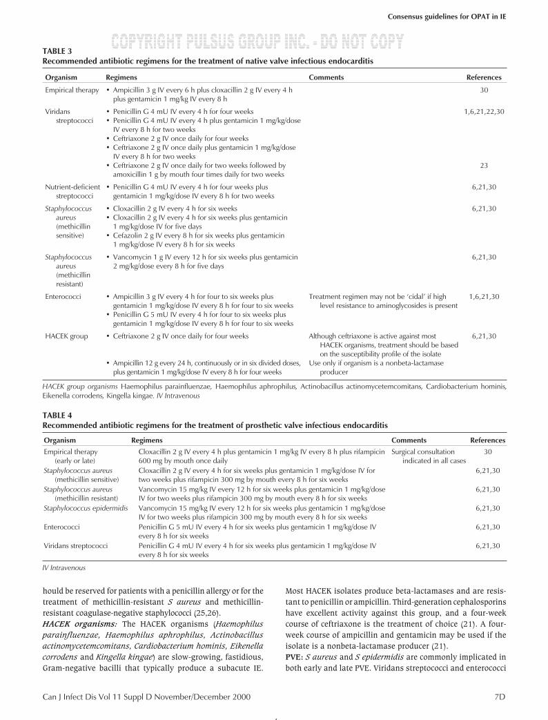
TABLE 3Recommended antibiotic regimens for the treatment of native valve infectious endocarditis
Organism Regimens Comments References
Empirical therapy • Ampicillin 3 g IV every 6 h plus cloxacillin 2 g IV every 4 hplus gentamicin 1 mg/kg IV every 8 h
30
Viridansstreptococci
• Penicillin G 4 mU IV every 4 h for four weeks• Penicillin G 4 mU IV every 4 h plus gentamicin 1 mg/kg/dose
IV every 8 h for two weeks• Ceftriaxone 2 g IV once daily for four weeks• Ceftriaxone 2 g IV once daily plus gentamicin 1 mg/kg/dose
IV every 8 h for two weeks• Ceftriaxone 2 g IV once daily for two weeks followed by
amoxicillin 1 g by mouth four times daily for two weeks
1,6,21,22,30
23
Nutrient-deficientstreptococci
• Penicillin G 4 mU IV every 4 h for four weeks plusgentamicin 1 mg/kg/dose IV every 8 h for two weeks
6,21,30
Staphylococcusaureus(methicillinsensitive)
• Cloxacillin 2 g IV every 4 h for six weeks• Cloxacillin 2 g IV every 4 h for six weeks plus gentamicin
1 mg/kg/dose IV for five days• Cefazolin 2 g IV every 8 h for six weeks plus gentamicin
1 mg/kg/dose IV every 8 h for six weeks
6,21,30
Staphylococcusaureus(methicillinresistant)
• Vancomycin 1 g IV every 12 h for six weeks plus gentamicin2 mg/kg/dose every 8 h for five days
6,21,30
Enterococci • Ampicillin 3 g IV every 4 h for four to six weeks plusgentamicin 1 mg/kg/dose IV every 8 h for four to six weeks
• Penicillin G 5 mU IV every 4 h for four to six weeks plusgentamicin 1 mg/kg/dose IV every 8 h for four to six weeks
Treatment regimen may not be ‘cidal’ if highlevel resistance to aminoglycosides is present
1,6,21,30
HACEK group • Ceftriaxone 2 g IV once daily for four weeks
• Ampicillin 12 g every 24 h, continuously or in six divided doses,plus gentamicin 1 mg/kg/dose IV every 8 h for four weeks
Although ceftriaxone is active against mostHACEK organisms, treatment should be basedon the susceptibility profile of the isolate
Use only if organism is a nonbeta-lactamaseproducer
6,21,30
HACEK group organisms Haemophilus parainfluenzae, Haemophilus aphrophilus, Actinobacillus actinomycetemcomitans, Cardiobacterium hominis,Eikenella corrodens, Kingella kingae. IV Intravenous
TABLE 4Recommended antibiotic regimens for the treatment of prosthetic valve infectious endocarditis
Organism Regimens Comments References
Empirical therapy(early or late)
Cloxacillin 2 g IV every 4 h plus gentamicin 1 mg/kg IV every 8 h plus rifampicin600 mg by mouth once daily
Surgical consultationindicated in all cases
30
Staphylococcus aureus(methicillin sensitive)
Cloxacillin 2 g IV every 4 h for six weeks plus gentamicin 1 mg/kg/dose IV fortwo weeks plus rifampicin 300 mg by mouth every 8 h for six weeks
6,21,30
Staphylococcus aureus(methicillin resistant)
Vancomycin 15 mg/kg IV every 12 h for six weeks plus gentamicin 1 mg/kg/doseIV for two weeks plus rifampicin 300 mg by mouth every 8 h for six weeks
6,21,30
Staphylococcus epidermidis Vancomycin 15 mg/kg IV every 12 h for six weeks plus gentamicin 1 mg/kg/doseIV for two weeks plus rifampicin 300 mg by mouth every 8 h for six weeks
6,21,30
Enterococci Penicillin G 5 mU IV every 4 h for six weeks plus gentamicin 1 mg/kg/dose IVevery 8 h for six weeks
6,21,30
Viridans streptococci Penicillin G 4 mU IV every 4 h for six weeks plus gentamicin 1 mg/kg/dose IVevery 8 h for six weeks
6,21,30
IV Intravenous
4
G:...choudhri.vpMon Dec 18 15:48:54 2000
Color profile: DisabledComposite Default screen
0
5
25
75
95
100
0
5
25
75
95
100
0
5
25
75
95
100
0
5
25
75
95
100

may also produce late PVE. A combination of intravenous van-
comycin, gentamicin and oral rifampicin is a good empirical
regimen for PVE (21). Specific regimens are given in Table 4.
All treatment courses are for six weeks unless otherwise indi-
cated. Early surgical consultation should be obtained in all
cases of PVE (14).
Endocarditis in IDUs: Staphylococcus aureus is the most com-
mon etiological organism in this situation. A two-week regimen
of cloxacillin plus gentamicin is curative in most cases of un-
complicated infection, especially if a clinical and bacteriological
response is seen within 96 h of initiating therapy (27). A longer
treatment course should be given if there is evidence of compli-
cations such as hemodynamic compromise, systemic emboli or
a metastatic focus of infection. A four-week oral regimen of
ciprofloxacin plus rifampicin is also effective, although this
regimen should not be used for treating methicillin-resistant S
aureus infections (28).
OPAT FOR IEOPAT for IE is an increasingly attractive option in the cur-
rent climate of cost containment. The availability of single,
daily dose regimens and computerized ambulatory infusion
pumps has made it possible to provide optimal home intrave-
nous therapy for most cases of IE while minimizing the need
for a prolonged hospital admission. The approach to placing a
patient on home intravenous therapy is outlined in Figure 1.
The choice of the antibiotic regimen for OPAT will depend
on the type of infecting organism, the method of administra-
tion (ie, with or without a programmable infusion pump) and the
ability of the patient to self-administer the antibiotics. Penicillin-
and cloxacillin-based regimens are the most economical if the
patient is able to self-administer the drugs or if it is possible to
use an infusion pump. If a nursing visit is required to administer
each dose, then the use of more expensive once-a-day agents
such as ceftriaxone becomes more economical.
8D Can J Infect Dis Vol 11 Suppl D November/December 2000
Choudhri
TABLE 5Recommended antibiotic regimens for the treatment of infectious endocarditis in injection drug users
Organism Regimens Comments References
Empirical therapy Cloxacillin 2 g IV every 4 h plus gentamicin 1 mg/kg IV every 8 hVancomycin 15 mg/kg IV every 12 h plus gentamicin 1 mg/kg IV every 8 h Regimen for patients
with penicillin allergy
30
Staphylococcus aureus(methicillin-sensitive)
Cloxacillin 2 g IV every 4 h for four weeks plus gentamicin 1 mg/kg/dose for five daysCloxacillin 2 g IV every 4 h for two weeks plus gentamicin 1 mg/kg/dose for two weeksCiprofloxacin 750 mg by mouth bid for four weeks plus rifampicin 600 mg by mouthbid for four weeks
27,28
Staphylococcus aureus(methicillin-resistant)
Vancomycin 1 g IV every 12 h for four weeks plus gentamicin 2 mg/kg/dose every 8 hfor five days
IV Intravenous
TABLE 6Treatment of infectious endocarditis in penicillin-allergic patients
Organism Regimens Comments References
Empirical therapy Vancomycin 15 mg/kg IV every 12 h plus gentamicin 1 mg/kg IV every 8 h
Native valve endocarditisStaphylococcus aureus
(methicillin sensitiveor methicillin resistant)
Viridans streptococciNutrient-deficient streptococci
Enterococci
Vancomycin 1 g IV every 12 h for six weeks plus gentamicin 2 mg/kg/doseevery 8 h for five days
Ceftriaxone 2 g IV once daily for four weeksCeftriaxone 2 g IV once daily plus gentamicin 1 mg/kg/dose IV every 8 hfor two weeksVancomycin 15 mg/kg IV every 12 h for four weeksVancomycin 15 mg/kg IV every 12 h for six weeks plus gentamicin1 mg/kg/dose IV every 8 h for six weeks
Ceftriaxone-basedregimens may beused in penicillin-allergic patients,because the riskof cross-reactivityis minimal
1,6,21
1,6,21,22
1,6,21
Prosthetic valve endocarditisEmpirical therapy
Staphylococcus aureus(methicillin-sensitiveor methicillin-resistant)
Staphylococcus epidermidis
Vancomycin 15 mg/kg IV every 12 h plus gentamicin 1 mg/kg IV every 8 hplus rifampicin 600 mg by mouth once dailyVancomycin 15 mg/kg IV every 12h for six weeks plus gentamicin1 mg/kg/dose IV for two weeks plus rifampicin 300 mg by mouth every 8 hfor six weeks
30
6
Endocarditis in injection drug usersEmpirical therapyStaphylococcus aureus
(methicillin sensitiveor methicillin resistant)
Vancomycin 15 mg/kg IV every 12 h plus gentamicin 1 mg/kg IV every 8 hCiprofloxacin 750 mg by mouth bid for four weeks plus rifampicin 600 mgby mouth bid for four weeksVancomycin 1 g IV every 12 h for four weeks plus gentamicin2 mg/kg/dose every 8 h for five days
3028
5
G:...choudhri.vpMon Dec 18 15:48:54 2000
Color profile: DisabledComposite Default screen
0
5
25
75
95
100
0
5
25
75
95
100
0
5
25
75
95
100
0
5
25
75
95
100

Can J Infect Dis Vol 11 Suppl D November/December 2000 9D
Consensus guidelines for OPAT in IE
Figure 1) Approach to the diagnosis and management of infection endocarditis. + Positive result; – Negative result; CBC Complete blood cell count; ESR
Erythrocyte sedimentation rate; MBC Minimum bactericidal concentration; MIC Minimum inhibitory concentration; OPAT Outpatient parental antibiotic
therapy; TEE Transesophageal echocardiography
6
G:...choudhri.vpMon Dec 18 15:49:01 2000
Color profile: DisabledComposite Default screen
0
5
25
75
95
100
0
5
25
75
95
100
0
5
25
75
95
100
0
5
25
75
95
100

Patients selected for home intravenous therapy must be
medically stable and should be monitored in hospital for at
least the first week of intravenous therapy. They should also
meet the general eligibility criteria for home intravenous ther-
apy. The patients and their caregivers should be familiar with
the risk of embolic complications and informed that the risk of
complications is the same with both inpatient and outpatient
therapy. The patients should be instructed to report immedi-
ately any new symptoms that develop and should have prompt
access to medical care in the event of complications.
The patient should report any new fever, systemic symptoms
(eg, night sweats, malaise, weakness) or embolic complications
that develop during therapy. All patients should have complete
blood counts and renal function tests (urea and creatinine)
performed weekly. Patients being managed with aminogly-
cosides should have the peak antibiotic level measured at the
start of therapy, followed by weekly measurements of trough lev-
els to minimize the risk of toxicity. The desired peak and trough
levels for gentamicin are 3 to 4 mg/L and less than 1.0 mg/L, re-
spectively. Patients on oral rifampin should also have weekly
measurements of the transaminases (aspartase aminotransfe-
rase, alanine aminotransferase) and alkaline phosphatase.
There have been no prospective, controlled trials to deter-
mine whether inpatient therapy and OPAT are equivalent in
terms of mortality and morbidity. Three small studies with a
cumulative total of 100 patients with streptococcal IE suggest
that home intravenous therapy is safe and efficacious for this
indication (22,23,29). Only two patients relapsed while on
therapy in these reports. A fourth retrospective study reported
that only four of seven patients (56%) completed the antibiotic
course because of the development of complications (30).
None of these patients relapsed, but three patients did report
intravenous-related complications.
In conclusion, most cases of IE can be managed safely, eco-
nomically and effectively with OPAT. Most of the recommended
treatment regimens can be delivered as effectively at home as in
the hospital. While there are no prospective, controlled trials to
document the benefits of this approach, the authors’ own per-
sonal experience and the evidence from several published reports
suggests that home intravenous therapy is safe and effective.
REFERENCES1. Stamboulian D, Carbone E. Recognition, management
and prophylaxis of endocarditis. Drugs1997;54:730-44.
2. Selton-Suty C, Hoen B, Grentzinger A, et al. Clinical andbacteriological characteristics of infective endocarditis in theelderly. Heart 1997;77:260-3.
3. Chambers HF, Morris DL, Tauber MG, et al. Cocaine use and therisk for endocarditis in intravenous drug users. Ann Intern Med1987;106:833-6.
4. Griffin MR, Wilson WR, Edwards WD. Infective endocarditis,Olmstead County, Minnesota, 1950 through 1981. JAMA1985;254:1199-202.
5. Kay KM, Kay D. Laboratory findings including blood cultures.In: Kaye D, ed. Infective Endocarditis, 2nd edn. New York:Raven Press, 1992:117-24.
6. Bansal RC. Infective endocarditis. Med Clin North Am1995;79:1205-40.
7. Mckinsey DS, Ratts TE, Bisno AL. Underlying cardiac lesionsin adults with infective endocarditis. The changing spectrum.Am J Med 1987;82:681-8.
8. Rutledge R, Kim BJ, Applebaum RE. Actuarial analysis of therisk of prosthetic valve endocarditis in 1,598 patients withmechanical and bioprosthetic valves. Arch Surg1985;120:469-72.
9. Douglas JL, Cobbs CG. Prosthetic valve endocarditis.In: Kaye D, ed. Infective Endocarditis, 2nd edn. New York:Raven Press, 1992:375-96.
10. Karchmer AW, Dismukes WE, Buckley MJ, Austen WG. Lateprosthetic valve endocarditis: clinical features influencingtherapy. Am J Med 1978;64:199-206.
11. Hermans PE. The clinical manifestations of infectiveendocarditis. Mayo Clin Proc 1982;57:15-21.
12. Sande MA, Lee BL, Mills J, Chambers HF. Endocarditis inintravenous drug users. In: Kaye D, ed. Infective Endocarditis,2nd edn. New York: Raven Press, 1992:345-59.
13. Bush LM, Johnson CC. Clinical syndrome and diagnosis.In: Kaye D, ed. Infective Endocarditis, 2nd edn. New York:Raven Press, 1992:99-115.
14. Moon MR, Stinson EB, Miller DC. Surgical treatment ofendocarditis. Prog Cardiovasc Dis 1997;40:239-64.
15. Steckelberg JM, Murphy JG, Ballard D, et al. Emboli in infectiveendocarditis: the prognostic value of echocardiography.Ann Intern Med 1991;114:635-40.
16. Wilson WR, Liezt Hoset DW, Piepgras DG, et al. The management
of patients with mycotic aneurysm. Curr Clin Top Infect Dis1981;2:151-83.
17. Johnson D. The clinical syndrome. In: Kaye D, ed. InfectiveEndocarditis, 2nd edn. New York: Raven Press, 1992:87-100.
18. Gurpreet SK, Maniet AR. Echocardiographic findings in infectiveendocarditis. Curr Opin Infect Dis 1992;5:642-6.
19. Daniel WG, Mugge A, Martin RP, et al. Improvement in thediagnosis of abscesses associated with endocarditis bytransesophageal echocardiography. N Engl J Med1991;324:795-800.
20. Durack DT, Lukes AS, Bright DR. New criteria for diagnosis ofendocarditis. Am J Med 1994;96:200-9.
21. Wilson WR, Karchmer AW, Dajani AS, et al. Antibiotic treatmentof adults with infective endocarditis due to streptococci,enterococci, staphylococci, and HACEK microorganisms.JAMA 1995;274:1706-13.
22. Francioli P, Etienne J, Hoigne R, Thys JP, Gerber A. Treatment ofstreptococcal endocarditis with a single daily dose of ceftriaxonesodium for four weeks. JAMA 1992;267:264-7.
23. Stamboulian D, Bonvehi P, Arevalo C, et al. Antibioticmanagement of outpatients with endocarditis due topenicillin-susceptible streptococci. Rev Infect Dis1991;13(Suppl):S160-3.
24. Korzeniowski O, Sande MA. The national collaborativeendocarditis study group. Combination antimicrobial therapyfor Staphylococcus aureus endocarditis in patients addicted toparenteral drugs and in nonaddicts: a prospective study.Ann Intern Med 1982;97:496-503.
25. Karchmer AW. Staphylococcus aureus and vancomycin:the sequel. Ann Intern Med 1991;115:739-41.
26. Bayer AS. Infective endocarditis. Clin Infect Dis 1993;17:313-22.27. DiNubile MJ. Short-course antibiotic therapy for right-sided
endocarditis caused by Staphylococcus aureus in injection drugusers. Ann Intern Med 1994;121:873-6.
28. Heldman AW, Hartert TV, Ray SC, et al. Oral antibiotic treatmentof right-sided staphylococcal endocarditis in injection drugusers: prospective randomized comparison with parenteraltherapy. Am J Med 1996;101:68-75.
29. New PB, Swanson GF, Bulich RG, Taplin GC. Ambulatoryantibiotic infusion devices: extending the spectrum of outpatienttherapies. Am J Med 1991;91:455-61.
30. Colford JM Jr, Corelli RL, Ganz JW, Guglielmo BJ, Jacobs RA.Home antibiotic therapy for streptococcal endocarditis: a call fora controlled trial. Am J Med 1993;94:111-2.
10D Can J Infect Dis Vol 11 Suppl D November/December 2000
Choudhri
7
G:...choudhri.vpMon Dec 18 15:49:01 2000
Color profile: DisabledComposite Default screen
0
5
25
75
95
100
0
5
25
75
95
100
0
5
25
75
95
100
0
5
25
75
95
100

Submit your manuscripts athttp://www.hindawi.com
Stem CellsInternational
Hindawi Publishing Corporationhttp://www.hindawi.com Volume 2014
Hindawi Publishing Corporationhttp://www.hindawi.com Volume 2014
MEDIATORSINFLAMMATION
of
Hindawi Publishing Corporationhttp://www.hindawi.com Volume 2014
Behavioural Neurology
EndocrinologyInternational Journal of
Hindawi Publishing Corporationhttp://www.hindawi.com Volume 2014
Hindawi Publishing Corporationhttp://www.hindawi.com Volume 2014
Disease Markers
Hindawi Publishing Corporationhttp://www.hindawi.com Volume 2014
BioMed Research International
OncologyJournal of
Hindawi Publishing Corporationhttp://www.hindawi.com Volume 2014
Hindawi Publishing Corporationhttp://www.hindawi.com Volume 2014
Oxidative Medicine and Cellular Longevity
Hindawi Publishing Corporationhttp://www.hindawi.com Volume 2014
PPAR Research
The Scientific World JournalHindawi Publishing Corporation http://www.hindawi.com Volume 2014
Immunology ResearchHindawi Publishing Corporationhttp://www.hindawi.com Volume 2014
Journal of
ObesityJournal of
Hindawi Publishing Corporationhttp://www.hindawi.com Volume 2014
Hindawi Publishing Corporationhttp://www.hindawi.com Volume 2014
Computational and Mathematical Methods in Medicine
OphthalmologyJournal of
Hindawi Publishing Corporationhttp://www.hindawi.com Volume 2014
Diabetes ResearchJournal of
Hindawi Publishing Corporationhttp://www.hindawi.com Volume 2014
Hindawi Publishing Corporationhttp://www.hindawi.com Volume 2014
Research and TreatmentAIDS
Hindawi Publishing Corporationhttp://www.hindawi.com Volume 2014
Gastroenterology Research and Practice
Hindawi Publishing Corporationhttp://www.hindawi.com Volume 2014
Parkinson’s Disease
Evidence-Based Complementary and Alternative Medicine
Volume 2014Hindawi Publishing Corporationhttp://www.hindawi.com