consensus guidelines for the management and treatment of
TRANSCRIPT

1
Consensus Guidelines for the Management and Treatment
of Neuroendocrine Tumors
Pamela L. Kunz, MD,* Diane Reidy-Lagunes, MD, MS,† Lowell B. Anthony, MD,‡ Erin M. Bertino, MD,§ Kari Brendtro, BS,|| Jennifer A. Chan, MD,¶ Herbert Chen, MD,# Robert T.
Jensen, MD,** Michelle Kang Kim, MD, MSc,† † David S. Klimstra, MD,‡ ‡ Matthew H. Kulke, MD,§ § Eric H. Liu, MD,|||| David C. Metz, MD,¶¶ Alexandria T. Phan, MD,## Rebecca S. Sippel,
MD,# Jonathan R. Strosberg, MD,*** and James C. Yao, MD† † †
From the *Department of Medicine, Stanford University School of Medicine, Stanford, CA; †Department of Medicine, Memorial Sloan Kettering Cancer Center, New York, NY; ‡Department of Medicine University of Kentucky Medical Center, Lexington, KY; §Department of Medicine, The James, Ohio State Medical Center, Columbus, OH; ||NANETS and NET Patient Advocate, Vancouver, WA; ¶Department of Medicinal Oncology, Dana-Farber Cancer Institute, Boston, MA; #Department of Surgery, University of Wisconsin, Madison, WI; **Digestive Diseases Branch, NIDDK, Bethesda, MD; ††Department of Medicine, Mount Sinai School of Medicine, New York, NY; ‡‡Department of Pathology, Memorial Sloan Kettering Cancer Center, New York, NY; §§Department of Medicine, Dana-Farber Cancer Institute, Boston, MA; ||||Department of Surgery, Vanderbilt University, Nashville, TN; ¶¶Department of Medicine, University of Pennsylvania, Philadelphia, PA; ##Department of Medicine, MD Anderson Cancer Center, Houston, TX; ***Department of Medicine, Moffit Cancer Center, Tampa, FL; and †††Department of Medicine, University of Texas MD Anderson Cancer Center, Houston, TX.
Received for publication February 4, 2013; accepted February 19, 2013.
Reprints: Pamela L. Kunz, MD, Department of Medicine, Stanford University School of Medicine, 875 Blake Wilbur Dr,
Stanford, CA 94305-5826 (e-mail: [email protected]).
Pamela L. Kunz and Diane Reidy-Lagunes: The authors contributed equally to this work and are co-first authors.
Pamela L. Kunz receives grant funding from Genentech, Merck, and Sanofi, is a consultant for OncoMed and Guardant Health, and has stock options from Guardant Health. Diane Reidy-Lagunes receives grant funding from Novartis and Merck, is a consultant for Novartis and Pfizer, and receives honorarium from Novartis. Jennifer A. Chan receives grant funding from Novartis, Onyx/Bayer, and Merck and has stock options from Merck. Eric H. Liu is a consultant for Novartis. Lowell B. Anthony receives grant funding from Novartis. David C. Metz is a consultant for Novartis and receives grant funding from Ipsen. Alexandria T. Phan is a consultant for Ipsen. Jonathan R. Strosberg is a consultant for Novartis and Pfizer, receives grant funding from Genentech and Novartis, and receives honorarium from Genentech, Pfizer and Sanofi. Matthew H. Kulke is a consultant for Novartis, Ipsen, Pfizer, and Lexicon and receives grant funding from Novartis. James C. Yao serves as a consultant for Novartis, Ipsen, and Pfizer and receives grant funding from Novartis. The remaining authors declare no conflicts of interest.
Copyright © 2013 by Lippincott Williams & Wilkins

2
Neuroendocrine tumors (NETs) are a heterogeneous group of tumors originating in various locations, including gastrointestinal tract, lung, and pancreas. The disease management poses a significant challenge because of the heterogeneous clinical presentations and varying degree of aggressiveness. The recent completion of several phase 3 trials, including those evaluating octreotide, sunitinib, and everolimus, demonstrate that rigorous evaluation of novel agents in this disease is possible and can lead to practice-changing outcomes. Nevertheless, there are many aspects to the treatment of NETs that remain unclear and controversial.
The North American Neuroendocrine Tumor Society (NANETS) was founded in 2006; and at that time, its board members convened a consensus guidelines committee in an effort to develop an expert consensus opinion on the treatment of these uncommon diseases. Although other comprehensive guidelines exist (ie, National Comprehensive Cancer Network Neuroendocrine Tumor guidelines, European Neuroendocrine Tumor Society (ENETs) guidelines), it was felt that the NANETS guidelines could enhance and complement these existing guidelines through the use of expert opinion added to evidenced-based recommendations. The first set of consensus guidelines1—7 was published in 2010 and were intentionally comprehensive in scope. Here, we present a set of consensus tables intended to complement these guidelines and serve as a quick, accessible reference for the practicing physician. Consensus tables were developed and revised during a series of meetings between October 2011 and October 2012. Eight tables were created to define treatment and workup recommendations. These tables include the following: (1) Pathology; (2) NETs of the thorax; (3) Gastric NETs; (4) Pancreatic NETs; (5) NETs of the small bowel and cecum (‘‘midgut’’); (6) NETs of the
colon and rectum (‘‘hindgut’’); (7) Pheochromocytoma, paraganglioma, and medullary thyroid cancer; and (8) High-grade neuroendocrine carcinoma. The tables include 2 categories of recommendations as either Consider or Recommend. Emphasis was placed on the development of sound guidelines based on the data when available and consensus expert opinion; controversial topics were also addressed. Each table includes guidelines for workup, treatment, and follow-up. When the disease-specific full consensus guidelines documents are next updated these consensus tables will be incorporated.
It should be noted that there was unanimous decision that all patients should be considered for clinical trials when possible. In addition, all members believe that the approach to patient management should include a team of experts that include, but are not limited to, medical and surgical oncologists, radiologists, gastroenterologists, interventional radiologists, and pathologists. Additionally, some of the controversial topics included in the tables were brought back to NANETS members and further refined during subsequent meetings and teleconferences. This introduction has been structured to further address some of these key issues.
Key Updates Since Publication of 2010 NANETS Consensus GuidelinesSince the 2010 publication of the NANETS Consensus Guidelines in Pancreas, a number of practice-changing studies have been published.
The RAD001 in Advanced Neuroendocrine Tumors-3 (RADIANT-3) study,8 published in 2011, is a randomized phase 3 study evaluating the efficacy of everolimus in advanced pancreatic NETs. In this international multisite
AbstractNeuroendocrine tumors are a heterogeneous group of tumors originating in various anatomic locations. The management of this disease poses a significant challenge because of the heterogeneous clinical presentations and varying degrees of aggressiveness. The recent completion of several phase 3 trials, including those evaluating octreotide, sunitinib, and everolimus, demonstrate that rigorous evaluation of novel agents in this disease is possible and can lead to practice-changing outcomes. Nevertheless, there are many aspects to the treatment of neuroendocrine tumors that remain unclear and controversial. The North American Neuroendocrine Tumor Society published a set of consensus guidelines
in 2010, which provided an overview for the treatment of patients with these malignancies. Here, we present a set of consensus tables intended to complement these guidelines and serve as a quick, accessible reference for the practicing physician. Key Wordsneuroendocrine tumors, carcinoid, neuroendocrine/diagnosis, neuroendocrine/treatment, neuroendocrine/pathology, pheochromocytoma (Pancreas 2013;42: 557)

3
study, 410 patients with low- or intermediate-grade, progressive, advanced pancreatic NETs were randomized to receive everolimus, 10 mg oral daily, or placebo. The median progression-free survival (PFS) was 11.0 months with everolimus compared with 4.6 months with placebo (hazard ratio, 0.35; 95% confidence interval, 0.27—0.45; P G 0.001). The response rate was 5% in the everolimus arm compared with 2% in the placebo arm. The median overall survival has not been reached.
In another phase 3 study published in 2011, 171 patients with advanced, well-differentiated, progressive pancreatic NETs were randomized to receive sunitinib, 37.5 mg orally daily, or placebo.9 The study was discontinued prematurely after an independent data and safety monitoring committee observed more serious adverse events and deaths in the placebo arm and a difference in PFS that favored the sunitinib arm during an unplanned interim analysis. The median PFS was 11.4 months in the sunitinib arm compared with 5.5 months in the placebo arm (hazard ratio, 0.42; 95% confidence interval, 0.26—0.66; P G0.001). Response rates in the sunitinib and placebo arms were 9.3% and 0%, respectively. The median overall survival could not be estimated given the high number of censored events in both groups.
In addition to the aforementioned treatment advances, there were 2 key publications on NET pathology reporting.4,10 A formal assessment of grade and differentiation using the minimum pathology data set described below in the pathology consensus table should be required for all patients before initiating therapy given the implications on treatment. There are different treatment algorithms for well-differentiated versus poorly differentiated NETs.
Key Controversial TopicsSeveral controversial topics were identified during the course of guidelines development (Table 1). A few of these topics are highlighted here.
Indications for Targeted TherapiesBased on the aforementioned phase 3 clinical trials, sunitinib and everolimus are Food and Drug Administration approved and recommended for patients with progressive metastatic pancreatic NETs. Everolimus was also studied in metastatic functional (ie, hormone secreting) carcinoid tumors in a large phase 3 clinical trial. Although this study did not meet its primary endpoint of PFS, there was a trend toward longer PFS in the treatment arm.11 At the current time, we do not have sufficient evidence to recommend routine use of everolimus in carcinoid tumors; the level
of recommendation for everolimus in the treatment of advanced carcinoid is listed as ‘‘consider’’.
Indications for Cytotoxic TherapiesCytotoxic therapies such as streptozocin, 5-fluorouracil, or temozolomide should be considered in the palliation of patients with advanced pancreatic NET and symptoms related to tumor bulk. There are no prospective randomized data for a temozolomidebased regimen; however, a single-institution series showed promising activity,12 and randomized clinical trials using temozolomide are planned. Cytotoxic therapies are currently listed as ‘‘consider’’ for pancreaticNET. There is currently no known role for cytotoxic therapies in advanced carcinoid.
Indication and Dosing of Somatostatin Analogs Refractory carcinoid syndrome is an unmet medical need. Carcinoid syndrome is caused by the secretion of serotonin and other bioactive amines into the systemic circulation and is manifested by flushing and diarrhea, fibrosis of the right-sided heart valves, and intestinal mesentery. Currently available somatostatin analogs include octreotide and lanreotide and can ameliorate the symptoms of carcinoid syndrome. Over time, however, patients with the carcinoid syndrome may become refractory to somatostatin analogs. For this reason, NET physicians often increase the dose and/or frequency of somatostatin analogs in an attempt to control refractory carcinoid syndrome. Such an approach has anecdotally improved symptoms although has never been tested in a rigorous and/or randomized fashion. The committee ‘‘recommends’’ that somatostatin analog doses could be escalated or interval shortened in an attempt to control these symptoms, but note that no prospective data exist.
The placebo-controlled, double-blind, prospective, randomized study on the effect of octreotide LAR in the control of tumor growth in patients with metastatic neuroendocrine midgut tumors PROMID trial also demonstrated antitumor efficacy of octreotide in advanced midgut carcinoid tumors.13 Despite this evidence in midgut tumors, there are no prospective data for the use of somatostatin analogs as antiproliferative agents in pancreatic NETs, although ongoing clinical trials are poised to answer this question.
Serum Biomarkers in Diagnosis and Surveillance Plasma chromogranin A (CgA) and 24-hour urinary 5- hydroxyindoleacetic acid (5-HIAA) levels can be elevated as surrogate markers of possible progression or response. 5- Hydroxyindoleacetic acid is not as useful in patients

4
with foregut (bronchial or gastric) or hindgut (rectal) NETs or in most patients with pancreatic NETs that do not secrete serotonin. Chromogranin A is a 49-kd protein that is contained in the neurosecretory vesicles of the NET cells and is commonly detected in the plasma of patients with endocrine neoplasms. Elevated plasma CgA levels have been associated with poor overall prognosis in patients with NETs.14 Additionally, early decreases may be associated with favorable treatment outcomes in some studies. The committee ‘‘recommends’’ following CgA levels in patients with advanced disease in patients who have elevated CgA levels at diagnosis and ‘‘considers’’ following CgA in resected disease.
Role of Surgical DebulkingProgression of liver metastases is the predominant cause of mortality in many NET patients. The median survivals of24 to 128 months are reported with treatment.15—17 For this reason, hepatic resection, radiofrequency ablation, and hepatic arterial embolization have been used to control tumor burden. In patients in whom all hepatic metastases seem to be resectable, and in whom no (or mild nonclinically significant) extrahepatic disease is observed, resection should be ‘‘considered’’.18—21 The lack of randomized data and selection bias may confound quantitative interpretation of reported results. Nevertheless, resection should be considered in carefully selected patients, particularly with functional tumors, where the tumors can be removed safely. Asymptomatic patients, in the setting of resectable disease, should also be ‘‘considered’’ as candidates for surgical debulking.
In recent years, we have witnessed many advances in NET trial design, conduct, and accrual—culminating in the FDA approval of 2 new biologic agents in this disease. There is ongoing research in biomarkers, imaging, and novel agents. Below we present 8 consensus tables summarizing available data and expert consensus in the field of NETs (Tables 2—9).
AcknowledgementsThe following NANETS members participated in several of NANETS meetings and were instrumental in the development of these tables. The authors thank them for their invaluable contributions and insights. J Phillip Boudreaux, MD; Thomas M O’Dorisio, MD; George A Fisher, MD, PhD; Vay Liang W Go, MD; Larry K Kvols, MD; William J Maples, MD; Susan O’Dorisio, MD, PhD; Rodney F Pommier, MD; and Karel Pacak, MD, PhD, DSc.

5
TABLES
Pancreas
Use of octreotide for tumor control in patients with advanced pancreatic NETs
Indications for initiating targeted therapies or cytotoxic chemotherapy in patients with advanced pancreatic NETs
Midgut
Specific recommendations for dosing of octreotide LAR in refractory carcinoid syndrome
Indications for initiating octreotide for tumor control in patients with advanced carcinoid tumors
Dose escalation of octreotide for tumor control in patients with advanced carcinoid tumors
Indications for right hemicolectomy in patients with appendiceal carcinoids with high-risk features, which could be defined by size, infiltration into mesentery, located at base, and higher grade of tumor
Frequency of echocardiograms in functional midgut tumors
Pheochromocytoma
Indications for systemic chemotherapy in patients with advanced pheochromocytoma/paraganglioma
Surgery
Role of surgical debulking in asymptomatic patients with metastatic liver predominant NET
Role of surgical debulking in patients where an R0 resection cannot be achieved
Embolization
Role of bland embolization, radioembolization and chemoembolization
All
Use and frequency of chromogranin A in following patients on or off treatment
Use of everolimus and sunitinib in patients without pN
Use of somatostatin scintigraphy imaging to follow disease
Table 1: Controversial Topics

6
Thoracic NET Pathology
Mitotic rate should be obtained. Use of the World Health Organization (WHO) and International Association for the Study of Lung Cancer grading system is recommended. If specimen is inadequate, a second biopsy is recommended.
Test or Procedure Recommendation Comment
Grading (proliferative rate)
Mitotic rate Recommend Mitoses/10 HPF*
Ki67 Consider
Typical carcinoid Recommend < 2 mitoses/10 HPF
Atypical carcinoid Recommend ≥ 10 mitoses/10 HPF
High grade (small cell or large cell neuroendocrine carcinoma)
Recommend > 10 mitoses/10 HPF
Presence of necrosis Recommend Absent: typical carcinoid; present: atypical carcinoid
Immunohistochemistry
CgA Recommend Marker neuroendocrine phenotype
Synaptophysin Recommend May be appropriate
Biopsy or resection of primary tumor
Anatomic site of tumor Recommend
Size Recommend In 3 dimensions
Depth of invasion Recommend
Lung primary: invasion into pleura, main stem bronchus, pericardium, chest wall, or diaphragm. Thymic primary: invasion through tumor capsule, invasion into pleura, lung, pericardium, or adjacent structures
Nodal metastases Recommend
Resection margins Recommend Positive/negative
Vascular or perineural invasion Recommend Present/absent
Presence of nonneuroendocrine components
Recommend Present/absent
Table 2: Neuroendocrine Tumor Pathology
Continued on next page
7
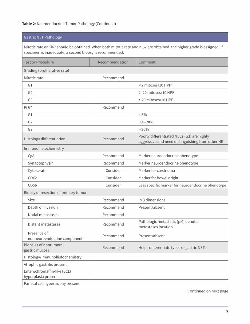
Gastric NET Pathology
Mitotic rate or Ki67 should be obtained. When both mitotic rate and Ki67 are obtained, the higher grade is assigned. If specimen is inadequate, a second biopsy is recommended.
Test or Procedure Recommendation Comment
Grading (proliferative rate)
Mitotic rate Recommend
G1 < 2 mitoses/10 HPF*
G2 2–20 mitoses/10 HPF
G3 > 20 mitoses/10 HPF
Ki 67 Recommend
G1 < 3%
G2 3%–20%
G3 > 20%
Histology differentiation RecommendPoorly differentiated NECs (G3) are highly aggressive and need distinguishing from other NE
Immunohistochemistry
CgA Recommend Marker neuroendocrine phenotype
Synaptophysin Recommend Marker neuroendocrine phenotype
Cytokeratin Consider Marker for carcinoma
CDX2 Consider Marker for bowel origin
CD56 Consider Less specific marker for neuroendocrine phenotype
Biopsy or resection of primary tumor
Size Recommend In 3 dimensions
Depth of invasion Recommend Present/absent
Nodal metastases Recommend
Distant metastases RecommendPathologic metastasis (pM) denotes metastases location
Presence of nonneuroendocrine components
Recommend Present/absent
Biopsies of nontumoral gastric mucosa
Recommend Helps differentiate types of gastric NETs
Histology/immunohistochemistry
Atrophic gastritis present
Enterochromaffin-like (ECL) hyperplasia present
Parietal cell hypertrophy present
Table 2: Neuroendocrine Tumor Pathology (Continued)
Continued on next page

8
Pancreatic NET Pathology
Mitotic rate or Ki67 should be obtained. When both mitotic rate and Ki67 are obtained, the higher grade is assigned. If specimen is inadequate, repeat biopsy is recommended.
Test or Procedure Recommendation Comment
Subtype
Small cell, non–small cell (ie, large cell)
Recommend
Grading (proliferative rate) Recommend
Mitotic rate
G1 < 2 mitoses/10 HPF*
G2 2–20 mitoses/10 HPF
G3 > 20 mitoses/10 HPF
Ki 67
G1 < 3%
G2 3%–20%
G3 > 20%
Histology differentiation RecommendPoorly differentiated NECs (G3) are highly aggressive and need to be distinguished from other NETs
Immunohistochemistry
CgA Recommend Marker neuroendocrine phenotype
Synaptophysin Recommend Marker neuroendocrine phenotype
Cytokeratin Consider Marker for carcinoma
CDX2 Consider Marker for bowel origin
CD56 Consider Less specific marker for neuroendocrine phenotype
Biopsy or resection of primary tumor
Size Recommend In 3 dimensions
Depth of invasion Recommend
Nodal metastases Recommend
Distant metastases Recommend pM denotes metastases location
Presence of nonneuroendocrine components
Recommend Present/absent
Table 2: Neuroendocrine Tumor Pathology (Continued)
Continued on next page

9
Midgut NET Pathology
Mitotic rate or Ki67 should be obtained. When both mitotic rate and Ki67 are obtained, overall grade is defined by the higher of the two. If specimen is inadequate, a second biopsy is recommended.
Test or Procedure Recommendation Comment
Grading (proliferative rate) Recommend
Mitotic rate
G1 < 2 mitoses/10 HPF*
G2 2–20 mitoses/10 HPF
G3 > 20 mitoses/10 HPF
Ki 67
G1 < 3%
G2 3%–20%
G3 > 20%
Immunohistochemistry
CgA Recommend Marker neuroendocrine phenotype
Synaptophysin Recommend Marker neuroendocrine phenotype
Cytokeratin Consider Marker for carcinoma
CDX2 Consider Marker for bowel origin
CD56 Consider Less specific marker for neuroendocrine phenotype
Resection of primary tumor
Size Recommend In 3 dimensions
Depth of invasion Recommend
Nodal metastases Recommend
Distant metastases Recommend pM should be used to denote metastases location
Presence of nonneuroendocrine components
Recommend Present/absent
Table 2: Neuroendocrine Tumor Pathology (Continued)
Continued on next page

10
Hindgut NET Pathology
Mitotic rate or Ki67 should be obtained. When both mitotic rate and Ki67 are obtained grade is the higher of grade determined by mitotic rate or Ki67. If specimen is inadequate, a second biopsy is recommended.
Test or Procedure Recommendation Comment
Grading (proliferative rate) Recommend
Mitotic rate
G1 < 2 mitoses/10 HPF*
G2 2–20 mitoses/10 HPF
G3 > 20 mitoses/10 HPF
Ki 67
G1 < 3%
G2 3%–20%
G3 > 20%
Immunohistochemistry
CgA Recommend Marker neuroendocrine phenotype
Synaptophysin Recommend Marker neuroendocrine phenotype
CDX2 Consider Marker for bowel origin
CD56 Consider Less specific marker for neuroendocrine phenotype
Resection of primary tumor
Size Recommend In 3 dimensions
Depth of invasion Recommend
Nodal metastases Recommend
Distant metastases Recommend pM denotes metastases location
Presence of nonneuroendocrine components
Recommend Present/absent
Table 2: Neuroendocrine Tumor Pathology (Continued)
Continued on next page

11
Pheochromocytoma/Paraganglioma Pathology
Distinction between benign and malignant disease is difficult to ascertain pathologically.
Test or Procedure Recommendation Comment
Patient and tumor characteristics
Age Recommend Younger age increases suspicion of genetic disease
Extra-adrenal location RecommendExtra-adrenal location increases the risk of malignancy
Pathology reporting
Multicentricity Recommend Can increase suspicion of genetic disease
Accompanying medullary hyperplasia
Recommend Can increase suspicion of genetic disease
Ki67 Consider Rates >2%–3% can be associated with malignancy
Periadrenal adipose tissue Consider
Large nests/diffuse growth Consider
Focal or confluent necrosis Consider Can be associated with malignancy
Cellularity Consider
Tumor cell spindling Consider
Cellular monotony Consider
Mitotic rate Consider>3/10 HPF* can be associated with more aggressive behavior
Atypical mitosis Consider
Hyperchromasia Consider
Profound nuclear pleomorphism Consider
Immunohistochemistry
CgA Recommend Marker of neuroendocrine phenotype
Synaptophysin Consider Marker of neuroendocrine phenotype
S-100 Consider Marker for sustentacular supporting framework
Cytokeratin ConsiderNegative staining supports pheochromocytoma/paraganglioma over carcinoid tumor or NET
Table 2: Neuroendocrine Tumor Pathology (Continued)
Continued on next page

12
Poorly Differentiated NET Pathology
Test or Procedure Recommendation Comment
Subtype
Small cell, non–small cell (ie, large cell)
Recommend
Grading (proliferative rate)
Mitotic rate (G3) Recommend > 10 mitoses/10 HPF* for lung
> 20 mitoses/10 HPF for Gastroenteropancreatic-NET
Ki 67 Recommend > 20%
Immunohistochemistry
CgA Recommend Marker neuroendocrine phenotype
Synaptophysin Recommend Marker neuroendocrine phenotype
Cytokeratin Consider Marker for epithelial carcinoma
CDX2 Consider Marker for bowel origin
CD56 Consider Less specific marker for neuroendocrine phenotype
Resection of primary tumor
Size Recommend In 3 dimensions
Depth of invasion Recommend
Nodal metastases Recommend
Distant metastases Recommend pM should be used to denote metastases location
Presence of nonneuroendocrine components
Recommend Present/Absent
Table 2: Neuroendocrine Tumor Pathology (Continued)
*Based on a 0.5-mm field diameter at high power, which yields a total area of 2 mm2 for 10 high power fields. ECL indicates enterochromaffin-like; GEP, gastroenteropancreatic.
13
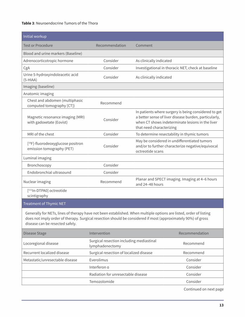
Initial workup
Test or Procedure Recommendation Comment
Blood and urine markers (Baseline)
Adrenocorticotropic hormone Consider As clinically indicated
CgA Consider Investigational in thoracic NET, check at baseline
Urine 5-hydroxyindoleacetic acid (5-HIAA)
Consider As clinically indicated
Imaging (baseline)
Anatomic imaging
Chest and abdomen (multiphasic computed tomography [CT])
Recommend
Magnetic resonance imaging (MRI) with gadoxetate (Eovist)
Consider
In patients where surgery is being considered to get a better sense of liver disease burden, particularly, when CT shows indeterminate lesions in the liver that need characterizing
MRI of the chest Consider To determine resectability in thymic tumors
[18F]-fluorodeoxyglucose positron emission tomography (PET)
ConsiderMay be considered in undifferentiated tumors and/or to further characterize negative/equivocal octreotide scans
Luminal imaging
Bronchoscopy Consider
Endobronchial ultrasound Consider
Nuclear imaging RecommendPlanar and SPECT imaging. Imaging at 4–6 hours and 24–48 hours
[111In-DTPA0] octreotide scintigraphy
Table 3: Neuroendocrine Tumors of the Thora
Continued on next page
Treatment of Thymic NET
Generally for NETs, lines of therapy have not been established. When multiple options are listed, order of listing does not imply order of therapy. Surgical resection should be considered if most (approximately 90%) of gross disease can be resected safely.
Disease Stage Intervention Recommendation
Locoregional diseaseSurgical resection including mediastinal lymphadenectomy
Recommend
Recurrent localized disease Surgical resection of localized disease Recommend
Metastatic/unresectable disease Everolimus Consider
Interferon ɑ Consider
Radiation for unresectable disease Consider
Temozolomide Consider

14
Table 3: Neuroendocrine Tumors of the Thora (Continued)
Follow-Up
Follow-up for resected disease is recommended 3 to 6 months after curative resection and then every 6 to 12 months for at least 7 years. Maximum duration of follow-up is not defined; late recurrence can occur in some patients. Follow-up for advanced disease is recommended every 3 to 6 months; may lengthen interval to every 6 months for patient with long duration (912 month) of stable disease.
Test or Procedure Recommendation Comment
Blood and urine markers
Adrenocorticotropic hormone Consider Consider following if abnormal at baseline
CgA Consider Consider following if abnormal at baseline
Urine 5-HIAA Consider Consider following if abnormal at baseline
Imaging
Anatomic imaging (CT or MRI) Recommend See initial imaging for details
Nuclear imaging Consider As clinically indicated for suspected recurrence
[111In-DTPA0]octreotide scintigraphy (see initial imaging for details)
Treatment of Lung/Bronchial NET
Generally for NETs, lines of therapy have not been established. When multiple options are listed, order of listing does not imply order of therapy. Surgical resection should be considered if most (approximately 90%) of gross disease can be resected safely. Clinical trials should always be considered.
Disease Stage Intervention Recommendation
Locoregional diseaseSurgical resection with hilar/mediastinal lymph node sampling is recommended
Recommend
Recurrent disease, resectable Surgical resection Recommend
Metastatic/unresectable disease Everolimus
Interferon ɑ Consider
Radiation for unresectable disease Consider
Temozolomide Consider

15
Table 4: Gastric NETs
Initial workup
Test or Procedure Recommendation Comment
Blood and urine markers (Baseline)
Gastric pH Recommend
Gastric pH helps differentiate type I (gastric pH >4) from type II (gastric pH <2). Type II requires workup for Multiple Endocrine Neoplasia (MEN) 1 syndrome. Type III gastric pH <4
Gastrin RecommendShould be fasting and off PPI when feasible (types I and II will have elevated gastrin levels; type III will have normal gastrin level)
5-HIAA ConsiderAs indicated for atypical type III foregut tumors or if symptoms suggestive of carcinoid syndrome. Need to follow diet during collection.
Anti-intrinsic factor and antiparietal cell antibodies
ConsiderOnly in type I. Consider workup for polyglandular syndrome
CgA ConsiderRecommended for type III (normogastrinemic) gastric carcinoids; false positive with proton pump inhibitor use and renal insufficiency
Imaging (baseline)
Anatomic imaging
Abdomen and pelvis (multiphasic CT or MRI)
Recommend For types II and III only
MRI with gadoxetate (Eovist) Consider
In patients where surgery is being considered to get a better sense of liver disease burden, particularly, when CT shows indeterminate lesions in the liver that need characterizing
Luminal imaging
Esophagogastroduodenoscopy (EGD)
RecommendPermits sampling of gastric mucosa and determination of disease extent
Endoscopic ultrasound (EUS) ConsiderBest procedure to determine tumor size/infiltration and to identify possible lymph node metastases
Nuclear imaging
[111In-DTPA0] octreotide scintigraphy
Consider

16
Table 4: Gastric NETs (Continued)
Surgery of Primary Tumors
In general, resection is recommended for local regional disease and in setting of impending obstruction and should still be considered for patients with advanced disease. Ability to resect primary tumors depends on the number, size, depth of invasion, and institutional expertise. In patients with suspected carcinoid syndrome who undergo major procedures, a preoperative bolus of octreotide, 250 to 500 Hg intravenous, is recommended with additional bolus doses available throughout the procedure.
Tumor Type Intervention Recommendation
Type I
<1 cm Surveillance or endoscopic removal Recommend
1–2* cm (up to 6 polyps)
Surveillance with repeat endoscopy approximately every 3 years or endoscopic resection. Endoscopic US could be used to assess depth of invasion but should be individualized. If submucosal invasion, endoscopic mucosal resection is increasingly used.
Recommend
>2* cm (up to 6 polyps)Endoscopic resection (if possible) or surgical resection
Recommend
>2* cm (>6 polyps)Must be individualized and could include surveillance, endoscopic resection or surgical resection
Recommend
Type II
<1 cm Surveillance or endoscopic removal Recommend
1–2 cm
Endoscopic resection. EUS should be used to assess depth of invasion. If submucosal invasion, endoscopic mucosal resection is increasingly used.
Recommend
>2 cmSurgical resection or endoscopic resection (if possible)
Recommend
Type IIIPartial gastrectomy and lymph node dissection
Recommend

17
Advanced Disease—Oncologic Control of Gastric NETs
Advanced disease is typically limited to type III only. Generally for NETs, lines of therapy have not been established. When multiple options are listed, order of listing does not imply order of therapy. Surgical resection should be considered if most (approximately 90%) of gross disease can be resected safely. Clinical trials should always be considered.
Indication Intervention Recommendation
Newly diagnosed with low or intermediate tumor volume
Observation if no hormonal symptoms present
Recommend
Octreotide LAR Consider
Newly diagnosed with high-volume disease
Everolimus Consider
Liver-directed therapies when liver- dominant disease
Consider
Octreotide LAR Consider
Stable disease Observation if no hormonal symptoms Consider
Progressive disease Everolimus Consider
Liver-directed therapies when liver- dominant disease
Consider
Octreotide LAR Consider
Refer to specialty center Consider
Hormonal Syndrome Control
Carcinoid syndrome is rarely found in gastric NETs (type III only).
Carcinoid syndrome
Initial or nonrefractory
Long-acting somatostatin analogs; octreotide LAR, 20–30 mg IM is available in the United States. Immediate release octreotide can be used for breakthrough symptoms.
Recommend
Refractory syndrome with stable tumor volume
Antidiarrheal agentsRecommend
Debulk tumor with liver-directed therapy if possible
Recommend
Escalate dose or shorten dosing interval of long-acting somatostatin analog. No prospective data exist.
Recommend
Add low-dose interferon ɑ (short-acting or pegylated form)
Consider
Referral to specialty center Consider
Rotate somatostatin analog as available Consider
Table 4: Gastric NETs (Continued)

18
Indication Intervention Recommendation
Refractory syndrome with increasing tumor volume
Measures for refractory syndrome Recommend
Measures for oncologic control See Oncologic control section.
Recommend
Refer to specialty center Consider
Table 4: Gastric NETs (Continued)
Follow-Up
Follow-up for resected gastric NET disease is recommended 3 to 6 months after curative resection and then every 6 to 12 months for at least 7 years. Maximum duration of follow-up is not defined; late recurrence can occur in some patients. Follow-up for advanced disease is recommended every 3 to 6 months; may lengthen interval to every 6 months for patient with long duration (>12 months) of stable disease.
Test or Procedure Recommendation Comment
Blood and urine markers
CgA Consider Consider following if abnormal at baseline
Specific hormone marker Consider Consider following if abnormal at baseline
Imaging
Anatomic imaging (multiphasic CT or MRI)
Recommend See initial imaging for details
Luminal Imaging
EGD
Gastric pH
Nuclear imaging ConsiderAs clinically indicated for suspected recurrence (see initial imaging for details)
[111In-DTPA0]octreotide scintigraphy
*Multiple lesions that are larger than 1 to 2 cm should be individually decided and could include local resection, surgical resection, or watchful waiting. EGD indicates esophagogastroduodenoscopy; MEN, multiple endocrine neoplasia.
19
Table 5: Pancreatic NETs
Continued on next page
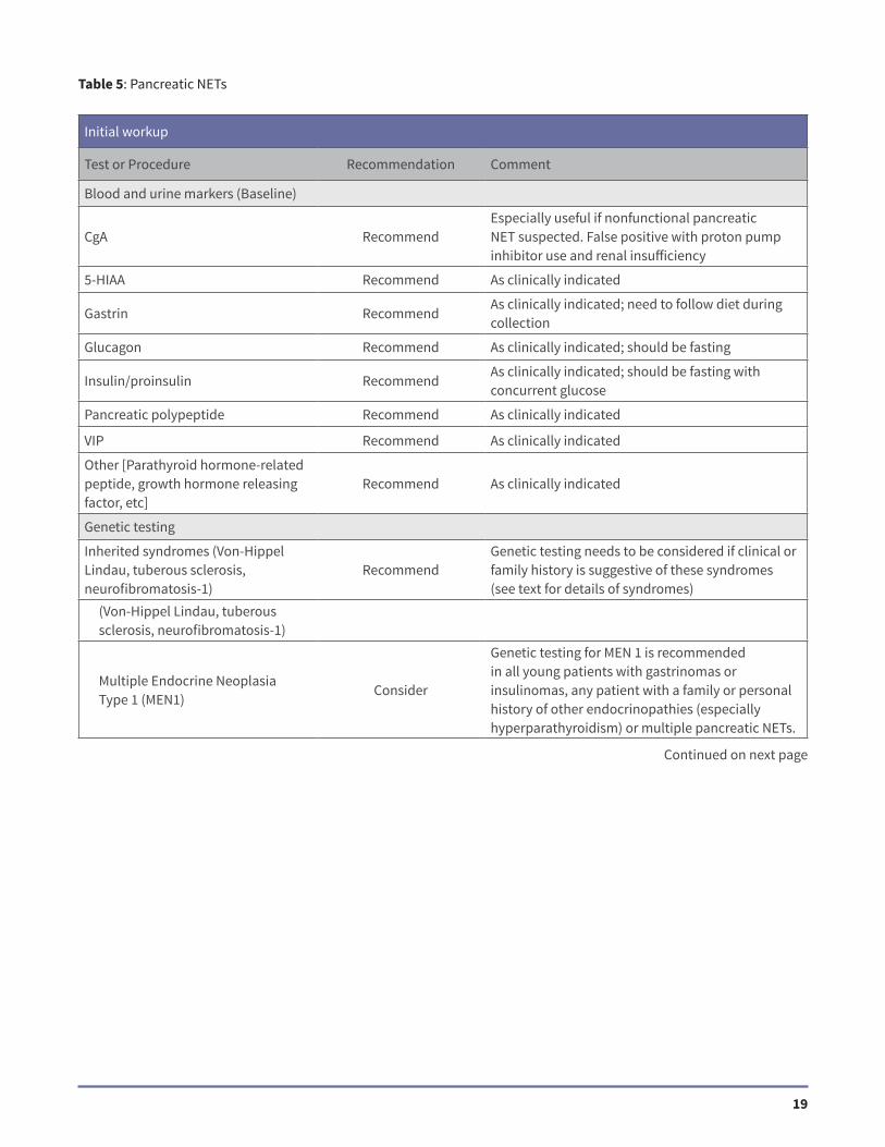
Initial workup
Test or Procedure Recommendation Comment
Blood and urine markers (Baseline)
CgA RecommendEspecially useful if nonfunctional pancreatic NET suspected. False positive with proton pump inhibitor use and renal insufficiency
5-HIAA Recommend As clinically indicated
Gastrin RecommendAs clinically indicated; need to follow diet during collection
Glucagon Recommend As clinically indicated; should be fasting
Insulin/proinsulin RecommendAs clinically indicated; should be fasting with concurrent glucose
Pancreatic polypeptide Recommend As clinically indicated
VIP Recommend As clinically indicated
Other [Parathyroid hormone-related peptide, growth hormone releasing factor, etc]
Recommend As clinically indicated
Genetic testing
Inherited syndromes (Von-Hippel Lindau, tuberous sclerosis, neurofibromatosis-1)
RecommendGenetic testing needs to be considered if clinical or family history is suggestive of these syndromes (see text for details of syndromes)
(Von-Hippel Lindau, tuberous sclerosis, neurofibromatosis-1)
Multiple Endocrine Neoplasia Type 1 (MEN1)
Consider
Genetic testing for MEN 1 is recommended in all young patients with gastrinomas or insulinomas, any patient with a family or personal history of other endocrinopathies (especially hyperparathyroidism) or multiple pancreatic NETs.

20
Imaging (baseline)
Anatomic imaging
Abdomen and pelvis (multiphasic CT or MRI)
Recommend Thin sections
MRI with gadoxetate (Eovist) Consider
In patients where surgery is being considered to get a better sense of liver disease burden, particularly, when CT shows indeterminate lesions in the liver that need characterizing
Additional sites Consider As clinically indicated
Luminal imaging
EGD Consider
In patients suspected of gastrinoma to visualize prominent gastric folds in Zollinger EllisonSyndrome; also with duodenal NETs (often nonfunctional) and in MEN 1 who have submucosal duodenal lesions
EUS Consider
Should be performed for diagnostic purposes if pancreatic NET is suspected and no primary identified on cross-sectional imaging; helps identify small pancreatic NET lesions
Nuclear imaging
[111In-DTPA0] octreotide scintigraphy
RecommendPlanar and SPECT imaging. Imaging at 4–6 hours and 24–48 hours
Table 5: Pancreatic NETs (Continued)
Surgery of Primary Tumors
In general, resection is recommended for local regional disease and should still be considered for patients with advanced disease. Optimize nutritional status and control of hormone excess state medically preoperatively as outlined in the functional pancreatic NET section.
Functional Status Intervention Recommendation
Functional pancreatic NET
Gastrinoma
Sporadic
Surgical removal with enucleation, resection, or occasionally a pancreaticoduodenectomy. Routine duodenotomy and periduodenal/tumoral nodal dissection required
Recommend
With MEN1
If imaged tumor is <2–2.5 cm, most observe, although some recommend enucleation or resection. Pancreaticoduodenectomy rarely indicated
Recommend
Other functional tumor (sporadic or with MEN1)
Enucleation or surgical resection/enucleation
Recommend
Continued on next page

21
Table 5: Pancreatic NETs (Continued)
Functional Status Intervention Recommendation
Nonfunctional pancreatic NET
SporadicEnucleation or surgical resection with lymph node dissection. Observation in elderly or comorbid conditions
Recommend
With MEN1
If imaged tumor is <2–2.5 cm, most observe, although some recommend enucleation or resection. Pancreaticoduodenectomy rarely indicated. If >2–2.5 cm, enucleation or surgical resection with adjacent lymph node dissection.
Recommend
With VHLIf imaged tumor is >3 cm, surgical resection is recommended.
Recommend
Advanced Disease—Oncologic Control of Pancreatic NETs
Generally for NETs, lines of therapy have not been established. When multiple options are listed, order of listing does not imply order of therapy. Surgical resection should be considered if most (approximately 90%) of gross disease can be resected safely. Clinical trials should always be considered.
Indication Intervention Recommendation
Newly diagnosed with low or intermediate tumor volume
Observation if no hormonal syndrome Recommend
Newly diagnosed with high-volume disease
Observation for a brief 3-month period if no hormonal syndrome
Recommend
Everolimus Consider
Hepatic artery embolization when liver dominant disease (bland embolization,chemoembolization, or radioembolization per institutional practice)
Consider
Sunitinib Consider
Stable disease Observation if no hormonal syndrome Recommend
Progressive disease Sunitinib Recommend
Everolimus Recommend
Cytotoxic chemotherapy Consider
Hepatic artery embolization when liver dominant disease (bland embolization,chemoembolization, or radioembolization per institutional practice)
Consider
Octreotide LAR Consider
Continued on next page

22
Hormonal Syndrome Control
Please also see the section entitled, ‘‘Neuroendocrine Tumors of the Jejunum, Ileum, Appendix, and Colon’’ for control of hormonal syndromes in the carcinoid syndrome.
Indication Intervention Recommendation
Insulinoma
Initial or nonrefractory Dietary modification Recommend
Diazoxide 200–600 mg/d Recommend
Everolimus Recommend
Medicalert bracelet Recommend
Glucagon pen Consider
Somatostatin analogs. May worsen hypoglycemia in some cases; therefore, consider short-acting octreotide trial before initiation of octreotide LAR).
Consider
Steroids (ie, decadron) Consider
Gastrinoma Oral proton pump inhibitors Recommend
Initial and long-term BID or TID dosing of Proton Pump Inhibitor Recommend
Medicalert bracelet Consider
Octreotide LAR Consider
Other functioning PETs Octreotide LAR Recommend
Refractory syndrome with stable tumor volume
Nonspecific antidiarrheal agents as clinically indicated
Recommend
Escalate dose or shorten dosing interval of octreotide LAR
Consider
Liver-directed therapy if possible Consider
Surgical debulking Consider
Refractory syndrome with increasing tumor volume
Measures for refractory syndrome Recommend
Measures for oncologic control (see Oncologic control section).
Recommend
Referral to specialty center Recommend
Table 5: Pancreatic NETs (Continued)
Continued on next page

23
Table 5: Pancreatic NETs (Continued)
Follow-Up
Follow-up for resected pancreatic is recommended 3 to 6 months after curative resection and then every 6 to 12 months for at least 7 years. Maximum duration of follow-up is not defined; late recurrence can occur in some patients. Follow-up for advanced disease is recommended every 3 to 6 months; may lengthen interval to every 6 months for patient with long duration (>12 months) of stable disease.
Test or Procedure Recommendation Comment
Blood and urine markers
CgA Consider Consider following if abnormal at baseline
Specific hormone marker Consider Consider following if abnormal at baseline
Imaging
Anatomic imaging (multiphasic CT or MRI)
Recommend See initial imaging for details
Nuclear imaging ConsiderAs clinically indicated for suspected recurrence (see initial imaging for details)
[111In-DTPA0]octreotide scintigraphy
GRF indicates growth hormone releasing factor; PPI, proton pump inhibitor; PTH, parathyroid hormone; VHL, Von-Hippel Lindau; ZES, Zollinger Ellison syndrome.
Initial workup
Test or Procedure Recommendation Comment
Blood and urine markers (Baseline)
CgA RecommendOften negative in those with localized tumors.False positive with proton pump inhibitor use and renal insufficiency
Urine 5-HIAA Recommend Need to follow diet during collection
Imaging (baseline)
Anatomic imaging
Abdomen and pelvis (multiphasic CT or MRI)
Recommend
Thin section with negative bowel contrast if attempting to identify primary tumor.Consider MRI if unable to give iodine-based contrast. Consider specific enterography protocols if available.
MRI with gadoxetate (Eovist) Recommend
In patients where surgery is being considered to get a better sense of liver disease burden, particularly when CT shows indeterminate lesions in the liver that need characterizing
Additional sites Recommend As clinically indicated
Table 6: Neuroendocrine Tumors of the Jejunum, Ileum, Appendix, and Cecum
Continued on next page

24
Test or Procedure Recommendation Comment
Luminal Imaging
Colonoscopy Recommend Terminal ileal intubation
Deep enteroscopy ConsiderBest approached bidirectionally; tattoo location if identified
Nuclear imaging
[111In-DTPA0]octreotide scintigraphy
RecommendPlanar and SPECT imaging. Imaging at 4–6 hours and 24–48 hours
Cardiac imaging
Echocardiogram ConsiderIf symptoms of carcinoid heart are suspected or as clinically indicated
Table 6: Neuroendocrine Tumors of the Jejunum, Ileum, Appendix, and Cecum (Continued)
Surgery of Primary Tumors
In general, resection is recommended for local regional disease and in setting of impending obstruction and should still be considered for patients with advanced disease. Ability to resect primary depends on size, depth of invasion, and institutional expertise. In patients with suspected carcinoid syndrome who undergo major procedures, a preoperative bolus of octreotide 250 to 500 µg IV is recommended with additional bolus doses available throughout procedure.
Primary Site/Size Intervention Recommendation
Appendix, cm
<1 Excision Recommend
1–2 Excision Recommend
Right hemicolectomy with node dissection if high risk features present
Consider
>2 Right hemicolectomy with node dissection Recommend
Cecum Right hemicolectomy with node dissection Recommend
Ileum
Resection with node dissection. Ileocecal valve and right colon can be preserved for more proximal tumors. Full bowel examination required at time of surgery in case of lateral metastases.
Recommend
JejunumResection with node dissection. Full bowel examination required at time of surgery in case of lateral metastases.
Recommend
Root of mesentery disease
Refer to expert center for assessment when nodal disease approaches branches of Superior Mesenteric Vein or Superior Mesenteric Artery.
Recommend
Continued on next page

25
Advanced Disease—Oncologic Control
Generally for NETs, lines of therapy have not been established. When multiple options are listed, order of listing does not imply order of therapy. Surgical resection should be considered if most (approximately 90%) of gross disease can be resected safely. Clinical trials should always be considered.
Indication Intervention Recommendation
Newly diagnosed with low or intermediate tumor volume
Observation if no hormonal symptoms present
Recommend
Octreotide LAR Consider
Newly diagnosed with high-volume disease
Everolimus Consider
Liver-directed therapies when liver-dominant disease
Consider
Octreotide LAR Consider
Stable disease Observation if no hormonal symptoms Consider
Progressive disease Refer to specialty center Recommend
Everolimus Consider
Liver-directed therapies when liver-dominant disease
Consider
Octreotide LAR Consider
Hormonal Syndrome Control
Carcinoid syndrome
Initial or nonrefractory
Long-acting somatostatin analogs; octreotide LAR 20–30 mg IM is available in the United States. Immediate release octreotide can be used for breakthrough symptoms.
Recommend
Refractory syndrome
Stable tumor volume Antidiarrheal agents Recommend
Debulk tumor with liver-directed therapy if possible
Recommend
Escalate dose or shorten dosing interval of long-acting somatostatin analog. No prospective data exist.
Recommend
Add low-dose interferon ɑ (short-acting or pegylated form)
Consider
Referral to specialty center Consider
Rotate somatostatin analog as available Consider
Table 6: Neuroendocrine Tumors of the Jejunum, Ileum, Appendix, and Cecum (Continued)
Continued on next page

26
Increasing tumor volume Measures for refractory syndrome Recommend
Measures for oncologic control(see Oncologic control section)
Recommend
Refer to specialty center Consider
Table 6: Neuroendocrine Tumors of the Jejunum, Ileum, Appendix, and Cecum (Continued)
Follow-Up
Follow-up for resected disease is recommended 3 to 6 months after resection with curative intent and then every 6 to 12 months for at least 7 years. Maximum duration of follow-up is not defined; late recurrence can occur in some patients. Follow-up for advanced disease is recommended every 3 to 6 months; may lengthen interval to every 6 months for patient with long duration (>12 months) of stable disease.
Test or Procedure Recommendation Comment
Blood and urine markers
CgA Consider Consider following if abnormal at baseline
Urine 5-HIAA Consider Consider following if abnormal at baseline
Imaging
Anatomic imaging (multiphasic CT or MRI)
Recommend See initial imaging for details
Nuclear imaging ConsiderAs clinically indicated for suspected recurrence (see initial imaging for details)
[111In-DTPA0]octreotide scintigraphy
SMA indicates superior mesenteric artery; SMV, superior mesenteric vein.
Table 7: Neuroendocrine Tumors of the Distal Colon and Rectum
Initial workup
Test or Procedure Recommendation Comment
Blood and urine markers (Baseline)
CgA RecommendOften negative in those with localized tumors.False positive with proton pump inhibitor use and renal insufficiency
Urine 5-HIAA Consider Need to follow diet during collection
Imaging (baseline)
Anatomic imaging
Abdomen and pelvis (multiphasic CT or MRI)
Recommend
Recommended for patients with tumors 92 cm, invasion beyond submucosa, or lymph node involvement. Could also consider for tumors with elevated mitotic rate or poor differentiation.

27
MRI with gadoxetate (Eovist) Consider
In patients where surgery is being considered to get a better sense of liver disease burden, particularly when CT shows indeterminate lesions in the liver that need characterizing.
Additional sites Consider As clinically indicated
Luminal imaging
Colonoscopy RecommendOften detected incidentally on colonoscopy; consider tattoo for localization.
EUS
Nuclear imaging
[111In-DTPA0]octreotide scintigraphy RecommendPlanar and SPECT imaging. Imaging at4–6 hours and 24–48 hours
Table 7: Neuroendocrine Tumors of the Distal Colon and Rectum (Continued)
Surgery of Primary Tumors
In general, resection is recommended for local regional disease and in setting of impending obstruction and should still be considered for patients with advanced disease. Ability to resect primary depends on size, depth of invasion, and institutional expertise.
Primary Site/Size Intervention Recommendation
<1
Endoscopic resection (polypectomy, endoscopic mucosal resection, endoscopic submucosal dissection) for those with mucosal or submucosal tumors
Recommend
1–2Transanal excision via rigid or flexible dissection. Could also consider after endoscopic resection with positive margins
Recommend
>2
Surgical resection (low anterior resection orabdominoperineal resection) for larger tumors, tumors invading muscularis propria, or those with lymphadenopathy
Recommend
Incidentally discoveredTattoo location if polyp has unusual features suggestive of carcinoid at screening colonoscopy.
Consider
Advanced Disease—Oncologic Control
Generally for NETs, lines of therapy have not been established. When multiple options are listed, order of listing does not imply order of therapy. Surgical resection should be considered if most (approximately 90%) of gross disease can be resected safely. Clinical trials should always be considered.
Indication Intervention Recommendation
Newly diagnosed with low or intermediate tumor volume
Observation if no hormonal syndrome Recommend
Octreotide LAR Consider
Continued on next page

28
Newly diagnosed with high-volume disease
Liver-directed therapies when liver-dominant disease
Consider
Octreotide LAR Consider
Stable disease Observation if no hormonal syndrome Consider
Progressive disease Refer to specialty center Recommend
Everolimus Consider
Liver-directed therapies when liver-dominant disease
Consider
Octreotide LAR Consider
Table 7: Neuroendocrine Tumors of the Distal Colon and Rectum (Continued)
Follow-Up
Intensity and duration of surveillance depends on stage of disease. Stage I tumors require no surveillance. Stage II or III should be followed 3 to 6 months after curative resection and then every 6 to 12 months for at least 7 years. Maximum duration of follow-up is not defined; late recurrence can occur in some patients. Follow-up for advanced disease is recommended every 3 to 6 months; may lengthen interval to every 6 months for patients with long duration (>12 months) of stable disease.
Test or Procedure Recommendation Comment
Blood and urine markers
CgA Consider Consider following if abnormal at baseline
Urine 5-HIAA Consider Consider following if abnormal at baseline
Imaging
Anatomic imaging (multiphasic CT or MRI)
Recommend See initial imaging for details
Nuclear imaging ConsiderAs clinically indicated for suspected recurrence (see initial imaging for details)
[111In-DTPA0]octreotide scintigraphy

29
Table 8: Pheochromocytoma/paraganglioma, Medullary Thyroid Cancer
Initial workup (Pheochromocytoma/Paraganglioma)
Test or Procedure Recommendation Comment
Blood and urine markers (Baseline)
Hormonal markers
Fractionated or free metanephrines (ie, normetanephrine and metanephrines) in urine or plasma, respectively, or both
Recommend
It is preferred to measure fractionated or free metanephrines versus the parent catecholamines. Blood sampling should be done in the supine position after a 20-minute rest.
>4 upper reference range Diagnostic of pheochromocytoma
1–4 upper reference range
Needs further evaluation. First, exclude drug effect, and then use clonidine suppression test coupled with the measurement of plasma normetanephrine (does not work if coupled with the measurement of plasma metanephrine).
Genetic counseling/genetic testing when appropriate
Recommend
To choose the proper genetic testing sequence, consider the biochemical profile of catecholamine secretion, age of the patient, localization of the primary tumor, and previous family history.
Methoxytyramine ConsiderMarker of dopamine secreting tumors, associated with malignancy and mutations in the succinate dehydrogenase complexYrelated tumors
Imaging (baseline)
Anatomic imaging
Abdomen and pelvis (multiphasic CT or MRI)
RecommendBoth modalities are effective for localizing and characterizing adrenal masses.
MRI with gadoxetate (Eovist) Consider
In patients where surgery is being considered to get a better sense of liver disease burden, particularly, when CT shows indeterminate lesions in the liver that need characterizing
Additional sites ConsiderAs clinically indicated, if no lesion is seen on abdomen and pelvis imaging
Nuclear imagingPlanar and SPECT imaging. Imaging at 4–6 hours and 24–48 hours
Iodine 123
[123I]- metaiodobenzylguanide (MIBG) ([123]I-MIBG) scintigraphy
Consider
Should be used on all functional tumors except adrenal pheochromocytomas G5 cm that are associated with elevations of plasma and urine metanephrine (rarely metastatic). Also to be used when treatment with 131I-MIBG is considered (metastatic disease already proven by anatomic imaging)
Continued on next page

30
[18F]-fluorodeoxyglucose PET ConsiderObtain if [123]I-MIBG scan is negative and there is concern for metastatic disease.
[111In-DTPA0]octreotide scintigraphy (octreotide scan)
Consider
Obtain if [123]-MIBG scan is negative and there is concern for metastatic disease as well as when treatment with octreotide is considered (metastatic disease already proven by anatomic imaging).
Table 8: Pheochromocytoma/paraganglioma, Medullary Thyroid Cancer (Continued)
Surgery of Primary Tumors (Pheochromocytoma/Paraganglioma)
For major procedures, start phenoxybenzamine at 10 mg oral 2 times a day and titrate to control hypertension. May also use ɑ-1 adrenoceptor blockers. Also consider calcium channel blocker or angiotensin receptor blockers, especially in patients with mild hypertension, and treatment should be for at least 10 to 14 days before surgery. Use volume expansion through hydration before surgery. If tachycardia is present, add β-adrenoceptor blocker (atenolol preferred). Only start after appropriate ɑ-blockade has started.
Surgical Approach Intervention Recommendation
Laparoscopic resection
Procedure of choice if no evidence of local invasion or malignancy. Consider cortical sparing adrenalectomy if familial or bilateral disease.
Recommend
Test or Procedure Recommendation Comment
Open resectionProcedure of choice if evidence of local invasion or malignancy or recurrent disease.
Recommend
Cytoreductive resection when locallyunresectable or distant metastases present
Cytoreductive surgery should be considered in all patients to help aid in symptom control.
Consider
Advanced Disease–Oncologic Control (Pheochromocytoma/Paraganglioma)
Generally for NETs, lines of therapy have not been established. When multiple options are listed, order of listing does not imply order of therapy. Surgical resection should be considered if most (approximately 90%) of gross disease can be resected safely. Clinical trials should always be considered.
Indication Intervention Recommend
Locally unresectable Cytoreductive surgery, if feasible Recommend
External beam radiation therapy Consider
Distant Disease Cytoreductive surgery, if feasible Recommend
[131I]-MIBG treatment if [123I]-MIBG–positive disease
Consider
Radiofrequency ablation Consider
Systemic chemotherapy (cyclophosphamide, vincristine, and dacarbazine,) if [123I]-MIBG–negative disease or rapidly progressing
Consider
Continued on next page

31
Hormonal Syndrome Control (Pheochromocytoma/Paraganglioma)
Indication Intervention Recommendation
Treatment of catecholamine overproduction
Alpha-blockade for symptom control.May change to selective ɑ-1 blockers for long-term treatment
Recommend
Beta-blockade if necessary after adequateɑ-blockade in patients with tachycardia
Recommend
Treatment of catecholamine crisis Alpha-methyl-para-tyrosine
Phentolamine IV bolus 2.5–5 mg at1 mg/min, may repeat every 5 minutes or run as an infusion (100 mg in 500 mL of Dextrose 5% water). Alternative isnitroprusside infusion at 0.5–5.0 µg/kg per minute. (do not exceed 3.0 µg/kg per minute for long-term use)
Recommend
Table 8: Pheochromocytoma/paraganglioma, Medullary Thyroid Cancer (Continued)
Follow-up (Pheochromocytoma/Paraganglioma)
Follow-up for resected disease is recommended 6 and 12 months after curative resection and then annually. Maximum duration of follow-up is not defined; late recurrence can occur in some patients. Follow-up for advanced disease is recommended every 3 to 6 months; may lengthen interval to every 6 months for patient with long duration (>12 months) of stable disease.
Test or Procedure Recommendation Comment
Blood and urine markers
Fractionated or free metanephrines Recommend
CgA Consider
May be used if tumor does not produce significant levels of plasma metanephrines, especially those with succinate dehydrogenase complex gene mutations
Imaging
Anatomic imaging (multiphasic CT or MRI)
Recommend As clinically indicated for suspected recurrence.
PET scan, octreotide scan, MIBG scan Consider As clinically indicated for suspected recurrence.
Initial workup (Medullary Thyroid Cancer)
Blood and urine markers (baseline)
Tumor markers
Calcitonin Recommend Correlates with tumor burden
Carcinoembryonic antigen (CEA) ConsiderPreferentially expressed in less differentiated tumors
Refer for genetic counseling/testing Recommend
Continued on next page

32
Test or Procedure Recommendation Comment
Test for associated tumors (pheochromocytoma and hyperparathyroidism)
Fractionated or free metanephrines(ie, normetanephrine and metanephrines) in urine or plasma, respectively, or both
Recommend
Fractionated or free metanephrines preferred over the parent catecholamines. Blood sampling should be done in the supine position after a 20-minute rest.
Calcium Recommend If abnormal, obtain a PTH level.
Imaging (baseline)
Anatomic imaging
CT of chest, mediastinum, and abdomen
RecommendEvaluate for metastatic disease, especially if evidence of nodal disease on neck ultrasound or calcitonin is significantly elevated.
Neck ultrasound RecommendTo assess for additional thyroid masses and neck lymphadenopathy
Laparoscopy of liver ConsiderAs clinically indicated if concerned aboutmicrometastatic disease in the liv
Surgery of Primary Tumors (Medullary Thyroid Cancer)
Intervention Recommendation Comment
Primary tumor resection
Locoregional disease Recommend
Advanced disease Consider
Nodal disease
Bilateral central neck dissection Recommend For locoregional disease
Consider For advanced disease
Ipsilateral lateral neck dissection RecommendIf evidence of nodal disease on preoperative imaging
ConsiderIf tumor is >1 cm or there is evidence of positive nodes in the central neck
Contralateral lateral neck dissection RecommendIf evidence of nodal disease on preoperative imaging
ConsiderIf bilateral tumors, or extensive lateraladenopathy on the side of the tumor
Prophylactic surgery (medullary thyroid cancer)
Preoperative
Test for pheochromocytoma, hyperparathyroidism
Recommend
All patients should be tested for a pheochromocytoma (fractionated metanephrines in plasma or urine) and hyperparathyroidism (serum calcium) preoperatively.
Baseline tumor markers (calcitonin and CEA)
Recommend
Neck ultrasound Recommend Evaluate for tumors and/or lymphadenopathy
Table 8: Pheochromocytoma/paraganglioma, Medullary Thyroid Cancer (Continued)
Continued on next page

33
Intervention Recommendation Comment
Surgical treatment
Total thyroidectomy RecommendShould be performed by age 1 in MEN 2B and by age 5 in MEN 2 and Familial Medullary Thyroid Carcinoma.
Bilateral central neck dissection ConsiderIf elevated preoperative calcitonin or evidence of tumor on neck ultrasound
Table 8: Pheochromocytoma/paraganglioma, Medullary Thyroid Cancer (Continued)
Advanced Disease–Oncologic Control (Medullary Thyroid Cancer)
Generally for NETs, lines of therapy have not been established when multiple options are listed. Surgical resection should be considered if most (approximately 90%) of gross disease can be resected safely. Clinical trials should always be considered.
Disease Stage Intervention Recommendation
Locally unresectable Cytoreductive surgery, if feasible Recommend
Vandetanib Recommend
External beam radiation therapy should be used only if surgical resection is not feasible or surgical resection is incomplete
Consider
Distant diseaseCytoreductive surgery, if patient is symptomatic and resection is feasible
Recommend
Vandetanib Recommend
Palliative regional therapy (RFA,embolization, etc.)e
Consider
Hormonal Syndrome Control (Medullary Thyroid Cancer)
Indication Intervention Recommendation
Refractory symptoms due tohypercalcitonemia
Long-acting somatostatin analogs Recommend
Cytoreductive surgery of unresectable disease
Consider
Follow-up (Medullary Thyroid Cancer)
Follow-up for resected disease is recommended 3 to 6 months after curative resection and then annually; maximum duration of follow-up is not defined; late recurrence can occur in some patients. Follow-up for advanced disease is recommended every 3 to 6 months; may lengthen interval to every 6 to 12 months for patient with long duration (>12 months) of stable disease. Follow-up after prophylactic thyroidectomy if no tumor present or only c-cell hyperplasia found is recommended every 1 to 2 years.
Test or Procedure Recommendation Comment
Biomarkers (calcitonin and CEA) Recommend
Fractionated plasma and/or urinarymetanephrines
Recommend Annually, if at risk for MEN 2A or 2B
Serum calcium Recommend Annually, if at risk for MEN2A
Continued on next page
34
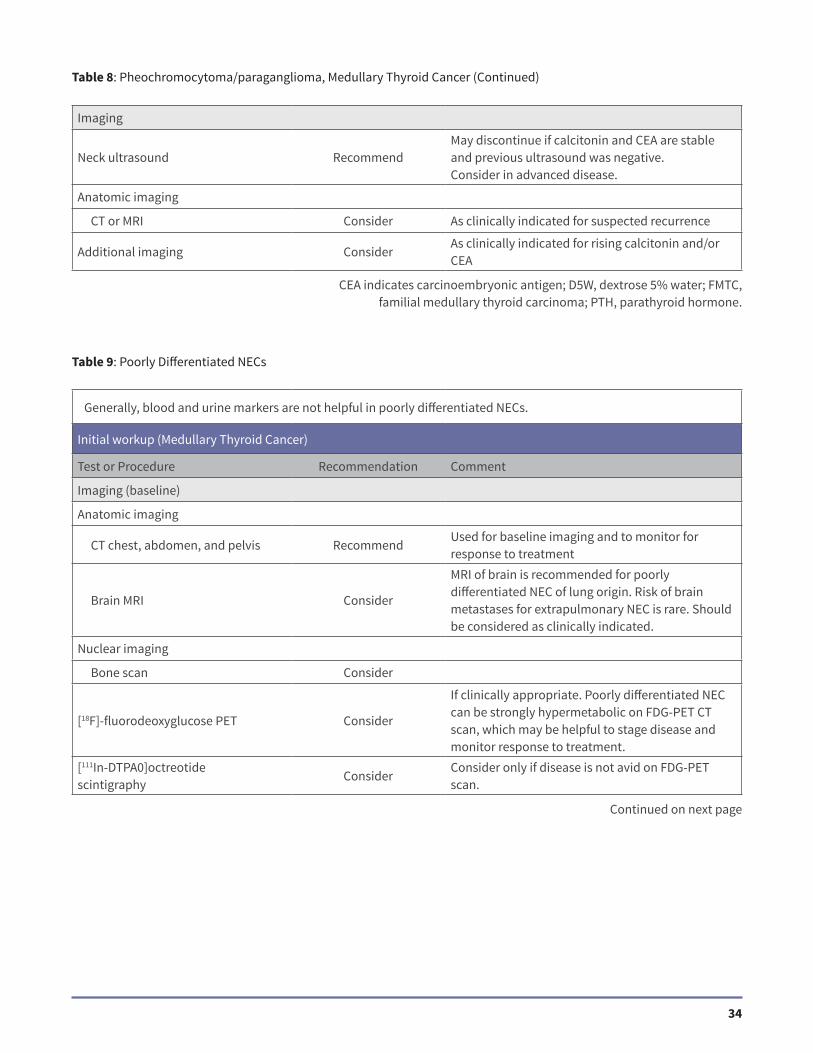
Imaging
Neck ultrasound RecommendMay discontinue if calcitonin and CEA are stable and previous ultrasound was negative.Consider in advanced disease.
Anatomic imaging
CT or MRI Consider As clinically indicated for suspected recurrence
Additional imaging ConsiderAs clinically indicated for rising calcitonin and/or CEA
Table 8: Pheochromocytoma/paraganglioma, Medullary Thyroid Cancer (Continued)
Table 9: Poorly Differentiated NECs
Generally, blood and urine markers are not helpful in poorly differentiated NECs.
Initial workup (Medullary Thyroid Cancer)
Test or Procedure Recommendation Comment
Imaging (baseline)
Anatomic imaging
CT chest, abdomen, and pelvis RecommendUsed for baseline imaging and to monitor for response to treatment
Brain MRI Consider
MRI of brain is recommended for poorly differentiated NEC of lung origin. Risk of brain metastases for extrapulmonary NEC is rare. Should be considered as clinically indicated.
Nuclear imaging
Bone scan Consider
[18F]-fluorodeoxyglucose PET Consider
If clinically appropriate. Poorly differentiated NEC can be strongly hypermetabolic on FDG-PET CT scan, which may be helpful to stage disease and monitor response to treatment.
[111In-DTPA0]octreotidescintigraphy
ConsiderConsider only if disease is not avid on FDG-PET scan.
CEA indicates carcinoembryonic antigen; D5W, dextrose 5% water; FMTC, familial medullary thyroid carcinoma; PTH, parathyroid hormone.
Continued on next page

35
Table 9: Poorly Differentiated NECs (Continued)
Continued on next page
Treatment of Poorly Differentiated NEC
Generally for NETs, lines of therapy have not been established. When multiple options are listed, order of listing does not imply order of therapy.
Disease Stage Intervention Recommendation
Locoregional disease, resectable
Clinical stage T1-2, NOSurgical resection, including removal of tumor with negative margins. Risk of recurrence is high, however.
Recommend
Postoperative therapy with 4Y6 cycles of cisplatin or carboplatin and etoposide.Radiation should only be considered in cases where risk of local recurrence is considered high and morbidity is low.
Recommend
Clinical stage in excess of T1-2, N0Chemotherapy with or without concurrent radiotherapy
Recommend
Surgery where morbidity is low, particularly where risk of obstruction is high. Risk of recurrence is high, however. Consider postoperative therapy with 4–6 cycles ofcisplatin or carboplatin and etoposide.Radiation should only be considered in cases where risk of local recurrence is considered high and morbidity is low.
Consider
Locoregional disease,unresectable
Platinum-based chemotherapy regimen(cisplatin or carboplatin and etoposide) for 4–6 cycles with concurrent or sequential radiation
Recommend
Metastatic: initial therapy Platinum-based chemotherapy* Recommend
Metastatic: progressive or relapsed disease
For relapse >6 months after termination of first-line therapy: original chemotherapy regimen
Recommend
For relapse <3–6 months: irinotecan ortopotecan, paclitaxel, docetaxel, vinorelbine, gemcitabine, temozolomide may be considered.
Consider
36
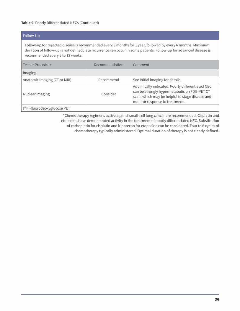
Table 9: Poorly Differentiated NECs (Continued)
Follow-Up
Follow-up for resected disease is recommended every 3 months for 1 year, followed by every 6 months. Maximum duration of follow-up is not defined; late recurrence can occur in some patients. Follow-up for advanced disease is recommended every 6 to 12 weeks.
Test or Procedure Recommendation Comment
Imaging
Anatomic imaging (CT or MRI) Recommend See initial imaging for details
Nuclear imaging Consider
As clinically indicated. Poorly differentiated NEC can be strongly hypermetabolic on FDG-PET CT scan, which may be helpful to stage disease and monitor response to treatment.
[18F]-fluorodeoxyglucose PET
*Chemotherapy regimens active against small-cell lung cancer are recommended. Cisplatin and etoposide have demonstrated activity in the treatment of poorly differentiated NEC. Substitution
of carboplatin for cisplatin and irinotecan for etoposide can be considered. Four to 6 cycles of chemotherapy typically administered. Optimal duration of therapy is not clearly defined.

37
REFERENCES
References
1. Anthony LB, Strosberg JR, Klimstra DS, et al. The NANETS consensus guidelines for the diagnosis and management of gastrointestinal neuroendocrine tumors (nets): well-differentiated nets of the distal colon and rectum. Pancreas. 2010;39(6):767–774.
2. Boudreaux JP, Klimstra DS, Hassan MM, et al. The NANETS consensus guideline for the diagnosis and management of neuroendocrine tumors: well-differentiated neuroendocrine tumors of the jejunum, ileum, appendix, and cecum. Pancreas. 2010;39(6):753–766.
3. Chen H, Sippel RS, O’Dorisio MS, et al. The North American Neuroendocrine Tumor Society consensus guideline for the diagnosis and management of neuroendocrine tumors: pheochromocytoma, paraganglioma, and medullary thyroid cancer. Pancreas. 2010;39(6):775–783.
4. Klimstra DS, Modlin IR, Coppola D, et al. The pathologic classification of neuroendocrine tumors: a review of nomenclature, grading, and staging systems. Pancreas. 2010;39(6):707–712.
5. Kulke MH, Anthony LB, Bushnell DL, et al. NANETS treatment guidelines: well-differentiated neuroendocrine tumors of the stomach and pancreas. Pancreas. 2010;39(6):735–752.
6. Phan AT, Oberg K, Choi J, et al. NANETS consensus guideline for the diagnosis and management of neuroendocrine tumors: well-differentiated neuroendocrine tumors of the thorax (includes lung and thymus). Pancreas. 2010;39(6):784–798.
7. Strosberg JR, Coppola D, Klimstra DS, et al. The NANETS consensus guidelines for the diagnosis and management of poorly differentiated (high-grade) extrapulmonary neuroendocrine carcinomas. Pancreas. 2010;39(6):799–800.
8. Yao JC, Shah MH, Ito T, et al. Everolimus for advanced pancreatic neuroendocrine tumors. N Engl J Med. 2011;364(6):514–523.
9. Raymond E, Dahan L, Raoul JL, et al. Sunitinib malate for the treatment of pancreatic neuroendocrine tumors. N Engl
J Med. 2011;364(6):501–513.10. Klimstra DS, Modlin IR, Adsay NV, et al. Pathology reporting of neuroendocrine tumors: application of the Delphic
consensus process to the development of a minimum pathology data set. Am J Surg Pathol. 2010;34(3):300–313.11. Pavel ME, Hainsworth JD, Baudin E, et al. Everolimus plus octreotide long-acting repeatable for the treatment of
advanced neuroendocrine tumours associated with carcinoid syndrome (RADIANT-2): a randomised, placebo-controlled, phase 3 study. Lancet. 2011;378(9808):2005–2012.
12. Strosberg JR, Fine RL, Choi J, et al. First-line chemotherapy with capecitabine and temozolomide in patients with metastatic pancreatic endocrine carcinomas. Cancer. 2011;117(2):268–275.
13. Rinke A, Muller HH, Schade-Brittinger C, et al. Placebo-controlled, double-blind, prospective, randomized study on the effect of octreotide LAR in the control of tumor growth in patients with metastatic neuroendocrine midgut tumors: a report from the PROMID Study Group. J Clin Oncol. 2009;27(28):4656–4663.
14. Yao JC, Pavel M, Phan AT, et al. Chromogranin A and neuron-specific enolase as prognostic markers in patients with advanced pNET treated with everolimus. J Clin Endocrinol Metab. 2011;96(12):3741–3749.
15. Kwekkeboom DJ, de Herder WW, Kam BL, et al. Treatment with the radiolabeled somatostatin analog [177 Lu-DOTA 0,Tyr3]octreotate: toxicity, efficacy, and survival. J Clin Oncol. 2008;26(13):2124–2130.
16. Madoff DC, Gupta S, Ahrar K, et al. Update on the management of neuroendocrine hepatic metastases. J Vasc Interv
Radiol. 2006;17(8):1235–1249; quiz 1250.17. Reidy DL, Tang LH, Saltz LB. Treatment of advanced disease in patients with well-differentiated neuroendocrine
tumors. Nat Clin Pract Oncol. 2009;6(3):143–152.18. Schurr P, Strate T, Rese K, et al. Aggressive surgery improves long-term survival in neuroendocrine pancreatic tumors:
an institutional experience. Ann Surg. 2007;245(2):273–281.

38
19. Touzios J, Kiely J, Pitt S, et al. Neuroendocrine hepatic metastases: does aggressive management improve survival? Ann Surg. 2005;241(5):776–783.
20. Sarmiento J, Heywood G, Rubin J. Surgical treatment of neuroendocrine metastases to the liver: a plea for resection to increase survival. J Am Coll Surg. 2003;197:29–37.
21. Chamberlain R, Canes D, Brown K, et al. Hepatic neuroendocrine metastases: does intervention affect outcome? J Am
Coll Surg. 2000;190:432–445.