connie j. mattera, m.s., r.n., emt-p...nch paramedic program stroke connie j. mattera, m.s., r.n.,...
TRANSCRIPT

NCH Paramedic Program Stroke Connie J. Mattera, M.S., R.N., EMT-P
National EMS Education standard: Anatomy, physiology, epidemiology, pathophysiology, psychosocial impact, presentations, prognosis, and management of (complex depth, comprehensive breadth) stroke/intracranial hemorrhage/transient ischemic attack.
Assigned readings: Bledsoe Vol. 4; pp. 197-202; this handout; NW EMSS SOPs (pp. 35-36); Procedure Manual: Neuro Assessment Stroke; Stroke Assessment checklist handout; Finding ELVO article
Goal: Strengthen participants’ ability to assess and recognize strokes and provide appropriate patient care and disposition based on evidence-based stroke management guidelines.
OBJECTIVES:
Upon completion of the assigned readings, class and study questions, each participant will do the following with at least an 80% degree of accuracy and no critical errors:
1. Define stroke and cite the incidence and epidemiology of stroke. 2. Differentiate the two main etiologies of stroke into ischemic and hemorrhagic. 3. Compare and contrast the types of ischemic stroke. 4. Explain the impact of modifiable and non-modifiable risk factors for stroke. 5. Sequence the impact of disrupted cerebral blood flow and how the brain becomes injured in stroke
explaining the importance of salvaging the penumbra. 6. Discuss each link in the stroke chain of survival and explain why these pts are time sensitive. 7. Identify and provide rationale for the EMS resources that must be prepared to identify and/or treat
stroke. 8. Explain the five goals of stroke management. 9. Explain the diagnostic importance of information to be obtained in a SAMPLE history for stroke. 10. Sequence the appropriate methods to secure an airway in a patient with a possible stroke. 11. Explain the dangers of hypoxia and hyperoxia when giving O2 to pts experiencing a stroke. 12. Discuss the indications, timing, and preferred sites for vascular access and IVFs in pts with stroke. 13. Explain the indications for dextrose and midazolam for pts experiencing a possible stroke. 14. Describe preferred positioning and environmental controls to protect a pt with stroke. 15. Explain anticipated changes in vital signs in pts with stroke and the thresholds for treatment. 16. Compare and contrast the components, timing, and predictive value of the Cincinnati Prehospital
Stroke Scale (CPSS), the Los Angeles Stroke Scale and the Miami Emergency Neurologic Deficit (MEND) exam.
17. Explain and sequence the components of a complete pt assessment for stroke. 18. Map the presenting S&S of ischemic and hemorrhage strokes, cerebral aneurysm, subarachnoid
hemorrhage (SAH) and acute intracerebral hemorrhage. 19. Identify diseases/conditions that must be considered in the differential diagnosis of stroke. 20. Recognize alternate S&S of stroke that may be present with or without alterations to the quick stroke
scales or exams. 21. Explain common complications of stroke and their implications for EMS care. 22. State the factors that contribute to poor outcomes in stroke. 23. Differentiate a TIA from a stroke. 24. Determine the most appropriate receiving hospital using the Stroke Decision Tree in the SOPs. 25. Explain the importance of calling a stroke alert to the appropriate receiving stroke center.
CJM: 7/13; JVD 7/13; CJM 1/16; 12/16; 1/18

NWC EMSS CE and NCH Paramedic Program Stroke – It’s no accident Definition: A stroke is a sudden focal, nonconvulsive neurologic event due to interruption of cerebral blood flow resulting in cell death. It is most often caused by an occlusion or rupture of an artery that supplies a specific region of the brain. The presentation is variable ranging from subtle to severe depending on the area of the brain involved and the nature of the attack. A stroke is no accident! To indicate the urgency for immediate emergency care, the terminology used to describe stroke was changed years ago from CVA (cerebrovascular accident) to brain attack. Approx. 80% are preventable and need to be treated with the same urgency as a heart attack with rapid detection and transport to specialty stroke centers.
Incidence of stroke in U.S. • Someone has a stroke every 40 seconds • > 795,000 each year • ~ 610,000 are first or new onset • ~185,00 (nearly one of four) are recurrent • # of adults who ever had a stroke: 6.3 million • % of adults who ever had a stroke: 2.6%
Morbidity and mortality • Deaths/yr: 133,103 (1 out of every 20 deaths). • Deaths per 100,000 population: 41.7 • On average, someone dies from stroke every 4 min • 5th leading cause of death: Only heart disease, cancer, chronic
lower respiratory dx, and unintentional injuries cause more deaths • 30-day death rate
o 40-84% after cerebral hemorrhage o 15-33% after cerebral infarction
https://www.cdc.gov/nchs/fastats/stroke.htm
Deaths have declined due to public education, early detection, better management of BP and stroke, increased rates of fibrinolytic therapy and use of endovascular procedures (CDC, 2014). These strides have been offset by growth in the aging, diabetic, and obese populations (more people at risk). Stroke is the leading cause of serious long-term adult disability such as paralysis, loss of speech. It is the leading diagnosis for transfers from hospital to rehab and long-term care facilities. Only 10% resolve completely with no lingering S&S. Morbidity in stroke survivors 31% need assistance with activities of daily living (ADLs) 20% need assistance to walk 16% need placement into assisted living 33% suffer from depression
Stroke morbidity factors • >65 years of age • Severe cognitive deficit to coma • Persistent incontinence • Severe visual/spacial deficits • Global aphasia • Co-morbid factors: HF, DM, AMI • Disability before stroke
Costs are staggering: $34 billion annually, including cost of healthcare services (initial hospitalization; rehabilitation, physician costs, possible readmission), medications,& lost productivity (Mozaffarian et al, 2015).
Demographics of stroke: Equal opportunity threat
While stroke kills and disables people of all ages, both sexes, and all races, prevalence, death and disability rates vary by race and ethnicity; median household income; urban vs nonmetropolitan locations; and geographic location (mortality inside and outside the Stroke Belt in the Southeastern U.S.). For the latest statistics see: https://www.cdc.gov/nchs/data/hus/hus15.pdf#023
Unmodifiable risk factors for stroke: age, gender, race/ethnicity, and stroke family history
Patient Age: It is a myth that stroke occurs only in elderly adults. Stroke strikes all age groups from fetuses to centenarians. Older persons do have a higher risk than the general population and that risk increases with age. For every decade after the age of 55, stroke risk doubles; 2/3 of all strokes occur in those >65. People >65 also have a 7 X greater risk of dying from stroke than the general population. The incidence of stroke is increasing proportionately with the increase in the older population (http://www.ninds.nih.gov/disorders/stroke/detail_stroke.htm#1105_10).
Gender Men: • 1.25 X higher risk; greater incidence
of stroke than women • Usually younger when they have a
stroke and therefore have a higher rate of survival. Women: • Higher mortality from stroke • More likely to dismiss symptoms and to seriously
underestimate their risk. • Those >30yrs who smoke and/or take oral
contraceptives/high levels of estrogen (hormone replacement therapy) are at 22X higher risk.
• Are often older when they have a stroke, are more physically frail, or have other health problems that impair their recovery (NINDS, 2016).
Do NOT use the term CVA anymore

NWC EMSS & NCH Paramedic Program page 2 Stroke
Anatomy and Physiology Cerebral blood supply is the most complex circulatory system in the body. The brain is 3 lbs of tissue but the most perfusion-sensitive organ. It receives ~20% the cardiac output and consumes 25% of available glucose.
Main blood vessels supplying the brain:
Blood is supplied to the brain, face, and scalp via two major sets of vessels: the right and left common carotid arteries and the right and left vertebral arteries.
The common carotid arteries have two divisions. • External carotids supply face and scalp with blood. • Internal carotids supply blood to most of the anterior
portion of the cerebrum (frontal lobe, anterior parietal lobes and temporal lobes).
• Patients with anterior circulation disruption may present with unilateral paralysis or numbness on the side opposite to the obstruction, language disturbance, visual disturbance, and/or monocular blindness. These strokes may also be referred to as carotid or anterior strokes.
The Vertebrobasilar arteries supply the posterior 2/5th of the cerebrum, part of the cerebellum, and the brain stem. Occlusion here can cause severe vertigo, nausea, vomiting, dysphagia, diplopia, visual field loss; gaze palsies, decreased pain and temperature sensation, same side (ipsilateral) cerebellar ataxia, loss of 2 point discrimination (posterior stroke).
Circle of Willis connects the two systems before entering the structures of the brain. Designed so disruption of any part will not cause significant loss of blood flow to tissues (Bledsoe, 2017) • Anterior communicating artery • Middle cerebral artery • Internal carotid artery • Post. communicating artery • Posterior cerebral artery
This unique circulation allows perfusion to continue in the event of blockage in either the anterior or the posterior circulations.
From this circle, other arteries (anterior cerebral (ACA), middle cerebral (MCA), and posterior cerebral (PCA)) arise and travel to all parts of the brain.
Anterior Cerebral Artery (ACA) The ACA extends upward and forward from the internal carotid artery. It supplies the frontal lobes, (controls logical thought, personality, and voluntary movement, especially of the legs). Stroke in the ACA results in leg weakness opposite to the side of brain injury. If both anterior cerebral territories are affected, profound mental symptoms may result (akinetic mutism).
Middle Cerebral Artery (MCA) Largest branch of the internal carotid and supplies a portion of the frontal lobe, lateral surface of the temporal and parietal lobes (including the primary motor and sensory areas of the face, throat, hand and arm), and in the dominant hemisphere, the areas for speech. The MCA is the one most often occluded in stroke.
Posterior Cerebral Artery (PCA) In most people, the PCAs stem from the basilar artery but sometimes originate from the ipsilateral internal carotid artery. The posterior arteries supply portions of the temporal and occipital lobes. Infarction occurring in the territory of the PCA, is usually secondary to embolism from lower segments of the vertebral basilar system or heart.
Lenticulostriate Arteries Beyond the Circle of Willis, collateral circulation is limited, supplied only by vessels in the dura mater & arachnoid membrane. Small, deep penetrating arteries (lenticulostriate arteries) branch from the MCA. Occlusions of these vessels or penetrating branches of the Circle of Willis or vertebral or basilar arteries are referred to as lacunar strokes. ~27% of stokes are lacunar. High incidence in pts with chronic HTN (http://www.strokecenter.org/professionals/brain-anatomy/blood-vessels-of-the-brain/).

NWC EMSS & NCH Paramedic Program page 3 Stroke
Risk factors for stroke: Need careful patient history
While you can’t control some risk factors like heredity, age, gender, and ethnicity (see demographics of stroke), >90% of stroke deaths are caused by modifiable risk factors due to patient lifestyle choices and are considered preventable.
SSBBPP DDBBPP
Normal <120 and <80
Elevated BP 120-129 and <80
Stage 1 HTN 130-139 or 80-89
Stage 2 HTN ≥140 or ≥ 90
HHTTNN ccrriissiiss >> 118800 >> 111100
• Hypertension: Persistently high BP >130/80 leads to
a diagnosis of HTN. Hypertension disrupts the structure of cerebral blood vessels, promotes atherosclerosis, and impairs vital cerebrovascular regulatory mechanisms. These vascular changes increase the susceptibility of the brain to ischemic injury. Based on the latest data available, 32.6% of US adults ≥20 yrs of age have HTN, which represents ~80 million people. African American adults have the highest prevalence of HTN in the world. Among non-Hispanic black men and women, the age-adjusted prevalence of HTN was 44.9% and 46.1%, respectively (Mozaffarian et al, 2016). Of all the risk factors, HTN is the most powerful. Stroke risk is 4-6 X higher in those with HTN; ~77% have a BP >140/90 at their first stroke. The impact of HTN on total stroke risk decreases with age. Factors other than HTN play a greater role in overall stroke risk in elderly adults. For those without HTN, the absolute risk of stroke increases over time until around age 90, when the absolute risk becomes the same as for people with HTN. Gender difference in the prevalence of HTN Younger people: HTN more common in men than women. With advancing age, more women than men have HTN. This gender-age difference probably has an impact on the incidence and prevalence of stroke. Researchers recommend patient-specific HTN treatment, considering care goals, age, menopause, APOE ε4 genotype, metabolic traits, insulin resistance, systemic inflammation, and comorbidities to protect vascular health, and therefore brain health. Antihypertensive medications can decrease a person's risk and incidence of stroke by 38% and decrease the stroke fatality rate by 40%. Common anti-HTN agents include beta-blockers, angiotensin converting enzyme (ACE) inhibitors, antiotensin
receptor blockers (ARBs), calcium channel blockers, diuretics, and vasodilators (See HF page of SOPs for lists of meds).
• Heart Disease: After HTN, the 2nd most powerful risk factor for stroke, especially if pt. has atrial fibrillation.
In AF, the left atrium beats up to four times faster than the rest of the heart. This leads to an irregular flow of blood and the occasional formation of blood clots (mural thrombi) that can leave the heart and travel to the brain, causing a stroke. AF may be asymptomatic and undetected, affects as many as 2.2 million Americans, and increases risk of stroke by 4-6%. About 15% of patients have AF before they experience a stroke. AF is more prevalent in older age groups. Unlike HTN and other risk factors that have a lesser impact on the absolute risk of stroke with advancing age, the influence of AF on total risk for stroke increases powerfully with age (1.5% at 50-59 to 23.5% at ages 80-89). In people >80, AF is the direct cause of one in four strokes.
• Heart valve disease/malfunction: Mitral stenosis, mitral annular calcification, valvular vegetation, and defective valve prostheses can double risk for stroke.
• Cardiac chamber lesions (LV aneurysm, intracardiac defects with paradoxic embolism (LV and left atrial thrombus with or without mitral stenosis)
• Atheromatous lesions of the ascending aorta; 30-70% stenosis of the internal carotid artery, or a clotting abnormality that promotes thrombosis. These patients may benefit from reduction of risk factors.
• Heart muscle malformations Patent foramen ovale (PFO) is a passage or a hole (sometimes called a "shunt") in the heart wall separating the two atria. Blood clots are usually filtered out by the lungs, but PFO could allow emboli to bypass the lungs and go directly through the arteries up to the brain, potentially causing a stroke. Atrial septal aneurysm (ASA), a congenital malformation of heart tissue, is a bulging of the septum or heart wall into one of the atria. PFO and ASA frequently occur together and therefore amplify the risk for stroke. Two other heart malformations that seem to increase the risk for stroke for unknown reasons are left atrial enlargement and LV hypertrophy. People with left atrial enlargement have a larger than normal left atrium. Those with LV hypertrophy have a thickening of the LV wall.
• Cardiac surgery to correct heart malformations or reverse the effects of heart disease: Strokes occurring in this situation are usually the result of surgically dislodged plaques from the aorta that travel to the arteries in the neck and head, causing stroke. Cardiac surgery increases risk of stroke by ~1%.

NWC EMSS & NCH Paramedic Program page 4 Stroke
• Diabetes Mellitus (DM): People with DM have 3X the risk of stroke as it accelerates atherosclerosis. This risk is highest in the 5th and 6th decades and decreases after that. Like HTN, the relative risk of stroke from DM is highest for men at an earlier age and highest for women at an older age. People with DM may also have other contributing risk factors that can amplify the overall risk for stroke. Ex: prevalence of HTN is 40% higher in diabetics. Control of hyperglycemia can reduce risk of microvascular complications and overall risk of stroke.
• High blood cholesterol/lipids: High cholesterol levels contribute to stroke risk. Cholesterol, a waxy substance produced by the liver, contributes to the production of hormones and vitamin D and is an integral component of cell membranes. The liver makes enough cholesterol to fuel the body's needs and this natural production alone is not a large contributing factor to atherosclerosis, heart disease, or stroke. The danger from cholesterol comes from high levels that contribute to the risk of atherosclerosis and thickening of the arteries. Cholesterol is a lipid (fat-soluble rather than water-soluble). Other lipids include fatty acids, glycerides, alcohol, waxes, steroids, and fat-soluble vitamins A, D, and E. Lipids and water, like oil and water, do not mix. Blood is a water-based liquid, therefore cholesterol does not mix with blood. In order to travel through the blood without clumping together, cholesterol needs to be covered by a layer of protein. The cholesterol and protein together are called a lipoprotein. There are two kinds of cholesterol, commonly called the "good" and the "bad." Good cholesterol is high-density lipoprotein, or HDL (healthy cholesterol); bad cholesterol is low-density lipoprotein, or LDL (lethal cholesterol). Together, these two forms make up a person's total serum cholesterol level. A level of <200 mg/dL is generally considered safe unless one is at high risk for heart attack or stroke, while a level of >240 is considered dangerous and places a person at risk for heart disease and stroke. Most cholesterol is in the form of LDL that circulates through the bloodstream, picking up excess cholesterol and depositing it where it is needed (production and maintenance of cell membranes). But when too much is present, the body cannot handle the
excessive LDLs, which build up along the inside of arterial walls, hardens, and turns into arterial plaque, leading to stenosis and
atherosclerosis. Plaque blocks blood vessels, can rupture, and contributes to the formation of blood clots. A person's LDL level should be <130 mg/dL to be safe. LDL levels between 130 and 159 put a person at a slightly higher risk for atherosclerosis, heart disease, and stroke. An LDL >160 puts a person at great risk for a heart attack or stroke.
HDL is beneficial and contributes to stroke prevention. It carries a small percentage of the cholesterol in the blood, but instead of depositing on the inside of artery walls, HDL returns to the liver to unload its cholesterol. The liver then eliminates the excess by passing it along to the kidneys. Currently, an HDL level >35 is considered desirable. High levels of HDL are associated with a reduced risk for heart disease and stroke and low levels (<35 mg/dL), even in people with normal levels of LDL, lead to an increased risk for heart disease and stroke. A healthy diet and regular exercise are the best ways to lower total cholesterol levels. In some cases, cholesterol-lowering medications are needed.
Core health behaviors: (diet, physical activity, smoking, and energy balance) • Smoking almost doubles
the risk for ischemic stroke, independent of other risk factors, and increases the risk for subarachnoid hemorrhage (SAH) by up to 3.5%. Smoking is directly responsible for a greater percentage of total strokes in younger than older adults. Risk factors other than smoking (HTN, heart disease, DM) account for more strokes in older adults. Heavy smokers are at greater risk than light smokers. Good news! The relative risk of stroke decreases immediately after quitting, with a major reduction seen after 2 to 4 years. It may take several decades for a former smoker's risk to drop to that of a nonsmoker. Smoking accelerates the rate of atherosclerosis progression by 50%, particularly in those with DM & HTN and increases clotting factors, such as fibrinogen. Smoking also increases the damage that results from stroke by weakening the endothelial wall of the cerebrovascular system. This leads to greater damage to the brain from events that occur in the secondary stage of stroke. Carbon monoxide in smoke displaces O2 from hemoglobin, interfering with O2 delivery to tissues.
• Physical inactivity; obesity: Obesity strains the entire cardiovascular system. Pts are likely to have high cholesterol, HTN, & DM. Moderate to vigorous-intensity exercise is associated with an overall 35% reduced risk of stroke.
• High alcohol consumption: Generally, an increase in alcohol intake leads to an increase in BP. While heavy drinking is a risk for both hemorrhagic and ischemic strokes, studies affirm that daily consumption of smaller amounts of alcohol provides a protective

NWC EMSS & NCH Paramedic Program page 5 Stroke
influence against ischemic stroke by decreasing the clotting ability of platelets (similar to the effects of aspirin). Heavy alcohol consumption may seriously deplete platelets and compromise blood clotting and blood viscosity, leading to hemorrhage. Heavy (or binge) drinking can lead to a rebound effect after the alcohol is purged from the body causing blood viscosity and platelet levels to skyrocket, increasing the risk for ischemic stroke.
• Cocaine enhances other risk factors, such as HTN, heart and vascular dx. It decreases relative CBF by up to 30%, causes vasoconstriction and inhibits vascular relaxation. Repeated exposure can lead to blood flow deficits that persist long after use has ended, causing permanent damage. Also causes dysrhythmias rapid HR; and activates platelets promoting thrombosis. Heavy users (6-20 X/wk) have elevated levels of C-reactive protein which is associated with risk of plaque rupture.
Marijuana smoking: Decreases BP and may interact with other risk factors, such as HTN and cigarette smoking, to cause rapidly fluctuating BP
levels, damaging blood vessels.
• Other drugs of abuse (amphetamines, heroin, anabolic steroids; some common legal drugs, such as caffeine, L-asparaginase and pseudoephedrine found in over-the-counter decongestants; and phenylpropanolamine (over-the-counter "diet pills" and cold and flu pills) increase incidence of stroke in those < 40. Many are vasoconstrictors, causing BPs to rise. Ecstasy (MDMA): deaths are related to massive cerebral hemorrhage.
• Head and neck injuries: May damage the cerebrovascular system and cause a small number of strokes. Traumatic brain injury may cause bleeding within the brain leading to damage like a hemorrhagic stroke. Neck injury, when associated with spontaneous tearing of the vertebral or carotid arteries caused by sudden and severe extension of the neck, neck rotation, or pressure on the artery, is a contributing cause of stroke, especially in young adults. Often called "beauty-parlor syndrome," referring to extending the neck backwards over a sink for hair-washing. Neck calisthenics, "bottoms-up" drinking, and improperly performed chiropractic manipulation of the neck can also put strain on the vertebral and carotid arteries, possibly leading to ischemic stroke.
• Infections: Recent infections may act with other risk factors to add a small risk. The immune system responds to infection by increasing inflammation and the infection-fighting properties of blood. This increases the number of clotting factors leading to an increased risk of embolic-ischemic stroke.
• Genetic risk factors: Although there may not be a single genetic factor associated with stroke, genes play a large role in the expression of stroke risk factors such as HTN, heart disease, DM, and vascular malformations. It’s possible that increased familial risk for stroke is due to environmental factors, such as a common sedentary lifestyle or poor eating habits, rather than heredity.
• Vascular malformations may have the strongest genetic link of all risk factors. A vascular malformation is an abnormally formed blood vessel or group of blood vessels. One genetic vascular disease called CADASIL (cerebral autosomal dominant arteriopathy with subcortical infarcts and leukoencephalopathy) is a rare, genetically inherited, congenital vascular disease of the brain that causes strokes, subcortical dementia, migraine-like headaches, and psychiatric disturbances. CADASIL is debilitating and symptoms usually surface around age 45. The exact incidence of CADASIL is unknown. (Circulation, Jan. 2013 )
Previous stroke or transient ischemic attack (TIA) also raises the risk of (recurrent) stroke.
Classifications of stroke – 2 main types (plugs & leaks)
Strokes are broadly classified into two types: (1) Ischemic: blood supply to part of the brain is suddenly interrupted by plugs [thrombus or embolus] or a hemodynamic failure as a consequence of decreased blood volume flowing through cerebral vessels. (2) Hemorrhagic: a vessel in the brain leaks, spilling blood into spaces surrounding brain cells (NINDS, 2016).
Brain cells become injured (ischemia) when they receive inadequate O2 (perfusion) or when they are damaged by sudden bleeding into or around them. Unchecked, ischemia ultimately leads to infarction (death of brain cells).

NWC EMSS & NCH Paramedic Program page 6 Stroke
When blood flow is interrupted, some brain cells die quickly (within minutes), while others remain at risk for death. These damaged cells make up the ischemic penumbra and can linger in a compromised state for several hours. Collateral circulation to the penumbra occurs but is hard to predict by looking at the patient. Core necrosis cells will probably die no matter what is done. Collateral blood flow to the penumbra may be sufficient to preserve brain, but not enough to sustain normal function until perfusion is restored. With timely treatment these cells can be saved.
Within a few months of the infarction, the necrotic brains cells are reabsorbed by macrophage (WBC) activity, leaving a very small fluid-filled cavity referred to as a lake (or lacune in French) in the injured brain (NINDS, 2016).
Ischemic strokes: 87% are caused by a blood vessel occlusion due to a clot, plaque or embolus. AHA reports that for 30-40% of ischemic strokes, no cause is found (Circulation, 2013). This type is called a cryptogenic stroke.
Thrombotic Stroke Central thrombus: Large-vessel occlusion due to atherothrombotic disease causes 14% of ischemic strokes. A clot forms in turbulent flow areas of a large artery or plaque suddenly obstructs CBF. The clot may partially or completely block CBF. Both large and small vessel occlusions cause reduced tissue perfusion leading to neurotransmitter failure, anaerobic glycolysis, cerebral anoxia, and cerebral edema.
Embolic Stroke Definition: Partial or complete blockage in a cerebral artery from embolic material, generally composed of cholesterol (usually smaller than 500 micrograms), plaque (usually larger than 500 micrograms), blood, air, or tumor tissue that arose elsewhere in the body (usually in the heart) and migrated to the brain. Incidence: 59% S&S: These strokes often occur suddenly, without warning. Their S&S may fluctuate due to the continuing movement of the embolic matter within the blood vessel.
Hemorrhagic - 13% of all strokes
Most unfavorable type of stroke
Incidence: Affects 37,000-52,000 persons/ yr in US. Most common in middle aged, African Americans, Asian (particularly Japanese), and those with HTN. About 50% of strokes in younger patients are hemorrhagic (ruptured congenital aneurysms or AV malformation); only 20%-25% are hemorrhagic in older patients. Onset is abrupt. S&S depend on the size and location of the hemorrhage and worsen as more brain tissue is affected.
Two forms: Subarachnoid Hemorrhage Refers to the nontraumatic presence of blood within the subarachnoid space from some pathologic process, usually from rupture of a berry aneurysm or arteriovenous malformation (AVM) (Zebian, 2015). Especially prevalent in 35-65 yr olds, SAH accounts for about 3% of all strokes; afflicting ~30,000/yr in U.S. Risk factors for SAH: HTN, smoking, heavy alcohol use, use of sympathomimetic drugs and presence of familial genetic predisposition. Mortality rate reaches as high as 40% within the first week, and ~50% die in the first 6 months (Zebian, 2015).
The most common and potentially treatable cause of secondary neurological injury in this population is delayed ischemic deficit (DID). As the name implies, this phenomenon is fundamentally a reduction of cerebral blood flow (CBF) and O2 delivery below critical ischemic thresholds, occurring days after the onset of hemorrhage. Three inter-related physiological processes appear to be involved in the reduced O2 delivery: severe narrowing of intracranial arteries (arterial vasospasm), intravascular volume depletion and a loss of normal autoregulatory function in the distal circulation. DID occurs in up to 40% of patients surviving SAH. One third of these patients will die from this phenomenon and another third will be left with permanent and severe disability (Bradely, 2015).
Clinical presentation of a higher grade SAH is “one of the most distinctive in medicine” (Stroke Assoc).
S&S can range from a mild headache with or without meningeal irritation to severe headache (worse headache of their life present in ~80% of pts) and nonfocal findings with or without a dilated pupil. Worsening S&S include mild alteration in neuro exam, including AMS, obviously depressed level of consciousness or focal deficit and finally, a patient that is either posturing or comatose.
About 20% experience a warning or “sentinel” headache between 2 and 8 weeks before the SAH event. Headache w/ the warning bleed is usually milder than that of the SAH event, and may be accompanied by nausea & vomiting.

NWC EMSS & NCH Paramedic Program page 7 Stroke
SAH often occurs during physical exertion or stress. Associated S&S may include nausea &/or vomiting (77%), stiff neck (35%), brief loss of consciousness (53%), and focal neuro deficits. Twenty percent of SAH patients experience seizure(s) within the first 24 hrs after the event.
Emergent management of SAH, including prehospital care, is critical. EMS is the first medical contact for about 2/3 of SAH patients. An estimated 10-15% die before reaching the hospital. Rapid assessment & transport with advanced notification of the ED is crucial for patients presenting with more than one S&S for SAH, including headache, change in level of consciousness, or vomiting.
Spontaneous intracerebral hemorrhage (ICH) ICH accounts for 10% of all strokes. Small, deeply penetrating arteries into brain tissue (parenchyma) are susceptible to loss of elasticity w/ HTN and are easily ruptured. ICH most commonly occurs in the cerebral lobes, periventricular white matter or basal ganglia (40-60%), thalamus (20%), the pons, and the cerebellum. Acute neurologic deterioration results from early hematoma growth, peri-hematoma injury, and obstructive hydrocephalus. Delayed deterioration is usually due to
edema and toxic effects of blood on the brain tissue. The expanding mass of blood can project 2-3 cm into the brain tissue and can grow to the size of a golf ball, plum or larger. This expansion can be
caused by continued bleeding, blood-brain barrier breakdown, or formation of a local coagulopathic state. The mass causes pressure on cerebral tissues and nerves leading to neuron death.
The hematoma also has the potential to disturb normal intracranial dynamics causing a sudden rise in ICP, tissue compression, displacement, and herniation from mass effect. Cerebral spasm contributes to further ischemia.
Rapid EMS assessment and transport are crucial because deterioration is common in the first few hours after ICH. Many patients with smaller ICHs will survive if prompt and aggressive medical care is received.
The patient’s GCS may decrease ≥ 2 points between initial EMS assessments and the ED evaluation. Early deterioration without aggressive intervention is associated with poor long term outcomes (> 75% mortality). Advance Stroke Alert notification correlates with shortened time to evaluation and CT scan.
ICH risk factors ■ Uncontrolled HTN (primary & most easily modified ■ Advancing age ■ Cerebral amyloid angiopathy ■ Neoplasms (tumors) with fragile blood vessels ■ Trauma ■ Vascular anomalies (AVMs and aneurysms)
■ Coagulation disorders; sickle cell disease, hemophilia ■ Collagen-vascular disease ■ Venous thrombosis ■ Drugs: Anticoagulants, amphetamines (vasculitis),
cocaine, oral contraceptives; excessive alcohol intake ■ Septic emboli, infective endocarditis, infected valve
prosthesis Disruption to cerebral blood flow (CBF) & perfusion in stroke The brain depends on a constant level of perfusion (cerebral blood flow of ~750 mL/ min or 55 mL/100 gm tissue/minute). One needs a balance of flow and metabolism (O2 demand & supply). The brain only functions normally if CBF is >20 mL/100 gm of brain tissue/min.
Perfusion is disrupted by changes in blood flow and cerebral perfusion pressure (MAP- ICP). Any condition that affects perfusion has a rapid effect on brain function depending on the areas that are impacted (global or focal).
Vascular occlusion or disruption results in decreased O2 and glucose delivery to the affected cells and increased CO2 and lactic acid accumulation.
CBF is autoregulated over a wide range of blood pressures (60 to 180 mmHg). It is lost with extremes of hyper or hypotension, severe brain damage with cerebral edema and increased ICP. When stroke occurs and the brain becomes ischemic, blood flow is regulated by “whatever head of pressure is pushing it” and small changes in MAP and cardiac output can cause drastic alterations in cerebral blood flow.
If flow drops <12-20 mL/100 gm/min, brain cells become electrically silent (as manifested by paralysis) but not dead. Early mgt is critical to restore perfusion and function.
Chemical controls – very important to EMS
• Excess CO2 – dilates vessels • Low CO2 (hyperventilation) constricts vessels • Low O2 – (hypoxia) dilates vessels • Excess O2 (hyperoxia) constricts vessels Evolution in stroke care Until the 1990s, care of patients experiencing stroke was largely supportive, focusing on treatment of respiratory and cardiovascular complications while the effects of the stroke became complete. No specific therapy was available to alter the course and extent of the evolving physical losses. Thus, limited emphasis was placed on rapid EMS interventions or transport. We also thought oxygen was good and not harmful for everybody!
THIS HAS CHANGED! Fibrinolytic drugs and advanced interventional therapies at stroke centers now offer an opportunity to possibly limit the extent or reverse neurological damage and improve outcomes in select patients with ischemic stroke. It’s a whole new era of standards and care!
NWC EMSS & NCH Paramedic Program page 8 Stroke
STROKE SYSTEMS OF CARE Expected benefits • Improved efficiency of care • Increased use of acute
stroke therapies • Fewer peri-stroke
complications • Reduced morbidity and
mortality • Improved long-term outcomes • Reduced health care costs (NMH)
Identification of stroke patients and rapid mobilization of stroke teams is now commonplace in US • Average door to needle times < 60 minutes • Opportunities still exist to achieve radical changes (< 20 min) • Workflow efficiencies are engineered into
endovascular interventions at comprehensive centers • Will continue to improve stroke care as systems
approaches are adopted and implemented
The best team approach to stroke management starts with dispatch and EMS and continues at a hospital within a system of care capable of delivering evidence-based stroke care from the ED to a dedicated stroke unit.
Detection 85% of strokes occur at home. Early recognition depends on patient, family, or other bystanders witnessing the event and recognizing the urgency of the symptoms.
Barriers: Lack of stroke awareness Symptoms are often subtle. Many fail to seek help quickly enough to be eligible for fibrinolytic therapy. Mellor et al (2015) found that delays arose from 3 sources: Patients’ lack of recognition (or perhaps denial) of the
significance of their symptoms, A decision to first contact primary rather than
emergency medical services care, and
Lack of recognition of the significance of S&S by the initial health care providers.
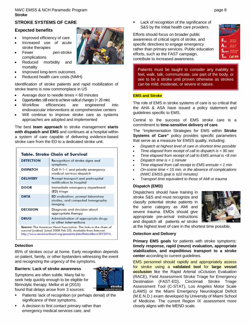
Efforts should focus on broader public awareness of critical signs of stroke, and specific directives to engage emergency rather than primary services. Public education efforts, such as the FAST campaign, contribute to increased awareness.
EMS and Stroke
The role of EMS in stroke systems of care is so critical that the AHA & ASA have issued a policy statement and guidelines specific to EMS.
Central to the success of EMS stroke care is a commitment to time-sensitive delivery of care. The “Implementation Strategies for EMS within Stroke Systems of Care” policy provides specific parameters that serve as a measure for EMSS quality, including: • Dispatch at highest level of care in shortest time possible • Time elapsed from receipt of call to dispatch is < 90 sec • Time elapsed from receipt of call to EMS arrival is <8 min • Dispatch time is < 1 minute • Time elapsed from call receipt to EMS enroute < 1 min • On-scene time < 15 min. in the absence of complications
(NWC EMSS goal is ≤10 minutes) • Transport time equivalent to those of AMI or trauma
Dispatch (EMD) Dispatchers should have training in stroke S&S and must recognize and classify potential stroke patients in the same category as AMI and severe trauma. EMDs should give appropriate pre-arrival instructions and dispatch all patients w/ stroke at the highest level of care in the shortest time possible.
Detection and Delivery
Primary EMS goals for patients with stroke symptoms: timely response, rapid (neuro) evaluation, appropriate stabilization, and expeditious transport to a stroke center according to current guidelines. EMS personnel should rapidly and appropriately assess for stroke using a validated tool for large vessel occlusion like the Rapid Arterial oCclusion Evaluation (RACE), Field Assessment Stroke Triage for Emergency Destination (FAST-ED), Cincinnati Stroke Triage Assessment Tool (C-STAT), Los Angeles Motor Scale (LAMS) or the Miami Emergency Neurological Deficit (M.E.N.D.) exam developed by University of Miami School of Medicine. The current Region IX assessment more closely aligns with the MEND scale.
Patients must be taught to consider any inability to feel, walk, talk, communicate, use part of the body, or see to be a stroke until proven otherwise as strokes can be mild, moderate, or severe in nature.

NWC EMSS & NCH Paramedic Program page 9 Stroke
Time-sensitive patient
TIME IS BRAIN! In cardiac arrest, the brain must be resuscitated in minutes to prevent irreversible brain damage (entire brain is not perfused). In most forms of stroke, the patient has focal ischemia. The core area must receive resuscitation within minutes, but a large area around the core (ischemic penumbra) does not die for hours. Each hour of ischemia increases the degree of irreversible brain damage. For every minute stroke is left untreated, ~1.9 million neurons die. Each hour, in which treatment fails to occur, the brain loses as many neurons as it does in almost 3.6 years of normal aging (Miller, 2012).
Expedite transport: EMS personnel must be aware of the possibilities for early stroke interventions at hospitals and appreciate the need for short scene times.
In the interest of saving time, some EMS actions can be accomplished enroute. Limit assessments/care on scene to those that are urgently needed or clearly indicated by the patient’s presentation.
If scene time is longer than 10 minutes, include information in the comments/narrative to support why.
Hospital stroke alert: Notify the nearest System hospital for OLMC ASAP if S&S of stroke started within the past 6 hours. Patients whose arrival is expected are more likely to receive early CT and appropriate interventional therapy. OLMC should either prepare for the patient’s arrival or call report to the receiving hospital.
Door, Data and Decision – Hospitals own this Upon arrival at the hospital, the patient should be rapidly evaluated by the stroke team (goal ≤ 10 minutes).
Time barriers at the hospital: ■ Time to treatment area ■ Time to physician assessment ■ Time to CT (goal ≤ 25 min.) ■ Time for CT interpretation (goal ≤ 45 min.) ■ Time for consultation and decision ■ Time for consent ■ Time for preparation
Remove barriers to rapid treatment: prioritize CTs, early read of CT, immediate consult with neurologists, location of fibrinolytics, pharmacists in the ED.
Drug: Ideally, tPA should be administered (“door to drug”) in less than 60 minutes after ED arrival. However, it must
be given within 4.5 hours from the onset of symptoms Evidence confirms that the sooner tPA is administered, the more optimal the results.
Disposition: Outcomes are best when patients are admitted directly to and receive care in a dedicated stroke unit, where recovery and rehab can begin immediately. Hospitals without full stroke care capabilities should have official transfer agreements with hospitals offering a full complement of stroke care, and patients should be transferred as soon as reasonably possible. The SOPs have a decision tree to transport to a Comprehensive vs. Primary Stroke Center destination.
Primary assessment/resuscitative interventions See Stroke SOP: IMC special considerations p.35
o Support ABCs as needed Ideally, avoid sedating these
patients Airway obstruction is a major
problem, especially if pt loses consciousness. Paralysis of the muscles of the throat, tongue, or mouth can lead to partial/complete upper airway obstruction. Saliva may pool in throat and be aspirated.
Aspiration is a serious complication, associated with considerable morbidity and mortality.
Open the airway with BLS manual maneuvers and secure with nasopharyngeal and/or oral pharyngeal airways as indicated.
Do not allow a patient to eat or drink until swallowing ability has been assessed by ED staff.
If GCS ≤ 8 (must do an accurate GCS!) assess need for DAI. If airway is patent and O2 sats can be maintained at 94%, BLS may be sufficient.
Assess ventilatory status (ETCO2)/ gas exchange (SpO2 ) at least q. 15 min. Give O2 if SpO2 < 94% or O2 sat unknown; avoid hypoxia and hyperoxia. Respiratory compromise may occur from aspiration, upper airway obstruction, hypoventilation, and pneumonia. Note length and frequency of apnea. Abnormal ventilatory patterns are especially prevalent in comatose patients and usually reflect serious brain dysfunction. Hypoxia and hypercarbia result from hypoventilation and can contribute to ↑ ICP and cardiac and respiratory instability. Patients with hx of cardiac and/or pulmonary disease are at ↑ risk for hypoxia. Assist ventilations at 10 BPM prn w/ 15 L O2/BVM prn.
The NWC EMSS has a scene time target of 10 minutes or less for patients with
suspected stroke.
NWC EMSS & NCH Paramedic Program page 10 Stroke
o Seizure/vomiting precautions; suction prn. Use
gentle technique, as trauma to tissues may later lead to significant bleeding if patient is treated with fibrinolytics. Avoid vagal stimulation during suctioning, as increases in ICP may worsen the effects of stroke.
o Positioning: Can affect O2 sats, CPP, and ICP. Maintain head/neck in neutral alignment to optimize perfusion and venous outflow from head. Do not use pillows. If SBP > 100: Elevate head of bed 10° - 15°. May need to use lateral immobilization devices to keep head from turning to the side. Do not flex knees or hips to avoid elevating ICP.
o Assess cardiovascular status/ perfusion adequacy Monitor ECG; acquire 12L. Cardiac
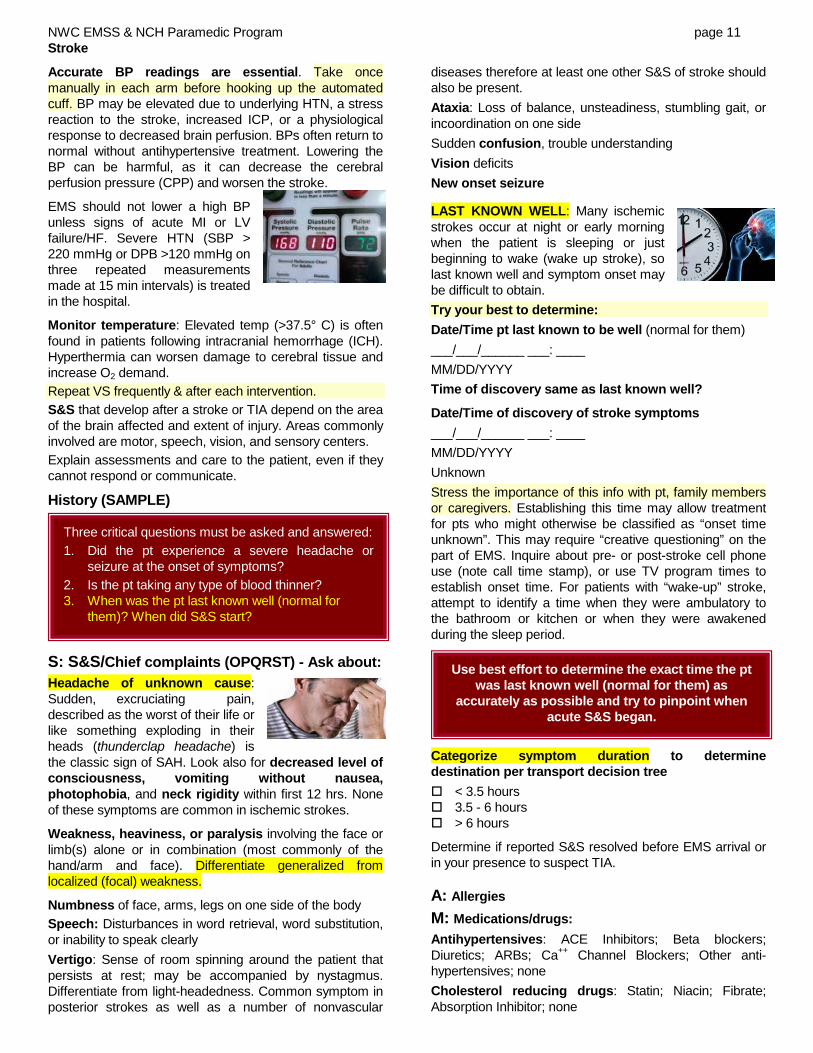
dysrhythmias may have caused the stroke or may be a side effect of brain injury. Life-threatening dysrhythmias are a potential early complication, particularly of intracranial hemorrhages. Bradycardia may indicate hypoxia or ↑ ICP. Bradycardia w/ AMS + SBP >90 should NOT be treated with atropine, norepinephrine, or pacing. Tachycardia may indicate hypoxia/hypercarbia. A-Fib: 15% of pts with stroke have AF SAH. Pathological Q waves, ST elevation or depression; prolonged QTc, wide, large & deeply inverted (neurogenic or cerebral) T waves; prominent U waves > 1 mm amplitude common causing incorrect suspicion of myocardial ischemia
IV usually unnecessary at
scene unless DAI is indicated or hypoglycemic. Avoid multiple attempts. Hospital may ask for lg. (18-20 g) antecubital IV to be placed so pt. can go directly to CT (prepped for infusion). Hydrate as necessary to maintain CPP but do not give large fluid boluses unless hypotensive. Stroke pts experience cerebral edema as a result of hypoxic tissue damage. Excess fluids (IV) will worsen this edema and increase damage to brain tissue.
o Disability/mental status exam: See p.12
If AMS: Consider possible causes of AMS using the AEIOU-TIPs mnemonic in the SOP p. 26.
o If generalized tonic/clonic seizure activity: Observe and record seizure activity per SOP p. 37 MIDAZOLAM 2 mg increments IVP/IO q. 30-60 sec (0.2 mg/kg IN) up to 10 mg prn titrated to stop seizure. If IV unable and IN contraindicated: IM 5-10 mg (0.1-0.2 mg/kg) max 10 mg single dose. All routes: May repeat prn to a total of 20 mg if SBP ≥ 90 (MAP≥ 65) unless contraindicated If hypovolemic, elderly, debilitated, chronic dx (HF/COPD); and/or on opiates or CNS depressants: ↓ total dose to 0.1 mg/kg.
Observe/record the following if seizure activity present: ■ Presence of aura ■ Focus of origin: one limb or whole body ■ Conscious or loss of consciousness? ■ Partial/generalized ■ Progression and duration of seizure activity ■ Eye deviation prior to or during seizure ■ Abnormal behaviors (lip smacking) ■ Incontinence or oral trauma ■ Duration and degree of postictal confusion
If AMS, seizure activity, or neurologic deficit: Assess blood glucose
Midazolam administration takes precedence over bG determination in pts who are actively seizing If < 70 or low reading: DEXTROSE 10% Do not give D10% without evidence of hypoglycemia. If no vascular access: Glucagon per SOP • Hypoglycemia = life threat • Hyperglycemia is harmful
Glucose levels >200 during stoke are associated with poor outcomes. Hyperglycemia increases brain water content and edema related to vascular dysfunction (Stroke, 2013).
o Provide comfort and reassurance; establish means of communicating with aphasic patients
o Limit activity; do not allow pt to walk; protect limbs from injury
SECONDARY ASSESSMENT Vital signs Compare rate and quality of peripheral and carotid pulses (do not rely on SpO2 monitor for rate alone). Assess for carotid bruits if skilled in that procedure. HTN and bradycardia signal ↑ ICP; reassess frequently!
Hypoglycemia may mimic stroke symptoms, especially in the elderly
Excess fluid loading is harmful in stroke; scene times are shortened if IVs are deferred or started enroute
The AHA does not recommend use of O2 in a patient with suspected stroke who has adequate oxygenation and SpO2 readings (94%) to help prevent reperfusion release of free oxygen radicals and vasoconstriction.
NWC EMSS & NCH Paramedic Program page 11 Stroke
Accurate BP readings are essential. Take once manually in each arm before hooking up the automated cuff. BP may be elevated due to underlying HTN, a stress reaction to the stroke, increased ICP, or a physiological response to decreased brain perfusion. BPs often return to normal without antihypertensive treatment. Lowering the BP can be harmful, as it can decrease the cerebral perfusion pressure (CPP) and worsen the stroke.
EMS should not lower a high BP unless signs of acute MI or LV failure/HF. Severe HTN (SBP > 220 mmHg or DPB >120 mmHg on three repeated measurements made at 15 min intervals) is treated in the hospital.
Monitor temperature: Elevated temp (>37.5° C) is often found in patients following intracranial hemorrhage (ICH). Hyperthermia can worsen damage to cerebral tissue and increase O2 demand. Repeat VS frequently & after each intervention. S&S that develop after a stroke or TIA depend on the area of the brain affected and extent of injury. Areas commonly involved are motor, speech, vision, and sensory centers. Explain assessments and care to the patient, even if they cannot respond or communicate.
History (SAMPLE)
S: S&S/Chief complaints (OPQRST) - Ask about: Headache of unknown cause: Sudden, excruciating pain, described as the worst of their life or like something exploding in their heads (thunderclap headache) is the classic sign of SAH. Look also for decreased level of consciousness, vomiting without nausea, photophobia, and neck rigidity within first 12 hrs. None of these symptoms are common in ischemic strokes.
Weakness, heaviness, or paralysis involving the face or limb(s) alone or in combination (most commonly of the hand/arm and face). Differentiate generalized from localized (focal) weakness.
Numbness of face, arms, legs on one side of the body Speech: Disturbances in word retrieval, word substitution, or inability to speak clearly Vertigo: Sense of room spinning around the patient that persists at rest; may be accompanied by nystagmus. Differentiate from light-headedness. Common symptom in posterior strokes as well as a number of nonvascular
diseases therefore at least one other S&S of stroke should also be present. Ataxia: Loss of balance, unsteadiness, stumbling gait, or incoordination on one side Sudden confusion, trouble understanding Vision deficits New onset seizure
LAST KNOWN WELL: Many ischemic strokes occur at night or early morning when the patient is sleeping or just beginning to wake (wake up stroke), so last known well and symptom onset may be difficult to obtain. Try your best to determine: Date/Time pt last known to be well (normal for them) ___/___/______ ___: ____ MM/DD/YYYY Time of discovery same as last known well?
Date/Time of discovery of stroke symptoms ___/___/______ ___: ____ MM/DD/YYYY Unknown Stress the importance of this info with pt, family members or caregivers. Establishing this time may allow treatment for pts who might otherwise be classified as “onset time unknown”. This may require “creative questioning” on the part of EMS. Inquire about pre- or post-stroke cell phone use (note call time stamp), or use TV program times to establish onset time. For patients with “wake-up” stroke, attempt to identify a time when they were ambulatory to the bathroom or kitchen or when they were awakened during the sleep period.
Categorize symptom duration to determine destination per transport decision tree < 3.5 hours 3.5 - 6 hours > 6 hours
Determine if reported S&S resolved before EMS arrival or in your presence to suspect TIA. A: Allergies M: Medications/drugs: Antihypertensives: ACE Inhibitors; Beta blockers; Diuretics; ARBs; Ca++ Channel Blockers; Other anti-hypertensives; none Cholesterol reducing drugs: Statin; Niacin; Fibrate; Absorption Inhibitor; none
Use best effort to determine the exact time the pt was last known well (normal for them) as
accurately as possible and try to pinpoint when acute S&S began.
Three critical questions must be asked and answered: 1. Did the pt experience a severe headache or
seizure at the onset of symptoms? 2. Is the pt taking any type of blood thinner? 3. When was the pt last known well (normal for
them)? When did S&S start?

NWC EMSS & NCH Paramedic Program page 12 Stroke
Anticoagulants/antiplatelet drugs
“Anticoagulants” warfarin / Coumadin apixaban / Eliquis argatroban dabigatran / Pradaxa desirudin / Iprivask edoxaban / Savaysa enoxaparin / Lovenox fondaparinux / Arixtra *full dose LMW heparin Lepirudin / Refludan rivaroxaban / Xarelto
Antiplatelet agents NOT considered an “anticoagulant”
for stroke SOP Aspirin clopidogrel / Plavix ASA/dipyridamole / Aggrenox prasugel / Effient ticagrelor / Brilinta ticlopidine (Ticlid)
*Low molecular weight heparin dalteparin Fragmin enoxaparin Lovenox tinzaparin Innohep
Diabetic drugs; Insulin; Oral agents; Other subcutaneous/ injectable agents; None Antidepressants; cocaine and other vasoconstrictors, e.g. amphetamines: PCP (Phencyclidine AKA angel dust, ozone, wack, rocket fuel) – increases BP; oral contraceptives; hormone replacement therapy (HRT)
P: Past Medical History: Ask about risk factors as they may provide clues to presence/type of stroke: None Atrial Fib/Flutter AV malformation, tumor or aneurysm Bleeding disorders: Protein S & C deficiency; Sickle
cell disease (occludes small arteries); Polycythemia (abnormally large number of RBCs); Hemophilia: risk factor for hemorrhagic stroke regardless of age
CAD/Prior MI: Heart/vascular dx (Within 5 yrs after an AMI, 8% men & 11% women will have a stroke)
Carotid stenosis Current Pregnancy (or up to 6 weeks post- partum) Depression Diabetes Mellitus Drugs/Alcohol Abuse Dyslipidemia (high cholesterol) Family history of stroke HF HRT (hormone replacement therapy) Hypertension Migraine Obesity/Overweight Previous Stroke Previous TIA: Advanced imaging techniques identify
small ischemic brain lesions in up to 20% of pts whose symptoms subsided and who were diagnosed w/ TIA (based on cessation or short duration of stroke symptoms within 24 hrs). Up to 15% of strokes are preceded by a TIA that is ignored by over half of pts whose symptoms resolve spontaneously. Prevalence increases with age. Urgent follow-up is recommended for TIA pts to begin anticoagulation and management of risk factors.
Following TIA: • 12% experience stroke within next 30 days • 3-17% have stroke within 90 days • 25% die within a year
Previous intracranial or intraspinal surgery, intracranial hemorrhage, or serious head trauma
Prosthetic Heart Valve PVD (peripheral vascular disease) Renal insufficiency – chronic (SCr>2.0) Sleep Apnea Smoker Helpful to document patient’s baseline function prior to this acute event: (if able) Able to ambulate independently (no help from another
person) w/ or w/o device With assistance (from person) Unable to ambulate L: Last oral intake E: Events surrounding this incident (HPI)
Age of pt: Obtain DOB (age) and patient weight. Age > 55 is an important unmodifiable risk factor. Hereditary: Family hx CV disease or stroke; ethnicity. Social history: (Core health behaviors): diet, exercise, alcohol consumption.
Prehospital Stroke Screen See SOP. Clinical S&S of acute stroke vary widely based on type of vessel occluded (large or small) and type and location of focal ischemia (anterior vs. poster stroke). See p. 8 for EMS stroke screens validated to assess for large vessel occlusions. Need to complete and repeat a sufficient neuro exam to find/suspect posterior as well as anterior strokes. Exam does not take that long and yields important information.
Determine patient’s normal neuro baseline if possible to detect new onset changes. Also assess for head trauma, infection, meningeal irritation, and vertigo (room spinning).
Mental status overview
Assess and score GCS. Also assess Orientation X4, memory, affect, behavior, cognition, Hallucinations? Note if sedated.
GCS ≤ 8 + new onset S&S of stroke is a candidate for a Comprehensive stroke center if transport time ≤ 30 min. A patient with ischemic stroke may be drowsy, but rarely unconscious unless the infarct is large. A patient with a depressed level of consciousness or coma is more likely to have experienced severe brain injury with increased ICP. Coma is the result of damage to both
o Level of consciousness; GCS Response to commands: Open/close eyes
o Questions: Age, month (orientation) o Speech: “You can’t teach an old dog new tricks.”
Slurred? Uses wrong words? Mute?

NWC EMSS & NCH Paramedic Program page 13 Stroke
cerebral hemispheres or to the brain stem. The most critical patient is comatose or becomes flaccid on the affected side.
Speech assessment Speech abnormality is often one of the first signs of stroke and is generally characteristic of a left hemisphere lesion of any size due to involvement of Brocca's area and the frontal lobe motor strip.
Assess their ability to form words. Have the pt repeat a simple phrase like, “You can't teach an old dog new tricks," or have them sing, "Happy Birthday to You".
Assess for difficulty with articulation, phonation, pacing, stuttering, and proper matching of respirations to speech.
Listen for hoarseness. Note slowness or explosiveness. Listen for abnormal speech patterns
SPEECH DYSFUNCTIONS
Dysarthria: Imperfect articulation, slurred speech or mis-pronunciation of words due to disturbances of muscular control (also common with intoxicated persons).
Listen for difficulty pronouncing the following: Groups: F, G, R – most common Labials: Lips form B, M, W Linguals: Tongue forms L, T, N (CN XII) Guttural: G, K
Don't assume lack of speech or response means coma (when scoring GCS). Patient may have receptive or expressive aphasia. Motor (expressive) aphasia: Inability to express oneself, trouble with word retrieval or selecting correct words, using inappropriate words, or inability to speak. Receptive aphasia: Inability to understand written, spoken, or tactile speech symbols. Hold up two objects. Ask them to point to one of them using an arm with good motor strength. Document speech ■ Normal: Can speak and is able to repeat the sentence
clearly and easily. ■ Abnormal: Cannot answer at all, cannot repeat
sentence correctly, slurs words, mumbles, or is difficult to understand.
Use effective alternative means of communicating w/ aphasic patients like blinking once for yes or twice for no or writing on a note pad.
Research says: BMC Neurol. 2013 Jul 15;13:87. Speech disturbance at stroke onset is correlated with stroke early mortality. Abstract Speech disturbance is a common symptom of stroke and is important as a prompt identifier of the event. The frequency of the symptom among each stroke subtype, differences between patients
with and without speech disturbance and its correlation to early mortality remain unclear. RESULTS: Speech disturbance was observed in 52.6% of cerebral infarction (CI), 47.5% of cerebral hemorrhage (CH), and 8.0% of subarachnoid hemorrhage cases. Characteristics showing statistically significant differences between patients with and without speech disturbance and patients were age, blood pressure, history of HTN, arrhythmia and DM, habit of tobacco and alcohol, and paresis. CONCLUSION: Speech disturbance was frequently observed in stroke patients at the onset and therefore could be useful to identify the problem at the earliest stage. Hazard ratio for death was higher in stroke patients with speech disturbance than patients without. Speech disturbance is a prompt predictor of stroke early mortality. Cranial nerves – overview
Other deficits to assess: pupil changes; light or sound sensitivity, deviated uvula; hoarse voice; vertigo/dizziness.
Smile/facial asymmetry (CN VII):
Exam: Inspect face for asymmetry, tics, or abnormal movements. Have patient smile, show their teeth, tightly close eyelids, puff out cheeks, and pucker lips. Observe for weakness on one side or flattening of the nasolabial fold. When eyelids are tightly closed, assess if one side does not close as well (shows more lashes than the other).
Normal: No facial droop; both sides move symmetrically and both eyes close the same
Abnormal/dysfunction: Stroke generally presents with weakness/paralysis of the lower face (below the eye) on one side due to bilateral nerve connections to the forehead and eyelids from each hemisphere. The patient has difficulty elevating one corner of the mouth (asymmetric smile), but can lift both eyebrows and wrinkle both sides of the forehead. Bell's palsy or peripheral 7th nerve damage results in total ipsilateral hemi-facial paralysis and they cannot wrinkle their forehead on one side.
Bell’s Palsy Stroke
o Facial asymmetry/droop: smile, show teeth o Vision deficits: loss of visual fields; diplopia o Horizontal gaze abnormalities: dysconjugate
gaze, forced or crossed gaze o Other: pupil changes; light/sound sensitivity,
deviated uvula; hoarse voice; vertigo/dizziness
NWC EMSS & NCH Paramedic Program page 14 Stroke
Any facial asymmetry (new or old), is documented as abnormal on the stroke screen.
Assess for vision disturbances
■ (CN II) Monocular blindness: Sudden, painless vision deficit in one eye due to a central retinal artery occlusion (CRAO) may involve loss of all or part of the vision. Described as a curtain dropping, fog, grayout or blackout of vision. The involved eye is on the same side as the diseased/obstructed artery.
■ Diplopia: Seeing two images. If the eyes don’t track together or are not on the same plane, the pt will experience dysfunction in their binocular vision. The pt. may have a sense of bouncing and moving visual images. Cover one eye. Double vision should resolve unless the patient has a dislocated lens, retinal detachment, or high brainstem lesion.
■ Blurred/indistinct vision in one/both eyes (new or old?)
■ Lose ability to see in one or more fields of vision. With one hemisphere disease, neither eye sees the contra-lateral environment (hemianopia).
■ The involved visual field loss is opposite to the side of the diseased artery. Testing one eye at a time, introduce a visual stimulus in all visual field quadrants. Rate from no loss to bilateral hemianopia.
Pupil size, shape, symmetry, reactivity (CN II & III)
■ Unilateral pupil dilation may be a sign of cranial nerve III (Oculomotor) and midbrain compression.
■ Fixed, dilated pupil, ptosis, and eye pulled to the ear in a patient c/o intense headache suggests cerebral aneurysm with loss of CN III function.
■ Oval pupils w/ hippus: Usually indicates ↑ ICP and
impending brain herniation. Hippus: Pupil rapidly dilates and constricts like it is jiggling up and down when tested for the light reflex. If seen in both eyes, herniation has occurred.
Eye movements (CN III, IV, & VI): gaze abnormalities Ask pt to hold head still and follow your finger with their eyes as you move it to the extreme gaze limits: Lt & Rt, up & down (trace large H). If pt moves their head, place your finger on their chin to hold head still.
Eyes should move together without stuttering or pain. Nystagmus does not affect a normal score, but should be noted. If either/both eyes are unable to track completely in one direction or another, note assessment as abnormal (Whitehead, 2012). In a Rt hemispheric stroke, the pt may have difficulty moving their eyes across midline to the left (ocular palsy) and may try to move their entire head instead. In a left hemispheric stroke, the pt may have trouble understanding and executing your verbal instructions. Document best gaze in the comments section as normal, or describe abnormal findings. Gaze deviation helps localize a stroke. Eyes will look toward the affected side of the brain. Right sided stroke; eyes look to the right.
VIII: Hearing/balance loss? Sound sensitivity? Inspect back of the throat. Look for deviation of the uvula and listen for hoarse speech which can indicate dysfunction of CN IX & X (glossopharyngeal and vagus).
Ask a conscious patient to stick out their tongue and look for deviation. Indicates dysfunction of CN XII (hypoglossal). Pearl…Palate away; tongue towards the affected side MOTOR assessment overview
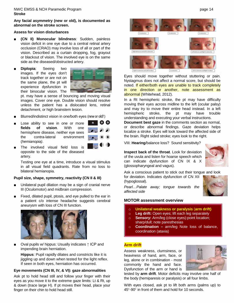
Arm drift Assess weakness, clumsiness, or heaviness of hand, arm, face, or leg, alone or in combination - most commonly the hand and face. Dysfunction of the arm or hand is tested by arm drift. Motor deficits may involve one half of the body (hemiparesis or paralysis) or all four limbs.
With eyes closed, ask pt to lift both arms (palms up) to 45°-90° in front of them and hold for 10 seconds.
o Unilateral weakness or paralysis (arm drift) o Leg drift: Open eyes; lift each leg separately o Sensory: Arm/leg (close eyes):point location;
sharp/dull; note paresthesias o Coordination – arm/leg Note loss of balance,
coordination (ataxia)
NWC EMSS & NCH Paramedic Program page 15 Stroke
The examiner may need to raise the patient’s arms and observe for asymmetrical drift, weakness or flaccidity. Note: Arm flexors are stronger than extensors; leg extensors are stronger than flexors. If hands pronate or fingers curl, these are subtle signs of weakness. Normal: Patient can hold position for 10 seconds with no movement or no drift at all Abnormal (new or old) ■ Drift: Limb holds 90° (or 45°) briefly, but drifts down
before full 10 sec; does not hit bed or other support ■ Some effort again gravity; limb cannot get to or
maintain 90° or 45°, drifts down but has some effort ■ No effort against gravity ■ No voluntary movement: Paralysis If either frontal lobe has a dysfunction, the opposite (contralateral) side of the body will experience motor loss as 80% of the motor fibers cross in the medulla. If both arms or legs are affected, consider spinal cord lesion although some TIAs/strokes can cause bilateral losses.
SENSORY deficits ■ Assess for sensory loss, tingling, or abnormal
sensations involving the face, arms, or legs; alone or in combination. Usually occurs simultaneously and on the same side as the weakness. The involved body parts are opposite to the side of the diseased artery.
■ Point location. Alternately touch patient on face, arms, and legs with the dull and sharp sides of a partially opened paperclip (or end of monofilament like fish line). Patient should be able to localize where the stimulus is being applied and discriminate if stimulus is dull or sharp. Rate sensory integrity from normal/no loss to total sensory loss/not aware of touch.
Alterations in sensory function ■ Hypalgesia: Decreased sensation ■ Analgesia: No sensation ■ Hyperalgesia: All touch is painful ■ Paresthesia: Alteration in sensation (pins and needles)
Extinction and inattention: Observe visual, tactile, or auditory inattention to one side or the other. The pt may not recognize own hand and will orient to only one side. Test extinction by touching the
patient on both sides at once. If they are able to feel only one side, they have parietal lobe disease.
Rate from no abnormality to profound hemi-inattention.
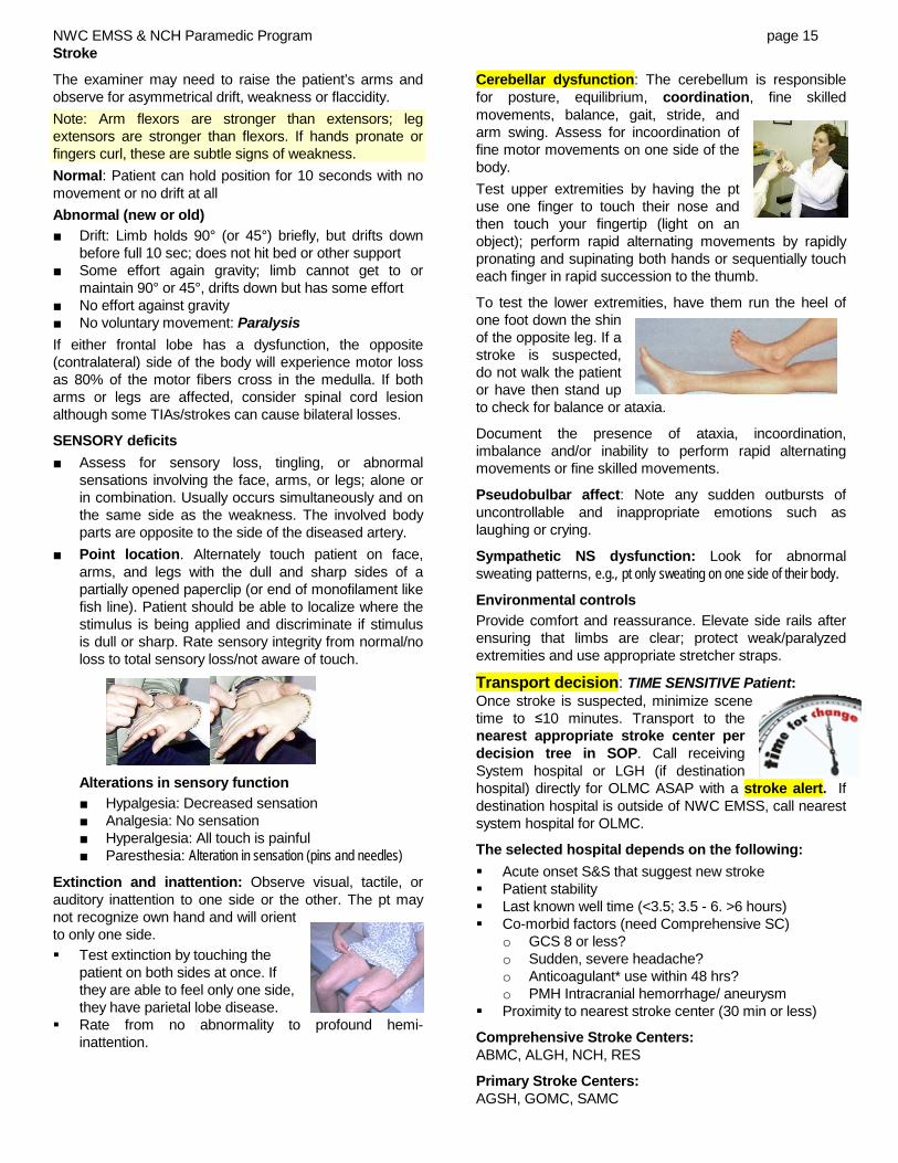
Cerebellar dysfunction: The cerebellum is responsible for posture, equilibrium, coordination, fine skilled movements, balance, gait, stride, and arm swing. Assess for incoordination of fine motor movements on one side of the body. Test upper extremities by having the pt use one finger to touch their nose and then touch your fingertip (light on an object); perform rapid alternating movements by rapidly pronating and supinating both hands or sequentially touch each finger in rapid succession to the thumb.
To test the lower extremities, have them run the heel of one foot down the shin of the opposite leg. If a stroke is suspected, do not walk the patient or have then stand up to check for balance or ataxia.
Document the presence of ataxia, incoordination, imbalance and/or inability to perform rapid alternating movements or fine skilled movements.
Pseudobulbar affect: Note any sudden outbursts of uncontrollable and inappropriate emotions such as laughing or crying.
Sympathetic NS dysfunction: Look for abnormal sweating patterns, e.g., pt only sweating on one side of their body.
Environmental controls Provide comfort and reassurance. Elevate side rails after ensuring that limbs are clear; protect weak/paralyzed extremities and use appropriate stretcher straps.
Transport decision: TIME SENSITIVE Patient: Once stroke is suspected, minimize scene time to ≤10 minutes. Transport to the nearest appropriate stroke center per decision tree in SOP. Call receiving System hospital or LGH (if destination hospital) directly for OLMC ASAP with a stroke alert. If destination hospital is outside of NWC EMSS, call nearest system hospital for OLMC.
The selected hospital depends on the following: Acute onset S&S that suggest new stroke Patient stability Last known well time (<3.5; 3.5 - 6. >6 hours) Co-morbid factors (need Comprehensive SC)
o GCS 8 or less? o Sudden, severe headache? o Anticoagulant* use within 48 hrs? o PMH Intracranial hemorrhage/ aneurysm
Proximity to nearest stroke center (30 min or less)
Comprehensive Stroke Centers: ABMC, ALGH, NCH, RES
Primary Stroke Centers: AGSH, GOMC, SAMC

NWC EMSS & NCH Paramedic Program page 16 Stroke
If witnesses/reliable historian cannot accompany patient to the ED, EMS must attempt to obtain their contact information/call back number in the event hospital personnel wish to obtain more information after patient arrival.
Differential diagnosis of stroke: Consider other causes of presenting S&S (“Stroke Mimics”)
■ Cervical/head trauma ■ Infections (TB, fungal, herpes
simplex encephalitis, meningitis) ■ Hypertensive encephalopathy ■ Intracranial mass
• Tumors (primary and secondary) • Epidural/subdural hematomas
■ Seizure disorder with persistent neurological signs (Todd's paralysis) (Tonic clonic seizures can occur simultaneous with hemorrhagic stroke.)
■ Migraine headaches with persistent neurological signs can mimic SAH but usually appear gradually and classically present with an aura. Basilar artery (brainstem) and hemiplegic migraines (often associated with aphasia if left hemisphere is involved) may be difficult to differentiate from stroke until angiography is performed). Hemiplegic migraine is more common in young women (20-40) as opposed to older patient (40-70) who are more likely to have atherosclerosis and thrombotic, ischemic strokes.
■ Metabolic disturbances • Hyperglycemia (DKA/HHNS); hypoglycemia • Post-cardiac arrest ischemia • Toxicological cause • Endocrine disorder (myxedema) • Uremia
■ Psychiatric syndromes ■ Shock and CNS hypoperfusion ■ Cardiac abnormalities (dysrhythmia, AMI, prolapsed mitral valve) ■ Degenerative disorder (Alzheimer's) ■ Cranial arteritis
Complications & Consequences of Stroke Stroke affects many body systems. Some stroke sequelae are permanent, some are not. Many impact the patient’s ability to be independent, safe and productive.
Conditions that patients may experience after stroke:
Neurological ■ Cerebral edema; ↑ ICP ■ Hydrocephalus ■ Hemorrhagic transformation ■ Seizures ■ Balance problems ■ Spasticity(causing stiff and awkward movement) ■ Contractures (permanent contraction of a muscle
due to spasm or paralysis ■ Speech problems
■ Visual deficits (w/ associated safety concerns) ■ Sensory deficits (numbness) w/ assoc. safety concerns ■ Perceptual problems: impaired judgment, lack of
awareness of unsafe environment, impaired understanding of surroundings
■ Cognitive deficits: learning of new information, problem solving, impulsiveness, decreased attention, distractibility
■ Memory problems ■ Communication problems ■ Personality changes ■ Behavior changes/problems: inability to inhibit
inappropriate behavior or “read” context of body language ■ Apathy; Pseudobulbar affect Medical ■ Dysphagia (problems swallowing), resulting in poor
nutrition, aspiration pneumonia ■ Hypoventilation ■ Atelectasis; ARDS ■ Myocardial ischemia, dysrhythmias ■ Neurogenic pulmonary edema ■ Decubitus ulcers ■ Deep vein thrombosis (DVT); Venous
thromboembolism (VTE)/pulmonary embolism ■ UTI ■ Depression
Documentation: see screens in appendix Patient demographics: DOB, gender, weight Chief complaint
Note if acute S&S resolved prior to hospital arrival Primary assessment/resuscitative interventions Vital signs (at least 2 sets; more if unstable) History: SAMPLE – especially last known well; onset
S&S; on anticoagulant?
Physical exam findings o GCS; orientation o Speech disturbances; slurred vs. aphasia o Cranial nerve disturbances: vision, visual
fields, diplopia, EOMs, gaze palsy (one or both eyes), facial droop
o Motor exam: Weakness/Paresis (arm drift) o Cerebellar exam: Ataxia; incoordination; vertigo o Sensory changes o Other neurological signs/symptoms
All elements leading to hospital selection (see above p. 15 and SOP)
EMS interventions Patient responses OLMC contact; called stroke alert Call back number for reliable historian
Hospital categorization of stroke etiology Large-artery occlusion/atherosclerosis (e.g., carotid or
basilar) Cardioembolism (e.g., AF/flutter, prosthetic heart
valve, recent MI)
NWC EMSS & NCH Paramedic Program page 17 Stroke
Small-vessel occlusion (e.g., subcortical or brain stem lacunar infarction <1.5 cm)
Stroke of other determined etiology (e.g., dissection, vasculopathy, hypercoagulable or hematologic disorders.
Cryptogenic stroke (stroke of undetermined etiology) Hospital interventions: (Nice to know only for EMS)
Goals of emergency management ■ Support vital functions ■ Restore cerebral circulation ■ Reduce neurological deficits ■ Prevent progression and cell death ■ Restore patient to optimal level of pre-stroke function Tissue Plasminogen Activator (tPA )
TIME IS BRAIN! In most forms of stroke, the patient has focal ischemia. The core area must receive resuscitation within minutes, but a large area around the core (ischemic penumbra) does not die for hours. Each hour of ischemia increases the degree of irreversible brain damage.
Tissue plasminogen activator (tPA). Goal of therapy for ischemic stroke: safely maximize functional recovery to pre-stroke baseline by rapidly reperfusing ischemic penumbra. IV-tPA remains the mainstay of acute stroke treatment for patients presenting within 4.5 hours of symptom onset.
Studies continue to support that time from onset to treatment with tPA is directly related to outcomes in ischemic stroke. The sooner tPA is given, the greater the benefit (Stroke, 2013). Beyond the approved and recommended time windows for tPA use, risk of complications rises.
IV tPA is no different from other medications in that it has potential adverse effects, which makes it essential that careful consideration is given to exclusion criteria, risks and benefits for use in each individual patient. The most major of its complications is symptomatic intracranial hemorrhage which occurs at a rate of roughly 5.2%. Other complications include orolingual angioedema (allergic reaction), acute hypotension, and systemic bleeding.
Endovascular therapies include intra-arterial tPA and mechanical clot extraction. Profoundly positive results from five endovascular trials published in 2015 mark an extremely encouraging year in the history of stroke care.
Patients who do not qualify for IV tPA may still be candidates for other, advanced therapies for stroke.
Intra-arterial tPA delivers fibrinolytic medication directly to the thrombus. It can be used for qualified patients up to 6 hours from onset of symptoms (Class I). As is the case with IV tPA, the best clinical outcomes with intra-arterial therapy are likely to be in those patients
who receive treatment as soon after symptom onset as is possible. This therapy can only be done in specialty centers by specially trained interventionalists.
Clot retrieval devices: There are currently four FDA-approved devices for clot disruption or removal.
The mainstay of treatment has long been tPA. But for people with blood clots in larger arteries, tPA often does not dissolve them completely.
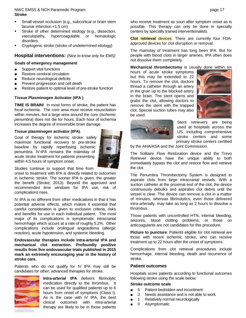
Mechanical thrombectomy is usually done within six hours of acute stroke symptoms but this may be extended to 22 hours. To remove the clot, doctors thread a catheter through an artery in the groin up to the blocked artery in the brain. The stent opens and grabs the clot, allowing doctors to remove the stent with the trapped clot. Special suction tubes may also be used.
Stent retrievers are being used at hospitals across the US, including comprehensive stroke centers and some primary stroke centers certified
by the AHA/ASA and the Joint Commission. The Solitaire Flow Restoration device and the Trevo Retriever device have the unique ability to both immediately bypass the clot and restore flow and retrieve the clot.
The Penumbra Thrombectomy System is designed to aspirate clots from large intracranial vessels. With a suction catheter at the proximal end of the clot, the device continuously debulks and aspirates clot debris until the vessel is clear. The device can remove a clot in a matter of minutes, whereas fibrinolytics, even those delivered intra-arterially, may take as long as 2 hours to dissolve a thrombus.
Those patients with uncontrolled HTN, internal bleeding, seizures, blood clotting problems, or those on anticoagulants are not candidates for this procedure.
Picture to puncture: Patients eligible for clot retrieval are those with recent ischemic stroke, who can receive treatment up to 22 hours after the onset of symptoms.
Complications from clot retrieval procedures include hemorrhage, internal bleeding, death and recurrence of stroke.
Patient outcomes Hospitals score patients according to functional outcomes following stroke using the scale below: Stroke outcome scale ■ 5 Patient bedridden and incontinent ■ 3 Needs assistance and is not able to work ■ 1 Relatively normal neurologically ■ 0 Asymptomatic

NWC EMSS & NCH Paramedic Program page 18 Stroke
Poorer outcomes are experienced if ■ > 65 years old ■ Severe cognitive deficit → coma ■ Persistent incontinence ■ Severe visual/spatial deficits (inattention) ■ Global aphasia ■ Co-morbid factors: HF, DM, AMI, previous stroke ■ Disability before stroke Advances and treatments on the horizon 'Wake-up' Stroke: Between 20 - 25% of all strokes occur while patients are asleep. Current guidelines for administration of IV tPA exclude these patients because time of symptom onset must be less than 3 (up to 4.5) hours. A 24-month study done in London, compared symptom improvement, functional outcome, and incidence of intracranial hemorrhage (ICH) (the most worrisome of adverse events associated with IV tPA) in 2 groups of patients treated with IV tPA. Among 326 patients with ischemic stroke, and 68 patients with wake up stroke, there were no differences in early improvement, functional outcome at 90 days, or intracranial hemorrhage rates between the two groups.

Distinctive signs and symptoms of stroke
Weakness of paralysis of the face, arms, or legs, especially on one side of the body; asymmetric smile or mouth drawn to one side; loss of facial expression; possibly a drooping eyelid; one eyelid does not close as tightly as the other
Inability to speak or understand speech (expressive or receptive aphasia) or distorted speech (dysarthria)
Sensory or autonomic symptoms affecting half of the body, such as numbness or abnormal sweat patterns
Visual loss in one half (or one quarter) of the visual field, or dimming or loss of vision (especially in only one eye); constricted pupils; pupils unequal in size or reactivity; double vision
Severe headache with no apparent cause, often accompanied by a stiff neck
Dizziness, confusion, unsteadiness, or falls with no apparent cause; seizures
Drowsiness or decreased consciousness
Other symptoms: nausea or vomiting, respiratory distress, unusually flushed or pale skin, loss of bowel and/or bladder control
Characteristics of stroke Thrombosis Embolism Intracerebral Hemorrhage SAH
Prodromal warning Common No No Rare Onset during sleep Sometimes Rare Rare Rare
Development Gradual Sudden Gradual or sudden Sudden Quick reversal Possible Possible No Possible Bloody CSF No Rare Common Yes
Coma Rare Rare Common Common Decreased consciousness Mild Mild Severe Moderate
Headache Mild Mild Severe Severe Hypertension Common Possible Not always Common Nuchal rigidity No No Sometimes Yes
Vomiting Rare Rare Sometimes Sometimes
GCS Level of
Dysfunction Level of
Consciousness Motor
Response Pupils Resp Pulse/ BP
15-8 cortex and cerebrum
Normal to drowsiness, stupor, light coma
Localizes pain withdraws
Normal sluggish & midsize
Normal Cheyne-Stokes
Stable
7-5 upper brainstem deepening coma Abnormal flexion or
extension unreactive &
enlarging Cheyne-Stokes
Central Neurog. Hyp. Becoming Unstable
4-3 lower brainstem profound coma Abnormal extension;
flaccid fixed & dilated Failing Unstable
failing
Table from: Shea, J. Altered States, Unpublished lecture outline.
Features of clinical situations mimicking Stroke (Stroke , 2013) Etiology History and Exam Findings
Psychogenic Lack of objective CN findings, neuro findings in nonvascular distribution, inconsistent exam Seizures Hx of seizures, witnessed seizure activity, postical period Hypoglycemia Hx DM, low serum glucose, ↓ LOC Complicated Migraine/with aura Hx similar events, preceding aura, headache Hypertensive encephalopathy Headache, delirium, significant HTN, cortical blindness, cerebral edema, seizure Wernicke’s encephalopathy Hx alcohol abuse, ataxia, EOM paralysis, confusion CNS abscess Hx drug abuse, endocarditis, medical device implant w/ fever CNS tumor Gradual progression of symptoms, other primary malignancy, seizure at onset Drug toxicity Med hx includes Lithium, phenytoin, carbamazepine

NWC EMSS & NCH Paramedic Program page 20 Stroke
Alternate Prehospital Stroke Screens
The MEND exam Prehospital (© 2005, University of Miami) incorporates the Cincinnati Prehospital Stroke Scale (CPSS) and selected components of the NIH Stroke Scale (NIHSS) used at the hospital. The MEND exam arose because the NIHSS is not feasible to perform in the prehospital setting and the CSS has only a 70% sensitivity to detect stroke in the field. In addition to predicting probability of stroke, it also gauges stroke severity and may identify strokes not found using the CPSS. The MEND exam takes less than 3 minutes. A recent study found it had a 90% correlation with the NIHSS.
Glasgow Coma Scoring
Strengths Limitations
■ Simplicity ■ Provides a common language to report
neurologic findings based on bedside observations
■ Ability to trend over time
■ Variation in inter-rater reliability; inconsistent use by EMS and hospital staff ■ If SBP < 80, can’t assess GCS with respect to possible outcomes ■ Hypothermia; Hypoglycemia ■ Sedating or paralyzing drugs; Intubated patients ■ Hearing impairments; Language barrier ■ Children younger than 3 years ■ Alcohol/drug intoxication ■ Relationship to mortality differs considerably between blunt & penetrating head trauma
Left cerebral hemisphere L MCA involvement
Right cerebral hemisphere R MCA involvement
Aphasia: expressive/ receptive or mixed Eyes: deviate to left R visual field deficit R hemiparesis/hemiplegia R hemisensory loss
Neglect (L hemi-inattention) Eyes: deviate to R L visual field deficit L hemiparesis/hemiplegia L hemisensory loss
Lacunar Syndromes
Pure motor Pure sensory Clumsy hand/dysarthria Ataxic hemiparesis Multi-infarct dementia (Cesario & Szost, 2013)
Brainstem or posterior stroke
5 Ds: Dizziness, Diplopia, Dysarthria, Dysphagia (chewing & swallowing), Dystaxia (incoordination)
Severe vertigo; nausea, vomiting; Visual field loss; gaze palsies Partial or complete loss of hearing May have difficulty breathing Hallmark: Crossed findings; same side (ipsilateral)
ataxia Cranial nerve deficits (ipsilateral) Motor/sensory deficits (contralateral); decreased pain
and temperature sensation; loss of 2 point discrimination
Clinical findings: asymptomatic to comatose

NWC EMSS & NCH Paramedic Program page 21 Stroke

NWC EMSS & NCH Paramedic Program page 22 Stroke
References ACEP Clinical Policies Subcommittee (Writing Committee) on Suspected Transient Ischemic Attack. (2016). Clinical
Policy: Critical issues in the evaluation of adult patients with suspected transient ischemic attack in the Emergency Department. Annals of Emerg Med, 68(3), 354-370.
AHA/ASA. (2013) Guidelines for the early management of patients with acute ischemic stroke: A guideline for healthcare professionals (pp 2, 4-6).
Bledsoe B.E., Casey M.J., Feldman, J., Johnson, L., Diel, S., Forred, W., Gorman, C. (2015). Glasgow coma scale scoring is often inaccurate. Prehosp Disaster Med, 30(1),1-8 .
Cheng, D. et al. (2013). Hyperglycemia exacerbates intracerebral hemorrhage via the downregulation of aquaporin-4. Stroke, 44: 1682-1689.
Iadecola, C., Yaffe, K., Biller, J. et al. on behalf of the AHA Council on Hypertension; Council on Clinical Cardiology; Council on Cardiovascular Disease in the Young; Council on Cardiovascular and Stroke Nursing; Council on Quality of Care and Outcomes Research; and Stroke Council.(2016). Impact of hypertension on cognitive function A scientific statement from the AHA. Accessed on line:http://hyper.ahajournals.org DOI: 10.1161/HYP.0000000000000053.
Jauch, E.C. et al. (2013). Guidelines for the early management of patients with acute ischemic stroke: A guideline for healthcare professionals from the AHA/ASA. Accessed on line: http://stroke.ahajournals.org/ at.
Krieger, D., et al. (April, 2013). Cryptogenic Stroke: What, When, and How to Treat for Secondary Prevention CME. 2013 Medscape, LLC. [On-line] Available at http://www.medscape.org/viewarticle/782001.
Lerario, M.P., Segal, A.Z. (2016). Acute ischemic stroke: Focus on reperfusion. Accessed on line: AHC Media.
Manawadu, K. et al (2013). Stroke 2013. Abstract 56: Thrombolysis in selected patients with wake up stroke is feasible with similar safety as thrombolysis in 0-4.5 Hours. Accessed online at http://stroke.ahajournals.org/cgi/content/meeting_abstract/43/2_MeetingAbstracts/A56
Mehta, B.P. et al. (2017). Finding ELVO: New stroke scales for EMS. EMS World. Accessed on line at: http://emsworld.com/node/218774
Mellor, R.M., Bailey, S., Sheppard, J., et al. (2015). Decisions and delays within stroke patients’ route to the hospital: A qualitative study. Annals of Emerg Med, 65(3), 279-287.
Miller, D. (2012). Stroke: Time is brain in delivering EMS care. Accessed on line EMS-1, http://www.ems1.com/air-medical-transport/articles/1259279-Stroke-Time-is-brain-in-delivering-EMS-care/
Mozaffarian, D., Benjamin, E.J., Go, A.S. et al. on behalf of the AHA Statistics Committee and Stroke Statistics Subcommittee. (2016). Heart disease and stroke statistics-2016 update: a report from the AHA. Circulation, e38-e360.
Powers, W.J., Derdeyn, C.P., Biller, J. et al. (2015). 2015 AHA/ASA Focused update of the 2013 guidelines for the early management of patients with acute ischemic stroke regarding endovascular treatment. Stroke. Accessed on line: http://stroke.ahajournals.org DOI: 10.1161/STR.0000000000000074.
Saver, J.L., Goyal, M., van der Lugt, A. et al. (2016). Time to treatment with endovascular thrombectomy and outcomes from ischemic stroke: a meta-analysis. JAMA, 316(12):1279-1289. doi: 10.1001/jama.2016.13647
University of Miami Gordon Center for Research in Medical Educational. (2013). About the MEND. Accessed online at http://www.asls.net/mend_about.php
Whitehead, S. (Sept. 12, 2013) The MEND Stroke Assessment for Prehospital Care. Accessed online at http://theemtspot.com/2012/09/12/the-mend-stroke-assessment-for-prehospital-care/
Zebian, R.C. (2015) Emergent management of subarachnoid hemorrhage. Accessed on line: http://emedicine.medscape.com/article/794076-overview?pa=8U9teNAxhIRLlK%2BbUIrk%2BKghiddatYY%2F94YDSC93d%2FBzeExZp%2Fy7ErQ%2BjgNkMp9V#a2
http://www.slideshare.net/romduck/the-big-picture-of-stroke-care National Institutes of health (NIH): www.health.nih.gov The National Institute of Neurological Disorders and Stroke (NINDS): www.ninds.nih.gov/disorders/stroke/stroke.htm National Stroke Association (NSA): www.stroke.org Stroke education for EMS professionals: http://www.strokeawareness.com/ems4stroke http://www.neurocriticalcare.org/science-research/subarachnoid-hemorrhage

NWC EMSS & NCH Paramedic Program page 23 Stroke

NWC EMSS & NCH Paramedic Program page 24 Stroke

NWC EMSS & NCH Paramedic Program page 25 Stroke
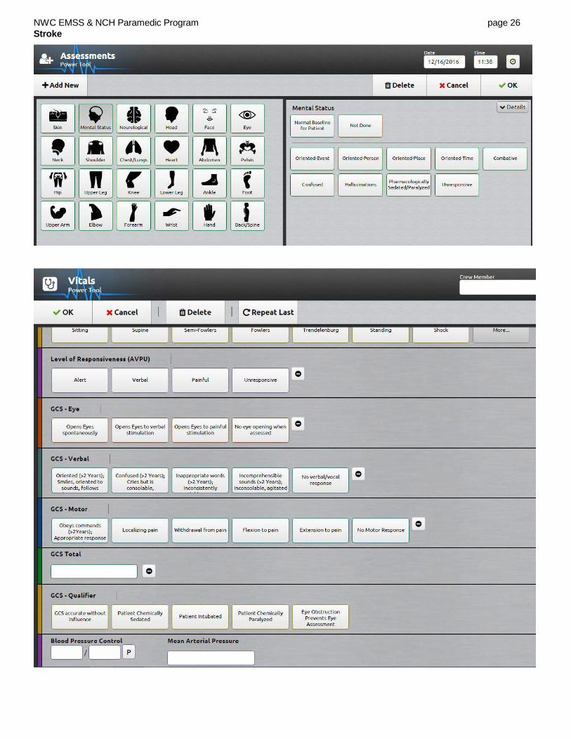
NWC EMSS & NCH Paramedic Program page 26 Stroke
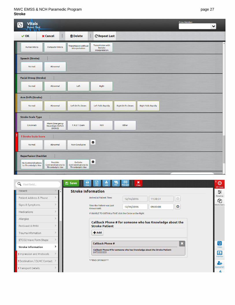
NWC EMSS & NCH Paramedic Program page 27 Stroke

NWC EMSS & NCH Paramedic Program page 28 Stroke
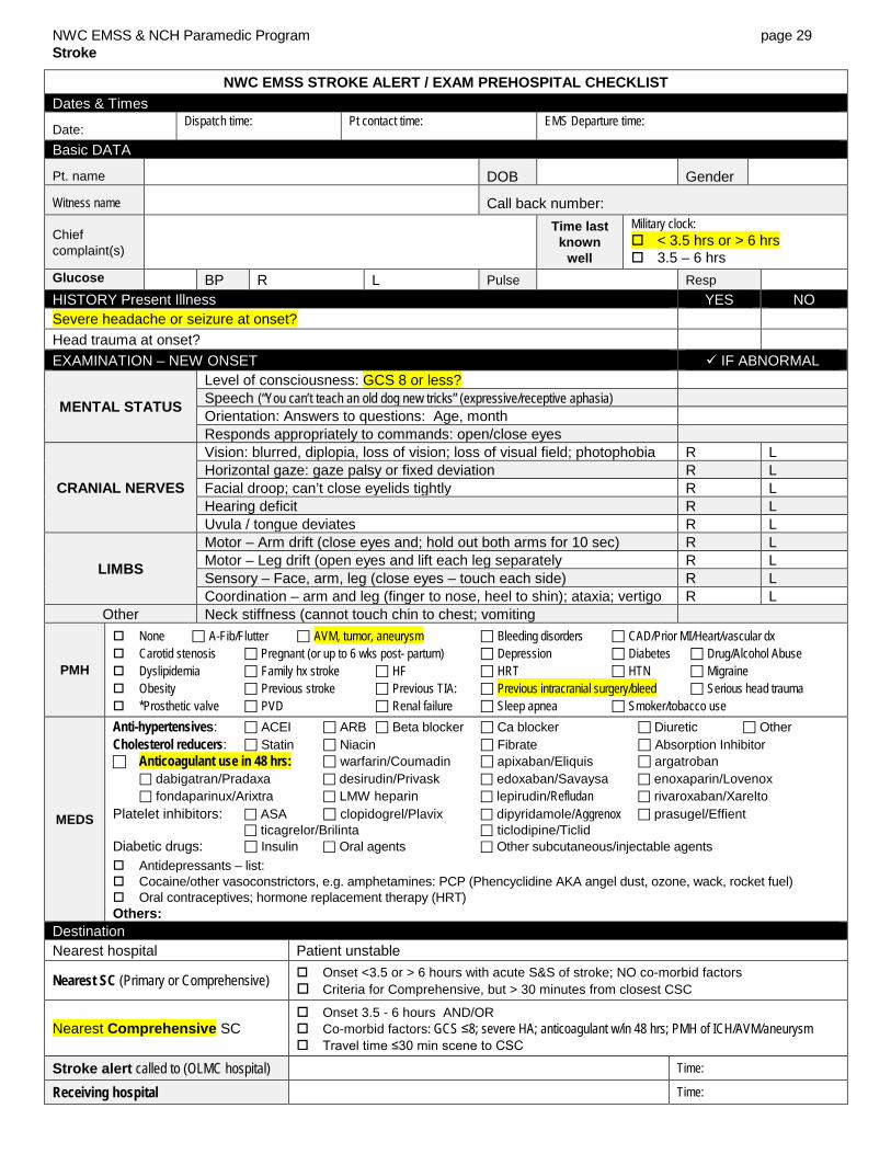
NWC EMSS & NCH Paramedic Program page 29 Stroke
NWC EMSS STROKE ALERT / EXAM PREHOSPITAL CHECKLIST Dates & Times
Date: Dispatch time: Pt contact time: EMS Departure time:
Basic DATA
Pt. name DOB Gender
Witness name Call back number:
Chief complaint(s)
Time last known
well
Military clock: < 3.5 hrs or > 6 hrs 3.5 – 6 hrs
Glucose BP R L Pulse Resp HISTORY Present Illness YES NO Severe headache or seizure at onset? Head trauma at onset? EXAMINATION – NEW ONSET IF ABNORMAL
MENTAL STATUS
Level of consciousness: GCS 8 or less? Speech (“You can’t teach an old dog new tricks” (expressive/receptive aphasia) Orientation: Answers to questions: Age, month Responds appropriately to commands: open/close eyes
CRANIAL NERVES
Vision: blurred, diplopia, loss of vision; loss of visual field; photophobia R L Horizontal gaze: gaze palsy or fixed deviation R L Facial droop; can’t close eyelids tightly R L Hearing deficit R L Uvula / tongue deviates R L
LIMBS
Motor – Arm drift (close eyes and; hold out both arms for 10 sec) R L Motor – Leg drift (open eyes and lift each leg separately R L Sensory – Face, arm, leg (close eyes – touch each side) R L Coordination – arm and leg (finger to nose, heel to shin); ataxia; vertigo R L
Other Neck stiffness (cannot touch chin to chest; vomiting
PMH
None A-Fib/Flutter AVM, tumor, aneurysm Bleeding disorders CAD/Prior MI/Heart/vascular dx Carotid stenosis Pregnant (or up to 6 wks post- partum) Depression Diabetes Drug/Alcohol Abuse Dyslipidemia Family hx stroke HF HRT HTN Migraine Obesity Previous stroke Previous TIA: Previous intracranial surgery/bleed Serious head trauma *Prosthetic valve PVD Renal failure Sleep apnea Smoker/tobacco use
MEDS
Anti-hypertensives: ACEI ARB Beta blocker Ca blocker Diuretic Other Cholesterol reducers: Statin Niacin Fibrate Absorption Inhibitor Anticoagulant use in 48 hrs: warfarin/Coumadin apixaban/Eliquis argatroban
dabigatran/Pradaxa desirudin/Privask edoxaban/Savaysa enoxaparin/Lovenox fondaparinux/Arixtra LMW heparin lepirudin/Refludan rivaroxaban/Xarelto
Platelet inhibitors: ASA clopidogrel/Plavix dipyridamole/Aggrenox prasugel/Effient ticagrelor/Brilinta ticlodipine/Ticlid
Diabetic drugs: Insulin Oral agents Other subcutaneous/injectable agents Antidepressants – list: Cocaine/other vasoconstrictors, e.g. amphetamines: PCP (Phencyclidine AKA angel dust, ozone, wack, rocket fuel) Oral contraceptives; hormone replacement therapy (HRT) Others:
Destination Nearest hospital Patient unstable
Nearest SC (Primary or Comprehensive) Onset <3.5 or > 6 hours with acute S&S of stroke; NO co-morbid factors Criteria for Comprehensive, but > 30 minutes from closest CSC
Nearest Comprehensive SC Onset 3.5 - 6 hours AND/OR Co-morbid factors: GCS ≤8; severe HA; anticoagulant w/in 48 hrs; PMH of ICH/AVM/aneurysm Travel time ≤30 min scene to CSC
Stroke alert called to (OLMC hospital) Time:
Receiving hospital Time: