comprehensive case studykellykirk.yolasite.com/resources/kirk comprensive case... · web...
TRANSCRIPT
Running head: COMPREHENSIVE CASE STUDY 1
Comprehensive Case Study
Kelly Kirk
Wright State University
COMPREHENSIVE CASE STUDY 2
Comprehensive Case Study
History and Physical
Patient Information
Name: A.E., D.O.B: 7/5/1942
Source
Patient-reliable source
Chief Complaint
“Car accident”
HPI
A.E. is a 72-year-old female who presents to the emergency department via ambulance
after being involved in a motor vehicle crash. Patient was a restrained driver and was T-boned at
an intersection when another car ran a red light at approximately 30 mph. She denies loss of
consciousness, but complains of left leg pain and left-sided chest pain. Vital signs remain stable.
The left lower extremity has an obvious deformity. The x-ray reveals proximal femur fracture,
and the orthopedic surgery team has been consulted. Chest x-ray reveals non-displaced rib
fractures x3 on the left (# 3, 4, and 6). Computed tomography (CT) of chest, abdomen, and
pelvis is negative for any additional injuries. A 12 lead EKG reveals sinus tachycardia (HR
115). Patient remains in c-spine precautions with a c-collar in place. She could not be clinically
cleared secondary to distracting pain. The orthopedic surgery team was able to reduce her femur
fracture without difficulty. Her vitals remained stable and her airway patent throughout
conscious sedation. The orthopedic resident placed the patient in skeletal traction, and
COMPREHENSIVE CASE STUDY 3
tentatively arranged for operative plans for tomorrow. A.E. was transferred to the trauma ward
with husband at bedside. Hospital day one, she remained hemodynamically stable, and the
trauma ACNP clinically cleared her cervical spine and cleared her to go to the operating room
for the repair of her femur under general anesthesia.
Patient is now POD#4 s/p open reduction internal fixation (ORIF) of left femur. Patient
was progressing well and mobilizing with physical therapy (PT). She initially had difficulty with
adequate pain control for her rib fractures, but she is reporting adequate pain control since the
addition of Tordol to her pain regimen. She has been on 2 liters NC oxygen with SpO2 ranging
from 94-97%. While working with PT today, patient reports sudden onset of shortness of breath
that began 3 hours ago and has progressively gotten worse. Her oxygen saturation decreased to
86% on 2 liters NC. Respiratory therapy titrated her oxygen up to 6 liters high flow nasal
cannula without improvement of O2 saturation. The patient is now on 100% non-rebreather with
O2 saturations 92-94%. She is complaining of dyspnea, new onset chest pain that feels different
from the pain from her rib fractures, and anxiety.
Past Medical History
Hypertension
Hyperlipidemia
Coronary artery disease
Past Surgical History
C-section x2
Tonsillectomy

COMPREHENSIVE CASE STUDY 4
Family History
Mother- Breast cancer (deceased)
Father-hypertension, CAD, hyperlipidemia (deceased)
Brother- hypertension
Daughters x 2- healthy
Social History
Patient is married and works an administrative assistant at a car dealership locally, and cares for
her grand-daughter once a week. She is a life-long non-smoker, and uses alcohol socially (less
than 1 drink a week). No illicit drug use. Patient walks 2-3 blocks almost daily.
Allergies
PCN- rash
Medications (Home)
Lisinopril 10 mg daily
Lipitor 20 mg daily
Hospital Administered Medications
Lovenox 30 mg sub Q BID
Torodol 15 mg IV every 6 hours for 6 doses
Lipitor 20 mg PO daily
COMPREHENSIVE CASE STUDY 5
Colace 100 mg PO BID
Senna 1 tab PO daily
Tylenol 650 mg PO every 8 hours
Oxycodone 5-10 mg PO every 4 hours PRN pain
ROS (at time of admission)
General: Complains of pain all over. Prior to accident patient denies weight change, fatigue,
weakness, fever, chills, or night sweats.
Skin: Denies rashes, itching, moles, and non-healing wounds. Denies changes in skin, hair, and
nails.
HEENT: Denies headaches, loss of consciousness, visual changes, eye discharge, hearing loss,
tinnitus, rhinorrhea, epistaxis, bleeding gums, mouth sores, sore throat, and hoarseness.
Neck: Complains of generalized neck stiffness secondary to c-collar and backboard.
Cardiac: Reports past medical history of hypertension and hyperlipidemia. Denies angina,
palpitations, orthopnea, and edema.
Chest/respiratory: Reports sharp, localized, left-sided chest pain, and mild shortness of breath.
Denies hemoptysis, cough, asthma, wheezing, and history of pneumonia.
GI: Occasional heartburn. Denies abdominal pain, change in bowel habits or appetite, nausea,
vomiting, and diarrhea. Denies history of hepatitis, ulcers, or melena.
GU: Stress incontinence. Denies frequency, urgency, dysuria, hematuria, nocturia, and UTIs.

COMPREHENSIVE CASE STUDY 6
Musculoskeletal: Denies arthritis, gout, and joint pain or stiffness or redness. Reports inability
to move left leg secondary to pain.
Neurologic: Denies seizures, fainting, tremors, numbness and tingling. Left lower extremity
weakness secondary to injury.
Physical Exam (POD # 4)
Vital Signs: Temp- 99.8, BP- 103/62, HR- 126, RR- 33, SpO2- 92% (100% Face mask)
General: Well developed Caucasian female in moderate distress. She appears her stated age.
She is awake, alert, and oriented to person, place, and time. Appears anxious, but is cooperative
with exam.
HEENT: Head is midline and skull is normocephalic, atraumatic, with appropriate hair texture
and distribution. Eyes: Bilateral eyebrows and lashes full, no ptosis noted. Conjunctivae are
pink and sclerae are white and without jaundice. Cornea clear. Pupils are equal, round, and
reactive (3-2mm) to light and accommodation. Ears: Deferred Nose: No discharge, nares patent
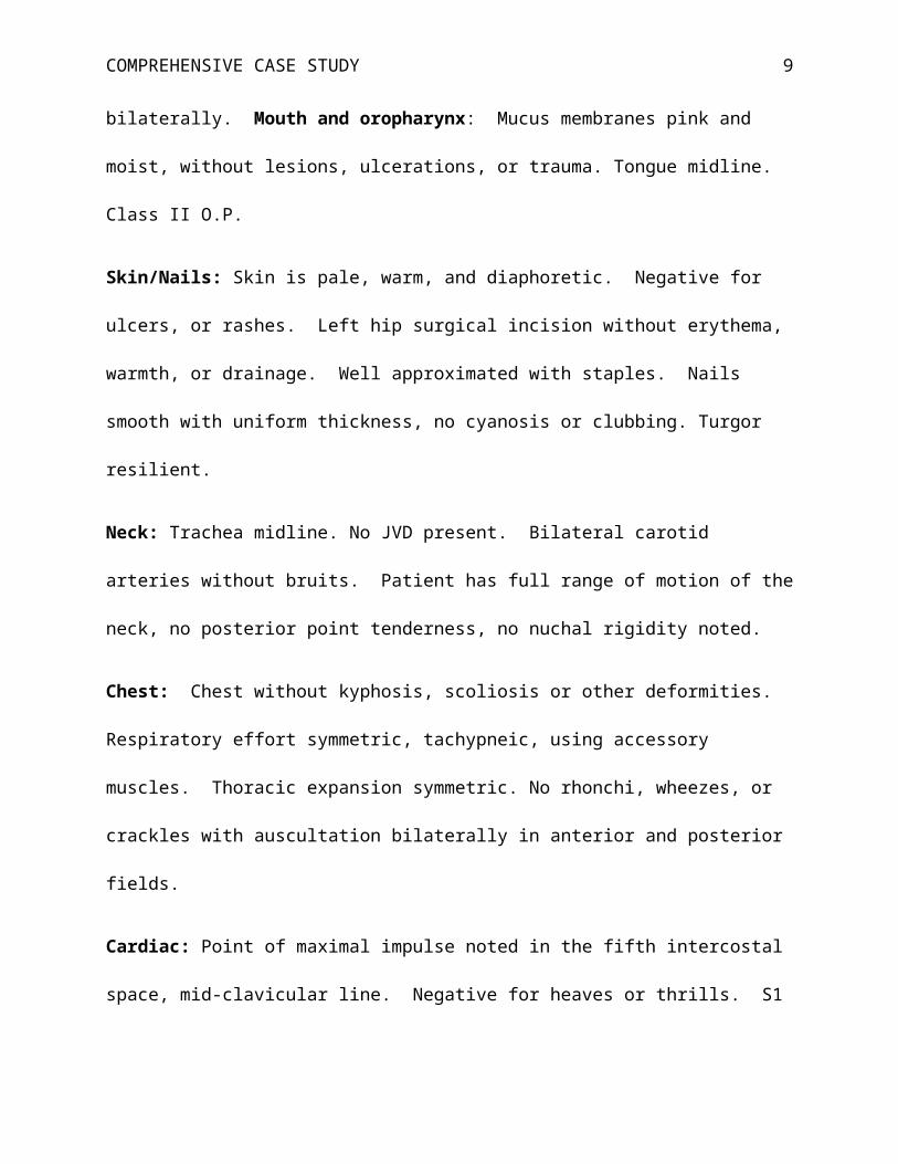
bilaterally. Mouth and oropharynx: Mucus membranes pink and moist, without lesions,
ulcerations, or trauma. Tongue midline. Class II O.P.
Skin/Nails: Skin is pale, warm, and diaphoretic. Negative for ulcers, or rashes. Left hip surgical
incision without erythema, warmth, or drainage. Well approximated with staples. Nails smooth
with uniform thickness, no cyanosis or clubbing. Turgor resilient.
Neck: Trachea midline. No JVD present. Bilateral carotid arteries without bruits. Patient has
full range of motion of the neck, no posterior point tenderness, no nuchal rigidity noted.

COMPREHENSIVE CASE STUDY 7
Chest: Chest without kyphosis, scoliosis or other deformities. Respiratory effort symmetric,
tachypneic, using accessory muscles. Thoracic expansion symmetric. No rhonchi, wheezes, or
crackles with auscultation bilaterally in anterior and posterior fields.
Cardiac: Point of maximal impulse noted in the fifth intercostal space, mid-clavicular line.
Negative for heaves or thrills. S1 S2 regular, tachycardic. Negative for murmurs, clicks, gallops,
or rubs.
Peripheral Vascular: Bilateral upper extremities and right lower extremity with capillary refill
time < 2 seconds and negative for edema. Left lower extremity with 1+ edema up to hip.
Brachial, carotid, radial, femoral, dorsalis pedis, and posterior tibial pulses +2/4 and regular, but
rapid. Homan’s sign negative right lower extremity. Unable to perform homan’s sign testing on
left lower extremity secondary to pain.
Abdomen: Flat, soft, and symmetric without lesions, or rashes or areas of visible pulsations or
peristalsis. Hypoactive bowel sounds in all four quadrants with auscultation. Negative for
bruits, venous hums, or friction rubs. Percussion tones tympanic over epigastrium and dull over
remainder of abdomen. No splenomegaly. Musculature soft and relaxed to light palpation. No
masses or tenderness to deep palpation.
Neuro: Cranial nerves II-XII intact. Deep tendon reflexes 2+ bilaterally at biceps, brachioradial,
triceps, and 2+ right knee. Left lower extremity reflexes deferred secondary to injury.
Musculoskeletal: Bilateral hands, wrists, elbows, shoulders, and ankles without joint
deformities. Spine straight without deformities. Shoulders and scapula symmetric. Strength 5/5
throughout. No erythema, warmth, or tenderness to palpation of upper and lower extremity
joints.

COMPREHENSIVE CASE STUDY 8
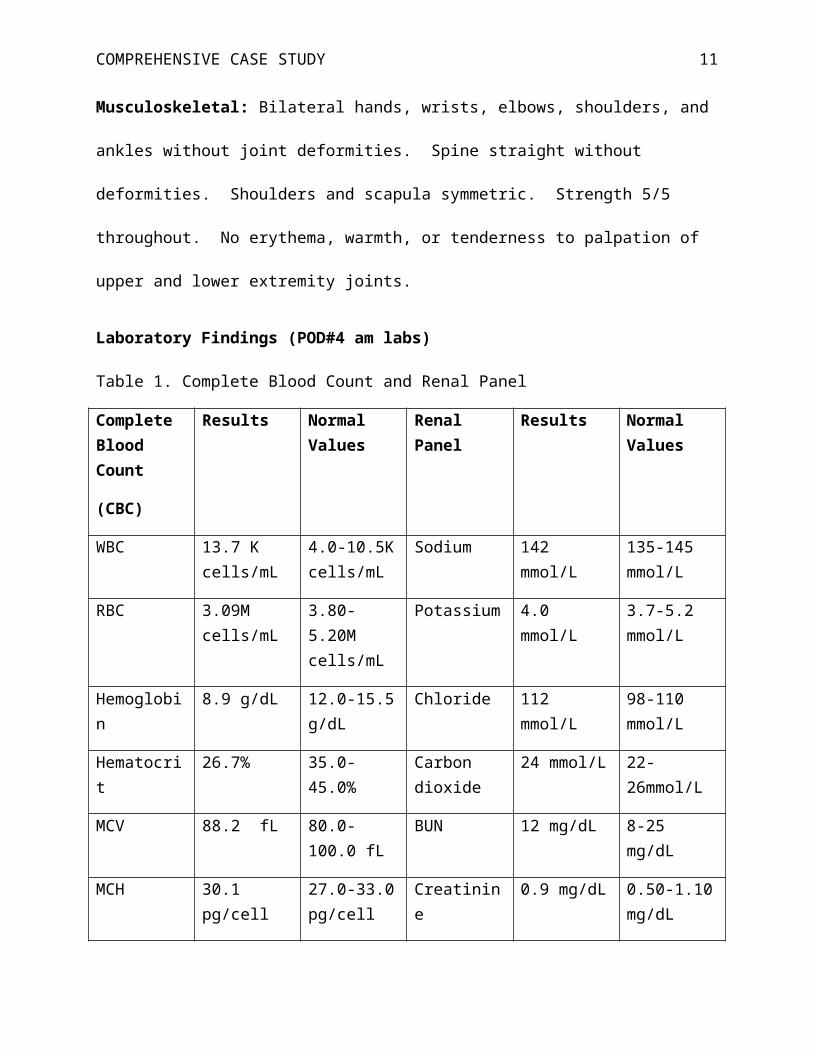
Laboratory Findings (POD#4 am labs)
Table 1. Complete Blood Count and Renal Panel
Complete Blood Count
(CBC)
Results Normal Values
Renal Panel Results Normal Values
WBC 13.7 K cells/mL
4.0-10.5K cells/mL
Sodium 142 mmol/L 135-145 mmol/L
RBC 3.09M cells/mL
3.80-5.20M cells/mL
Potassium 4.0 mmol/L 3.7-5.2 mmol/L
Hemoglobin 8.9 g/dL 12.0-15.5 g/dL
Chloride 112 mmol/L 98-110 mmol/L
Hematocrit 26.7% 35.0-45.0% Carbon dioxide
24 mmol/L 22-26mmol/L
MCV 88.2 fL 80.0-100.0 fL BUN 12 mg/dL 8-25 mg/dL
MCH 30.1 pg/cell 27.0-33.0 pg/cell
Creatinine 0.9 mg/dL 0.50-1.10 mg/dL
MCHC 33.6 g/dL 32.0-36.0 g/dL
Glucose 125 mg/dL 65-100 mg/dL
RDW 15.3% 11.0-15.0% Calcium 7.8 mg/dL 8.5-10.3 mg/dL
Platelet 115 K/uL 140-400 K/uL Phosphorus 2.3 mg/dL 2.5-4.5 mg/dL
MPV 9.7 fL 7.5-11.5 fL Magnesium 1.9 mg/dL 1.8-2.5 mg/dL
COMPREHENSIVE CASE STUDY 9
Arterial blood gas (ABG) at the time of the episode on 100% non-rebreather:
pH PaCO2 PaO2 HCO3 O2 Saturation
Base Excess
Result 7.48 29 54 24 91% 0.8
Normal 7.35-7.45 35-45
mm Hg
80-100
mm Hg
21-27
mEq/L
95-98% -2.0 - +2.0
mEq/L
Differential Diagnosis
There are multiple differential diagnoses to consider for this patient. A.E.’s age
contributes to the complexity of this scenario. The list of differential diagnosis for this patient is
identical to a patient that is 30 years younger who presents with the same injuries and symptoms,
and includes pneumonia, pneumothorax, hemothorax, acute pulmonary edema, hemorrhage,
atelectasis, pulmonary embolism (PE), heart failure, pleural effusions, acute coronary syndrome
(ACS), and fat embolism. However, the degree of probability of each diagnosis is different for
A.E. than for a patient who is 35-years-old. Patients over 65-years-old with chest wall trauma
and rib fractures are more likely to develop pulmonary complications, have higher mortality
rates, and spend more days in the hospital, when compared to younger adults with similar
injuries (Sahr, Webb, Renner, Sokol, & Swegle, 2013). The likelihood of PE or fat emboli with
sudden onset dyspnea in a post-operative patient is high, however, pneumonia is almost twice as
likely to complicate the hospital course for an elderly patients with rib fractures, than a younger
person with the same number of rib fractures (Bulger, Arneson, Mock, & Jurkovich, 2000). A.E.
has a history of hypertension and coronary artery disease, which moves ACS higher on the list of
differentials, and makes her less tolerant of post-operative bleeding and anemia.

COMPREHENSIVE CASE STUDY 10
The diagnosis that falls at or near the top of the list of differentials in terms of degree of
probability is PE. Before even looking at the patient’s symptomatology, one can determine that
A.E. is at high risk for developing a venous thromboembolism (VTE). VTE (includes deep vein
thrombosis and PE) affects 900,000 people each year in the United States. One third of these
cases are secondary to a PE and result in sudden death (Raskob, Hull, & Pineo, 2010). Deep
vein thrombosis (DVT) of the lower extremity is present in almost all clinically significant PEs,
therefore, the presence of a DVT places patients at high risk for PE (Raskob et al., 2010). The
patient’s age increases her risk of VTE. The increased risk of VTE in patients over age 65 is
likely multifactorial; proposed mechanisms include endothelial dysfunction, predisposition to
venous stasis, higher concentrations of coagulation factors, and impaired fibrinolysis (Kim et al.,
2012). Lower extremity fracture, rib fractures, and the need for a major operation further
increase A.E.’s risk of VTE (Kim et al., 2012). A.E. presents with the classic findings of PE
including sudden onset dyspnea and hypoxemia unexplained by chest x-ray findings (Kline,
2011). Clinicians should determine the pretest probability of PE to guide diagnostic evaluation.
The Wells score is widely used to determine the pretest probability of PE; scores greater than six,
indicate high likelihood of PE (Kline, 2011). A.E.’s score is at least six, and could be as high as
nine if one suspects DVT based the unilateral lower extremity edema.
Fat emboli likely occur with all long-bone fractures, as 90% of these patients have fat
particles in their pulmonary vascular bed. Fat embolism syndrome is far less common, but can
be fatal (Mariano, 2013). Fat emboli typically present with confusion, dyspnea, and petechiae
over the chest, axilla, upper extremities, and conjunctiva (Mariano, 2013). Although this patient
is dyspneic, without the presence of petechiae or fat globules in the urine, sputum, or retina, the
diagnosis of fat emboli is unlikely.
COMPREHENSIVE CASE STUDY 11
The probability of ACS is higher for A.E. because of her age and her history of
hypertension and coronary artery disease. The rupture of an existing thrombus is usually the
cause of postoperative myocardial infarction (POMI). Surgical stress creates demand ischemia
and lesions that would not cause ischemia preoperatively, can become very problematic both
peri- and post-operatively (Cooper & Ashley, 2012). Patients with heart failure, preexisting
pulmonary disease, cerebrovascular disease, diabetes, renal insufficiency, prior ischemia, and
poor functional capacity (less than 4 METs), are at a greater risk for developing POMI (Fleisher
et al., 2007). Cardiac risk is also dependent on the type of surgery that is being performed.
Highest risk surgeries are vascular surgeries. Orthopedic surgeries are considered intermediate
risk, meaning the combined incidence of cardiac death and nonfatal myocardial infarction is 1%-
5% (Fleisher et al., 2007). The symptoms of POMI are similar to a myocardial infarction that is
unrelated to surgery, however, effects from anesthesia and narcotics can blunt typical symptoms
(Cooper & Ashley, 2012). A.E. is experiencing classic symptoms of acute coronary syndrome,
chest pain, dyspnea, and diaphoresis, however she possess few of the aforementioned risk
factors. She does have a history of coronary artery disease and hypertension, her functional
capacity is considered moderate (4-7 METs). Her preoperative EKG was normal, and her EKG
at the time of the episode reveals sinus tachycardia without ST-wave changes. POMI cannot be
ruled out entirely, however, based on these findings the degree of probability is less than PE.
Pneumothorax and hemothorax can be associated with rib fractures, and often present
with chest pain and dyspnea. Approximately 20% of patients with chest wall trauma will have
associated pneumothorax, but do not cause significant symptoms until it occupies more than 40%
of the hemithorax (Brunett, Yarris, & Cevik, 2011). Both pneumothorax and hemothorax can be
missed on chest x-ray, however this patient did have a chest CT in the ED, which only revealed

COMPREHENSIVE CASE STUDY 12
the three rib fractures. It is very unlikely that a pneumo/hemothorax was missed on the CT scan.
A.E. also received positive pressure ventilation when she was under general anesthesia for repair
of her femur. If a pneumothorax were present, it is likely that it would have become problematic
at the time of her operation or shortly thereafter. An occult pneumothorax is often well tolerated
in patients who are spontaneously breathing, however they can convert into tension
pneumothorax with positive pressure ventilation (Brunett et al., 2011). There is a risk of delayed
hemothorax and pneumothorax, although it is relatively low. Plourde et. al., found that delayed
hemothorax and pneumothorax occurred in 11.8% and 0.9% respectively after blunt chest
trauma, and majority of cases were in the first week (Plourde et al., 2014). A chest x-ray can
help rule out a delayed pneumo/hemothorax, however they are not likely diagnoses in this case.
Diagnostic Tests
Hemodynamic stability guides the diagnostic work-up for PE. Patients who present with
hypotension or shock require rapid risk stratification to determine right ventricular dysfunction.
The most useful test in this situation is echocardiogram, which helps rule out other differential
diagnoses such as acute valvular dysfunction, aortic dissection, and tamponade (Torbicki et al.,
2008). The clinician must carefully evaluate the patient to determine if he/she is stable enough
to travel to diagnostic testing areas, or wait for laboratory tests to result. In this case, the patient
is stable from a hemodynamic standpoint, however her marginal oxygenation despite 100%
oxygen, raises concern for traveling to diagnostic areas. Tests that are done at bedside and can
quickly exclude other reasons for her dyspnea and chest pain are prioritized initially.

COMPREHENSIVE CASE STUDY 13
Chest Radiography
A chest x-ray is mandatory for the hypoxic inpatient with dyspnea. Fortunately A.E. had
a recent chest x-ray just over 24 hours ago, as comparison is important. A portable chest x-ray is
helpful to exclude pneumonia and pneumothorax, especially when recent images are available
for comparison (Jacobson & McKean, 2012). A chest x-ray is not highly sensitive for
pneumonia or pneumothoracies, a negative chest x-ray cannot entirely rule out either diagnosis
(Eisenhuber, Schaefer-Prokop, Prosch, & Schima, 2012). A chest x-ray can identify acute
pulmonary edema, and may help to determine cardiac versus non-cardiac edema. Clues that
pulmonary edema is cardiac in nature include an enlarged cardiac silhouette, enlarged vascular
pedicle, and the presence of perbronchial cuffs and pleural effusions (Cardinale, Volpicelli,
Lamorte, Martino, & Veltri, 2012). The sensitivity and specificity of the chest x-ray is 67-68%
and 76-83% respectively for the diagnosis of heart failure (Cardinale et al., 2012).
The role of the chest x-ray for the diagnosis of a PE is primarily to exclude other
differentials, as opposed to rule in PE based on chest x-ray findings. The clinician should have a
high suspicion of index for the diagnosis of PE for the patient with dyspnea and hypoxia with a
normal chest x-ray (Cardinale et al., 2012). Non-specific findings associated with PE are
atelectasis, small pleural effusions, elevated hemi-diaphragm, and Hampton’s hump (Cardinale
et al., 2012). The Westermark sign is highly specific for PE, however it is rarely seen (Cardinale
et al., 2012).
Electrocardiogram
A 12-lead ECG may help determine if chest pain and dyspnea are related to cardiac
ischemia or infarction. Comparison to the previous 12-lead ECG that was performed in the ED

COMPREHENSIVE CASE STUDY 14
improves its diagnostic value. The presence of ST-segment elevation of one mm or more in two
contiguous leads, or a new left bundle branch block, in a patient with ischemic symptoms
confirms the diagnosis of an ST-elevation myocardial infarction (STEMI) (O’Gara et al., 2013).
However, only about half (13%-69%) of patients with an acute myocardial infarction (AMI) will
meet these criteria on their initial 12 lead ECG (Green & Hill, 2011). The positive predictive
value for AMI is greater than 90% for patients with new ST-segment elevation, however a
normal or inconclusive ECG cannot rule out the diagnosis of AMI (Green & Hill, 2011).
The use of ECGs for the diagnosis of PE is limited, abnormal findings are neither
sensitive nor specific for the diagnosis of PE. The role of the ECG when PE is suspected,
primarily serves to exclude other causes of chest pain. Most patients with PE will present with
sinus tachycardia. Larger emboli may produce right ventricular strain patterns on 12-lead ECG
(Nana-Sinkam, 2003). Right axis deviation, t-wave inversion, and complete or incomplete right
bundle branch block suggest right heart strain. Hypoxia and right atrial enlargement may
produce atrial arrhythmias (Nana-Sinkam, 2003).
Computed Tomography with Angiography
Improvement in imaging technology has made the CT angiogram the test of choice for
the diagnosis of PE (Torbicki et al., 2008). The introduction of multidetector computed
tomography (MDCT) improved the sensitivity and specificity of this diagnostic tool. The
PIOPED II series found a sensitivity of 83% and specificity of 96% for the diagnosis of PE
(Torbicki et al., 2008), however Winer-Muram et al. observed a sensitivity and specificity
greater than 90% (Winer-Muram et al., 2004). Sensitivity of CT angiogram decreases in the case
of small, peripheral emboli (Torbicki et al., 2008). CT scan can also help determine the severity
COMPREHENSIVE CASE STUDY 15
of the PE by assessing clot burden and right ventricular dysfunction, and rule out diagnoses that
would be a contraindication for anticoagulation, such as aortic dissection. The contraindications
to CT angiogram are contrast allergy, severe renal insufficiency or failure, and the inability to
travel to the CT scanner (Torbicki et al., 2008). Although her hypoxemia is concerning, she is
not experiencing profound distress, travel to the CT scanner, on a monitor, and accompanied by
the ACNP will likely be well tolerated.
Compression Ultrasonography
PE originates from a DVT in a lower extremity in the vast majority of patients, therefore
a compression ultrasonography (CUS) is necessary to evaluate whether A.E.’s PE originated
from a DVT in her lower extremities (Torbicki et al., 2008). Ultrasound of the lower extremities
is 90% sensitive and 95% specific for the diagnosis of a proximal DVT. Although the
recommended treatment may ultimately be identical in the case of a PE versus a PE with lower
extremity DVT, it may be advantageous to know if the DVT originated from a lower extremity
DVT. If anticoagulation becomes intolerable or contraindicated in the future, an inferior vena
cava (IVC) filter may be necessary (Torbicki et al., 2008). However, if it was never determined
if the PE originated from a lower extremity DVT, the benefit of a IVC filter may be
questionable. Compression stockings are recommended for two years for patients with lower
extremity DVT (Guyatt, Akl, Crowther, Gutterman, & Schunemann, 2012). If A.E. is treated for
a PE without evaluating for DVT, this recommended intervention would not be done. Lower
extremity compression stockings help prevent post-thrombotic syndrome and they are a level 2B
recommendation (Guyatt et al., 2012).
COMPREHENSIVE CASE STUDY 16
Laboratory Testing
There are multiple laboratory studies that may help to narrow the list of differentials.
Cardiac enzymes are mandatory in this scenario to rule out acute coronary syndrome, and
complete blood count will identify post-operative bleeding. An elevated d-dimer level indicates
the presence of an acute clot and is very specific for fibrin. However, an elevated D-dimer is not
specific for PE as fibrin is present in many conditions (Torbicki et al., 2008). The negative
predictive value of the D-dimer is quite high, therefore a negative D-dimer can exclude the
diagnosis of PE in patients with low or moderate probability of PE (Torbicki et al., 2008). This
does not apply in this scenario, as A.E. has a high probability of PE based on her wells score. A
basic metabolic panel is necessary to evaluate the creatinine before administration of contrast for
CT angiogram (Torbicki et al., 2008).
Prioritized Plan
The prioritized plan for this patient is based on the suspicion of index for the diagnosis of
PE, hemodynamic stability, bleeding risk, and overall prognosis. Fortunately this patient
hemodynamically stable, therefore American College of Chest Physicians made a level 1C
recommendation against the administration of systemic thrombolytic therapy in the most recent
guidelines (Guyatt et al., 2012). Ultimately this patient should be treated immediately with
anticoagulation for highly suspected PE. The clinician should order a stat chest x-ray, 12-lead
EKG, CT angiogram, and all aforementioned labs, however awaiting results of diagnostics
should not delay the initiation of anticoagulation in this scenario. In patients with a high
suspicion for PE, it is a grade 2C recommendation to administer parental anticoagulation
regardless of the availability of the results of diagnostic testing (Guyatt et al., 2012). The

COMPREHENSIVE CASE STUDY 17
differential diagnosis that was of highest probability (secondary to PE) is ACS; anticoagulation
is within the treatment regimen for ACS, further strengthening the recommendation to avoid
delays in anticoagulation administration. A.E. also needs a higher level of care and requires
transfer to the intensive care unit. Although she is stable from a hemodynamic standpoint, she
requires close monitoring and frequent vital signs, as the potential of hemodynamic compromise
is high. The CT angiogram revealed RV dysfunction, however at no point did she have a
systolic blood pressure less than 90 mm Hg. The American Heart Association classifies this as a
submassive PE (Jaff et al., 2011). An echocardiogram is appropriate if she becomes hypotensive
to evaluate for worsening RV function (Jaff et al., 2011). In the case of deteriorating RV
function and hemodynamic instability, the patient may benefit from a surgical or catheter
assisted embolectomy over thrombolysis considering the surgery she had four days prior
(Torbicki et al., 2008). She is also quite hypoxemic and minimally responsive to oxygen. The
ACNP should make efforts to avoid intubation and mechanical ventilation, as it may exacerbate
adverse hemodynamic effects by decreasing preload (Torbicki et al., 2008). Fever and agitation
reduction are important for reducing oxygen consumption in patients with PE (Torbicki et al.,
2008), nursing care on the general ward unequipped to care for such a tenuous patient.
Anticoagulation with either intravenous (IV) unfractionated heparin (UFH), low
molecular weight heparin (LMWH) or fondaparinux, reduces additional thrombus formation
while endogenous clot degradation mechanisms lyse the existing thrombus (Chesnutt,
Prendergast, & Tavan, 2014). Guidelines recommend the use of LMWH or fondaparinux over
IV UFH (grade 2B), however in this case IV UFH is a more appropriate choice. It’s important to
remember that anticoagulants are being administered prior to receiving the results of the
diagnostic testing. If CBC reveals a significant drop in the patient’s hemoglobin or hematocrit,

COMPREHENSIVE CASE STUDY 18
post-operative bleeding should be suspected. For patients with high risk of bleeding, the
recommended anticoagulant is IV UFH, because of its short half-life and reversibility (Torbicki
et al., 2008). Post-operative bleeding may require a surgical intervention or by interventional
angiography. Additional dye loads from angiography or hypotension in the OR can be taxing on
the kidneys and renal insufficiency is another circumstance in which IV UFH is the anticoagulant
of choice (Guyatt et al., 2012). Dosing recommendations for IV UFH require a 80-units/kg
bolus followed by an infusion at the rate of 18 units/kg/hour. Activated partial thromboplastin
time (aPTT) should be measured three hours after the bolus dose and every six hours thereafter.
The recommended goal for aPTT is 1.5-2.5 times the normal limit, or 46-70 seconds depending
on the lab (Guyatt et al., 2012). There is an increased risk of heparin-induced thrombocytopenia,
therefore platelets must be monitored.
Vitamin K antagonists (VKA) are the long term anticoagulants of choice for VTE,
typically warfarin, and it should be started as early as possible, preferably the same day;
concurrent therapy of warfarin and parenteral anticoagulation for a minimum of 5 days and/or
until the international normalized ratio (INR) is greater than 2.0 for 2 consecutive days is a grade
1B recommendation (Guyatt et al., 2012). In this case, the ACNP should await the results of the
diagnostic testing prior to initiating warfarin. One should also collaborate with the surgical team
to ensure that warfarin is the long-term anticoagulant of choice from their standpoint. Guidelines
recommend starting doses of 5 mg or 7.5 mg, especially in patients over the age of 60 to avoid
supra-therapeutic anticoagulation (Torbicki et al., 2008). While A.E. is in the hospital, INR
levels should be monitored daily. Outpatient monitoring will be discussed below. Given her
recent surgery, the ACNP elects to start Coumadin at 5 mg once daily. Without long term
anticoagulation, the risk of VTE reoccurrence is high. The risk of VTE reoccurrence determines

COMPREHENSIVE CASE STUDY 19
the duration of long-term anticoagulation therapy and is multifactorial. Patients with cancer are
at very high risk for reoccurrence, and they are appropriate for indefinite anticoagulation
(Torbicki et al., 2008). An “unprovoked” PE is a spontaneous PE in a patient without reversible
risk factors such as surgery, trauma, estrogen therapy, pregnancy, and medical illness (Torbicki
et al., 2008). In this case, the patient had recent lower extremity surgery, trauma, and on bed rest
for 2-3 days, therefore she has reversible risk factors. Patients with a first occurrence of PE with
reversible risk factors have a 2.5% chance of reoccurrence per year, this figure jumps to 4.5% in
patients with unprovoked PEs (Torbicki et al., 2008). In patients with PE provoked by a risk
factor (eg surgery and bed rest), current guidelines recommend anticoagulation for three months
provided that the risk factor has been reduced; this is a grade 1B recommendation (Guyatt et al.,
2012).
This patient should remain in the ICU for at least 24 hours to monitor for hemodynamic
and or respiratory compromise. If her work of breathing improves and oxygen demands
decrease, she can be transferred out of the ICU to the general floor until her INR is therapeutic
and she is ready for discharge from a surgical standpoint. A.E. should continue physical therapy
as tolerated; guidelines recommend early ambulation over bed rest (Guyatt et al., 2012).
Follow Up
Prior to discharge A.E. and her spouse need education regarding safe anticoagulation
management, including taking the Coumadin as prescribed, what to do when doses are missed,
and how frequently to have her INR drawn. Initially INR levels should be drawn every 2-3 days
until a stable dose is achieved and then testing can be reduced to every four to six weeks (Guyatt
et al., 2012). P450 cytochromes are responsible for warfarin metabolization, consequently there

COMPREHENSIVE CASE STUDY 20
are many medications that will affect INR levels. Patients must be taught to notify other
prescribers that they are on warfarin before starting on any new medications, especially
antibiotics. If the ANCP adjusts the dosing regimen, the patient will need more frequent
monitoring until the dose is again stable (Guyatt et al., 2012). There are multiple supplement
and dietary considerations for patients on Coumadin. Alcohol consumption should be kept to a
minimum and foods high in vitamin K don’t need to be avoided, but consistent consumption is
important for dosing stability.
It’s important to remember general health promotion education for A.E. For patients
with coronary artery disease and hypertension, class IA recommendations on lifestyle
management to reduce cardiovascular risk include: consume a diet of fruits, vegetables, whole
grains, fish, legumes, and low fat dairy products; limit sweets and red meat; reduce saturated fat
intake; and lower sodium intake (Eckel et al., 2013). Guidelines also recommend 40-minute
sessions of moderate to vigorous aerobic physical activity 3-4 times a week, this is a level IIA
recommendation (Eckel et al., 2013). Physical therapy will be working with A.E. after discharge
and her level of vigorous physical activity will likely be limited by her injury, however, this are
recommendations to work towards as therapy progresses. It is also important that A.E. that
receives regular immunizations, including the flu vaccine yearly and pneumococcal vaccine
every five years. Prior to discharge A.E. was provided with education about warfarin, including
how to take it, how to handle missed doses, and food and drug interactions. It is important to
reiterate these teaching points at follow up appointments. Patient medication lists (including
over the counter and supplements) must be reviewed and confirmed with the patient at every
appointment. It becomes particularly important to review the signs and symptoms of PE and

COMPREHENSIVE CASE STUDY 21
DVT once the warfarin is discontinued because of reoccurrence, especially within the first year
when reoccurrence is most likely.

COMPREHENSIVE CASE STUDY 22
References
Brunett, P., Yarris, L., & Cevik, A. (2011). Pulmonary trauma. In J. Tintinalli, J. Stapczynski, O.
Ma, D. Cline, R. Cydulka, & G. Meckler (Eds.), Tintinalli’s Emergency Medicine: A
Comprehensive Study Guide (7th ed., Ch. 258). Retrieved from
http://accessmedicine.mhmedical.com.ezproxy.libraries.wright.edu:2048/content.aspx?
bookid=348&Sectionid=40381746. Accessed July 01, 2014.
Bulger, E., Arneson, M., Mock, C., & Jurkovich, G. (2000). Rib fractures in the elderly. The
Journal of Trauma: Injury, Infection, and Critical Care, 48, 1040-1047.
Cardinale, L., Volpicelli, G., Lamorte, A., Martino, J., & Veltri, A. (2012). Revisiting signs,
strengths, and weakness of standard chest radiography in patients of acute dyspnea in the
emergency department . Journal of Thoracic Disease, 4, 398-407.
http://dx.doi.org/10.3978/j.issn.2072-1439.2012.05.05
Chesnutt, M., Prendergast, T., & Tavan, E. (2014). Pulmonary disorders. In M. Papadakis, S.
McPhee, & M. Rabow (Eds.), Current Medical Diagnosis and Treatment (53rd ed., pp.
234-314). United States: McGraw Hill.
Cooper, Z., & Ashley, S. (2012). Postoperative Complications. In S. McKean, J. Ross, D.
Dressler, D. Brotman, & J. Ginsberg (Eds.), Principles and Practice of Hospital Medicine
(Ch. 46). Retrieved from
http://accessmedicine.mhmedical.com.ezproxy.libraries.wright.edu:2048/content.aspx?
bookid=496&Sectionid=41304010.
Eckel, R., Jakicic, J., Ard, J., Hubbard, V., De Jesus, J., Lee, I., ... Yanovski, S. (2013). 2013
AHA/ACC guideline of lifestyle management to reduce cardiovascular risk: A report of
COMPREHENSIVE CASE STUDY 23
the American College of Cardiology/American Heart Association Task Force on Practice
Guidelines. Circulation. http://dx.doi.org/10.1161/01.cir.0000437740.48606.d1
Eisenhuber, E., Schaefer-Prokop, C., Prosch, H., & Schima, W. (2012). Bedside chest
radiography. Respiratory Care, 57, 427-443.
Fleisher, L., Beckman, J., Brown, K., Calkins, H., Chaikof, E., Fleischmann, K., ... Robb, J.
(2007). ACC/AHA 2007 guidelines on perioperative cardiovascular evaluation and care
for non-cardiac surgery. Circulation, 116, e418-e500.
http://dx.doi.org/10.1161/_CIRCULATIONAHA.107.185699
Green, G., & Hill, P. (2011). Chest pain: cardiac or not. In J. Stapcz, O. Ma, D. Cline, R.
Cydulka, & G. Meckler (Eds.), Tintinalli’s Emergency Medicine: A Comprehensive Study
Guide (7th ed., Ch. 52). Retrieved from
http://accessmedicine.mhmedical.com.ezproxy.libraries.wright.edu:2048/content.aspx?
bookid=348&Sectionid=40381517.
Guyatt, G., Akl, E., Crowther, M., Gutterman, D., & Schunemann, H. (2012). Antithrombotic
therapy and prevention of thrombosis, 9th ed: American College of Chest Physicians
evidenced-based clinical practice guidelines. Chest, 141, 7-47.
Jacobson, F., & McKean, S. (2012). Basic Chest Radiography. In S. McKean, J. Ross, D.
Dressler, D. Brotman, & J. Ginsberg (Eds.), Principles and Practice of Hospital Medicine
(Ch. 107). Retrieved from
http://accessmedicine.mhmedical.com.ezproxy.libraries.wright.edu:2048/content.aspx?
bookid=496&Sectionid=41304082.
Jaff, M., McMurty, S., Archer, S., Cushman, M., Goldenberg, N., Goldhaber, S., ... Zierler, B.
(2011). Management of massive and submassive pulmonary embolism, illiofemoral deep

COMPREHENSIVE CASE STUDY 24
vein thrombosis, and chronic thromboembolic pulmonary hypertension. Circulation, 123,
1788-1830. http://dx.doi.org/10.1161/_CIR.0b013e318214914f
Kim, D., Kobayashi, L., Barmparas, G., Fortlage, D., Ba, D., Curry, T., & Coimbra, R. (2012).
Venous thromboembolism in the elderly: The result of comorbid conditions or a
consequence of injury? Trauma and Acute Care Surgery, 75, 1286-1291.
http://dx.doi.org/10.1097/TA.0b013e31824ef9ec
Kline, J. (2011). Thromboembolism. In J. Tintinalli, J. Stapczynski, O. Ma, D. Cline, R.
Cydulka, & G. Meckler (Eds.), Tintinalli’s Emergency Medicine: A Comprehensive Study
Guide (7th ed., Ch. 60). Retrieved from
http://accessmedicine.mhmedical.com.ezproxy.libraries.wright.edu:2048/content.aspx?
bookid=348&Sectionid=40381525.
Mariano, E. (2013). Anesthesia for Orthopedic surgery. In J. Butterworth, D. Mackey, & J.
Wasnick (Eds.), Morgan and Mikhail’s Clinical Anesthesiology (5th ed., Ch. 38).
Retrieved from
http://accessmedicine.mhmedical.com.ezproxy.libraries.wright.edu:2048/content.aspx?
bookid=564&Sectionid=42800570.
Nana-Sinkam, P. (2003). Pulmonary thromboembolism. In M. Hanley, & C. Welsh (Eds.),
Current Diagnosis and Treatment in Pulmonary Medicine (Ch. 19). Retrieved from
http://accessmedicine.mhmedical.com.ezproxy.libraries.wright.edu:2048/content.aspx?
bookid=346&Sectionid=39883269.
O’Gara, P., Kushner, F., Ascheim, D., Casey, D., Chung, M., De Lemos, J., ... Zhao, D. (2013).
2013 ACCF/AHA guideline for the management of ST-elevation myocardial infarction.
Circulation, 127, e362-e425. http://dx.doi.org/10.1161/_CIR.0b013e3182742cf6

COMPREHENSIVE CASE STUDY 25
Plourde, M., Emond, M., Lavoie, A., Guimont, C., LeSage, N., Chauny, J., ... Dufresne, M.
(2014). Cohort study on the prevalence and risk factors for delayed pulmonary
complications in adults following minor blunt thoracic trauma. Canadian Journal of
Emergency Medicine , 16, 136-143.
Raskob, G., Hull, R., & Pineo, G. (2010). Venous thrombosis. In M. Lichtman, T. Kipps, U.
Seligsohn, K. Kaushansky, & J. Prchal (Eds.), Williams Hematology (8th ed., Ch. 134).
Retrieved from
http://accessmedicine.mhmedical.com.ezproxy.libraries.wright.edu:2048/content.aspx?
bookid=358&Sectionid=39835958.
Sahr, S., Webb, M., Renner, C., Sokol, R., & Swegle, J. (2013). Implementation of a rib fracture
triage protocol in elderly trauma patients. Journal of Trauma Nursing, 20, 172-175.
http://dx.doi.org/10.1097/JTN.OœOOOOOOOOOO(X)8
Torbicki, A., Perrier, A., Konstantinides, S., Agnelli, G., Galie, N., Pruszczyk, P., ... Bassand, J.
(2008). Guidelines on the diagnosis and management of acute pulmonary embolism.
European Heart Journal, 29, 2276-2315. http://dx.doi.org/10.1093/eurheartj/ehn310
Winer-Muram, H., Rydberg, J., Johnson, M., Tarver, R., Williams, M., & Shah, H. (2004).
Suspected acute pulmonary embolism: evaluation with multi-detector row CT versus
digital subtraction pulmonary arteriography . Radiology, 233, 806-815.