completion of the seven-step pathway from … of the seven-step pathway from tabersonine to the...
TRANSCRIPT

Completion of the seven-step pathway fromtabersonine to the anticancer drug precursorvindoline and its assembly in yeastYang Qua, Michael L. A. E. Eassona, Jordan Froeseb, Razvan Simionescub, Tomas Hudlickyb, and Vincenzo De Lucaa,1
aDepartment of Biological Sciences and bDepartment of Chemistry, Brock University, St. Catharines, Ontario, Canada L2S 3A1
Edited by Jerrold Meinwald, Cornell University, Ithaca, NY, and approved March 31, 2015 (received for review February 3, 2015)
Antitumor substances related to vinblastine and vincristine areexclusively found in the Catharanthus roseus (Madagascar peri-winkle), a member of the Apocynaceae plant family, and continueto be extensively used in cancer chemotherapy. Although in highdemand, these valuable compounds only accumulate in traceamounts in C. roseus leaves. Vinblastine and vincristine are con-densed from the monoterpenoid indole alkaloid (MIA) precursorscatharanthine and vindoline. Although catharanthine biosynthesisremains poorly characterized, the biosynthesis of vindoline fromthe MIA precursor tabersonine is well understood at the molecularand biochemical levels. This study uses virus-induced gene silenc-ing (VIGS) to identify a cytochrome P450 [CYP71D1V2; tabersonine3-oxygenase (T3O)] and an alcohol dehydrogenase [ADHL1; taber-sonine 3-reductase (T3R)] as candidate genes involved in the con-version of tabersonine or 16-methoxytabersonine to 3-hydroxy-2,3-dihydrotabersonine or 3-hydroxy-16-methoxy-2,3-dihydrotabersonine,which are intermediates in the vindorosine and vindoline pathways,respectively. Biochemical assays with recombinant enzymes confirmthat product formation is only possible by the coupled action of T3Oand T3R, as the reaction product of T3O is an epoxide that is not usedas a substrate by T3R. The T3O and T3R transcripts were identified in aC. roseus database representing genes preferentially expressed in leafepidermis and suggest that the subsequent reaction products aretransported from the leaf epidermis to specialized leaf mesophyllidioblast and laticifer cells to complete the biosynthesis of theseMIAs. With these two genes, the complete seven-gene pathwaywas engineered in yeast to produce vindoline from tabersonine.
monoterpenoid indole alkaloid | tabersonine 3-oxygenase |tabersonine 3-reductase | virus-induced gene silencing |tabersonine-to-vindoline metabolic engineering
Monoterpenoid indole alkaloids (MIAs), composed of asecoiridoid moiety and an indole moiety, constitute an
important class of alkaloids with very diverse structures. MIAsare well known for their complicated biosynthetic routes, rep-resented by the model plant Catharanthus roseus (Madagascarperiwinkle), in which biosynthetic pathways spread among mul-tiple cell types, leading to a complex pool of different structures(1–7). Many MIAs and iridoids have been demonstrated for theirecological and pharmaceutical functions, including the bisindolealkaloid anticancer drugs vinblastine and vincristine fromC. roseus (1). Vinblastine and vincristine are condensed from twoMIA moieties, catharanthine and vindoline. Their complicatedstructures make the total chemical synthesis difficult and costly.Despite their medicinal importance, these drugs are still har-vested from cultivated periwinkles. The elucidation of the bio-synthesis of catharanthine and vindoline becomes critical: toeither increase their production in planta or to engineer thesepathways in microorganisms to allow industrial production ofmedicinally relevant compounds. The identification of relevantMIA pathway genes in this model plant is also facilitating thediscovery of pathways in a number of other species that accu-mulate other MIAs of medicinal value.
Recently, the entire nine-step biosynthetic pathway frommonoterpene precursor geranyl diphosphate leading to the for-mation of the iridoidal MIA precursor secologanin has been fullyelucidated in C. roseus (8–13). Biochemically specialized leafinternal phloem associated parenchyma (IPAP) cells convertgeranyl diphosphate through multiple oxidation and cyclizationsteps to yield loganic acid that is transported to leaf epidermis,where it is converted to secologanin (4). Cells in the leaf epi-dermis express strictosidine synthase (STR) to form strictosidinefrom this iridoid and tryptamine (14). Subsequent cleavage ofstrictosidine by strictosidine β-glucosidase (SGD) (15) yields labileaglycone precursors required for the assembly of Corynanthe,iboga, and aspidosperma skeletons represented by MIAs such asajmalicine, catharanthine, and vindoline that accumulate inC. roseus. Although the pathways leading to the formation ofcatharanthine or tabersonine remain to be discovered, it is knownthat tabersonine is converted to vindoline via hydroxylation andO-methylation to give rise to 16-methoxytabersonine (16, 17), fol-lowed by an unknown C-3-oxidation and C-2/C-3-reduction step,N-methylation (18, 19), C-4-hydroxylation, and C-4-O-acetylation(3, 20, 21) (Fig. S1). Small amounts of tabersonine can also beconverted to vindorosine (16-desmethoxyvindoline) when thefirst two steps in the vindoline pathway are bypassed (Fig. S1).Recent discoveries have also shown that catharanthine accu-mulates entirely in the wax exudates on the leaf surface (6),mediated by a leaf epidermis-localized plasma membrane-asso-ciated member of the pleiotropic drug-resistance family of ATP-binding cassette transporters (7) compared with vindoline, which
Significance
Bioinformatics and virus-induced gene silencing (VIGS)-guidedgene discovery combined with biochemical enzyme assaysshow that tabersonine 3-oxygenase (T3O) and tabersonine3-reductase (T3R) are required to form 3-hydroxy-16-methoxy-2,3-dihydrotabersonine, an intermediate in the formation ofanticancer drug precursor vindoline from tabersonine. In theabsence of T3R, tabersonine is converted by T3O to a series ofbyproducts that can no longer be used by T3R, suggesting aconcerted reaction mechanism. Engineering the seven-genepathway in yeast demonstrated a prototype platform of highpotential for industrial production of the anticancer drugprecursor vindoline.
Author contributions: Y.Q., T.H., and V.D.L. designed research; Y.Q., M.L.A.E.E., and J.F. per-formed research; Y.Q. contributed new reagents/analytic tools; Y.Q., M.L.A.E.E., J.F., R.S., T.H.,and V.D.L. analyzed data; and Y.Q. and V.D.L. wrote the paper.
The authors declare no conflict of interest.
This article is a PNAS Direct Submission.
Freely available online through the PNAS open access option.
Data deposition: The sequences reported in this paper have been deposited in the Gen-Bank database (accession nos. KP122967 and KP122966).1To whom correspondence should be addressed. Email: [email protected].
This article contains supporting information online at www.pnas.org/lookup/suppl/doi:10.1073/pnas.1501821112/-/DCSupplemental.
6224–6229 | PNAS | May 12, 2015 | vol. 112 | no. 19 www.pnas.org/cgi/doi/10.1073/pnas.1501821112

is found within specialized internal leaf cells that preferentiallyexpress the terminal pathway reactions (3, 6). Enzymes requiredfor the first two steps in the pathway, tabersonine 16-hydroxylase2 (T16H2) and 16-hydroxytabersonine O-methyltransferase(16OMT), are localized in leaf epidermis (16, 17). In com-parison, the last two steps catalyzed by desacetoxyvindoline-4-hydroxylase (D4H) and deacetylvindoline-4-O-acetyltransferase(DAT) are localized in two biochemically specialized leaf mesophyllcells known as idioblasts and laticifers (3). To determine the lo-calization of the remaining steps, sucrose gradient density centri-fugation showed that 3-hydroxy-16-methoxy-2,3-dihydrotabersonineN-methyltransferase (NMT) activity may be associated withchloroplasts (19), suggesting its preferential expression in chloroplast-containing mesophyll cells. In addition, the intermediate 16-methoxytabersonine mainly exists in leaf body. These evidencessuggest that the majority of the pathway, with the exception of thefirst two steps, occurs in leaf mesophyll cells.The present study identified two enzymes, tabersonine
3-oxygenase (T3O) and tabersonine 3-reductase (T3R), thatconvert 16-methoxytabersonine to 3-hydroxy-16-methoxy-2,3-dihy-drotabersonine through bioinformatics-guided virus-induced genesilencing (VIGS) and biochemical studies. Expression studies alsosuggested that T3O, T3R, and NMT are localized in leaf epider-mis, leaving D4H and DAT the only enzymes in leaf mesophyll.Assembly of the complete seven-gene pathway in Saccharomycescerevisiae (Baker’s yeast) demonstrated the prototype microbialplatform for industrial vindoline production from tabersonine.
ResultsBioinformatics-Assisted Gene Discovery. Despite the biochemicallocalization of NMT in leaf mesophyll (19), NMT transcripts werefound in a C. roseus leaf epidermal EST database (4). To reevaluatethe pathway localization, quantitative reverse transcription PCR(qRT-PCR) analyses were conducted. Consistent with in situlocalization and biochemical studies (3, 16, 17), expression ofearly vindoline biosynthesis genes T16H2 and 16OMT is enriched(four- to fivefold) in leaf epidermis compared with that in wholeleaf tissues, whereas the last two genes, D4H and DAT, areexpressed with a ratio of 0.5 or less in the leaf epidermiscompared to the whole leaf (Fig. 1). However, qRT-PCR sug-gested that NMT expression is threefold higher in leaf epi-dermis over whole leaf tissues. These results suggest that onlythe last two steps in vindoline biosynthesis may occur inspecialized leaf mesophyll cells, whereas the conversion of
tabersonine to desacetoxyvindoline takes place in the leafepidermis (Fig. S1).Candidate genes that encode “hydratase” activity for con-
verting 16-methoxytabersonine to 3-hydroxy-16-methoxy-2,3-dihydrotabersonine were selected from an EST database en-riched in transcripts preferentially expressed in C. roseus leafepidermis (4). This database, shown to be enriched in transcriptsfor numerous genes involved in triterpene, very long chain fattyacid, flavonoid, and MIA biosynthesis, has been successfully usedto identify candidate genes for loganic acid O-methyltransferase(LAMT) (4) and for the ATP-binding cassette transporterresponsible for secretion of the catharanthine to the leaf sur-face (7). The database also shows that six known biochemicallycharacterized genes involved in MIA biosynthesis [tryptophandecarboxylase (TDC), STR, SGD, T16H2, 16OMT, and NMT] aremostly well represented, together with the late iridoid pathwaygenes LAMT and secologanin synthase (SLS), as well as selectedleaf epidermal markers for triterpene biosynthesis, amyrin syn-thase (AS) and amyrin oxidase (AO) (Table S1). In contrast,G10H and 10HGO, which initiate biosynthesis of iridoids inspecialized leaf mesophyll IPAP cells, as well as D4H and DAT,which complete the biosynthesis of vindoline in specialized leafmesophyll idioblast or laticifer cells, are poorly or not repre-sented (Table S1). The realization that NMT, which catalyzedthe third to last step in vindoline biosynthesis, is likely expressedin the leaf epidermis raised the possibility that the “hydratase”might also be preferentially expressed in these cells. For thisproposed reaction, the C-3-oxidation might involve a cytochromeP450, whereas C-2/C-3 reduction might be simultaneously cat-alyzed by this CYP or by another reductase. As a result, acytochrome p450 gene (CYP71D1V2, GenBank: KP122967;Table S1, represented eight times) and an alcohol dehydrogenase-type gene (ADHL1, GenBank: KP122966; Table S1, represented59 times) were identified as likely candidate genes. CYP71D1V2and ADHL1 are well represented compared with other known leafepidermal MIA pathway transcripts (Table S1), and they have lessthan 65% amino acid sequence identity to genes from the leaftranscriptomes of three other MIA producing-members of theApocynaceae family (Vinca minor, Amsonia hubrichtii, and Tab-ernaemontana elegans; PhytoMetaSyn Project, www.phytometasyn.ca)that do not make vindoline. In addition, as CYP71D1V2 andADHL1 are preferentially enriched 30- and twofold in leaf epi-dermis compared with whole leaf tissues (Fig. 1), consistent withthe patterns of epidermal enrichment of T16H2, 16OMT, andNMT, both were selected for further characterization by VIGS.The CYP71D1V2 has been described as inactive in convertingamyrin to ursolic acid (22), whereas ADHL1 has not beenpreviously described.
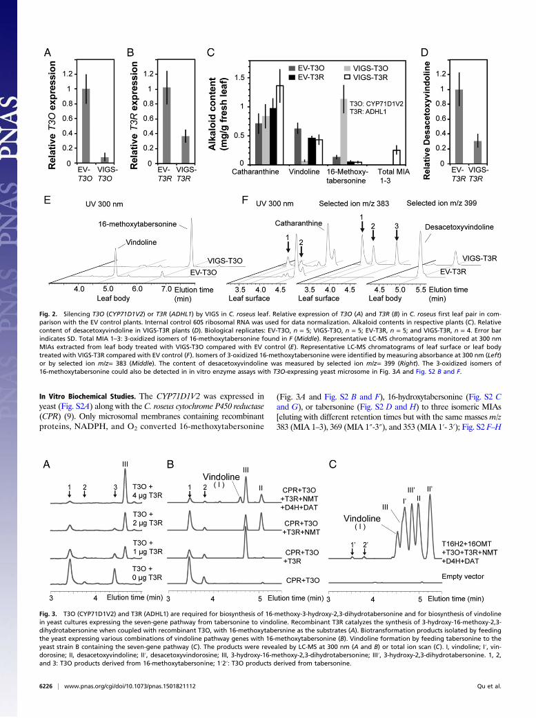
VIGS of Candidate Genes. VIGS of CYP71D1V2 and ADHL1 inC. roseus leaves was monitored by comparative metabolite profilingof MIAs secreted to the leaf surface (such as catharanthine) tothose found within the leaf body (such as vindoline and16-methoxytabersonine) (6, 7). qRT-PCR analyses showed thatplants could be silenced by an average of 92% for CYP71D1V2and by 63% for ADHL1 compared with the empty vector (EV)controls (Fig. 2 A and B). The 89% decline in vindoline and850% increase of 16-methoxytabersonine (m/z = 367) levelsobserved in CYP71D1V2-silenced plants compared with the EVcontrols (Fig. 2 C and E) suggest that 16-methoxytabersonine is alikely substrate of CYP71D1V2. Although the milder silencing ofADHL1 did not reduce vindoline levels (Fig. 2C), desacetoxy-vindoline, the N-methylated MIA produced immediately afterthe 3-hydroxylation step, was reduced accordingly by 69% (Fig. 2D and F). The leaf surface extracts of VIGS-ADHL1 plants alsoaccumulated three new unidentified isomeric MIAs (m/z = 383)that were not detected in the EV controls (Fig. 2F).
Fig. 1. Expression ratios of 7 C. roseus genes that convert tabersonine tovindoline in leaf epidermis compared with those in whole leaves. The tran-script abundance of selected genes in RNA extracts from young leaf epi-dermis and whole mature leaves was compared with those from wholeyoung leaves. Young leaf refers to LP-1 (youngest leaf pair on a vegetativemeristem), whereas manure leaf refers to LP-4 (fourth leaf pair from theyoungest leaf pair). Internal control 60S ribosomal RNA was used for datanormalization. Error bars indicate the SD from three technical replicates.
Qu et al. PNAS | May 12, 2015 | vol. 112 | no. 19 | 6225
PLANTBIOLO
GY
In Vitro Biochemical Studies. The CYP71D1V2 was expressed inyeast (Fig. S2A) along with the C. roseus cytochrome P450 reductase(CPR) (9). Only microsomal membranes containing recombinantproteins, NADPH, and O2 converted 16-methoxytabersonine
(Fig. 3A and Fig. S2 B and F), 16-hydroxytabersonine (Fig. S2 Cand G), or tabersonine (Fig. S2 D and H) to three isomeric MIAs[eluting with different retention times but with the same masses m/z383 (MIA 1–3), 369 (MIA 1″-3″), and 353 (MIA 1′- 3′); Fig. S2 F–H
Fig. 2. Silencing T3O (CYP71D1V2) or T3R (ADHL1) by VIGS in C. roseus leaf. Relative expression of T3O (A) and T3R (B) in C. roseus first leaf pair in com-parison with the EV control plants. Internal control 60S ribosomal RNA was used for data normalization. Alkaloid contents in respective plants (C). Relativecontent of desacetoxyvindoline in VIGS-T3R plants (D). Biological replicates: EV-T3O, n = 5; VIGS-T3O, n = 5; EV-T3R, n = 5; and VIGS-T3R, n = 4. Error barindicates SD. Total MIA 1–3: 3-oxidized isomers of 16-methoxytabersonine found in F (Middle). Representative LC-MS chromatograms monitored at 300 nmMIAs extracted from leaf body treated with VIGS-T3O compared with EV control (E). Representative LC-MS chromatograms of leaf surface or leaf bodytreated with VIGS-T3R compared with EV control (F). Isomers of 3-oxidized 16-methoxytabersonine were identified by measuring absorbance at 300 nm (Left)or by selected ion m/z= 383 (Middle). The content of desacetoxyvindoline was measured by selected ion m/z= 399 (Right). The 3-oxidized isomers of16-methoxytabersonine could also be detected in in vitro enzyme assays with T3O-expressing yeast microsome in Fig. 3A and Fig. S2 B and F.
Fig. 3. T3O (CYP71D1V2) and T3R (ADHL1) are required for biosynthesis of 16-methoxy-3-hydroxy-2,3-dihydrotabersonine and for biosynthesis of vindolinein yeast cultures expressing the seven-gene pathway from tabersonine to vindoline. Recombinant T3R catalyzes the synthesis of 3-hydroxy-16-methoxy-2,3-dihydrotabersonine when coupled with recombinant T3O, with 16-methoxytabersnine as the substrates (A). Biotransformation products isolated by feedingthe yeast expressing various combinations of vindoline pathway genes with 16-methoxytabersonine (B). Vindoline formation by feeding tabersonine to theyeast strain B containing the seven-gene pathway (C). The products were revealed by LC-MS at 300 nm (A and B) or total ion scan (C). I, vindoline; I′, vin-dorosine; II, desacetoxyvindoline; II′, desacetoxyvindorosine; III, 3-hydroxy-16-methoxy-2,3-dihydrotabersonine; III′, 3-hydroxy-2,3-dihydrotabersonine. 1, 2,and 3: T3O products derived from 16-methoxytabersonine; 1′2′: T3O products derived from tabersonine.
6226 | www.pnas.org/cgi/doi/10.1073/pnas.1501821112 Qu et al.

and Fig. S3]. The mass increase of 16 m/z obtained for each MIAsuggested that CYP71D1V2 catalyzed a monooxygenation re-action instead of the mass increase of 18 m/z that would be ex-pected to generate 2,3-dihydro-3-hydroxylatedMIAs. In comparison,2,3-dihydrotabersonine was not a substrate of CYP71D1V2 (Fig.S2E), suggesting monooxygenation only occurs across the 2,3-double bond. When the living yeast expressing CYP71D1V2 wasincubated with 16-methoxytabersonine, 16-hydroxytabersonine,or tabersonine, only MIA 1 and 2 (Fig. S4A), 1″ and 2″ (Fig.S4B), or 1′ and 2′ (Fig. S4C) could be isolated, suggesting MIA 3(Fig. S2F), 3″ (Fig. S2G), or 3′ (Fig. S2H) occurring in microsomalenzyme assays is not stable within live cells. Further analyses oftabersonine-derived MIA 1′ by NMR suggested it to be 2,3-epoxy-tabersonine (Fig. S5). This structure was further supported by thesodium borohydride reduction of MIA 1′ to the respective diol thathad the expected mass of 327 m/z, as measured by LC-MS. To-gether, these analyses also suggest that MIA 2′ may be the stereo-isomer of MIA 1′.The three isomeric MIAs (1–3, m/z = 383) identified in
CYP71D1V2 expressing yeast microsomal enzyme assays with16-methoxytabersonine (Fig. 3A and Fig. S2 B and F) showedidentical LC-MS profiles with the three MIAs (m/z = 383) foundon leaf surfaces of VIGS-ADHL1 plants (Fig. 2F). These resultssuggested that ADHL1might catalyze a subsequent reduction of theepoxides to form NMT substrate (Fig. S1). Although recombinantADHL1 purified from Escherichia coli (Fig. S6D) was not activewith either MIA 1 or 2 generated from CYP71D1V2-expressingyeast incubated with 16-methoxytabersonine (Fig. S6A, Left) ortabersonine (Fig. S6A, Right), ADHL1 assays containing all thethree isomeric MIAs produced in CYP71D1V2-expressing yeastmicrosomal enzyme assays with 16-methoxytabersonine (Fig.S6B, Left) or tabersonine (Fig. S6B, Right) yielded small amountsof respective 2,3-dihydro-3-hydroxylated MIAs with a slight de-cline in the levels of MIA 3 or 3′. When CYP71D1V2-containingmicrosomes were combined with different levels of recombinantADHL1 in the same reaction, the formation of the three isomericMIAs derived from 16-methoxytabersonine (Fig. 3A and Fig. S6C,Left) or tabersonine (Fig. S6C, Right) declined with increasing levelsof ADHL1 protein in favor of formation of large amounts of therespective 2,3-dihydro-3-hydroxylated MIAs. Substrate saturationkinetics was conducted with CYP71D1V2-containing microsomescombined with ADHL1 to give an apparent Km for tabersonine of3.1 μM (Fig. S7). On the basis of the results, CYP71D1V2 andADHL1 were renamed as T3O and T3R, respectively.
Assembly of Tabersonine-to-Vindoline Pathway in Yeast. To confirmthat the products of the T3O and T3R reactions are true in-termediates for the biosynthesis of vindoline and vindorosine,the tabersonine-to-vindoline pathway was assembled in yeast.Yeast strains coexpressing CPR, T3O, and T3R (Fig. S8A) con-verted 16-methoxytabersonine (Fig. 3B and Fig. S8B, Left) ortabersonine (Fig. S8B, Right) mostly into 2,3-dihydro-3-hydroxy-lated MIAs, whereas additional expression of NMT allowed thedetection of desacetoxyvindoline or desacetoxyvindorosine, andadditional expression of D4H and DAT (strain A) triggered theformation of vindoline (Fig. 3B and Fig. S8B, Left) or vindor-osine (Fig. S8B, Right). When yeast expressing all seven genes forthe vindoline pathway (addition of T16H2/16OMT and omissionof CPR, strain B) was fed with tabersonine, vindoline and vin-dorosine were clearly formed together with several other path-way intermediates (Fig. 3C).Under tested conditions, yeast strain A was able to convert
16-methoxytabersonine at 37 mg·L−1 12 h−1 or 8 mg·g dry weight(dwt)−1 12 h−1 rate and to produce vindoline at 2.7 mg·L−1 12 h−1
or 0.6 mg·g dwt−1 12 h−1 rate. Yeast strain B was able to converttabersonine at 17 mg·L−1 12 h−1 or 3.7 mg·g dwt−1 12 h−1 andto produce vindoline at 1.1 mg·L−1 12 h−1 or 0.24 mg·g dwt−1 12 h−1.In comparison, vindoline content in C. roseus total leaves is
2 mg·g dwt−1 or less (23). In addition, more than 95% of theproduced vindoline and other intermediates are secreted by yeastto the medium (Fig. S9) and are easily extracted with ethyl acetate.Therefore, the yeast system could be a powerful platform for in-dustrial production of vindoline from tabersonine.
DiscussionThe identification of T3O and T3R completes the molecular andbiochemical characterization of the only remaining unknownreactions in the tabersonine-to-vindoline pathway. It was shownthat saturation of T3O with T3R eliminated the accumulation ofepoxide byproduct in favor of 2,3-dihydro-3-hydroxylated MIAsand that a proposed reactive intermediate (Fig. 4) required forthe T3R reaction may spontaneously be converted into unusableepoxide derivatives in its absence. The formation of the 2,3-epoxide significantly alters the UV absorption from the longerwavelengths typical of the reaction substrates to those typical ofan indolenine moiety, whereas the UV absorption of MIA 3 and3′ resembles the profile of 3-hydroxylated MIA end products(Fig. S2). We therefore speculate that MIA 3 or 3′ may be theperoxide of the ring-opened 2,3-epoxide (Fig. 4), but it appearsas 2,3-epoxide isomers when analyzed by mass spectrometrybecause of the unstable nature of peroxide bond. Although theexact mechanism for this reaction remains to be discovered, it islikely that T3O and T3R catalyze the 3-hydroxylation step in aconcerted manner in planta.A related study (24) that appeared while this report was in
review showed that a vincamine-type MIA, rather than the
Fig. 4. Conversion of tabersonine to 3-hydroxy-2,3-dihydrotabersonine bythe combined action of T3O and T3R enzymes. In the absence of T3R, theproposed intermediate is spontaneously converted to byproduct MIAs 1′-3′.Reduction of MIA 1′ with sodium borohydride yields the expected diol.
Qu et al. PNAS | May 12, 2015 | vol. 112 | no. 19 | 6227
PLANTBIOLO
GY

2,3-epoxide, was detected when a T3O-expressing yeast strainwas cultured in the presence of 16-methoxytabsersonine. This led tothe suggestion by these authors that the 2,3-epoxide can undergorearrangement to yield the vincamine–eburnamine backbone inthe absence of T3H and suggesting a biosynthetic relationshipbetween the aspidosperma- and eburnamine-type MIAs. Thepresent study shows, in contrast, that the 2,3-epoxide is clearlydetected within VIGS-T3R-silenced plants as well as in T3O-expressing cultured yeast and in microsomal assays when taber-sonine, 16-hydroxytabersonine, or 16-methoxytabersonine weresupplied as MIA substrates. The proposed rearrangement in ref.24 is likely to be chemical and well-known to occur, dependingon the acidity of the growth medium or alkaloid extractionconditions, and occurs when the aspidosperma nucleus is in theindolenine oxidation state (25–27).Previous biochemical localization studies involving organ-
elle isolation by sucrose gradient centrifugation followed byfractionation suggested that NMT activity was associated withCatharanthus chloroplasts (19). In addition, the phylogeneticrelationship of the NMT gene to nuclear-encoded but chloro-plast-associated tocopherol C-methyltransferases (18) furtherimplied that the NMT and possibly “hydratase” reactions mightbe associated leaf mesophyll chloroplasts. The preferential en-richment of T16H2, 16OMT, NMT, T3O, and T3R transcripts inleaf epidermis compared with whole leaf tissues (Fig. 1) and theexclusive and restricted detection of T3O-derived MIA products(Fig. 3A) to leaf surfaces of VIGS-T3R plants (Fig. 2F) impliesthat both reactions take place in the epidermis of Catharanthus
leaves. In contrast to the leaf surface accumulation of T3O-derived MIA products (Fig. 2F), 16-methoxytabersonine was onlydetected in the leaf body (Fig. 2E). This raises questions aboutthe different possible biological mechanisms that might accountfor the results obtained. This study further suggests that the MIAtransported to idioblasts or laticifers for elaboration by D4H andDAT into vindoline is desacetoxyvindoline (Fig. 5). Taking intoaccount this new information, it is possible that NMT is associ-ated with plastids in the leaf epidermis, but further studies arerequired to determine this.As the model plant most studied for MIA biosynthesis,
C. roseus revealed a fascinating picture of cell-type specificcompartmentation of different parts of the vindoline pathwaythat would require movement of different biosynthetic inter-mediates for its assembly (Fig. 5). Remarkably, specialized IPAPcells preferentially express the plastid-specific methylerythritolphosphate pathway to supply geraniol for elaboration into loganicacid via seven key biochemical reactions. Loganic acid must thenbe transported by an uncharacterized process to the leaf epider-mis, where it is converted to secologanin (3, 4, 8–13). Leaf epi-dermal cells preferentially express tryptophan decarboxylasetogether with all MIA pathway genes leading to biosynthesis ofcatharanthine, desacetoxyvindorosine, and desacetoxyvindoline.At this point, catharanthine is secreted via CrTPT2-mediatedtransport (6, 7) and accumulated on the leaf surface, and desa-cetoxyvindorosine and desacetoxyvindoline are transported byan unidentified process to idioblasts or laticifers for a two-stepelaboration to form vindorosine and vindoline. The completion
Fig. 5. Cellular compartmentation of iridoid and MIA biosynthesis. 7DLS, 7-deoxyloganetic acid synthase; 10HGO, 10-hydroxygeraniol oxidoreductase; DL7H,7-deoxyloganic acid 7-hydroxylase; DLGT, 7-deoxyloganetic acid glucosyltransferase; G10H, geraniol 10-hydroxylase; IS, iridoid synthase; SGD, strictosidineβ-glucosidase; SLS, secologanin synthase; STR, strictosidine synthase; TDC, tryptophan decarboxylase.
6228 | www.pnas.org/cgi/doi/10.1073/pnas.1501821112 Qu et al.

of the tabersonine-to-vindoline pathway from this and otherstudies (5, 16–18, 20, 21) highlights the complex nature of plantsecondary metabolism.The assembly of the seven-gene tabersonine-to-vindoline path-
way in yeast for vindoline production demonstrates the feasibilityof microbial MIA production. The discrepancy between thetabersonine consumption and the final vindoline yield in yeastshowed great potential for further production optimization. Theresults suggested that T16H, NMT, and D4H activities might bebottlenecks that limit flux, as N-desmethy- and N-methyl inter-mediates mainly accumulated in strain B (Fig. 3C). Further geneexpression optimization on these three enzymes may significantlyincrease vindoline yield to achieve an industrial strain superior tothe plant. In the future, completion of strictosidine-to-taber-sonine/catharanthine branch pathways may lead to the total syn-thesis of plant-derived MIA in controlled microbial systems.
Materials and MethodsVIGS. Seeds of C. roseus cv. Little Delicata were sowed and grown in agreenhouse at 28 °C, 16/8 h photoperiod. Four-week-old seedlings with twoto three pairs of true leaves were used for VIGS as described (28), withmodifications. Cells of an overnight culture of Agrobacterium tumefacienscarrying pTRV1 (tobacco rattle virus) or various pTRV2 constructs wereresuspended in infection buffer (10 mM Mes at pH 5.6, 10 mM MgCl2,200 μM acetosyringone) to an OD600 1.5, and incubated at 28 °C for 2 h. Thesuspension of pTRV1 and pTRV2 strains were mixed in equal volume justbefore the infection. The stem of the seedling was poked with a steriletoothpick just underneath the apical meristem, and 150 μL infection mixturewas flooded over the wound. After infection, the plants were transferred toa greenhouse at 20 °C, 16/8 h photoperiod. VIGS-phytoene desaturase wasused to indicate the silencing effect (9).
Leaf Alkaloid Extraction. The first leaf pair (LP-1)were collected and split in halfalong the vertical axis 25–30 d after infection. One half of the LP-1 werefrozen in liquid nitrogen for RNA extraction, and the other half wereweighed (10–30 mg) and submerged in 20 times chloroform (wt/vol) for 1 min
to generate leaf surface extraction (6). The leaf material was thereaftertransferred to 20 times methanol (wt/vol) for extraction on ice for 1 h togenerate the leaf body extraction.
Yeast Biotransformation Assays. A single yeast [BY47471 (MATα his3Δ1 leu2Δ0met15Δ0 ura3Δ0 YPL154c::kanMX4) (29)] colony carrying various constructswas inoculated in 2 mL synthetic complementary (SC) medium with appro-priate amino acids dropped out, supplemented with 2% (wt/vol) glucoseovernight at 30 °C. Cells were harvested and inoculated in SC medium with2% (wt/vol) galactose and induced at 30 °C for 24 h. The cells were harvestedand suspended in 1 mL biotransformation buffer (10 mM Tris·HCl at pH 7.5,1 mM EDTA) with 20 μM substrates and incubated at 30 °C for 24 h. Fortabersonine conversion quantification, 225 μM tabersonine was used. Basifiedbuffer was extracted with ethyl acetate, dried, and redissolved in methanolfor LC-MS analysis. For large-scale product purification, tabersonine (50 mg)was fed to 2 L yeast expressing T3O and CPR for 12 h. The extracted productwas harvested from thin-layer chromatograph sheets (Silica gel 60 F254) withsolvent toluene: ethyl acetate: methanol (15:4:1, vol/vol). About 8 mg MIA 1′was subjected to further NMR and reduction analysis.
In Vitro Assay. The assay (100 μL) contains 50 mM Hepes at pH 7.5, 1 mMNADPH, 25 μM substrate (tabersonine, 16-hydroxytabersonine 16-methoxy-tabersonine, 2,3-dihydrotabersonine, or reaction products of T3O), 50 μgmicrosomal protein, and/or various amount of recombinant T3R. The re-action took place at 30 °C for 1 h. Ethyl acetate extracted products wereredissolved in methanol for LC-MS analysis. The kinetics assays (100 μL)contain 50 mM Hepes at pH 7.5, 0.25 mM NADPH, 25 μg microsomal proteins(T3O), 2 μg purified recombinant T3R, and various amount of tabersonine.The reaction took place at 30 °C, and the velocity was calculated as theconsumption of NADPH measured at OD340.
ACKNOWLEDGMENTS. We thank Dr. Dae-Kyun Ro from the University ofCalgary for providing the yeast strain and expression vectors. This work wassupported by a Natural Sciences and Engineering Research Council ofCanada Discovery Grant (to V.D.L.), Canada Research Chairs (to V.D.L.), andthe Canada Foundation for Innovation and the Ontario Ministry of Researchand Innovation.
1. De Luca V, Salim V, Thamm A, Masada SA, Yu F (2014) Making iridoids/secoiridoidsand monoterpenoid indole alkaloids: Progress on pathway elucidation. Curr OpinPlant Biol 19:35–42.
2. De Luca V, Salim V, Atsumi SM, Yu F (2012) Mining the biodiversity of plants: Arevolution in the making. Science 336(6089):1658–1661.
3. St-Pierre B, Vazquez-Flota FA, De Luca V (1999) Multicellular compartmentation ofCatharanthus roseus alkaloid biosynthesis predicts intercellular translocation of apathway intermediate. Plant Cell 11(5):887–900.
4. Murata J, Roepke J, Gordon H, De Luca V (2008) The leaf epidermome of Cathar-anthus roseus reveals its biochemical specialization. Plant Cell 20(3):524–542.
5. Murata J, De Luca V (2005) Localization of tabersonine 16-hydroxylase and 16-OHtabersonine-16-O-methyltransferase to leaf epidermal cells defines them as a majorsite of precursor biosynthesis in the vindoline pathway in Catharanthus roseus. Plant J44(4):581–594.
6. Roepke J, et al. (2010) Vinca drug components accumulate exclusively in leaf exudatesof Madagascar periwinkle. Proc Natl Acad Sci USA 107(34):15287–15292.
7. Yu F, De Luca V (2013) ATP-binding cassette transporter controls leaf surface secretionof anticancer drug components in Catharanthus roseus. Proc Natl Acad Sci USA110(39):15830–15835.
8. Geu-Flores F, et al. (2012) An alternative route to cyclic terpenes by reductive cycli-zation in iridoid biosynthesis. Nature 492(7427):138–142.
9. Salim V, Yu F, Altarejos J, De Luca V (2013) Virus-induced gene silencing identifiesCatharanthus roseus 7-deoxyloganic acid-7-hydroxylase, a step in iridoid and mono-terpene indole alkaloid biosynthesis. Plant J 76(5):754–765.
10. Asada K, et al. (2013) A 7-deoxyloganetic acid glucosyltransferase contributes a keystep in secologanin biosynthesis in Madagascar periwinkle. Plant Cell 25(10):4123–4134.
11. Salim V, Wiens B, Masada-Atsumi S, Yu F, De Luca V (2014) 7-deoxyloganetic acidsynthase catalyzes a key 3 step oxidation to form 7-deoxyloganetic acid in Cathar-anthus roseus iridoid biosynthesis. Phytochemistry 101:23–31.
12. Miettinen K, et al. (2014) The seco-iridoid pathway from Catharanthus roseus. NatCommun 5:3606.
13. Irmler S, et al. (2000) Indole alkaloid biosynthesis in Catharanthus roseus: New en-zyme activities and identification of cytochrome P450 CYP72A1 as secologanin syn-thase. Plant J 24(6):797–804.
14. Treimer JF, Zenk MH (1979) Purification and properties of strictosidine synthase, thekey enzyme in indole alkaloid formation. Eur J Biochem 101(1):225–233.
15. Geerlings A, Ibañez MM, Memelink J, van Der Heijden R, Verpoorte R (2000) Mo-lecular cloning and analysis of strictosidine beta-D-glucosidase, an enzyme in terpe-noid indole alkaloid biosynthesis in Catharanthus roseus. J Biol Chem 275(5):3051–3056.
16. Levac D, Murata J, Kim WS, De Luca V (2008) Application of carborundum abrasionfor investigating the leaf epidermis: Molecular cloning of Catharanthus roseus16-hydroxytabersonine-16-O-methyltransferase. Plant J 53(2):225–236.
17. Besseau S, et al. (2013) A pair of tabersonine 16-hydroxylases initiates the synthesis ofvindoline in an organ-dependent manner in Catharanthus roseus. Plant Physiol163(4):1792–1803.
18. Liscombe DK, Usera AR, O’Connor SE (2010) Homolog of tocopherol C methyl-transferases catalyzes N methylation in anticancer alkaloid biosynthesis. Proc NatlAcad Sci USA 107(44):18793–18798.
19. Deluca V, Balsevich J, Tyler RT, Kurz WG (1987) Characterization of a novel N-meth-yltransferase (NMT) from Catharanthus roseus plants : Detection of NMT and otherenzymes of the indole alkaloid biosynthetic pathway in different cell suspensionculture systems. Plant Cell Rep 6(6):458–461.
20. St-Pierre B, Laflamme P, Alarco AM, De Luca V (1998) The terminal O-acetyl-transferase involved in vindoline biosynthesis defines a new class of proteins re-sponsible for coenzyme A-dependent acyl transfer. Plant J 14(6):703–713.
21. Vazquez-Flota F, De Carolis E, Alarco AM, De Luca V (1997) Molecular cloning andcharacterization of desacetoxyvindoline-4-hydroxylase, a 2-oxoglutarate dependent-dioxygenase involved in the biosynthesis of vindoline in Catharanthus roseus (L.) G.Don. Plant Mol Biol 34(6):935–948.
22. Huang L, et al. (2012) Molecular characterization of the pentacyclic triterpenoidbiosynthetic pathway in Catharanthus roseus. Planta 236(5):1571–1581.
23. Chung I-M, et al. (2011) Screening 64 cultivars Catharanthus roseus for the productionof vindoline, catharanthine, and serpentine. Biotechnol Prog 27(4):937–943.
24. Kellner F, et al. (March 19, 2015) Discovery of a P450-catalyzed step in vindolinebiosynthesis: A link between the aspidosperma and eburnamine alkaloids. ChemCommun (Camb), 10.1039/C5CC01309G.
25. Wenkert E (1962) Biosynthesis of indole alkaloids. The aspidosperma and Iboga bases.J Am Chem Soc 84(1):98–102.
26. Wenkert E, Wickberg B (1965) General methods of synthesis of indole alkaloids. IV. Asynthesis of dl-eburnamonine. J Am Chem Soc 87:1580–1589.
27. Barton JED, Harley-Mason J (1965) Total synthesis of Hunteria and Aspidospermaalkaloids from a common intermediate. Chem Commun (London) 10:197–198.
28. Liscombe DK, O’Connor SE (2011) A virus-induced gene silencing approach to un-derstanding alkaloid metabolism in Catharanthus roseus. Phytochemistry 72(16):1969–1977.
29. Parr CL, Keates RAB, Bryksa BC, Ogawa M, Yada RY (2007) The structure and functionof Saccharomyces cerevisiae proteinase A. Yeast 24(6):467–480.
Qu et al. PNAS | May 12, 2015 | vol. 112 | no. 19 | 6229
PLANTBIOLO
GY