comparison of two methods for detecting intrathecal ...345841/fulltext01.pdf · degree project work...
TRANSCRIPT

School of Natural Sciences
Degree project work
Stephanie Holmqvist
Subject: Biomedical Laboratory Science
Level: First level
Nr: 2010:BL1
Comparison of Two Methods for Detecting
Intrathecal Synthesis of Borrelia Specific
Antibodies

Comparison of Two Methods for Detecting Intrathecal Synthesis of
Borrelia Specific Antibodies Stephanie Holmqvist
Examination project work in Biomedical Laboratory Science, 15 credits
Biomedical Laboratory Science Programme 180 credits
Linnaeus University
Kalmar Växjö
Supervisors Maria Nilsson Dept. of Clinical Microbiology
Ph D. Blekingesjukhuset
SE-371 85 KARLSKRONA
Inger Edfors School of Natural Sciences
Assoc. Professor Linnæus University
SE-391 82 KALMAR
Examiner
Britt-Inger Marklund School of Natural Sciences
Ph D. Linnæus University
SE-391 82 KALMAR
Abstract
In Europe, Lyme disease is caused by the species Borrelia (B.) burgdorferi sensu stricto, B.
garinii and B. afzelii. The disease is the most common vector-borne infection in Europe and
the United States, and the resulting manifestation can involve the skin, nervous system,
heart and joints. The symptoms that arise are associated with the Borrelia species causing
the infection. The species most associated with neuroborreliosis is B. garinii whilst B.
burgdorferi sensu stricto is associated with arthritis and B. afzelii is associated with
dermatological symptoms. Lyme disease normally has three phases in untreated patients.
The first phase is characterised by erythema migrans, a reddening of the skin around the
area of the tick bite. If the disease develops to the second phase the patient will suffer from
neuroborreliosis which is characterised by neurological symptoms such as headache and
peripheral facial paralysis. Cerebrospinal fluid (CSF) analysis is used to diagnose
neuroborreliosis. The diagnosis is complicated by variations between the different Borrelia
species and that many healthy individuals have antibodies directed against Borrelia.
Antibodies in CSF can be found in different diseases. The antibodies can be produced in the
central nervous system or come across the blood-brain barrier and thus derive originally
from the blood. By measuring the concentration of total albumin in serum and in CSF it can
be determined if the antibodies present in the CSF have been produced in the central
nervous system or if they originate from the blood. The typical manifestation in the last
phase of Lyme disease is severe arthritis. The aim of this examination project was to
compare two ELISAs for detection of antibodies directed to Borrelia. Indirect ELISAs from
DAKO and Euroimmun were compared for the diagnosis of neuroborreliosis in 100
individuals. Borrelia specific antibodies of class IgM or IgG were found in 16 of 100
patients by DAKO’s ELISA and in 20 of the same 100 patients by Euroimmun’s ELISA.
The reason that Euroimmun’s method detected more cases of neuroborreliosis is probably
that this method detects antibodies directed to all three pathological species of Borrelia
while DAKO’s method only detects antibodies directed to B. burgdorferi. In conclusion,
this study indicates that Euroimmun’s method to detect antibodies of class IgM and IgG
directed to Borrelia is superior to DAKO’s method. The obtained results were confirmed by
Western blot analysis which gave results in accordance with those of Euroimmun’s ELISA.

SUMMARY IN SWEDISH
Borrelios är en sjukdom som orsakas av bakterier av genuset Borrelia vilka överförs till
människor med hjälp av fästingar. I Europa finns det tre arter av Borrelia som är patogena för
människan och dessa är Borrelia burgdorferi sensu stricto, Borrelia garinii och Borrelia
afzelii. Studier har visat att symptomen som uppstår vid borrelios beror på vilken Borrelia-art
en människa blir infekterad med. Neuroborrelios, som drabbar 16% av alla patienter med en
diagnostiserad borrelios i Sverige, orsakas framförallt av Borrelia garinii medan Borrelia
burgdorferi sensu stricto och Borrelia afzelii är associerade med artrit respektive hudsymtom.
Borrelios karaktäriseras av tre faser där den första fasen kännetecknas av ett rött hudutslag, ett
så kallat erythema migrans, vid fästingbettet. Därefter kan sjukdomen utvecklas och övergå i
den andra fasen vilken karaktäriseras av neuroborrelios vilken ger upphov till neurologiska
symptom. Denna sjukdom diagnostiseras genom detektion av antikroppar som är riktade mot
Borrelia i serum samt i cerebrospinalvätska (CSF). Diagnosen försvåras av variation mellan
de olika Borrelia-arterna samt att många friska individer har antikroppar riktade mot Borrelia.
Antikroppar i CSF kan detekteras då B-celler infiltrerat det centrala nervsystemet vilket sker
vid olika sjukdomssymtom eller då antikropparna har kommit över blodhjärnbarriären och
härstammar således ursprungligen från blodet. Genom att mäta den totala albuminhalten i
serum samt i CSF kan det upptäckas om de antikroppar som finns i CSF har bildats intratekalt
eller om de härstammar från blodet. Den tredje fasen vid borrelios utgörs av en allvarlig artrit.
Syftet med examensarbetet var att jämföra två metoder för detektion av antikroppar av IgM-
och IgG-klass riktade mot Borrelia-antigen i CSF och serum. En indirekt enzyme linked
immunosorbent assay (ELISA) för detektion av Borrelia-antikroppar från företaget DAKO
och en från Euroimmun användes för att analysera prover från 100 patienter med en misstänkt
neuroborrelios. Av 100 patienter, kunde Borrelia-specifika antikroppar av IgM- eller IgG-
klass detekteras hos 16 patienter med DAKO:s metod medan 20 patienter blev positiva för
neuroborrelios med Euroimmun:s metod. Resultaten från studien talar för att Euroimmuns
metod detekterar fler fall av neuroborrelios. Detta beror troligtvis på att denna metod kan
detektera antikroppar mot samtliga tre patogena Borrelia-arter. Att DAKO:s metod inte kan
detektera antikroppar mot Borrelia garinii tros vara en orsak till att den ger falskt negativa
resultat då denna bakterieart har visat sig vara den mest sjukdomsframkallande vid
neuroborrelios.

CONTENTS INTRODUCTION ................................................................................................................................................ 1
BORRELIA ............................................................................................................................................................... 1
LYME DISEASE ......................................................................................................................................................... 1
IMMUNE RESPONSE TO BORRELIA BURGDORFERI ............................................................................................................ 2
SEROLOGIC TESTS ..................................................................................................................................................... 3
Enzyme Linked Immunosorbent Assay (ELISA) ............................................................................................. 3
Western Blot ................................................................................................................................................... 4
CEREBROSPINAL FLUID VARIABLES ............................................................................................................................... 4
THE AIM OF THE STUDY ............................................................................................................................................. 5
MATERIALS AND METHODS ............................................................................................................................. 6
PATIENTS AND SAMPLES ............................................................................................................................................ 6
EUROIMMUN’S ELISA – ANTIBODIES OF THE IGM AND IGG CLASS AGAINST BORRELIA ANTIGENS IN CSF AND SERUM ................. 6
TOTAL IGM, IGG AND ALBUMIN IN CSF AND SERUM ...................................................................................................... 7
CEREBROSPINAL FLUID VARIABLES ............................................................................................................................... 7
DAKO’S IDEIATM
LYME NEUROBORRELIOSIS ................................................................................................................. 8
EUROIMMUN’S WESTERN BLOT – BORRELIA IGM AND BORRELIA IGG ............................................................................... 9
EUROIMMUN’S ELISA – BORRELIA IGM AND BORRELIA IGG.......................................................................................... 10
RESULTS ......................................................................................................................................................... 11
DISCUSSION ................................................................................................................................................... 14
ACKNOWLEDGEMENTS .................................................................................................................................. 18
REFERENCES ................................................................................................................................................... 19

ABBREVATION LIST
ACA: Acrodermatitis chronica atrophicans
AI: Antibody Index
B.: Borrelia
CNS: Central Nervous System
CSF: Cerebrospinal Fluid
ELISA: Enzyme Linked Immunosorbent Assay
IFA: Immunofluorescence assay
IgA: Immunoglobulin A
IgG: Immunoglobulin G
IgM: Immunoglobulin M
NBT/BCIP: Nitro Blue Tetrazolium-Chloride-5-Bromo-4-Chloro-3-Indolyl-Phosphate
O.D.: Optical Density
OspC: Outer Surface Protein C
RU: Relative Units
SDS-PAG: Sodium Dodecyl Sulphate Polyacrylamide Gel
TMB: Tetrametylbenzidine
VlsE: Variable major protein-like sequence, expressed

1
INTRODUCTION
Borrelia
The bacterium Borrelia is a helical-shaped bacterium which affects humans by causing Lyme
disease or relapsing fever (1). Bacteria in the genus Borrelia are about 8-30 µm long and 0.2-
0.5 µm wide (2). Borrelia is mainly transmitted to humans by ticks. A tick in the genus Ixodes
can live for two years and during these two years they go through three stages of
development. The first stage in the tick’s life is when it is a larva with six legs. Then the larva
becomes a nymph with eight legs and finally the tick becomes an eight-legged adult. An
Ixodes tick attaches to an animal host during each stage of its lifecycle and the ticks feed on
different hosts during different stages. If the larva is infected after its first blood meal the
Borrelia in the tick’s gut replicates (1). The replication of Borrelia in the tick’s gut takes 8-12
hours during log-phase (3). After the replication, the spirochetes penetrate the midgut and
invade the hemolymph. From the hemolymph they can spread to the salivary glands (2).
When the larva develops to a nymph and feeds a second time the Borrelia is transmitted via
saliva to the new host and the host becomes infected. However, an uninfected nymph can in
this stage also be infected if it feeds on an infected host. The nymphs then fall off and develop
into adults which feed a last time (1). Thereafter, the female adult ticks mate and lay
approximately 2000 eggs and finally they die (3). The transmission of the bacteria in the
genus Borrelia usually occurs after the first 24-48 hours of feeding and the numbers of
infected mammals are dramatically increased with a prolonged tick feeding (2).
Lyme disease
In Europe, Lyme disease is caused by the species Borrelia (B.) burgdorferi sensu stricto, B.
garinii and B. afzelii while B. burgdorferi sensu stricto is the only species that causes Lyme
disease in the United States (3). This disease is the most common vector-borne infection in
temperate areas of the northern hemisphere, i.e. Europe and the United States, and it can be
manifested by a variety of symptoms (4). The symptoms can for example be neurological and
dermatological. The disease has normally three phases in untreated patients. The first phase is
characterised by a reddening of the skin around the area of the tick bite. This red rash, also
called erythema migrans, expands and occurs within 30 days and is the most frequently
observed symptom (77%) in Sweden (1, 4). Together with the red rash, patients in stage one
can also have influenza-like symptoms with headache, severe fatigue, fever, vomiting and
muscle and joint pain (1). This phase can heal spontaneously, but 15% of the untreated cases
develop to phase two of the Lyme disease (3). Among all patients in stage one, 50-90% have
IgM antibodies in serum directed to B. burgdorferi that can be detected serologically (5).
Phase two is characterised by neurological manifestations such as meningitis, encephalitis and
cranial nerve pareses (1). These symptoms are referred to as neuroborreliosis which is the
second most frequent clinical manifestation and occurs in 16% of all patients with a
diagnosed Lyme disease in Sweden (4). Shortly after a tick bite, weeks to months, a patient
with neuroborreliosis receives subclinical meningitis symptoms, for example headache and
neck pain, together with non-specific symptoms such as malaise, fatigue, and arthralgia. In the
later stage of neuroborreliosis more typical symptoms arise such as facial nerve palsy and

2
cranial nerve palsy (3). Involvement of the nervous system is the predominant result of
morbidity caused by Borrelia in Europe (6). Patients in this stage can also suffer from
arthritis, especially in the knee joints, or, more rarely, by cardiac dysfunction. The typical
manifestation in the last phase is severe arthritis (1).
Lyme disease distribution is associated with the distribution of the hard tick Ixodes. In Europe
it is primarily the tick Ixodes ricinus that favours the spread of the disease. In the central
regions and southern coastal areas of Sweden, Ixodes ricinus is especially abundant and 5-
26% of these ticks carry Borrelia (6). This makes Lyme disease an endemic disease in those
areas with a frequency of 69 cases per 100 000 residents per year (4). Studies have also shown
that patients have different clinical manifestations dependent on which Borrelia species
causes the infection. The species most associated with neuroborreliosis is B. garinii (6).
However, all three pathogenic species have been identified in patients with neuroborreliosis
(7). The species B. burgdorferi sensu stricto is associated with arthritis and B. afzelii is
associated with acrodermatitis chronica atrophicans (ACA) (6). The clinical manifestation is
also dependent on where the tick has bitten the patient. Patients who were bitten in the neck or
head area had significantly more neurological manifestations than patients who were bitten in
other parts of the body (4).
A diagnosis of Lyme disease is made by serological testing and not by detecting Borrelia in
blood smears. The diagnosis also takes into account the patient’s symptoms and tick bite.
Patients in phase one are usually treated with the antibiotics doxycycline or penicillin (1).
However, the dosage and duration of the chosen antibiotic are important for the treatment of
the disease (2). The treatment is very effective but must last for a long time and often in large
doses to be successful. To treat the later phases of Lyme disease is harder since these
symptoms often are caused by immune responses and not by the Borrelia bacterium itself.
The earlier a treatment begins, the more it prevents later illness (1). If the disease is severe or
if the patient has neurological manifestation, an intravenous treatment with penicillin G or
ceftriaxone can be necessary. (2)
Immune response to Borrelia burgdorferi
An immune response starts when the spirochetes invade the body. Early in the infections the
T cells are activated while the B cells’ response is slower. The first antibodies the plasma cells
produce are IgM. This IgM response can usually be detected 3-4 weeks after the onset of
infection and after 6-8 weeks the IgM response decreases. In the beginning of the infection
the IgM antibodies are usually directed against the outer surface protein C (OspC) and the
flagella-associated antigen. The humoral immune response to other Borrelia antigens
gradually develops as the disease is proceeding. Later in the infection, typically 2-3 months
after the onset of infection, specific IgG and IgA antibodies are produced. These types of
antibodies are thereafter detectable for several years. However, patients treated before the IgG
response has developed lack Borrelia-specific antibodies in diagnostics levels (8).

3
Enzyme Linked Immunosorbent Assay (ELISA) is a method that can be used to measure
antibodies produced during the immune response. Due to cross-reactions with antibodies
specific for the flagellin antigen; which can be bound to other spirochaetaceae and bacteria
having flagella, genetic diversity of the Borrelia species and slow immune response in the
early stage of the disease, serological analysis is difficult to interpret. Another problem which
makes the interpretation of serological analysis more difficult is that 8-14% of healthy
individuals from Kalmar have antibodies against Borrelia in their serum (9).
Serologic tests
In contrast to most infections caused by bacteria, the diagnosis of Borrelia is dependent on
indirect methods and can rarely be confirmed by Polymerase Chain Reaction (PCR) or
cultivation (8). However, presence of erythema migrans is enough to diagnose Lyme disease
(3). The most generally used indirect methods are Enzyme Linked Immunosorbent Assay
(ELISA) and indirect immunofluorescence assay (IFA). An advantage with the ELISA is its
ability to analyse a large number of patient samples every day (8).
To distinguish between inactive and active infections is difficult because the immune
response in serum and cerebrospinal fluid can persist for several years. Another disadvantage
with serological tests is serological cross-reactivity, which occurs typically in IgM analysis.
Cross-reactivity often arises in patients with rheumatoid arthritis or other autoimmune
diseases (3). This problem can be overcome if IgG antibodies, which have bound to
rheumatoid factors, are removed, for example by ultracentrifugation. This prevents false
positive IgM test results. Removal of IgG also prevents that IgG displaces IgM from the
antigen which would lead to false negative IgM test results (10).
Enzyme Linked Immunosorbent Assay (ELISA)
Enzyme Linked Immunosorbent Assay (ELISA) is a method for a qualitative detection and a
quantitative measurement of antibodies or antigens. There are different types of ELISA such
as indirect ELISA, sandwich ELISA and competitive ELISA, but all types can be used to
analyse presence or absence of antibodies or antigens. Besides qualitative detection, ELISA
can also be used for quantitative detection with the aid of known concentrations of a
substance. With these calibrators, a standard curve is constructed and an unknown sample
concentration can be determined. An indirect ELISA is often used when antibodies are
detected but antigens will mostly be detected with a sandwich ELISA or a competitive
ELISA. The principle of the ELISA is that an antibody, which is conjugated with an enzyme,
is used. The most commonly used enzymes are alkaline phosphatase and horseradish
peroxidase. A colourless material, which is chromogenic, is then added to the enzyme-
conjugated antibody and a reaction product, which is coloured, is formed. When an indirect
ELISA is used, serum and cerebrospinal fluid, with possible antibodies, is added to a
microtiter well which is coated with antigens (Figure 1). Specific antibodies react and
unbound antibodies are washed away. In order to detect bound antibodies a second, enzyme-
conjugated, antibody is added. This secondary antibody will bind to the primary antibody and

4
unbound antibodies are washed away. Thereafter a substrate is added and a coloured reaction
product is generated. The amount of reaction product is measured by a spectrophotometric
plate reader (11).
Figure 1: The principle of an indirect ELISA. To a well coated with antigen, samples with specific antibodies
are added. The unbound antibodies are washed away before the enzyme-conjugated secondary antibodies are
added. The secondary antibodies will bind to the primary antibodies. Thereafter the unbound antibodies are
washed away. Finally substrate is added and a coloured reaction product is generated which can be measured by
a spectrophotometric plate reader. The picture is modified from Kindt J.T., Goldsby, A.R., Osborne, A.B.,
(2007), Kuby Immunology, 6th
edition, W.H. Freeman and Company.
Western Blot
Western Blot is an analytical technique where a specific protein can be identified in a
complex mixture of proteins. First, the protein mixture is separated by electrophoresis on a
sodium dodecyl sulphate polyacrylamide gel (SDS-PAG) (11). Thereafter the separated
proteins on the gel are transferred onto a nitrocellulose membrane through a process called
electroblotting. The blot, which is another name for the nitrocellulose membrane with its
transferred proteins, is thereafter incubated in a protein solution. This solution will block all
remaining binding sites on the membrane. The blot is incubated again but now the blot is
incubated in a dilution of antiserum (primary antibody). These primary antibodies are directed
against the protein of interest. Subsequently, a new incubation in a solution with a secondary
antibody occurs. In order to visualise this interaction the secondary antibody is labelled with
for example an enzyme. A chromogenic substrate is thereafter added which produces an
insoluble coloured product. This product is precipitated onto the membrane and a coloured
band is visualised at the site of the target antigen, that is to say, the protein of interest (12).
Cerebrospinal fluid variables
Normally, cerebrospinal fluid (CSF) analysis is used to diagnose neurological diseases such as
neuroborreliosis which affect the central and peripheral nervous system (13). The immune
response in the central nervous system (CNS) lacks the switch from IgM class response to
IgG class response. This pattern is in contrast to the immune response which occurs in serum
where IgM switches to IgG in patients with an infection. Due to this, detection of IgM in CSF
is not an indication of an early infection because IgM levels can persist just like the IgG for
many years (14).

5
Different neurological diseases have different patterns of intrathecal synthesis of antibodies of
class IgG, IgM and IgA (13). The intrathecal production of antibodies in the central nervous
system derives from perivascular infiltrates of B cells that mature and proliferate locally. The
typically initial immune response pattern in the central nervous system in patients with a
neuroborreliosis is intrathecal production of IgG, IgM and IgA with a predominance of IgM.
These patients have also, normally, a lymphocytic pleocytosis in the CSF and a blood-brain
barrier dysfunction (14). These characteristics together give 70% diagnostics sensitivity and
98% diagnostic specificity for detection of neuroborreliosis. However, if these factors are
combined with cerebrospinal fluid variables the highest sensitivity and specificity will be
obtained (80% and 98% respectively) (13).
The concentration of proteins in CSF is affected by several factors such as CSF flow rate,
protein diffusion into CSF, and the blood-brain barrier. All blood proteins in CSF have been
transported over the blood-brain barrier. One way to come across the blood-brain barrier is by
passive diffusion. Smaller molecules, like albumin and IgG, are faster in exchange than the
larger molecule IgM. The factor that modulates the definite concentration in the CSF is the
CSF flow rate. An elevated protein concentration in the CSF will be seen when the CSF flow
rate is reduced, i.e. a blood-brain dysfunction is due to decreased CSF flow rate (14).
The aim of the study
Due to the diversity of symptoms indicating Lyme disease it is important to have reliable
analytical methods. The currently used method in routine diagnostics at the Department of
Clinical Microbiology at Blekingesjukhuset, Karlskrona, (DAKO, Cambridgeshire, United
Kingdom) can only detect IgG and IgM antibodies directed to B. burgdorferi. However,
several other Borrelia species can cause neuroborreliosis, and it is therefore possible that this
method gives false negative test results. A newly developed method provided by Euroimmun
(Lübeck, Germany), detects IgG and IgM directed to all Borrelia species occurring in Europe.
Furthermore, it also corrects for antibodies in the CSF caused by a blood-brain dysfunction.
The aim of this study was to compare these two methods, DAKO’s and Euroimmun’s, in the
aspect of detecting antibodies against Borrelia of IgM and IgG class in cerebrospinal fluid.

6
MATERIALS AND METHODS
Patients and samples
Cerebrospinal fluid (CSF) and serum sample pairs were collected from 100 unidentified
patients, from the county of Blekinge, with a suspected neuroborreliosis between 2006 and
Mars 2010. Sampling of paired CSF and serum samples did not differ by more than 72 hours.
Sample pairs were obtained from patients with the following symptoms: Peripheral facial
paralysis (9 patients), headache (17 patients), numbness (15 patients), erythema migrans (4
patients), neurological symptoms (6 patients), Bell’s palsy (3 patients), arthralgia (2 patients),
different eye problems (12 patients), dizziness (7 patients) and other different symptoms. No
clinical data was provided for one patient. Of 100 patients, 16 had reported a preceding tick
bite. The results obtained from DAKO’s method are from the routine diagnostic at department
of Clinical Microbiology, Blekingesjukhuset, during 2006-2010. Samples were stored at -
20ºC or -80ºC until tested with Euroimmun’s method. Since the samples were used for
development of a method was it not necessary to have approval from an ethics committee.
Euroimmun’s ELISA – Antibodies of the IgM and IgG class against
Borrelia antigens in CSF and serum
Patient serum, CSF and reagents were stored at room temperature for 30 minutes and the
patient samples were centrifuged at 15000 x g for ten minutes before starting the analysis. The
instrument, Siemens BEP 2000, was washed before the serum, cerebrospinal fluid and
controls were analysed and all required reagents from Euroimmun were loaded in the
instrument. The reagents used were an IgG sample buffer, an IgM sample buffer (IgG/RF-
Absorbent, anti-human IgG antibody preparation obtained from goat which will bind IgG
antibodies from serum and CSF samples. The antibodies in this buffer will bind to rheumatoid
factors which will be absorbed by the IgG/anti-human IgG complex), a wash buffer (10x
concentrate), IgG conjugate (peroxidase-labelled anti-human IgG from rabbit), IgM conjugate
(peroxidase-labelled anti-human IgM from rabbit), TMB substrate (diluted 3,3’-5,5’-
tetrametylbenzidine and stabilized peroxide (H2O2)), stop solution (0.50 M sulphuric acid)
and twelve different controls (six controls for IgM analysis and six for IgG analysis with the
concentrations 100 U, 50 U, 25 U, 5 U and two positive controls for serum and CSF). All
reagents were mixed thoroughly before loaded to the instrument. Then the paired patient
samples, both the serum and the CSF from the same patient, were loaded and the analysis was
started. First, 100 µl calibrators, positive controls and diluted patient sera (1:404 and 1:1616,
diluted with IgG sample buffer or IgM sample buffer) or diluted cerebrospinal fluid (1:2, 1:32
and 1:128, diluted with IgG sample buffer or IgM sample buffer), were transferred into an
individual microplate well. The microplate wells, for both IgM and IgG analysis, were coated
with a mixture of whole antigen extracts of B. burgdorferi, B. garinii and B. afzelii. The
microplates for IgG analysis were also coated with VlsE (variable major protein-like
sequence, expressed) since this antigen gives a high specificity for Borrelia. The controls, the
diluted serum and CSF were thereafter incubated for 60 minutes at room temperature. After
incubation, the microplate wells were washed 3 times with 450 µl wash buffer. To each
microplate well either 100 µl peroxidase-labelled anti-human IgG or 100 µl peroxidase-
labelled anti-human IgM were added and incubated for 60 minutes at room temperature. After
the incubation the microplate wells were washed 3 times with 450 µl wash buffer before 100

7
µl TMB substrate solution were added. Again, the controls, the diluted serum and
cerebrospinal fluids were incubated at room temperature for 15 minutes before 100 µl stop
solution was transferred to each well in the same order and speed as the TMB substrate
solution was added. Finally, the colour intensity was measured within 30 minutes. The
photometric measurement was made at 450 nm and with a reference wavelength between 620
and 650 nm. The reference value (CSQrel: 0.7-1.3) based on ratio between IgG, IgM and
albumin (cerebrospinal fluid variables) was used (11, 15).
The obtained results from some of the samples were above the measurement range and
therefore these samples were tested at a higher dilution. All samples were prediluted with IgG
sample buffer and mixed before the samples were loaded in the instrument. The final dilution
of the different serum samples were 1:1212 and 1:4848, 1:808 and 1:3232, 1:3232 and
1:12928 or 1:12928 and 1:51712. The final dilutions of the different CSF samples were 1:4;
1:64 and 1:256 or 1:32; 1:512 and 1:2048 (11, 15).
Total IgM, IgG and albumin in CSF and serum
All serum and cerebrospinal fluid (CSF) samples were centrifuged at 15000 x g for 10
minutes. The paired serum and CSF samples were sent to the Department of Clinical
Chemistry at Blekingesjukhuset Karlskrona where total IgM, IgG and albumin were measured
by nephelometry using BN ProSpec (DADE BEHRING, Marburg, Germany).
Cerebrospinal fluid variables
An intrathecal production of antibodies can be detected with an evaluation method which
involves the ratio between the CSF/serum quotients for specific antibodies (Antibody Index
(AI) or CSQspec) and total IgG or IgM (CSQIgG or CSQIgM) in serum and CSF (Equation 1).
When antibodies are found in the CSF is it important to know if they are produced
intrathecally, i.e. pathological, or if the antibodies are blood-derived. It is also important to
know if the increased concentration of proteins in CSF is due to blood-brain barrier disorder
or not. This problem can be solved by using the albumin CSF/serum quotient (CSQalb) since
albumin in CSF originates from the blood (16). CSQalb describes the patient’s blood-brain
barrier function and CSF flow rate and a high value indicates a blood-brain barrier
dysfunction. However, the reference CSQalb values are age-dependent
(CSQalb=(4+age)/15x10-3
, this formula is only valid for patients older than five years) (14).
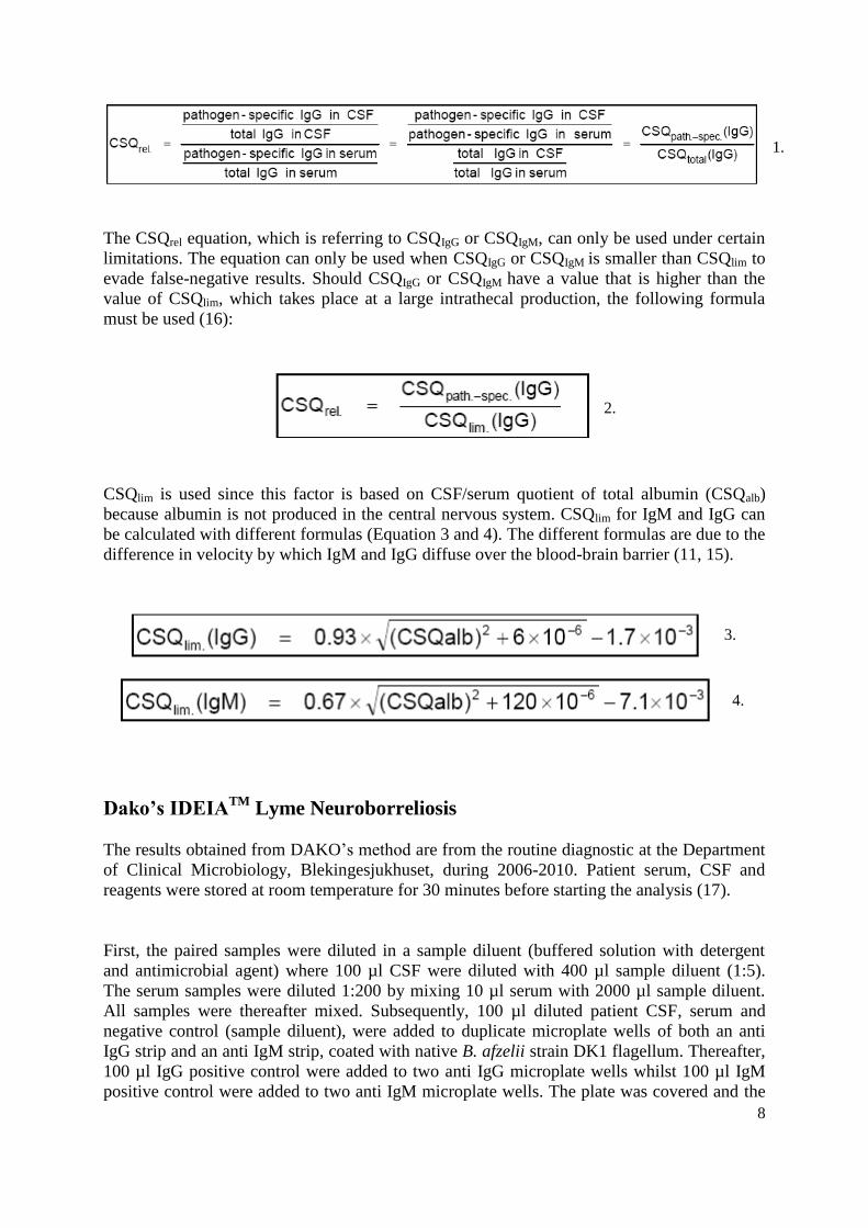
CSQrel is used to determine if there are any brain-derived fractions of specific antibodies in
CSF. The CSQrel value is the quotient of the portion of pathogen-specific antibodies in total
IgG or IgM of CSF and the portion of pathogen-specific antibodies in total IgG or IgM in
serum (15). The reference range of CSQrel is determined to be between 0.7 and 1.3. A normal
CSQrel value indicates absence of intrathecal production of antibodies in CNS while a CSQrel
value ≥1.5 indicates a local pathogen-specific antibody synthesis in the CNS (16).
8
The CSQrel equation, which is referring to CSQIgG or CSQIgM, can only be used under certain
limitations. The equation can only be used when CSQIgG or CSQIgM is smaller than CSQlim to
evade false-negative results. Should CSQIgG or CSQIgM have a value that is higher than the
value of CSQlim, which takes place at a large intrathecal production, the following formula
must be used (16):
CSQlim is used since this factor is based on CSF/serum quotient of total albumin (CSQalb)
because albumin is not produced in the central nervous system. CSQlim for IgM and IgG can
be calculated with different formulas (Equation 3 and 4). The different formulas are due to the
difference in velocity by which IgM and IgG diffuse over the blood-brain barrier (11, 15).
Dako’s IDEIATM
Lyme Neuroborreliosis
The results obtained from DAKO’s method are from the routine diagnostic at the Department
of Clinical Microbiology, Blekingesjukhuset, during 2006-2010. Patient serum, CSF and
reagents were stored at room temperature for 30 minutes before starting the analysis (17).
First, the paired samples were diluted in a sample diluent (buffered solution with detergent
and antimicrobial agent) where 100 µl CSF were diluted with 400 µl sample diluent (1:5).
The serum samples were diluted 1:200 by mixing 10 µl serum with 2000 µl sample diluent.
All samples were thereafter mixed. Subsequently, 100 µl diluted patient CSF, serum and
negative control (sample diluent), were added to duplicate microplate wells of both an anti
IgG strip and an anti IgM strip, coated with native B. afzelii strain DK1 flagellum. Thereafter,
100 µl IgG positive control were added to two anti IgG microplate wells whilst 100 µl IgM
positive control were added to two anti IgM microplate wells. The plate was covered and the
1.
2.
3.
4.

9
controls, the diluted serum and cerebrospinal fluids were incubated on a shaker (500 rpm) for
60 minutes at room temperature. After incubation, the wells were washed 4 times with at least
350 µl working strength wash buffer (buffered solution with detergent and antimicrobial
agent). After the final wash, the plate was tapped on an absorbent paper to remove the last
traces of wash buffer. To each microplate well, 100 µl flagellum conjugate were added and
the microplate was covered and incubated on a shaker (500 rpm) for 60 minutes at room
temperature. After the incubation, the microplate wells were washed 4 times with at least 350
µl working strength wash buffer and tapped on an absorbent paper before 100 µl TMB
substrate solution were added to each well. The microplate was covered and the controls, the
diluted serum and cerebrospinal fluids were incubated in a dark environment for 10 minutes at
room temperature. Thereafter, 100 µl stop solution (0.46 M sulphuric acid) were added to all
microplate wells in the same order and speed as the TMB substrate solution was added. The
microplate was shaken thoroughly for about 20 seconds before the controls, the diluted serum
and cerebrospinal fluids were incubated at room temperature for five minutes. Finally the
colour intensity was measured within 30 minutes after the stop solution was added. The
photometric measurement was made at 450 nm and with a reference wavelength between 620
and 650 nm. The obtained results were registered in the database. If the mean optical density
(O.D.) value for CSF was more than 0,150 O.D. a calculation of the index value was done
after Equation 5 (reference value: <0,3) (17).
serumCSF
serum
CSF DODODO
DOIndex ....
..
.. 5.
Euroimmun’s Western Blot – Borrelia IgM and Borrelia IgG
All the patient serum and reagents (all from Euroimmun, Lübeck, Germany) were kept at
room temperature at least 30 minutes before the experiment started. Thirty µl of the patient
serum, a positive control for IgM and a positive control for IgG were diluted 1:51 with 1,5 ml
universal buffer and were thereafter mixed. An IgM strip, a nitrocellulose membrane, with
electrophoretically separated Borrelia afzelii antigens, and an IgG strip, a nitrocellulose
membrane with electrophoretically separated Borrelia antigens and recombinant VlsE, were
placed in separate trays. One IgM strip and one IgG strip were used for each patient. To
prevent unspecific binding of antibodies to the nitrocellulose membrane, the strips were
incubated with 1,5 ml universal buffer on a rocking shaker, 30 rpm, for 15 minutes at room
temperature. The buffer was poured off and all the patient serum, the positive control for IgM
and the positive control for IgG in the test tube were applied to their respective channel in the
tray. The strips were incubated on a shaker at room temperature for 30 minutes at 30 rpm. The
diluted samples were aspirated and the strips were washed 3 times with 1,5 ml universal
buffer. During each wash the incubation trays were shaken for 5 minutes. Alkaline
phosphatase conjugated anti-IgM or anti-IgG antibodies were added to the IgG or IgM strip
respectively and the strips were incubated at room temperature for 30 minutes at 30 rpm. The
strips were subsequently washed three times with 1,5 ml universal buffer. During each wash
the trays were shaken for 5 minutes. After the third wash, 1,5 ml NBT/BCIP (Nitro Blue
Tetrazolium-Chloride-5-Bromo-4-Chloro-3-Indolyl-Phosphate) substrate was added to all
strips, and rose to a blue product with the aid of alkaline phosphatase, and the trays were
10
shaken at room temperature for 10 minutes at 30 rpm. Finally, the strips were washed with
distilled water 3 times and the strips were incubated one minute during each wash. After the
last wash, the strips were applied to an evaluation protocol. The strips were dried at room
temperature and were thereafter read by a scanner with the EUROLineScan program. The
results from the different samples were registered in the database (18, 19).
Euroimmun’s ELISA – Borrelia IgM and Borrelia IgG
Patient serum and reagents were stored at room temperature for 30 minutes and the patient
samples were centrifuged at 15000 x g for ten minutes before starting the analysis. The
instrument, Siemens BEP 2000, was washed before the serum and controls were analysed and
all required reagents from Euroimmun were loaded in the instrument. The reagents used were
an IgG sample buffer, an IgM sample buffer (IgG/RF-Absorbent, anti-human IgG antibody
preparation obtained from goat), a wash buffer (10x concentrate), IgG conjugate (peroxidase-
labelled anti-human IgG from rabbit), IgM conjugate (peroxidase-labelled anti-human IgM
from rabbit), TMB substrate (diluted 3,3’-5,5’-tetrametylbenzidine and stabilized peroxide
(H2O2)), stop solution (0.50 M sulphuric acid) and ten different controls (five controls for IgM
analysis and five for IgG analysis with the concentrations: 200 Relative Units (RU)/ml, 20
RU/ml, 2 RU/ml, one positive control and one negative control). All reagents were mixed
thoroughly before loaded to the instrument. Then the patient samples were loaded and the
analysis was started. First, 100 µl calibrators, positive controls, negative controls and diluted
patient sera (1:101, diluted with IgG sample buffer or IgM sample buffer), were transferred to
individual microplate wells. The microplate wells, for both IgM and IgG analysis, were
coated with a mixture of whole antigen extracts of B. burgdorferi, B. garinii and B. afzelii.
The microplates for IgG analysis were also coated with VlsE. The controls and the diluted
serum were thereafter incubated for 30 minutes at room temperature. After incubation, the
microplate wells were washed 3 times with 400 µl wash buffer. To each microplate well
either 100 µl peroxidase-labelled anti-human IgG or 100 µl peroxidase-labelled anti-human
IgM was added and incubated for 30 minutes at room temperature. After the incubation, the
microplate wells were washed 3 times with 400 µl wash buffer before 100 µl TMB substrate
solution had been added. Again, the controls and the diluted serum were incubated at room
temperature for 15 minutes before 100 µl stop solution was transferred to each well in the
same order and speed as the TMB substrate solution was added. Finally, the colour intensity
was measured within 30 minutes. The photometric measurement was made at 450 nm and
with a reference wavelength between 620 and 650 nm. The obtained results were registered in
the database (reference value: <20 RU/ml). (5, 20).

11
RESULTS
To compare the two methods for detection of intrathecal synthesis of Borrelia specific
antibodies, sample pairs (serum + CSF) from 100 patients with a suspected neuroborreliosis
were collected. The diagnosis neuroborreliosis is obtained when a patient is positive for either
IgM or IgG or when a patient is positive for both IgM and IgG. The results from the
Department of Clinical Microbiology Blekingesjukhuset routine diagnostic test, from DAKO,
determined 16 of the 100 patients to have neuroborreliosis. Of these 16 patients, 13 were
positive for IgG and 10 were positive for IgM. Of 16 patients, seven were positive for both
IgM and IgG. The results are summarised in Figure 2 and Table I.
Euroimmun’s method for detection of Borrelia antibodies was positive for 20 patients. All
these patients were positive for IgG and seven of them were also positive for IgM. Results are
summarised in Figure 2 and Table I.
Figure 2: The number of patients with samples positive for neuroborreliosis in DAKO’s and Euroimmun’s
ELISA. Of 100 patients, 16 were positive with DAKO’s ELISA whilst 20 patients were positive in Euroimmun’s
ELISA.
Since the two methods did not give identical results, conformational tests were performed. All
22 samples that were positive for neuroborreliosis, either in DAKO, Euroimmun or in both
tests, were examined further to confirm the results with a Western blot which detects
antibodies directed to B. burgdorferi sensu stricto, B. garinii and B. afzelii in serum. The
Western blot was positive for 18 patients and borderline for two patients who were positive in
Euroimmun’s IgG ELISA. A borderline result is obtained when unspecific antibodies are
detected in the patient’s serum. Of the seven patients who were positive in Euroimmun’s IgM
ELISA, one was positive in Western blot and two patients were determined to be borderline.
Patient 57/157 was determined to be borderline in Euroimmun’s Western blot IgG but was
negative in all ELISA-based methods. Results are summarised in Table I.
Of the 10 patients who were positive in DAKO’s method for IgM analysis, one patient was
positive, eight patients negative and one patient was determined borderline for IgM in
12
Euroimmun’s Western blot. Three samples which were negative for IgM in DAKO’s ELISA
were determined to be borderline for IgM in the Western blot analysis. Of 13 patients who
were positive for IgG in DAKO’s method, eleven were positive in Western blot whilst two
patients were determined to be borderline. One patient who was negative for IgG in DAKO’s
method was determined to be borderline in Euroimmun’s Western blot. Results are
summarised in Table I.
A serum ELISA, which detects antibodies directed to B. burgdorferi sensu stricto, B. garinii
and B. afzelii in serum, must be included in the conformation test because ELISA incubation
for serum serology is different from incubation for CSF diagnostic. The differences between
these two ELISAs are that serum ELISA has a different calibration curve, different time for
incubation and the dilution on the serum sample is different. But the main reason that serum
ELISA is included in the conformation test is that this ELISA has a cut off value for
discrimination of serologically positive and negative samples. The ELISA for CSF diagnostic
lacks this cut off value, i.e. a result will always be obtained but it can never be said to be
either positive or negative for borreliosis.
The serum ELISA was positive for 17 patients and negative for three patients who were
positive in Euroimmun’s IgG ELISA. Of the seven patients who were positive in
Euroimmun’s IgM ELISA, five were positive in serum ELISA and two patients were
negative. Two patients who were negative in Euroimmun’s IgM ELISA were positive in
serum ELISA. Results are summarised in Table I.
Of the 10 patients who were positive in DAKO’s method for IgM analysis four patients were
positive and six patients negative for IgM in Euroimmun’s serum ELISA. Three samples
which were negative for IgM in DAKO’s ELISA were determined to be positive for IgM in
the serum ELISA analysis. Of 13 patients, who were positive for IgG in DAKO’s method,
eleven were positive in serum ELISA whilst two patients were negative. Six patients who
were negative for IgG in DAKO’s method were positive in Euroimmun’s serum ELISA.
Results are summarised in Table I.
The results for IgM from the serum ELISA and the Western blot correspond to each other
except for seven samples. Sample 55/155 was determined to be a borderline in the Western
blot but was negative in the serum ELISA. The samples 63/163, 77/177 and 95/195 were
negative in the Western blot but were positive in the serum ELISA whilst samples 83/183,
86/186 and 97/197 were borderline in the Western blot but positive in the serum ELISA. The
results for IgG from the serum ELISA and the Western blot correspond to each other except
for the samples 57/157, 75/175 and 98/198 which were all determined to be a borderline in
Western blot but were negative in the serum ELISA. Sample 95/195 was positive in the
Western blot but negative in the serum ELISA. The results are summarised in Table I.

13
Table I: The obtained results for the 22 positive samples from Dako’s IDEIATM
Lyme Neuroborreliosis,
Euroimmun’s ELISA for detection of antibodies of the IgM and the IgG class against Borrelia antigens in CSF
and serum, Euroimmun’s anti-Borrelia Western blot and Euroimmun’s ELISA for detection of antibodies of the
IgM and the IgG class against Borrelia antigens in serum. The reference value for the different methods are:
DAKO’s <0,3; Euroimmun’s 0,7-1,3 (value ≥1,5 indicates a local pathogen-specific antibodies synthesis in the
central nervous system) and serum ELISA from Euroimmun <20 RU/ml (Equation 1-5). Sometimes
Euroimmun’s method gave two results, this is due to that antibodies were detectable in two dilutions. In the
Western blot test and serum ELISA only serum was analysed.
Sample
number
DAKO:s
IgM
(O.D.
ratio)
DAKO:s
IgG
(O.D.
ratio)
Euroimmun
IgM
(CSQrel)
Euroimmun
IgG
(CSQrel)
Euroimmun
Western
blot IgM
Euroimmun
Western
blot IgG
Euroimmun
Serum
ELISA IgM
(RU/ml)
Euroimmun
Serum
ELISA IgG
(RU/ml)
4/104 Negative 1.4 Negative 12.68;
12.83
Negative Positive Negative >200
28/128 Negative Negative Negative 1.54; 1.37 Negative Positive Negative >200
41/141 Negative Negative Negative 2.13 Negative Positive Negative 36.3
47/147 7.7 1.3 4.50; 4.98 9.67;
10.72
Positive Positive >200 174.4
52/152 Negative 0.4 Negative 2.14; 1.95 Negative Positive Negative 66.1
54/154 Negative Negative Negative 12.85;
11.69
Negative Positive Negative 53.4
55/155 Negative Negative Negative 1.55; 1.57 Borderline Positive Negative 50.3
57/157 4.77 Negative Negative Negative Negative Borderline Negative Negative
63/163 0.7 1.8 94.21;
88.54
12.46;
13.41
Negative Positive >200 167.1
65/165 37.5 191.8 Negative 147.53;
169.52
Negative Positive Negative 66.4
68/168 5.6 41.2 19.82;
22.59
56.63;
57.76
Negative Positive Negative 97.7
75/175 Negative 6.9 Negative 10.76;
11.35
Negative Borderline Negative Negative
77/177 Negative 34.4 Negative 17.52 Negative Positive 25.4 153.1
82/182 Negative Negative Negative 2.82 Negative Positive Negative 164.4
83/183 Negative 35.3 98.10;
83.74
33.82;
33.66
Borderline Positive 22.0 170.7
84/184 3.1 2.6 Negative 97.19;
94.44
Negative Positive Negative 42.8
86/186 Negative 0.6 0.35; 0.42 1.67; 1.70 Borderline Positive >200 >200
90/190 Negative Negative Negative 1.49; 1.89 Negative Positive Negative 173.3
95/195 4.0 Negative 5.64 5.10; 5.09 Negative Positive 38.3 Negative
96/196 3.0 Negative Negative Negative Negative Negative Negative Negative
97/197 1.1 1.9 3.51 19.27;
19.36
Borderline Positive 27.1 106.3
98/198 6.1 260.6 19.14 76.85;
103.68
Negative Borderline Negative Negative
14
DISCUSSION
This study indicates that Euroimmun’s method to detect antibodies directed to Borrelia is
superior to DAKO’s method. With Euroimmun’s method, 20 patients were positive for
neuroborreliosis while DAKO’s method only detected the infection in 16 patients.
Euroimmun’s method was also superior in the IgG analysis. Euroimmun detected 20 samples
which were positive for IgG whilst DAKO only was positive for 13 samples. This can be due
to that Euroimmun’s method can detect antibodies directed to all three pathological species of
Borrelia while DAKO only can detect antibodies directed to B. burgdorferi. Another
advantage with Euroimmun’s method is the correction of serum antibodies in the CSF caused
by a blood-brain barrier dysfunction by analysing the total albumin concentration in the CSF
and serum. All values from controls and calibrators for the analysis of IgM, IgG and albumin
were within the valid range (data not shown). In this study, the calibrators or patient samples
were not analysed in duplicates. However, the calibrators in the test can be used to improve
the reliability of the ELISA test if they are used in duplications.
Euroimmun’s method detects more positive cases of neuroborreliosis, however, DAKO’s
method detects more positive samples in the IgM analysis (10 versus 7). Why DAKO’s
method seems to be superior in the IgM analysis is unknown. However, the results from
Western blot and the serum ELISA support the obtained results from Euroimmun’s ELISA,
i.e. when DAKO’s method gave a positive result in the IgM analysis the results from
Euroimmun and the conformation tests were negative. These results indicate that DAKO’s
method can give false positive test results since the Western blot test is regarded to be a test
which confirms the results that are obtained from the ELISA. A false positive IgM test results
can be obtained when samples contain rheumatoid factors. However, the different results from
the IgM analysis between DAKO and Euroimmun can be due to other factors. Perhaps the
obtained results from Euroimmun’s ELISA and the conformation tests were in accordance
with each other since the used kits in this study were from the same company and these kits
contain the same Borrelia antigens and can therefore give rise to the same results. However,
the structure of the antigen is different in the Western blot and the ELISA and therefore the
results from these tests can be different. In a Western blot, the protein is straightened out, i.e.
denatured, and in this process the protein will loose their secondary and tertiary structure.
Therefore, the proteins will be more available for the antibody which binds specifically to the
protein in interested. But it is also possible that DAKO’s ELISA has detected patients with an
early neuroborreliosis that Euroimmun’s ELISA has missed since an early neuroborreliosis
can not be seen in analysis of serum and therefore the sample will be negative in the Western
blot. But it is also possible that some of the antibodies in the sample have been degraded and
therefore the results from the Euroimmun’s analysis could be false negative since the samples
which were used in this study had been stored for 0-4 years in -20 to -80°C. This problem
could explain the results obtained from sample 57/157, 65/165, 84/184 and 96/196 since these
samples were all positive in DAKO’s IgM analysis but negative in Euroimmun’s IgM
analysis. In these samples, the antibodies of class IgM could have been degraded during the
storage. However, the ELISA from Euroimmun detected more positive samples than the
ELISA from DAKO and this source of error is therefore considered to be small. Nevertheless,
a degradation of the antibodies is still a possibility.

15
Interestingly, all patients whose samples were determined to be negative with DAKO’s
method but positive in Euroimmun all had an impact of the central nervous system. The
symptoms the patients suffered from were for example headache, peripheral facial paralysis
and hemiparesis. A study has shown that B. garinii is the species which is mostly associated
with neuroborreliosis and gives rise to neurological symptoms (6). Because DAKO’s method
can not detects antibodies directed to B. garinii this can explain why Euroimmun’s method
detected more positive samples.
It is difficult to give the diagnosis neuroborreliosis to a patient as the diagnosis is not based
solely on the results of the ELISA and the Western blot, but also on the patient’s symptoms.
The symptoms in neuroborreliosis are very unspecific and symptoms from different parts of
the body can arise. This observation is in accordance with the symptoms from the patients
who are included in this study since all patients whose samples were determined to be positive
in the different methods suffered from different symptoms. For example, five of the 22
patients, who were determined to be positive in DAKO’s method, Euroimmun’s or in both,
suffered from headache; five patients suffered from back and neck pain, and four patients
suffered from peripheral facial paralysis. All patients who obtained a positive result suffered
from symptoms which are characteristic for neuroborreliosis; however, these symptoms are
also frequently occurring in other diseases. Another factor that is problematic in the diagnosis
of neuroborreliosis is the interpretation of the results from the ELISA and the Western blot.
Many factors can influence the final result, for example cross-reaction with rheumatoid
factors, genetic diversity of the Borrelia species and a slow immune response in the early
stage of the disease. In Sweden, many healthy individuals have antibodies directed to Borrelia
even though they do not suffer from neuroborreliosis and this can of course also be
problematic in the interpretation of serological tests. Furthermore, a patient who is treated
with antimicrobial drugs early in the infection can lack IgG antibodies but still have a
Borrelia infection, another factor that makes the interpretation of the results difficult.
Some samples were positive in DAKO’s and Euroimmun’s ELISA but were negative when
the serum was analysed by the confirmation tests. However, these samples can still be
positive because in the onset of neuroborreliosis, antibodies are only produced intrathecally
and not in serum. It takes some time before the antibodies are produced in the serum when the
neuroborreliosis is acute. The possible examples of an early neuroborreliosis is sample
57/157, 65/165, 68/168, 75/175, 84/184, 95/195, 96/196 and 98/198 because all these sample
are positive, i.e. have a high concentration of antibodies in CSF, in DAKO’s, in Euroimmun’s
or in both ELISA methods but are negative in the Western blot and serum ELISA. The
samples should therefore also be confirmed with a Western blot where the CSF is analysed so
that also the patients with an early neuroborreliosis can be detected. However, the volume of
CSF from the different patients was not enough to perform this analysis. Since CSF has not
been analysed in the Western blot, the obtained results from the Western blot can not entirely
be compared with the results from DAKO’s and Euroimmun’s method.
In Euroimmun’s method a sample is positive for neuroborreliosis when CSQrel is higher than
1.5 and in DAKO when O.D. ratio is higher than 0.3. When CSQrel is higher than 1.5
(Euroimmun) or O.D. ratio is higher than 0.3 (DAKO) this is an indication of specific
antibody production in CSF. However, a value above 1.5 or 0.3 can never be interpreted

16
without further information like additional results or clinical symptoms such as peripheral
facial paralysis, headache and numbness. A physician needs to consider the clinical symptoms
in the diagnosis of neuroborreliosis because a pathological CSQrel can remain for several
years even if the patient has been treated and is healthy again. If a patient with a suspected
acute neuroborreliosis lacks factors that indicate an acute infection, for example raised cell
number in CSF, elevated protein level in CSF and a high CSQalb but has a CSQrel which is
pathological this could be a late neuroborreliosis that does not require treatment anymore. It is
an advantage to use the different CSF/serum quotients in CSF diagnosis instead of antibody
titres since the quotients are method-independent when the CSF and serum samples are tested
simultaneously in the same analytical run. However, the quotients are unreliable when the
sample is contaminated with blood (>7000 erythrocytes/µl) (14).
If CSQrel is between 1.30 and 1.50 in Euroimmun’s method the sample is considered to be
borderline. This means that a pathogen-specific antibody production in the central nervous
system is questionable. Sample 28/128 and 90/190 gave results which were determined to be
borderline (1.37 and 1.49 respectively). However, the results from the other dilution were
clear positive (1.54 and 1.89 respectively) and therefore these patients have an “indication of
pathogen-specific antibody production in the central nervous system”. If CSQrel is between
0.7-1.3 antibodies are detectable but it is not an indication of specific antibody production in
the central nervous system. The level of the CSQrel cannot be interpreted in terms of acuteness
or severity of the disease. It just indicates whether there is an intrathecal production of
antibodies or not. In patients who are negative in CSF but positive in serum calculation is not
possible because the concentration of pathogen-specific antibodies in CSF is below
measurement range.
In the diagnosis of neuroborreliosis detection of antibodies of class IgG is more important
than antibodies of class IgM since IgG is more prevalent in patients with neuroborreliosis.
Detection of intrathecal production of specific IgG in the central nervous system is a finding
in about 96% in patients with neuroborreliosis whilst specific IgM production is seen in about
60% of the patients. An intrathecal IgM production is usually found together with a specific
IgG synthesis (21, 22). This finding is in accordance with the results from this study (Table I).
It is important that the collection of paired CSF and serum samples from the same patient
does not differ by more than 48 hours. All collection of paired samples in this study differed
by less than 48 hours except for sample 86/186 there the collection time differed by 72 hours.
This is important because the antibody production can differ between different days. Should
the CSF and serum sample differ by more than 48 hours, the antibody pattern can be
misleading. It is also important that the CSF sample and serum sample, obtained from the
same patient, are analysed together. The reason for this is that in order to compare CSF and
serum they must be incubated under the same conditions. ELISA is a method that is not so
standardised and results can vary between analysis. However, this problem is reduced because
calibrators are used in all test procedures. The calibrators’ values are then utilised in formulas
and this will compensate for variations between tests.
Sources of errors in this study can be inadequate washing, for example less than three wash
cycles or too small wash buffer volumes, which can lead to falsely high extinction values. If

17
residual liquid is left in the reagent wells after washing that can interfere with the substrate
and give falsely too low extinction values. Another source of error is if the temperature is
higher than room temperature during the incubation with substrate. If the temperature is too
high the extinction values can increase. However, the calibrators are subject to the same
influence which will compensate for such variations. The reason for the greater extinction
values is that the activity of the enzyme is temperature-dependent. In some samples, the
serum was negative but in the CSF an insignificant antibody production could be determined.
This reaction is unspecific and could be caused by antibodies against components of buffers,
i.e. acid and salt, which were used when the wells were coated with antigens. This problem
can especially arise when the dilution is small such as in the CSF diluted 1:2. Here, the
concentration of these antibodies will be higher than it would be in serum diluted 1:404. This
problem can also be caused by inadequate blocking of the microplate wells which can lead to
antibodies binding directly to the plastic.
The obtained results from this study are important for the routine diagnostics for the detection
of antibodies of class IgM and IgG directed to Borrelia at all departments of Clinical
Microbiology in Sweden. The results are important because this study shows that
Euroimmun’s method can detect more cases of neuroborreliosis than DAKO’s. However,
some improvement should be done before Euroimmun’s method becomes the method for
routine diagnostic detecting antibodies directed to Borrelia at Departments of Clinical
Microbiology in Sweden. For example, the dilution in the assay could be optimised. Many of
the samples which were determined to be positive in Euroimmun’s method had to be retested
with higher dilutions. The highest dilution for one serum sample was 1:51712 whilst the
highest dilution of CSF was 1:2048. However, is it difficult to say which dilution should be
best because it will always be some samples that need to be retested with a higher dilution. A
disadvantage with Euroimmun’s method is that it is slower than DAKO’s method.
In conclusion, Euroimmun’s method seems to be superior to DAKO’s method and this is
probably due to the detection of antibodies directed to all three pathological Borrelia species
by Euroimmun whilst DAKO only detects antibodies of class IgM and IgG directed to B.
burgdorferi. It is a disadvantage for DAKO that this method does not include antigens
specific to B. garinii when a study has shown that this species, of all three pathological
Borrelia strains, is the most associated with neuroborreliosis (6). However, it is difficult to
know with certainty which method of these two that is the superior one since we can not know
which results are accurate.

18
ACKNOWLEDGEMENTS
Thanks to my tutors, Maria Nilsson at the Department of Clinical Microbiology -
Blekingesjukhuset - and Inger Edfors, School of Natural Sciences – Linnæus University, for
guidance and support in this work.
Thanks to Sylvia Falke, Diana Eichhorst, Katja Steinhagen and Jessica Dobschanski at
Euroimmun for all the help with the arrangement of the new method and for guidance and
support in this work. I also want to thank Euroimmun for giving me the opportunity to make
this examination project.
Thanks to all the staff at Department of Clinical Microbiology, Blekingesjukhuset, especially
to Malin Wilén Pilto, Petra Michelsberg, Lena Rova and Kristin Mattsson for continuous
support during all my work.
Thanks to all the staff at Department of Clinical Chemistry at Blekingesjukhuset Karlskrona
and Karlshamn for the help with collection of CSF samples. Thanks to Helen and Bitte for the
help with analysing of the serum and CSF samples.
Thanks to my sister Angelika who let me live in her apartment during this examination project
work.

19
REFERENCES
1. Bauman, R., (2007), Microbiology With Diseases by Taxonomy, 2nd
, Benjamin Cummings.
2. Murray, R. P., Jo Baron, E., Jorgensen, H. J., Pfaller, A. M., Yolken, H. R., (2003), Manual
of Clinical Microbiology, 8th
edition, American Society for Microbiology.
3. Ornstein, K., (2002), Lyme Borreliosis: Detection and identification of the infecting
genotype, Faculty of Medicine, Lund University.
4. Berglund, J. et al, (1995), An epidemiological study of lyme disease in southern Sweden,
The New England Journal of Medicine, Volume 333:1319-1324.
5. Euroimmun’s test instruction: Anti-Borrelia ELISA (IgM), Medizinische Labordiagnostika
AG, 2009-10-08.
6. Ornstein, K., Berglund, J., Nilsson, I., Norrby, R. and Bergström, S., (2001),
Characterization of Lyme Borreliosis Isolates from Patients with Erythema Migrans and
Neuroborreliosis in Southern Sweden, Journal of Clinical Microbiology, Vol. 39, No. 4, page
1294-1298.
7. Ornstein, K., Berglund, J., Bergström, S., Norrby, R., Barbour, G. A., (2002), Three Major
Lyme Borrelia Genospecies (Borrelia burgdorferi sensu stricto, B. afzelii and B. garinii)
Identified by PCR in Cerebrospinal Fluid from Patients with Neuroborreliosis in Sweden,
Scandinavian Journal of Infectious Diseases, Vol. 34, No. 5, page 341-346.
8. Emmerson, A.M., Hawkey, P.M., Gillespie, S.H., (1997), Principles and practice of clinical
bacteriology, John Wiley & Sons.
9. Tjernberg, I., Krüger, G., Eliasson, I., (2007), C6 peptide ELISA test in the serodiagnosis
of Lyme borreliosis in Sweden, European Journal of Clinical Microbiology & Infectious
Diseases, Vol. 26, No. 1, page 37-42.
10. Euroimmun’s test instruction for the ELISA: Antibodies of the IgM class against Borrelia
antigens in cerebrospinal fluid, Medizinische Labordiagnostika AG, 2009-04-07.
11. Kindt J.T., Goldsby, A.R., Osborne, A.B., (2007), Kuby Immunology, 6th
edition, W.H.
Freeman and Company.
12. Wilson, K and Walker, J, (2005), Biochemistry and Molecular Biology, 6th
edition,
Cambridge University Press.
13. Tumani, H., Nölker, G., Reiber, H., (1995), Relevance of cerebrospinal fluid variables for
early diagnosis of neuroborreliosis, Neurology, Vol. 45, page 1663-1670.
14. Reiber, H., Peter, B.J., (2001), Cerebrospinal fluid analysis: disease-related data pattern
and evaluation programs, Journal of the Neurological Sciences, Vol. 184, page 101-122.
15. Euroimmun’s test instruction for the ELISA: Antibodies of the IgG class against Borrelia
antigens in cerebrospinal fluid, Medizinische Labordiagnostika AG, 2007-06-21.
16. Reiber, H., Lange, P., (1991), Quantification of Virus-Specific Antibodies in
Cerebrospinal Fluid and Serum: Sensitive and Specific Detection of Antibodies Synthesis in
Brain, Clinical Chemistry, Vol 37, No. 7, page 1153-1160.
17. Dako’s test instruction, IDEIATM
Lyme Neuroborreliosis, Oxois (Ely) Ltd, November
2006, version K602811-2EFG.
18. Euroimmun’s test instruction for the Westernblot: Antibodies against Borrelia afzelii
(IgM), Medizinische Labordiagnostika AG, 2008-03-13.
19. Euroimmun’s test instruction for the EUROLINE-WB: Antibodies against Borrelia (IgG),
Medizinische Labordiagnostika AG, 2008-03-13.
20. Euroimmun’s test instruction: Anti-Borrelia plus VlsE ELISA (IgG), Medizinische
Labordiagnostika AG, 2005-10-25.

20
21. EUROIMMUN Anti-Borrelia CSF ELISA (IgM): CSF/serum quotient (CSQrel) in sera of
patients with different neurological diseases, (2005), version EI_2132-1_O_UK_V01,
Medizinische Labordiagnostika AG.
22. EUROIMMUN Anti-Borrelia CSF ELISA (IgG): CSF/serum quotient (CSQrel) in sera of
patients with different neurological diseases, (2005), version EI_2132-1_O_UK_U01,
Medizinische Labordiagnostika AG.
