comparison of shoot induction ability of different explants in herbaceous peony (paeonia lactiflora...
TRANSCRIPT

Scientia Horticulturae 123 (2010) 385–389
Comparison of shoot induction ability of different explants in herbaceous peony(Paeonia lactiflora Pall.)
Daike Tian *, Ken M. Tilt, Fenny Dane, Floyd M. Woods, Jeff L. Sibley
101 Funchess Hall, Department of Horticulture, Auburn University, Auburn, AL 36849, United States
A R T I C L E I N F O
Article history:
Received 15 December 2008
Received in revised form 14 October 2009
Accepted 14 October 2009
Keywords:
Nodal stem
Meristematic region
Shoot elongation
Benzylaminopurine
Gibberellic acid
Thidiazuron
A B S T R A C T
Shoot induction ability of explants of herbaceous peony was investigated in semisolid MS medium
containing BA, TDZ and GA3. Callus was readily induced from stem without node and petiole explants
within 2 days of culture but failed to generate shoots. Adventitious shoots were successfully produced from
meristematic regions only: bud eyes on nodal stem sections, and junctions of petioles and petiolules. No
shoots were induced from internode sections, petiole without junctions, or leaf sections. Nodal sections
were the most efficient explants. There were up to 20 shoots in one explant generated within 20 days of
culture. TDZ was more effective than BA to induce shoots. The 100% shoot induction rate was obtained in
medium containing 0.1–3 mg L�1 of TDZ. However, higher concentrations of TDZ inhibited shoot
elongation and only large leaf clusters were produced. Combinations of BA and TDZ failed to increase shoot
induction rates but caused shoots shorter. The 2–60-min pretreatment of explants with 20 mg L�1 TDZ
solution was very effective to induce adventitious shoots directly, but both shoot number and shoot length
tended to decrease as treatment time increased. GA3 was beneficial for shoot and stem elongation.
� 2009 Elsevier B.V. All rights reserved.
Contents lists available at ScienceDirect
Scientia Horticulturae
journal homepage: www.e lsev ier .com/ locate /sc ihor t i
1. Introduction
Paeonia spp. typifies the family Paeoniaceae that produces thebeautiful, dramatic flowers known as garden peonies. Peonies arealso used as pot flowers, cut flowers and dry flowers. Herbaceouspeony (Paeonia lactiflora Pallas.) has been cultivated in China formore than 3900 years (Wang and Zhang, 2005). Today, herbaceouspeonies are back in fashion because of ease cultivation and diverseflower form and color. However, with the complex sequentialdormancy of peony seeds, it usually takes 2 years for germinationunder natural conditions (Griess and Meyer, 1976). Peonyseedlings must grow for several years before blooming. Peoniescan be propagated by division, cutting, grafting, and layering toobtain true-to-type plants. Herbaceous peonies are usuallypropagated by 3–5 eye divisions of underground stock plants. Agrower may be able to double their stock plants every 3 years byplanting in a 3-year rotation (Shannon and Kamp, 1959). Nodalstems and underground rhizomes can be used for cuttings (Arinoet al., 1981; Antanaitiene and Staniene, 2001; Wang and Zhang,2005). Layering is seldom used in herbaceous peony but theefficiency has been improved by a new approach of vertical
* Corresponding author. Current address: South China Botanical Garden, Chinese
Academy of Sciences, 723 Xinke Rd, Tianhe District, Guangzhou, 510520, China.
Tel.: +86 20 85232604; fax: +86 20 85232604.
E-mail address: [email protected] (D. Tian).
0304-4238/$ – see front matter � 2009 Elsevier B.V. All rights reserved.
doi:10.1016/j.scienta.2009.10.007
layering with application of a rooting chemical like UkorzeniaczB2, a mixture of PGRs (Czekalski and Jerzy, 2003). There aremultiple traditional choices for propagation of peony. However,the limited number of plants produced by these traditionalmethods cannot meet the increasing demands in the market,especially for a quick release of a new cultivar and massiveproduction of a favorite variety. The development of micropropa-gation methods for peonies is necessary to not only overcome thisproblem but also accelerate peony breeding progress.
Micropropagation of peony began in the middle 1960s. Calluswas induced successfully for the first time by Yamada and Sinoto(1966) from petal culture of Paeonia japonica. Commercial micro-propagation of peony plants is the result of the past 40 years of tissueculture research. Many advances have been made and several paperreviews on tissue culture of peony have been published (Buchheimand Meyer, 1992; Gabryszewska, 2004; Li and Luo, 2004; Beruto andCurir, 2007). Only since 2006 has one company, PlanteckBiotechnologies Inc. (based in Quebec, Canada) mass-producedone cultivar of herbaceous peonies and made its tissue culturedplants available to the market (Whysall, 2006). However, moreresearch is needed to make mass production of peony morecommercially successful. Optimization of procedures are necessary,from selection of explants, decontamination, screening of medium,application of plant growth regulators (PGRs), induction of callusand shoots, subculture, rooting, and to final transplanting.
Many kinds of explants from herbaceous peony are known togenerate callus, embryos and adventitious shoots in vitro. Mature
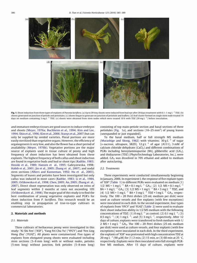
Fig. 1. Shoot induction from three types of explants of Paeonia lactiflora. (a) Up to 20 tiny shoots were induced from bud eye after 20 days treatment with 0.1–1 mg L�1 TDZ; (b)
shoots generated on junction of petiole and petiolules; (c) shoots began to generate on junction of petiolule and leaflets; (d) leaf cluster formed on single stem node treated 14
days on medium containing 3 mg L�1 TDZ; (e) shoots were obtained from stem nodes which were treated 10 h with TDZ (20 mg L�1) before inoculation.
D. Tian et al. / Scientia Horticulturae 123 (2010) 385–389386
and immature embryo tissues are good sources to induce embryosand shoots (Meyer, 1976a; Buchheim et al., 1994; Kim and Lee,1994; Shin et al., 1996; Kim et al., 2006; Stanys et al., 2007) but canonly be supplied by seeded varieties. Floral portions are moreeasily sterilized than vegetative organs. However, the efficiency oforganogenesis is very low, and also the flower has a short period ofavailability (Meyer, 1976b). Vegetative portions are the majorsource of explants used in tissue culture of peony and highfrequency of shoot induction has been obtained from theseexplants. The highest frequency of both callus and shoot inductionare found in vegetative buds and bud or shoot tips (Radtke, 1983;Hosoki et al., 1989; Hansen et al., 1995; Gabryzewska, 1998;Habib et al., 2001; Jin et al., 2005; Zhang et al., 2007), and nodalstem sections (Albers and Kunneman, 1992; Hu et al., 2003).Segments of leaves and petioles have been investigated but onlycallus was induced in most cases (Radtke, 1983; Li et al., 1984,2005; Orlikowska et al., 1998; Chen, 2005; An, 2005; Zhang et al.,2007). Direct shoot regeneration was only observed on veins ofleaf segments within 2 months at rates not exceeding 10%(Orlikowska et al., 1998). The major goal of this study is to find thebest combination of young vegetative explants and PGR’s forshoot induction from P. lactiflora. This research would be anenabling step in propagation of true-to-type cultivars incommercial laboratories.
2. Materials and methods
2.1. Materials
Three cultivars of herbaceous peony were investigated in thisstudy: ‘Xi Shi Fen’ (‘XSF’), ‘Yang Fei Chu Yu’ (‘YFCY’) and ‘Fen LingHong Zhu’ (‘FLHZ’). All plants were containerized. Five types ofexplants from elongated young shoots were evaluated includingstem sections (5–8 mm long) with or without nodes, petioles(5 mm long) without junction, fork petioles (5–8 mm long)
consisting of top main-petiole section and basal sections of threepetiolules (Fig. 1a), and sections (16–25 mm2) of young leaves(unexpanded or just expanded).
To the basal medium, half or full strength MS medium(Murashige and Skoog, 1962) with vitamins; 30 g L�1 of sugar(D-sucrose, ultrapure, S829), 12 g L�1 of agar (A111), 3 mM ofcalcium chloride dehydrate (CaCl2) and different combinations ofPGRs including benzylaminopurine (BA), gibberellic acid (GA3),and thidiazuron (TDZ) (PhytoTechnology Laboratories, Inc.), wereadded. GA3 was dissolved in 70% ethanol and added to mediumafter autoclaving.
2.2. Treatments
Three experiments were conducted simultaneously beginningin January, 2006. In experiment I, the response of five explant typesof ‘XSF’ (Table 1) to different PGRs were evaluated on medium (1)1/2 MS + 1 mg L�1 BA + 0.1 mg L�1 GA3; (2) 1/2 MS + 0.1 mg L�1
BA + 1 mg L�1 GA3; (3) 1/2 MS + 1 mg L�1 BA + 3 mg L�1 TDZ; and(4) 1/2 MS + 1 mg L�1 BA + 3 mg L�1 TDZ + l mg L�1 GA3, respec-tively. The 100 � 20 Petri dishes (25 mL medium per dish) wereused as culture vessels and five explants (with few exceptions)were inoculated in each dish. In the second experiment, four typesof explants from ‘YFCY’ and ‘FLHZ’ (Table 2) were used to evaluatetheir shoot induction ability in 1/2 MS medium with the followingconcentrations of TDZ: (1) 0 mg L�1 as control; (2) 0.1 mg L�1; (3)0.5 mg L�1; (4) 1 mg L�1; and (5) 3 mg L�1, respectively. After 12days of culture, explants were transferred to fresh medium with 1/2 MS + 1 mg L�1 GA3. The 100 � 20 Petri dishes (25 mL mediumper dish) were used as culture vessels, and four explants (with fewexceptions) were inoculated in each dish. In the third experiment,the explants of ‘XSF’ was pretreated with high concentration TDZ at20 mg L�1 (90 mM) for (1) 2 min, (2) 15 min, or (3) 60 min at 25 8C,respectively. Explants were then inoculated onto full strength PGR-free MS medium. After 15 days of culture, explants were

Table 1Callus and shoot induction of ‘Xi Shi Fen’ on MS medium containing PGRs.
Explant typea and
number/
treatment
Treatmentb Calli inducedc
3 days
Shoot induction
rate (%)d
30 days
Leaf 27 (1), (2), (3),
or (4)
� 0 d
Petiole 15 (1), (2), (3), or (4) � 0 d
Fork petiole 10 (1) + 50 c
10 (2) + 60 bc
10 (3) + 50 c
8 (4) + NA
Stem (� node) 10 (1), (2), (3),
or (4)
+ 0 d
Stem (+ node) 10 (1) + 100 a
10 (2) + 80 ab
10 (3) + 100 a
15 (4) + 93 a
a ‘� node’: stem section without node; ‘+ node’: stem section with node.b Treatment: explants are inoculated on media containing (1) (1/2) MS + 1 mg L�1
BA + 0.1 mg L�1 GA3; (2) (1/2) MS + 0.1 mg L�1 BA + 1 mg L�1 GA3; (3) (1/2) MS + 1
BA + 3 mg L�1 TDZ; and (4) (1/2) MS + 1 mg L�1 BA + 3 mg L�1 TDZ + 1 mg L�1 GA3,
respectively.c ‘�’: no callus was induced; ‘+’: callus was induced.d ‘NA’: the data are unavailable for evaluation because of heavy contamination of
explants. Means with different letters in the same column are significantly different
(HSK, a= 0.01).
D. Tian et al. / Scientia Horticulturae 123 (2010) 385–389 387
transferred to the fresh medium with 1/2 MS + 0.1 mg L�1
BA + 1 mg L�1 GA3 for shoot elongation or medium with 1/2MS + 1 mg L�1 GA3. The 25 mm � 95 mm tubes (10 mL mediumper dish) were used as culture vessels, and each tube contained oneexplant. An unbalanced randomized design was used in this studydue to limitation of some materials. The detailed information ontreatment and explant number of each treatment were seen inTables 1–3, respectively. Mean differences were examined byTukey’s test (HSD) with ANOVA procedure using SAS 9.1 (SASInstitute Inc.).
2.3. Surface sterilization, inoculation and culture
Young elongated shoots were washed in tap water three timesand cut into 2–4 cm long sections. Since a high contamination rate
Table 2Callus and shoot induction of ‘Yang Fei Chu Yu’ and ‘Fen Ling Hong Zhu’.
Cultivara Explant typeb and number per
treatment
Treatmentc
YFCY Stem (� node) 8 (1), (2), (3), (4),
Stem (+ node) 8 (1)
8 (2)
8 (3)
8 (4)
8 (5)
Petiole (� junct) 20 (1), (2), (3), (4),
Leaf 20 (1), (2), (3), (4),
FLHZ Stem (� node) 8 (1), (2), (3), (4),
Stem (+ node) 8 (1)
8 (2)
8 (3)
11 (4)
13 (5)
Petiole (� junct) 8 (1), (2), (3), (4),
Leaf 20 (1), (2), (3), (4),
a YFCY: ‘Yang Fei Chu Yu’ and FLHZ: ‘Fen Ling Hong Zhu’.b ‘� node’: without node, ‘+ node’: with node, ‘� junct’: without a junction of petiolc Treatment: explants are inoculated on media containing (1) (1/2) MS, (2) (1/2) MS + 0.
MS + 3 mg L�1 TDZ, respectively.d ‘�’: shooting rates should be higher than the actual observed values because shoot in
different letters in the same column are significantly different (HSK, a= 0.01).
was observed in previous studies using only bleach for steriliza-tion, a two-step method was used in this study. Namely, sectionswere soaked in 70% ethanol for 8–10 s followed by 15 min ofsterilization with 10% commercial bleach, to which Tween 20 (1drop per 100 mL) was added. Plant material was rinsed three timesfor 5 min in autoclaved distilled water. After sterilization, explantswere cut into shorter sections (3–8 mm); inoculated on Petridishes, and moved to 25 mm � 95 mm tubes or small plasticculture jars after initial culture. Culture vessels were maintainedunder cool fluorescence light, 60 lx, with a 16 h light cycle at25 � 1 8C. Data were collected after 3, 7, 15, and 30 days of culture,respectively.
3. Results
3.1. Contamination
Contamination of explants was problematic only on ‘YFCY’stem sections originating from the cooler, not from forcing-treated plants in greenhouse. Less than 10% of explants werecontaminated on ‘XSF’ and ‘FLHZ’. With a treatment of 8–10 s in70% ethanol before sterilization with 10% bleach, the contamina-tion of explants largely decreased compared to the one-stepsterilization method (using 10% bleach only) in previous experi-ments (data not shown).
3.2. Callus induction
The segments of stems and some petioles generatedcallus within 1 or 2 days of culture but little callus wasproduced on leaf blades even following 2–3 months of culture(Tables 1–3). Explants inoculated on medium with PGRsproduced more callus than those on medium without PGRs. Also,callus was pale white and waterlogged in medium with BA asonly PGR. Callus grew fast in the beginning but turned brown,and its growth stopped after 15 days of culture. Attempts toinduce adventitious shoots from this type of callus wereunsuccessful because of browning problems. On media withTDZ, callus was green and turgid. It grew slow and had lessbrowning problems. Whether transferred to fresh medium ornot, this type of callus continued to grow, but failed todifferentiate into adventitious shoots.
Calli induced (3 days) Shoot induction rate (%)d
or (5) + 0 d
+ 0 d
+ 100 a
+ 100 a
+ 100 a
+ 100 a
or (5) + 0 d
or (5) � 0 d
or (5) + 0 d
+ 0 d
+ �60 bc
+ �70 b
+ �40 c
+ �38 c
or (5) + 0 d
or (5) � 0 d
e and petiolules.
1 mg L�1 TDZ, (3) (1/2) MS + 0.5 mg L�1 TDZ, (4) (1/2) MS + 1 mg L�1 TDZ, and (5) (1/2)
duction ability of the explants was possibly inhibited by contamination. Means with

Table 3Callus and shoot induction of ‘Xi Shi Fen’ by soaking shock treatment with TDZ.
Explant typea and number per
treatment
Treatmentb Callus induction
(3 days)
Shoot induction
rate (%, 30 days)
Mean shoots
per explantc
Mean LS
length (mm)d
Petiole 10 (1), (2), or (3) + 0 b 0 b 0 b
Fork petiole 6 (1) + 100 a NA NA
6 (2) + 100 a NA NA
10 (3) + 100 a NA NA
Stem (� node) 10 (1), (2), or (3) + 0 b 0 b 0 b
Stem (+ node) 10 (1) + 100 a 3 a 20.4 a
10 (2) + 100 a 2.8 a 19.1 a
10 (3) + 100 a 2 a 15.8 a
a ‘� node’: without node, ‘+ node’: with node.b Treatment: explants were soaked in 20 mg L�1 TDZ for (1) 2 min, (2) 15 min, and (3) 60 min, respectively, then inoculated on PGR-free MS medium. After 15 days of
culture, explants were transferred to (1/2) MS + 0.1 mg L�1 BA + 1 mg L�1 GA3 for shoot elongation and multiplication.c ‘NA’: data unavailable.d Mean shoot number and the average length of longest shoots (LS) were calculated after 30 days of culture. Means with different letters in the same column are
significantly different (HSK, a= 0.01).
D. Tian et al. / Scientia Horticulturae 123 (2010) 385–389388
3.3. Shoot induction
Shoot primordia usually developed on nodal stems within 3days of culture and were followed by shoot generation in all PGRcontaining media. Shoots elongated to 2 cm length within 2 weeksof culture. New shoots or shoot clusters were only induced frommeristematic regions of the explants: eye of lateral buds (Fig. 1a),junction of rachis and petioles (Fig. 1b), and junction of petioluleand leaflets (Fig. 1c). Adventitious shoots were not induced onyoung stem segment without nodes, petiole segment withoutjunctions, and sections of leaf blades. Young nodal stems were themost efficient explants for shoot induction with nearly 100% shootinduction rate obtained (Tables 1–3). Each node can produceseveral to over 20 shoots in 20 days. However, most shootprimordia on fork petioles did not develop into normal shoots, andat most only three elongated shoots on one explant formedfollowing 30 days of culture. Callus from explants failed to formshoots.
Addition of BA to medium resulted in shoot induction of peony.A higher concentration (1 mg L�1) of BA was more effective than alower concentration (0.1 mg L�1) (Table 1). Application of TDZ toBA-containing medium did not significantly increase shooting ratebut made shoots visually shorter and stronger. GA3 had asignificant effect on shoot elongation (data not shown), but shootsinduced on medium with high GA3 concentrations were thin andtoo weak for root induction. TDZ showed very strong shootinduction ability and 100% shooting rate [(the number of explantswith induced shoots/the number of inoculated explants) � 100%]was obtained on the nodal stems of ‘YFCY’ with treatments of TDZranging from 0.1 to 3 mg L�1 (Table 2). Shooting rates of ‘FLHZ’were low. It was not clear if this large difference was caused bygenotype or by pretreatment of stock plants. Shooting rate of‘FLHZ’ also decreased at TDZ concentrations larger than 0.5 mg L�1.Pretreatment with high concentrations of TDZ inhibited stemelongation of induced shoots significantly and resulted in largerleaf clusters (Fig. 1d) after transfer to shoot elongation medium (1/2 MS + 0.1 mg L�1 BA + 1 mg L�1 GA3).
TDZ shock treatment (a short-time treatment with very highconcentrations of TDZ compared with the normal usage) of ‘XSF’resulted in same shoot induction rates for the same type ofexplants (Table 3). Treatments with 2, 15, or 60 min of 20 mg L�1
TDZ soak resulted in 100% shooting rates for explants of both nodalstems and fork petioles. However, no shoot initiation was observedon petioles without junctions and on stem sections without nodes.Although fork petioles also showed higher shoot-induction abilityafter shock treatment with high concentrations of TDZ, the induced
shoot primordia were difficult to form normal shoots, and onlythree shoots at most could be produced on a segment. Nodifference in shoot number and shoot length of ‘XSF’ was observedbetween shock treatments although longer shock treatment of20 mg L�1 TDZ tended to inhibit the number and elongation ofadventitious shoots (Table 3). Other experiments showed thatnormal elongated shoots could be obtained from nodal stemsections of ‘XSF’ even if explants were treated with 20 mg L�1 TDZat 8 8C for 10 h (Fig. 1e). This difference was possibly caused byeither the difference in treatment temperature or the differentresponse of meristematic tissue to TDZ in different growingseasons.
4. Discussion
The present study indicated that adventitious shoots were notinduced directly from non-nodal stem sections, petiole withoutsection, leaf sections without main vein. Whereas up to 100% shootinduction rate was obtained from bud eyes of nodal stem sectionsand from the junctions of fork petioles, while only two of morethan 300 leaf sections generated shoots. Therefore, meristematicregions play a critical role in direct shoot induction of herbaceouspeony. Although high frequency of shoot initiation was alsoinduced on fork petioles by the addition of TDZ in medium, a verylimited number of normal shoots formed. Thus, axillary buds andbud or shoot tips, and stem nodes are among the best vegetativeexplants to directly induce shoots in tissue culture of herbaceouspeony. TDZ has shown a much stronger ability than BA on shootinduction (Trigiano and Gray, 2000). But, it has only been reportedin a few cases of peony tissue culture (Gabryszewska, 1998, 2006;Orlikowska et al., 1998; Beruto et al., 2004). Axillary shootformation was significantly enhanced by the addition of TDZ at avery low concentration to medium containing a mixture of otherPGRs. TDZ influenced the existing meristems specifically andsubsequently de novo organogenesis was triggered in theneighboring cells (Metha et al., 2005). The combination of TDZwith other cytokinins in medium can be more effective ascompared to TDZ used alone for multiplication (Gabryszewska,1998). The high concentrations of TDZ led to inhibition of stemelongation. The inhibitive effect of TDZ on shoot or stem elongationhas also been reported in other tissue cultured plants (Ledbetterand Preece, 2004; Lee, 2005). TDZ shock method was very effectivein direct induction of adventitious shoots in tissue culture ofherbaceous peony. However, the optimal concentration of TDZ andtreatment time need further study. GA3 was necessary forelongation of shoot stems (Figueiredo et al., 2001) and also
D. Tian et al. / Scientia Horticulturae 123 (2010) 385–389 389
facilitated shoot induction (Yu et al., 2008). Therefore GA3 hasbeen often used in peony tissue culture. The present study alsoshowed that GA3 was beneficial for shoot elongation GA3. The rootswere obtained on semisolid medium or by paper bridge method(liquid medium) with IBA treatments (data not shown) in laterexperiments. However, effect of GA3 on rooting was notinvestigated in our studies. Both positive and negative effects ofGA3 rooting have been widely reported in other species. GA3
promoted rooting of somatic embryos of herbaceous peony (Shinet al., 1997).
References
Albers, M.R.J., Kunneman, B.P.A.M., 1992. Micropropagation of Paeonia. Acta Hortic.314, 85–92.
An, B.Y., 2005. Studies on the establishment of in vitro regeneration system ofPaeonia suffruticosa. MS Thesis, Andr. Northeast For. Univ.
Antanaitiene, R., Staniene, G., 2001. Investigation of propagation by segments ofrhizome and micropropagation of peony varieties. Sodininkyste ir darzinin-kyste 20, 44–54.
Arino, K., Mizukoshi, Y., Hatakeyama, J., 1981. Propagation by leaf-bud cutting anddevelopment of the method by utilizing of rooting-stocks in peony (Paeoniaalbiflora). 2. Propagation process of rooting stocks. Tohoku Agric. Res. 29, 217–218.
Beruto, M., Curir, P., 2007. In vitro culture of tree peony through axillary budding. In:Jain, S.M., Haggman, H. (Eds.), Protocols for Micropropagation of Woody Treesand Fruits. Springer, Dordrecht, The Netherlands, pp. 477–497.
Beruto, M., Lanteri, L., Portogallo, C., 2004. Micropropagation of tree peony (Paeoniasuffruticosa). Plant Cell Tissue Organ Cult. 79, 249–255.
Buchheim, J.A.T., Meyer Jr., M.M., 1992. Micropropagation of peony (Paeonia spp.).In: Bajaj, Y.P.S. (Ed.), High-tech and Micropropagation. IV. Biotechnology inAgriculture and Forestry, vol. 20. Springer-Verlag, Berlin Heidelberg, pp. 269–285.
Buchheim, J.A.T., Burkhart, L.F., Meyer M.M.Jr., 1994. Effect of exogenous gibberellicacid, abscisic acid, and benzylaminopurine on epicotyl dormancy of culturedherbaceous peony embryos. Plant Cell Tissue Organ Cult. 36, 35–43.
Chen, X.L., 2005. The preliminary research on tissue culture of Paeonia suffruticosa.MS Thesis, Henan Agr. Univ.
Czekalski, M., Jerzy, M., 2003. Propagation of Paeonia lactiflora with vertical layers.Acta Sci. Polonorum 2, 73–83.
Figueiredo, S.F.L., Alvarello, N., Viana, V.R.C., 2001. Micropropagation of Rolliniamucosa (Jacq.) Baill. In Vitro Cell. Dev. Biol.-Plant 37, 471–475.
Gabryszewska, E., 1998. The influence of cytokinins, thidiazuron, paclobutrazol andred light on shoot proliferation of herbaceous peony cv. Jadwiga in vitro. J. FruitOrnam. Plant Res. 6, 157–169.
Gabryszewska, E., 2004. Regeneration and growth of peony (Paeonia spp.) in vitro.Acta Agrobot. 57, 5–19.
Gabryszewska, E., 2006. Effects of temperature on the growth and dormancy oftissue-cultured herbaceous peony shoots. Acta Hortic. 725, 471–475.
Griess, J.L., Meyer, M.M., 1976. Dormancy and survival of perennial plants. Am.Peony Soc. Bull. 231, 39–42.
Habib, A., Donnelly, D.J., D’Aoust, L., 2001. Micropropagation of herbaceous peony.Am. Peony Soc. Bull. 319, 19–24.
Hansen, C., Stephens, L., Zhang, H., 1995. In vitro propagation of fern-leaf peony. Am.Peony Soc. Bull. 296, 7–10.
Hosoki, T., Ando, M., Kubara, T., Hamada, M., Itami, M., 1989. In vitro propagation ofherbaceous peony (Paeonia lactiflora Pall.) by a longitudinal shoot-split method.Plant Cell Rep. 8, 243–246.
Hu, Y.Q., Fen, H.H., Shi, B.L., 2003. Inducement of adventitious bud in Paeonialactiflora Pall. Shanxi For. Sci. Technol. Suppl. 23–24, 33.
Jin, B., He, X.D., Wu, J.H., Zhao, Y.H., 2005. Preliminary study on in vitro culture ofherbaceous peony. Jiangsu Agric. Sci. (4), 69–71.
Kim, Y.S., Lee, B.K., 1994. Effect of plant growth regulator and culture temperatureon embryo culture of Paeonia albiflora. Hortic. Abstr. 12, 146–147.
Kim, M.H., Shin, J.H., Sohn, J.K., 2006. Cryopreservation of somatic embryos of theherbaceous peony (Paeonia lactiflora Pall.) by air drying. Cryobiology 53, 69–74.
Ledbetter, D.L., Preece, J.E., 2004. Thidiazuron stimulates adventitious shoot pro-duction from Hydrangea quercifolia Bartr. leaf explants. Sci. Hortic. 101, 121–126.
Lee, S.W., 2005. Thidiazuron in the improvement of banana micropropagation. ActaHortic. (ISHS) 692, 67–74.
Li, Y.M., Luo, X.F., 2004. Research development of in vitro breeding and fastpropagation of Paeonia spp. J. Southwest For. College 24, 70–73.
Li, L.X., Qu, F.N., Cui, C.R., Zhao, J.Q., Duan, C.H., 2005. Selection of callus inductionmedium for tree peony by orthogonal design. J. Yantai Univ. (Natl. Sci. Eng. Ed.)18, 41–44 49.
Li, Y.L., Wu, D.Y., Pan, S.L., Xu, S.L., Wei, Z.M., Xu, Z.H., Li, X.J., 1984. In vitropropagation of Paeonia suffruticosa. Kexue Tongbao (Sci. Commun.) 29,1675–1678.
Metha, U.J., Sahasrabudhe, N., Hazra, S., 2005. Thidiazuron-induced morphogenesisin tamarind seedlings. In Vitro Cell. Dev. Biol.-Plant 41, 240–243.
Meyer, M.M., 1976a. Culture of Paeonia embryos by in vitro techniques. In: Kesse-nich, G.H. (Ed.), 1979. American Peony Society 75 Years. Am. Peony Soc., pp.135–136.
Meyer, M.M., 1976b. Culture of Paeonia callus by tissue culture techniques. In:Kessenich, G.H. (Ed.), 1979. American Peony Society 75 Years. Am. Peony Soc.,pp. 162–163.
Murashige, T., Skoog, F., 1962. A revised medium for rapid growth and bioassayswith tobacco tissue cultures. Physiol. Plant. 15, 473–497.
Orlikowska, T., Marasek, A., Kucharska, D., 1998. Regeneration of Paeonia mlokose-witschii Lom. and P. tenuifolia L. in vitro from different explants. Acta Soc. Bot.Poloniae 67, 223–227.
Radtke, G.W., 1983. Tissue culture of herbaceous peonies. Am. Peony Soc. Bull. 246,19–23.
Shannon, J., Kamp, J.R., 1959. Trials of various possible propagation methods onherbaceous peonies. Ill. State Florists’ Assoc. Bull. 197, 4–7.
Shin, J.H., Kim, J.C., Park, S.D., Sohn, J.K., 1996. Effect of GA3 on seed germination ofpeony (Paeonia lactiflora Pall.). Korean J. Plant Tissue Cult. 23, 231–234.
Shin, J.H., Park, S.D., Sohn, J.K., Kim, K.M., Kim, K.W., 1997. Plant regenerationthrough somatic embryogenesis from cotyledon of herbaceous peony (Paeonialactiflora Pall.). Korean J. Plant Tissue Cult. 24, 291–294.
Stanys, V., Mazeikiene, I., Staniene, G., Siksnianas, T., 2007. Effect of phytohormonesand stratification on morphogenesis of Paeonia lactiflora Pall. isolated embryos.Biologija 53, 27–30.
Trigiano, N.R., Gray, J.D., 2000. Plant Tissue Culture Concept and LaboratoryExercised, 2nd edition. CRC Press, Boca Raton, London, New York, Washington,DC.
Wang, J.G., Zhang, Z.S., 2005. Herbaceous Peony of China. China For. Press, Beijing,China.
Whysall, S., 2006. Pursuit of the perfect peony—new breed produces compact plantswith full, deciduous foliage, large, colourful blooms. CanWest News Service.http://www.canada.com.
Yamada, T., Sinoto, Y., 1966. On the variation of chromosome in the cultured cells ofPaeonia japonica. Jpn. J. Genet. (Abstr.) 41, 488.
Yu, H.X., Zhou, M.Y., Wang, M., Liu, Q.Q., Huang, Z.L., Gong, Z.Y., Liang, G.H., Gu, M.H.,2008. Optimization of culture medium for inducing axillary buds of herbaceouspeony by orthogonal and rotational combination design. J. Yangzhou Univ.(Agric. Life Sci. Ed.) 29, 84–89.
Zhang, Q.R., Yang, Q.S., Li, Y.H., 2007. Effects of different plant growth regulators ontissue culture of Paeonia lactiflora Pall. J. Henan Agric. Univ. 41, 25–28.