comparison of acellular dermal matrix allograft (adma) and
TRANSCRIPT
Research Article
Corresponding authors Parya Haghpanah Aski Tel+98- 09127204931 E-mail parya_haghpanahyahoocom copy 2020 The Author(s) This is an open access article distributed under the terms of the Creative Commons Attribution License (httpcreativecommonsorglicensesby40) which permits unrestricted use distribution and reproduction in any medium provided the original work is properly cited
Jenabian et al J Adv Periodontol Implant Dent 2020 12(1) 11-17
doi1034172japid2020004
httpsjapidtbzmedacir
Niloofar Jenabian1 Mohadese Yazdanpanahbahabadi1 Parya Haghpanah Aski 1 Ali Bijani2
1Department of Periodontics Dental School Babol University of Medical Sciences Babol Iran2Department of Non-communicable Pediatric Diseases Research Center School of Dentistry Babol University of Medical Sciences Babol Iran
Comparison of acellular dermal matrix allograft (ADMA) and a subepithelial connective tissue graft (SCTG) for the treatment of gingival recession
AbsrtactBackground This study aimed to evaluate the effect of acellular dermal matrix allograft (ADMA) for the treatment of gingival recession as a substitute for subepithelial connective tissue graft (SCTG)Methods In this controlled clinical trial 18 teeth were selected in nine subjects with bilateral gingival recession One side was treated with SCTG and a coronally displaced flap as the control group and the other side was treated with ADMA and a coronally displaced flap as the test group Probing pocket depth (PPD) clinical attachment level vertical recession depth recession width gingival thickness keratinized tissue width and the root coverage percentage were measured before the surgery and at -1 -3 and -6month postoperative intervals The healing index pain index and patient satisfaction were also investigated The data were analyzed with a general linear model (GLM) repeated measures and paired t-testResults All the parameters improved except for PPD however a comparison between the groups did not reveal statistically significant differences Only root coverage percentage and pain index were significantly lower in the test group The average percentage of root coverage in the control and test groups were 1662plusmn8201 and 94plusmn6444 respectively Conclusion Both methods resulted in improvements in the clinical results However the use of the ADMA led to less pain and root coverage in comparison with the SCTG method
Article History Received 11 May 2019Accepted 15 Mar 2020ePublished 21 Apr 2020
KeywordsAllograftAutologous Gingival recessionTooth root Transplantation Transplants
ARTICLE INFO
Introduction
ldquoGingival margin situated in apical of the cementoenamel junctionrdquo has been
considered as the gingival recession which could lead to the exposure of the root surface and loss of the attached gingiva1 In more than 50 of people one or more gingival recessions with lt1 mm height have been observed2 The gingival recession might cause an increase in tooth sensitivity3 pain an unaesthetic appearance of the gingiva and loss of periodontal attachment4 it may also make dental healthcare difficult5 Among all the factors which cause gingival recession the most common reason is the traumatic and abrasive use of the toothbrush which can involve the buccal surfaces of the teeth6 also gingival recession could happen as a result of inflammatory conditions and it generally presents in patients with periodontitis7
It seems that the most important factor which increases the risk of the gingival recession is the thin
gingival biotype covering the thin marginal tissue8
Various methods have been applied to cover the roots including the autogenous free gingival graft910 the autogenous connective tissue graft11 pedicle autografts (laterally12 and coronally13 positioned flaps) subepithelial connective tissue grafts (SCTG)14 tissue engineering techniques15 (such as acellular dermal matrix allograft) and the use of biological mediators to prevent the progression of gingival recession facilitate the plaque control protect the keratinized gingiva reduce the high frenum activity and reduce tooth hypersensitivity16 SCTG has the advantages of both free gingival autografts and pedicle grafts The high survival rate of the subepithelial connective tissue graft is attributed to the existence of two blood sources namely the facial gingival tissue flap and the exposed bed of the root zone environment Although the subepithelial connective tissue graft is the gold standard1718 pain the patientrsquos
Jenabian et al
J Adv Periodontol Implant Dent 2020 Volume 12 Issue 112 |
inconvenience the sufficiency of the donor area bleeding of the harvesting region and a need for a second surgery are the complications of this method Therefore the acellular dermal matrix allograft has been widely applied as a substitute for autogenous tissue grafts in mucogingival surgeries19-21 This allograft is made of human corpse skin through decellularization freezing and drying processes to prevent inflammation or rejection of the transplant However the essential factors of the tissue structure such as collagen elastin proteoglycans and vascular channels are maintained in the process22-24 According to the studies due to the presence of vascular channels in the region ADMA would be integrated with the host tissue and preserve its structural integrity25-30
Many studies concluded that the application of ADMA is comparable to subepithelial connective tissue graft The mean root coverage of these two methods did not show clinically significant differences30-33
In this regard it seems that this method could be an appropriate alternative to connective tissue grafts especially in young and older people and also those who are not systematically fit for intensive surgeries
Recently this allograft has been produced in Iran by the Tissue Regeneration Corporation (TRC) under the proprietary name Cenoderm It would be more appropriate for both the patient and clinician due to its availability and price because it is manufactured in Iran The present study was conducted to clinically compare the acellular dermal matrix allograft (Cenoderm) and subepithelial connective tissue graft
Methods
Eighteen teeth were selected in nine subjects with bilateral gingival recession for this randomized double-blind controlled split-mouth study The inclusion criteria were as follows (1) ge18 years of age (2) ability to maintain proper oral hygiene (OrsquoLeary plaque score34 le 20) (3) all recessions in either Miller I or II category (4) bilateral isolated buccal gingival recession with a depth of at least 2 mm from the cementoenamel junction (CEJ) in the anterior and premolar teeth (5) no filling and bleeding upon probing in the selected teeth (6) and adequate vestibular depth The exclusion criteria were defined as follows (1) pregnancy (2) confounding medications interfering with wound healing (eg anti-neoplastic agents and corticosteroids) (3) cigarette smoking (4) traumatic methods of brushing and application of abrasive toothpaste (5) a history of periodontal surgery in the past two years (6) wearing removable prostheses or orthodontic
appliances in the designated area (7) long-term (ie gt2 weeks) use of antibiotics in the last three months and (8) known allergies to the materials used during periodontal surgery
This study was reviewed and approved by the Ethics Committee of Babol University of Medical Sciences and registered at httpwwwirctir (registration number IRCT201305201760N23 registration date 2013-08-23) All the enrolled patients were provided with an oral explanation of the study and written informed consent was obtained
Randomization and blinding
The patients were treated either with subepithelial connective tissue graft and a coronally positioned flap or an acellular dermal matrix allograft with a coronally positioned flap
A 4-interval permuted block method was used to assign the patients to receive any type of intervention on the right side (ie arbitrarily selected) The assignments were sent in closed envelopes to the clinician in charge (PH) who was unaware of the assignment codes Further measurements of periodontal indices were performed by another clinician (NJ) who was also blinded to the study arms All the surgeries were performed by the same clinician and the measurements and randomization were made by another clinician
Study design
Phase I periodontal therapy was performed for all the patients All the procedures were performed by the same person (PH) After adequate and profound local anesthesia (ie infiltration method) with 2 lidocaine containing epinephrine at a concentration of 180000 all the exposed roots were carefully prepared by scaling and root planing
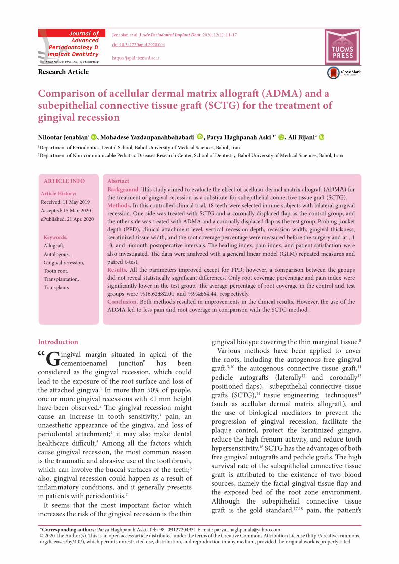
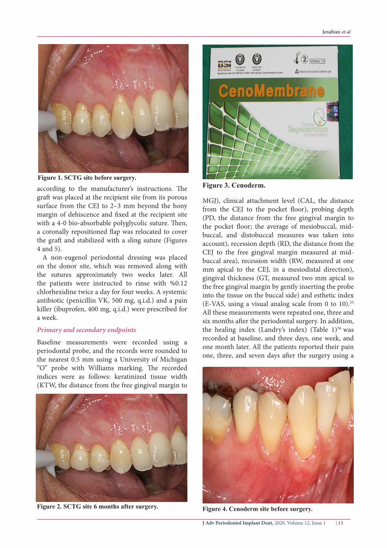
The sulcular incision was made at the recipient site The flap was beveled in the interdental papilla region adjacent to the tooth with the exposed root A partial-thickness flap was raised beyond the mucogingival junction The mesiodistal width of the incision was extended to the line angle of the adjacent teeth mesially and distally Mesial and distal vertical releasing incisions were also made The required graft width was measured using a periodontal probe A connective tissue graft was prepared from the palate measuring 1ndash15 mm in thickness and fixed at the recipient site with a 4-0 bio-absorbable polyglycolic suture Then a coronally repositioned flap was relocated to cover the graft and stabilized in the site with a sling suture (Figures 1 and 2) A 10times20-mm and 06ndash09-mm-thick graft was prepared from acellular dermal matrix graft [Cenoderm Tissue Regeneration Corporation (TRC) Iran] (Figure 3)
J Adv Periodontol Implant Dent 2020 Volume 12 Issue 1 | 13
Jenabian et al
according to the manufacturerrsquos instructions The graft was placed at the recipient site from its porous surface from the CEJ to 2ndash3 mm beyond the bony margin of dehiscence and fixed at the recipient site with a 4-0 bio-absorbable polyglycolic suture Then a coronally repositioned flap was relocated to cover the graft and stabilized with a sling suture (Figures 4 and 5)
A non-eugenol periodontal dressing was placed on the donor site which was removed along with the sutures approximately two weeks later All the patients were instructed to rinse with 012 chlorhexidine twice a day for four weeks A systemic antibiotic (penicillin VK 500 mg qid) and a pain killer (ibuprofen 400 mg qid) were prescribed for a week
Primary and secondary endpoints
Baseline measurements were recorded using a periodontal probe and the records were rounded to the nearest 05 mm using a University of Michigan ldquoOrdquo probe with Williams marking The recorded indices were as follows keratinized tissue width (KTW the distance from the free gingival margin to
MGJ) clinical attachment level (CAL the distance from the CEJ to the pocket floor) probing depth (PD the distance from the free gingival margin to the pocket floor the average of mesiobuccal mid-buccal and distobuccal measures was taken into account) recession depth (RD the distance from the CEJ to the free gingival margin measured at mid-buccal area) recession width (RW measured at one mm apical to the CEJ in a mesiodistal direction) gingival thickness (GT measured two mm apical to the free gingival margin by gently inserting the probe into the tissue on the buccal side) and esthetic index (E-VAS using a visual analog scale from 0 to 10)35 All these measurements were repeated one three and six months after the periodontal surgery In addition the healing index (Landryrsquos index) (Table 1)36 was recorded at baseline and three days one week and one month later All the patients reported their pain one three and seven days after the surgery using a
Figure 1 SCTG site before surgery
Figure 2 SCTG site 6 months after surgery
Figure 3 Cenoderm
Figure 4 Cenoderm site before surgery
Jenabian et al
J Adv Periodontol Implant Dent 2020 Volume 12 Issue 114 |
visual analog scale (VAS from 0 to 10) diagram35 The root coverage index was obtained from the following equation (Recession depth at baseline ndash Recession depth at the 6th month)Recession depth at baseline) times100 In all the sessions clinical photographs were also taken
Study population
The subjects were recruited from patients referred to the Department of Periodontics Dental School Babol University of Medical Sciences Babol Iran Patients with bilateral gingival recession were selected from enrolled patients and randomly recruited in the clinical trial
Statistical analysis
The sample size was estimated at nine subjects (n=9)
to achieve an 80 power for a standardized difference of 18 between the two study arms calculated for the primary endpoint (recession depth) using an Altmanrsquos nomogram Continuous data were expressed as means (plusmnstandard deviations) Kolmogorov-Smirnov test was used to assess the normal distribution of the data The changing trend of each index within each study arm group was traced using GLM repeated measures (RM) statistics Sphericity (one of the GLM-RM assumptions) was tested with Mauchlyrsquos test and the data were adjusted and reported with epsilon GreenhousendashGeisser correction in case of violation
Furthermore a Bonferroni test was used to evaluate differences between the groups The Friedman test was applied to trace the changing trends of nonparametric data Moreover the mean difference in each non-normally distributed index was calculated from the beginning to the end of the study and compared with a paired t-test A two-tailed α at Plt005 was considered statistically significant
Results
Baseline characteristics
Baseline indices of the patients at T0 were compared between the two groups to figure out any possible confounding effect due to the baseline differences The t-test showed no significant difference in any of the indices (Plt005)
Primary and secondary endpoints (Overall group analysis)
Changing trends of measured indices were all statistically significant as shown in Figures 6 and 7 Except for probing depth [X2(3)=1005 P=002)] others improved including recession depth (F(194)=8415 Plt0001) recession width (F(193)=7793 Plt0001) the width of keratinized gingiva (F(166)=3784 Plt0001) clinical attachment level (F(184)= 4504 Plt0001) pain (F(2)=8378 Plt0001) thickness of keratinized gingiva [X2(3)=4631
Figure 5 Cenoderm site- 6 months after surgery
Figure 6 The left side displays SCTG data and the right side displays ADMA data RW recession width RD recession depth KTW keratinized tissue width PD probing depth CAL clinical attachment level GT gingival thickness T0 baseline T1 first month T2 third month T3 sixth month)
1 Very poor
bull Tissue color ge50 of the gingiva redbull Response to palpation bleedingbull Granulation tissue presentbull Incision margin not epithelialized with loss
of epithelium beyond the incision marginbull Suppuration present
2 Poor bull Tissue color ge50 of the gingiva redbull Response to palpation bleedingbull Granulation tissue presentbull Incision margin not epithelialized with the
connective tissue exposed3 Good bull Tissue color ge25 and lt50 of the gingiva
redbull Response to palpation no bleedingbull Granulation tissue nonebull Incision margin no connective tissue exposed
4 Very good bull Tissue color lt25 of the gingiva redbull Response to palpation no bleedingbull Granulation tissue nonebull Incision margin no connective tissue exposed
5 Excellent bull Tissue color all the tissues pinkbull Response to palpation no bleedingbull Granulation tissue nonebull Incision margin no connective tissue exposed
Table 1 Healing index by Landry et al
J Adv Periodontol Implant Dent 2020 Volume 12 Issue 1 | 15
Jenabian et al
Plt0001)] healing index [X2(3)=4776 Plt0001)] and esthetic [X2(3)=5372 Plt0001)]
Primary and secondary endpoints (Inter-group analyses)
In intergroup comparisons patients in the SCTG group reported greater pain [(SCTG=419plusmn018) ADMA=319plusmn018 mean diff=096 P=0001 power=095)] and displayed better root coverage [(SCTG= 8201plusmn1662) (ADMA=6444plusmn94) (t(8)=343) (mean diff=543) P=0009)] Meanwhile other indices were not in contrast with the study groups esthetic improvement [(SCTG=733 (plusmn111) (ADMA=7plusmn087) (t(8)=2) (mean diff=033) P=008)] improved healing [(SCTG=188plusmn035) (ADMA=21plusmn06) (t(8)=15) (mean diff=022) P=017)] increased thickness of attached gingiva [(SCTG=089plusmn033 mm) (ADMA=06plusmn033 mm) (t(8)=151) (mean diff=022) P=017)] probing depth change [(SCTG=0plusmn071 mm) (ADMA= -006plusmn073 mm) t((8)= -019) (mean diff=005) P=086)] improved clinical attachment level [(SCTG=256plusmn03 mm) (ADMA=314plusmn03 mm) (mean diff= 058) P=019 power=025)] gained width of keratinized gingiva [(SCTG=348plusmn068 mm) (ADMA=322plusmn068 mm) (mean diff=026) P=079 power=006)] improved recession depth [(SCTG=129plusmn029 mm) (ADMA=163plusmn029 mm) (mean diff=034) P=042 power=012)] and improved recession depth [(SCTG=152plusmn027 mm] (ADMA=202plusmn028 mm) (mean diff=049) P= 023 power=040)] (Figure 6 and 7)
Discussion
In this research CAF + Cenoderm (test group) was compared with CAF + SCTG (control group) for
the treatment of Miller class Ill buccal gingival recession
No significant difference was observed between the two groups in any of the parameters at baseline The average percentage of root coverage in the Cenoderm group was significantly less than that in the SCTG group (6444plusmn94 and 8201plusmn1662 in test and control groups respectively) consistent with the findings reported by Novaes et al37 Barros et al38 Bouchard et al39 and Agarwal et al40 which were expected because the subepithelial connective tissue graft is a gold standard method
In the present study the average clinical attachment gain improved in both groups with no significant difference between them consistent with the result of the studies by Novaes et al37 Haghighati et al41 Hirsch et al42 and Juluri et al43
The changes in probing depth were not significant in the two groups indicating that PPD did not change during the study consistent with several previous studies37414445 but not all of them46 These differences in PPD values could be attributed to the variations in the initial PPD and differences in examiners
According to Gholami et al45 the use of SCTG leads to more gains in the keratinized gingiva compared to ADMA They believed that this is because ADMA needs more time for keratinization of the epithelial surface However a study by Novaes et al37 was consistent with this study which showed an increase in the keratinized tissue width in both methods (SCTG and ADMA) after six months with no significant difference between them
The mean average of depth and width reduction of gingival loss was enhanced in both groups with no significant difference between them consistent with the results reported by Novaes et al37 Rahmani et al44 Gholami et al45 and Shori et al46
Despite the difference in the amount of coverage between the two groups there was no difference between these two treatment methods in terms of the VAS index for esthetic
An assessment of tissue repair was carried out by Landry index in each group which showed an improvement with no significant difference between the two groups
The indices of healing esthetic and pain have not been investigated in other studies
The gingival thickness too improved in both groups with no significant difference between them VAS for pain showed an apparent difference between the two groups The pain was considerably less severe in the test group in comparison with the control group These results were somehow predictable because the surgical site was more
Figure 7 The left side displays SCTG data and the right side displays ADMA data HI healing index (T0 baseline T1 third day T2 first week T3 first month) EVAS esthetic visual analogue scale (T0 baseline T1 first month T2 third month T3 sixth month) PVAS pain visual analogue scale (T0 first day T1 third day T2 seventh day)
Jenabian et al
J Adv Periodontol Implant Dent 2020 Volume 12 Issue 116 |
extensive with the SCTG technique compared to the ADMA technique According to this study patients used more analgesics for a longer duration47 These results could be considered as one of the advantages of the ADMA method
Furthermore it is possible to treat a broader region in this technique in comparison with the other techniques such as SCTG The advantage of fewer complications of ADMA (Cenoderm) could be exploited in young people under orthodontic treatments and older people with systemic problems especially those with bleeding disorders
Conclusion
According to the results of this study both SCTG and Cenoderm resulted in improvements in the clinical outcomes However the use of Cenoderm led to less severe pain and less root coverage in comparison with the SCTG techniqueAcknowledgement
None declared
Authorsrsquo Contributions
NJ Executor MY Executor PHA Executor and corresponding author AB Statistics Consultant All authors have read and approved the final manuscript
Funding
Council of technology and research Babol university of Medical Sciences
Ethics Approval
The study protocol was approved by the Ethical Committee of Babol University of Medical Sciences and registered at httpwwwirctir (registration number IRCT201305201760N23 date registered March 23 2013)
Competing of Interests
Yes- Cenoderm (Tissue Regeneration Corporation)
References1 American Academy of Periodontology Glossary of
periodontal terms 4th ed Chicago2001- 4th edition2 Kassab MM Cohen RE The etiology and prevalence of
gingival recession J Am Dent Assoc 2003 Feb134(2)220-53 Rees JS Addy M A cross-sectional study of dentine
hypersensitivity J Clin Periodontol 2002 Nov29(11)997-1003
4 Goutoudi P Koidis PT Konstantinidis A Gingival recession a cross-sectional clinical investigation Eur J Prosthodont Restor Dent 1997 Jun5(2)57-61
5 Oliver RC Brown LJ Loe H Periodontal diseases in the United States population J Periodontol 1998 Feb69(2)269-78
6 Wennstrom JL Mucogingival therapy Ann Periodontol 1996 Nov1(1)671-701
7 Susin C Haas AN Oppermann RV Haugejorden O Albandar JM Gingival recession epidemiology and risk indicators in a representative urban Brazilian population J
Periodontol 2004 Oct75(10)1377-868 Muller HP Eger T Schorb A Gingival dimensions after
root coverage with free connective tissue grafts J Clin Periodontol 1998 May25(5)424-30
9 H B Free transplantation of gingiva propria Sven Tandlak Tidskr 196322684-9
10 Nabers JM Free gingival grafts Periodontics 1966 Sep-Oct4(5)243-5
11 Levine RA Covering denuded maxillary root surfaces with the subepithelial connective tissue graft Compendium 1991 Aug12(8)568 70 72 passim
12 Grupe H WR Repair of gingival defects by a sliding flap operation J Periodontol 195656715-20
13 Pini Prato G Pagliaro U Baldi C Nieri M Saletta D Cairo F et al Coronally advanced flap procedure for root coverage Flap with tension versus flap without tension a randomized controlled clinical study J Periodontol 2000 Feb71(2)188-201
14 Langer B Langer L Subepithelial connective tissue graft technique for root coverage J Periodontol 1985 Dec56(12)715-20
15 McGuire MK Nunn ME Evaluation of the safety and efficacy of periodontal applications of a living tissue-engineered human fibroblast-derived dermal substitute I Comparison to the gingival autograft a randomized controlled pilot study J Periodontol 2005 Jun76(6)867-80
16 McGuire MK Scheyer ET Schupbach P Growth factor-mediated treatment of recession defects a randomized controlled trial and histologic and microcomputed tomography examination J Periodontol 2009 Apr80(4)550-64
17 Wennstrom JL Zucchelli G Increased gingival dimensions A significant factor for successful outcome of root coverage procedures A 2-year prospective clinical study J Clin Periodontol 1996 Aug23(8)770-7
18 Paolantonio M Treatment of gingival recessions by combined periodontal regenerative technique guided tissue regeneration and subpedicle connective tissue graft A comparative clinical study J Periodontol 2002 Jan73(1)53-62
19 Park JB Increasing the width of keratinized mucosa around endosseous implant using acellular dermal matrix allograft Implant Dent 2006 Sep15(3)275-81
20 Yan JJ Tsai AY Wong MY Hou LT Comparison of acellular dermal graft and palatal autograft in the reconstruction of keratinized gingiva around dental implants a case report Int J Periodontics Restorative Dent 2006 Jun26(3)287-92
21 Imberman M Gingival augmentation with an acellular dermal matrix revisited surgical technique for gingival grafting Pract Proced Aesthet Dent 2007 Mar19(2)123-8
22 Achauer BM VanderKam VM Celikoz B Jacobson DG Augmentation of facial soft-tissue defects with Alloderm dermal graft Ann Plast Surg 1998 Nov41(5)503-7
23 Tobin HA Karas ND Lip augmentation using an alloderm graft J Oral Maxillofac Surg 1998 Jun56(6)722-7
24 Kridel RW Foda H Lunde KC Septal perforation repair with acellular human dermal allograft Arch Otolaryngol Head Neck Surg 1998 Jan124(1)73-8
25 Wainwright D Madden M Luterman A Hunt J Monafo W Heimbach D et al Clinical evaluation of an acellular allograft dermal matrix in full-thickness burns J Burn Care Rehabil 1996 Mar-Apr17(2)124-36
26 Wainwright DJ Use of an acellular allograft dermal matrix (AlloDerm) in the management of full-thickness burns Burns 1995 Jun21(4)243-8
27 Livesey SA Herndon DN Hollyoak MA Atkinson YH Nag A Transplanted acellular allograft dermal matrix Potential
J Adv Periodontol Implant Dent 2020 Volume 12 Issue 1 | 17
Jenabian et al
as a template for the reconstruction of viable dermis Transplantation 1995 Jul 1560(1)1-9
28 Callan DP Silverstein LH Use of acellular dermal matrix for increasing keratinized tissue around teeth and implants Pract Periodontics Aesthet Dent 1998 Aug10(6)731-4
29 Harris RJ A comparative study of root coverage obtained with an acellular dermal matrix versus a connective tissue graft results of 107 recession defects in 50 consecutively treated patients Int J Periodontics Restorative Dent 2000 Feb20(1)51-9
30 Henderson RD Greenwell H Drisko C Regennitter FJ Lamb JW Mehlbauer MJ et al Predictable multiple site root coverage using an acellular dermal matrix allograft J Periodontol 2001 May72(5)571-82
31 de Queiroz Cortes A Sallum AW Casati MZ Nociti FH Jr Sallum EA A two-year prospective study of coronally positioned flap with or without acellular dermal matrix graft J Clin Periodontol 2006 Sep33(9)683-9
32 Harris RJ Cellular dermal matrix used for root coverage 18-month follow-up observation Int J Periodontics Restorative Dent 2002 Apr22(2)156-63
33 Gapski R Parks CA Wang HL Acellular dermal matrix for mucogingival surgery a meta-analysis J Periodontol 2005 Nov76(11)1814-22
34 OrsquoLeary TJ Drake RB Naylor JE The plaque control record J Periodontol 1972 Jan43(1)38
35 Jenabian N Yazdanpanahbahabadi M Bijani A Rahimirad M Gingival unit graft versus free gingival graft for treatment of gingival recession A randomized controlled clinical trial JDT 2016 June 13(3) 184-92
36 Landry R TR Howley T Effectiveness of benzydamine HCL in the treatment of periodontal postsurgical patients Res Clin Forum 198810105-18
37 Novaes AB Jr Grisi DC Molina GO Souza SL Taba M Jr Grisi MF Comparative 6-month clinical study of a subepithelial connective tissue graft and acellular dermal matrix graft for the treatment of gingival recession J Periodontol 2001 Nov72(11)1477-84
38 Barros RR Novaes AB Grisi MF Souza SL Taba MJ Palioto DB A 6-month comparative clinical study of a conventional
and a new surgical approach for root coverage with acellular dermal matrix J Periodontol 2004 Oct75(10)1350-6
39 Bouchard P Etienne D Ouhayoun JP Nilveus R Subepithelial connective tissue grafts in the treatment of gingival recessions A comparative study of 2 procedures J Periodontol 1994 Oct65(10)929-36
40 Agarwal C Kumar BT Mehta DS An acellular dermal matrix allograft (Allodermreg) for increasing keratinized attached gingiva A case series JISP 201519(2)216-20
41 Haghighati F MM Moslemi N Comparative clinical evaluation of subepithelial connective tissue graft and acellular dermal matrix allograft for the treatment of gingival recession JDT 20063(4)159-66
42 Hirsch A Goldstein M Goultschin J Boyan BD Schwartz Z A 2-year follow-up of root coverage using sub-pedicle acellular dermal matrix allografts and subepithelial connective tissue autografts J Periodontol 2005 Aug76(8)1323-8
43 Juluri R Gopalakrishnan D Prasad N Chialastri SM Balasundaram A Martande S Augmentation of Keratinized Tissue around Implants using Acellular Dermal Matrix Allograft A Case Report J Dental Oral Health 2018 August4(3)1-4
44 Rahmani ME Lades MA Comparative clinical evaluation of acellular dermal matrix allograft and connective tissue graft for the treatment of gingival recession J Contemp Dent Pract 2006 May 17(2)63-70
45 Gholami GA Saberi A Kadkhodazadeh M Amid R Karami D Comparison of the clinical outcomes of connective tissue and acellular dermal matrix in combination with double papillary flap for root coverage A 6-month trial Dent Res J (Isfahan) 2013 Jul10(4)506-13
46 Shori T Kolte A Kher V Dharamthok S Shrirao T A comparative evaluation of the effectiveness of subpedicle acellular dermal matrix allograft with subepithelial connective tissue graft in the treatment of isolated marginal tissue recession A clinical study J Indian Soc Periodontol 2013 Jan17(1)78-81
47 Harris RJ Clinical evaluation of 3 techniques to augment keratinized tissue without root coverage J Periodontol 2001 Jul72(7)932-8

Jenabian et al
J Adv Periodontol Implant Dent 2020 Volume 12 Issue 112 |
inconvenience the sufficiency of the donor area bleeding of the harvesting region and a need for a second surgery are the complications of this method Therefore the acellular dermal matrix allograft has been widely applied as a substitute for autogenous tissue grafts in mucogingival surgeries19-21 This allograft is made of human corpse skin through decellularization freezing and drying processes to prevent inflammation or rejection of the transplant However the essential factors of the tissue structure such as collagen elastin proteoglycans and vascular channels are maintained in the process22-24 According to the studies due to the presence of vascular channels in the region ADMA would be integrated with the host tissue and preserve its structural integrity25-30
Many studies concluded that the application of ADMA is comparable to subepithelial connective tissue graft The mean root coverage of these two methods did not show clinically significant differences30-33
In this regard it seems that this method could be an appropriate alternative to connective tissue grafts especially in young and older people and also those who are not systematically fit for intensive surgeries
Recently this allograft has been produced in Iran by the Tissue Regeneration Corporation (TRC) under the proprietary name Cenoderm It would be more appropriate for both the patient and clinician due to its availability and price because it is manufactured in Iran The present study was conducted to clinically compare the acellular dermal matrix allograft (Cenoderm) and subepithelial connective tissue graft
Methods
Eighteen teeth were selected in nine subjects with bilateral gingival recession for this randomized double-blind controlled split-mouth study The inclusion criteria were as follows (1) ge18 years of age (2) ability to maintain proper oral hygiene (OrsquoLeary plaque score34 le 20) (3) all recessions in either Miller I or II category (4) bilateral isolated buccal gingival recession with a depth of at least 2 mm from the cementoenamel junction (CEJ) in the anterior and premolar teeth (5) no filling and bleeding upon probing in the selected teeth (6) and adequate vestibular depth The exclusion criteria were defined as follows (1) pregnancy (2) confounding medications interfering with wound healing (eg anti-neoplastic agents and corticosteroids) (3) cigarette smoking (4) traumatic methods of brushing and application of abrasive toothpaste (5) a history of periodontal surgery in the past two years (6) wearing removable prostheses or orthodontic
appliances in the designated area (7) long-term (ie gt2 weeks) use of antibiotics in the last three months and (8) known allergies to the materials used during periodontal surgery
This study was reviewed and approved by the Ethics Committee of Babol University of Medical Sciences and registered at httpwwwirctir (registration number IRCT201305201760N23 registration date 2013-08-23) All the enrolled patients were provided with an oral explanation of the study and written informed consent was obtained
Randomization and blinding
The patients were treated either with subepithelial connective tissue graft and a coronally positioned flap or an acellular dermal matrix allograft with a coronally positioned flap
A 4-interval permuted block method was used to assign the patients to receive any type of intervention on the right side (ie arbitrarily selected) The assignments were sent in closed envelopes to the clinician in charge (PH) who was unaware of the assignment codes Further measurements of periodontal indices were performed by another clinician (NJ) who was also blinded to the study arms All the surgeries were performed by the same clinician and the measurements and randomization were made by another clinician
Study design
Phase I periodontal therapy was performed for all the patients All the procedures were performed by the same person (PH) After adequate and profound local anesthesia (ie infiltration method) with 2 lidocaine containing epinephrine at a concentration of 180000 all the exposed roots were carefully prepared by scaling and root planing
The sulcular incision was made at the recipient site The flap was beveled in the interdental papilla region adjacent to the tooth with the exposed root A partial-thickness flap was raised beyond the mucogingival junction The mesiodistal width of the incision was extended to the line angle of the adjacent teeth mesially and distally Mesial and distal vertical releasing incisions were also made The required graft width was measured using a periodontal probe A connective tissue graft was prepared from the palate measuring 1ndash15 mm in thickness and fixed at the recipient site with a 4-0 bio-absorbable polyglycolic suture Then a coronally repositioned flap was relocated to cover the graft and stabilized in the site with a sling suture (Figures 1 and 2) A 10times20-mm and 06ndash09-mm-thick graft was prepared from acellular dermal matrix graft [Cenoderm Tissue Regeneration Corporation (TRC) Iran] (Figure 3)
J Adv Periodontol Implant Dent 2020 Volume 12 Issue 1 | 13
Jenabian et al
according to the manufacturerrsquos instructions The graft was placed at the recipient site from its porous surface from the CEJ to 2ndash3 mm beyond the bony margin of dehiscence and fixed at the recipient site with a 4-0 bio-absorbable polyglycolic suture Then a coronally repositioned flap was relocated to cover the graft and stabilized with a sling suture (Figures 4 and 5)
A non-eugenol periodontal dressing was placed on the donor site which was removed along with the sutures approximately two weeks later All the patients were instructed to rinse with 012 chlorhexidine twice a day for four weeks A systemic antibiotic (penicillin VK 500 mg qid) and a pain killer (ibuprofen 400 mg qid) were prescribed for a week
Primary and secondary endpoints
Baseline measurements were recorded using a periodontal probe and the records were rounded to the nearest 05 mm using a University of Michigan ldquoOrdquo probe with Williams marking The recorded indices were as follows keratinized tissue width (KTW the distance from the free gingival margin to
MGJ) clinical attachment level (CAL the distance from the CEJ to the pocket floor) probing depth (PD the distance from the free gingival margin to the pocket floor the average of mesiobuccal mid-buccal and distobuccal measures was taken into account) recession depth (RD the distance from the CEJ to the free gingival margin measured at mid-buccal area) recession width (RW measured at one mm apical to the CEJ in a mesiodistal direction) gingival thickness (GT measured two mm apical to the free gingival margin by gently inserting the probe into the tissue on the buccal side) and esthetic index (E-VAS using a visual analog scale from 0 to 10)35 All these measurements were repeated one three and six months after the periodontal surgery In addition the healing index (Landryrsquos index) (Table 1)36 was recorded at baseline and three days one week and one month later All the patients reported their pain one three and seven days after the surgery using a
Figure 1 SCTG site before surgery
Figure 2 SCTG site 6 months after surgery
Figure 3 Cenoderm
Figure 4 Cenoderm site before surgery
Jenabian et al
J Adv Periodontol Implant Dent 2020 Volume 12 Issue 114 |
visual analog scale (VAS from 0 to 10) diagram35 The root coverage index was obtained from the following equation (Recession depth at baseline ndash Recession depth at the 6th month)Recession depth at baseline) times100 In all the sessions clinical photographs were also taken
Study population
The subjects were recruited from patients referred to the Department of Periodontics Dental School Babol University of Medical Sciences Babol Iran Patients with bilateral gingival recession were selected from enrolled patients and randomly recruited in the clinical trial
Statistical analysis
The sample size was estimated at nine subjects (n=9)
to achieve an 80 power for a standardized difference of 18 between the two study arms calculated for the primary endpoint (recession depth) using an Altmanrsquos nomogram Continuous data were expressed as means (plusmnstandard deviations) Kolmogorov-Smirnov test was used to assess the normal distribution of the data The changing trend of each index within each study arm group was traced using GLM repeated measures (RM) statistics Sphericity (one of the GLM-RM assumptions) was tested with Mauchlyrsquos test and the data were adjusted and reported with epsilon GreenhousendashGeisser correction in case of violation
Furthermore a Bonferroni test was used to evaluate differences between the groups The Friedman test was applied to trace the changing trends of nonparametric data Moreover the mean difference in each non-normally distributed index was calculated from the beginning to the end of the study and compared with a paired t-test A two-tailed α at Plt005 was considered statistically significant
Results
Baseline characteristics
Baseline indices of the patients at T0 were compared between the two groups to figure out any possible confounding effect due to the baseline differences The t-test showed no significant difference in any of the indices (Plt005)
Primary and secondary endpoints (Overall group analysis)
Changing trends of measured indices were all statistically significant as shown in Figures 6 and 7 Except for probing depth [X2(3)=1005 P=002)] others improved including recession depth (F(194)=8415 Plt0001) recession width (F(193)=7793 Plt0001) the width of keratinized gingiva (F(166)=3784 Plt0001) clinical attachment level (F(184)= 4504 Plt0001) pain (F(2)=8378 Plt0001) thickness of keratinized gingiva [X2(3)=4631
Figure 5 Cenoderm site- 6 months after surgery
Figure 6 The left side displays SCTG data and the right side displays ADMA data RW recession width RD recession depth KTW keratinized tissue width PD probing depth CAL clinical attachment level GT gingival thickness T0 baseline T1 first month T2 third month T3 sixth month)
1 Very poor
bull Tissue color ge50 of the gingiva redbull Response to palpation bleedingbull Granulation tissue presentbull Incision margin not epithelialized with loss
of epithelium beyond the incision marginbull Suppuration present
2 Poor bull Tissue color ge50 of the gingiva redbull Response to palpation bleedingbull Granulation tissue presentbull Incision margin not epithelialized with the
connective tissue exposed3 Good bull Tissue color ge25 and lt50 of the gingiva
redbull Response to palpation no bleedingbull Granulation tissue nonebull Incision margin no connective tissue exposed
4 Very good bull Tissue color lt25 of the gingiva redbull Response to palpation no bleedingbull Granulation tissue nonebull Incision margin no connective tissue exposed
5 Excellent bull Tissue color all the tissues pinkbull Response to palpation no bleedingbull Granulation tissue nonebull Incision margin no connective tissue exposed
Table 1 Healing index by Landry et al
J Adv Periodontol Implant Dent 2020 Volume 12 Issue 1 | 15
Jenabian et al
Plt0001)] healing index [X2(3)=4776 Plt0001)] and esthetic [X2(3)=5372 Plt0001)]
Primary and secondary endpoints (Inter-group analyses)
In intergroup comparisons patients in the SCTG group reported greater pain [(SCTG=419plusmn018) ADMA=319plusmn018 mean diff=096 P=0001 power=095)] and displayed better root coverage [(SCTG= 8201plusmn1662) (ADMA=6444plusmn94) (t(8)=343) (mean diff=543) P=0009)] Meanwhile other indices were not in contrast with the study groups esthetic improvement [(SCTG=733 (plusmn111) (ADMA=7plusmn087) (t(8)=2) (mean diff=033) P=008)] improved healing [(SCTG=188plusmn035) (ADMA=21plusmn06) (t(8)=15) (mean diff=022) P=017)] increased thickness of attached gingiva [(SCTG=089plusmn033 mm) (ADMA=06plusmn033 mm) (t(8)=151) (mean diff=022) P=017)] probing depth change [(SCTG=0plusmn071 mm) (ADMA= -006plusmn073 mm) t((8)= -019) (mean diff=005) P=086)] improved clinical attachment level [(SCTG=256plusmn03 mm) (ADMA=314plusmn03 mm) (mean diff= 058) P=019 power=025)] gained width of keratinized gingiva [(SCTG=348plusmn068 mm) (ADMA=322plusmn068 mm) (mean diff=026) P=079 power=006)] improved recession depth [(SCTG=129plusmn029 mm) (ADMA=163plusmn029 mm) (mean diff=034) P=042 power=012)] and improved recession depth [(SCTG=152plusmn027 mm] (ADMA=202plusmn028 mm) (mean diff=049) P= 023 power=040)] (Figure 6 and 7)
Discussion
In this research CAF + Cenoderm (test group) was compared with CAF + SCTG (control group) for
the treatment of Miller class Ill buccal gingival recession
No significant difference was observed between the two groups in any of the parameters at baseline The average percentage of root coverage in the Cenoderm group was significantly less than that in the SCTG group (6444plusmn94 and 8201plusmn1662 in test and control groups respectively) consistent with the findings reported by Novaes et al37 Barros et al38 Bouchard et al39 and Agarwal et al40 which were expected because the subepithelial connective tissue graft is a gold standard method
In the present study the average clinical attachment gain improved in both groups with no significant difference between them consistent with the result of the studies by Novaes et al37 Haghighati et al41 Hirsch et al42 and Juluri et al43
The changes in probing depth were not significant in the two groups indicating that PPD did not change during the study consistent with several previous studies37414445 but not all of them46 These differences in PPD values could be attributed to the variations in the initial PPD and differences in examiners
According to Gholami et al45 the use of SCTG leads to more gains in the keratinized gingiva compared to ADMA They believed that this is because ADMA needs more time for keratinization of the epithelial surface However a study by Novaes et al37 was consistent with this study which showed an increase in the keratinized tissue width in both methods (SCTG and ADMA) after six months with no significant difference between them
The mean average of depth and width reduction of gingival loss was enhanced in both groups with no significant difference between them consistent with the results reported by Novaes et al37 Rahmani et al44 Gholami et al45 and Shori et al46
Despite the difference in the amount of coverage between the two groups there was no difference between these two treatment methods in terms of the VAS index for esthetic
An assessment of tissue repair was carried out by Landry index in each group which showed an improvement with no significant difference between the two groups
The indices of healing esthetic and pain have not been investigated in other studies
The gingival thickness too improved in both groups with no significant difference between them VAS for pain showed an apparent difference between the two groups The pain was considerably less severe in the test group in comparison with the control group These results were somehow predictable because the surgical site was more
Figure 7 The left side displays SCTG data and the right side displays ADMA data HI healing index (T0 baseline T1 third day T2 first week T3 first month) EVAS esthetic visual analogue scale (T0 baseline T1 first month T2 third month T3 sixth month) PVAS pain visual analogue scale (T0 first day T1 third day T2 seventh day)
Jenabian et al
J Adv Periodontol Implant Dent 2020 Volume 12 Issue 116 |
extensive with the SCTG technique compared to the ADMA technique According to this study patients used more analgesics for a longer duration47 These results could be considered as one of the advantages of the ADMA method
Furthermore it is possible to treat a broader region in this technique in comparison with the other techniques such as SCTG The advantage of fewer complications of ADMA (Cenoderm) could be exploited in young people under orthodontic treatments and older people with systemic problems especially those with bleeding disorders
Conclusion
According to the results of this study both SCTG and Cenoderm resulted in improvements in the clinical outcomes However the use of Cenoderm led to less severe pain and less root coverage in comparison with the SCTG techniqueAcknowledgement
None declared
Authorsrsquo Contributions
NJ Executor MY Executor PHA Executor and corresponding author AB Statistics Consultant All authors have read and approved the final manuscript
Funding
Council of technology and research Babol university of Medical Sciences
Ethics Approval
The study protocol was approved by the Ethical Committee of Babol University of Medical Sciences and registered at httpwwwirctir (registration number IRCT201305201760N23 date registered March 23 2013)
Competing of Interests
Yes- Cenoderm (Tissue Regeneration Corporation)
References1 American Academy of Periodontology Glossary of
periodontal terms 4th ed Chicago2001- 4th edition2 Kassab MM Cohen RE The etiology and prevalence of
gingival recession J Am Dent Assoc 2003 Feb134(2)220-53 Rees JS Addy M A cross-sectional study of dentine
hypersensitivity J Clin Periodontol 2002 Nov29(11)997-1003
4 Goutoudi P Koidis PT Konstantinidis A Gingival recession a cross-sectional clinical investigation Eur J Prosthodont Restor Dent 1997 Jun5(2)57-61
5 Oliver RC Brown LJ Loe H Periodontal diseases in the United States population J Periodontol 1998 Feb69(2)269-78
6 Wennstrom JL Mucogingival therapy Ann Periodontol 1996 Nov1(1)671-701
7 Susin C Haas AN Oppermann RV Haugejorden O Albandar JM Gingival recession epidemiology and risk indicators in a representative urban Brazilian population J
Periodontol 2004 Oct75(10)1377-868 Muller HP Eger T Schorb A Gingival dimensions after
root coverage with free connective tissue grafts J Clin Periodontol 1998 May25(5)424-30
9 H B Free transplantation of gingiva propria Sven Tandlak Tidskr 196322684-9
10 Nabers JM Free gingival grafts Periodontics 1966 Sep-Oct4(5)243-5
11 Levine RA Covering denuded maxillary root surfaces with the subepithelial connective tissue graft Compendium 1991 Aug12(8)568 70 72 passim
12 Grupe H WR Repair of gingival defects by a sliding flap operation J Periodontol 195656715-20
13 Pini Prato G Pagliaro U Baldi C Nieri M Saletta D Cairo F et al Coronally advanced flap procedure for root coverage Flap with tension versus flap without tension a randomized controlled clinical study J Periodontol 2000 Feb71(2)188-201
14 Langer B Langer L Subepithelial connective tissue graft technique for root coverage J Periodontol 1985 Dec56(12)715-20
15 McGuire MK Nunn ME Evaluation of the safety and efficacy of periodontal applications of a living tissue-engineered human fibroblast-derived dermal substitute I Comparison to the gingival autograft a randomized controlled pilot study J Periodontol 2005 Jun76(6)867-80
16 McGuire MK Scheyer ET Schupbach P Growth factor-mediated treatment of recession defects a randomized controlled trial and histologic and microcomputed tomography examination J Periodontol 2009 Apr80(4)550-64
17 Wennstrom JL Zucchelli G Increased gingival dimensions A significant factor for successful outcome of root coverage procedures A 2-year prospective clinical study J Clin Periodontol 1996 Aug23(8)770-7
18 Paolantonio M Treatment of gingival recessions by combined periodontal regenerative technique guided tissue regeneration and subpedicle connective tissue graft A comparative clinical study J Periodontol 2002 Jan73(1)53-62
19 Park JB Increasing the width of keratinized mucosa around endosseous implant using acellular dermal matrix allograft Implant Dent 2006 Sep15(3)275-81
20 Yan JJ Tsai AY Wong MY Hou LT Comparison of acellular dermal graft and palatal autograft in the reconstruction of keratinized gingiva around dental implants a case report Int J Periodontics Restorative Dent 2006 Jun26(3)287-92
21 Imberman M Gingival augmentation with an acellular dermal matrix revisited surgical technique for gingival grafting Pract Proced Aesthet Dent 2007 Mar19(2)123-8
22 Achauer BM VanderKam VM Celikoz B Jacobson DG Augmentation of facial soft-tissue defects with Alloderm dermal graft Ann Plast Surg 1998 Nov41(5)503-7
23 Tobin HA Karas ND Lip augmentation using an alloderm graft J Oral Maxillofac Surg 1998 Jun56(6)722-7
24 Kridel RW Foda H Lunde KC Septal perforation repair with acellular human dermal allograft Arch Otolaryngol Head Neck Surg 1998 Jan124(1)73-8
25 Wainwright D Madden M Luterman A Hunt J Monafo W Heimbach D et al Clinical evaluation of an acellular allograft dermal matrix in full-thickness burns J Burn Care Rehabil 1996 Mar-Apr17(2)124-36
26 Wainwright DJ Use of an acellular allograft dermal matrix (AlloDerm) in the management of full-thickness burns Burns 1995 Jun21(4)243-8
27 Livesey SA Herndon DN Hollyoak MA Atkinson YH Nag A Transplanted acellular allograft dermal matrix Potential
J Adv Periodontol Implant Dent 2020 Volume 12 Issue 1 | 17
Jenabian et al
as a template for the reconstruction of viable dermis Transplantation 1995 Jul 1560(1)1-9
28 Callan DP Silverstein LH Use of acellular dermal matrix for increasing keratinized tissue around teeth and implants Pract Periodontics Aesthet Dent 1998 Aug10(6)731-4
29 Harris RJ A comparative study of root coverage obtained with an acellular dermal matrix versus a connective tissue graft results of 107 recession defects in 50 consecutively treated patients Int J Periodontics Restorative Dent 2000 Feb20(1)51-9
30 Henderson RD Greenwell H Drisko C Regennitter FJ Lamb JW Mehlbauer MJ et al Predictable multiple site root coverage using an acellular dermal matrix allograft J Periodontol 2001 May72(5)571-82
31 de Queiroz Cortes A Sallum AW Casati MZ Nociti FH Jr Sallum EA A two-year prospective study of coronally positioned flap with or without acellular dermal matrix graft J Clin Periodontol 2006 Sep33(9)683-9
32 Harris RJ Cellular dermal matrix used for root coverage 18-month follow-up observation Int J Periodontics Restorative Dent 2002 Apr22(2)156-63
33 Gapski R Parks CA Wang HL Acellular dermal matrix for mucogingival surgery a meta-analysis J Periodontol 2005 Nov76(11)1814-22
34 OrsquoLeary TJ Drake RB Naylor JE The plaque control record J Periodontol 1972 Jan43(1)38
35 Jenabian N Yazdanpanahbahabadi M Bijani A Rahimirad M Gingival unit graft versus free gingival graft for treatment of gingival recession A randomized controlled clinical trial JDT 2016 June 13(3) 184-92
36 Landry R TR Howley T Effectiveness of benzydamine HCL in the treatment of periodontal postsurgical patients Res Clin Forum 198810105-18
37 Novaes AB Jr Grisi DC Molina GO Souza SL Taba M Jr Grisi MF Comparative 6-month clinical study of a subepithelial connective tissue graft and acellular dermal matrix graft for the treatment of gingival recession J Periodontol 2001 Nov72(11)1477-84
38 Barros RR Novaes AB Grisi MF Souza SL Taba MJ Palioto DB A 6-month comparative clinical study of a conventional
and a new surgical approach for root coverage with acellular dermal matrix J Periodontol 2004 Oct75(10)1350-6
39 Bouchard P Etienne D Ouhayoun JP Nilveus R Subepithelial connective tissue grafts in the treatment of gingival recessions A comparative study of 2 procedures J Periodontol 1994 Oct65(10)929-36
40 Agarwal C Kumar BT Mehta DS An acellular dermal matrix allograft (Allodermreg) for increasing keratinized attached gingiva A case series JISP 201519(2)216-20
41 Haghighati F MM Moslemi N Comparative clinical evaluation of subepithelial connective tissue graft and acellular dermal matrix allograft for the treatment of gingival recession JDT 20063(4)159-66
42 Hirsch A Goldstein M Goultschin J Boyan BD Schwartz Z A 2-year follow-up of root coverage using sub-pedicle acellular dermal matrix allografts and subepithelial connective tissue autografts J Periodontol 2005 Aug76(8)1323-8
43 Juluri R Gopalakrishnan D Prasad N Chialastri SM Balasundaram A Martande S Augmentation of Keratinized Tissue around Implants using Acellular Dermal Matrix Allograft A Case Report J Dental Oral Health 2018 August4(3)1-4
44 Rahmani ME Lades MA Comparative clinical evaluation of acellular dermal matrix allograft and connective tissue graft for the treatment of gingival recession J Contemp Dent Pract 2006 May 17(2)63-70
45 Gholami GA Saberi A Kadkhodazadeh M Amid R Karami D Comparison of the clinical outcomes of connective tissue and acellular dermal matrix in combination with double papillary flap for root coverage A 6-month trial Dent Res J (Isfahan) 2013 Jul10(4)506-13
46 Shori T Kolte A Kher V Dharamthok S Shrirao T A comparative evaluation of the effectiveness of subpedicle acellular dermal matrix allograft with subepithelial connective tissue graft in the treatment of isolated marginal tissue recession A clinical study J Indian Soc Periodontol 2013 Jan17(1)78-81
47 Harris RJ Clinical evaluation of 3 techniques to augment keratinized tissue without root coverage J Periodontol 2001 Jul72(7)932-8
Jenabian et al
J Adv Periodontol Implant Dent 2020 Volume 12 Issue 114 |
visual analog scale (VAS from 0 to 10) diagram35 The root coverage index was obtained from the following equation (Recession depth at baseline ndash Recession depth at the 6th month)Recession depth at baseline) times100 In all the sessions clinical photographs were also taken
Study population
The subjects were recruited from patients referred to the Department of Periodontics Dental School Babol University of Medical Sciences Babol Iran Patients with bilateral gingival recession were selected from enrolled patients and randomly recruited in the clinical trial
Statistical analysis
The sample size was estimated at nine subjects (n=9)
to achieve an 80 power for a standardized difference of 18 between the two study arms calculated for the primary endpoint (recession depth) using an Altmanrsquos nomogram Continuous data were expressed as means (plusmnstandard deviations) Kolmogorov-Smirnov test was used to assess the normal distribution of the data The changing trend of each index within each study arm group was traced using GLM repeated measures (RM) statistics Sphericity (one of the GLM-RM assumptions) was tested with Mauchlyrsquos test and the data were adjusted and reported with epsilon GreenhousendashGeisser correction in case of violation
Furthermore a Bonferroni test was used to evaluate differences between the groups The Friedman test was applied to trace the changing trends of nonparametric data Moreover the mean difference in each non-normally distributed index was calculated from the beginning to the end of the study and compared with a paired t-test A two-tailed α at Plt005 was considered statistically significant
Results
Baseline characteristics
Baseline indices of the patients at T0 were compared between the two groups to figure out any possible confounding effect due to the baseline differences The t-test showed no significant difference in any of the indices (Plt005)
Primary and secondary endpoints (Overall group analysis)
Changing trends of measured indices were all statistically significant as shown in Figures 6 and 7 Except for probing depth [X2(3)=1005 P=002)] others improved including recession depth (F(194)=8415 Plt0001) recession width (F(193)=7793 Plt0001) the width of keratinized gingiva (F(166)=3784 Plt0001) clinical attachment level (F(184)= 4504 Plt0001) pain (F(2)=8378 Plt0001) thickness of keratinized gingiva [X2(3)=4631
Figure 5 Cenoderm site- 6 months after surgery
Figure 6 The left side displays SCTG data and the right side displays ADMA data RW recession width RD recession depth KTW keratinized tissue width PD probing depth CAL clinical attachment level GT gingival thickness T0 baseline T1 first month T2 third month T3 sixth month)
1 Very poor
bull Tissue color ge50 of the gingiva redbull Response to palpation bleedingbull Granulation tissue presentbull Incision margin not epithelialized with loss
of epithelium beyond the incision marginbull Suppuration present
2 Poor bull Tissue color ge50 of the gingiva redbull Response to palpation bleedingbull Granulation tissue presentbull Incision margin not epithelialized with the
connective tissue exposed3 Good bull Tissue color ge25 and lt50 of the gingiva
redbull Response to palpation no bleedingbull Granulation tissue nonebull Incision margin no connective tissue exposed
4 Very good bull Tissue color lt25 of the gingiva redbull Response to palpation no bleedingbull Granulation tissue nonebull Incision margin no connective tissue exposed
5 Excellent bull Tissue color all the tissues pinkbull Response to palpation no bleedingbull Granulation tissue nonebull Incision margin no connective tissue exposed
Table 1 Healing index by Landry et al
J Adv Periodontol Implant Dent 2020 Volume 12 Issue 1 | 15
Jenabian et al
Plt0001)] healing index [X2(3)=4776 Plt0001)] and esthetic [X2(3)=5372 Plt0001)]
Primary and secondary endpoints (Inter-group analyses)
In intergroup comparisons patients in the SCTG group reported greater pain [(SCTG=419plusmn018) ADMA=319plusmn018 mean diff=096 P=0001 power=095)] and displayed better root coverage [(SCTG= 8201plusmn1662) (ADMA=6444plusmn94) (t(8)=343) (mean diff=543) P=0009)] Meanwhile other indices were not in contrast with the study groups esthetic improvement [(SCTG=733 (plusmn111) (ADMA=7plusmn087) (t(8)=2) (mean diff=033) P=008)] improved healing [(SCTG=188plusmn035) (ADMA=21plusmn06) (t(8)=15) (mean diff=022) P=017)] increased thickness of attached gingiva [(SCTG=089plusmn033 mm) (ADMA=06plusmn033 mm) (t(8)=151) (mean diff=022) P=017)] probing depth change [(SCTG=0plusmn071 mm) (ADMA= -006plusmn073 mm) t((8)= -019) (mean diff=005) P=086)] improved clinical attachment level [(SCTG=256plusmn03 mm) (ADMA=314plusmn03 mm) (mean diff= 058) P=019 power=025)] gained width of keratinized gingiva [(SCTG=348plusmn068 mm) (ADMA=322plusmn068 mm) (mean diff=026) P=079 power=006)] improved recession depth [(SCTG=129plusmn029 mm) (ADMA=163plusmn029 mm) (mean diff=034) P=042 power=012)] and improved recession depth [(SCTG=152plusmn027 mm] (ADMA=202plusmn028 mm) (mean diff=049) P= 023 power=040)] (Figure 6 and 7)
Discussion
In this research CAF + Cenoderm (test group) was compared with CAF + SCTG (control group) for
the treatment of Miller class Ill buccal gingival recession
No significant difference was observed between the two groups in any of the parameters at baseline The average percentage of root coverage in the Cenoderm group was significantly less than that in the SCTG group (6444plusmn94 and 8201plusmn1662 in test and control groups respectively) consistent with the findings reported by Novaes et al37 Barros et al38 Bouchard et al39 and Agarwal et al40 which were expected because the subepithelial connective tissue graft is a gold standard method
In the present study the average clinical attachment gain improved in both groups with no significant difference between them consistent with the result of the studies by Novaes et al37 Haghighati et al41 Hirsch et al42 and Juluri et al43
The changes in probing depth were not significant in the two groups indicating that PPD did not change during the study consistent with several previous studies37414445 but not all of them46 These differences in PPD values could be attributed to the variations in the initial PPD and differences in examiners
According to Gholami et al45 the use of SCTG leads to more gains in the keratinized gingiva compared to ADMA They believed that this is because ADMA needs more time for keratinization of the epithelial surface However a study by Novaes et al37 was consistent with this study which showed an increase in the keratinized tissue width in both methods (SCTG and ADMA) after six months with no significant difference between them
The mean average of depth and width reduction of gingival loss was enhanced in both groups with no significant difference between them consistent with the results reported by Novaes et al37 Rahmani et al44 Gholami et al45 and Shori et al46
Despite the difference in the amount of coverage between the two groups there was no difference between these two treatment methods in terms of the VAS index for esthetic
An assessment of tissue repair was carried out by Landry index in each group which showed an improvement with no significant difference between the two groups
The indices of healing esthetic and pain have not been investigated in other studies
The gingival thickness too improved in both groups with no significant difference between them VAS for pain showed an apparent difference between the two groups The pain was considerably less severe in the test group in comparison with the control group These results were somehow predictable because the surgical site was more
Figure 7 The left side displays SCTG data and the right side displays ADMA data HI healing index (T0 baseline T1 third day T2 first week T3 first month) EVAS esthetic visual analogue scale (T0 baseline T1 first month T2 third month T3 sixth month) PVAS pain visual analogue scale (T0 first day T1 third day T2 seventh day)
Jenabian et al
J Adv Periodontol Implant Dent 2020 Volume 12 Issue 116 |
extensive with the SCTG technique compared to the ADMA technique According to this study patients used more analgesics for a longer duration47 These results could be considered as one of the advantages of the ADMA method
Furthermore it is possible to treat a broader region in this technique in comparison with the other techniques such as SCTG The advantage of fewer complications of ADMA (Cenoderm) could be exploited in young people under orthodontic treatments and older people with systemic problems especially those with bleeding disorders
Conclusion
According to the results of this study both SCTG and Cenoderm resulted in improvements in the clinical outcomes However the use of Cenoderm led to less severe pain and less root coverage in comparison with the SCTG techniqueAcknowledgement
None declared
Authorsrsquo Contributions
NJ Executor MY Executor PHA Executor and corresponding author AB Statistics Consultant All authors have read and approved the final manuscript
Funding
Council of technology and research Babol university of Medical Sciences
Ethics Approval
The study protocol was approved by the Ethical Committee of Babol University of Medical Sciences and registered at httpwwwirctir (registration number IRCT201305201760N23 date registered March 23 2013)
Competing of Interests
Yes- Cenoderm (Tissue Regeneration Corporation)
References1 American Academy of Periodontology Glossary of
periodontal terms 4th ed Chicago2001- 4th edition2 Kassab MM Cohen RE The etiology and prevalence of
gingival recession J Am Dent Assoc 2003 Feb134(2)220-53 Rees JS Addy M A cross-sectional study of dentine
hypersensitivity J Clin Periodontol 2002 Nov29(11)997-1003
4 Goutoudi P Koidis PT Konstantinidis A Gingival recession a cross-sectional clinical investigation Eur J Prosthodont Restor Dent 1997 Jun5(2)57-61
5 Oliver RC Brown LJ Loe H Periodontal diseases in the United States population J Periodontol 1998 Feb69(2)269-78
6 Wennstrom JL Mucogingival therapy Ann Periodontol 1996 Nov1(1)671-701
7 Susin C Haas AN Oppermann RV Haugejorden O Albandar JM Gingival recession epidemiology and risk indicators in a representative urban Brazilian population J
Periodontol 2004 Oct75(10)1377-868 Muller HP Eger T Schorb A Gingival dimensions after
root coverage with free connective tissue grafts J Clin Periodontol 1998 May25(5)424-30
9 H B Free transplantation of gingiva propria Sven Tandlak Tidskr 196322684-9
10 Nabers JM Free gingival grafts Periodontics 1966 Sep-Oct4(5)243-5
11 Levine RA Covering denuded maxillary root surfaces with the subepithelial connective tissue graft Compendium 1991 Aug12(8)568 70 72 passim
12 Grupe H WR Repair of gingival defects by a sliding flap operation J Periodontol 195656715-20
13 Pini Prato G Pagliaro U Baldi C Nieri M Saletta D Cairo F et al Coronally advanced flap procedure for root coverage Flap with tension versus flap without tension a randomized controlled clinical study J Periodontol 2000 Feb71(2)188-201
14 Langer B Langer L Subepithelial connective tissue graft technique for root coverage J Periodontol 1985 Dec56(12)715-20
15 McGuire MK Nunn ME Evaluation of the safety and efficacy of periodontal applications of a living tissue-engineered human fibroblast-derived dermal substitute I Comparison to the gingival autograft a randomized controlled pilot study J Periodontol 2005 Jun76(6)867-80
16 McGuire MK Scheyer ET Schupbach P Growth factor-mediated treatment of recession defects a randomized controlled trial and histologic and microcomputed tomography examination J Periodontol 2009 Apr80(4)550-64
17 Wennstrom JL Zucchelli G Increased gingival dimensions A significant factor for successful outcome of root coverage procedures A 2-year prospective clinical study J Clin Periodontol 1996 Aug23(8)770-7
18 Paolantonio M Treatment of gingival recessions by combined periodontal regenerative technique guided tissue regeneration and subpedicle connective tissue graft A comparative clinical study J Periodontol 2002 Jan73(1)53-62
19 Park JB Increasing the width of keratinized mucosa around endosseous implant using acellular dermal matrix allograft Implant Dent 2006 Sep15(3)275-81
20 Yan JJ Tsai AY Wong MY Hou LT Comparison of acellular dermal graft and palatal autograft in the reconstruction of keratinized gingiva around dental implants a case report Int J Periodontics Restorative Dent 2006 Jun26(3)287-92
21 Imberman M Gingival augmentation with an acellular dermal matrix revisited surgical technique for gingival grafting Pract Proced Aesthet Dent 2007 Mar19(2)123-8
22 Achauer BM VanderKam VM Celikoz B Jacobson DG Augmentation of facial soft-tissue defects with Alloderm dermal graft Ann Plast Surg 1998 Nov41(5)503-7
23 Tobin HA Karas ND Lip augmentation using an alloderm graft J Oral Maxillofac Surg 1998 Jun56(6)722-7
24 Kridel RW Foda H Lunde KC Septal perforation repair with acellular human dermal allograft Arch Otolaryngol Head Neck Surg 1998 Jan124(1)73-8
25 Wainwright D Madden M Luterman A Hunt J Monafo W Heimbach D et al Clinical evaluation of an acellular allograft dermal matrix in full-thickness burns J Burn Care Rehabil 1996 Mar-Apr17(2)124-36
26 Wainwright DJ Use of an acellular allograft dermal matrix (AlloDerm) in the management of full-thickness burns Burns 1995 Jun21(4)243-8
27 Livesey SA Herndon DN Hollyoak MA Atkinson YH Nag A Transplanted acellular allograft dermal matrix Potential
J Adv Periodontol Implant Dent 2020 Volume 12 Issue 1 | 17
Jenabian et al
as a template for the reconstruction of viable dermis Transplantation 1995 Jul 1560(1)1-9
28 Callan DP Silverstein LH Use of acellular dermal matrix for increasing keratinized tissue around teeth and implants Pract Periodontics Aesthet Dent 1998 Aug10(6)731-4
29 Harris RJ A comparative study of root coverage obtained with an acellular dermal matrix versus a connective tissue graft results of 107 recession defects in 50 consecutively treated patients Int J Periodontics Restorative Dent 2000 Feb20(1)51-9
30 Henderson RD Greenwell H Drisko C Regennitter FJ Lamb JW Mehlbauer MJ et al Predictable multiple site root coverage using an acellular dermal matrix allograft J Periodontol 2001 May72(5)571-82
31 de Queiroz Cortes A Sallum AW Casati MZ Nociti FH Jr Sallum EA A two-year prospective study of coronally positioned flap with or without acellular dermal matrix graft J Clin Periodontol 2006 Sep33(9)683-9
32 Harris RJ Cellular dermal matrix used for root coverage 18-month follow-up observation Int J Periodontics Restorative Dent 2002 Apr22(2)156-63
33 Gapski R Parks CA Wang HL Acellular dermal matrix for mucogingival surgery a meta-analysis J Periodontol 2005 Nov76(11)1814-22
34 OrsquoLeary TJ Drake RB Naylor JE The plaque control record J Periodontol 1972 Jan43(1)38
35 Jenabian N Yazdanpanahbahabadi M Bijani A Rahimirad M Gingival unit graft versus free gingival graft for treatment of gingival recession A randomized controlled clinical trial JDT 2016 June 13(3) 184-92
36 Landry R TR Howley T Effectiveness of benzydamine HCL in the treatment of periodontal postsurgical patients Res Clin Forum 198810105-18
37 Novaes AB Jr Grisi DC Molina GO Souza SL Taba M Jr Grisi MF Comparative 6-month clinical study of a subepithelial connective tissue graft and acellular dermal matrix graft for the treatment of gingival recession J Periodontol 2001 Nov72(11)1477-84
38 Barros RR Novaes AB Grisi MF Souza SL Taba MJ Palioto DB A 6-month comparative clinical study of a conventional
and a new surgical approach for root coverage with acellular dermal matrix J Periodontol 2004 Oct75(10)1350-6
39 Bouchard P Etienne D Ouhayoun JP Nilveus R Subepithelial connective tissue grafts in the treatment of gingival recessions A comparative study of 2 procedures J Periodontol 1994 Oct65(10)929-36
40 Agarwal C Kumar BT Mehta DS An acellular dermal matrix allograft (Allodermreg) for increasing keratinized attached gingiva A case series JISP 201519(2)216-20
41 Haghighati F MM Moslemi N Comparative clinical evaluation of subepithelial connective tissue graft and acellular dermal matrix allograft for the treatment of gingival recession JDT 20063(4)159-66
42 Hirsch A Goldstein M Goultschin J Boyan BD Schwartz Z A 2-year follow-up of root coverage using sub-pedicle acellular dermal matrix allografts and subepithelial connective tissue autografts J Periodontol 2005 Aug76(8)1323-8
43 Juluri R Gopalakrishnan D Prasad N Chialastri SM Balasundaram A Martande S Augmentation of Keratinized Tissue around Implants using Acellular Dermal Matrix Allograft A Case Report J Dental Oral Health 2018 August4(3)1-4
44 Rahmani ME Lades MA Comparative clinical evaluation of acellular dermal matrix allograft and connective tissue graft for the treatment of gingival recession J Contemp Dent Pract 2006 May 17(2)63-70
45 Gholami GA Saberi A Kadkhodazadeh M Amid R Karami D Comparison of the clinical outcomes of connective tissue and acellular dermal matrix in combination with double papillary flap for root coverage A 6-month trial Dent Res J (Isfahan) 2013 Jul10(4)506-13
46 Shori T Kolte A Kher V Dharamthok S Shrirao T A comparative evaluation of the effectiveness of subpedicle acellular dermal matrix allograft with subepithelial connective tissue graft in the treatment of isolated marginal tissue recession A clinical study J Indian Soc Periodontol 2013 Jan17(1)78-81
47 Harris RJ Clinical evaluation of 3 techniques to augment keratinized tissue without root coverage J Periodontol 2001 Jul72(7)932-8
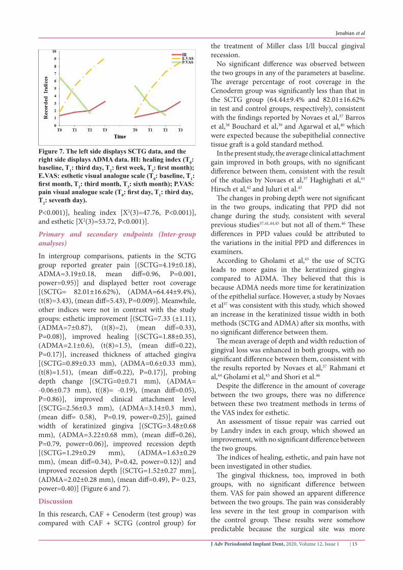
Jenabian et al
J Adv Periodontol Implant Dent 2020 Volume 12 Issue 116 |
extensive with the SCTG technique compared to the ADMA technique According to this study patients used more analgesics for a longer duration47 These results could be considered as one of the advantages of the ADMA method
Furthermore it is possible to treat a broader region in this technique in comparison with the other techniques such as SCTG The advantage of fewer complications of ADMA (Cenoderm) could be exploited in young people under orthodontic treatments and older people with systemic problems especially those with bleeding disorders
Conclusion
According to the results of this study both SCTG and Cenoderm resulted in improvements in the clinical outcomes However the use of Cenoderm led to less severe pain and less root coverage in comparison with the SCTG techniqueAcknowledgement
None declared
Authorsrsquo Contributions
NJ Executor MY Executor PHA Executor and corresponding author AB Statistics Consultant All authors have read and approved the final manuscript
Funding
Council of technology and research Babol university of Medical Sciences
Ethics Approval
The study protocol was approved by the Ethical Committee of Babol University of Medical Sciences and registered at httpwwwirctir (registration number IRCT201305201760N23 date registered March 23 2013)
Competing of Interests
Yes- Cenoderm (Tissue Regeneration Corporation)
References1 American Academy of Periodontology Glossary of
periodontal terms 4th ed Chicago2001- 4th edition2 Kassab MM Cohen RE The etiology and prevalence of
gingival recession J Am Dent Assoc 2003 Feb134(2)220-53 Rees JS Addy M A cross-sectional study of dentine
hypersensitivity J Clin Periodontol 2002 Nov29(11)997-1003
4 Goutoudi P Koidis PT Konstantinidis A Gingival recession a cross-sectional clinical investigation Eur J Prosthodont Restor Dent 1997 Jun5(2)57-61
5 Oliver RC Brown LJ Loe H Periodontal diseases in the United States population J Periodontol 1998 Feb69(2)269-78
6 Wennstrom JL Mucogingival therapy Ann Periodontol 1996 Nov1(1)671-701
7 Susin C Haas AN Oppermann RV Haugejorden O Albandar JM Gingival recession epidemiology and risk indicators in a representative urban Brazilian population J
Periodontol 2004 Oct75(10)1377-868 Muller HP Eger T Schorb A Gingival dimensions after
root coverage with free connective tissue grafts J Clin Periodontol 1998 May25(5)424-30
9 H B Free transplantation of gingiva propria Sven Tandlak Tidskr 196322684-9
10 Nabers JM Free gingival grafts Periodontics 1966 Sep-Oct4(5)243-5
11 Levine RA Covering denuded maxillary root surfaces with the subepithelial connective tissue graft Compendium 1991 Aug12(8)568 70 72 passim
12 Grupe H WR Repair of gingival defects by a sliding flap operation J Periodontol 195656715-20
13 Pini Prato G Pagliaro U Baldi C Nieri M Saletta D Cairo F et al Coronally advanced flap procedure for root coverage Flap with tension versus flap without tension a randomized controlled clinical study J Periodontol 2000 Feb71(2)188-201
14 Langer B Langer L Subepithelial connective tissue graft technique for root coverage J Periodontol 1985 Dec56(12)715-20
15 McGuire MK Nunn ME Evaluation of the safety and efficacy of periodontal applications of a living tissue-engineered human fibroblast-derived dermal substitute I Comparison to the gingival autograft a randomized controlled pilot study J Periodontol 2005 Jun76(6)867-80
16 McGuire MK Scheyer ET Schupbach P Growth factor-mediated treatment of recession defects a randomized controlled trial and histologic and microcomputed tomography examination J Periodontol 2009 Apr80(4)550-64
17 Wennstrom JL Zucchelli G Increased gingival dimensions A significant factor for successful outcome of root coverage procedures A 2-year prospective clinical study J Clin Periodontol 1996 Aug23(8)770-7
18 Paolantonio M Treatment of gingival recessions by combined periodontal regenerative technique guided tissue regeneration and subpedicle connective tissue graft A comparative clinical study J Periodontol 2002 Jan73(1)53-62
19 Park JB Increasing the width of keratinized mucosa around endosseous implant using acellular dermal matrix allograft Implant Dent 2006 Sep15(3)275-81
20 Yan JJ Tsai AY Wong MY Hou LT Comparison of acellular dermal graft and palatal autograft in the reconstruction of keratinized gingiva around dental implants a case report Int J Periodontics Restorative Dent 2006 Jun26(3)287-92
21 Imberman M Gingival augmentation with an acellular dermal matrix revisited surgical technique for gingival grafting Pract Proced Aesthet Dent 2007 Mar19(2)123-8
22 Achauer BM VanderKam VM Celikoz B Jacobson DG Augmentation of facial soft-tissue defects with Alloderm dermal graft Ann Plast Surg 1998 Nov41(5)503-7
23 Tobin HA Karas ND Lip augmentation using an alloderm graft J Oral Maxillofac Surg 1998 Jun56(6)722-7
24 Kridel RW Foda H Lunde KC Septal perforation repair with acellular human dermal allograft Arch Otolaryngol Head Neck Surg 1998 Jan124(1)73-8
25 Wainwright D Madden M Luterman A Hunt J Monafo W Heimbach D et al Clinical evaluation of an acellular allograft dermal matrix in full-thickness burns J Burn Care Rehabil 1996 Mar-Apr17(2)124-36
26 Wainwright DJ Use of an acellular allograft dermal matrix (AlloDerm) in the management of full-thickness burns Burns 1995 Jun21(4)243-8
27 Livesey SA Herndon DN Hollyoak MA Atkinson YH Nag A Transplanted acellular allograft dermal matrix Potential
J Adv Periodontol Implant Dent 2020 Volume 12 Issue 1 | 17
Jenabian et al
as a template for the reconstruction of viable dermis Transplantation 1995 Jul 1560(1)1-9
28 Callan DP Silverstein LH Use of acellular dermal matrix for increasing keratinized tissue around teeth and implants Pract Periodontics Aesthet Dent 1998 Aug10(6)731-4
29 Harris RJ A comparative study of root coverage obtained with an acellular dermal matrix versus a connective tissue graft results of 107 recession defects in 50 consecutively treated patients Int J Periodontics Restorative Dent 2000 Feb20(1)51-9
30 Henderson RD Greenwell H Drisko C Regennitter FJ Lamb JW Mehlbauer MJ et al Predictable multiple site root coverage using an acellular dermal matrix allograft J Periodontol 2001 May72(5)571-82
31 de Queiroz Cortes A Sallum AW Casati MZ Nociti FH Jr Sallum EA A two-year prospective study of coronally positioned flap with or without acellular dermal matrix graft J Clin Periodontol 2006 Sep33(9)683-9
32 Harris RJ Cellular dermal matrix used for root coverage 18-month follow-up observation Int J Periodontics Restorative Dent 2002 Apr22(2)156-63
33 Gapski R Parks CA Wang HL Acellular dermal matrix for mucogingival surgery a meta-analysis J Periodontol 2005 Nov76(11)1814-22
34 OrsquoLeary TJ Drake RB Naylor JE The plaque control record J Periodontol 1972 Jan43(1)38
35 Jenabian N Yazdanpanahbahabadi M Bijani A Rahimirad M Gingival unit graft versus free gingival graft for treatment of gingival recession A randomized controlled clinical trial JDT 2016 June 13(3) 184-92
36 Landry R TR Howley T Effectiveness of benzydamine HCL in the treatment of periodontal postsurgical patients Res Clin Forum 198810105-18
37 Novaes AB Jr Grisi DC Molina GO Souza SL Taba M Jr Grisi MF Comparative 6-month clinical study of a subepithelial connective tissue graft and acellular dermal matrix graft for the treatment of gingival recession J Periodontol 2001 Nov72(11)1477-84
38 Barros RR Novaes AB Grisi MF Souza SL Taba MJ Palioto DB A 6-month comparative clinical study of a conventional
and a new surgical approach for root coverage with acellular dermal matrix J Periodontol 2004 Oct75(10)1350-6
39 Bouchard P Etienne D Ouhayoun JP Nilveus R Subepithelial connective tissue grafts in the treatment of gingival recessions A comparative study of 2 procedures J Periodontol 1994 Oct65(10)929-36
40 Agarwal C Kumar BT Mehta DS An acellular dermal matrix allograft (Allodermreg) for increasing keratinized attached gingiva A case series JISP 201519(2)216-20
41 Haghighati F MM Moslemi N Comparative clinical evaluation of subepithelial connective tissue graft and acellular dermal matrix allograft for the treatment of gingival recession JDT 20063(4)159-66
42 Hirsch A Goldstein M Goultschin J Boyan BD Schwartz Z A 2-year follow-up of root coverage using sub-pedicle acellular dermal matrix allografts and subepithelial connective tissue autografts J Periodontol 2005 Aug76(8)1323-8
43 Juluri R Gopalakrishnan D Prasad N Chialastri SM Balasundaram A Martande S Augmentation of Keratinized Tissue around Implants using Acellular Dermal Matrix Allograft A Case Report J Dental Oral Health 2018 August4(3)1-4
44 Rahmani ME Lades MA Comparative clinical evaluation of acellular dermal matrix allograft and connective tissue graft for the treatment of gingival recession J Contemp Dent Pract 2006 May 17(2)63-70
45 Gholami GA Saberi A Kadkhodazadeh M Amid R Karami D Comparison of the clinical outcomes of connective tissue and acellular dermal matrix in combination with double papillary flap for root coverage A 6-month trial Dent Res J (Isfahan) 2013 Jul10(4)506-13
46 Shori T Kolte A Kher V Dharamthok S Shrirao T A comparative evaluation of the effectiveness of subpedicle acellular dermal matrix allograft with subepithelial connective tissue graft in the treatment of isolated marginal tissue recession A clinical study J Indian Soc Periodontol 2013 Jan17(1)78-81
47 Harris RJ Clinical evaluation of 3 techniques to augment keratinized tissue without root coverage J Periodontol 2001 Jul72(7)932-8