intercalary allograft
DESCRIPTION
orthoTRANSCRIPT

1
BACKGROUND■ An intercalary reconstruction is defined as replacement ofthe diaphyseal portion of a long bone after segmental skeletalresection (diaphysectomy).■ Intercalary reconstructions typically result in superior func-tion compared to other limb-sparing procedures because thepatient’s own joints above and below the reconstruction areleft undisturbed.■ Most intercalary reconstructions are performed with bulkallografts, although various endoprosthetic intercalary pros-theses are available.■ Allograft reconstructions require osseous healing for long-term stability; initial allograft stability must be obtained withintramedullary nailing or internal fixation.■ Allografts have a long-term risk of fracture likely related tothe size and type of fixation.■ Intercalary allografts are often supplemented with vascular-ized fibular grafts.■ Endoprosthetic reconstruction has typically been limited tosmall, central tumors as significant lengths of bone proximaland distal to the lesion are required for successful fixation oftraditional prosthetic stems.■ New implant designs have expanded the indication for inter-calary prostheses by drastically reducing the length of boneneeded to achieve stable fixation.■ Segmental prostheses typically provide immediate stable fixa-tion, allowing early rehabilitation and rapid return of function.■ Vascularized fibular grafts are not required for intercalaryimplants.■ Although aseptic loosening has been reported after endo-prosthetic reconstruction, it is rarely if ever seen with inter-calary implants; the lack of bearing surfaces contributing towear debris formation and the lack of exposure to joint fluidhelp to protect the implant.
INDICATIONS■ Intercalary reconstructions are indicated for osseous joint-sparing reconstructions.■ Intercalary reconstructions are performed most commonlyin the femur, tibia, humerus, and forearm.■ Intercalary reconstructions require adequate remainingbone stock to allow stable fixation; if inadequate bone re-mains, the procedure should be converted to a joint-replacingreconstruction.■ Intercalary allograft reconstructions require osseous heal-ing for long-term stability, which can be compromised bychemotherapy or radiation during the healing period.■ Intercalary allografts in areas with poor vascularity, withsmall residual host segments, in heavy or very active patients,
or with planned radiation or chemotherapy likely benefit fromsupplemental vascularized fibular allografts.■ Intercalary endoprosthetic reconstruction is an option ifallograft reconstruction is contraindicated due to poor hostfactors or in patients with prior failed allograft reconstruction.■ Modular intercalary prostheses provide the flexibility toeasily convert to a conventional endoprosthetic replacement ei-ther at initial surgery or for later revisions by using joint-replac-ing modules at either end of the implant.
ANATOMY■ Anatomic considerations in the femur include the anteriorbow, femoral anteversion, and the proximity of the superficialfemoral artery to the distal femur near the adductor hiatus.■ Anatomic considerations in the tibia include the limited softtissue coverage anteromedially and the proximity of the poste-rior tibial neurovascular bundle to the posteromedial tibia.■ Anatomic considerations in the humerus include the prox-imity of the radial nerve to the posterior midhumeral shaft.■ Anatomic considerations in the forearm include the radialbow.
PATIENT HISTORY AND PHYSICAL FINDINGS■ Table 1 outlines methods for examining the patient with amass or suspected tumor.■ Important aspects of the patient history include:
■ Onset of mass or pain■ Progression of growth or pain■ Constitutional symptoms (fevers, chills, night sweats,weight loss)■ Personal or family history of cancer
IMAGING AND OTHER STAGING STUDIES■ Radiographs are the primary imaging study used in forminga differential diagnosis of bone lesions.■ Radiographs are used to assess the geometry and size of thehost bone and the lesion to allow appropriate sizing of the al-lograft reconstruction.■ Radiographs are helpful in determining the response tochemotherapy, which influences the decision making for limbsalvage.■ Bone scan is used to determine if there are additional os-seous sites of disease.■ Computed tomography (CT) scanning is used to determine thebone geometry and to assess bone destruction by a lytic lesion.■ Magnetic resonance imaging (MRI) is used to assess the softtissue extension of a lesion. It is also used to assess the prox-
Chapter 29
Walter W. Virkus, Robert M. Henshaw, Benjamin Miller, and Steven Gitelis
Use of Allografts andSegmental Prostheses forReconstruction of SegmentalBone Defects
13282_ON-29.qxd 5/13/09 9:13 AM Page 1

imity of neurologic and vascular structures, the intraosseous ormarrow extent of tumor, and extension into an adjacent joint.■ Sagittal and coronal MRI images are extremely useful in plan-ning the resection length; measurements from the adjacent jointsto the planned levels of osteotomy are made to provide a repro-ducible method of identifying the levels intraoperatively.■ The role of positron emission tomography (PET) scanninghas yet to be defined for sarcomas. It likely plays a role in theassessment of metastatic disease.
SURGICAL MANAGEMENT■ The main surgical decisions to be made when performing anintercalary resection are the type and length of fixation and theneed for supplemental vascularized fibula graft.■ Plate fixation allows for reconstruction with standard os-teosynthesis techniques.■ Plate fixation allows for compression across the allograft–hostjunctions, which likely improves healing.■ Plate fixation results in screw holes in the allograft, which isthought to contribute to late allograft fracture.■ Intramedullary fixation of allografts requires additional in-cisions but likely provides stronger fixation.■ Intramedullary fixation provides long-term protection of theallograft without placing screw holes in the allograft (FIG 1).■ Intramedullary fixation makes it difficult to obtain compres-sion at the allograft–host junctions, which may impede healing.
■ Allograft and host cuts can be transverse or stepped (FIG 2).■ Transverse cuts make rotational adjustments easier and likelyresult in less periosteal dissection, which may improve healing.■ Stepped cuts are more technically difficult, add surgical time,and may cause more damage to the local periosteum. However,they likely increase host–allograft bone contact, which may im-prove healing.■ Intercalary implants require careful attention to resectionlength and canal preparation to ensure optimal fixation of theimplant stems.■ Regardless of the method of skeletal reconstruction, carefulattention to the soft tissues and use of rotational muscle flapswhen indicated are necessary to reduce the risk of wound com-plications and subsequent infection.
Preoperative Planning■ Preoperative planning is extraordinarily critical in thesecomplex reconstructive procedures.■ Multiple imaging techniques should be used to fully assess thetumor, length of resection, and the remaining bone anatomynecessary to support the planned reconstruction.■ Standard instruments for tumor resection are required, in-cluding bone saws; equipment for additional procedures suchas vascular bypass, vascularized graft, or specialized soft tissuereconstruction should be available.■ Some surgeons prefer to use a separate group of instrumentsfor the resection and the reconstruction.
Examination Technique SignificancePalpation Feel the borders of the mass to evaluate size and whether Lack of mobility suggests adherence to surrounding structures.
it is mobile from the surrounding structures.Joint range of motion Assess range of motion of joints adjacent to mass. Large masses may restrict joint motion.Soft tissue evaluation Assess soft around mass or biopsy to determine resection Alternative methods of wound closure may be required at
approach and whether sufficient soft tissue remains to the time of resection.allow primary closure.
Vascular examination Assess circulation of extremity. Vascular compromise may complicate limb reconstruction.
Table 1 Methods for Examining the Patient with a Mass or Suspected Tumor
A B
FIG 1 • Placement of intramedullary nail and compressionof junction. A. Fluoroscopic view of intramedullary nailthrough allograft in the proximal femur. The K-wire forrotational reference can be seen. Gaps in the host–graftjunction are present without compression of the junction.B. Fluoroscopic view after placement and tightening of com-pression screw in intramedullary nail. Gap at host–allograftjunction is minimized.
2 Part 4 ONCOLOGY • Section IV LOWER EXTREMITIES
13282_ON-29.qxd 5/13/09 9:13 AM Page 2

Chapter 29 ALLOGRAFTS AND SEGMENTAL PROSTHESES FOR RECONSTRUCTION OF SEGMENTAL BONE DEFECTS 3
■ The allograft needs to be ordered from a bone bank. Somesurgeons prefer to size-match allografts used for intercalary re-constructions, requiring radiographs of the allograft to be ob-tained before ordering.■ If a vascularized fibula graft is planned, the timing and hostlocation of the graft must be determined.■ Endoprosthetic implants may require customized compo-nents, typically requiring a 3- to 4-week lead time to allow fordesign and manufacturing of the implant.■ An examination under anesthesia focusing on the rotationalprofile of the nonoperative leg can be useful in ensuring properrotation of the operative leg.
■ Intraoperative C-arm fluoroscopy is useful to measure andselect the levels of planned bone osteotomy.
Positioning■ Positioning depends on the tumor location and the surgicalapproach required for oncologic resection.■ Femoral resections are typically performed either in thesupine or lateral position; tibial cases are performed supine.■ Femoral reconstructions may be facilitated by a bump underthe buttock, but this may make clinical assessment of rotationmore difficult.■ Forearm reconstructions are performed with the patientsupine and the arm on a hand table.■ Humeral reconstructions can be performed with the patientin the supine or beach chair position.
Approach■ The approach for intercalary reconstructions is determinedby the incision needed to perform an adequate resection of thetumor.■ Femoral reconstructions are ideally performed through alateral or anterolateral approach to the thigh; medial incisionsmay be necessary to isolate and protect the superficial femoralartery and vein during the resection.■ Tibial reconstructions can be performed through an antero-lateral or anteromedial approach.■ An anteromedial approach is more likely to require compli-cated soft tissue coverage.■ Humeral resections are performed through an extensile an-teromedial approach; care must be taken to identify and pro-tect the radial nerve when oncologically possible.■ Forearm reconstructions of the radius are ideally performedthrough an anterior approach.
FIG 2 • Preparation of allograft. Distal allograft cut is placed inmetaphysis to match host location (host location not seen). Careis taken to make smooth and perpendicular cuts in both thehost and allograft bone.
PREPARATION OF THE ALLOGRAFT
RESECTION
TECH
NIQ
UES
■ The approach is performed as indicated by the locationof the tumor and the biopsy.
■ Pertinent vascular and neurologic dissection is per-formed to identify and protect critical structures; soft tis-sue dissection around the tumor mass is then performed.
■ Dissection is performed down to bone at the distal andproximal extent of the planned bone resection.
■ Using fluoroscopy or other measurements from preoper-ative imaging, K-wires placed perpendicular to the shaftof the bone may be used to mark the proximal and dis-tal osteotomy level. Care should be taken to avoid exces-sive damage to the periosteum adjacent to the plannedosteotomy.
■ Before osteotomy, the bone should be carefully exam-ined and marked to ensure proper rotational orientationduring the reconstruction. The linea aspera of the femuris a convenient anatomic landmark; K-wires or cuts in thebone may be placed proximal and distal to the resectionarea as well.
■ Transverse osteotomy is performed with a power saw.Cooling the saw blade with irrigant prevents excessiveheat from causing thermal damage to the host bone.
■ The specimen is removed, the length is measured andrecorded, and margins are assessed.
■ The allograft is thawed in antibiotic-laden lactatedRinger solution.
■ The section of the allograft that most closely matches theshape and size of the resected bone is marked. A few mil-limeters are added to the resected length to allow addi-tional small cuts to be made to improve the bone contactand alignment with the host bone.
■ The proximal and distal allograft cuts are made with apower saw.
■ If intramedullary nail fixation is planned, the allograft isthen reamed to a diameter 2 mm greater than theplanned nail diameter.
■ Reaming should be done slowly and progressively toavoid cracking the very brittle allograft.
13282_ON-29.qxd 5/13/09 9:13 AM Page 3
4 Part 4 ONCOLOGY • Section IV LOWER EXTREMITIESTE
CH
NIQ
UES
A CB D
F
E G H
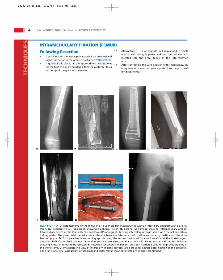
TECH FIG 1 • A–D. Osteosarcoma of the femur in a 14-year-old boy reconstructed with an intercalary allograft with plate fix-ation. A. Preoperative AP radiograph showing diaphyseal lesion. B. Coronal MRI image showing intramedullary and ex-tramedullary extent of the lesion. C. Postoperative AP radiograph showing intercalary reconstruction with medial and laterallocking plates. The most distal medial screw in the epiphysis was later removed to allow continued growth across the distalfemoral physis. D. Postoperative lateral radiograph showing the reconstruction with callus formation at the host–allograftjunctions. E–G. Customized modular femoral intercalary reconstruction in a patient with Ewing sarcoma. E. Sagittal MRI scanshowing length of tumor to be resected. F. Resection specimen and implant; cross-pin fixation is used for improved stability ofthe short stems. G. Intraoperative view of intercalary implant; surfaces are porous for extraskeletal fixation at the prostheticbone junctions. H,I. Radiographs of proximal and distal femur showing intercalary implant. (continued)
INTRAMEDULLARY FIXATION (FEMUR)
Following Resection■ A small incision is made approximately 8 cm proximal and
slightly posterior to the greater trochanter (TECH FIG 1).■ A guidewire is placed at the appropriate starting point
for the type of nail being used, either the piriformis fossaor the tip of the greater trochanter.
■ Alternatively, if a retrograde nail is planned, a smallmedial arthrotomy is performed and the guidewire isinserted into the distal femur in the intercondylarnotch.
■ After confirming the wire position with fluoroscopy, anentry reamer is used to open a portal into the proximal(or distal) femur.
13282_ON-29.qxd 5/13/09 9:13 AM Page 4

Chapter 29 ALLOGRAFTS AND SEGMENTAL PROSTHESES FOR RECONSTRUCTION OF SEGMENTAL BONE DEFECTS 5TEC
HN
IQU
ES
TECH FIG 1 • (continued) J–P. Salvage of multiply failed intercalary allograft with conversion to intercalary endo-prosthesis. J. Chronic painful nonunion of distal allograft–host junction despite repeated surgery and vascularizedfibular bone grafting. Patient had been braced for 5 years and unable to bear weight for over 2 years. K. Resectionof allograft with failed blade plate; cultures of allograft showed infection with methicillin-sensitive Staphylococcusaureus. L. Use of high-dose antibiotic spacer for sterilization of soft tissues while preserving limb length and tissuepocket for planned reconstruction. M. Preparation of intramedullary canal using facing reamer and (N) use of jigsfor anchor plug fixation with bicortical transfixion pins. O. Intraoperative view of assembled prosthesis; locking col-lar and screws are used to hold proximal and distal body segments together. P. Radiograph showing final recon-struction. The patient is completely pain-free and ambulatory without braces or crutches.
I J K
M
ON P
L
■ A beaded guidewire is inserted into the femur, across theresection bed, and into the femoral segment on the farside of the resection bed.
■ The femur is then progressively reamed to 1.5 mm largerthan the planned nail diameter.
■ The beaded guidewire is partially pulled out and the al-lograft is inserted into the resection bed.
■ The guidewire is then passed back through the allograftinto the far femoral segment.
■ A preliminary check of the bone cuts is made by manu-ally compressing the allograft against the host bone and
inspecting bone contact and alignment. Small revisioncuts may be required on the allograft.
■ The nail is then inserted. With the nail in place, a final in-spection of the junctions is made and revision trimmingof the allograft is performed to improve bone contact.
■ The nail is fully seated.■ The locking screws in the “distal” tip of the nail are
placed. These may be the screws in the proximal femur ifa retrograde nail is being used.
■ The nail is then backtapped to close down any gaps inthe host–allograft junctions.
13282_ON-29.qxd 5/13/09 9:13 AM Page 5

6 Part 4 ONCOLOGY • Section IV LOWER EXTREMITIESTE
CH
NIQ
UES
TECH FIG 2 • Grade 3 aneurysmal bone cyst with intercalaryreconstruction. A. Preoperative AP radiograph showing le-sion. B. Postoperative AP radiograph showing intercalary re-construction with long plate.
A B
TECH FIG 3 • Oblique compression screws used for fixation ofa very distal host–allograft junction. Small plates can be usedas an alternative.
TECH FIG 4 • Provisional fixation of plate along femoral inter-calary reconstruction. Use of clamps allows assessment ofalignment and bone contact of junctions. Fluoroscopic evalua-tion of reconstruction should also be undertaken at this time.
TECH FIG 5 • Compression of intercalary reconstruction witha plate. The tensioning device is seen proximally affixed tothe host bone with a single screw with a distal hook into thelast hole in the plate.
■ Rotation is checked either clinically or by aligning thepreviously placed K-wires.
■ The screws in the “proximal” end of the nail are placed.If a compressible nail is being used, the compressionscrew is applied after placement of the dynamic lockingscrew (TECH FIG 2). Because of the extended time oftenrequired for healing, two locking screws should beplaced proximally and distally.
■ If compression has been applied with a compression nailand the allograft is rotationally stable, no additional fix-ation is required.
■ If the allograft is rotationally unstable around the nail, ashort plate can be applied with unicortical screws to im-prove stability. This can be placed at one or both host–al-lograft junctions depending on the amount of instability.
Plate and Screw Fixation■ The allograft is placed into the resection bed and gross
alignment is checked (TECH FIG 3).■ If necessary, the plate is contoured to match the align-
ment of the femoral segment being reconstructed.■ A 4.5-mm plate should be used (except in the forearm).
The type of plate is dictated by the location. Specialplates are available for the proximal and distal femur.
■ A slight prebend of the plate is performed to maximizecompression of the junction on the side opposite theplate.
■ The plate is positioned and fixed to one side of the hostbone with one screw.
■ The host–allograft segment is aligned and the plate ispositioned appropriately (TECH FIG 4).
■ Rotational alignment is checked clinically or with thepreviously placed K-wires.
■ Compression is applied across the host–allograft seg-ments either with a tensioning device or a Verbruggeclamp and a pull screw (TECH FIG 5).
■ Sagittal and coronal plane alignment is verified with flu-oroscopy.
■ Allograft trimming is performed as necessary to improvebone contact and alignment.
13282_ON-29.qxd 5/13/09 9:13 AM Page 6

Chapter 29 ALLOGRAFTS AND SEGMENTAL PROSTHESES FOR RECONSTRUCTION OF SEGMENTAL BONE DEFECTS 7TEC
HN
IQU
ES
■ Compression screws are placed into the plate to maxi-mize compression across the host–allograft junctions.
■ Two screws are placed through the plate into the allo-graft to prevent dislodgement. If a locking plate is beingused, these can be unicortical locking screws.Alternatively, cerclage cables can be placed to avoidholes in the allograft.
■ In resections resulting in very small residual host seg-ments, large plate fixation may not be possible. In thesecases, small hand or foot plates may be beneficial for fix-ation. Alternatively, oblique compression screws fromthe host through the allograft may be used.
Closure■ Irrigation of the wound is performed and hemostasis
obtained.■ If small gaps remain at the host–allograft junctions,
grafting can be performed. Usually cancellous bone canbe curetted from unused allograft for this purpose.
■ The wound is closed in layers over suction drains.■ Efforts should be made to obtain circumferential muscle
coverage over the reconstructed bone.■ A gentle compression dressing is placed. Splinting of the
knee may improve patient comfort for the first few post-operative days.
VASCULARIZED FIBULAR GRAFT■ Vascularized fibular grafts are used to supplement inter-
calary allografts, improving healing and adding strengthand living bone to the reconstruction.
■ Vascularized grafts add complexity and time to the oper-ative reconstruction.
■ Vascularized graft can be harvested from the ipsilateralor contralateral leg.
■ In the femur, vascularized grafts can be placed medial or
lateral, bridging across the allograft and affixed to theproximal and distal host segment.
■ In the tibia, soft tissue constraints often require the vas-cularized graft to be placed within the allograft. Agroove made in the allograft facilitates placement of thevascular graft.
■ For a more complete explanation of this procedure, seeChapter ON-28.
TIBIA, HUMERUS, AND FOREARM■ Reconstructions in the tibia, humerus, and forearm are
similar.■ Intramedullary fixation may be possible in the tibia and
humerus.■ Plate fixation for the humerus and tibia should be done
with 4.5-mm plates.
■ Plate fixation in the forearm is performed with 3.5-mmplates.
■ Special locking plates designed for the proximal tibia areoften useful in tibial reconstructions.
■ Care must be taken in the radius to recreate the radialbow.
INTERCALARY ENDOPROSTHETIC RECONSTRUCTION■ After tumor resection, frozen sections of the remaining
medullary contents are performed to ensure margins arefree of tumor.
■ Careful measurement of the specimen length is necessaryto ensure proper selection of components (if using amodular system).
■ Preparation of the bone canal is critical to ensure ade-quate fixation; use of rigid instead of flexible in-tramedullary reamers is preferred to obtain best fit ofthe prosthetic stem.
■ C-arm fluoroscopy is used to monitor the reamingprocess to avoid penetration into the adjacent joint.
■ Facing reamers are used to machine the proper radius ofcurvature of the exposed cortical bone to ensure flush fitof the implant’s stem–body junction.
■ Trial reduction is performed to check length and rota-tion and to ensure that reduction of the prosthesis bod-ies is possible; the specific connection between theproximal and distal bodies varies depending on the im-plant manufacturer.
■ For implants with cemented stems, proper canal prepa-ration using cement restrictors, pulsatile lavage, andpacking for hemostasis is performed before injection ofcement and impaction of the stem.
■ Uncemented systems may require further canal prepara-tion; Compress™ implants use a number of interlockedjigs for centralization of the intramedullary anchor plugand placement of transfixion pins through the adjacentcortical bone.
■ Final assembly of the prosthesis is performed per implantrequirements; care must be taken to ensure that rotationis proper and that all locking mechanisms are tightenedor impacted.
■ Supplemental bone graft, collected during reaming, isplaced at the implant–bone junction (over the porouscoated surface) to provide extraskeletal fixation of theimplant.
■ Soft tissue reconstruction over closed suction drains isthen performed. When necessary, local rotation flaps areused to ensure muscular coverage of the implant.
13282_ON-29.qxd 5/13/09 9:13 AM Page 7

8 Part 4 ONCOLOGY • Section IV LOWER EXTREMITIES
POSTOPERATIVE CARE■ Proper wound healing is critical to avoid infection ofthe reconstruction; use of antibiotics during the periopera-tive phase is extremely important, particularly with allograftreconstructions.■ Allograft patients are maintained on touch-down weightbearing until healing of the host–allograft junctions is seen ra-diographically (usually 3 to 12 months).■ Additional bracing is necessary only if a stable construct isnot obtained. This can occur in plate reconstructions with smallresidual host segments after resection.■ Physical therapy should be directed toward range of motionof the hip and knee. Muscle strengthening can be performedwith minimal resistance as dictated by the stability of the con-struct achieved in surgery.■ Intercalary implants permit immediate weight bearing;short-stemmed components around the knee are protected byplacing the patient in a knee immobilizer and limiting jointrange of motion to gentle assisted exercises under supervisionduring the initial healing stages.
OUTCOMES■ Most studies report combined results for intercalary, os-teoarticular, and allograft prosthesis composites.■ Nonunion rates are reported to be 10% to 15%.■ Late fracture rates are reported to be 7% to 20%.■ Union rates with nails and plates are nearly equal, but thelate fracture rate is higher with plates.■ Functional outcomes are reported as 78% to 86% good orexcellent.■ Graft survival is reported as 79% at 5 years.■ Few reports of intercalary implants have been published; in-dividual institutional experience has been very favorable, with100% implant survival and near to complete functional restora-tion of the limb.
COMPLICATIONS■ Nonunion of the allograft–bone junction frequently occursand may be treated with secondary bone grafting or allograftrevision. Use of vascularized fibular grafts in high-risk patientsshould be considered to minimize this complication.
PEARLS AND PITFALLSWound healing ■ Aggressive surgical débridement and reclosure of wounds after development of flap necrosis is
critical to minimize the risk of infection. Use of flaps and skin grafts should always be consid-ered whenever tissue appears inadequate for an easy closure without tension.
Limb-length discrepancy ■ Particular attention to resection length is necessary to ensure restoration of limb length; this isless important in the upper extremity. Trial reductions are useful in ensuring proper lengthand rotation of the reconstruction. Marking the bone with pins before resection provides use-ful landmarks for the reconstruction.
Allograft fixation ■ Intramedullary fixation is stronger, provides more protection against late allograft fracture,and makes restoration of sagittal and coronal plate alignment easier.
■ Intramedullary fixation can potentially contaminate the entire osseous segment being recon-structed if clean margins were not obtained during the resection.
■ Screw placement into the allograft should be minimized to decrease the risk of late fracture.■ Healing time in these reconstructions can be prolonged, so every effort should be made to
gain maximally stable fixation.
Allograft compression ■ Compression across the host–allograft junctions should be obtained when possible.■ Compression with intramedullary fixation can be obtained with various commercially avail-
able nails.■ Compression with plates can be obtained with a tensioning device, clamp and pull screw, or
(as a last resort) compression screws.
Allograft alignment ■ Coronal and sagittal alignment can be difficult with plate fixation. This should be confirmedfluoroscopically after preliminary fixation and revision bone cuts made if necessary.
■ Rotational malalignment can occur with intramedullary or plate fixation. Use of alignment K-wires or clinical comparison to the contralateral limb should be performed before final fixation.
Host–allograft bone contact ■ Large gaps at the host–allograft junctions can dramatically increase the healing time andunion rate. Revision bone cuts or grafting should be considered if gaps remain after fixation.
Allograft–bone healing ■ Healing problems are common in these reconstructions. Careful preservation of the periosteumaround the resection area and avoidance of thermal injury during resection osteotomy may im-prove healing rates. Additional surgery to bone graft nonunions of the junctions is common.
Late fracture ■ The risk of late fracture appears to be related to holes or damage to the allograft. Screws anddamage to the allograft should be minimized. Use of cerclage cabling or intramedullary fixa-tion minimizes fracture risk.
Intercalary implants ■ Use of transfixion pins or bolts can greatly increase the stability of the construct, allowinggreater lengths of bone to be replaced.
13282_ON-29.qxd 5/13/09 9:13 AM Page 8

Chapter 29 ALLOGRAFTS AND SEGMENTAL PROSTHESES FOR RECONSTRUCTION OF SEGMENTAL BONE DEFECTS 9
■ Infection is the most common cause of allograft and pros-thetic failure and can lead to secondary amputation. Aggressivesurgical débridement with removal of all foreign bone or mate-rials is necessary.■ Late fracture of massive allografts can lead to chronic painand loss of function. Use of intramedullary fixation andavoiding screws in the allograft may help to minimize thiscomplication.■ Prosthetic loosening rarely if ever occurs for intercalaryimplants. Implant failure can be revised with use of a newprosthesis.■ Degeneration of an adjacent joint after either allograft or in-tercalary implant reconstruction can be treated with surface-replacing total joint arthroplasty.
REFERENCES1. Henshaw RM. Complex massive intercalary endoprosthetic recon-
struction of the femur and tibia: a new technique using customizedcompress implants augmented with cement. Musculoskeletal TumorSociety Annual Meeting 2006;45:61.
2. Makely JT. The use of allografts to reconstruct intercalary defects oflong bones. Clin Orthop Relat Res 1988;197:58–75.
3. Muscolo LD, Ayerza MA, Aponte-Tinao LA, et al. Intercalary femurand tibia segmental allografts provide an acceptable alternative inreconstructing tumor resections. Clin Orthop Relat Res 2004;426:97–102.
4. Muscolo LD, Ayerza MA, Aponte-Tinao LA, et al. Partial epiphysealpreservation and intercalary allograft reconstruction in high-grademetaphyseal osteosarcoma of the knee. J Bone Joint Surg Am 2004;86A:2686–2692.
5. Sorger JI, Hornicek FJ, Zavatta M, et al. Allograft fractures revisited.Clin Orthop Relat Res 2001;382:66–74.
6. VanderGriend RA. The effect of internal fixation on the healing oflarge allografts. J Bone Joint Surg Am 1994;76A:657–663.
13282_ON-29.qxd 5/13/09 9:13 AM Page 9