comparative vegetative anatomy and systematics of the
TRANSCRIPT

Eastern Illinois University
From the SelectedWorks of Barbara S. Carlsward
2006
Comparative vegetative anatomy and systematicsof the angraecoids (Vandeae, Orchidaceae) withan emphasis on the leafless habitBarbara S. Carlsward, Eastern Illinois UniversityWilliam Louis SternBenny Bytebier
Available at: https://works.bepress.com/barbara_carlsward/7/

Comparative vegetative anatomy and systematics of the angraecoids (Vandeae, Orchidaceae) with an emphasis on the leafless habit BARBARA S. CARLSWARD, WILLIAM LOUIS STERN, and BENNY BYTEBIER Abstract The vegetative anatomy and morphology of 142 species of the angraecoid orchids (Angraecinae + Aerangidinae) and 18 species of Aeridinae were examined using light and scanning electron microscopy. Leafless members of Vandeae were of particular interest because of their unique growth habit. Leafy and leafless members of Angraecinae and Aerangidinae were examined and compared with specimens of Aeridinae. Vandeae were homogeneous in both leaf and root anatomy. A foliar hypodermis and fibre bundles were generally absent. Stegmata with spherical silica bodies were found associated with sclerenchyma and restricted to leaves in almost all specimens examined. Distinct inner tangential wall thickenings of the endovelamen occurred in several vandaceous genera. Exodermal proliferations and aeration units commonly occurred in both leafy and leafless Vandeae. Cladistic analyses of Angraecinae and Aerangidinae with members of Aeridinae and Polystachyinae as outgroups using 26 structural characters resulted in 20 000+ equally parsimonious trees. Vandeae formed the only well-‐supported clade in bootstrap analyses and were characterized by having a monopodial growth habit, spherical stegmata, loss of mucilage, and loss of tilosomes. INTRODUCTION Vandeae comprise a large group of epiphytic monopodial taxa. These exhibit a wide variety of habits from plants with elongate stems and well-‐developed photosynthetic leaves (Fig. 1), to those with abbreviated stems and caducous leaves, to those with abbreviated stems and small nonphotosynthetic scale leaves (Fig. 2). This range of variation can be seen in all three traditionally circumscribed subtribes (Dressler, 1993): Aeridinae, Angraecinae and Aerangidinae. Following Carlsward (2004) and Carlsward et al. (2006), Vandeae consists of only two subtribes: Aeridinae and a broadly defined Angraecinae that includes members of Aerangidinae (often referred to as angraecoid orchids). The angraecoid subtribes include approximately 50 genera in Africa and Madagascar and two genera in tropical America (Dressler, 1993; Carlsward, 2004; Carlsward et al., 2006). Although several regional or familial anatomical surveys have included miscellaneous vandaceous taxa (Curtis, 1917; Engard, 1944; Mulay & Panikkar, 1956; Dycus & Knudson, 1957; Chiang & Chou, 1971; Sanford & Adanlawo, 1973; Williams, 1979; Olatunji & Nengim, 1980; Barthlott, 1981; Cheadle & Kosakai, 1982; Kaushik, 1983; Pridgeon, Stren & Benzing, 1983; Møller & Rasmussen, 1984; Rasmussen, 1986; Singh, 1986; Porembski & Barthlott, 1988; Mohana Rao et al.,

1989; Das & Paria, 1992; Oliveira & Sajo, 1999), few anatomical investigations of Vandeae have been published. One of the most comprehensive studies of a group of leafless Vandeae was that of Jonsson (1981), in his monograph of Microcoelia. The anatomy and morphology of Chiloschista lunifera were examined briefly by Jeyamurthy, Dagar & Rathore (1990). In 1982, M. A. Clements & S. P. Claypole (unpubl. data) performed an ontogenetic investigation of Taeniophyllum glandulosum and were particularly interested in how roots are converted into shoots during vegetative propagation via plantlet formation.
Figure 1–2. Fig. 1. Angraecum germinyanum illustrating a leafy plant with an elongate stem. Fig. 2. Dendrophylax funalis illustrating the most common type of leafless habit found in Vandeae, with a reduced stem (arrowhead) bearing scale leaves, photosynthetic roots, and an inflorescence axis above. Although several regional or familial anatomical surveys have included miscellaneous vandaceous taxa (Curtis, 1917; Engard, 1944; Mulay & Panikkar, 1956; Dycus & Knudson, 1957; Chiang & Chou, 1971; Sanford & Adanlawo, 1973; Williams, 1979; Olatunji & Nengim, 1980; Barthlott, 1981; Cheadle & Kosakai, 1982; Kaushik, 1983; Pridgeon, Stren & Benzing, 1983; Møller & Rasmussen, 1984; Rasmussen, 1986; Singh, 1986; Porembski & Barthlott, 1988; Mohana Rao et al., 1989; Das & Paria, 1992; Oliveira & Sajo, 1999), few anatomical investigations of Vandeae have been published. One of the most comprehensive studies of a group of leafless Vandeae was that of Jonsson (1981), in his monograph of Microcoelia. The anatomy and morphology of Chiloschista lunifera were examined briefly by Jeyamurthy, Dagar & Rathore (1990). In 1982, M. A. Clements & S. P. Claypole (unpubl. data) performed an ontogenetic investigation of Taeniophyllum

glandulosum and were particularly interested in how roots are converted into shoots during vegetative propagation via plantlet formation. In order to clarify taxonomic issues, Arends & Stewart (1989) and Sulistiarini (1986) examined the leaf anatomy of Aerangis gracillima and Luisia latipetala (respectively) and compared their results with other members of the same genus. In their study of the leaf anatomy of Ossiculum aurantiacum, Gasson & Cribb (1986) also surveyed leaf anatomical characters for many other angraecoid taxa to make systematic comparisons with O. aurantiacum.Nseya & Arends (1995) examined the vegetative anatomy and chromosome number of ten species in the African genus Cyrtorchis. In the leaves they found abaxial, paracytic stomata; generally no hypodermis; mesophyll homogeneous to heterogeneous only above the midrib or heterogeneous throughout; and mechanical cells (sclerites) present in several species of the genus. Most of their observations for roots were quantified, and they recorded the following range of variation for Cyrtorchis: velamen 0.12–1.89 mm; exodermis 0.07–0.20 mm; cortical parenchyma 2.1–7.4 mm; and central cylinder 0.39–2.28 mm wide. Members of Aeridinae are by far the most common vandaceous taxa included in orchid anatomical surveys. The anatomy of Luisia filiformis was examined during a survey of the ecological anatomy of several Sikkim Himalayan orchids (Mohana Rao et al., 1989). They found L. filiformis possessed paracytic stomatal complexes, no absorbing trichomes, no hypodermis, an undifferentiated mesophyll, leaf idioblasts with ‘multispiral cellulosic thickenings’, and in roots a ∪-‐thickened endodermis and no mycorrhizae. Kaushik (1983) published an extensive survey of the vegetative anatomy for Himalayan Orchidaceae, in which he included the following taxa of Aeridinae (Sarcanthinae): Aerides multiflora, A. odorata, A. vandarum, Camarotis obtusa, C. purpurea, Gastrochilus calceolaris, Luisia trichorhiza, Kingidium taenialis, Rhynchostylis retusa, Cleisostoma micranthum, C. gemmatum, Vanda cristata and Vandopsis undulata. Kaushik was able to define Aeridinae as plants without pseudobulbs, with adventitious roots formed along the entire length of the stem, and with distichous, nonplicate leaves. He broke the subtribe into the following groups, based on water-‐storage idioblasts in leaves, roots and stems: (A) Kingidium+Camarotis, idioblasts absent; (B) Vandopsis+Gastrochilus, idioblasts large with smooth cell walls; (C) Cleisostoma+Aerides+Rhynchostylis+Vanda+Luisia, idioblasts club-‐ or barrel-‐shaped with various banded cell wall thickenings. In a survey of developmental root anatomy for ten Taiwanese orchid species, Chiang & Chou (1971) examined the following vandaceous taxa: Haraella odorata, Phalaenopsis amabilis, Sarcanthus fuscomaculatus, Saccolabium formosana and Thrixspermum saruwatarii. Roots possessed two to four velamen layers with endophytic fungi and algae in all specimens examined.

Singh (1986) examined the root anatomy of four species of Aeridinae: Rhynchostylis retusa, Vanda cristata, V. roxburghii and Aerides odoratum. Velamen layers ranged from two to eight; exodermal walls were either ∪-‐ or ○-‐thickened; endodermal cells were either thin-‐walled or ○-‐thickened; and the number of vascular groups (xylem + phloem) ranged from 11 to 46. In their survey of leaf anatomy in nine epiphytic orchids, Oliveira & Sajo (1999) examined Vanda tricolor and found lignified cells in the mesophyll as well as in the subepidermal layer and no water-‐storage tissue. In his monograph of aerial orchid roots, Leitgeb (1864) examined several genera of Vandeae, mainly of the subtribe Aeridinae. In this extensive treatment, Leitgeb was first to describe ‘Deckzellen’, or cover cells, in the velamen of many vandaceous genera. Cover cells form a tiered mantle, or covering, of two to four wedge-‐shaped cells that develop from the inner velamen above the passage (short) cells of the exodermis. Cover cells may be associated with tilosomes in other orchid tribes, and several other anatomists mention their occurrence within Vandeae (Solereder & Meyer, 1930; Mulay & Panikkar, 1956). Although Curtis (1917) did not observe cover cells (his ‘pyramidal cells’) in Sarcochilus adversus (his representative of Vandeae), he does mention these structures in Dendrobium cunninghamii (which lacks tilosomes). Areas of the velamen that remain dry after wetting were first observed by Leitgeb (1864) and subsequently termed ‘pneumathodes’ by Jost (1887). Haberlandt (1914) also described the structure of pneumathodes in Taeniophyllum zollingeri. According to Haberlandt (1914), the pneumathode was composed of three parts: a wedge-‐shaped area of air-‐filled velamen cells; one (usually) to several thin-‐walled exodermal cells; and several specialized cortical cells he called ‘complementary cells’ (a term typically used for lenticel tissue in the bark of trees). Dycus & Knudson (1957), in their study of the role of the velamen in aerial roots, described pneumathode cells as more heavily lignified than surrounding velamen cells, but they did not detect any specialized cortical complexes below these specialized velamen areas. In a survey of plant epidermal surfaces using scanning electron microscopy (SEM), Barthlott (1981) described the ‘spotted’ appearance of the root surface for Taeniophyllum sp. While Barthlott does not describe them as such, these spots probably correspond to pneumathodes in the velamen, which are able to retain air when wetted with water and are thus different from surrounding areas. Jonsson (1981) observed pneumathodes in the velamen of Microcoelia exilis using transmission electron microscopy (TEM). He found that velamen cells of the pneumathode possessed pores covered by a thin membrane, which would explain the ‘spotted’ appearance of the velamen as viewed by SEM (Barthlott, 1981). Benzing et al. (1983) later examined pneumathodes in several leafless and leafy vandaceous taxa and compared them to nonvandaceous taxa (Encyclia tampensis

and Epidendrum radicans). Their definition of pneumathode was more restrictive than Haberlandt’s (1914) and only included the air-‐filled cells of the velamen, while the adjacent cortical layers (exodermal cell + cells of the assimilatory cortical region) were defined as ‘aeration units’. These aeration units potentially act as cortical stomatal complexes, regulating gas exchange between the photosynthetic cortex and air-‐filled velamen cells, a function of pneumathodes that had been hypothesized by Schimper in 1888 (cited in Pridgeon, 1987). Pridgeon et al. (1983) examined tilosomes (rod-‐bodies, or Stabkörper) and speculated on their systematic occurrence in Orchidaceae, creating a classification scheme of tilosome types. Of the 31 vandaceous genera examined, only one unidentified species of Saccolabium (SEL77-‐2791) possessed broadly lamellate tilosomes. In their extensive study of velamen types in Orchidaceae,Porembski & Barthlott (1988) did not find any tilosomes in Vandeae. The root multiple epidermis itself has been characterized in an extensive survey of velamen micromorphology assembled by Porembski & Barthlott (1988). They found a consistent and unique type of velamen, the ‘Vanda type’, for the 46 vandaceous genera surveyed. Velamina consisted of two to five cell layers with a distinct epivelamen, the cells of which are typically thinner walled than those of the endovelamen. Cell wall thickenings were large, helical and anastomosing; unthickened walls may show small pores. Sanford & Adanlawo (1973) previously examined the velamen and exodermis of several West African epiphytic orchids and related these characteristics to habitat tolerance. They found that velamina with more cell layers were typically found in taxa that grew in drier, harsher habitats than those with fewer cell layers. They also classified the velamen into three categories based on its wall striations: type I with broad parallel unbranched and uncrossing striations; type II with broad branched and occasionally crossing striations; and type III with fine striations that usually crossed one another. Vandeae generally possessed two velamen layers with type I or type II thickenings (Sanford & Adanlawo, 1973). Olatunji & Nengim (1980) examined the occurrence and distribution of ‘tracheoidal’ idioblasts among Orchidaceae. They speculated on the function of these cortical idioblasts and concluded that they most likely provide mechanical support to surrounding thin-‐walled parenchyma via their wall thickenings. Solereder & Meyer (1930) described these idioblasts as water-‐storage cells with various thickenings (spiral, reticulate, ring, or ridge-‐like). The term ‘tracheoidal idioblast’ was originally coined by Foster (1956) to describe those idioblasts that resembled tracheids because of their wall thickenings. Although these idioblasts may serve in water storage and/or mechanical support, their function is in no way associated with water or mineral conduction (as the term ‘tracheoidal’ might suggest). Burr & Barthlott (1991) further confused the issue by collectively calling these tracheoidal cells a ‘pseudovelamen’, because their thickenings were similar to those of velamen cells. The development of these cortical cells is independent of the velamen’s development.

Olatunji & Nengim (1980) conducted the most extensive survey of Vandeae; they examined roots, stems and leaves for 44 West African species of Angraecinae and Aerangidinae (sensuDressler, 1993). They found two general types of idioblasts in roots, stems and leaves: the Cyrtorchis type with broad, widely spaced thickenings, and the Bulbophyllum type with thinner, closer thickenings. The most common type of idioblast in all taxa, excluding Bulbophyllum, was the Cyrtorchis type. Idioblasts occurred in all organs of Vandeae but were most commonly found in roots and leaves (Olatunji & Nengim, 1980). Perhaps because of the term ‘pseudovelamen’, Porembski & Barthlott (1988) included the presence of tracheoidal idioblasts in their survey of the velamen radicum in Orchidaceae. Among the 46 vandaceous taxa examined, they found idioblasts in the roots of Aerides multiflorum, Armodorum siamense, Holcoglossum quasipinifolium, Smitinandia micrantha and Calyptrochilum christyanum. Cheadle & Kosakai (1982) surveyed the occurrence of vessels in roots, stems, inflorescence axes and leaves of Orchidaceae including Angraecum conchiferum, Cyrtorchis arcuata, C. praetermissa, Diaphananthe xanthopollinia (=Rhipidoglossum xanthopollinium), Mystacidium capense, Phalaenopsis sp., Renanthera matutina, Sarcochilus falcatus, Tridactyle bicaudata and Vanda tricolor. They found vessels with scalariform perforation plates in the roots, stems, inflorescence axes and leaves of Vandeae sensuDressler (1993). Tracheids were restricted to stems, inflorescence axes and leaves. The most comprehensive surveys of stegmata in Orchidaceae were completed by Møller & Rasmussen (1984) and more recently by Prychid, Rudall & Gregory (2003). The three conditions that exist among Orchidaceae are: stegmata present with conical silica bodies; stegmata present with spherical silica bodies; and stegmata absent. Spherical silica bodies were consistently found in Vandeae, as well as in the genera Apostasia, Eria and Dendrobium. According to Rasmussen (1986), conical silica bodies are probably the ancestral condition for the family, with spherical silica bodies secondarily derived from either the conical or the absent condition. Prychid et al. (2003) provided alternatives to this hypothesis and suggested that the presence of both conical and spherical silica bodies may be ancestral or that both independently originated in Apostasia and the epidendroid groups. Studies of leaf anatomy that include Vandeae are less common than those of the root. Williams (1979) examined subsidiary cell development and distribution in several groups of Orchidaceae. Although the emphasis of this study was on Oncidieae, 24 species of Vandeae were included. Most Vandeae possessed stomata distributed along the abaxial surface only; leaves of Doritis pulcherrima, Saccolabium hendersonianum, Vanda tricolor and Chamaeangis orientalis (= C. sarcophylla) were amphistomatal. Subsidiary cells ranged from two to (less commonly) four, and their cell wall divisions were consistently oblique. There was no attempt to derive systematic implications for Vandeae.

Das & Paria (1992) also examined the stomatal structure of several Vandeae in their survey of Orchidaceae from India. Most taxa were hypostomatic, except for Aerides multiflorum, Rhynchostylis retusa and Vanda tessellata, which were amphistomatic. They found three categories of stomata: type I consisted of four similar subsidiary cells forming a circle around the guard cells; type II consisted of two elongate and parallel + two short and perpendicular subsidiary cells surrounding the guard cells; and type III consisted of undifferentiated subsidiary cells. Among the seven species of Aeridinae studied, type I stomatal complexes were most common, type II stomatal complexes were less common, and type III stomatal complexes were absent in Vandeae. MATERIAL AND METHODS Fresh and dry plant materials in our study were obtained from various botanical gardens, commercial and private horticultural collections, herbarium specimens, and wild-‐collected plants. When possible, vouchers for these materials were deposited at FLAS and/or SEL (Appendix 1). Prepared slides on loan from the Royal Botanic Gardens, Kew, were also used. Fresh plant materials were preserved in FAA (9 parts 40% formalin, 0.5 parts 70% ethanol, and 0.5 parts glacial acetic acid) for at least 48 h and stored in 70% ethanol before sectioning. Materials obtained from herbarium specimens were rehydrated in concentrated ammonia for at least 24 h, washed thoroughly in tap water, and stored in 70% ethanol (Toscano de Brito, 1996). Stems were unavailable for most specimens, primarily because the monopodial nature of vandaceous orchids usually necessitates destroying the entire plant in order to collect stem parts. Preliminary observations of stem anatomy for the few available specimens showed little variation among taxa, as noted in previous studies (Withner, Nelson & Wejksnora, 1974; Kaushik, 1983). Therefore, stem anatomy was omitted from anatomical descriptions and phylogenetic analyses. Transverse (TS) and longitudinal (LS) sections of unembedded leaves and roots were made using a sliding microtome at a thickness of 50–90 µm. Root and leaf specimens that could not be cut with the sliding microtome were embedded in paraffin (melting point = 55 °C) using a graduated tertiary butyl alcohol/ethyl alcohol series and sectioned using an rotary microtome at a thickness of 10 µm. All sections for light microscopy were stained with Heidenhain’s iron-‐alum haematoxylin and safranin (Carlsward et al., 1997). Differentiation and dehydration of stained sections were carried out in a graduated ethanol series followed by clearing in limonene (CitriSolv, Fisher Scientific Company). Sections were then mounted on microscope slides with Canada balsam and photographed through an Olympus BH-‐2 epifluorescent microscope using a Pixera 120C digital camera with Studio Pro software. In order to examine tilosomes and velamen wall thickenings for SEM, transverse sections of roots measuring 90–120 µm were dehydrated in a graded ethanol series

and dried in a critical point dryer using liquid CO2. Dried sections were then mounted on clean aluminium stubs with double-‐sided adhesive graphite tabs. Mounted sections were coated with gold-‐palladium for approximately 60 s in an argon vacuum. Sections were photographed digitally using an Hitachi S-‐4000 scanning electron microscope attached to a computer utilizing Spectrum Mono software. Descriptions of tilosomes follow Pridgeon et al. (1983). Slides prepared at Kew were usually stained with safranin and alcian blue (or fast green), a combination which frequently makes cellular organelles undetectable. When possible, only non-‐Kew specimens were used to report the presence of organelles within tissues such as chlorenchyma. The terms ‘small’ and ‘large’ are used in reference to cell or space sizes relative to the size of surrounding cells of the plant organ. Where root hairs are present, the velamen and exodermis are thinner-‐walled than when hairs are absent. Therefore, for the sake of consistency, data for phylogenetic analyses of the velamen and exodermis have been recorded from areas lacking root hairs when possible. Mesophyll and epidermal cells above the midrib are usually different from surrounding cells. The categories used to describe the degree of modification in Vandeae are: (1) unmodified, cells above midrib not differentiable from surrounding cells (Fig. 3); (2) slightly modified, cells anticlinally elongate to isodiametric and ± differentiable from surrounding cells (Fig. 4); and (3) distinctly modified, cells anticlinally elongate and clearly differentiable from surrounding tissue (Fig. 5). Distinctly dense inner tangential wall thickenings of the endovelamen were found over both long and short cells of the exodermis and superficially resembled tilosomes. These structures differed from regular velamen wall thickenings primarily because of their thickness and morphology. The differentiation between these pronounced endovelamen thickenings and true tilosomes was not usually clear using just light microscopy and often required SEM observations. The two types of endovelamen thickenings observed in Vandeae were smooth (Figs 6, 7) and ridged (Figs 8, 9). In many taxa studied, large areas of thin-‐walled, empty, lignified and/or suberized cells can be found localized along the periphery of the root cortex (Fig. 10) or root periphery when the velamen has been sloughed off the root. These areas seem to be derived from the exodermis and are therefore referred to as exodermal proliferations. The wall structure of water-‐storage cells in leaves and roots formed a complete continuum between unthickened and smooth to secondarily thickened and banded. Birefringence of these water-‐storage cells refers to the refractive quality, or isotropy, of the cell walls as viewed with polarized light. Anisotropic walls (which may be primary and/or secondary in nature) are referred to as birefringent. Thickenings of the anisotropic cell walls varied across a complete continuum: spiral

thickenings; evenly banded thickenings; wide bands anastomosing to form pits; and edge-‐thickened (Figs 11–14). Edge-‐thickened cells are water-‐storage cells with secondary thickenings along the cell’s ridges, as viewed in thick sections three-‐dimensionally (described on p. 42 of Solereder & Meyer, 1930).
Figure 3–5. Leaf TS showing mesophyll above midrib. Scale bars = 100 µm. Fig. 3. Cribbia brachyceras with mesophyll unmodified. Fig. 4. Rhipidoglossum kamerunense with mesophyll slightly modified. Fig. 5. Angraecum dives showing distinctly modified mesophyll.

Figure 6–9. Root TS showing endovelamen thickenings (arrowheads). Figs 6, 7. Angraecum eburneum.Fig. 6. Light micrograph. Scale bar = 50 µm. Fig. 7. Scanning electron micrograph. Scale bar = 12 µm. Figs 8, 9. Jumellea arachnantha. Fig. 8. Light micrograph. Scale bar = 50 µm. Fig. 9. Scanning electron micrograph. Scale bar = 8.75 µm. Owing to the use of ferric ammonium sulphate mordant, raphides and other calcium oxalate crystals were difficult to observe in leaf and root specimens stained with Heidenhain’s iron-‐alum haematoxylin, and their absence in leaves and roots could not be reliably determined in our study. Raphides are found throughout Orchidaceae (Solereder & Meyer, 1930) and are not generally of value in phylogenetic analyses of orchid subgroups (Holtzmeier, Stern & Judd, 1998; Stern & Judd, 2000, 2001, 2002). For all specimens examined there were anatomical constants similar to those discussed by Stern & Judd (2002) in their treatment of Cymbidieae and in Stern & Carlsward (2004). These constants are described in a general anatomical treatment of Vandeae below. Subsequent descriptions of genera within subtribes Angraecinae

(including Aerangidinae) and Aeridinae primarily include patterns of variation from the general orchidaceous anatomy.
Figure 10–14. Fig. 10. Root TS showing exodermal proliferation (e) below the original exodermis (arrowhead) in Cyrtorchis praetermissa. Scale bar = 100 µm. Figs 11–14. Water-‐storage idioblasts with various thickenings. Scale bars = 50 µm. Fig. 11. Polarized leaf TS of Cryptopus paniculatus with spiral thickenings. Fig. 12. Root TS of Calyptrochilum christyanum with evenly banded thickenings. Fig. 13. Root TS of Jumellea filicornoides with pitted wall (pits indicated with arrowheads). Fig. 14. Root TS of Dendrophylax porrectus with edge-‐thickened wall (arrowheads).

Twenty-‐six features of vegetative anatomy and morphology (Table 1) were used to construct a character matrix in the computer program MacClade v.4.0.6 for Mac OS X (Maddison & Maddison, 2000). These characters were then employed in maximum parsimony cladistic analyses of 142 species (207 specimens) of Angraecinae + Aerangidinae (Appendix 2). Quantitative characters were used only when discrete states could be delimited by gaps in the range of variation. Eighteen species (23 specimens) of Aeridinae were employed as sister group comparisons, and four species of Polystachyinae (Polystachya and Neobenthamia) were used as more distantly related outgroups on the basis of molecular analyses (Cameron et al., 1999; Chase et al., 2003) as well as morphological studies of Orchidaceae (Freudenstein & Rasmussen, 1999). When multiple specimens of a species were available, the characters were scored as a composite of all specimens for that species. For leafless species, states for leaf characters were coded as missing data (?) in the data matrix. The computer program PAUP* v.4.0b10 (Swofford, 2003) was used to perform parsimony analyses in which all characters were unordered. Heuristic searches were performed with 1000 random-‐addition sequence replicates, saving 10 trees per replicate. For branch-‐swapping, the subtree pruning and regrafting (SPR) algorithm was used, saving multiple trees (MULTREES on). The maximum number of trees saved for all analyses was limited to 20 000. All resulting trees were then swapped to completion. Bootstrap analyses utilized 1000 replicates, with 10 random-‐addition replicates (SPR swapping) per bootstrap replicate. Organization of the anatomical descriptions and structure for all phylogenetic analyses will follow the two main taxonomic conclusions (based on molecular sequence data, Fig. 15) made in the first author’s dissertation (Carlsward, 2004; Carlsward et al., 2006), the most important of which was the inclusion of Aerangidinae (sensuDressler, 1993) within Angraecinae. The second conclusion from Carlsward (2004) followed here is the transfer of Solenangis aphylla to Microcoelia. ANATOMY Synopsis of Vandeae Leaf Cuticle smooth to ridged along the contours of the epidermal cells. Hairs glandular, multicellular with a small basal cell sunken into the epidermis. Surrounding epidermal cells form a crypt around the hair base (Fig. 16). Stomata superficial, restricted to the abaxial surface. Outer ledges thin, inner ledges thick. Cuticular horns large, prominent. Substomatal chambers small, irregularly shaped. Epidermal cells periclinally orientated to isodiametric. Cell walls evenly thin-‐walled to differentially thickened along the outer tangential wall. Abaxial cells smaller than adaxial cells. Hypodermis usually absent, when present composed of thin-‐walled water-‐storage cells (Fig. 17) or thick-‐walled fibrous idioblasts (Fig. 18). Fibrous idioblasts dead, empty cells elongate in LS (Fig. 19) and rounded to angular in TS.


Table 1. Anatomical and morphological characters used in cladistic analyses of Vandeae. Plesiomorphic states are listed first (0), followed by unordered apomorphic states (1, 2, 3) Fibre bundles absent. Mesophyll homogeneous, composed of thin-‐walled chlorenchyma. Water-‐storage cell walls birefringent and smooth to pitted. Nonbirefringent water-‐storage cell walls always smooth to pitted. ‘Pits’ of water-‐storage cells represent small slits of the primary wall. Raphide idioblasts thin-‐walled, rounded in TS and elongate in LS (Fig. 20). Mesophyll and epidermal cells above the midrib variously modified from surrounding cells. Vascular bundles collateral, in one row. Sclerenchyma associated with both xylem and phloem poles, thicker-‐walled and better developed near phloem. Bundle sheath distinct, composed of thin-‐walled chlorenchyma cells. Stegmata contain spherical, rough-‐surfaced silica bodies (Fig. 21) found in association with sclerenchyma of phloem and xylem poles.

Figure 15. Bootstrap consensus tree of a combined multigene dataset (internal transcribed spacer nrDNA region + plastid trnL-‐F and matK regions) for Angraecinae and Aerangidinae (from Carlsward, 2004). A, traditional Angraecinae taxa excluding Beclardia macrostachya (star) plus Aeridinae (open rectangle) and Polystachyinae (black rectangle). B, traditional Aerangidinae taxa excluding Angraecum distichum (star). Bootstrap percentages above branches are based on 1000 replicates.

Figure 16. Leaf TS of Angraecum multinominatum showing multicellular, glandular hair within epidermal crypt. Scale bar = 50 µm.

Figure 17–19. Hypodermal cells in leaf. Scale bars = 100 µm. Fig. 17. TS showing hypodermis composed of water-‐storage cells in Angraecum conchiferum. Fig. 18. TS showing hypodermis composed of thick-‐walled fibres in Jumellea filicornoides. Fig. 19. Scraping of Tridactyle tridactylites showing elongate fibres of thick-‐walled hypodermis.
Figure 20–22. Figs 20, 21. Leaf LS of Plectrelminthus caudatus showing mesophyll features. Scale bars = 50 µm. Fig. 20. Elongate, rounded raphide idioblast. Fig. 21. Spherical silica bodies (arrowheads) within stegmata. Fig. 22. Solenangis clavata root TS showing radially elongate epivelamen cells and cover cells (arrowheads) arranged above passage cells of ∩-‐thickened exodermal cells. Scale bar = 100 µm. Root Velamen two to three cells wide. Epivelamen cells radially elongate, ∪-‐thickened in TS with fine anastomosing radial wall thickenings. Endovelamen cells angular with undulate to straight-‐sided radial walls and isodiametric to radially elongate. Tangential cell walls evenly thickened to unevenly thickened. Distinct inner tangential wall thickenings of the endovelamen absent. Cover cells present over short cells of the exodermis (Fig. 22). Hairs single-‐celled and thin-‐walled. Where root hairs are present, cells of the velamen and exodermis thinner walled than where hairs are absent. Exodermal cells radially elongate to isodiametric. Most

commonly, long cells are radially elongate, whereas short (passage) cells range from radially elongate to isodiametric. Walls of long cells ○-‐thickened to moderately ∩-‐thickened. Short cells thin-‐walled to slightly ∩-‐thickened with densely stained protoplast (Figs 6, 22). Exodermal proliferations present. Tilosomes absent. Cortical cells thin-‐walled, primarily chlorenchymatous, and isodiametric to radially elongate. Chloroplasts most abundant in outer cortical region, closest to the exodermis. The two to three outermost and innermost cortical layers (nearest the exodermis and endodermis, respectively) composed of small isodiametric cells with few intercellular spaces. Water-‐storage cell walls birefringent and smooth-‐walled to pitted. Nonbirefringent water-‐storage cells always smooth-‐walled to pitted. Aeration units present, composed of one (or rarely two) exodermal cell with a thin inner-‐tangential wall and usually two differentially thickened cortical cells below (Figs 23, 24). Endodermis uniseriate and ○-‐thickened with thin-‐walled to slightly ○-‐thickened passage cells found opposite xylem arms. Cells isodiametric to radially elongate. Pericycle uniseriate with thin-‐walled cells opposite xylem and thick-‐walled cells opposite phloem. Vascular cylindercomposed of alternating clusters of primary xylem and phloem cells. Xylem rays possess one to several large, distinct metaxylem elements. Phloem cells arranged in clusters that vary from rounded to elliptical in TS. Vascular tissue embedded in sclerenchyma. Pith sclerenchymatous. Cells circular in TS, with small to large intercellular spaces.
Figure 23–24. Root TS showing aeration units: a, aeration cell; c, modified cortical cell; g, cortical region of gas exchange. Scale bars = 50 µm. Fig. 23. Campylocentrum fasciola. Fig. 24. Chamaeangis sarcophylla. Angraecinae (including Aerangidinae) Aerangis

Leaf Cuticle generally smooth to slightly ridged along the contours of the epidermal cells, papillose on both surfaces in A. verdickii, and infrequently papillose abaxially in A. luteoalba var. rhodosticta. Adaxial cuticle 1.25–7.5 µm thick; abaxial cuticle 1.25–5.0 µm thick. Hairs glandular and multicellular; rare to infrequent and often only sunken epidermal areas present in A. confusa, A. coriacea, A. kirkii, A. luteoalba var. rhodosticta and A. ugandensis; absent in A. biloba, A. kotschyana, A. macrocentra, A. somalensis, A. thomsonii and A. verdickii. Hairs supported by a raised buttress of several epidermal cells in A. coriacea (BSC 212) and A. ugandensis. Stomata abaxial; ad-‐ and abaxial in A. coriacea, A. somalensis and A. verdickii. Outer ledges thin to thick; inner ledges moderate to thick. Substomatal chambers small, irregularly shaped; large in A. biloba, A. confusa (BSC 265) and A. kirkii. Epidermal cells periclinally orientated to isodiametric; isodiametric to conical in A. thomsonii. Hypodermis absent. Fibre bundles absent. Mesophyll homogeneous, 12–20 cells thick. Heterogeneous with columnar anticlinal adaxial cells and isodiametric abaxial cells in A. verdickii(Fig. 25, illustrated by Listrostachys pertusa). Water-‐storage cells with birefringent bands (Fig. 11) in A. kirkii, A. kotschyana, A. macrocentra and A. somalensis; edges secondarily thickened (Fig. 14) in A. coriacea (BSC 269); all other species with smooth to pitted walls. Cells above midrib slightly modified in A. biloba, A. confusa, A. kirkii, A. somalensis, A. ugandensis (BSC 238) and A. verdickii; distinctly modified in A. coriacea, A. kotschyana, A. macrocentra, A. luteoalba var. rhodosticta, A. thomsonii and A. ugandensis (WLS 1004, K 45064); unmodified in all other species examined. Vascular bundles collateral, in one row. Sclerenchyma associated with both phloem and xylem poles, surrounded by ± distinct bundle sheaths with large and darkly stained chloroplasts in A. biloba, A. coriacea, A. luteoalba var. rhodosticta and A. verdickii. Stegmata contain spherical, rough-‐surfaced silica bodies found in association with phloem and xylem sclerenchyma. Root Velamen two to six cells wide. Epivelamen cells isodiametric in A. confusa (K 345-‐81-‐03740), A. kotschyana, A. macrocentra, A. luteoalba var. rhodosticta, A. thomsonii and A. ugandensis; radially elongate to isodiametric in A. kirkii; only radially elongate in all other species. Endovelamen cells angular, isodiametric to radially elongate. Inner endovelamen cells thin-‐walled with undulate radial walls and angular tangential walls. Distinct endovelamen wall thickenings absent. Cover cells present over short cells of the exodermis. Hairs present in A. biloba, A. confusa, A. kirkii, A. macrocentra and A. luteoalba var. rhodosticta. Hyphae present in A. biloba, A. kirkii and A. luteoalba var. rhodosticta. Algal cells (Fig. 26, illustrated by Angraecum cultriforme) present A. ugandensis (WLS 1004, K 45064). Exodermal cells radially elongate to isodiametric. Long cell walls ∩-‐thickened in A. confusa, A. coriacea, A. kirkii, A. macrocentra, A. luteoalba var. rhodosticta, A. somalensis, A. thomsonii; ∩-‐ to ○-‐thickened in A. biloba; primarily ○-‐thickened in all other specimens examined. Proliferations present in A. confusa (K 345-‐81-‐03740), A.

luteoalba var. rhodosticta and A. ugandensis (BSC 238, K 45064). Cortex 13–30 cells wide. Starch grains present in A. luteoalba var. rhodosticta and A. ugandensis. Hyphae present in A. kirkii and A. luteoalba var. rhodosticta. Birefringent bands in water-‐storage cells of A. coriacea (BSC 269), A. kotschyana, A. somalensis and A. thomsonii; absent in A. biloba, A. coriacea (BSC 212) and A. luteoalba var. rhodosticta; all other species with smooth to pitted birefringent walls. Aeration units present in A. biloba, A. kirkii and A. ugandensis (BSC 238). Endodermal cells primarily ○-‐thickened to infrequently ∪-‐thickened in A. biloba and A. ugandensis (WLS 1004). Pericyclic cells thin-‐walled opposite xylem and thick-‐walled opposite phloem, all cells thin-‐walled in A. confusa (K 345-‐81-‐03740). Vascular cylinder 7-‐ to 24-‐arch. Vascular tissue embedded in sclerenchyma only in most specimens examined; embedded in thin-‐ and thick-‐walled sclerenchyma in A. confusa (BSC 256) and A. coriacea where thick-‐walled cells surround phloem clusters and thin-‐walled cells surround xylem clusters. Pith usually sclerenchymatous, parenchymatous in A. confusa (K 345-‐81-‐03740), A. ugandensis (WLS 1004) and A. verdickii. Cells circular in TS.

Figure 25. Leaf TS of Listrostachys pertusa showing heterogeneous mesophyll, scattered fibrous idioblasts (arrowheads) and thick-‐walled hypodermal cells. Scale bar = 100 µm.
Figure 26–27. Root TS. Fig. 26. Scanning electron micrograph of Angraecum cultriforme showing algal cells (arrowheads) within the epivelamen. Scale bar = 20 µm. Fig. 27. Beclardia macrostachya showing ∪-‐thickened exodermal cells. Scale bar = 100 µm.

Aeranthes Leaf Cuticle smooth to ridged along the contours of the epidermal cells. Adaxial cuticle 2.5–23.7 µm thick; abaxial cuticle 2.5–16.25 µm thick. Hairs multicellular, glandular. Stomata abaxial. Outer ledges thin to thick; inner ledges thin to thick in A. arachnites. Cuticular ledges small in A. arachnites, A. caudata and A. grandiflora. Substomatal chambers small, irregularly shaped. Epidermal cells periclinally orientated to isodiametric; conical abaxially in A. arachnites (WLS 1081) and A. peyrotii. Hypodermis adaxial, composed of one or two rows of large, thin-‐walled, birefringent, usually pleated water-‐storage cells. Fibre bundles distributed in a single row along the abaxial mesophyll, composed of several thick-‐walled lignified cells surrounding one to three small thin-‐walled cells. Bundles absent only in A. arachnites (BSC 129); this leaf appears to be immature with much less lignification than all other specimens examined. Mesophyll 13–19 cells wide; heterogeneous with columnar adaxial cells and smaller isodiametric abaxial cells in A. ramosa; isobilateral in A. peyrotii, composed of isodiametric cells near ad-‐ and abaxial surfaces and anticlinally orientated cells in the central mesophyll; homogeneous in all other specimens examined. Water-‐storage cells with birefringent bands in A. caudata and A. ramosa; restricted to hypodermis in A. arachnites (WLS 1081). Cells above midrib distinctly modified in A. arachnites (WLS 1081), A. caudata, A. grandiflora, A. peyrotii and A. ramosa; slightly modified in A. arachnites (BSC 129). Vascular bundles collateral, in one row. Sclerenchyma associated with both xylem and phloem poles. Bundle sheath distinct. Stegmata contain spherical, rough-‐surfaced silica bodies found in association with phloem and xylem sclerenchyma. Stegmata surround the entire surface of fibre bundles, except in A. arachnites (BSC 129) where fibre bundles are absent. Root Velamen one to two cells wide; often sloughed off in A. peyrotii. Epivelamen cells radially elongate to isodiametric in A. grandiflora and A. peyrotii; only radially elongate in all other species examined. Endovelamen cells angular, isodiametric to radially elongate. Distinct endovelamen thickenings ridged in A. arachnites, A. caudata and A. ramosa. Cover cells present over short cells of exodermis. Hairs present in A. arachnites, A. caudata, A. grandiflora and A. peyrotii. Algal cells present in A. peyrotii and A. ramosa. Exodermal cells radially elongate to isodiametric. Long cell walls ∩-‐thickened. Proliferations present in all specimens examined except A. grandiflora (K 559-‐69-‐04918). Cortex 8–15 cells wide. Starch grains present in A. arachnites (WLS 1081) and A. peyrotii. Hyphae present in A. caudata and A. peyrotii. Water-‐storage cell walls birefringent and smooth to pitted. Single modified cortical layer of thin-‐ to thick-‐walled cells surrounding the endodermis in A. ramosa. Aeration units present in A. arachnites, A. caudata and A. grandiflora. Endodermal cells○-‐thickened. Pericyclic cells thin-‐walled opposite xylem and thick-‐walled

opposite phloem. Vascular cylinder 6-‐ to 16-‐arch. Vascular tissue embedded in sclerenchyma. Pith sclerenchymatous. Cells circular in TS. Ancistrorhynchus Leaf Cuticle smooth to ridged along the contours of the epidermal cells. Adaxial cuticle 2.5–8.75 µm thick; abaxial cuticle 1.25–8.75 µm thick. Hairs glandular and multicellular in A. metteniae and A. refractus, absent in A. clandestinus. Stomata abaxial in A. clandestinus and A. metteniae; ad-‐ and abaxial in A. refractus. Outer ledges thick, cuticular horns small in A. metteniae. Substomatal chambers small, irregularly shaped. Epidermal cells periclinally orientated to isodiametric in A. metteniae; isodiametric and conical in A. clandestinus and A. refractus. Hypodermis ad-‐ and abaxial, composed of a single row of fibrous idioblasts scattered among thin-‐walled chlorenchyma cells. Fibre bundles absent. Mesophyll 9–14 cells thick. Mesophyll heterogeneous with columnar anticlinal adaxial cells and isodiametric abaxial cells in A. metteniae; isobilateral in A. clandestinus and A. refractus with columnar anticlinal cells situated on either side of a central row of isodiametric cells. Water-‐storage cells with spirally thickened birefringent bands in A. clandestinus and A. refractus, smooth to pitted in A. metteniae. Cells above the midrib slightly modified. Vascular bundles collateral, in one row. Sclerenchyma associated with both xylem and phloem poles. Bundle sheath indistinct in A. clandestinus and A. refractus, distinct in all other species examined. Stegmata contain spherical, rough-‐surfaced silica bodies found in association with phloem and xylem sclerenchyma. Root Velamen two to seven cells wide. Epivelamen cells isodiametric in A. metteniae and A. refractus (WLS 1026), radially elongate in A. refractus (K 424-‐70-‐04125). Endovelamen cells angular and isodiametric to radially elongate. Distinct endovelamen wall thickenings absent. Cover cells present over short cells of exodermis. Hairs present. Hyphae abundant in A. refractus. Exodermal cells radially elongate to isodiametric. Long cell walls ∩-‐thickened in A. metteniae and A. refractus (K 424-‐70-‐04125); ∩-‐ to ○-‐thickened in A. refractus (WLS 1026); primarily ○-‐thickened in all other specimens examined. Proliferations present in A. metteniae and A. refractus (K 424-‐70-‐04125). Cortex 15–20 cells wide. Starch grains abundant throughout in A. refractus. Water-‐storage cells with birefringent bands in A. refractus (WLS 1026); pitted to banded in A. metteniae; smooth to pitted in A. refractus (K 424-‐70-‐04125). Aeration units present in A. metteniae and A. refractus (WLS 1026). Endodermal cells○-‐thickened to infrequently ∪-‐thickened in A. metteniae. Pericycle thin-‐walled opposite xylem and thick-‐walled opposite phloem. Vascular cylinder 12-‐ to 18-‐arch. Vascular tissue embedded in sclerenchyma, cell walls of embedding tissue in A. metteniae thickest around phloem clusters. Pith sclerenchymatous. Cells circular in TS.

Angraecopsis Leaf Cuticle papillose along the adaxial surface in A. parviflora (BSC 220); smooth to ridged along the contours of the epidermal cells in all other specimens examined. Adaxial cuticle 1.25–6.25 µm; abaxial cuticle 1.25–2.5 µM. Hairs multicellular, glandular. Stomata abaxial in A. parviflora; ad-‐ and abaxial in A. breviloba. Outer ledges thick in A. breviloba and A. parviflora (BSC 220). Substomatal chambers small, irregularly shaped. Epidermal cells isodiametric to periclinally orientated in A. parviflora; isodiametric and conical abaxially in A. breviloba. Hypodermis absent. Fibre bundles absent. Mesophyll homogeneous, 10–13 cells wide. Birefringent walls absent in water-‐storage cells of A. breviloba, present and smooth to pitted in A. parviflora. Mucilage globules present throughout mesophyll of A. parviflora. Cells above midrib distinctly modified in A. breviloba, only slightly modified in A. parviflora. Vascular bundles collateral, in one row. Sclerenchyma associated with both xylem and phloem poles. Bundle sheath distinct. Stegmata contain spherical, rough-‐surfaced silica bodies found in association with phloem and xylem sclerenchyma. Root Velamen two to three cells wide. Epivelamen cells isodiametric. Endovelamen cells angular and isodiametric to radially elongate. Distinct endovelamen wall thickenings absent. Cover cells present over short cells of exodermis in A. amaniensis and A. parviflora, absent in A. breviloba. Hairs present in A. breviloba and A. parviflora. Hyphae present in A. breviloba. Exodermal cells radially elongate to isodiametric. Long cell walls primarily ○-‐thickened. Proliferations absent. Cortex 10–13 cells wide. Mucilage abundant throughout in A. parviflora. Water-‐storage cell walls birefringent and smooth to pitted in A. amaniensis and A. parviflora, edge-‐thickened in A. breviloba. Aeration units present in A. amaniensis, A. breviloba and A. parviflora (BSC 220). Endodermal cells thin-‐walled in A. parviflora (K 366-‐80-‐03819) ○-‐thickened in all other specimens examined. Pericycle thin-‐walled opposite xylem and thick-‐walled opposite phloem. Vascular cylinder 6-‐ to 10-‐arch. Xylem clusters without distinct metaxylem elements in A. breviloba. Vascular tissue embedded in sclerenchyma, cell walls of embedding sclerenchyma thickest around phloem clusters in A. breviloba. Pith sclerenchymatous. Cells circular in TS. Angraecum Leaf Angraecum distichum, A. subulatum and A. teres possess semiterete leaves with an adaxial groove. In A. teres the adaxial groove is reduced to a sulcus of six or seven cells, so the leaf superficially appears terete. All other species examined possess

flattened, bilateral leaves. Cuticle smooth to ridged along the contours of the epidermal cells, papillose in A. eburneum ssp. superbum var. longicalcar and A. eburneum ssp. xerophilum. Adaxial cuticle 1.25–18.75 µm thick; abaxial cuticle 1.25–30.0 µm thick. Hairs multicellular, glandular; absent in A. birrimense and A. eichlerianum. Stomata abaxial; slightly sunken in A. chevalieri. Outer ledges thin to thick, inner ledges thick; cuticular horns small in A. calceolus, A. cultriforme, A. eichlerianum and A. sacciferum. Substomatal chamber small, irregularly shaped; large in A. conchiferum. Epidermal cells periclinally orientated to isodiametric; conical adaxially in A. chevalieri, A. conchiferum, A. cultriforme, A. dives and A. eburneum ssp. superbum (BSC 141); papillose adaxially in A. eburneum ssp. xerophilum; conical abaxially in A. chevalieri, A. conchiferum, A. cultriforme, A. distichum, A. dives, A. eburneum ssp. xerophilum, A. erectum, A. gabonense, A. pungens, A. subulatum and A. teres. Hypodermis ad-‐ and abaxial, forming a single row of cells and composed of fibrous idioblasts in groups of one to several cells interrupted by chlorenchyma in A. birrimense, A. chevalieri, A. cultriforme, A. eichlerianum, A. erectum and A. multinominatum; ∩-‐thickened fibres forming an uninterrupted row in A. sesquipedale. Hypodermis only adaxial and composed of one to several rows of smooth to pleated water-‐storage cells with birefringent walls in A. conchiferum, A. eburneum ssp. superbum (BSC 141), A. gabonense, A. germinyanum and A. sacciferum; along the ad-‐ and abaxial surfaces in A. calceolus, A. eburneum ssp. superbum (BSC 154), A. eburneum ssp. xerophilum, A. pungens and A. subulatum. Fibre bundles present in a single abaxial peripheral row and scattered among vascular bundles in A. teres; alternating with vascular bundles in a single abaxial row in A. subulatum; solitary bundles present only at leaf margins in A. distichum and A. sesquipedale; bundles absent in all other species examined. Bundles composed only of thick-‐walled sclerenchyma in A. distichum, A. sesquipedale and A. teres; composed of abaxially thick-‐walled cells and adaxially thinner-‐walled cells in A. subulatum. Mesophyll 8–50 cells wide; heterogeneous with columnar adaxial cells and isodiametric abaxial cells in A. conchiferum, A. distichum, A. dives, A. eburneum ssp. superbum, A. eburneum ssp. superbum var. longicalcar, A. eburneum ssp. xerophilum, A. gabonense, A. germinyanum, A. pungens, A. sesquipedale, A. subulatum and A. teres; heterogeneous with large, empty adaxial cells and small, assimilatory abaxial cells in A. calceolus and A. erectum; homogeneous in all other specimens examined. Water-‐storage cells with smooth to pitted birefringent walls in most specimens; birefringent walls banded in A. birrimense, A. cultriforme, A. dives, A. eichlerianum and A. erectum; birefringent walls thickened along the edges in A. erectum; water-‐storage cells with birefringent walls absent in A. eburneum ssp. superbum var. longicalcar, A. eburneum ssp. xerophilum, A. gabonense, A. germinyanum and A. sesquipedale. Fibrous idioblasts scattered throughout mesophyll in A. gabonense and A. subulatum. Cells above midrib distinctly modified in all specimens examined except A. calceolus, A. cultriforme, A. distichum and A. teres, where they are only slightly modified. Vascular bundles collateral; in two rows in A. teres (one central ring of large bundles surrounded by a ring of smaller bundles interspersed with fibre bundles); in one row in all other species examined. Sclerenchyma usually associated with xylem and

phloem poles; not associated with xylem poles in A. eichlerianum and A. gabonense. Bundle sheath distinct. Stegmata of vascular bundles associated with only phloem pole sclerenchyma in A. eichlerianum, A. gabonense, A. pungens and A. subulatum; associated with both xylem and phloem sclerenchyma in all other specimens examined. Stegmata encircling fibre bundles in A. distichum, A. sesquipedale and A. teres; only found on the abaxial surface of fibre bundles in A. subulatum. Root Velamen two to four cells wide, completely sloughed off in A. conchiferum (BSC 241). Epivelamen cells isodiametric in A. birrimense, A. distichum, A. eichlerianum, A. gabonense, A. germinyanum, A. rutenbergianum and A. teres; radially elongate in all other specimens examined. Endovelamen cells angular, isodiametric to radially elongate; outer layers usually thicker-‐walled than those of inner layers. Distinct endovelamen thickenings ridged in A. distichum; smooth in A. eburneum, A. eburneum ssp. giryamae, A. eburneum ssp. superbum, A. eburneum ssp. superbum var. longicalcar, A. eburneum ssp. xerophilum and A. sesquipedale; absent in all other species examined. Cover cells absent in A. distichum, A. gabonense, A. sacciferum and A. subulatum; present over short cells of exodermis in all other species examined. Hairs present in A. calceolus, A. erectum, A. gabonense, A. rutenbergianum, A. sacciferum and A. subulatum. Algal cells present in A. conchiferum (BSC 241), A. eburneum ssp. superbum (BSC 141), A. eichlerianum and A. teres. Hyphae present in A. eburneum ssp. xerophilum and A. teres. Exodermal cells radially elongate to isodiametric. Long cell walls ∩-‐thickened in A. calceolus, A. conchiferum (K 120-‐82-‐01054), A. eburneum ssp. xerophilum, A. germinyanum, A. sesquipedale and A. teres; primarily ○-‐thickened in all other specimens examined. Proliferations present in A. calceolus, A. conchiferum (BSC 241), A. distichum, A. eburneum, A. gabonense, A. germinyanum, A. pungens, A. rutenbergianum, A. sacciferum, A. sesquipedale and A. teres. Cortex 8–26 cells wide. Hyphae present in A. chevalieri, A. rutenbergianum, A. sacciferum and A. subulatum. Water-‐storage cell walls usually birefringent and smooth to pitted; with birefringent bands in A. birrimense, A. chevalieri, A. cultriforme, A. dives, A. eburneum ssp. xerophilum, A. eichlerianum, A. erectum, A. multinominatum, A. sacciferum, A. sesquipedale and A. teres; cells with birefringent walls absent in A. distichum. Single modified cortical layer of thin-‐ to thick-‐walled cells surrounding the endodermis in A. distichum and A. subulatum. Aeration units present in A. calceolus, A. conchiferum, A. cultriforme, A. eburneum, A. eburneum ssp. superbum (BSC 141), A. eburneum ssp. xerophilum, A. eichlerianum, A. erectum, A. germinyanum, A. pungens, A. sacciferum and A. teres. Endodermal cells primarily ○-‐thickened to infrequently ∪-‐thickened in A. eburneum, A. eburneum ssp. superbum (BSC 141) and A. multinominatum. Pericyclic cells usually thin-‐walled opposite xylem and thick-‐walled opposite phloem; all cells completely lignified in A. subulatum. Vascular cylinder 7-‐ to 28-‐arch. Xylem clusters without distinct metaxylem elements in A. distichum, A. gabonense, and A. teres. Vascular tissue embedded in sclerenchyma. Pith sclerenchymatous with scattered thin-‐walled parenchyma cells in A. germinyanum.

Cells primarily circular in TS, but isodiametric and slightly angular in A. multinominatum. Beclardia Leaf Cuticle smooth to ridged along the contours of the epidermal cells. Adaxial cuticle 2.5 µm thick; abaxial cuticle 1.25 µm thick. Hairs multicellular, glandular. Stomata primarily abaxial, rarely adaxial. Outer ledges moderate. Substomatal chambers small, irregularly shaped. Epidermal cells periclinally orientated to isodiametric. Hypodermis absent. Fibre bundles absent. Mesophyll homogeneous, 14 cells wide. Adaxial cells generally larger with fewer chloroplasts than cells closest to the abaxial surface. Water-‐storage cells with birefringent walls infrequent, smooth-‐walled, usually found within adaxial mesophyll. Cells above midrib distinctly modified. Vascular bundles collateral, in one row. Sclerenchyma associated with both xylem and phloem poles. Bundle sheath distinct. Stegmata contain spherical, rough-‐surfaced silica bodies found in association with phloem and xylem sclerenchyma. Root Velamen two to three cells wide. Epivelamen cells mainly isodiametric. Endovelamen cells angular, isodiametric to radially elongate. Distinct endovelamen wall thickenings absent. Cover cells present over short cells of the exodermis. Hairs and hyphae present. Exodermal cells often radially elongate to isodiametric. Long cell walls ∪-‐thickened (Fig. 27). Proliferations absent. Cortex 10 cells wide. Water-‐storage cells large, radially elongate, birefringent, and edge-‐thickened. Aeration units present. Endodermal cells strongly ○-‐thickened. Pericyclic cells thin-‐walled opposite xylem and thick-‐walled opposite phloem. Vascular cylinder 6-‐arch. Vascular tissue embedded in sclerenchyma, cell walls of embedding sclerenchyma thickest around phloem clusters. Pith sclerenchymatous. Cells circular in TS. Bolusiella Leaf Bolusiella batesii and B. maudiae possess terete leaves with no discernible adaxial epidermis. Leaves of B. iridifolia are deeply sulcate with the adaxial epidermis restricted to the sulcus. Cuticle smooth to ridged along the contours of the epidermal cells. Adaxial cuticle 6.25 µm thick in sulcate leaves of B. iridifolia; 3.75–15.0 µm thick abaxially. Hairs absent. Stomata ad-‐ and abaxial in dorsiventral leaves of B. iridifolia. Outer ledges moderate in B. batesii and thin to moderate in B. iridifolia; inner ledges moderate to thick in B. iridifolia. Substomatal chamber forming a distinct gap between hypodermal fibres in B. batesii. Epidermal cells isodiametric, infrequently conical in B. iridifolia. Hypodermis composed of a single

row of fibrous idioblasts in B. batesii and B. iridifolia; absent in B. maudiae. Idioblasts thick-‐walled and tightly packed in B. batesii, thin-‐walled and interspersed among thin-‐walled chlorenchyma cells in B. iridifolia. Fibre bundles absent. Mesophyll homogeneous, 25–41 cells wide. Central suture of flattened cells in B. batesii and B. maudiae(Fig. 28). One to three thick-‐walled fibrous idioblasts present near the adaxial pole in B. maudiae (Fig. 29). Cells above midrib in B. iridifolia slightly modified, leaves in all other species terete. Vascular bundles arranged in a ∪-‐shaped arc. Sclerenchyma associated with xylem and phloem poles relatively thin-‐walled. Bundle sheath indistinct. Stegmata contain spherical, rough-‐surfaced silica bodies found in association with phloem and xylem sclerenchyma.
Figure 28–29. Leaf TS showing features of the mesophyll in Bolusiella spp. Fig. 28. Bolusiella batesii showing flattened mesophyll cells forming a central suture between paired vascular bundles (arrowheads). Scale bar = 200 µm. Fig. 29. Bolusiella maudiae showing two solitary, thick-‐walled fibres (arrowheads) near the adaxial pole. Scale bar = 100 µm.

Root Velamen two to four cells wide. Epivelamen cells radially elongate in B. iridifolia and B. maudiae; isodiametric and ∪-‐thickened in B. batesii. Endovelamen cells angular, isodiametric to radially elongate; strongly thick-‐walled in B. batesii. Distinct endovelamen wall thickenings absent. Cover cells absent in B. batesii and B. maudiae; present over short cells of exodermis in B. iridifolia. Hairs present in B. batesii and B. maudiae. Hyphae abundant in B. batesii. Exodermal cells radially elongate to isodiametric. Long cell walls primarily ○-‐thickened to infrequently ∩-‐thickened in B. iridifolia. Proliferations present in B. batesii. Cortex five to nine cells wide. Cells isodiametric to tangentially elongate in B. batesii and B. iridifolia. Water-‐storage cell walls birefringent and smooth to pitted. Aeration units present in B. iridifolia. Endodermal cells heavily ○-‐thickened in B. batesii and B. iridifolia. Pericyclic cells all thick-‐walled in B. batesii and B. iridifolia; mainly thin-‐walled in B. maudiae. Vascular cylinder 7-‐ to 12-‐arch. Xylem arms without distinct metaxylem elements in B. maudiae. Vascular tissue embedded in sclerenchyma, thick-‐walled in B. batesii and B. iridifolia. Pith sclerenchymatous, thick-‐walled in B. batesii. Cells circular in TS. Bonniera Leaf Cuticle smooth to ridged along the contours of the epidermal cells. Adaxial cuticle 5.0 µm thick; abaxial cuticle 2.5 µm thick. Hairs multicellular, glandular. Stomata abaxial. Outer ledges thin, inner ledges thick. Substomatal chambers small, irregularly shaped. Epidermal cells periclinally orientated to isodiametric. Hypodermis adaxial, one row of large frequently birefringent water-‐storage cells. Fibre bundles absent. Mesophyll 11 cells wide, heterogeneous with columnar adaxial cells and isodiametric abaxial cells. Birefringent water-‐storage cells smooth to pitted. Cells above midrib slightly modified. Vascular bundles collateral, in one row. Sclerenchyma associated with both xylem and phloem poles. Bundle sheath distinct. Stegmata contain spherical, rough-‐surfaced silica bodies found in association with phloem and xylem sclerenchyma. Root Velamen two to three cells wide. Epivelamen cells radially elongate. Endovelamen cells angular, isodiametric to radially elongate. Distinct endovelamen thickenings absent. Cover cells present over short cells of exodermis. Abundant algal cells present, especially in epivelamen. Exodermal cells radially elongate to isodiametric. Long cell walls primarily ○-‐thickened. Proliferations present. Cortex 11 cells wide. Water-‐storage cell walls birefringent and smooth to pitted. Aeration units present. Endodermal cells strongly ○-‐thickened. Pericyclic cells thin-‐walled opposite xylem

and thick-‐walled opposite phloem. Vascular cylinder 14-‐arch. Vascular tissue embedded in thick-‐walled sclerenchyma. Pith sclerenchymatous. Cells circular in TS. Calyptrochilum Leaf Cuticle smooth to ridged along the contours of the epidermal cells. Adaxial cuticle 12.5–22.5 µm thick; abaxial cuticle 5.0–7.5 µm thick. Hairs multicellular, glandular; absent in C. emarginatum. Stomata abaxial. Outer ledges thin, inner ledges thick. Substomatal chambers small, irregularly shaped. Epidermal cells periclinally orientated to isodiametric; often conical abaxially in C. christyanum (BSC 137, BSC 148). Hypodermis absent. Fibre bundles absent. Mesophyll 13–23 cells wide; homogeneous in C. emarginatum; heterogeneous with columnar adaxial cells and isodiametric abaxial cells in C. christyanum. Water-‐storage cells slightly lignified, birefringent bands present. Cells above midrib slightly modified in C. christyanum, distinctly modified in C. emarginatum. Vascular bundles collateral, in one row. Sclerenchyma associated with both xylem and phloem poles. Bundle sheath distinct. Stegmata contain spherical, rough-‐surfaced silica bodies found in association with phloem and xylem sclerenchyma. Root Velamen three to seven cells wide. Epivelamen cells radially elongate. Endovelamen cells angular, isodiametric to radially elongate; outer layers possess thicker-‐walled cells than those of inner layers. Distinct endovelamen thickenings absent. Cover cells present over short cells of exodermis. Hairs present in C. christyanum (BSC 20). Algal cells present in C. christyanum (BSC 137) and C. emarginatum (WLS 3). Exodermal cells radially elongate to isodiametric. Long cell walls ○-‐thickened in C. emarginatum, ∪-‐thickened in C. christyanum. Proliferations present in C. christyanum (BSC 137) and C. emarginatum (WLS 3). Cortex 12–21 cells wide. Hyphae present in C. emarginatum (WLS 3). Water-‐storage cells with birefringent bands often slightly lignified. Single modified cortical layer of thin-‐ to thick-‐walled cells surrounding the endodermis in C. christyanum (BSC 20) and C. emarginatum where lignification is most pronounced. Aeration units present in C. christyanum (BSC 137) and C. emarginatum (K 57248). Endodermal cells○-‐thickened. Pericyclic cells thin-‐walled opposite xylem and thick-‐walled opposite phloem. Vascular cylinder 13-‐ to 35-‐arch. Xylem clusters without distinct metaxylem elements in C. christyanum (BSC 137). Vascular tissue embedded in sclerenchyma; sclerenchyma forming caps of thick-‐walled cells over phloem clusters in C. emarginatum. Pith sclerenchymatous. Cells circular in TS. Campylocentrum Leaf

Campylocentrum micranthum was the only species examined with well-‐developed photosynthetic leaves. All other species examined possessed brown, scale leaves along the stem. The following description applies to the only leafy species examined, C. micranthum. Cuticle smooth to ridged along the contours of the epidermal cells. Adaxial cuticle 7.5 µm thick; abaxial cuticle 6.25 µm thick. Hairs absent. Stomata abaxial. Outer ledges thin, inner ledges thick. Substomatal chambers small, irregularly shaped. Epidermal cells periclinally orientated to isodiametric adaxially; conical abaxially. Hypodermis ad-‐ and abaxial, composed of one row of fibrous idioblasts scattered among mesophyll chlorenchyma cells. Fibre bundles absent. Mesophyll 15 cells wide, heterogeneous with several rows of ad-‐ and abaxial columnar cells surrounding central isodiametric cells. Water-‐storage cells with birefringent walls absent. Cells above midrib slightly modified. Vascular bundles collateral, in one row. Sclerenchyma associated with both xylem and phloem poles. Bundle sheath distinct. Stegmata contain spherical, rough-‐surfaced silica bodies found in association with phloem and xylem sclerenchyma. Root Velamen one to three cells wide. Epivelamen cells isodiametric to tangentially flattened. Endovelamen cells angular, isodiametric to radially elongate. Distinct endovelamen thickenings absent. Cover cells present over short cells of exodermis, absent in C. fasciola. Hairs present in C. pachyrrhizum. Algal cells present in C. fasciola, C. poeppigii and C. sullivanii. Hyphae present in C. micranthum. Exodermal cells radially elongate to isodiametric. Long cell walls ∩-‐thickened in C. fasciola, C. pachyrrhizum, C. poeppigii, and C. sullivanii; ∩-‐ to ○-‐thickened in C. micranthum. Proliferations present in C. fasciola, C. pachyrrhizum, C. poeppigii and C. sullivanii. Cortex 8–14 cells wide. Starch grains abundant in C. poeppigii and C. sullivanii. Hyphae present in C. micranthum. Water-‐storage cell walls birefringent and smooth to pitted in C. fasciola and C. sullivanii; with birefringent bands in C. micranthum, C. pachyrrhizum, and C. poeppigii. Aeration units present in all species examined. Endodermal cells○-‐thickened. Pericyclic cells thin-‐walled opposite xylem and thick-‐walled opposite phloem. Vascular cylinder 6-‐ to 9-‐arch. Xylem clusters without distinct metaxylem elements in C. micranthum, C. pachyrrhizum and C. poeppigii. Vascular tissue embedded in sclerenchyma. Pith sclerenchymatous. Cells circular in TS. Chamaeangis Leaf Cuticle smooth to ridged along the contours of the epidermal cells. Ad-‐ and abaxial cuticle 6.25–15.0 µm thick. Hairs absent. Stomata ad-‐ and abaxial. Inner ledges moderate to thick, cuticular horns exceptionally large in C. lanceolata and C. odoratissima. Substomatal chamber large and radially elongate in C. sarcophylla.

Epidermal cells periclinally orientated to isodiametric. Hypodermis absent. Fibre bundles absent. Mesophyll homogeneous, 15–33 cells thick; isobilateral in C. sarcophylla with several peripheral rows of columnar anticlinal cells surrounding a central core of isodiametric cells. Water-‐storage cells with birefringent bands, cell walls often slightly lignified. Cells above midrib distinctly modified in C. sarcophylla, slightly modified in C. lanceolata, C. odoratissima and C. vesicata. Vascular bundles collateral, in one row. Sclerenchyma associated with both xylem and phloem poles. Bundle sheath distinct. Stegmata contain spherical, rough-‐surfaced silica bodies found in association with phloem and xylem sclerenchyma. Root Velamen two to three cells wide. Epivelamen cells primarily isodiametric. Endovelamen cells angular, isodiametric to radially elongate. Distinct endovelamen thickenings ridged. Cover cells present over short cells of exodermis. Hairs present in C. vesicata (BSC 219, BSC 267). Exodermal cells radially elongate to isodiametric. Long cell walls ∩-‐thickened. Proliferations present in C. odoratissima. Cortex 8–22 cells wide. Water-‐storage cells with birefringent smooth to pitted walls in C. lanceolata, C. odoratissima, C. sarcophylla and C. vesicata (BSC 219); with variously formed birefringent bands in C. vesicata (BSC 267). Aeration units present in C. sarcophylla and C. vesicata. Endodermal cells heavily ○-‐thickened in C. sarcophylla and C. vesicata (BSC 219). Pericyclic cells thin-‐walled opposite xylem and thick-‐walled opposite phloem. Vascular cylinder 9-‐ to 28-‐arch. Xylem clusters without distinct metaxylem elements in C. odoratissima and C. vesicata (BSC 219). Vascular tissue embedded in sclerenchyma, cell walls of embedding tissue thickest around phloem groups in C. sarcophylla. Pith sclerenchymatous. Cells circular in TS. Cribbia Leaf Cuticle smooth to ridged along the contours of the epidermal cells. Adaxial cuticle 1.25–8.75 µm thick; abaxial cuticle 1.25–6.25 µm thick. Hairs multicellular, glandular; rare in C. brachyceras (K 084-‐81-‐01175, WLS 1057) and C. confusa, basal cell sunken into a buttress of raised epidermal cells. Stomata abaxial. Outer ledges thin to thick in C. brachyceras, moderate in C. confusa. Inner ledges thin to thick in C. brachyceras. Substomatal chambers small, irregularly shaped. Epidermal cells periclinally orientated to isodiametric, occasionally conical along abaxial surface in C. confusa. Hypodermis most prominent on the adaxial surface with one to two complete rows of smooth to pleated water-‐storage cells, less prominent abaxially with scattered water-‐storage cells interspersed within mesophyll chlorenchyma. Fibre bundles absent. Mesophyll homogeneous, 11–13 cells wide. Water-‐storage cell walls birefringent and smooth to pitted. Cells above midrib slightly modified in C. brachyceras (K 084-‐81-‐01175, WLS 1057) and C. confusa, unmodified in C. brachyceras (BSC 236). Vascular bundles collateral, in one row. Sclerenchyma

associated with both xylem and phloem poles. Bundle sheath distinct. Stegmata contain spherical, rough-‐surfaced silica bodies found in association with phloem and xylem sclerenchyma. Root Velamen two to three cells wide. Epivelamen cells isodiametric and thin-‐walled to slightly ∪-‐thickened. Endovelamen cells angular, isodiametric to radially elongate, and thin-‐walled. Distinct endovelamen thickenings absent. Cover cells present over exodermal short cells in C. brachyceras, absent in C. confusa. Hairs present in C. brachyceras (BSC 236, WLS 1057) and C. confusa. Hyphae present in C. brachyceras (BSC 236) and C. confusa. Algal cells present in C. brachyceras (WLS 1057). Exodermal cells radially elongate to isodiametric. Long cell walls primarily ○-‐thickened. Proliferations present in C. brachyceras (WLS 1057) and C. confusa. Cortex 8–12 cells wide. Abundant starch grains in C. confusa. Hyphae present in C. brachyceras (BSC 236). Water-‐storage cell walls birefringent and smooth to pitted. Aeration units present in C. brachyceras (BSC 236, WLS 1057) and C. confusa. Endodermal cells strongly ○-‐thickened in C. brachyceras (BSC 236, WLS 1057) and C. confusa, occasionally ∩-‐thickened in C. brachyceras (K 084-‐81-‐01175). Pericyclic cells thin-‐walled opposite xylem and thick-‐walled opposite phloem. Vascular cylinder 7-‐ to 11-‐arch. Xylem clusters without distinct metaxylem elements in C. brachyceras. Vascular tissue embedded in sclerenchyma, cells thick-‐walled in C. confusa. Pith sclerenchymatous. Cells circular in TS. Cryptopus Leaf Cuticle smooth to ridged along the contours of the epidermal cells. Ad-‐ and abaxial cuticle 1.25 µm thick. Hairs multicellular, glandular; rare with only sunken base of hair present in C. paniculatus; absent in C. elatus. Stomata abaxial. Outer ledges thin, inner ledges thick; cuticular horns small. Substomatal chambers small, irregularly shaped. Epidermal cells periclinally orientated to isodiametric. Small globules of a brown ergastic substance (probably tannins) present in scattered ad-‐ and abaxial cells in C. paniculatus. Hypodermis absent. Fibre bundles absent. Mesophyll homogeneous, 10–11 cells wide. Water-‐storage cells with birefringent bands. Cells above midrib slightly modified. Vascular bundles collateral, in one row. Sclerenchyma associated with both xylem and phloem poles. Bundle sheath distinct. Stegmata contain spherical, rough-‐surfaced silica bodies found in association with phloem and xylem sclerenchyma. Root Velamen two to three cells wide. Epivelamen cells isodiametric to tangentially flattened. Endovelamen cells angular, isodiametric to radially elongate. Distinct

endovelamen thickenings absent. Cover cells present over short cells of exodermis. Hairs present in C. paniculatus (BSC 117). Exodermal cells radially elongate to isodiametric. Long cell walls ∩-‐ to ○-‐thickened. Proliferations absent. Cortex 8–13 cells wide. Water-‐storage cell walls birefringent and smooth to pitted in C. elatus and C. paniculatus (BSC 117); with birefringent bands in C. paniculatus (BSC 223). Aeration units present in C. elatus and C. paniculatus (BSC 117). Endodermal cells primarily ○-‐thickened to infrequently ∪-‐thickened in C. paniculatus (BSC 117). Pericyclic cells thin-‐walled opposite xylem and thick-‐walled opposite phloem. Vascular cylinder 7-‐ to 13-‐arch. Xylem rays without distinct metaxylem elements in C. elatus and C. paniculatus (BSC 117). Vascular tissue embedded in sclerenchyma. Pith sclerenchymatous. Cells circular in TS. Cyrtorchis Leaf Cuticle smooth to ridged along the contours of the epidermal cells. Adaxial cuticle 2.5–11.25 µm thick; abaxial cuticle 1.25–8.75 µm thick. Hairs multicellular, glandular; absent in C. chailluana. Stomata abaxial. Outer ledges thin to thick. Substomatal chambers small, irregularly shaped. Epidermal cells isodiametric and conical in C. arcuata, C. aschersonii and C. praetermissa; conical cells restricted to the adaxial surface in C. ringens. Hypodermis absent; adaxial in C. praetermissa and C. ringens. Fibrous idioblasts arranged in a single row and interspersed among mesophyll chlorenchyma cells. Fibre bundles absent. Mesophyll 15–25 cells wide; homogeneous in C. arcuata and C. arcuata ssp. whytei; heterogeneous with columnar adaxial cells and isodiametric abaxial cells in C. aschersonii, C. praetermissa and C. ringens. Water-‐storage cells without birefringent walls in C. arcuata ssp. whytei; birefringent walls present and smooth to pitted in all other specimens examined. Fibrous idioblasts scattered throughout mesophyll in C. aschersonii, C. praetermissa and C. ringens. Cells above midrib distinctly modified in C. arcuata ssp. whytei, C. chailluana, C. praetermissa and C. ringens, slightly modified in C. arcuata and C. aschersonii. Vascular bundles collateral, in one row. Sclerenchyma associated with both xylem and phloem poles. Bundle sheath distinct. Stegmata contain spherical, rough-‐surfaced silica bodies found in association with phloem and xylem sclerenchyma. Root Velamen two to four cells wide. Epivelamen cells isodiametric in C. praetermissa (BSC 203); radially elongate in all other specimens examined. Endovelamen cells angular, isodiametric to radially elongate; walls of outer layers thicker than those of inner layers. Distinct endovelamen thickenings ridged in C. praetermissa and C. ringens. Cover cells present over short cells of the exodermis. Hairs present in C. aschersonii. Algal cells present in C. chailluana and C. praetermissa (BSC 203). Exodermal cells radially elongate to isodiametric. Long cell walls primarily ○-‐

thickened. Proliferations present in C. aschersonii and C. praetermissa (BSC 203). Cortex 15–26 cells wide. Water-‐storage cells with birefringent smooth to pitted walls in C. aschersonii and C. chailluana; with variously formed birefringent bands in C. arcuata, C. arcuata ssp. whytei, C. praetermissa and C. ringens. Mucilage present in cells of C. arcuata, C. arcuata ssp. whytei and C. chailluana. Aeration units present in C. arcuata, C. chailluana and C. praetermissa (BSC 203). Endodermal cells primarily ○-‐thickened to rarely ∪-‐thickened in C. praetermissa. Pericyclic cells thin-‐walled opposite xylem and thick-‐walled opposite phloem. Vascular cylinder 17-‐ to 34-‐arch. Xylem rays without distinct metaxylem elements in C. aschersonii, C. ringens and C. praetermissa (WLS 1213). Vascular tissue embedded in sclerenchyma, cell walls of embedding sclerenchyma thickest around phloem clusters in C. arcuata ssp. whytei and C. praetermissa. Pith sclerenchymatous. Cells circular in TS. Dendrophylax Leaf All species of Dendrophylax are leafless and only possess brown, nonphotosynthetic scales along the stem. Root Velamen one to four cells wide. Epivelamen cells isodiametric in D. barrettiae and D. porrectus; isodiametric to radially elongate in D. alcoa, D. gracilis, D. lindenii (BSC 156) and D. varius; radially elongate in all other specimens examined. Anastomosing thickenings of radial walls often fused to form what appear to be pits in D. alcoa, D. funalis (BSC 32), D. gracilis and D. lindenii. Endovelamen cells angular, isodiametric to radially elongate; outer layer with thicker cell walls than inner layers. Distinct endovelamen thickenings absent. Cover cells present over short cells of exodermis, absent in D. barrettiae and D. varius. Hairs present in D. funalis (BSC 32), D. gracilis, D. lindenii, D. porrectus and D. varius. Hyphae present in D. barrettiae, D. funalis (BSC 32), D. porrectus (BSC 151) and D. varius. Algal cells present in D. barrettiae, D. funalis (BSC 32), D. gracilis, D. lindenii (BSC 71), D. porrectus (BSC 151) and D. varius. Exodermal cells radially elongate to isodiametric. Long cell walls ∩-‐thickened in all specimens except D. gracilis, where the walls are primarily ○-‐thickened. Proliferations present in D. funalis, D. gracilis, D. lindenii, D. porrectus (all specimens except BSC 142) and D. varius. Cortex 5–20 cells wide. Cells irregularly shaped and chloroplasts absent in rehydrated specimens (D. gracilis and D. varius). Hyphae present in D. barrettiae, D. gracilis, D. porrectus and D. varius. Water-‐storage cells with birefringent bands. In addition to the banded wall ornamentation, water-‐storage cells of D. porrectus are also thickened along the wall edges. Aeration units present in all species examined. Endodermal cells○-‐thickened. Pericyclic cells thin-‐walled opposite xylem and thick-‐walled opposite phloem. Vascular cylinder 4-‐ to 13-‐arch. Xylem arms without distinct metaxylem elements in D. alcoa, D. barrettiae, D. lindenii (BSC 156), D. porrectus and D. varius. Vascular tissue

embedded in sclerenchyma. Pith sclerenchymatous. Cells circular in TS or isodiametric and often ± angular in D. alcoa and D. lindenii (BSC 71). Diaphananthe Leaf Cuticle smooth to ridged along the contours of the epidermal cells, occasionally papillose in D. millarii. Ad-‐ and abaxial cuticle less than 1.25 µm to 10.0 µm thick. Hairs multicellular and glandular, absent in D. fragrantissima. Stomata abaxial and rarely adaxial in D. bidens and D. lorifolia; frequently ad-‐ and abaxial in D. fragrantissima. Outer ledges thin to moderate, inner ledges moderate to thick. Substomatal chamber large and irregular in D. fragrantissima and D. lorifolia. Epidermal cells periclinally orientated to isodiametric, papillose in D. millarii. Hypodermis absent. Fibre bundles absent. Mesophyll homogeneous, 11–23 cells thick. Water-‐storage cells with variously formed birefringent bands in D. fragrantissima and D. lorifolia; smooth to pitted in all other specimens examined. Cells above midrib unmodified in D. fragrantissima; slightly modified in D. bidens, D. lorifolia and D. millarii. Vascular bundles collateral, in one row. Sclerenchyma associated with both xylem and phloem poles. Bundle sheath distinct. Stegmata contain spherical, rough-‐surfaced silica bodies found in association with phloem and xylem sclerenchyma, rare in D. millarii. Root Velamen two to four cells wide. Epivelamen cells isodiametric in D. bidens (K 431-‐81-‐05022) and D. lorifolia; radially elongate in all other specimens examined. Endovelamen cells angular, isodiametric to radially elongate; outermost layers of cells thicker-‐walled than inner layers. Distinct endovelamen thickenings ridged in D. fragrantissima. Cover cells absent in D. fragrantissima, present over short cells of exodermis in all other specimens examined. Hairs present in D. fragrantissima. Algal cells present in D. fragrantissima and D. lorifolia. Exodermal cells radially elongate to isodiametric. Long cell walls ∩-‐thickened in D. bidens (K 431-‐81-‐05022), D. fragrantissima, D. lorifolia and D. millarii; primarily ○-‐thickened in D. bidens (BSC 33). Proliferations present in D. lorifolia. Cortex 16–22 cells wide. Water-‐storage cells with variously banded birefringent cell walls in D. bidens, D. fragrantissima and D. lorifolia; with smooth to pitted walls in D. millarii. Single modified cortical layer of thin-‐ to thick-‐walled cells surrounding the endodermis (Fig. 30) in D. bidens and D. lorifolia. Aeration units present in D. lorifolia and D. millarii. Endodermal cells primarily ○-‐thickened; strongly ○-‐thickened in D. lorifolia; rarely ∪-‐thickened in D. bidens (BSC 33). Pericyclic cells thin-‐walled opposite xylem and thick-‐walled opposite phloem. Vascular cylinder 16-‐ to 27-‐arch. Xylem clusters without distinct metaxylem elements in D. millarii. Vascular tissue embedded in sclerenchyma, cell walls of embedding sclerenchyma thickest around phloem clusters in D.

fragrantissima. Pith usually sclerenchymatous, but parenchymatous in D. bidens (BSC 33). Cells circular in TS.

Figure 30–32. Figs 30, 31. Root TS. Fig. 30. Jumellea sagittata showing modified layer of cortical cells surrounding the endodermis. Scale bar = 100 µm. Fig. 31. Microcoelia macrantha showing grooves (arrowhead) in ∩-‐thickened exodermal cells. Scale bar = 50 µm. Fig. 32. Leaf TS of Sphyrarhynchus schliebenii showing large raphide idioblast (arrowhead) forming raised area just below epidermis. Scale bar = 50 µm. Eggelingia Leaf Cuticle smooth to ridged along the contours of the epidermal cells. Adaxial cuticle 13.75 µm thick; abaxial cuticle 11.25 µm thick. Hairs multicellular, glandular. Stomata abaxial. Outer ledges thin, inner ledges thick. Substomatal chambers small, irregularly shaped. Epidermal cells isodiametric and often conical. Hypodermis ad-‐ and abaxial. Fibrous idioblasts in one discontinuous row scattered among mesophyll chlorenchyma cells. Fibre bundles absent. Mesophyll 14 cells wide, heterogeneous. Adaxial mesophyll cells columnar, abaxial cells isodiametric. Birefringent water-‐storage cells smooth to pitted. Solitary fibrous idioblasts scattered throughout. Cells above midrib slightly modified. Vascular bundles collateral, in one row. Sclerenchyma associated with both xylem and phloem poles. Bundle sheath distinct. Stegmata contain spherical, rough-‐surfaced silica bodies found in association with phloem and xylem sclerenchyma. Root Roots not available. Eurychone Leaf Cuticle smooth to ridged along the contours of the epidermal cells. Ad-‐ and abaxial cuticle 1.25 µm thick. Hairs multicellular, glandular. Stomata abaxial. Outer ledges thin, inner ledges thick. Substomatal chambers small, irregularly shaped. Epidermal cells periclinally orientated to isodiametric. Hypodermis absent. Fibre bundles absent. Mesophyll homogeneous, 15 cells wide. Birefringent water-‐storage cells smooth to pitted. Cells above midrib distinctly modified. Vascular bundles collateral, in one row. Sclerenchyma associated with both xylem and phloem poles. Bundle sheath distinct. Stegmata contain spherical, rough-‐surfaced silica bodies found in association with phloem and xylem sclerenchyma. Root

Velamen two to three cells wide. Epivelamen cells isodiametric. Endovelamen cells angular, isodiametric to radially elongate. Distinct endovelamen thickenings absent. Cover cells infrequent over short cells of the exodermis. Hairs and hyphae present. Exodermal cells radially elongate to isodiametric. Long cell walls primarily ○-‐thickened. Proliferations present. Cortex 15 cells wide. Water-‐storage cells birefringent and smooth to pitted. Aeration units present. Endodermal cells○-‐thickened. Pericyclic cells thin-‐walled opposite xylem and thick-‐walled opposite phloem. Vascular cylinder 11-‐arch. Vascular tissue embedded in sclerenchyma. Pith sclerenchymatous. Cells circular in TS. Jumellea Leaf Cuticle smooth to ridged along the contours of the epidermal cells. Adaxial cuticle 2.5–8.75 µm thick; abaxial cuticle 1.25–7.5 µm thick. Hairs multicellular, glandular; absent in J. confusa. Stomata abaxial. Outer ledges thin, inner ledges thin to thick in J. sagittata (BSC 43). Substomatal chambers small, irregularly shaped. Epidermal cells periclinally orientated to isodiametric. Hypodermis ad-‐ and abaxial, composed of fibrous idioblasts in a single incomplete row scattered among chlorenchyma cells. Fibre bundles absent. Mesophyll 11–17 cells wide; heterogeneous with columnar adaxial cells and isodiametric abaxial cells in J. arborescens, J. confusa and J. flavescens; homogeneous in J. arachnantha, J. phalaenophora and J. sagittata; isobilateral with columnar cells surrounding a central region of isodiametric cells in J. filicornoides. Water-‐storage cells with birefringent walls smooth to pitted in J. filicornoides and J. flavescens; absent in J. arachnantha, J. arborescens, J. confusa, J. phalaenophora and J. sagittata. Fibrous idioblasts scattered throughout in J. arachnantha, J. arborescens, J. filicornoides, J. flavescens, J. phalaenophora and J. sagittata. Cells above midrib distinctly modified in J. arachnantha, J. arborescens, J. confusa, J. flavescens, J. phalaenophora and J. sagittata; slightly modified in J. filicornoides. Vascular bundles collateral, in one row. Sclerenchyma associated with both xylem and phloem poles. Bundle sheath cells distinct, walls occasionally slightly lignified in J. arachnantha, J. flavescens, J. phalaenophora and J. sagittata. Stegmata contain spherical, rough-‐surfaced silica bodies found in association with phloem and xylem sclerenchyma. Root Velamen two to three cells wide. Epivelamen cells isodiametric in J. arborescens and J. phalaenophora; isodiametric to radially elongate in J. sagittata (BSC 229); radially elongate in all other specimens examined. Endovelamen cells angular, isodiametric to radially elongate; cells of the outer layers often thicker-‐walled than those of the inner layers. Distinct endovelamen thickenings ridged. Cover cells present over short cells of exodermis. Hairs present in J. arachnantha, J. arborescens, J. phalaenophora and J. sagittata (BSC 229). Hyphae present in J. confusa, J. flavescens,

J. phalaenophora and J. sagittata. Exodermal cells radially elongate to isodiametric. Long cell walls ∩-‐thickened in J. confusa; ○-‐thickened in all other species examined. Proliferations present in J. arachnantha, J. confusa, J. flavescens, and J. sagittata. Cortex 8–27 cells wide. Hyphae present in J. confusa and J. sagittata (BSC 229). Water-‐storage cell walls birefringent and smooth to pitted in J. arachnantha, J. confusa, J. filicornoides and J. flavescens; with birefringent bands in J. arborescens, J. confusa and J. flavescens. Single modified cortical layer of thin-‐ to thick-‐walled cells surrounding the endodermis in J. arachnantha, J. arborescens, J. phalaenophora and J. sagittata. Aeration units present in all species examined except J. flavescens. Endodermal cells usually ○-‐thickened to infrequently ∪-‐thickened in J. confusa. Pericyclic cells thin-‐walled opposite xylem and thick-‐walled opposite phloem. Vascular cylinder 13-‐ to 38-‐arch. Vascular tissue embedded in sclerenchyma; cell walls of embedding tissue thickest over phloem groups in J. confusa, J. flavescens, J. phalaenophora and J. sagittata (BSC 229). Pith sclerenchymatous. Cells circular in TS. Lemurella Leaf Cuticle papillose to less frequently ridged. Adaxial cuticle 11.25 µm thick; abaxial 6.25 µm thick. Hairs rare, multicellular, glandular. Stomata abaxial; superficial to slightly raised. Outer ledges thin, inner ledges thick; cuticular horns small. Substomatal chambers small, irregularly shaped. Epidermal cells papillose adaxially; frequently conical abaxially. Hypodermis absent. Fibre bundles absent. Mesophyll 14 cells wide, heterogeneous with columnar adaxial cells and isodiametric abaxial cells. Birefringent water-‐storage cells smooth to pitted. Cells above midrib slightly modified. Red ergastic substance (possibly tannins) scattered in epidermal cells and present in mesophyll cells adjacent to the epidermises. Vascular bundles collateral, in one row. Sclerenchyma associated with both xylem and phloem poles. Bundle sheath distinct. Stegmata contain spherical, rough-‐surfaced silica bodies found in association with phloem and xylem sclerenchyma. Root Velamen two cells wide. Epivelamen cells tangentially elongate and scalloped. Endovelamen cells angular, isodiametric to radially elongate. Distinct endovelamen thickenings absent. Cover cells present over short cells of exodermis. Hairs and hyphae present. Exodermal cells radially elongate to isodiametric, ∩-‐thickened. Proliferations present. Cortex 13 cells wide. Water-‐storage cell walls birefringent and smooth to pitted. Aeration units present. Endodermal cells strongly ○-‐thickened. Pericyclic cells thin-‐walled opposite xylem and thick-‐walled opposite phloem. Vascular cylinder 8-‐arch. Vascular tissue embedded in thick-‐walled sclerenchyma. Pith sclerenchymatous. Cells thick-‐walled and circular in TS.
Lemurorchis Leaf Cuticle smooth to ridged along the contours of the epidermal cells. Ad-‐ and abaxial cuticle 2.5 µm thick. Hairs multicellular, glandular. Stomata abaxial. Outer ledges thick, inner ledges thick; cuticular horns small. Substomatal chambers small, irregularly shaped. Epidermal cells periclinally orientated to isodiametric. Hypodermis absent. Fibre bundles absent. Mesophyll 11 cells wide, heterogeneous with greater numbers of large adaxial water-‐storage cells and fewer small abaxial assimilatory cells. Water-‐storage cells with birefringent bands. Cells above midrib distinctly modified. Vascular bundles collateral, in one row. Sclerenchyma associated with both xylem and phloem poles. Bundle sheath distinct. Stegmata contain spherical, rough-‐surfaced silica bodies primarily associated with phloem sclerenchyma, rarely associated with xylem sclerenchyma. Root Velamen frequently absent or two cells wide. Epivelamen cells isodiametric. Endovelamen cells angular, isodiametric to radially elongate. Distinct endovelamen thickenings ridged. Cover cells present over short cells of exodermis. Exodermal cells radially elongate to isodiametric. Long cell walls ○-‐thickened. Proliferations present. Cortex 8 cells wide. Water-‐storage cell walls birefringent and smooth to pitted. Aeration units absent. Endodermal cells∪-‐ to ○-‐thickened. Pericyclic cells thin-‐walled opposite xylem and thick-‐walled opposite phloem. Vascular cylinder 18-‐arch. Vascular tissue embedded in sclerenchyma. Pith sclerenchymatous. Cells circular in TS. Listrostachys Leaf Cuticle smooth to ridged along the contours of the epidermal cells. Ad-‐ and abaxial cuticle 6.25–12.5 µm thick. Hairs multicellular, glandular. Stomata abaxial. Outer ledges thin, inner ledges thick. Substomatal chambers small, irregularly shaped. Epidermal cells periclinally orientated to isodiametric. Hypodermis ad-‐ and abaxial. Fibrous idioblasts occurring singly or in groups of two to three, forming a single row with interspersed chlorenchyma. Fibre bundles absent. Mesophyll 12–15 cells wide, heterogeneous with columnar adaxial cells and isodiametric abaxial cells. Water-‐storage cells with birefringent walls absent in WLS 2 and rare in BSC 194. Solitary fibrous idioblasts concentrated in abaxial mesophyll, generally thinner-‐walled than hypodermal idioblasts. Cells above midrib slightly modified. Vascular bundles collateral, in one row. Sclerenchyma associated with both xylem and phloem poles. Bundle sheath distinct. Stegmata contain spherical, rough-‐surfaced silica bodies found in association with phloem and xylem sclerenchyma.

Root Velamen three to four cells wide. Epivelamen cells primarily isodiametric to infrequently radially elongate in WLS 2. Endovelamen cells angular, isodiametric to radially elongate. Distinct endovelamen thickenings absent. Cover cells absent. Hairs and hyphae present. Exodermal cells radially elongate to isodiametric. Long cell walls primarily ○-‐thickened. Proliferations present. Cortex 12 cells wide. Water-‐storage cell walls birefringent and smooth-‐walled to pitted. Aeration units present. Endodermal cells strongly ○-‐thickened. Pericyclic cells thin-‐walled opposite xylem and thick-‐walled opposite phloem. Vascular cylinder 7-‐ to 17-‐arch. Vascular tissue embedded in sclerenchyma. Pith sclerenchymatous. Cells circular in TS. Microcoelia (including Solenangis aphylla = Microcoelia aphylla) Leaf All species of Microcoelia are leafless and stems only possess brown, nonphotosynthetic scales. Root Velamen two to three cells wide. Epivelamen cells isodiametric in M. bulbocalcarata, M. caespitosa, M. corallina, M. exilis, M. globulosa (BSC 177), M. macrantha, M. megalorrhiza, M. perrieri and M. smithii; radially elongate in all other specimens examined. Endovelamen cells angular, isodiametric to radially elongate. Distinct endovelamen thickenings absent. Cover cells present over short cells of the exodermis in all species except M. aphylla. Hairs present in M. aphylla (BSC 174), M. caespitosa, M. corallina, M. exilis (BSC 175) and M. macrorrhynchia (WLS 80). Hyphae present in M. globulosa (BSC 243) and M. macrantha (BSC 232). Algal cells present in M. aphylla, M. exilis (BSC 175), M. globulosa (BSC 177) and M. macrorrhynchia. Exodermal cells radially elongate to isodiametric. Long cell walls ∩-‐thickened. Microcoelia aphylla, M. caespitosa, M. exilis (BSC 275) and M. megalorrhiza possess radial wall swellings (Fig. 22, illustrated by Solenangis clavata); M. macrantha, M. obovata and M. physophora possess radial wall grooves (Fig. 31). Proliferations present in M. aphylla (BSC 174), M. bulbocalcarata, M. exilis (BSC 175), M. globulosa (BSC 177) and M. macrantha (BSC 232). Cortex 6–16 cells wide. Chloroplasts absent in M. globulosa (BSC 177). Starch grains abundant in M. aphylla (BSC 174). Water-‐storage cells with birefringent bands in M. aphylla, M. megalorrhiza, M. obovata, M. physophora and M. stolzii (WLS 981); with edge thickenings in M. corallina, M. exilis, M. globulosa (BSC 243), M. obovata, M. physophora and M. stolzii (WLS 981); smooth-‐walled to pitted in M. caespitosa, M. globulosa (BSC 177), M. macrantha, M. macrorrhynchia, M. perrieri and M. smithii;

cell walls not birefringent in M. bulbocalcarata and M. stolzii (BSC 196). Single layer of thin-‐ to thick-‐walled cells surrounding the endodermis in M. exilis (BSC 184, BSC 275), M. obovata, M. perrieri and M. smithii. Aeration units present in all species examined. Endodermal cells primarily ○-‐thickened; strongly ○-‐thickened in M. aphylla; thin-‐walled only in M. bulbocalcarata, M. corallina and M. stolzii. Pericyclic cells thin-‐walled opposite xylem and thick-‐walled opposite phloem. Vascular cylinder 3-‐ to 15-‐arch. Xylem clusters without distinct metaxylem elements in M. aphylla (BSC 174), M. corallina, M. perrieri and M. stolzii (BSC 196). Vascular tissue embedded in sclerenchyma, cell walls of embedding sclerenchyma thickest around phloem clusters in M. globulosa (BSC 177) and M. stolzii (WLS 981). Pith usually sclerenchymatous, but parenchymatous in M. corallina. Cells circular in TS. Microterangis Leaf Cuticle smooth to ridged along the contours of the epidermal cells. Ad-‐ and abaxial cuticle 2.5 µm thick. Hairs rare, multicellular, glandular. Stomata abaxial. Outer ledges thin to moderate, inner ledges thick. Substomatal chambers small, irregularly shaped. Epidermal cells periclinally orientated to isodiametric. Hypodermis absent. Fibre bundles absent. Mesophyll homogeneous, 14 cells wide. Birefringent water-‐storage cells smooth to pitted. Cells above midrib distinctly modified. Vascular bundles collateral, in one row. Sclerenchyma associated with both xylem and phloem poles. Bundle sheath distinct. Stegmata contain spherical, rough-‐surfaced silica bodies found in association with phloem and xylem sclerenchyma. Root Velamen two to three cells wide. Epivelamen cells radially elongate. Endovelamen cells angular, isodiametric to radially elongate. Distinct endovelamen thickenings absent. Cover cells present over short cells of the exodermis. Algal cells present. Exodermal cells radially elongate. Long cell walls ∩-‐ to infrequently ∩+∪-‐thickened. Proliferations absent. Cortex 6 cells wide. Starch grains abundant throughout. Water-‐storage cells with birefringent bands. Aeration units present. Endodermal cells strongly ○-‐thickened. Pericyclic cells thin-‐walled opposite xylem and thick-‐walled opposite phloem. Vascular cylinder 6-‐arch. Xylem rays without distinct metaxylem elements. Vascular tissue embedded in thick-‐walled sclerenchyma. Pith sclerenchymatous. Cells circular in TS. Mystacidium Leaf Cuticle smooth to ridged along the contours of the epidermal cells. Ad-‐ and abaxial cuticle 1.25–3.75 µm thick. Hairs multicellular, glandular. Stomata abaxial in M.

braybonae and M. capense; ad-‐ and abaxial in M. flanaganii. Outer ledges thin to moderate, inner ledges thick. Substomatal chambers small, irregularly shaped. Epidermal cells periclinally orientated to isodiametric. Hypodermis absent. Fibre bundles absent. Mesophyll 12–15 cells wide; homogeneous in M. capense and M. flanaganii; heterogeneous with columnar adaxial cells and isodiametric abaxial cells in M. braybonae. Water-‐storage cell walls not birefringent in M. capense and M. flanaganii; birefringent and smooth to pitted in M. braybonae. Cells above midrib slightly modified in M. braybonae and M. capense, unmodified in M. flanaganii. Vascular bundles collateral, in one row. Sclerenchyma associated with both xylem and phloem poles. Bundle sheath distinct. Stegmata contain spherical, rough-‐surfaced silica bodies found in association with phloem and xylem sclerenchyma. Root Velamen two to four cells wide. Endovelamen cells angular, isodiametric to radially elongate. Distinct endovelamen thickenings absent. Cover cells present over short cells of the exodermis. Hairs present in M. braybonae (BSC 134) and M. flanaganii. Algal cells present in M. braybonae (BSC 134). Exodermal cells radially elongate to isodiametric. Long cell walls ○-‐thickened to infrequently ∩-‐thickened in M. braybonae (BSC 135). Proliferations present in M. capense. Cortex 11–14 cells wide. Water-‐storage cell walls birefringent and smooth to pitted with edge thickenings. Aeration units present. Endodermal cells○-‐thickened. Pericyclic cells thin-‐walled opposite xylem and thick-‐walled opposite phloem. Vascular cylinder 8-‐ to 10-‐arch. Vascular tissue embedded in sclerenchyma. Pith sclerenchymatous. Cells circular in TS. Neobathiea Leaf Cuticle smooth to papillose. Ad-‐ and abaxial cuticle 3.75–5.0 µm thick. Hairs absent. Stomata abaxial. Outer ledges thin to thick, inner ledges thick; cuticular horns small. Substomatal chambers small, irregularly shaped. Epidermal cells periclinally orientated to isodiametric. Hypodermis absent. Fibre bundles absent. Mesophyll homogeneous, 11–13 cells wide. Birefringent water-‐storage cells smooth to pitted. Cells above midrib slightly modified. Vascular bundles collateral, in one row. Sclerenchyma associated with both xylem and phloem poles. Bundle sheath distinct. Stegmata contain spherical, rough-‐surfaced silica bodies associated most frequently with phloem sclerenchyma in BSC 216; only associated with phloem sclerenchyma in BSC 193. Root Velamen two cells wide. Epivelamen cells isodiametric. Endovelamen cells angular, isodiametric to radially elongate. Distinct endovelamen thickenings absent. Cover

cells present over short cells of exodermis. Hairs present. Hyphae abundant in BSC 193. Exodermal cells radially elongate to isodiametric. Long cell walls ∩-‐thickened. Proliferations absent. Cortex 10–16 cells wide. Starch abundant in BSC 216. Water-‐storage cell walls birefringent and smooth to pitted. Aeration units present. Endodermal cells○-‐thickened. Pericyclic cells thin-‐walled opposite xylem and thick-‐walled opposite phloem. Vascular cylinder 5-‐ to 8-‐arch. Xylem arms without distinct metaxylem elements in BSC 193. Vascular tissue embedded in sclerenchyma. Pith sclerenchymatous. Cells circular in TS. Oeonia Leaf Cuticle smooth to ridged along the contours of the epidermal cells. Adaxial cuticle 5.0 µm thick; abaxial cuticle 3.75 µm thick. Hairs rare, multicellular, glandular. Stomata abaxial. Outer ledges thin, inner ledges thick; cuticular horns small. Substomatal chambers small, irregularly shaped. Epidermal cells periclinally orientated to isodiametric. Hypodermis absent. Fibre bundles absent. Mesophyll homogeneous, 12 cells wide. Birefringent water-‐storage cells smooth to pitted. Cells above midrib slightly modified. Vascular bundles collateral, in one row. Sclerenchyma associated with both xylem and phloem poles. Bundle sheath distinct. Stegmata contain spherical, rough-‐surfaced silica bodies found in association with phloem and xylem sclerenchyma. Root Velamen two cells wide. Endovelamen cells angular, isodiametric to radially elongate. Distinct endovelamen thickenings absent. Cover cells present over short cells of exodermis. Algal cells present in BSC 166. Exodermal cells radially elongate to isodiametric. Long cell walls primarily ○-‐thickened. Proliferations absent. Cortex 9–11 cells wide. Cells isodiametric to tangentially elongate in BSC 166. Water-‐storage cell walls birefringent and smooth to pitted. Aeration units present. Endodermal cells○-‐thickened. Pericyclic cells thin-‐walled opposite xylem and thick-‐walled opposite phloem. Vascular cylinder 7-‐arch. Vascular tissue embedded in sclerenchyma. Pith sclerenchymatous. Cells circular in TS. Oeoniella Leaf Cuticle smooth to ridged along the contours of the epidermal cells. Adaxial cuticle 6.25 µm thick; abaxial cuticle 1.25–5.0 µm thick. Hairs multicellular, glandular. Stomata abaxial. Outer ledges thin to thick, inner ledges thick. Substomatal chambers small, irregularly shaped. Epidermal cells periclinally orientated to isodiametric and infrequently conical adaxially in BSC 123; conical abaxially in all

specimens examined. Hypodermis absent. Fibre bundles absent. Mesophyll homogeneous, 13–18 cells wide. Water-‐storage cells with birefringent bands in BSC 213 and K 433-‐75-‐04420; birefringent walls absent in BSC 123. Cells above midrib slightly modified in BSC 123 and K 433-‐75-‐04420; distinctly modified in BSC 213. Vascular bundles collateral, in one row. Sclerenchyma associated with both xylem and phloem poles. Bundle sheath distinct. Stegmata contain spherical, rough-‐surfaced silica bodies found in association with phloem and xylem sclerenchyma. Root Velamen two to three cells wide. Endovelamen cells angular, isodiametric to radially elongate. Distinct endovelamen thickenings absent. Cover cells present over short cells of exodermis. Hairs present in BSC 123. Algal cells present in BSC 213 and K 433-‐75-‐04420. Hyphae present in BSC 213. Exodermal cells radially elongate to isodiametric. Long cell walls ∩-‐thickened in K 433-‐75-‐04420; ∩-‐ to ○-‐thickened in BSC 123; and primarily ○-‐thickened in BSC 213. Proliferations present in BSC 123 and K 433-‐75-‐04420. Cortex 14–16 cells wide. Water-‐storage cells with pitted to banded birefringent walls, often slightly thickened. Aeration units present. Endodermal cells○-‐thickened. Pericyclic cells usually thin-‐walled opposite xylem and thick-‐walled opposite phloem; completely thin-‐walled in BSC 123. Vascular cylinder 18-‐ to 25-‐arch. Vascular tissue embedded in parenchyma and thin-‐walled sclerenchyma in BSC 123; embedded in only sclerenchyma in BSC 213 and K 433-‐75-‐04420. Pith parenchymatous or thin-‐walled and sclerenchymatous in BSC 123. Cells circular in TS. Ossiculum Leaf Cuticle smooth to ridged along the contours of the epidermal cells. Ad-‐ and abaxial cuticle 6.25 µm thick. Hairs absent. Sunken areas in epidermis abundant. Stomata abaxial. Outer ledges thin, inner ledges thin; cuticular horns small to ± prominent. Substomatal chambers small, irregularly shaped. Epidermal cells periclinally orientated to isodiametric. Hypodermis absent. Fibre bundles present singly along each margin, composed of many thick-‐walled fibres surrounding two to five small thin-‐walled cells. Mesophyll homogeneous, 12–19 cells wide. Water-‐storage cells with birefringent bands, often slightly thickened. Cells above midrib unmodified. Vascular bundles arranged in a V-‐shaped pattern as a result of leaf TS shape. Sclerenchyma associated with both xylem and phloem poles. Bundle sheath distinct. Stegmata contain spherical, rough-‐surfaced silica bodies found associated with phloem sclerenchyma only; encircling fibre bundles. Root Roots not available.

Plectrelminthus Leaf Cuticle smooth to ridged along the contours of the epidermal cells. Ad-‐ and abaxial cuticle 1.25–2.50 µm thick. Hairs multicellular, glandular. Stomata abaxial; rarely adaxial in K 391-‐83-‐04788. Outer ledges thin to thick, inner ledges thick; cuticular horns small. Substomatal chambers small, irregularly shaped. Epidermal cells periclinally orientated to isodiametric. Hypodermis ad-‐ and abaxial. Fibrous idioblasts forming a single row and occurring singly or in pairs scattered among mesophyll chlorenchyma cells. Adaxial fibrous idioblasts larger than abaxial idioblasts. Fibre bundles absent. Mesophyll 13–17 cells wide, heterogeneous with columnar adaxial cells and isodiametric abaxial cells. Water-‐storage cell walls not birefringent. Thick-‐walled fibrous idioblasts concentrated near hypodermis. Cells above midrib slightly modified in BSC 69, distinctly modified in K 391-‐83-‐04788 and WLS 624. Vascular bundles collateral, in one row. Sclerenchyma associated with both xylem and phloem poles. Bundle sheath distinct. Stegmata contain spherical, rough-‐surfaced silica bodies found in association with phloem and xylem sclerenchyma. Root Velamen four to five cells wide. Endovelamen cells angular, isodiametric to radially elongate; outer layers composed of smaller, thicker-‐walled cells than inner layers. Distinct endovelamen thickenings absent. Cover cells present over short cells of the exodermis. Exodermal cells radially elongate to isodiametric. Long cell walls primarily ∩-‐thickened in BSC 69 and K 391-‐83-‐04788; primarily ○-‐thickened in WLS 624. Proliferations absent. Cortex 19–23 cells wide. Water-‐storage cell walls with birefringent bands. Aeration units present in BSC 69. Endodermal cells usually ○-‐thickened to rarely ∪-‐thickened in K 391-‐83-‐04788. Pericyclic cells thin-‐walled opposite xylem and thick-‐walled opposite phloem. Vascular cylinder 17-‐ to 40-‐arch. Vascular tissue embedded in thin-‐walled parenchyma in K 391-‐83-‐04788 and WLS 624; embedded in sclerenchyma in BSC 69. Pith sclerenchymatous. Cells circular in TS. Podangis Leaf Podangis dactyloceras possesses terete leaves with no discernible adaxial epidermis. Cuticle smooth to ridged along the contours of the epidermal cells; 5.0–6.25 µm thick. Hairs rare, multicellular, glandular. Stomata abaxial. Outer ledges thin to moderate, inner ledges thick. Substomatal chambers small, irregularly shaped. Epidermal cells conical. Hypodermis abaxial. Fibrous idioblasts forming an almost

continuous layer infrequently interrupted by chlorenchyma cells. Fibre bundles absent. Mesophyll 27–45 cells wide, heterogeneous with several rows of columnar cells surrounding a central suture of isodiametric to periclinally flattened cells. Water-‐storage cell walls birefringent, concentrated in central suture of mesophyll. Cells above midrib unmodified. Vascular bundles forming a circle around central suture. Sclerenchyma associated with both xylem and phloem poles. Bundle sheath distinct. Stegmata contain spherical, rough-‐surfaced silica bodies found in association with phloem and xylem sclerenchyma. Root Velamen two to three cells wide. Endovelamen cells angular, isodiametric to radially elongate; outer layer composed of thicker-‐walled cells than inner layer. Distinct endovelamen thickenings absent. Cover cells present over short cells of the exodermis. Hairs present in BSC 70 and BSC 227. Algal cells present in BSC 227 and K 16654. Exodermal cells radially elongate to isodiametric. Long cell walls primarily ○-‐thickened. Proliferations absent. Cortex 12–18 cells thick. Water-‐storage cells birefringent and smooth to pitted. Aeration units present in BSC 227. Endodermal cells strongly ○-‐thickened in BSC 227 and K 16654. Pericyclic cells thin-‐walled opposite xylem and thick-‐walled opposite phloem. Vascular cylinder 20-‐ to 30-‐arch. Xylem rays without distinct metaxylem elements in BSC 70 and BSC 227. Vascular tissue embedded in sclerenchyma. Pith sclerenchymatous. Cells circular in TS. Rangaeris Leaf Cuticle smooth to ridged along the contours of the epidermal cells. Adaxial cuticle 1.25–15.0 µm thick; abaxial cuticle 2.5–8.75 µm thick. Hairs infrequent, multicellular, glandular. Stomata abaxial; ad-‐ and abaxial in R. muscicola; slightly sunken in R. muscicola (K 181164). Outer ledges thin to thick but primarily moderate, inner ledges thick. Substomatal chambers small, irregularly shaped. Epidermal cells periclinally orientated to isodiametric in R. longicaudata and R. schliebenii; frequently conical ad-‐ and abaxially in R. muscicola; conical abaxially in R. amaniensis. Hypodermis adaxial only in R. longicaudata; ad-‐ and abaxial in R. amaniensis, R. muscicola and R. schliebenii. Fibrous idioblasts thin-‐ to thick-‐walled, distributed in a single row below the epidermis interrupted by chlorenchyma. Fibre bundles absent. Mesophyll 13–20 cells wide; homogeneous in R. amaniensis and R. muscicola; heterogeneous with columnar adaxial cells and isodiametric abaxial cells in R. longicaudata and R. schliebenii. Lignified, moderately thickened fibrous idioblasts scattered throughout mesophyll in R. schliebenii; cells isodiametric and angular in TS (Fig. 25), elongate in LS. Water-‐storage cells with birefringent walls found only above the midrib in R. muscicola; water-‐storage cell walls not birefringent in all other species examined. Cells above the midrib distinctly modified. Vascular bundles collateral, in one row. Sclerenchyma associated with

both xylem and phloem poles. Bundle sheath indistinct. Stegmata contain spherical, rough-‐surfaced silica bodies found in association with phloem and xylem sclerenchyma. Root Velamen two to four cells wide. Endovelamen cells angular, isodiametric to radially elongate; outer layer composed of thicker-‐walled cells than inner layer. Distinct endovelamen thickenings absent. Cover cells present over short cells of the exodermis. Algal cells abundant in R. amaniensis (WLS 975) and R. muscicola (K 181164). Hyphae present in R. longicaudata and R. schliebenii. Exodermal cells radially elongate to isodiametric. Long cell walls primarily ○-‐thickened. Proliferations present in R. amaniensis (K 084-‐81-‐01290) and R. longicaudata. Cortex 14–34 cells wide. Mucilage globules abundant in R. longicaudata. Water-‐storage cell walls with birefringent bands. Aeration units present in R. amaniensis (WLS 975) and R. muscicola. Endodermal cells○-‐thickened. Pericyclic cells thin-‐walled opposite xylem and thick-‐walled opposite phloem. Vascular cylinder 15-‐ to 29-‐arch. Xylem clusters without distinct metaxylem elements in R. muscicola (BSC 86). Vascular tissue embedded in sclerenchyma, cell walls of embedding sclerenchyma thickest around phloem clusters in R. amaniensis (WLS 975) and above phloem clusters in R. schliebenii. Pith sclerenchymatous. Cells circular in TS. Rhipidoglossum Leaf Cuticle smooth to ridged along the contours of the epidermal cells, occasionally papillose in R. bilobatum and R. xanthopollinium. Adaxial cuticle 1.25 µm to 12.5 µm thick; abaxial cuticle less than 1.25 µm to 7.5 µm thick. Hairs glandular and multicellular; basal cell sunken into buttress of raised epidermal cells in R. bilobatum; hairs absent in R. curvatum and R. obanense. Stomata abaxial; ad-‐ and abaxial in R. obanense. Outer ledges thin to thick, inner ledges moderate to thick. Substomatal chambers small, irregularly shaped. Epidermal cells periclinally orientated to isodiametric; conical in R. subsimplex and R. xanthopollinium. Hypodermis absent. Fibre bundles absent. Mesophyll 9–19 cells thick; heterogeneous with columnar anticlinal adaxial cells and isodiametric abaxial cells in R. bilobatum, R. pulchellum and R. xanthopollinium (BSC 191); homogeneous in all other specimens examined. Water-‐storage cells with variously formed birefringent bands in R. obanense; absent R. kamerunense, R. pulchellum, R. subsimplex and R. xanthopollinium. Cells above midrib unmodified in R. curvatum; slightly modified in R. bilobatum, R. kamerunense, R. obanense, R. rutilum and R. xanthopollinium; distinctly modified in R. pulchellum and R. subsimplex. Vascular bundles with thin-‐walled sclerenchyma at xylem and phloem groups in R. kamerunense and R. xanthopollinium. Bundle sheath indistinct in R. curvatum, R.

kamerunense and R. obanense. Stegmata contain spherical, rough-‐surfaced silica bodies found in association with phloem and xylem sclerenchyma. Root Velamen two to four cells wide. Epivelamen cells isodiametric in R. curvatum and R. kamerunense; radially elongate in all other specimens examined. Endovelamen cells angular, isodiametric to radially elongate; outermost layers of cells thicker-‐walled than inner layers. Distinct endovelamen thickenings ridged in R. obanense, absent in all other species examined. Cover cells absent in R. obanense and R. pulchellum; present over short cells of exodermis in all other specimens examined. Hairs present in R. xanthopollinium. Hyphae present in R. pulchellum, R. rutilum and R. subsimplex. Algal cells present in R. bilobatum, R. curvatum and R. xanthopollinium. Exodermal cells radially elongate to isodiametric. Long cell walls ∩-‐thickened in R. kamerunense, R. obanense and R. xanthopollinium (K 396-‐82-‐04315); ○-‐thickened in all other specimens examined. Proliferations present in R. bilobatum, R. curvatum, R. pulchellum and R. subsimplex. Cortex 11–28 cells wide. Starch grains abundant in R. pulchellum. Hyphae present in R. rutilum. Water-‐storage cell walls birefringent and smooth to pitted in R. bilobatum, R. curvatum, R. pulchellum, R. rutilum, R. subsimplex and R. xanthopollinia; birefringent walls absent in R. kamerunense and R. obanense. Single modified cortical layer of thin-‐ to thick-‐walled cells surrounding the endodermis (Fig. 30) in R. curvatum and R. pulchellum. Aeration units present in R. bilobatum, R. kamerunense, R. obanense, R. subsimplex, and R. xanthopollinium (BSC 191). Endodermal cells strongly ○-‐thickened in R. bilobatum, R. curvatum, R. pulchellum and R. subsimplex. Pericyclic cells thin-‐walled opposite xylem and thick-‐walled opposite phloem. Vascular cylinder 7-‐ to 20-‐arch. Xylem clusters without distinct metaxylem elements in R. curvatum and R. obanense. Vascular tissue embedded in sclerenchyma, cell walls of embedding sclerenchyma thickest around phloem clusters in R. pulchellum, R. rutilum, R. subsimplex and R. xanthopollinium (BSC 191). Pith usually sclerenchymatous but parenchymatous in R. kamerunense. Cells circular in TS. Sobennikoffia Leaf Cuticle ridged to papillose. Adaxial cuticle 6.25–13.75 µm thick; abaxial 6.25–12.5 µm thick. Hairs multicellular, glandular. Stomata abaxial. Outer ledges thin to primarily thick, inner ledges thick; cuticular horns small in S. humbertiana (BSC 230) and S. robusta (WLS 977). Substomatal chamber small, anticlinally orientated. Epidermal cells typically papillose to occasionally conical. Hypodermis absent. Fibre bundles absent. Mesophyll 14–19 cells wide; heterogeneous with columnar adaxial cells and isodiametric abaxial cells in S. robusta; isobilateral in S. humbertiana, with columnar cells surrounding a central region of isodiametric cells. Water-‐storage cells with birefringent bands. Cells above midrib distinctly modified. Vascular

bundles collateral, in one row. Sclerenchyma associated with xylem and phloem poles. Bundle sheath distinct, cell walls occasionally thickened. Stegmata contain spherical, rough-‐surfaced silica bodies found in association with phloem and xylem sclerenchyma. Root Velamen two to four cells wide. Epivelamen cells isodiametric in S. robusta (WLS 977); radially elongate in all other specimens examined. Endovelamen cells angular, isodiametric to radially elongate; cells of the outermost layers often thicker-‐walled than those of the inner layers. Distinct endovelamen thickenings smooth. Cover cells present over short cells of exodermis. Hairs and hyphae present in S. robusta (WLS 977). Algal cells present in S. robusta (BSC 138). Exodermal cells radially elongate to isodiametric. Long cell walls primarily ○-‐thickened. Proliferations absent. Cortex 15–22 cells wide. Water-‐storage cells with birefringent bands. Aeration units present. Endodermal cells○-‐thickened. Pericyclic cells thin-‐walled opposite xylem and thick-‐walled opposite phloem in S. robusta; thin-‐walled with thinly lignified cells opposite phloem clusters in S. humbertiana. Vascular cylinder 8-‐ to 18-‐arch. Xylem without distinct metaxylem elements in S. robusta (WLS 977). Vascular tissue embedded in thin-‐walled parenchyma or thinly lignified cells in S. humbertiana; embedded in thick-‐walled sclerenchyma in S. robusta. Pith sclerenchymatous, thin-‐walled in S. humbertiana. Cells circular in TS. Solenangis (excluding S. aphylla = Microcoelia aphylla) Leaf Cuticle smooth to ridged along the contours of the epidermal cells. Adaxial cuticle 5.0–12.5 µm thick; abaxial cuticle 5.0–7.5 µm thick. Hairs multicellular, glandular; rare to infrequent in S. clavata, with basal cell sunken into raised buttress of epidermal cells; absent in S. wakefieldii. Stomata abaxial; infrequently sunken in S. clavata (WLS 593). Outer ledges thin, inner ledges thick. Substomatal chambers small, irregularly shaped. Epidermal cells periclinally orientated to isodiametric adaxially and conical abaxially in S. clavata (WLS 182, WLS 593); conical ad-‐ and abaxially in S. wakefieldii. Hypodermis ad-‐ and abaxial, composed of thick-‐walled fibrous idioblasts occurring singly or in groups of two or three cells forming a single layer interspersed among thin-‐walled chlorenchyma. Fibre bundles absent. Mesophyll homogeneous, 12–15 cells wide. Water-‐storage cells with birefringent walls infrequent in S. clavata, absent in S. wakefieldii. Cells above midrib distinctly modified in S. clavata, slightly modified in S. wakefieldii. Vascular bundles collateral, in one row. Sclerenchyma associated with both xylem and phloem poles. Bundle sheath distinct. Stegmata contain spherical, rough-‐surfaced silica bodies found in association with phloem and xylem sclerenchyma.

Root Velamen two to three cells wide. Epivelamen cells isodiametric in S. wakefieldii; radially elongate in S. clavata. Endovelamen cells angular, isodiametric to radially elongate. Distinct endovelamen wall thickenings absent. Cover cells present over short cells of exodermis. Hairs present in S. clavata (K 431-‐81-‐05035). Algal cells present in S. clavata (WLS 182). Exodermal cells radially elongate to isodiametric. Long cell walls ∪-‐thickened in S. wakefieldii, ∩-‐thickened with radial wall swellings in S. clavata. Proliferations present in S. clavata (K 431-‐81-‐05035). Cortex 9–10 cells wide. Water-‐storage cell walls smooth to pitted. Aeration units present. Endodermal cells○-‐thickened. Pericyclic cells thin-‐walled opposite xylem and thick-‐walled opposite phloem. Vascular cylinder 11-‐ to 20-‐arch. Xylem rays without distinct metaxylem elements in S. clavata (WLS 593). Vascular tissue embedded in sclerenchyma, cell walls of embedding sclerenchyma thickest around phloem clusters in S. clavata (WLS 182, K 431-‐81-‐05035). Pith sclerenchymatous. Cells circular in TS. Sphyrarhynchus Leaf Cuticle smooth to ridged along the contours of the epidermal cells, infrequently papillose on the abaxial surface in K 356-‐81-‐03860. Adaxial cuticle 2.5–3.75 µm thick; abaxial cuticle 2.5–5.0 µm thick. Hairs glandular and multicellular with basal cell sunken into raised buttress of epidermal cells. Stomata ad-‐ and abaxial. Outer ledges thin to moderate. Substomatal chambers small, irregularly shaped. Epidermal cells usually conical. Hypodermis absent. Fibre bundles absent. Mesophyll homogeneous, 13–14 cells wide. Water-‐storage cells with birefringent walls absent. Large raphide idioblasts situated just below the epidermal surfaces, forming raised areas (Fig. 32). Cells above midrib slightly modified. Vascular bundles collateral, in one row. Sclerenchyma associated with both xylem and phloem poles. Bundle sheath distinct. Stegmata contain spherical, rough-‐surfaced silica bodies found in association with phloem and xylem sclerenchyma. Root Velamen two to three cells wide. Epivelamen cells isodiametric. Endovelamen cells angular, isodiametric to radially elongate. Distinct endovelamen wall thickenings absent. Cover cells present over short cells of exodermis. Hairs and hyphae present. Exodermal cells radially elongate to isodiametric. Long cell walls primarily ○-‐thickened to slightly ∩-‐thickened. Proliferations absent. Cortex 10 or 11 cells wide. Water-‐storage cell walls birefringent and edge thickened. Aeration units present. Endodermal cells usually ○-‐thickened to infrequently ∪-‐thickened. Pericyclic cells thin-‐walled opposite xylem and thick-‐walled opposite phloem. Vascular cylinder 7-‐

to 9-‐arch. Vascular tissue embedded in sclerenchyma. Pith sclerenchymatous. Cells circular in TS. Summerhayesia Leaf Cuticle smooth to ridged along the contours of the epidermal cells. Adaxial cuticle 2.5 µm thick; abaxial cuticle 3.75 µm thick. Hairs absent. Stomata abaxial. Outer ledges moderate; inner ledges moderate to thick. Substomatal chambers small, irregularly shaped. Epidermal cells periclinally orientated to isodiametric adaxially, conical abaxially. Hypodermis ad-‐ and abaxial. Fibrous idioblasts in one row interrupted by thin-‐walled chlorenchyma. Abaxial idioblasts thicker-‐walled than adaxial idioblasts. Fibre bundles absent. Mesophyll 18 cells wide, heterogeneous with columnar adaxial cells and isodiametric abaxial cells. Water-‐storage cells with birefringent walls absent. Fibrous idioblasts scattered throughout mesophyll, similar in appearance to hypodermal idioblasts but generally thinner-‐walled. Cells above midrib distinctly modified. Vascular bundles collateral, in one row. Sclerenchyma associated with both xylem and phloem poles. Bundle sheath distinct. Stegmata contain spherical, rough-‐surfaced silica bodies found in association with phloem and xylem sclerenchyma. Root Velamen four cells wide. Epivelamen cells isodiametric and thin-‐walled. Endovelamen cells isodiametric to radially elongate, thin-‐walled with fine anastomosing radial wall thickenings. Distinct endovelamen wall thickenings absent. Cover cells present over short cells of the exodermis. Exodermal cells radially elongate to isodiametric. Long cell walls primarily ○-‐thickened. Proliferations absent. Cortex 16 cells wide. Water-‐storage cells with birefringent bands. Aeration units absent. Endodermal cells usually ○-‐thickened to infrequently ∪-‐thickened. Pericyclic cells thin-‐walled opposite xylem and thick-‐walled opposite phloem. Vascular cylinder 11-‐arch. Vascular tissue embedded in sclerenchyma. Pith sclerenchymatous. Cells circular in TS. Tridactyle Leaf Cuticle smooth to ridged along the contours of the epidermal cells. Adaxial cuticle 1.25–12.5 µm thick; abaxial less than 1.25–7.5 µm thick. Hairs multicellular, glandular. Stomata abaxial; ad-‐ and abaxial in T. filifolia and T. tridentata; slightly sunken in T. crassifolia and T. tridentata. Outer ledges thin to thick, inner ledges moderate to thick. Substomatal chambers small, irregularly shaped. Epidermal cells periclinally orientated to isodiametric; conical adaxially in T. bicaudata (BSC 208), T.

scottellii, and occasionally in T. tridentata; conical abaxially in T. bicaudata (BSC 262, K 366-‐80-‐03811) and T. tridentata; papillose abaxially in T. bicaudata (BSC 208) and T. scottellii. Hypodermis ad-‐ and abaxial. Thick-‐walled fibrous idioblasts occurring singly or in groups of two or three forming a single row interrupted by mesophyll chlorenchyma. Abaxial idioblasts usually thicker-‐walled than adaxial idioblasts. Fibre bundles absent. Mesophyll 10–23 cells wide; heterogeneous with columnar adaxial cells and isodiametric abaxial cells in T. bicaudata (BSC 208, BSC 262), T. crassifolia, T. filifolia, T. furcistipes, T. scottellii, T. tanneri and T. tridentata; homogeneous in T. bicaudata (K 366-‐80-‐03811) and T. tridactylites. Water-‐storage cells with birefringent walls infrequent and banded in T. tanneri; smooth to pitted in T. crassifolia and T. furcistipes; absent in T. bicaudata, T. filifolia, T. scottellii, T. tridactylites and T. tridentata. Fibrous idioblasts scattered throughout mesophyll in all species examined except T. crassifolia. Cells above midrib distinctly modified in T. bicaudata, T. furcistipes, T. scottellii, T. tanneri and T. tridactylites; slightly modified in T. crassifolia, T. filifolia and T. tridentata. Vascular bundles collateral, in one row. Sclerenchyma associated with both xylem and phloem poles. Bundle sheath distinct. Stegmata contain spherical, rough-‐surfaced silica bodies found in association with phloem and xylem sclerenchyma in T. bicaudata (BSC 208, BSC 262), T. crassifolia, T. filifolia and T. tridentata; only associated with phloem sclerenchyma in T. bicaudata (K 366-‐80-‐03811), T. furcistipes, T. scottellii, T. tanneri and T. tridactylites. Root Velamen two to five cells wide; sloughed off in T. filifolia. Epivelamen cells radially elongate in all species except T. tanneri, where they are isodiametric to infrequently radially elongate. Cells thin-‐walled in T. scottellii and T. tridentata. Tufts of several anticlinally orientated cells scattered along the epivelamen of T. bicaudata (BSC 208, BSC 262), T. crassifolia and T. furcistipes(Fig. 33). Endovelamen cells angular, isodiametric to radially elongate; outer layers composed of thicker-‐walled cells than inner layers. Distinct endovelamen wall thickenings absent. Cover cells absent in T. tanneri (BSC 271), present over short cells of exodermis in all other specimens examined. Hairs present in T. scottellii and T. tanneri. Hyphae present in T. scottellii, T. tanneri (BSC 271) and T. tridentata. Algal cells present in T. bicaudata (BSC 208, K 366-‐80-‐03811), T. scottellii and T. tridactylites (K 25166). Exodermal cells radially elongate to isodiametric. Long cell walls ○-‐thickened to infrequently ∩-‐thickened in T. bicaudata (BSC 208, BSC 262) and T. tanneri (K 366-‐80-‐03811). Proliferations present in T. filifolia, T. scottellii, T. tanneri (BSC 271) and T. tridactylites (K 25166); absent in T. bicaudata, T. crassifolia, T. furcistipes, T. tanneri (K 097-‐76-‐00664), T. tridactylites (BSC 96) and T. tridentata. Cortex 14–28 cells wide. Cells often irregularly shaped in T. tridentata. Water-‐storage cell walls with birefringent edge thickenings in T. bicaudata (BSC 206, BSC 262); birefringent bands in T. bicaudata (K 366-‐80-‐03811), T. crassifolia, T. filifolia, T. furcistipes, T. scottellii, T. tanneri, T. tridactylites and T. tridentata. Aeration units present in T. bicaudata, T. furcistipes, T. scottellii, T. tanneri (BSC 271) and T. tridactylites (BSC 96). Endodermal cells

primarily ○-‐thickened to rarely ∪-‐thickened in T. bicaudata (BSC 208). Pericyclic cells thin-‐walled opposite xylem and thick-‐walled opposite phloem. Vascular cylinder 11-‐ to 33-‐arch. Vascular tissue usually embedded in sclerenchyma; embedding tissue parenchymatous and thin-‐walled in T. furcistipes. In T. tridactylites (K 25166), thin-‐walled parenchyma cells form wings of tissue around phloem clusters. Pith sclerenchymatous. Cells circular in TS. Jumellea Leaf Cuticle smooth to ridged along the contours of the epidermal cells. Adaxial cuticle 2.5–8.75 µm thick; abaxial cuticle 1.25–7.5 µm thick. Hairs multicellular, glandular; absent in J. confusa. Stomata abaxial. Outer ledges thin, inner ledges thin to thick in J. sagittata (BSC 43). Substomatal chambers small, irregularly shaped. Epidermal cells periclinally orientated to isodiametric. Hypodermis ad-‐ and abaxial, composed of fibrous idioblasts in a single incomplete row scattered among chlorenchyma cells. Fibre bundles absent. Mesophyll 11–17 cells wide; heterogeneous with columnar adaxial cells and isodiametric abaxial cells in J. arborescens, J. confusa and J. flavescens; homogeneous in J. arachnantha, J. phalaenophora and J. sagittata; isobilateral with columnar cells surrounding a central region of isodiametric cells in J. filicornoides. Water-‐storage cells with birefringent walls smooth to pitted in J. filicornoides and J. flavescens; absent in J. arachnantha, J. arborescens, J. confusa, J. phalaenophora and J. sagittata. Fibrous idioblasts scattered throughout in J. arachnantha, J. arborescens, J. filicornoides, J. flavescens, J. phalaenophora and J. sagittata. Cells above midrib distinctly modified in J. arachnantha, J. arborescens, J. confusa, J. flavescens, J. phalaenophora and J. sagittata; slightly modified in J. filicornoides. Vascular bundles collateral, in one row. Sclerenchyma associated with both xylem and phloem poles. Bundle sheath cells distinct, walls occasionally slightly lignified in J. arachnantha, J. flavescens, J. phalaenophora and J. sagittata. Stegmata contain spherical, rough-‐surfaced silica bodies found in association with phloem and xylem sclerenchyma. Root Velamen two to three cells wide. Epivelamen cells isodiametric in J. arborescens and J. phalaenophora; isodiametric to radially elongate in J. sagittata (BSC 229); radially elongate in all other specimens examined. Endovelamen cells angular, isodiametric to radially elongate; cells of the outer layers often thicker-‐walled than those of the inner layers. Distinct endovelamen thickenings ridged. Cover cells present over short cells of exodermis. Hairs present in J. arachnantha, J. arborescens, J. phalaenophora and J. sagittata (BSC 229). Hyphae present in J. confusa, J. flavescens, J. phalaenophora and J. sagittata. Exodermal cells radially elongate to isodiametric. Long cell walls ∩-‐thickened in J. confusa; ○-‐thickened in all other species examined. Proliferations present in J. arachnantha, J. confusa, J. flavescens, and J.

sagittata. Cortex 8–27 cells wide. Hyphae present in J. confusa and J. sagittata (BSC 229). Water-‐storage cell walls birefringent and smooth to pitted in J. arachnantha, J. confusa, J. filicornoides and J. flavescens; with birefringent bands in J. arborescens, J. confusa and J. flavescens. Single modified cortical layer of thin-‐ to thick-‐walled cells surrounding the endodermis in J. arachnantha, J. arborescens, J. phalaenophora and J. sagittata. Aeration units present in all species examined except J. flavescens. Endodermal cells usually ○-‐thickened to infrequently ∪-‐thickened in J. confusa. Pericyclic cells thin-‐walled opposite xylem and thick-‐walled opposite phloem. Vascular cylinder 13-‐ to 38-‐arch. Vascular tissue embedded in sclerenchyma; cell walls of embedding tissue thickest over phloem groups in J. confusa, J. flavescens, J. phalaenophora and J. sagittata (BSC 229). Pith sclerenchymatous. Cells circular in TS. Lemurella Leaf Cuticle papillose to less frequently ridged. Adaxial cuticle 11.25 µm thick; abaxial 6.25 µm thick. Hairs rare, multicellular, glandular. Stomata abaxial; superficial to slightly raised. Outer ledges thin, inner ledges thick; cuticular horns small. Substomatal chambers small, irregularly shaped. Epidermal cells papillose adaxially; frequently conical abaxially. Hypodermis absent. Fibre bundles absent. Mesophyll 14 cells wide, heterogeneous with columnar adaxial cells and isodiametric abaxial cells. Birefringent water-‐storage cells smooth to pitted. Cells above midrib slightly modified. Red ergastic substance (possibly tannins) scattered in epidermal cells and present in mesophyll cells adjacent to the epidermises. Vascular bundles collateral, in one row. Sclerenchyma associated with both xylem and phloem poles. Bundle sheath distinct. Stegmata contain spherical, rough-‐surfaced silica bodies found in association with phloem and xylem sclerenchyma. Root Velamen two cells wide. Epivelamen cells tangentially elongate and scalloped. Endovelamen cells angular, isodiametric to radially elongate. Distinct endovelamen thickenings absent. Cover cells present over short cells of exodermis. Hairs and hyphae present. Exodermal cells radially elongate to isodiametric, ∩-‐thickened. Proliferations present. Cortex 13 cells wide. Water-‐storage cell walls birefringent and smooth to pitted. Aeration units present. Endodermal cells strongly ○-‐thickened. Pericyclic cells thin-‐walled opposite xylem and thick-‐walled opposite phloem. Vascular cylinder 8-‐arch. Vascular tissue embedded in thick-‐walled sclerenchyma. Pith sclerenchymatous. Cells thick-‐walled and circular in TS. Lemurorchis Leaf

Cuticle smooth to ridged along the contours of the epidermal cells. Ad-‐ and abaxial cuticle 2.5 µm thick. Hairs multicellular, glandular. Stomata abaxial. Outer ledges thick, inner ledges thick; cuticular horns small. Substomatal chambers small, irregularly shaped. Epidermal cells periclinally orientated to isodiametric. Hypodermis absent. Fibre bundles absent. Mesophyll 11 cells wide, heterogeneous with greater numbers of large adaxial water-‐storage cells and fewer small abaxial assimilatory cells. Water-‐storage cells with birefringent bands. Cells above midrib distinctly modified. Vascular bundles collateral, in one row. Sclerenchyma associated with both xylem and phloem poles. Bundle sheath distinct. Stegmata contain spherical, rough-‐surfaced silica bodies primarily associated with phloem sclerenchyma, rarely associated with xylem sclerenchyma. Root Velamen frequently absent or two cells wide. Epivelamen cells isodiametric. Endovelamen cells angular, isodiametric to radially elongate. Distinct endovelamen thickenings ridged. Cover cells present over short cells of exodermis. Exodermal cells radially elongate to isodiametric. Long cell walls ○-‐thickened. Proliferations present. Cortex 8 cells wide. Water-‐storage cell walls birefringent and smooth to pitted. Aeration units absent. Endodermal cells∪-‐ to ○-‐thickened. Pericyclic cells thin-‐walled opposite xylem and thick-‐walled opposite phloem. Vascular cylinder 18-‐arch. Vascular tissue embedded in sclerenchyma. Pith sclerenchymatous. Cells circular in TS. Listrostachys Leaf Cuticle smooth to ridged along the contours of the epidermal cells. Ad-‐ and abaxial cuticle 6.25–12.5 µm thick. Hairs multicellular, glandular. Stomata abaxial. Outer ledges thin, inner ledges thick. Substomatal chambers small, irregularly shaped. Epidermal cells periclinally orientated to isodiametric. Hypodermis ad-‐ and abaxial. Fibrous idioblasts occurring singly or in groups of two to three, forming a single row with interspersed chlorenchyma. Fibre bundles absent. Mesophyll 12–15 cells wide, heterogeneous with columnar adaxial cells and isodiametric abaxial cells. Water-‐storage cells with birefringent walls absent in WLS 2 and rare in BSC 194. Solitary fibrous idioblasts concentrated in abaxial mesophyll, generally thinner-‐walled than hypodermal idioblasts. Cells above midrib slightly modified. Vascular bundles collateral, in one row. Sclerenchyma associated with both xylem and phloem poles. Bundle sheath distinct. Stegmata contain spherical, rough-‐surfaced silica bodies found in association with phloem and xylem sclerenchyma. Root

Velamen three to four cells wide. Epivelamen cells primarily isodiametric to infrequently radially elongate in WLS 2. Endovelamen cells angular, isodiametric to radially elongate. Distinct endovelamen thickenings absent. Cover cells absent. Hairs and hyphae present. Exodermal cells radially elongate to isodiametric. Long cell walls primarily ○-‐thickened. Proliferations present. Cortex 12 cells wide. Water-‐storage cell walls birefringent and smooth-‐walled to pitted. Aeration units present. Endodermal cells strongly ○-‐thickened. Pericyclic cells thin-‐walled opposite xylem and thick-‐walled opposite phloem. Vascular cylinder 7-‐ to 17-‐arch. Vascular tissue embedded in sclerenchyma. Pith sclerenchymatous. Cells circular in TS. Microcoelia (including Solenangis aphylla = Microcoelia aphylla) Leaf All species of Microcoelia are leafless and stems only possess brown, nonphotosynthetic scales. Root Velamen two to three cells wide. Epivelamen cells isodiametric in M. bulbocalcarata, M. caespitosa, M. corallina, M. exilis, M. globulosa (BSC 177), M. macrantha, M. megalorrhiza, M. perrieri and M. smithii; radially elongate in all other specimens examined. Endovelamen cells angular, isodiametric to radially elongate. Distinct endovelamen thickenings absent. Cover cells present over short cells of the exodermis in all species except M. aphylla. Hairs present in M. aphylla (BSC 174), M. caespitosa, M. corallina, M. exilis (BSC 175) and M. macrorrhynchia (WLS 80). Hyphae present in M. globulosa (BSC 243) and M. macrantha (BSC 232). Algal cells present in M. aphylla, M. exilis (BSC 175), M. globulosa (BSC 177) and M. macrorrhynchia. Exodermal cells radially elongate to isodiametric. Long cell walls ∩-‐thickened. Microcoelia aphylla, M. caespitosa, M. exilis (BSC 275) and M. megalorrhiza possess radial wall swellings (Fig. 22, illustrated by Solenangis clavata); M. macrantha, M. obovata and M. physophora possess radial wall grooves (Fig. 31). Proliferations present in M. aphylla (BSC 174), M. bulbocalcarata, M. exilis (BSC 175), M. globulosa (BSC 177) and M. macrantha (BSC 232). Cortex 6–16 cells wide. Chloroplasts absent in M. globulosa (BSC 177). Starch grains abundant in M. aphylla (BSC 174). Water-‐storage cells with birefringent bands in M. aphylla, M. megalorrhiza, M. obovata, M. physophora and M. stolzii (WLS 981); with edge thickenings in M. corallina, M. exilis, M. globulosa (BSC 243), M. obovata, M. physophora and M. stolzii (WLS 981); smooth-‐walled to pitted in M. caespitosa, M. globulosa (BSC 177), M. macrantha, M. macrorrhynchia, M. perrieri and M. smithii; cell walls not birefringent in M. bulbocalcarata and M. stolzii (BSC 196). Single layer of thin-‐ to thick-‐walled cells surrounding the endodermis in M. exilis (BSC 184, BSC 275), M. obovata, M. perrieri and M. smithii. Aeration units present in all species

examined. Endodermal cells primarily ○-‐thickened; strongly ○-‐thickened in M. aphylla; thin-‐walled only in M. bulbocalcarata, M. corallina and M. stolzii. Pericyclic cells thin-‐walled opposite xylem and thick-‐walled opposite phloem. Vascular cylinder 3-‐ to 15-‐arch. Xylem clusters without distinct metaxylem elements in M. aphylla (BSC 174), M. corallina, M. perrieri and M. stolzii (BSC 196). Vascular tissue embedded in sclerenchyma, cell walls of embedding sclerenchyma thickest around phloem clusters in M. globulosa (BSC 177) and M. stolzii (WLS 981). Pith usually sclerenchymatous, but parenchymatous in M. corallina. Cells circular in TS. Microterangis Leaf Cuticle smooth to ridged along the contours of the epidermal cells. Ad-‐ and abaxial cuticle 2.5 µm thick. Hairs rare, multicellular, glandular. Stomata abaxial. Outer ledges thin to moderate, inner ledges thick. Substomatal chambers small, irregularly shaped. Epidermal cells periclinally orientated to isodiametric. Hypodermis absent. Fibre bundles absent. Mesophyll homogeneous, 14 cells wide. Birefringent water-‐storage cells smooth to pitted. Cells above midrib distinctly modified. Vascular bundles collateral, in one row. Sclerenchyma associated with both xylem and phloem poles. Bundle sheath distinct. Stegmata contain spherical, rough-‐surfaced silica bodies found in association with phloem and xylem sclerenchyma. Root Velamen two to three cells wide. Epivelamen cells radially elongate. Endovelamen cells angular, isodiametric to radially elongate. Distinct endovelamen thickenings absent. Cover cells present over short cells of the exodermis. Algal cells present. Exodermal cells radially elongate. Long cell walls ∩-‐ to infrequently ∩+∪-‐thickened. Proliferations absent. Cortex 6 cells wide. Starch grains abundant throughout. Water-‐storage cells with birefringent bands. Aeration units present. Endodermal cells strongly ○-‐thickened. Pericyclic cells thin-‐walled opposite xylem and thick-‐walled opposite phloem. Vascular cylinder 6-‐arch. Xylem rays without distinct metaxylem elements. Vascular tissue embedded in thick-‐walled sclerenchyma. Pith sclerenchymatous. Cells circular in TS. Mystacidium Leaf Cuticle smooth to ridged along the contours of the epidermal cells. Ad-‐ and abaxial cuticle 1.25–3.75 µm thick. Hairs multicellular, glandular. Stomata abaxial in M. braybonae and M. capense; ad-‐ and abaxial in M. flanaganii. Outer ledges thin to moderate, inner ledges thick. Substomatal chambers small, irregularly shaped. Epidermal cells periclinally orientated to isodiametric. Hypodermis absent. Fibre

bundles absent. Mesophyll 12–15 cells wide; homogeneous in M. capense and M. flanaganii; heterogeneous with columnar adaxial cells and isodiametric abaxial cells in M. braybonae. Water-‐storage cell walls not birefringent in M. capense and M. flanaganii; birefringent and smooth to pitted in M. braybonae. Cells above midrib slightly modified in M. braybonae and M. capense, unmodified in M. flanaganii. Vascular bundles collateral, in one row. Sclerenchyma associated with both xylem and phloem poles. Bundle sheath distinct. Stegmata contain spherical, rough-‐surfaced silica bodies found in association with phloem and xylem sclerenchyma. Root Velamen two to four cells wide. Endovelamen cells angular, isodiametric to radially elongate. Distinct endovelamen thickenings absent. Cover cells present over short cells of the exodermis. Hairs present in M. braybonae (BSC 134) and M. flanaganii. Algal cells present in M. braybonae (BSC 134). Exodermal cells radially elongate to isodiametric. Long cell walls ○-‐thickened to infrequently ∩-‐thickened in M. braybonae (BSC 135). Proliferations present in M. capense. Cortex 11–14 cells wide. Water-‐storage cell walls birefringent and smooth to pitted with edge thickenings. Aeration units present. Endodermal cells○-‐thickened. Pericyclic cells thin-‐walled opposite xylem and thick-‐walled opposite phloem. Vascular cylinder 8-‐ to 10-‐arch. Vascular tissue embedded in sclerenchyma. Pith sclerenchymatous. Cells circular in TS. Neobathiea Leaf Cuticle smooth to papillose. Ad-‐ and abaxial cuticle 3.75–5.0 µm thick. Hairs absent. Stomata abaxial. Outer ledges thin to thick, inner ledges thick; cuticular horns small. Substomatal chambers small, irregularly shaped. Epidermal cells periclinally orientated to isodiametric. Hypodermis absent. Fibre bundles absent. Mesophyll homogeneous, 11–13 cells wide. Birefringent water-‐storage cells smooth to pitted. Cells above midrib slightly modified. Vascular bundles collateral, in one row. Sclerenchyma associated with both xylem and phloem poles. Bundle sheath distinct. Stegmata contain spherical, rough-‐surfaced silica bodies associated most frequently with phloem sclerenchyma in BSC 216; only associated with phloem sclerenchyma in BSC 193. Root Velamen two cells wide. Epivelamen cells isodiametric. Endovelamen cells angular, isodiametric to radially elongate. Distinct endovelamen thickenings absent. Cover cells present over short cells of exodermis. Hairs present. Hyphae abundant in BSC 193. Exodermal cells radially elongate to isodiametric. Long cell walls ∩-‐thickened. Proliferations absent. Cortex 10–16 cells wide. Starch abundant in BSC 216. Water-‐

storage cell walls birefringent and smooth to pitted. Aeration units present. Endodermal cells○-‐thickened. Pericyclic cells thin-‐walled opposite xylem and thick-‐walled opposite phloem. Vascular cylinder 5-‐ to 8-‐arch. Xylem arms without distinct metaxylem elements in BSC 193. Vascular tissue embedded in sclerenchyma. Pith sclerenchymatous. Cells circular in TS. Oeonia Leaf Cuticle smooth to ridged along the contours of the epidermal cells. Adaxial cuticle 5.0 µm thick; abaxial cuticle 3.75 µm thick. Hairs rare, multicellular, glandular. Stomata abaxial. Outer ledges thin, inner ledges thick; cuticular horns small. Substomatal chambers small, irregularly shaped. Epidermal cells periclinally orientated to isodiametric. Hypodermis absent. Fibre bundles absent. Mesophyll homogeneous, 12 cells wide. Birefringent water-‐storage cells smooth to pitted. Cells above midrib slightly modified. Vascular bundles collateral, in one row. Sclerenchyma associated with both xylem and phloem poles. Bundle sheath distinct. Stegmata contain spherical, rough-‐surfaced silica bodies found in association with phloem and xylem sclerenchyma. Root Velamen two cells wide. Endovelamen cells angular, isodiametric to radially elongate. Distinct endovelamen thickenings absent. Cover cells present over short cells of exodermis. Algal cells present in BSC 166. Exodermal cells radially elongate to isodiametric. Long cell walls primarily ○-‐thickened. Proliferations absent. Cortex 9–11 cells wide. Cells isodiametric to tangentially elongate in BSC 166. Water-‐storage cell walls birefringent and smooth to pitted. Aeration units present. Endodermal cells○-‐thickened. Pericyclic cells thin-‐walled opposite xylem and thick-‐walled opposite phloem. Vascular cylinder 7-‐arch. Vascular tissue embedded in sclerenchyma. Pith sclerenchymatous. Cells circular in TS. Oeoniella Leaf Cuticle smooth to ridged along the contours of the epidermal cells. Adaxial cuticle 6.25 µm thick; abaxial cuticle 1.25–5.0 µm thick. Hairs multicellular, glandular. Stomata abaxial. Outer ledges thin to thick, inner ledges thick. Substomatal chambers small, irregularly shaped. Epidermal cells periclinally orientated to isodiametric and infrequently conical adaxially in BSC 123; conical abaxially in all specimens examined. Hypodermis absent. Fibre bundles absent. Mesophyll homogeneous, 13–18 cells wide. Water-‐storage cells with birefringent bands in BSC 213 and K 433-‐75-‐04420; birefringent walls absent in BSC 123. Cells above midrib

slightly modified in BSC 123 and K 433-‐75-‐04420; distinctly modified in BSC 213. Vascular bundles collateral, in one row. Sclerenchyma associated with both xylem and phloem poles. Bundle sheath distinct. Stegmata contain spherical, rough-‐surfaced silica bodies found in association with phloem and xylem sclerenchyma. Root Velamen two to three cells wide. Endovelamen cells angular, isodiametric to radially elongate. Distinct endovelamen thickenings absent. Cover cells present over short cells of exodermis. Hairs present in BSC 123. Algal cells present in BSC 213 and K 433-‐75-‐04420. Hyphae present in BSC 213. Exodermal cells radially elongate to isodiametric. Long cell walls ∩-‐thickened in K 433-‐75-‐04420; ∩-‐ to ○-‐thickened in BSC 123; and primarily ○-‐thickened in BSC 213. Proliferations present in BSC 123 and K 433-‐75-‐04420. Cortex 14–16 cells wide. Water-‐storage cells with pitted to banded birefringent walls, often slightly thickened. Aeration units present. Endodermal cells○-‐thickened. Pericyclic cells usually thin-‐walled opposite xylem and thick-‐walled opposite phloem; completely thin-‐walled in BSC 123. Vascular cylinder 18-‐ to 25-‐arch. Vascular tissue embedded in parenchyma and thin-‐walled sclerenchyma in BSC 123; embedded in only sclerenchyma in BSC 213 and K 433-‐75-‐04420. Pith parenchymatous or thin-‐walled and sclerenchymatous in BSC 123. Cells circular in TS. Ossiculum Leaf Cuticle smooth to ridged along the contours of the epidermal cells. Ad-‐ and abaxial cuticle 6.25 µm thick. Hairs absent. Sunken areas in epidermis abundant. Stomata abaxial. Outer ledges thin, inner ledges thin; cuticular horns small to ± prominent. Substomatal chambers small, irregularly shaped. Epidermal cells periclinally orientated to isodiametric. Hypodermis absent. Fibre bundles present singly along each margin, composed of many thick-‐walled fibres surrounding two to five small thin-‐walled cells. Mesophyll homogeneous, 12–19 cells wide. Water-‐storage cells with birefringent bands, often slightly thickened. Cells above midrib unmodified. Vascular bundles arranged in a V-‐shaped pattern as a result of leaf TS shape. Sclerenchyma associated with both xylem and phloem poles. Bundle sheath distinct. Stegmata contain spherical, rough-‐surfaced silica bodies found associated with phloem sclerenchyma only; encircling fibre bundles. Root Roots not available. Plectrelminthus

Leaf Cuticle smooth to ridged along the contours of the epidermal cells. Ad-‐ and abaxial cuticle 1.25–2.50 µm thick. Hairs multicellular, glandular. Stomata abaxial; rarely adaxial in K 391-‐83-‐04788. Outer ledges thin to thick, inner ledges thick; cuticular horns small. Substomatal chambers small, irregularly shaped. Epidermal cells periclinally orientated to isodiametric. Hypodermis ad-‐ and abaxial. Fibrous idioblasts forming a single row and occurring singly or in pairs scattered among mesophyll chlorenchyma cells. Adaxial fibrous idioblasts larger than abaxial idioblasts. Fibre bundles absent. Mesophyll 13–17 cells wide, heterogeneous with columnar adaxial cells and isodiametric abaxial cells. Water-‐storage cell walls not birefringent. Thick-‐walled fibrous idioblasts concentrated near hypodermis. Cells above midrib slightly modified in BSC 69, distinctly modified in K 391-‐83-‐04788 and WLS 624. Vascular bundles collateral, in one row. Sclerenchyma associated with both xylem and phloem poles. Bundle sheath distinct. Stegmata contain spherical, rough-‐surfaced silica bodies found in association with phloem and xylem sclerenchyma. Root Velamen four to five cells wide. Endovelamen cells angular, isodiametric to radially elongate; outer layers composed of smaller, thicker-‐walled cells than inner layers. Distinct endovelamen thickenings absent. Cover cells present over short cells of the exodermis. Exodermal cells radially elongate to isodiametric. Long cell walls primarily ∩-‐thickened in BSC 69 and K 391-‐83-‐04788; primarily ○-‐thickened in WLS 624. Proliferations absent. Cortex 19–23 cells wide. Water-‐storage cell walls with birefringent bands. Aeration units present in BSC 69. Endodermal cells usually ○-‐thickened to rarely ∪-‐thickened in K 391-‐83-‐04788. Pericyclic cells thin-‐walled opposite xylem and thick-‐walled opposite phloem. Vascular cylinder 17-‐ to 40-‐arch. Vascular tissue embedded in thin-‐walled parenchyma in K 391-‐83-‐04788 and WLS 624; embedded in sclerenchyma in BSC 69. Pith sclerenchymatous. Cells circular in TS. Podangis Leaf Podangis dactyloceras possesses terete leaves with no discernible adaxial epidermis. Cuticle smooth to ridged along the contours of the epidermal cells; 5.0–6.25 µm thick. Hairs rare, multicellular, glandular. Stomata abaxial. Outer ledges thin to moderate, inner ledges thick. Substomatal chambers small, irregularly shaped. Epidermal cells conical. Hypodermis abaxial. Fibrous idioblasts forming an almost continuous layer infrequently interrupted by chlorenchyma cells. Fibre bundles absent. Mesophyll 27–45 cells wide, heterogeneous with several rows of columnar cells surrounding a central suture of isodiametric to periclinally flattened cells.

Water-‐storage cell walls birefringent, concentrated in central suture of mesophyll. Cells above midrib unmodified. Vascular bundles forming a circle around central suture. Sclerenchyma associated with both xylem and phloem poles. Bundle sheath distinct. Stegmata contain spherical, rough-‐surfaced silica bodies found in association with phloem and xylem sclerenchyma. Root Velamen two to three cells wide. Endovelamen cells angular, isodiametric to radially elongate; outer layer composed of thicker-‐walled cells than inner layer. Distinct endovelamen thickenings absent. Cover cells present over short cells of the exodermis. Hairs present in BSC 70 and BSC 227. Algal cells present in BSC 227 and K 16654. Exodermal cells radially elongate to isodiametric. Long cell walls primarily ○-‐thickened. Proliferations absent. Cortex 12–18 cells thick. Water-‐storage cells birefringent and smooth to pitted. Aeration units present in BSC 227. Endodermal cells strongly ○-‐thickened in BSC 227 and K 16654. Pericyclic cells thin-‐walled opposite xylem and thick-‐walled opposite phloem. Vascular cylinder 20-‐ to 30-‐arch. Xylem rays without distinct metaxylem elements in BSC 70 and BSC 227. Vascular tissue embedded in sclerenchyma. Pith sclerenchymatous. Cells circular in TS. Rangaeris Leaf Cuticle smooth to ridged along the contours of the epidermal cells. Adaxial cuticle 1.25–15.0 µm thick; abaxial cuticle 2.5–8.75 µm thick. Hairs infrequent, multicellular, glandular. Stomata abaxial; ad-‐ and abaxial in R. muscicola; slightly sunken in R. muscicola (K 181164). Outer ledges thin to thick but primarily moderate, inner ledges thick. Substomatal chambers small, irregularly shaped. Epidermal cells periclinally orientated to isodiametric in R. longicaudata and R. schliebenii; frequently conical ad-‐ and abaxially in R. muscicola; conical abaxially in R. amaniensis. Hypodermis adaxial only in R. longicaudata; ad-‐ and abaxial in R. amaniensis, R. muscicola and R. schliebenii. Fibrous idioblasts thin-‐ to thick-‐walled, distributed in a single row below the epidermis interrupted by chlorenchyma. Fibre bundles absent. Mesophyll 13–20 cells wide; homogeneous in R. amaniensis and R. muscicola; heterogeneous with columnar adaxial cells and isodiametric abaxial cells in R. longicaudata and R. schliebenii. Lignified, moderately thickened fibrous idioblasts scattered throughout mesophyll in R. schliebenii; cells isodiametric and angular in TS (Fig. 25), elongate in LS. Water-‐storage cells with birefringent walls found only above the midrib in R. muscicola; water-‐storage cell walls not birefringent in all other species examined. Cells above the midrib distinctly modified. Vascular bundles collateral, in one row. Sclerenchyma associated with both xylem and phloem poles. Bundle sheath indistinct. Stegmata contain spherical, rough-‐surfaced silica bodies found in association with phloem and xylem sclerenchyma.

Root Velamen two to four cells wide. Endovelamen cells angular, isodiametric to radially elongate; outer layer composed of thicker-‐walled cells than inner layer. Distinct endovelamen thickenings absent. Cover cells present over short cells of the exodermis. Algal cells abundant in R. amaniensis (WLS 975) and R. muscicola (K 181164). Hyphae present in R. longicaudata and R. schliebenii. Exodermal cells radially elongate to isodiametric. Long cell walls primarily ○-‐thickened. Proliferations present in R. amaniensis (K 084-‐81-‐01290) and R. longicaudata. Cortex 14–34 cells wide. Mucilage globules abundant in R. longicaudata. Water-‐storage cell walls with birefringent bands. Aeration units present in R. amaniensis (WLS 975) and R. muscicola. Endodermal cells○-‐thickened. Pericyclic cells thin-‐walled opposite xylem and thick-‐walled opposite phloem. Vascular cylinder 15-‐ to 29-‐arch. Xylem clusters without distinct metaxylem elements in R. muscicola (BSC 86). Vascular tissue embedded in sclerenchyma, cell walls of embedding sclerenchyma thickest around phloem clusters in R. amaniensis (WLS 975) and above phloem clusters in R. schliebenii. Pith sclerenchymatous. Cells circular in TS. Rhipidoglossum Leaf Cuticle smooth to ridged along the contours of the epidermal cells, occasionally papillose in R. bilobatum and R. xanthopollinium. Adaxial cuticle 1.25 µm to 12.5 µm thick; abaxial cuticle less than 1.25 µm to 7.5 µm thick. Hairs glandular and multicellular; basal cell sunken into buttress of raised epidermal cells in R. bilobatum; hairs absent in R. curvatum and R. obanense. Stomata abaxial; ad-‐ and abaxial in R. obanense. Outer ledges thin to thick, inner ledges moderate to thick. Substomatal chambers small, irregularly shaped. Epidermal cells periclinally orientated to isodiametric; conical in R. subsimplex and R. xanthopollinium. Hypodermis absent. Fibre bundles absent. Mesophyll 9–19 cells thick; heterogeneous with columnar anticlinal adaxial cells and isodiametric abaxial cells in R. bilobatum, R. pulchellum and R. xanthopollinium (BSC 191); homogeneous in all other specimens examined. Water-‐storage cells with variously formed birefringent bands in R. obanense; absent R. kamerunense, R. pulchellum, R. subsimplex and R. xanthopollinium. Cells above midrib unmodified in R. curvatum; slightly modified in R. bilobatum, R. kamerunense, R. obanense, R. rutilum and R. xanthopollinium; distinctly modified in R. pulchellum and R. subsimplex. Vascular bundles with thin-‐walled sclerenchyma at xylem and phloem groups in R. kamerunense and R. xanthopollinium. Bundle sheath indistinct in R. curvatum, R. kamerunense and R. obanense. Stegmata contain spherical, rough-‐surfaced silica bodies found in association with phloem and xylem sclerenchyma. Root

Velamen two to four cells wide. Epivelamen cells isodiametric in R. curvatum and R. kamerunense; radially elongate in all other specimens examined. Endovelamen cells angular, isodiametric to radially elongate; outermost layers of cells thicker-‐walled than inner layers. Distinct endovelamen thickenings ridged in R. obanense, absent in all other species examined. Cover cells absent in R. obanense and R. pulchellum; present over short cells of exodermis in all other specimens examined. Hairs present in R. xanthopollinium. Hyphae present in R. pulchellum, R. rutilum and R. subsimplex. Algal cells present in R. bilobatum, R. curvatum and R. xanthopollinium. Exodermal cells radially elongate to isodiametric. Long cell walls ∩-‐thickened in R. kamerunense, R. obanense and R. xanthopollinium (K 396-‐82-‐04315); ○-‐thickened in all other specimens examined. Proliferations present in R. bilobatum, R. curvatum, R. pulchellum and R. subsimplex. Cortex 11–28 cells wide. Starch grains abundant in R. pulchellum. Hyphae present in R. rutilum. Water-‐storage cell walls birefringent and smooth to pitted in R. bilobatum, R. curvatum, R. pulchellum, R. rutilum, R. subsimplex and R. xanthopollinia; birefringent walls absent in R. kamerunense and R. obanense. Single modified cortical layer of thin-‐ to thick-‐walled cells surrounding the endodermis (Fig. 30) in R. curvatum and R. pulchellum. Aeration units present in R. bilobatum, R. kamerunense, R. obanense, R. subsimplex, and R. xanthopollinium (BSC 191). Endodermal cells strongly ○-‐thickened in R. bilobatum, R. curvatum, R. pulchellum and R. subsimplex. Pericyclic cells thin-‐walled opposite xylem and thick-‐walled opposite phloem. Vascular cylinder 7-‐ to 20-‐arch. Xylem clusters without distinct metaxylem elements in R. curvatum and R. obanense. Vascular tissue embedded in sclerenchyma, cell walls of embedding sclerenchyma thickest around phloem clusters in R. pulchellum, R. rutilum, R. subsimplex and R. xanthopollinium (BSC 191). Pith usually sclerenchymatous but parenchymatous in R. kamerunense. Cells circular in TS. Sobennikoffia Leaf Cuticle ridged to papillose. Adaxial cuticle 6.25–13.75 µm thick; abaxial 6.25–12.5 µm thick. Hairs multicellular, glandular. Stomata abaxial. Outer ledges thin to primarily thick, inner ledges thick; cuticular horns small in S. humbertiana (BSC 230) and S. robusta (WLS 977). Substomatal chamber small, anticlinally orientated. Epidermal cells typically papillose to occasionally conical. Hypodermis absent. Fibre bundles absent. Mesophyll 14–19 cells wide; heterogeneous with columnar adaxial cells and isodiametric abaxial cells in S. robusta; isobilateral in S. humbertiana, with columnar cells surrounding a central region of isodiametric cells. Water-‐storage cells with birefringent bands. Cells above midrib distinctly modified. Vascular bundles collateral, in one row. Sclerenchyma associated with xylem and phloem poles. Bundle sheath distinct, cell walls occasionally thickened. Stegmata contain spherical, rough-‐surfaced silica bodies found in association with phloem and xylem sclerenchyma.

Root Velamen two to four cells wide. Epivelamen cells isodiametric in S. robusta (WLS 977); radially elongate in all other specimens examined. Endovelamen cells angular, isodiametric to radially elongate; cells of the outermost layers often thicker-‐walled than those of the inner layers. Distinct endovelamen thickenings smooth. Cover cells present over short cells of exodermis. Hairs and hyphae present in S. robusta (WLS 977). Algal cells present in S. robusta (BSC 138). Exodermal cells radially elongate to isodiametric. Long cell walls primarily ○-‐thickened. Proliferations absent. Cortex 15–22 cells wide. Water-‐storage cells with birefringent bands. Aeration units present. Endodermal cells○-‐thickened. Pericyclic cells thin-‐walled opposite xylem and thick-‐walled opposite phloem in S. robusta; thin-‐walled with thinly lignified cells opposite phloem clusters in S. humbertiana. Vascular cylinder 8-‐ to 18-‐arch. Xylem without distinct metaxylem elements in S. robusta (WLS 977). Vascular tissue embedded in thin-‐walled parenchyma or thinly lignified cells in S. humbertiana; embedded in thick-‐walled sclerenchyma in S. robusta. Pith sclerenchymatous, thin-‐walled in S. humbertiana. Cells circular in TS. Solenangis (excluding S. aphylla = Microcoelia aphylla) Leaf Cuticle smooth to ridged along the contours of the epidermal cells. Adaxial cuticle 5.0–12.5 µm thick; abaxial cuticle 5.0–7.5 µm thick. Hairs multicellular, glandular; rare to infrequent in S. clavata, with basal cell sunken into raised buttress of epidermal cells; absent in S. wakefieldii. Stomata abaxial; infrequently sunken in S. clavata (WLS 593). Outer ledges thin, inner ledges thick. Substomatal chambers small, irregularly shaped. Epidermal cells periclinally orientated to isodiametric adaxially and conical abaxially in S. clavata (WLS 182, WLS 593); conical ad-‐ and abaxially in S. wakefieldii. Hypodermis ad-‐ and abaxial, composed of thick-‐walled fibrous idioblasts occurring singly or in groups of two or three cells forming a single layer interspersed among thin-‐walled chlorenchyma. Fibre bundles absent. Mesophyll homogeneous, 12–15 cells wide. Water-‐storage cells with birefringent walls infrequent in S. clavata, absent in S. wakefieldii. Cells above midrib distinctly modified in S. clavata, slightly modified in S. wakefieldii. Vascular bundles collateral, in one row. Sclerenchyma associated with both xylem and phloem poles. Bundle sheath distinct. Stegmata contain spherical, rough-‐surfaced silica bodies found in association with phloem and xylem sclerenchyma. Root

Velamen two to three cells wide. Epivelamen cells isodiametric in S. wakefieldii; radially elongate in S. clavata. Endovelamen cells angular, isodiametric to radially elongate. Distinct endovelamen wall thickenings absent. Cover cells present over short cells of exodermis. Hairs present in S. clavata (K 431-‐81-‐05035). Algal cells present in S. clavata (WLS 182). Exodermal cells radially elongate to isodiametric. Long cell walls ∪-‐thickened in S. wakefieldii, ∩-‐thickened with radial wall swellings in S. clavata. Proliferations present in S. clavata (K 431-‐81-‐05035). Cortex 9–10 cells wide. Water-‐storage cell walls smooth to pitted. Aeration units present. Endodermal cells○-‐thickened. Pericyclic cells thin-‐walled opposite xylem and thick-‐walled opposite phloem. Vascular cylinder 11-‐ to 20-‐arch. Xylem rays without distinct metaxylem elements in S. clavata (WLS 593). Vascular tissue embedded in sclerenchyma, cell walls of embedding sclerenchyma thickest around phloem clusters in S. clavata (WLS 182, K 431-‐81-‐05035). Pith sclerenchymatous. Cells circular in TS. Sphyrarhynchus Leaf Cuticle smooth to ridged along the contours of the epidermal cells, infrequently papillose on the abaxial surface in K 356-‐81-‐03860. Adaxial cuticle 2.5–3.75 µm thick; abaxial cuticle 2.5–5.0 µm thick. Hairs glandular and multicellular with basal cell sunken into raised buttress of epidermal cells. Stomata ad-‐ and abaxial. Outer ledges thin to moderate. Substomatal chambers small, irregularly shaped. Epidermal cells usually conical. Hypodermis absent. Fibre bundles absent. Mesophyll homogeneous, 13–14 cells wide. Water-‐storage cells with birefringent walls absent. Large raphide idioblasts situated just below the epidermal surfaces, forming raised areas (Fig. 32). Cells above midrib slightly modified. Vascular bundles collateral, in one row. Sclerenchyma associated with both xylem and phloem poles. Bundle sheath distinct. Stegmata contain spherical, rough-‐surfaced silica bodies found in association with phloem and xylem sclerenchyma. Root Velamen two to three cells wide. Epivelamen cells isodiametric. Endovelamen cells angular, isodiametric to radially elongate. Distinct endovelamen wall thickenings absent. Cover cells present over short cells of exodermis. Hairs and hyphae present. Exodermal cells radially elongate to isodiametric. Long cell walls primarily ○-‐thickened to slightly ∩-‐thickened. Proliferations absent. Cortex 10 or 11 cells wide. Water-‐storage cell walls birefringent and edge thickened. Aeration units present. Endodermal cells usually ○-‐thickened to infrequently ∪-‐thickened. Pericyclic cells thin-‐walled opposite xylem and thick-‐walled opposite phloem. Vascular cylinder 7-‐ to 9-‐arch. Vascular tissue embedded in sclerenchyma. Pith sclerenchymatous. Cells circular in TS.

Summerhayesia Leaf Cuticle smooth to ridged along the contours of the epidermal cells. Adaxial cuticle 2.5 µm thick; abaxial cuticle 3.75 µm thick. Hairs absent. Stomata abaxial. Outer ledges moderate; inner ledges moderate to thick. Substomatal chambers small, irregularly shaped. Epidermal cells periclinally orientated to isodiametric adaxially, conical abaxially. Hypodermis ad-‐ and abaxial. Fibrous idioblasts in one row interrupted by thin-‐walled chlorenchyma. Abaxial idioblasts thicker-‐walled than adaxial idioblasts. Fibre bundles absent. Mesophyll 18 cells wide, heterogeneous with columnar adaxial cells and isodiametric abaxial cells. Water-‐storage cells with birefringent walls absent. Fibrous idioblasts scattered throughout mesophyll, similar in appearance to hypodermal idioblasts but generally thinner-‐walled. Cells above midrib distinctly modified. Vascular bundles collateral, in one row. Sclerenchyma associated with both xylem and phloem poles. Bundle sheath distinct. Stegmata contain spherical, rough-‐surfaced silica bodies found in association with phloem and xylem sclerenchyma. Root Velamen four cells wide. Epivelamen cells isodiametric and thin-‐walled. Endovelamen cells isodiametric to radially elongate, thin-‐walled with fine anastomosing radial wall thickenings. Distinct endovelamen wall thickenings absent. Cover cells present over short cells of the exodermis. Exodermal cells radially elongate to isodiametric. Long cell walls primarily ○-‐thickened. Proliferations absent. Cortex 16 cells wide. Water-‐storage cells with birefringent bands. Aeration units absent. Endodermal cells usually ○-‐thickened to infrequently ∪-‐thickened. Pericyclic cells thin-‐walled opposite xylem and thick-‐walled opposite phloem. Vascular cylinder 11-‐arch. Vascular tissue embedded in sclerenchyma. Pith sclerenchymatous. Cells circular in TS. Tridactyle Leaf Cuticle smooth to ridged along the contours of the epidermal cells. Adaxial cuticle 1.25–12.5 µm thick; abaxial less than 1.25–7.5 µm thick. Hairs multicellular, glandular. Stomata abaxial; ad-‐ and abaxial in T. filifolia and T. tridentata; slightly sunken in T. crassifolia and T. tridentata. Outer ledges thin to thick, inner ledges moderate to thick. Substomatal chambers small, irregularly shaped. Epidermal cells periclinally orientated to isodiametric; conical adaxially in T. bicaudata (BSC 208), T. scottellii, and occasionally in T. tridentata; conical abaxially in T. bicaudata (BSC 262, K 366-‐80-‐03811) and T. tridentata; papillose abaxially in T. bicaudata (BSC 208) and T. scottellii. Hypodermis ad-‐ and abaxial. Thick-‐walled fibrous idioblasts

occurring singly or in groups of two or three forming a single row interrupted by mesophyll chlorenchyma. Abaxial idioblasts usually thicker-‐walled than adaxial idioblasts. Fibre bundles absent. Mesophyll 10–23 cells wide; heterogeneous with columnar adaxial cells and isodiametric abaxial cells in T. bicaudata (BSC 208, BSC 262), T. crassifolia, T. filifolia, T. furcistipes, T. scottellii, T. tanneri and T. tridentata; homogeneous in T. bicaudata (K 366-‐80-‐03811) and T. tridactylites. Water-‐storage cells with birefringent walls infrequent and banded in T. tanneri; smooth to pitted in T. crassifolia and T. furcistipes; absent in T. bicaudata, T. filifolia, T. scottellii, T. tridactylites and T. tridentata. Fibrous idioblasts scattered throughout mesophyll in all species examined except T. crassifolia. Cells above midrib distinctly modified in T. bicaudata, T. furcistipes, T. scottellii, T. tanneri and T. tridactylites; slightly modified in T. crassifolia, T. filifolia and T. tridentata. Vascular bundles collateral, in one row. Sclerenchyma associated with both xylem and phloem poles. Bundle sheath distinct. Stegmata contain spherical, rough-‐surfaced silica bodies found in association with phloem and xylem sclerenchyma in T. bicaudata (BSC 208, BSC 262), T. crassifolia, T. filifolia and T. tridentata; only associated with phloem sclerenchyma in T. bicaudata (K 366-‐80-‐03811), T. furcistipes, T. scottellii, T. tanneri and T. tridactylites. Root Velamen two to five cells wide; sloughed off in T. filifolia. Epivelamen cells radially elongate in all species except T. tanneri, where they are isodiametric to infrequently radially elongate. Cells thin-‐walled in T. scottellii and T. tridentata. Tufts of several anticlinally orientated cells scattered along the epivelamen of T. bicaudata (BSC 208, BSC 262), T. crassifolia and T. furcistipes(Fig. 33). Endovelamen cells angular, isodiametric to radially elongate; outer layers composed of thicker-‐walled cells than inner layers. Distinct endovelamen wall thickenings absent. Cover cells absent in T. tanneri (BSC 271), present over short cells of exodermis in all other specimens examined. Hairs present in T. scottellii and T. tanneri. Hyphae present in T. scottellii, T. tanneri (BSC 271) and T. tridentata. Algal cells present in T. bicaudata (BSC 208, K 366-‐80-‐03811), T. scottellii and T. tridactylites (K 25166). Exodermal cells radially elongate to isodiametric. Long cell walls ○-‐thickened to infrequently ∩-‐thickened in T. bicaudata (BSC 208, BSC 262) and T. tanneri (K 366-‐80-‐03811). Proliferations present in T. filifolia, T. scottellii, T. tanneri (BSC 271) and T. tridactylites (K 25166); absent in T. bicaudata, T. crassifolia, T. furcistipes, T. tanneri (K 097-‐76-‐00664), T. tridactylites (BSC 96) and T. tridentata. Cortex 14–28 cells wide. Cells often irregularly shaped in T. tridentata. Water-‐storage cell walls with birefringent edge thickenings in T. bicaudata (BSC 206, BSC 262); birefringent bands in T. bicaudata (K 366-‐80-‐03811), T. crassifolia, T. filifolia, T. furcistipes, T. scottellii, T. tanneri, T. tridactylites and T. tridentata. Aeration units present in T. bicaudata, T. furcistipes, T. scottellii, T. tanneri (BSC 271) and T. tridactylites (BSC 96). Endodermal cells primarily ○-‐thickened to rarely ∪-‐thickened in T. bicaudata (BSC 208). Pericyclic cells thin-‐walled opposite xylem and thick-‐walled opposite phloem. Vascular cylinder 11-‐ to 33-‐arch. Vascular tissue usually embedded in sclerenchyma;

embedding tissue parenchymatous and thin-‐walled in T. furcistipes. In T. tridactylites (K 25166), thin-‐walled parenchyma cells form wings of tissue around phloem clusters. Pith sclerenchymatous. Cells circular in TS.
Figure 33–34. Fig. 33. Root TS of Tridactyle crassifolia showing tufts in epivelamen. Scale bar = 100 µm. Fig. 34. Leaf TS of Ypsilopus viridiflorus showing twisted vascular bundles (phloem poles indicated by arrowheads). Scale bar = 200 µm. Ypsilopus Leaf
Leaves of Y. viridiflorus are terete along most of their length, except for the leaf sheath which encircles the stem. Adaxial epidermal characteristics were derived from sections of this leaf sheath. Cuticle smooth to ridged along the contours of the epidermal cells. Adaxial cuticle less than 1.25–7.5 µm thick; abaxial cuticle less than 1.25–6.25 µm thick. Hairs multicellular, glandular in Y. viridiflorus; absent in Y. longifolius. Stomata abaxial. Outer ledges moderate to thick, inner ledges thick. Substomatal chambers small, irregularly shaped. Epidermal cells periclinally orientated to isodiametric; rarely conical abaxially in Y. longifolius. Hypodermis ad-‐ and abaxial in Y. longifolius, abaxial only in Y. viridiflorus. Fibrous idioblasts occurring singly or in groups of two to several cells, distributed among mesophyll chlorenchyma cells in a single row. Fibre bundles absent. Mesophyll 10–17 cells wide, homogeneous in Y. longifolius (K 224-‐84-‐01904) and Y. viridiflorus; heterogeneous with columnar adaxial cells and isodiametric abaxial cells in Y. longifolius (BSC 273). Water-‐storage cells with birefringent walls rare in Y. viridiflorus, absent in Y. longifolius. Cells above midrib distinctly modified in Y. longifolius and unmodified in Y. viridiflorus. Vascular bundles collateral, in one row in Y. longifolius; arranged in a single row with a small vascular bundle situated above the larger midrib bundle in the leaf sheath of Y. viridiflorus. During development, the bundles presumably twist to form a single row of variously orientated vascular strands. In all sections examined, the midrib has rotated 90° from its configuration within the leaf sheath (so the xylem and phloem units of different vascular bundles face the leaf margins; Fig. 34). Sclerenchyma associated with xylem and phloem poles. Bundle sheath distinct. Stegmata only associated with phloem sclerenchyma in Y. longifolius (K 224-‐84-‐01904) and Y. viridiflorus, associated with xylem and phloem sclerenchyma in Y. longifolius (WLS 625). Root Velamen two to four cells wide. Epivelamen cells thin-‐walled and isodiametric to radially elongate in Y. longifolius; isodiametric only in Y. viridiflorus. Endovelamen cells angular, isodiametric to radially elongate. Cell walls thin to slightly thickened; outer layers often thicker-‐walled than inner layers. Distinct endovelamen thickenings absent. Cover cells present over exodermal short cells. Hairs present in Y. longifolius (BSC 273). Hyphae present in Y. viridiflorus, algal cells in Y. longifolius. Exodermal cells radially elongate to isodiametric. Long cell walls ○-‐thickened in Y. viridiflorus, ∪-‐thickened in Y. longifolius. Proliferations present in Y. viridiflorus. Cortex 9–23 cells wide. Water-‐storage cell walls birefringent and smooth to pitted in Y. longifolius; with birefringent bands in Y. viridiflorus. Aeration units present in Y. longifolius (BSC 273) and Y. viridiflorus. Endodermal cells○-‐thickened. Pericyclic cells thin-‐walled opposite xylem and thick-‐walled opposite phloem. Vascular cylinder 8-‐ to 15-‐arch. Vascular tissue embedded in sclerenchyma, cell walls of embedding sclerenchyma thickest around phloem groups in Y. longifolius (WLS 625). Pith mainly sclerenchymatous, central-‐most region of Y. longifolius (WLS 625) parenchymatous. Cells circular in TS.

Aeridinae Acampe Leaf Cuticle smooth to ridged along the contours of the epidermal cells. Adaxial cuticle 11.25 µm thick; abaxial cuticle 8.75 µm thick. Hairs multicellular, glandular. Stomata abaxial, superficial to sunken. Outer ledges thin, inner ledges thick. Substomatal chambers small, irregularly shaped. Epidermal cells periclinally orientated to isodiametric. Hypodermis absent. Fibre bundles absent. Mesophyll homogeneous, 16 cells wide. Water-‐storage cells with birefringent bands. Cells above midrib distinctly modified. Vascular bundles collateral, in one row. Sclerenchyma associated with both xylem and phloem poles. Bundle sheath distinct. Stegmata contain spherical, rough-‐surfaced silica bodies found in association with phloem and xylem sclerenchyma. Root Velamen two to three cells wide. Epivelamen cells isodiametric in K 23465; radially elongate in BSC 147. Endovelamen cells angular, isodiametric to radially elongate; cells of outer layer thicker-‐walled than those of the inner layer. Distinct endovelamen thickenings absent. Cover cells present over short cells of exodermis. Hairs present. Algal cells present in K 23465. Exodermal cells radially elongate to isodiametric. Long cell walls primarily ○-‐thickened. Proliferations present in K 23465. Cortex 17 cells wide. Water-‐storage cells with birefringent bands. Aeration units absent. Endodermal cells○-‐thickened. Pericyclic cells thin-‐walled opposite xylem and thick-‐walled opposite phloem. Vascular cylinder 12-‐ to 14-‐arch. Xylem rays without distinct metaxylem elements in K 23465. Vascular tissue embedded in sclerenchyma. Pith sclerenchymatous. Cells circular in TS. Amesiella Leaf Cuticle smooth to ridged along the contours of the epidermal cells. Adaxial cuticle 7.5 µm thick; abaxial cuticle 5.0 µm thick. Hairs multicellular, glandular. Stomata ad-‐ and abaxial. Outer ledges thin, inner ledges thick. Substomatal chamber large, irregularly shaped. Epidermal cells periclinally orientated to isodiametric, infrequently conical abaxially. Hypodermis absent. Fibre bundles absent. Mesophyll 20 cells wide, isobilateral with columnar anticlinal cells surrounding central core of isodiametric cells. Water-‐storage cells with finely banded birefringent wall thickenings. Cells above midrib unmodified. Vascular bundles collateral, in one row. Sclerenchyma associated with both xylem and phloem poles. Bundle sheath distinct. Stegmata absent. Root

Velamen three cells wide. Endovelamen cells angular, isodiametric to radially elongate. Distinct endovelamen thickenings absent. Cover cells present over short cells of exodermis. Hairs present. Exodermal cells radially elongate to isodiametric. Long cell walls ○-‐thickened to infrequently ∩-‐thickened. Proliferations present. Cortex 17 cells wide. Water-‐storage cells with finely banded birefringent wall thickenings. Aeration units present. Endodermal cells○-‐thickened. Pericyclic cells thin-‐walled opposite xylem and thick-‐walled opposite phloem. Vascular cylinder 11-‐arch. Vascular tissue embedded in sclerenchyma. Pith sclerenchymatous. Cells circular in TS. Chiloschista Leaf Species of Chiloschista possess caducous leaves, and most specimens sampled in our study were leafless at the time material was harvested for anatomical study. Therefore, only two of the six specimens (C. parishii and C. lunifera) were used to describe the leaf anatomy of Chiloschista. Cuticle smooth to ridged along the contours of the epidermal cells. Ad-‐ and abaxial cuticle less than 1.25 µm thick. Hairs simple, eglandular. Epidermal cells slightly raised around base of hairs. Stomata ad-‐ and abaxial. Outer ledges thin to moderate, inner ledges moderate to thick; cuticular horns inconspicuous. Substomatal chambers small, irregularly shaped. Epidermal cells periclinally orientated to isodiametric. Hypodermis absent. Fibre bundles absent. Mesophyll homogeneous, 10–14 cells wide. Water-‐storage cells with birefringent walls absent. Cells above midrib unmodified. Vascular bundles collateral, in one row. Thin-‐walled sclerenchyma associated with phloem and xylem poles. Stegmata absent. Root Velamen often absent to three cells wide. Epivelamen cells isodiametric in C. lunifera (BSC 149); isodiametric to radially elongate in C. parishii (BSC 22); radially elongate in all other specimens examined. Endovelamen cells angular, isodiametric to radially elongate. Distinct endovelamen thickenings absent. Cover cells present over short cells of exodermis. Hairs present in C. lunifera and C. parishii. Hyphae present in C. lunifera (BSC 179) and C. parishii (BSC 22). Algal cells present in C. lunifera and C. usneoides. Exodermal cells radially elongate to isodiametric. Long cell walls primarily ○-‐thickened. Proliferations present in C. lunifera, C. parishii (all specimens except BSC 163) and C. usneoides. Cortex 10–13 cells wide. Water-‐storage cells with birefringent walls absent. Aeration units present. Endodermal cells○-‐thickened. Pericyclic cells thin-‐walled opposite xylem and thick-‐walled opposite phloem. Vascular cylinder 5-‐ to 8-‐arch. Vascular tissue embedded in sclerenchyma. Pith sclerenchymatous. Cells circular in TS.

Microtatorchis Leaf Although many species of Microtatorchis are leafless, the specimen examined in our study (M. iboetii) generally possesses leaves, but these were not available for examination because only roots were available from the spirit collection of the National Herbarium Netherlands. Root Velamen often absent to two cells wide. Epivelamen cells isodiametric to tangentially flattened and thin-‐walled. Endovelamen cells angular, thin-‐walled, isodiametric to radially elongate. Distinct endovelamen thickenings absent. Cover cells absent. Hairs present. Exodermal cells radially elongate to isodiametric. Long cell walls primarily ○-‐thickened. Proliferations present. Cortex 11 cells wide. Water-‐storage cell walls birefringent and smooth to pitted. Aeration units absent. Endodermal cells○-‐thickened. Pericyclic cells thin-‐walled opposite xylem and thick-‐walled opposite phloem. Vascular cylinder 3-‐arch. Vascular tissue embedded in sclerenchyma. Pith sclerenchymatous. Cells isodiametric and ± angular in TS. Neofinetia Leaf Cuticle smooth to ridged along the contours of the epidermal cells. Adaxial cuticle 13.75 µm thick; abaxial cuticle 8.75–13.75 µm thick. Hairs absent. Stomata abaxial, superficial to slightly sunken. Outer ledges thin to moderate, inner ledges moderate to thick. Substomatal chambers small, irregularly shaped. Epidermal cells conical. Hypodermis ad-‐ and abaxial, composed of thick-‐walled fibrous idioblasts forming one row adaxially and two rows abaxially, interrupted by thin-‐walled chlorenchyma. Fibre bundles absent. Mesophyll homogeneous, 15–16 cells wide. Birefringent water-‐storage cells smooth to pitted. Thick-‐walled fibrous idioblasts scattered throughout. Cells above midrib slightly modified. Vascular bundles collateral, in one row. Sclerenchyma associated with both xylem and phloem poles. Bundle sheath distinct. Stegmata contain spherical, rough-‐surfaced silica bodies found in association with phloem and xylem sclerenchyma. Root Velamen three cells wide. Epivelamen cells radially elongate. Endovelamen cells angular, isodiametric to radially elongate; cells of outer layer usually thicker-‐walled than those of the inner layers. Distinct endovelamen thickenings ridged. Cover cells present over short cells of exodermis. Algal cells present. Exodermal cells radially elongate to isodiametric. Long cell walls primarily ○-‐thickened. Proliferations

present. Cortex 16–19 cells wide. Water-‐storage cells with birefringent bands. Large, fibrous idioblasts scattered throughout. Aeration units absent. Endodermal cells○-‐thickened. Pericyclic cells thin-‐walled opposite xylem and thick-‐walled opposite phloem. Vascular cylinder 9-‐ to 12-‐arch. Vascular tissue embedded in sclerenchyma, cell walls of embedding sclerenchyma thickest around phloem clusters. Pith sclerenchymatous. Cells circular in TS. Phalaenopsis Leaf Although most species of Phalaenopsis are leafy, of the three species examined only P. deliciosa had well-‐developed, photosynthetic leaves. The other two species were deciduous and leafless when harvested for anatomical study. The following description applies only to leaves of P. deliciosa. Cuticle smooth to ridged along the contours of the epidermal cells. Ad-‐ and abaxial cuticle 2.5 µm thick. Hairs absent. Stomata abaxial. Outer ledges thin to moderate, inner ledges moderate to thick; cuticular horns inconspicuous. Substomatal chambers small, irregularly shaped. Epidermal cells periclinally orientated to isodiametric. Hypodermis absent. Fibre bundles absent. Mesophyll homogeneous, 15 cells wide. Water-‐storage cells with birefringent walls absent. Cells above midrib distinctly modified. Vascular bundles collateral, in one row. Thin-‐walled sclerenchyma associated with xylem and phloem poles. Bundle sheath distinct. Stegmata contain spherical, rough-‐surfaced silica bodies found in association with phloem and xylem sclerenchyma. Root Velamen two cells wide. Epivelamen cells isodiametric to tangentially flattened in P. deliciosa and P. hainanensis; radially elongate in all other species examined. Endovelamen cells angular, isodiametric to radially elongate. Distinct endovelamen thickenings absent. Cover cells present over short cells of exodermis. Hairs present in P. deliciosa. Algal cells present in P. deliciosa and P. wilsonii. Exodermal cells radially elongate to isodiametric. Long cell walls ∩-‐thickened in P. hainanensis and P. wilsonii; strongly ∩-‐thickened in P. deliciosa. Proliferations present in P. deliciosa and P. wilsonii. Cortex 10–21 cells wide. Starch grains present in P. deliciosa. Cells isodiametric to tangentially elongate in P. hainanensis. Water-‐storage cells with birefringent walls infrequent in P. wilsonii and absent in P. deliciosa and P. hainanensis. Aeration units present. Endodermal cells heavily ○-‐thickened in P. deliciosa. Pericyclic cells thin-‐walled opposite xylem and thick-‐walled opposite phloem. Vascular cylinder 7-‐ to 9-‐arch. Vascular tissue embedded in sclerenchyma. Pith parenchymatous to thick-‐walled and sclerenchymatous in P. deliciosa and P. hainanensis. Cells circular in TS. Taeniophyllum

Leaf All species of Taeniophyllum are leafless and only possess brown, nonphotosynthetic scales along the stem. Root Velamen often absent to two cells wide. Epivelamen cells isodiametric in T. biocellatum, T. fasciola, T. sp. (BSC 170); isodiametric to tangentially flattened in T. smithii and T. sp. (BSC 171). Endovelamen cells angular, isodiametric to radially elongate. Distinct endovelamen thickenings ridged in T. biocellatum and T. fasciola, absent in T. sp. (BSC 170, 171) and T. smithii. Cover cells absent. Hairs present in T. biocellatum, T. fasciola and T. sp. (BSC 171). Hyphae present in T. sp. and T. biocellatum. Exodermal cells radially elongate to isodiametric. Long cell walls ∩-‐thickened in T. fasciola, T. smithii and T. sp. (BSC 170); ○-‐thickened in T. sp. (BSC 171) and T. biocellatum. Proliferations present in T. sp. (BSC 170) and T. biocellatum. Cortex 4–24 cells wide. Cells isodiametric to radially and tangentially elongate. Starch grains present in T. sp. (BSC 170) and T. biocellatum. Water-‐storage cell walls birefringent and smooth to pitted in T. sp. (BSC 171), T. biocellatum, T. fasciola and T. smithii (BSC 181); with birefringent bands in T. smithii (BSC 182); birefringent walls absent in T. sp. (BSC 170). Aeration units present. Endodermal cells heavily ○-‐thickened. Pericyclic cells thin-‐walled opposite xylem and thick-‐walled opposite phloem. Vascular cylinder 5-‐ to 8-‐arch. Xylem arms without distinct metaxylem elements in T. fasciola. Vascular tissue embedded in thick-‐walled sclerenchyma. Pith sclerenchymatous. Cells thick-‐walled and circular in TS. Trichoglottis Leaf Cuticle smooth to ridged along the contours of the epidermal cells. Ad-‐ and abaxial cuticle 8.75 µm thick. Hairs rare, multicellular, glandular. Stomata ad-‐ and abaxial. Outer ledges thin, inner ledges moderate to thick. Substomatal chamber large, irregularly shaped. Epidermal cells periclinally orientated to isodiametric. Hypodermis absent. Fibre bundles absent. Mesophyll homogeneous, 16 cells wide. Water-‐storage cells with birefringent finely banded walls. Cells above midrib distinctly modified. Vascular bundles collateral, in one row. Sclerenchyma associated with both xylem and phloem poles. Bundle sheath distinct. Stegmata contain spherical, rough-‐surfaced silica bodies found in association with phloem and xylem sclerenchyma. Root Velamen three cells wide. Epivelamen cells radially elongate. Endovelamen cells angular, isodiametric to radially elongate; cells in the outer layer thicker-‐walled than those of the inner layers. Distinct endovelamen thickenings absent. Cover cells present over short cells of exodermis. Exodermal cells radially elongate to

isodiametric. Long cell walls primarily ○-‐thickened. Proliferations absent. Cortex 20 cells wide. Water-‐storage cells with birefringent finely banded walls, often slightly thickened. Aeration units present. Endodermal cells○-‐thickened. Pericyclic cells thin-‐walled opposite xylem and thick-‐walled opposite phloem. Vascular cylinder 21-‐arch. Vascular tissue embedded in thick-‐walled sclerenchyma. Pith sclerenchymatous. Cells circular in TS. Vanda Leaf Cuticle smooth to ridged along the contours of the epidermal cells. Adaxial cuticle 10.0 µm thick; abaxial cuticle 6.25 µm thick. Hairs absent. Stomata abaxial. Outer ledges thin to moderate, inner ledges thick. Substomatal chamber small, usually wedged between hypodermal fibres. Epidermal cells conical. Hypodermis ad-‐ and abaxial, composed of one or two rows of thick-‐walled fibrous idioblasts interspersed with thin-‐walled chlorenchyma. Fibre bundles absent. Mesophyll homogeneous, 16 cells wide. Birefringent water-‐storage cells smooth to pitted. Thick-‐walled fibrous idioblasts scattered throughout abaxial mesophyll. Cells above midrib slightly modified. Vascular bundles collateral, in one row. Sclerenchyma associated with both xylem and phloem poles. Bundle sheath distinct. Stegmata contain spherical, rough-‐surfaced silica bodies found in association with phloem and xylem sclerenchyma. Root Velamen three to four cells wide. Epivelamen cells radially elongate. Endovelamen cells angular, isodiametric to radially elongate; cells of the outer layer thicker-‐walled than those of the inner layer. Distinct endovelamen walls absent. Cover cells present over short cells of exodermis. Exodermal cells radially elongate to isodiametric. Long cell walls primarily ○-‐thickened. Proliferations absent. Cortex 21 cells wide. Cells often irregularly shaped. Thick-‐walled fibrous idioblasts abundant throughout. Water-‐storage cell walls birefringent and smooth to pitted. Single modified cortical layer of thin-‐ to thick-‐walled cells surrounding the endodermis. Aeration units present. Endodermal cells○-‐thickened. Pericyclic cells thin-‐walled opposite xylem and thick-‐walled opposite phloem. Vascular cylinder 31-‐arch. Xylem rays without distinct metaxylem elements. Vascular tissue embedded in sclerenchyma, cell walls of embedding sclerenchyma thickest around phloem clusters. Pith mainly sclerenchymatous with scattered thin-‐walled parenchyma in the centre. Cells circular in TS. Polystachyinae (Epidendreae) Neobenthamia Leaf
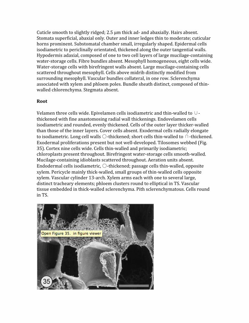
Cuticle smooth to slightly ridged; 2.5 µm thick ad-‐ and abaxially. Hairs absent. Stomata superficial, abaxial only. Outer and inner ledges thin to moderate; cuticular horns prominent. Substomatal chamber small, irregularly shaped. Epidermal cells isodiametric to periclinally orientated, thickened along the outer tangential walls. Hypodermis adaxial, composed of one to two cell layers of large mucilage-‐containing water-‐storage cells. Fibre bundles absent. Mesophyll homogeneous, eight cells wide. Water-‐storage cells with birefringent walls absent. Large mucilage-‐containing cells scattered throughout mesophyll. Cells above midrib distinctly modified from surrounding mesophyll. Vascular bundles collateral, in one row. Sclerenchyma associated with xylem and phloem poles. Bundle sheath distinct, composed of thin-‐walled chlorenchyma. Stegmata absent. Root Velamen three cells wide. Epivelamen cells isodiametric and thin-‐walled to ∪-‐thickened with fine anastomosing radial wall thickenings. Endovelamen cells isodiametric and rounded, evenly thickened. Cells of the outer layer thicker-‐walled than those of the inner layers. Cover cells absent. Exodermal cells radially elongate to isodiametric. Long cell walls ○-‐thickened; short cells thin-‐walled to ∩-‐thickened. Exodermal proliferations present but not well-‐developed. Tilosomes webbed (Fig. 35). Cortex nine cells wide. Cells thin-‐walled and primarily isodiametric; chloroplasts present throughout. Birefringent water-‐storage cells smooth-‐walled. Mucilage-‐containing idioblasts scattered throughout. Aeration units absent. Endodermal cells isodiametric, ○-‐thickened; passage cells thin-‐walled, opposite xylem. Pericycle mainly thick-‐walled, small groups of thin-‐walled cells opposite xylem. Vascular cylinder 13-‐arch. Xylem arms each with one to several large, distinct tracheary elements; phloem clusters round to elliptical in TS. Vascular tissue embedded in thick-‐walled sclerenchyma. Pith sclerenchymatous. Cells round in TS.

Figure 35. Scanning electron micrograph of root TS of Polystachya longiscapa showing webbed tilosome. Scale bar = 23.1 µm. Polystachya Leaf Cuticle smooth to ridged. Adaxial cuticle less than 1.25–2.5 µm thick; abaxial cuticle up to 1.25 µm thick. Hairs rare, glandular, multicellular with sunken basal cell in P. modesta; absent in P. concreta and P. longiscapa. Stomata superficial, abaxial only. Outer ledges thin to thick; inner ledges moderate to thick; cuticular horns small. Substomatal chamber small, irregularly shaped. Epidermal cells isodiametric to periclinally orientated. Walls evenly thickened to thickened only along the outer tangential surface. Hypodermis adaxial, composed of one to three layers of large, thin-‐walled, mucilage-‐containing water-‐storage idioblasts. Fibre bundles absent. Mesophyll homogeneous, 10–15 cells wide. Water-‐storage cells with birefringent walls absent. Large, mucilage-‐containing idioblasts scattered throughout. Cells above midrib distinctly modified from surrounding mesophyll, most prominent in P. longiscapa. Vascular bundles collateral, in one row. Sclerenchyma associated with xylem and phloem clusters, cells thicker-‐walled near phloem. Bundle sheath distinct, composed of thin-‐walled chlorenchyma. Stegmata absent. Root Velamen three to four cells wide. Epivelamen cells isodiametric to tangentially flattened, thin-‐walled in P. concreta and P. modesta to ∪-‐thickened in P. longiscapa. Radial wall thickenings fine and anastomosing. Endovelamen cells isodiametric and rounded; walls thin to evenly thickened. Cover cells absent. Hairs present in P. concreta. Exodermal cells isodiametric to radially elongate. Long cell walls ∩-‐thickened in P. concreta and P. modesta, ○-‐thickened in P. longiscapa. Short cells thin-‐walled to ∩-‐thickened. Exodermal proliferations absent. Tilosomes webbed. Cortex 10–20 cells wide. Cells thin-‐walled, isodiametric to radially elongate; chloroplasts present throughout in P. modesta and P. longiscapa. Water-‐storage cells with birefringent smooth to banded walls in P. longiscapa; birefringent walls smooth with edge thickenings in P. concreta; birefringent walls absent in P. modesta. Mucilage-‐containing idioblasts present throughout. Aeration units absent. Endodermal cells isodiametric; thin-‐walled in P. modesta, slightly ○-‐thickened in P. concreta; and ○-‐thickened in P. longiscapa. Passage cells thin-‐walled, opposite xylem. Pericycle mainly thin-‐walled with small groups of lignified cells opposite phloem in P. concreta and P. modesta. Cells thin-‐walled opposite xylem and thick-‐walled opposite phloem in P. longiscapa. Vascular cylinder 11-‐ to 14-‐arch. Xylem rays each with one to several large, distinct tracheary elements; phloem clusters round to elliptical in TS. Vascular tissue embedded in parenchyma in P. modesta; thin-‐walled sclerenchyma and parenchyma in P. concreta; sclerenchyma in P. longiscapa. Pith completely parenchymatous in P. concreta and P. modesta; outer

ring of sclerenchymatous and inner ring of parenchymatous tissue in P. longiscapa. Cells round in TS. CLADISTICS Heuristic analyses of 164 species using 26 vegetative anatomical and morphological characters (Table 1 and Appendix 2) initially produced 100 equally parsimonious trees. These trees were then swapped to completion, resulting in 20 000+ equally parsimonious trees with L = 227, CI = 0.154, RI = 0.745 and RC = 0.115 (Fig. 36). A bootstrap consensus revealed little resolution among the 169 taxa examined. The monophyly of Vandeae, however, was well-‐supported (90% bootstrap support) using members of Polystachyinae as outgroups. The characters supporting Vandeae as a clade were: loss of tilosomes; presence of stegmata; loss of mucilage; and presence of a monopodial stem (Fig. 37). Three ingroup clades were maintained with at least 50% bootstrap support (BS): Sobennikoffia humbertiana+S. robusta (77% BS); Cyrtorchis praetermissa+C. ringens (55% BS); and Angraecopsis amaniensis+A. breviloba (50% BS). However, Angraecopsis amaniensis+A. breviloba was the only clade not collapsed in the strict consensus (Fig. 37). These two species of Angraecopsis were supported by the presence of deciduous leaves. DISCUSSION Anatomy Vandeae can be characterized by several vegetative features including a monopodial habit, the presence of spherical stegmata within the leaves, a loss of mucilage throughout the plant body; and a loss of tilosomes (Fig. 37). Of these characters, the presence of a single, continuously growing stem apex is probably one of the most important evolutionary steps toward the unique type of leaflessness within Vandeae. If a reduction in vegetative output by decreasing stem length and reducing leaves to vestigial organs is controlled by one or two genes, it could then be switched on or off relatively easily. This vegetative reduction could allow plants to allot more energy towards sexual reproduction, giving leafless orchids a novel way of thriving within the harsh canopy environment (Benzing et al., 1983). Root aeration units were commonly observed throughout Vandeae and may have been another important evolutionary step toward the development of leaflessness. Although many orchids possess photosynthesizing roots, leaves remain the primary organs responsible for fixing carbon in the majority of Orchidaceae (including most Vandeae). In order to shift the evolutionarily function of photosynthesis from leaf to root, the root must have developed some method of retarding water loss while, at the same time, exchanging CO2 and O2. In conjunction with pneumathodes within the velamen (which occur throughout Epidendroideae), aeration units probably serve as the only means of gas exchange in roots of leafless Vandeae and are potentially analogous to the stomatal complex of leaves. Aeration units were also observed in many leafy vandaceous taxa, but they have not been detected in any other group of Orchidaceae. This suggests a preadaptive significance within

Vandeae to the formation of aeration units in the process of becoming leafless. While their presence and possible function within roots was noted as far back as the late 19th century (Leitgeb, 1864; Schimper, 1888; Haberlandt, 1914), their physiology was not fully understood for another 100 years (Benzing et al., 1983; Cockburn, Goh & Avadhani, 1985). The distinct thickenings of the inner tangential endovelamen cell wall superficially resemble tilosomes and occurred scattered throughout Vandeae in the following genera: Aeranthes, Angraecum, Chamaeangis, Cyrtorchis, Diaphananthe, Jumellea, Lemurorchis, Neofinetia, Rhipidoglossum, Sobennikoffia and Taeniophyllum. However, endovelamen thickenings differed from tilosomes in that they occur over both long and short cells (as opposed to only short cells) of the exodermis, and they appear to be distinctive wall thickenings (as opposed to the excrescences of the endovelamen wall). Smooth endovelamen wall thickenings are only known to occur in species of Angraecum and the closely related genus Sobennikoffia. These smooth thickenings are structurally distinct from ridged endovelamen thickenings, which appear scattered primarily in angraecoid genera (Aeranthes, Chamaeangis, Cyrtorchis, Diaphananthe, Jumellea, Lemurorchis, Neofinetia, Rhipidoglossum and Taeniophyllum). The function of endovelamen thickenings is unknown but may be similar to that hypothesized for tilosomes; namely, as a means to prevent water loss via transpiration (Benzing, Ott & Friedman, 1982; Pridgeon et al., 1983). Exodermal proliferations are common throughout Vandeae as well as in the outgroup Polystachyinae. They occur where the velamen has been lost, are superficially similar in cell type to the velamen (i.e. large, empty, lignified and/or suberized cells), and may function similarly in mechanical protection (A. M. Pridgeon, pers. comm.). Exodermal cell walls were most commonly ∩-‐thickened, frequently grading into ○-‐thickened. Notable exceptions to this thickening pattern were limited to four genera. Microcoelia and Solenangis possessed radial swellings and/or grooves (Figs 22, 31) along their primarily ∩-‐thickened walls. Exodermal walls were ∪-‐thickened in Beclardia, Solenangis wakefieldii, Calyptrochilum christyanum and Ypsilopus longifolius. Foliar fibre bundles exhibited a variety of distributional patterns within the mesophyll but were restricted to a few angraecoid taxa. Bundles were most commonly composed of only thick-‐walled cells that were either restricted to leaf margins (Angraecum distichum, A. sesquipedale and Ossiculum) or found as a single row below the epidermis and scattered among vascular bundles (Angraecum teres). Fibre bundles of A. subulatum were composed of both thin-‐ and thick-‐walled cells (cf. fig. 18 in Stern & Judd, 2002) and found alternating in a single row with the vascular bundles. All species of Aeranthes examined (with the exception of an immature specimen of A. arachnites) possessed fibre bundles distributed in a single

abaxial row and composed of thick-‐walled cells surrounding one to three small, thin-‐walled cells.
Figure 36. One of 20 000+ equally parsimonious trees using anatomical and morphological data. A, Angraecinae + Aerangidinae (italics) and Aeridinae (bold

type). Polystachyinae (outgroup) are indicated with a black rectangle. B, subset of Angraecinae + Aerangidinae + Aeridinae. C, subset of Angraecinae + Aerangidinae + Aeridinae. Branches that do not collapse in the strict consensus are indicated with an open circle. Filled boxes received at least 50% bootstrap support.
Figure 37. Strict consensus of 20 000+ equally parsimonious trees. Aeridinae are in bold type, Angraecinae + Aerangidinae are in italics, and Polystachyinae are indicated as the outgroup. Circled letters along the cladogram represent characters supporting each node of the consensus tree. Letters surrounded by squares are those nodes with at least 50% bootstrap support. Numbers outside parentheses represent character numbers, numbers inside parentheses represent character state: a = 2(1), 18(1), 21(1), 22(1); b = 6(0); c = 16(1); d = 8(2); e = 13(1); f = 1(0), 12(1); g = 24(1); h = 11(1); i = 14(3); j = 15(1); k = 12(1); l = 5(0); m = 14(2); n =

14(0); o = 3(1); p = 7(0), 12(1); q = 23(0); r = 3(1), 11(2); s = 3(1); t = 23(0); u = 15(0); v = 3(2). Epidermal and mesophyll cells above the midrib in leaves were frequently modified in most genera of Vandeae. This collection of cells most closely resembles the ‘median unfolding tissue’ described by Löv (1926) and then by Solereder & Meyer (1930), which functions in the unfolding of leaves during development. Esau (1958, 1977) described similar ‘bulliform’ cells occurring in several monocot families, especially Poaceae. Similar cells were also found in leaves of Maxillaria by Holtzmeier et al. (1998). Cladistics Based on vegetative structure, Vandeae formed a well-‐supported (90% BS) clade distinguished from the closely related Polystachyinae (Cameron et al., 1999; Chase et al., 2003) by the loss of mucilage and tilosomes (features that are almost universal within Polystachyinae), the development of spherical silica bodies along sclerenchymatous cells within leaves, and a monopodial growth habit (Fig. 37). The monophyly of Vandeae is also strongly supported by chloroplast and nuclear DNA sequence data from the first author’s dissertation (Carlsward, 2004) and recent publication (Carlsward et al., 2006), as well as by floral anatomy and morphology (Freudenstein & Rasmussen, 1999). However, the utility of vegetative anatomy and morphology seems to be limited for this distinct tribe of orchids (Fig. 37). There were only three moderately to weakly supported clades: Angraecopsis amaniensis+A. breviloba (50% BS); Cyrtorchis praetermissa+C. ringens (55% BS); and Sobennikoffia humbertiana+S. robusta (77% BS). Of the remaining unsupported groups (those with less than 50% BS) within the strict consensus tree (Fig. 37), two clades were in agreement with molecular analyses of Carlsward (2004) and Carlsward et al. (2006): (1) Angraecopsis, Sphyrarhynchus and Mystacidium, supported by the presence of edge-‐thickened water-‐storage cells and (2) Neobenthamia gracilis and Polystachya longiscapa, supported by a reversal to ○-‐thickened exodermal cell walls. Jumellea arachnantha, J. phalaenophora and J. sagittata (Angraecinae) were grouped together based on the presence of distinct ridged endovelamen wall thickenings. However, the leafless Taeniophyllum biocellatum (Aeridinae) was also included in this clade. Species of Angraecum formed several clades, together with Sobennikoffia, based on the presence of distinct, smooth endovelamen wall thickenings: A. eburneum ssp. giryamae+A. eburneum ssp. superbum var. longicalcar, A. eburneum ssp. xerophilum+Sobennikoffia, A. eburneum+A. eburneum ssp. superbum. All of the remaining unsupported clades within the strict consensus tree were not consistent with traditional or molecular hypotheses of phylogeny and were probably based on homoplasious similarities instead of true synapomorphies.

Rhipidoglossum has traditionally been segregated from Diaphananthe based on floral features (Schlechter, 1918; Garay, 1972; Senghas, 1986), but a closer examination of the floral morphology by Cribb (1989) and Summerhayes (1960) led them to believe the two genera were conspecific. However, both Cribb (1989) and Summerhayes (1960) kept Rhipidoglossum a distinct section within Diaphananthe. Although molecular data from Carlsward (2004) supported the two genera as distinct (Fig. 15), vegetative anatomy was not helpful in distinguishing these genera. ACKNOWLEDGEMENTS We thank Norris H. Williams and W. Mark Whitten for their thoughtful advice in preparing this manuscript; Walter S. Judd for his help in character state delimitation; Alec M. Pridgeon for his counsel with the interpretation of root anatomy; and Lars Jonsson for his advice on leafless Vandeae, especially Microcoelia. Robert L. Dressler and James D. Ackerman were also extremely generous with their taxonomic advice. We acknowledge the tremendous generosity of the following commercial and hobbyist orchid growers: Isobyl La Croix (Uzumara Orchids, Gairloch, Scotland); Robert Fuchs (R. F. Orchids, Inc., Homestead, Florida); Martin Motes (Motes Orchids, Homestead, Florida); Claude Hamilton (Hamlyn Orchids, Kingston, Jamaica); Gaspar Silvera (Orquideas Tropicales, Panama City, Panama); William A. (Andy) Phillips (Andy’s Orchids, Encinitas, California); Jim Rose (Cal-‐Orchid, Santa Barbara, California); Erich Michel (Hoosier Orchid Company, Indianapolis, Indiana); Rick Reese (Rick’s Tropica, New Port Richey, Florida); Countryside Orchids (Corrales, New Mexico); Johan Hermans (Royal Botanic Gardens, Kew, London, England); James D. Ackerman (University of Puerto Rico, Puerto Rico); Germán Carnevali (Centro de Investigación Cient’fica de Yucatán, Mexico); Paul Simon (private grower, Los Altos Hills, California); and James Watts (private grower, Hollywood, Florida). Harry Luther, Bruce Holst and Wesley Higgins of the Marie Selby Botanical Gardens were all extremely generous with their resources and time. We also acknowledge the help of Paula J. Rudall, Mark W. Chase, Edith Kapinos, and the late Timothy Lawrence at the Royal Botanic Gardens, Kew, for their help in processing, vouchering, and shipping the specimens donated by Johan Hermans. Assistance and use of equipment provided by the Electron Microscopy Core Laboratory at the University of Florida (especially Karen L. Kelley) were essential to the success of our anatomical work. Greenhouse space for living specimens was provided by the Florida Museum of Natural History, for which we are extremely grateful. This research was supported by the National Science Foundation (Doctoral Dissertation Improvement Grant, DEB-‐0104566), the Lewis and Varina Vaughn Fellowship in Orchid Biology, the American Orchid Society’s 11th World Orchid

Conference Fellowship, and the College of Liberal Arts and Sciences Dissertation Fellowship. Appendices APPENDIX 1 Taxa of Vandeae examined for anatomical study


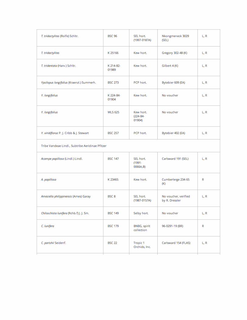


APPENDIX 2 Character states for taxa used in cladistic analyses of Vandeae


REFERENCES Arends JC, Stewart J. 1989. Aerangis gracillima: a definitive account of a rare African orchid of Cameroon and Gabon. Lindleyana 4: 23–29. Barthlott W. 1981. Epidermal and seed surface characters of plants: systematic applicability and some evolutionary aspects. Nordic Journal of Botany 1: 345–355. Benzing DH, Ott DW, Friedman WE. 1982. Roots of Sobralia macrantha (Orchidaceae): structure and function of the velamen-‐exodermis complex. American Journal of Botany 69: 608–614. Benzing DH, Friedman WE, Peterson G, Renfrow A. 1983. Shootlessness, velamentous roots, and the pre-‐eminence of Orchidaceae in the epiphytic biotope. American Journal of Botany 70: 121–133. Brummitt RK, Powell CE. 1992. Authors of plant names. Kew: Royal Botanic Gardens. Burr B, Barthlott W. 1991. On a velamen-‐like tissue in the root cortex of orchids. Flora 185: 313–323. Cameron KM, Chase MW, Whitten WM, Kores PJ, Jarrell DC, Albert VA, Yukawa T, Hills HG, Goldman DH. 1999. A phylogenetic analysis of the Orchidaceae: evidence from rbcL nucleotide sequences. American Journal of Botany 86: 208–224. Carlsward BS. 2004. Molecular systematics and anatomy of Vandeae (Orchidaceae): the evolution of monopodial leaflessness. Unpubl. DPhil thesis, University of Florida. Carlsward BS, Stern WL, Judd WS, Lucansky TW. 1997. Comparative leaf anatomy and systematics in Dendrobium, sections Aporum and Rhizobium (Orchidaceae). International Journal of Plant Sciences 158: 332–342. Carlsward BS, Whitten WM, Williams NH, Bytebier B. 2006. Molecular phylogenetics of Vandeae (Orchidaceae) and the evolution of leaflessness. American Journal of Botany 93: 770–786. Chase MW, Freudenstein JV, Cameron KM, Barrett RL. 2003. DNA data and Orchidaceae systematics: a new phylogenetic classification. In: DixonKW, KellSP, BarrettRL, CribbPJ, eds. Orchid conservation. Kota Kinabalu, Malaysia: Natural History Publications, 69–89. Cheadle VI, Kosakai H. 1982. The occurrence and kinds of vessels in Orchidaceae. Phyta (India), Studies on Living and Fossil Plants, Plant Commemoration 1982: 45–57.

Chiang SHT, Chou T. 1971. Histological studies on the roots of orchids in Taiwan. Taiwania 16: 1–29. Cockburn W, Goh JC, Avadhani PN. 1985. Photosynthetic carbon assimilation in a shootless orchid, Chiloschista usneoides (Don) Lindl. Plant Physiology 77: 83–86. Cribb P. 1989. Flora of tropical East Africa, Orchidaceae (Part 3). Rotterdam: A.A. Balkema. Curtis KM. 1917. The anatomy of the six epiphytic species of the New Zealand Orchidaceae. Annals of Botany 31: 133–149. Das S, Paria N. 1992. Stomatal structure of some Indian orchids with reference to taxonomy. Bangladesh Journal of Botany 21: 65–72. Dressler RL. 1993. Phylogeny and classification of the orchid family. Portland, OR: Dioscorides Press. Dycus AM, Knudson L. 1957. The role of the velamen of the aerial roots of orchids. Botanical Gazette 119: 78–87. Engard CJ. 1944. Morphological identity of the velamen and exodermis in orchids. Botanical Gazette 105: 457–462. Esau K. 1958. Plant anatomy. New York: John Wiley. Esau K. 1977. Anatomy of seed plants. New York: John Wiley. Foster AS. 1956. Plant idioblasts: remarkable examples of cell specialization. Protoplasma 46: 184–193. Freudenstein JV, Rasmussen FN. 1999. What does morphology tell us about orchid relationships? A cladistic analysis. American Journal of Botany 86: 225–248. Garay LA. 1972. On the systematics of the monopodial orchids I. Botanical Museum Leaflets, Harvard University 23: 149–212. Gasson P, Cribb PJ. 1986. The leaf anatomy of Ossiculum aurantiacum Cribb & van der Laan (Orchidaceae: Vandoideae). Kew Bulletin 41: 827–832. Haberlandt G. 1914. Physiological plant anatomy. London: Macmillan. Holmgren PK, Holmgren NH, Barnett LC. 1990. Index herbariorum, Part I: the herbaria of the world. New York: NY Botanical Garden.

Holtzmeier MA, Stern WL, Judd WS. 1998. Comparative anatomy and systematics of Senghas’s cushion species of Maxillaria (Orchidaceae). Botanical Journal of the Linnean Society 127: 43–82. Jeyamurthy A, Dagar JC, Rathore RKS. 1990. Some morphological and anatomical observations on an interesting and rare orchid Chiloschista lunifera (Reichb.f.) J.J.Sm. from Andamans. Journal of the Indian Botanical Society 69: 335–338. Jonsson L.1981. A monograph of the genus Microcoelia (Orchidaceae). Motala: Borgströms Tryckeri AB. Jost L. 1887. Ein Beitrag zur Kenntniss der Athmungsorgane der Pflanzen. Botanische Zeitung (Berlin) 45: 601–606, 617–628, 633–642. Kaushik P. 1983. Ecological and anatomical marvels of the Himalayan orchids. New Delhi: Today and Tomorrow’s Printers and Publishers. Leitgeb H. 1864. Die Luftwurzeln der Orchideen. Denkschriften der Kaiserlichen Akademie der Wissenschaften, Mathematischnaturwissenschaftlichen Klasse 24: 179–222. Löv L. 1926. Zur Kenntnis der Entfaltungszellen monokotyler Blätter. Flora 120: 332–343. Maddison DR, Maddison WP. 2000. Macclade 4: Analysis of Phylogeny and Character Evolution, v.4.06. Sunderland, MA: Sinauer Associates. Mohana Rao PR, Kumari SVL, Khasim SM, Isaiah JM. 1989. Anatomy of some Sikkim Himalayan orchids with reference to their ecological adaptability. Acta Botanica Indica 17: 229–232. Møller JD, Rasmussen H. 1984. Stegmata in Orchidales: character state distribution and polarity. Botanical Journal of the Linnean Society 89: 53–76. Mulay BN, Panikkar TKB. 1956. Origin, development, and structure of velamen in the roots of some species of terrestrial orchids. Proceedings of the Rajasthan Academy of Sciences 6: 31–48. Nseya AM, Arends JC. 1995. Contribution à l’étude anatomique et caryologique des Orchidaceae: le genre Cyrtorchis Schltr. Bulletin du Muséum National d’Histoire Naturelle, Paris. Section B, Adansonia 17: 75–93. Olatunji OA, Nengim RO. 1980. Occurrence and distribution of tracheoidal elements in the Orchidaceae. Botanical Journal of the Linnean Society 80: 357–370.

Oliveira VC, Sajo MG. 1999. Anatomia foliar de especies de Orchidaceae. Revista Brasileira de Botanica 22: 365–374. Porembski S, Barthlott W. 1988. Velamen radicum micromorphology and classification of Orchidaceae. Nordic Journal of Botany 8: 117–137. Pridgeon AM. 1987. The velamen and exodermis of orchid roots. In: ArdittiJ, ed. Orchid biology. Reviews and perspectives IV. Ithaca, NY: Cornell University Press, 139–192. Pridgeon AM, Stern WL, Benzing DH. 1983. Tilosomes in roots of Orchidaceae: morphology and systematic occurrence. American Journal of Botany 70: 1365–1377. Prychid CJ, Rudall PJ, Gregory M. 2003. Systematics and biology of silica bodies in monocotyledons. Botanical Review 69: 377–440. Rasmussen H. 1986. An aspect of orchid anatomy and adaptationism. Lindleyana 1: 102–107. Royal Botanic Gardens, Kew. 2004. World checklist of monocotyledons. http://www.kew.org/wcsp/home.do The Board of Trustees of the Royal Botanic Gardens, Kew [accessed December 2004]. Sanford WW, Adanlawo I. 1973. Velamen and exodermis characters of West African epiphytic orchids in relation to taxonomic grouping and habitat tolerance. Botanical Journal of the Linnean Society 66: 307–321. Schimper AFW. 1888. Die epiphytische Vegetation Amerikas. Jena: Gustav Fischer. Schlechter R. 1918. Versuch einer natürlichen Neuordnung der afrikanischen agraekoiden Orchidaceen. Beihefte zum Botanischen Centralblatt. Abt. II 36: 62–181. Senghas K. 1986. Rhipidoglossum. Die Orchidee 18: 1110–1112. Singh H. 1986. Anatomy of root in some Orchidaceae. Acta Botanica Indica 14: 24–32. Solereder H, Meyer FJ. 1930. Systematic anatomy of the monocotyledons. VI. Microspermae. Jerusalem: Israel Program for Scientific Translations. Stern WL, Carlsward BS. 2004. Vegetative constants in the anatomy of epiphytic orchids. Orchid Review 112: 119–122. Stern WL, Judd WS. 2000. Comparative anatomy and systematics of the orchid tribe Vanilleae excluding Vanilla. Botanical Journal of the Linnean Society 134: 179–202.

Stern WL, Judd WS. 2001. Comparative anatomy and systematics of Catasetinae (Orchidaceae). Botanical Journal of the Linnean Society 136: 153–178. Stern WL, Judd WS. 2002. Systematic and comparative anatomy of Cymbidieae (Orchidaceae). Botanical Journal of the Linnean Society 139: 1–27. Sulistiarini D. 1986. Leaf anatomy and the taxonomy status of Luisia latipetala. Berita Biologi 3: 143–145. Summerhayes VS. 1960. African Orchids. XXVII. Kew Bulletin 14: 126–157. CrossRef Swofford DL. 2003. paup*: phylogenetic analysis using parsimony (*and other methods), v.4.0b10. Sunderland, MA: Sinauer Associates. Toscano de Brito ALV. 1996. The use of concentrated ammonia as an excellent medium for the restoration of orchid pollinaria: an example from the subtribe Ornithocephalinae (Orchidaceae). Lindleyana 11: 205–210. Williams NH. 1979. Subsidiary cells in the Orchidaceae: their general distribution with special reference to development in the Oncidieae. Botanical Journal of the Linnean Society 78: 41–66. Withner CL, Nelson PK, Wejksnora PJ. 1974. The anatomy of orchids. In: WithnerCL, ed. The orchids: scientific studies. New York: John Wiley, 267–347.