comparative hepatotoxicity of fluconazole, ketoconazole...
TRANSCRIPT

Research ArticleComparative Hepatotoxicity of Fluconazole, Ketoconazole,Itraconazole, Terbinafine, and Griseofulvin in Rats
Star Khoza,1 Ishmael Moyo,1 and Denver Ncube2
1Department of Clinical Pharmacology, College of Health Sciences, University of Zimbabwe, Harare, Zimbabwe2Department of Anatomy, College of Health Sciences, University of Zimbabwe, Harare, Zimbabwe
Correspondence should be addressed to Star Khoza; [email protected]
Received 7 October 2016; Revised 19 December 2016; Accepted 18 January 2017; Published 5 February 2017
Academic Editor: Brad Upham
Copyright © 2017 Star Khoza et al. This is an open access article distributed under the Creative Commons Attribution License,which permits unrestricted use, distribution, and reproduction in any medium, provided the original work is properly cited.
Oral ketoconazole was recently the subject of regulatory safety warnings because of its association with increased risk of inducinghepatic injury. However, the relative hepatotoxicity of antifungal agents has not been clearly established. The aim of this study wasto compare the hepatotoxicity induced by five commonly prescribed oral antifungal agents. Rats were treated with therapeutic oraldoses of griseofulvin, fluconazole, itraconazole, ketoconazole, and terbinafine. After 14 days, only ketoconazole had significantlyhigher ALT levels (𝑝 = 0.0017) and AST levels (𝑝 = 0.0008) than the control group. After 28 days, ALT levels were highest inthe rats treated with ketoconazole followed by itraconazole, fluconazole, griseofulvin, and terbinafine, respectively. The AST levelswere highest in the rats treated with ketoconazole followed by itraconazole, fluconazole, terbinafine, and griseofulvin, respectively.All drugs significantly elevated ALP levels after 14 days and 28 days of treatment (𝑝 < 0.0001). The liver enzyme levels suggestedthat ketoconazole had the highest risk in causing liver injury followed by itraconazole, fluconazole, terbinafine, and griseofulvin.However, histopathological changes revealed that fluconazole was the most hepatotoxic, followed by ketoconazole, itraconazole,terbinafine, and griseofulvin, respectively. Given the poor correlation between liver enzymes and the extent of liver injury, it isimportant to confirm liver injury through histological examination.
1. Introduction
In 2013, the United States Food and Drug Administration(US FDA) and the European Medicines Agency’s Commit-tee on Medical Products for Human Use (EMA-CHMP)concurrently issued safety warnings and limited the use oforal ketoconazole because of its association with increasedrisk of inducing hepatic injury, risk of drug interactions,and increased risk of adrenal insufficiency [1, 2]. The twoagencies recommended that ketoconazole should be used“only when alternative antifungal therapies are not availableor tolerated.” In addition to the safety warning, the FDAissued another directive recommending that drug companiesand researchers should avoid using oral ketoconazole in druginteraction studies [3]. The regulatory safety warnings onoral ketoconazole have serious implications on its use inthe clinical and drug development settings. Ketoconazolehas been widely used for more than three decades in thetreatment of fungal infections and has been the principal
prototype human cytochrome P450 3A inhibitor in druginteraction studies and drug metabolism research duringdrug development [4–6].
The link between ketoconazole and hepatotoxicity is wellestablished [7–10]. However, for a long time the evidencesuggested that the hepatotoxicity was mild, rarely fatal, andreversible upon discontinuation of the drug [7, 8, 10]. Anestimated prevalence of serious hepatotoxicity of one in15,000 patients was reported in the United Kingdom inthe first decade of oral ketoconazole market authorization[10]. Incidence data on ketoconazole induced hepatotoxicityis scarce. In a randomized controlled study, subclinicalhepatic dysfunction was observed in 17.5% of patients treatedwith ketoconazole while none of the patients treated withgriseofulvin had evidence of hepatic dysfunction [11]. Bycontrast, a recent meta-analysis of 204 studies reported anoverall incidence of ketoconazole-associated hepatotoxicityof between 3.6% and 4.2% [12].
HindawiJournal of ToxicologyVolume 2017, Article ID 6746989, 9 pageshttps://doi.org/10.1155/2017/6746989

2 Journal of Toxicology
Although the hepatotoxicity of antifungal agents is wellestablished [9, 13–19], their relative hepatotoxicity has notbeen extensively evaluated [20]. Two epidemiological studiesreported contrasting findings regarding the relative hepa-totoxicity of antifungal agents [21, 22]. One of the studiescited by the FDA in its regulatory decision reported thatketoconazole was associated with the highest relative risk(RR = 228; 95% CI: 33.9–933.0) when compared to nonusers,followed by itraconazole (RR = 17.7; 95% CI: 2.6–72.6) andterbinafine (RR = 4.2; 95% CI: 0.2–24.9) [21]. This studyincluded a cohort of 69,830 patients in the United Kingdomwho had received at least one prescription for flucona-zole, griseofulvin, itraconazole, ketoconazole, or terbinafinebetween 1991 and 1996. Of the 69,830 patients included inthe study, only 1052 received ketoconazole. The incidencerates of hepatotoxicity were highest in patients treated withketoconazole (19.0 per 10,000), followed by itraconazole (1.0per 10,000) and terbinafine (0.7 per 10,000) [21]. In contrast tothe study byGarcıa Rodrıguez et al. (1999), Kao and associatesreported the highest incidence rate of drug-induced liverinjury of 31.6 per 10,000 patients in individuals who receivedfluconazole, compared to 4.9 for ketoconazole, 4.3 for grise-ofulvin, 3.6 for itraconazole, and 1.6 for terbinafine [22]. Thestudy included 90,847 patients in Taiwan who received oralantifungal agents between 2002 and 2008. Of these patients,57,321 received oral ketoconazole [22].
Based on the currently available evidence, it is uncertainwhich antifungal agent poses the greatest risk of hepatotox-icity. The small number of cases in the two epidemiolog-ical studies that reported on the relative hepatotoxicity ofantifungal agents limits the interpretation of their findings[21, 22]. The findings by Garcıa Rodrıguez et al. (1999) werebased on 16 cases of acute liver injury [21]. Of these 16 cases,five occurred during current use of oral antifungal agents:two were using ketoconazole, two were using itraconazole,and one was using terbinafine. Out of the ten remainingcases, only one had a history of using an antifungal agentwhile the other nine cases occurred before the use of anyantifungal agent. Similarly, the study by Kao et al. (2014)was based on only 52 cases of drug-induced liver injury[22]. Of the 52 cases, 28 used ketoconazole, 14 were offluconazole, 8 were of griseofulvin, 3 were of itraconazole,and 2 were of terbinafine. In addition to the failure by thesetwo epidemiologic studies to provide conclusive evidenceregarding the antifungal agent with the greatest risk ofhepatotoxicity, few head-to-head experimental studies haveevaluated the relative hepatotoxicity of oral antifungal agentsin clinical settings or using animal models [11, 23–25].Furthermore, the higher number of cases of liver injuryreported with ketoconazole than fluconazolemight be relatedto the higher number of prescriptions for ketoconazole thanfluconazole. Given the implications of the safety warningsissued in 2013 on the use of ketoconazole in clinical settingsand during drug development research, there is need forexperimental studies that evaluate the relative hepatotoxicityof azole antifungal agents. The objective of this study wasto compare the hepatotoxicity effects of the five commonlyprescribed oral antifungal agents (ketoconazole, fluconazole,itraconazole, terbinafine, and griseofulvin). We hypothesized
that fluconazole is more hepatotoxic than ketoconazole basedon histological examination.
2. Materials and Methods
2.1. Materials. All biochemical kits for alanine amino-transferase (ALT), aspartate amino transferases (AST), andalkaline phosphatase (ALP) were sourced from BeckmanCoulter Inc. (California, USA). Terbinafine (Lamisil�; batchnumber U0638; marketed by Novartis Pharma Ltd., UnitedKingdom), itraconazole (Canditral�; batch number 01141282;marketed by Glenmark Pharmaceuticals Ltd., India), grise-ofulvin (Griseon�; batch number 178046; marketed by PlusFive Pharmaceuticals Ltd., Zimbabwe), fluconazole (Flumyc-200�; batch number AHP054014; marketed by Ipca Labo-ratories Ltd., India), and ketoconazole (Nizol�; batch num-ber 13312; marketed by Intas Pharmaceuticals Ltd., India)were all sourced from a local pharmaceutical wholesaler.Standard diet pellets were obtained from National FoodsPvt. Ltd., Zimbabwe. Formaldehyde (37% solution), paraf-fin wax, haematoxylin, eosin, and other standard labora-tory chemicals were sourced from Sigma-Aldrich (UnitedKingdom). Blood collection tubes (Vacuette� Z serum clotactivator tubes) were sourced from Greiner Bio-One (UnitedKingdom). Microcentrifuge tubes (LW2075; batch number110488) were sourced from Alpha Laboratories (Hampshire,United Kingdom).
2.2. Animals and Dosing Procedures. Sixty-six 6-week-oldmale Sprague Dawley rats weighing 180–200 g were adaptedto laboratory conditions for five days before experimentation.The rats were housed in plastic cages in groups of six withwood shavings as bedding under a 12-hour light/12-hourdark cycle. The rats were maintained in a conventionalanimal house with an ambient temperature of 25 ± 2∘Cand were given commercial standard diet rat pellets and tapwater ad libitum. Ethical clearance to conduct the study wasobtained from the Joint ParirenyatwaHospital and College ofHealth Sciences Research Ethics Committee (approval num-ber: JREC/328/14). The animals were handled and treatedfollowing the principles outlined in the “Guide for the Careand Use of Laboratory Animals” prepared by the NationalAcademy of Sciences and published by theNational Institutesof Health (NIH publication 86-23 Rev. 1985).
The rats were divided into eleven groups (each groupwithsix rats) including the control group. The rats in the controlgroup were sacrificed one day before drug administration(day 0) in the active treatment groups. Five groups of ratsreceived a daily single oral antifungal agent dose for 14 days.The other five groups of rats received a daily single oralantifungal agent dose for 28 days. The intragastric method(oral gavage) was used during drug administration. Thetreatment interventionswere 20mg/kg fluconazole, 50mg/kggriseofulvin, 20mg/kg ketoconazole, 20mg/kg itraconazole,and 25mg/kg terbinafine. Antifungal agents are frequentlyprescribed for two weeks or four weeks for most systemicand topical fungal infections. The equivalent doses in rats forcommon adult dose ranges for systemic and topical infections
Journal of Toxicology 3
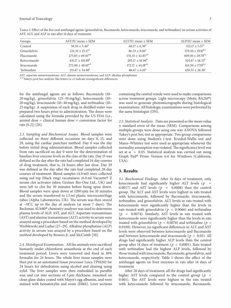
Table 1: Effect of the five oral antifungal agents (griseofulvin, fluconazole, ketoconazole, itraconazole, and terbinafine) on serum activities ofAST, ALT, and ALP in rats after 14 days of treatment.
Groups AST/IU mean ± SEM ALT/IU mean ± SEM ALP/IU mean ± SEMControl 58.50 ± 5.40a 60.17 ± 6.30a 152.17 ± 5.57a
Griseofulvin 124.33 ± 25.17a 86.33 ± 8.06a 370.50 ± 19.81b,c
Fluconazole 275.83 ± 69.45a,b 178.33 ± 42.85a,b 409.50 ± 29.78b,c
Ketoconazole 431.17 ± 101.89b 283.17 ± 61.96b 324.67 ± 18.33b
Itraconazole 272.00 ± 48.10a,b 172.17 ± 44.18a,b 414.50 ± 17.19b,c
Terbinafine 135.67 ± 34.98a 88.67 ± 6.10a 420.33 ± 26.30c
AST: aspartate aminotransferase, ALT: alanine aminotransferase, and ALP: alkaline phosphatasea–eTukey’s post hoc analysis: like letters (a–e) indicate nonsignificant differences.
for the antifungal agents are as follows: fluconazole (10–20mg/kg), griseofulvin (25–50mg/kg), ketoconazole (10–20mg/kg), itraconazole (10–40mg/kg), and terbinafine (10–25mg/kg). A suspension of each drug in distilled water wasprepared two hours prior to administration. The doses werecalculated using the formula provided by the US FDA (i.e.,animal dose = clinical human dose × conversion factor forrats [6.2]) [26].
2.3. Sampling and Biochemical Assays. Blood samples werecollected on three different occasions on days 0, 15, and29, using the cardiac puncture method. Day 0 was the daybefore initial drug administration. Blood samples collectedfrom rats sacrificed on day 0 were for the determination ofbaseline liver enzyme levels in the clan of the rats. Day 15 wasdefined as the day after the rats had completed 14-day coursesof drug treatment, that is, 24 hours after last dose. Day 29was defined as the day after the rats had completed 28-daycourses of treatment. Blood samples (4.0ml) were collectedusing red top (black ring) vacutainers (6.0ml Vacuette� Zserum clot activator tubes; Greiner Bio-One Ltd., UK) andwere left to clot for 30 minutes before being spun down.Blood samples were spun down at 1200 rpm for 10 minutesand the serum transferred to 1.5ml plastic microcentrifugetubes (Alpha Laboratories, UK). The serum was then storedat −18∘C up to the day of analysis (at most 7 days). TheBeckman AU680� chemistry analyser was used to determineplasma levels of ALP, AST, and ALT. Aspartate transaminase(AST) and alanine transaminase (ALT) activity in serumwereassayed using a procedure based on themethod developed byWroblewski and Ladue [27–29]. Alkaline phosphatase (ALP)activity in serum was assayed by a procedure based on themethod developed by Bowers Jr. and McComb [30].
2.4. Histological Examination. All the animals were sacrificedhumanly under chloroform anaesthesia at the end of eachtreatment period. Livers were removed and fixed in 10%formalin for 24 hours. The whole liver tissue samples werethen put in an automated tissue processer Leica TP10202 for24 hours for dehydration using alcohol and clearing usingxylol. The liver samples were then embedded in paraffinwax and cut into sections of 5 𝜇m thickness, mounted onclean glass slides coated with Mayer’s egg albumin, and werestained with hematoxylin and eosin (H&E). Liver sections
containing the central venule were used tomake comparisonsacross treatment groups. Light microscopy (Motic BA210�)was used to generate photomicrographs during histologicalexaminations. All histologic examinationswere performed bythe same histologist (DN).
2.5. Statistical Analysis. Data are presented as the mean value± standard error of the mean (SEM). Comparisons amongmultiple groups were done using one way ANOVA followedTukey’s post hoc test as appropriate. Two group comparisonswere done using Student’s 𝑡-test. Kruskal-Wallis test andMann–Whitney test were used as appropriate whenever thenormality assumptionwas violated.The significance level wasset at 𝛼 = 0.05. Statistical analysis was carried out usingGraph Pad� Prism Version 6.0 for Windows (California,USA).
3. Results
3.1. Biochemical Findings. After 14 days of treatment, onlyketoconazole had significantly higher ALT levels (𝑝 =0.0017) and AST levels (𝑝 = 0.0008) than the controlgroup. The ALT and AST levels were highest in rats treatedwith ketoconazole, followed by fluconazole, itraconazole,terbinafine, and griseofulvin. ALT levels in rats treated withketoconazole were significantly higher than the levels inrats treated with griseofulvin (𝑝 = 0.0066) and terbinafine(𝑝 = 0.0074). Similarly, AST levels in rats treated withketoconazole were significantly higher than the levels in ratstreated with griseofulvin (𝑝 = 0.0076) and terbinafine (𝑝 =0.0109). However, no significant differences in ALT and ASTlevels were observed between ketoconazole and fluconazoleand between ketoconazole and itraconazole (𝑝 > 0.05). Alldrugs had significantly higher ALP levels than the controlgroup after 14 days of treatment (𝑝 < 0.0001). Rats treatedwith terbinafine had the highest ALP levels, followed bythose treatedwith itraconazole, fluconazole, griseofulvin, andketoconazole, respectively. Table 1 shows the effect of theantifungal agents on liver enzymes in rats after 14 days oftreatment.
After 28 days of treatment, all the drugs had significantlyhigher AST levels compared to the control group (𝑝 <0.001). The AST levels were highest in the rats treatedwith ketoconazole followed by itraconazole, fluconazole,
4 Journal of Toxicology
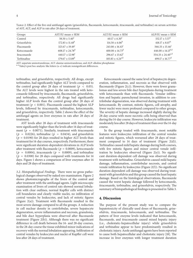
Table 2: Effect of the five oral antifungal agents (griseofulvin, fluconazole, ketoconazole, itraconazole, and terbinafine) on serum activitiesof AST, ALT, and ALP in rats after 28 days of treatment.
Groups AST/IU mean ± SEM ALT/IU mean ± SEM ALP/IU mean ± SEMControl 58.50 ± 5.40a 60.17 ± 6.30a 152.17 ± 5.57a
Griseofulvin 172.83 ± 11.48b 114.50 ± 6.06b 418.17 ± 8.07b
Fluconazole 323.67 ± 19.49c 243.00 ± 16.43c 566.33 ± 15.46c
Ketoconazole 608.17 ± 24.78d 400.00 ± 14.73d 446.00 ± 14.13b,d
Itraconazole 440.17 ± 12.98e 296.67 ± 13.62e 554.33 ± 24.40c,d
Terbinafine 179.67 ± 13.89b 105.83 ± 4.24a,b 499.17 ± 16.77d
AST: aspartate aminotransferase, ALT: alanine aminotransferase, and ALP: alkaline phosphatasea–eTukey’s post hoc analysis: like letters (a–e) indicate nonsignificant differences.
terbinafine, and griseofulvin, respectively. All drugs, exceptterbinafine, had significantly higher ALT levels compared tothe control group after 28 days of treatment (𝑝 < 0.05).The ALT levels were highest in the rats treated with keto-conazole followed by itraconazole, fluconazole, griseofulvin,and terbinafine, respectively. All drugs had significantlyhigher ALP levels than the control group after 28 days oftreatment (𝑝 < 0.001). Fluconazole caused the highest ALPlevels, followed by itraconazole, terbinafine, ketoconazole,and griseofulvin, respectively. Table 2 shows the effect of theantifungal agents on liver enzymes in rats after 28 days oftreatment.
AST levels after 28 days of treatment with itraconazolewere significantly higher than the levels after 14 days of treat-ment (𝑝 = 0.0071). Similarly, treatment with itraconazole(𝑝 = 0.0226), terbinafine (𝑝 = 0.0434), and griseofulvin(𝑝 = 0.0190) for 28 days resulted in higher ALT levels thantreatment for 14-day courses, respectively. Furthermore, therewere significant duration-dependent elevations in ALP levelsafter treatment with fluconazole (𝑝 = 0.0009), ketoconazole(𝑝 = 0.0004), itraconazole (𝑝 = 0.0009), and terbinafine(𝑝 = 0.0300) for 28 days compared with treatments for 14days. Figure 1 shows a comparison of liver enzymes after 14days and 28 days of treatment.
3.2. Histopathological Findings. There were no gross patho-logical changes observed by naked eye examination. Figure 2shows photomicrographs of the livers of the control andafter treatment with the antifungal agents. Light microscopicexamination of livers of control rats showed normal lobula-tion with clear outlines, normal Kupffer cells with distinctcell boundaries and clearly visible nuclei, no infiltration ofcentral venules by leukocytes, and lack of mitotic figures(Figure 2(a)). Treatment with fluconazole resulted in themost severe damage compared to all the groups. A reductionin cell nuclear density in centrilobular, severe hepatocytedegeneration, severe inflammation and necrosis, granuloma,and bile duct hyperplasia were observed after fluconazoletreatment (Figure 2(b)). Although there was no significantdifference in cell death between the 14- and 28-day courses,in the 28-day course the tissue exhibited minor indications ofrecovery with the normal lobulation appearing. Infiltration ofcentral venules by leukocytes and nuclei of Kupffer cell wereless after 28 days of treatment.
Ketoconazole caused the same level of hepatocyte degen-eration, inflammation, and necrosis as that observed withfluconazole (Figure 2(c)). However, there were fewer granu-lomas and less severe bile duct hyperplasia during treatmentwith ketoconazole than with fluconazole. Venular infiltra-tion and hepatic parenchymal invasion, in addition to cen-trilobular degeneration, was observed during treatment withketoconazole. By contrast, mitotic figures, cell atrophy, andfewer nuclei were more profound compared to other groups.The severity of hepatic damage increased slightly during the28-day course with more necrotic cells being observed thanduring the 14-day course. However, leukocyte infiltration wasmoderately less after 28 days of treatment than over the 14-daycourse.
In the group treated with itraconazole, most notablefeatures were leukocytes infiltration of the central venulesand mitotic figures, which worsened after 28 days of treat-ment compared to 14 days of treatment (Figure 2(d)).Terbinafine causedmild hepatic damage during both courses,with few mitotic figures and minor central venule infil-tration by leukocytes observed (Figure 2(e)). No signifi-cant duration-dependent cell damage was observed duringtreatment with terbinafine. Griseofulvin caused mild hepaticdamage, inflammation, centrilobular necrosis, and centralvenule infiltration by leukocytes (Figure 2(f)). No significantduration-dependent cell damage was observed during treat-mentwith griseofulvin and this group caused the least hepaticdamage. Based on the histological observations, fluconazolecaused the worst hepatic damage followed by ketoconazole,itraconazole, terbinafine, and griseofulvin, respectively. Thesummary of histopathological findings is presented inTable 3.
4. Discussion
The purpose of the present study was to compare thehepatotoxicity of clinically used doses of fluconazole, grise-ofulvin, itraconazole, ketoconazole, and terbinafine. Thepattern of liver enzyme levels indicated that ketoconazole,fluconazole, and itraconazole caused mixed hepatic injury(i.e., cholestatic-hepatocellular injury) while griseofulvinand terbinafine appear to have predominantly resulted incholestatic injury. Azole antifungal agents have been reportedto cause both hepatocellular and cholestatic injury [18]. Theincrease in liver enzymes with longer treatment duration

Journal of Toxicology 5
0
200
400
600
800 Effects on AST after 14-day courseA
ST/IU
0
200
400
600
800
Itrac
onaz
ole
Terb
inafi
ne
Keto
cona
zole
Gris
eofu
lvin
Fluc
onaz
ole
Con
trol
Itrac
onaz
ole
Terb
inafi
ne
Keto
cona
zole
Gris
eofu
lvin
Fluc
onaz
ole
Con
trol
Itrac
onaz
ole
Terb
inafi
ne
Keto
cona
zole
Gris
eofu
lvin
Fluc
onaz
ole
Con
trol
Itrac
onaz
ole
Terb
inafi
ne
Keto
cona
zole
Gris
eofu
lvin
Fluc
onaz
ole
Con
trol
Itrac
onaz
ole
Terb
inafi
ne
Keto
cona
zole
Gris
eofu
lvin
Fluc
onaz
ole
Con
trol
Itrac
onaz
ole
Terb
inafi
ne
Keto
cona
zole
Gris
eofu
lvin
Fluc
onaz
ole
Con
trol
Effects on AST after 28-day course
AST
/IU
0
100
200
300
400
500 Effects on ALT after 28-day course
ALT
/IU
0
200
400
600
800 Effects on ALP levels after 14-day course
ALP
/IU
0
200
400
600
800 Effects on ALP after 28-day course
ALP
/IU
0
100
200
300
400
500 Effects on ALT after 14-day course
ALT
/IU
Figure 1: Serum levels of AST, ALT, and ALP, for the control group and the groups that received 14- and 28-day courses of fluconazole,griseofulvin, ketoconazole, itraconazole, and terbinafine.
was noted with all antifungal agents, with itraconazole andterbinafine recording the highest changes in liver enzymes. Incontrast, significant histological changes were observed withitraconazole while only slight worsening in hepatic damagewas observed with ketoconazole treatment. The increase
in the risk of hepatotoxicity during longer treatment withantifungal agents has been reported in several studies [22–24, 31] and regular monitoring of liver enzymes in patientsthat require long treatment with antifungal agents is standardpractice.

6 Journal of Toxicology
(a1) Control (a2) Control
(b1) Fluconazole (14-day course) (b2) Fluconazole (28-day course)
(c1) Ketoconazole (14-day course) (c2) Ketoconazole (28-day course)
(d1) Itraconazole (14-day course) (d2) Itraconazole (28-day course)
(e1) Terbinafine (14-day course) (e2) Terbinafine (28-day course)
(f1) Griseofulvin (14-day course) (f2) Griseofulvin (28-day course)
Figure 2: Photomicrographs of liver sections of the control group (a) and after 14- and 28-day courses of fluconazole (b), ketoconazole (c),itraconazole (d), terbinafine (e), and griseofulvin (f). (a) Normal lobulation with clear outlines, normal cells with visible outlines, and singlenuclei. (b) In 14-day plate the rectangular area shows a marked reduction of cell numbers in the perivenular area. Circular areas indicateinfiltration of venules by leukocytes. 28-day course plate shows reduced cellular density similar to 14-day plate. (c) Rectangular area indicatesreduced cell density; circular areas indicate perivenular region with minor necrotic figures, that is, dark spots on plate. Triangular regionindicates scattered necrotic foci (28-day course). (d) Minor venular distortion indicated by the circular demarcations here; triangular areasshow apoptotic cells. Rectangular area shows reduced cell density. (e) Rectangular area shows paucity of cells; circular areas show clusteringof cells which is a possible indication of stress. (f) Rectangular areas show a reduction in cell numbers and tissue striations (28 d). Perivenularcell paucity is indicated by the circular area on the plate (14 d). Also apparent are necrotic foci around the central venue (arrows). All platesare ×200 magnification.

Journal of Toxicology 7
Table 3: Histopathological findings in livers of treatment with antifungal agents.
Histological findings in the liverTreatment group Hepatocyte degeneration Necrosis Inflammation Bile duct hyperplasia and granulomaKetoconazole +++ +++ +++ ++Fluconazole +++ +++ +++ +++Itraconazole ++ ++ ++ +Griseofulvin + + + +Terbinafine + + + ++Normal (<4 lesions); + mild (4–7 lesions); ++ moderate (8–11 lesions); +++ severe lesions (≥12 lesions per slide); inflammation was determined based on thepresence of macrophages and scattered neutrophils and eosinophils in central venules.
Based on liver enzyme levels observed in this study,ketoconazole had the highest risk in causing liver injuryfollowed by itraconazole, fluconazole, terbinafine, and grise-ofulvin.The relative hepatotoxicity of antifungal agents basedon liver enzymes is consistent with several studies. Thehigher ALT levels during treatment with ketoconazole thanduring treatment with fluconazole observed in this studyis consistent with an in vitro study that reported thatketoconazole significantly increased the levels of ALT andlactate dehydrogenase (LDH) in cultured rat hepatocyteswhile fluconazole had minimal effects on both biomarkers[23]. Hepatotoxicity produced by ketoconazole and its mainmetabolite (N-deacetyl ketoconazole) presents as elevationof ALT or lactate dehydrogenase (Rodriguez and Acosta Jr.,1997) [32, 33]. Similarly, in concordance with the presentstudy, an in vivo study reported that itraconazole treatmentresulted in significantly higher ALT and ALP levels thanfluconazole in rats treated for 14 days [24]. An in vitrostudy using rat hepatocyte cultures also reported similarfindings regarding the relative hepatotoxicity of itraconazoleand fluconazole [34]. More recently, a meta-analysis of 39studies incorporating more than 8,000 patients reported that17.4%of patients treatedwith itraconazole had elevated serumliver enzymes compared to 2.0% of fluconazole users [19].Griseofulvin has also been observed to have a lower risk ofcausing hepatotoxicity than ketoconazole in clinical studies[11].
The observation that fluconazole causes more hepaticdamage than ketoconazole based on histological examina-tions is not consistent with an in vitro study which reportedthat ketoconazole caused more hepatotoxicity than flucona-zole in cultured rat hepatocytes (Rodriguez and Acosta Jr.1995) [23]. Another in vitro study reported that itracona-zole caused more hepatic damage than fluconazole in rathepatocyte cultures while the present study observed thatfluconazole causes more hepatic damage than itraconazolebased on histology examinations [34]. Similarly, in an invivo study by Somchit et al. (2004), hepatocellular necrosis,degeneration of periacinar and midzonal hepatocytes, bileduct hyperplasia, biliary cirrhosis, and giant cell granu-loma were observed in rats treated with itraconazole whilemild degenerative changes of centrilobular hepatocytes wereobserved in the rats treated with fluconazole [24]. However,the study by Somchit et al. (2004) used doses that rangedbetween 7 and 70 times higher than the recommended daily
human doses in humans while the present study used dosesequivalent to human therapeutic doses [24]. Similarly, thehepatocytes in the in vitro studies were exposed to doses thatwere higher than those used therapeutically. Therefore, thedifference between observations made in the present studyand the study by Somchit et al. (2004) and the in vitro studiesmay be explained by the differences in the hepatotoxicitymechanisms at therapeutic doses compared to toxic doses.Secondly, the different routes of administration in the presentstudy and the study by Smochit et al. (2004) may also explainthe differences in the findings. In the study by Somchit et al.(2004), drugs were administered intraperitoneally while theoral route was used in the present study [24].
The observation during histological examination thatfluconazole is more hepatotoxic than ketoconazole anditraconazole is consistent with the results from a largepopulation-based study of 90,847 users of antifungal agentsin Taiwan [22]. In this Taiwanese population, the incidencerate of drug-induced liver injury in patients treated withfluconazole was more than sixfold higher than in patientstreated with ketoconazole, griseofulvin, itraconazole, andterbinafine. In addition, fatality after acute liver injury wasassociated with fluconazole. Out of six fatal drug-inducedliver injury cases, five were current users of fluconazolewhile one was using both fluconazole and ketoconazole [22].In contrast to the observations made in our study usinghistology reports and the study by Garcıa Rodrıguez et al.(2014), an epidemiologic study of 69,830 patients in theUnited Kingdom observed that the incidence rate of acuteliver injury was more than 13-fold higher in patients treatedketoconazole than in patients treated with itraconazole andterbinafine [21]. In this population of patients who filledprescriptions for oral antifungal agents between 1991 and1996, ketoconazole had the highest risk for causing acute liverinjury followed by itraconazole, terbinafine, fluconazole, andgriseofulvin. None of the 35,833 current users of fluconazoleexperienced acute liver injury while one case was associatedwith past use of fluconazole [21].
In the present study, biochemical assays revealed thatketoconazole was the most hepatotoxic antifungal agentwhile histological examinations indicated that fluconazolewas themost hepatotoxic. Similarly, a population-based studythat used biochemical assays as the only diagnostic toolreported that ketoconazole was the most hepatotoxic whilethe study that used a combination of biochemical assays,

8 Journal of Toxicology
biopsy, and tissue pathology reported that fluconazole wasthe most hepatotoxic [21, 22]. The discrepancy betweenhistological examinations and biochemical assays in thisstudy and the difference between the two epidemiologicalstudies that used different diagnostic approaches for acuteliver injury suggests that relative hepatotoxicity of antifungalagentsmay depend on the diagnostic tests used. Furthermore,the fact that most of the drug-induced liver hepatotoxicitycases in clinical use are usually based on liver enzymesand not histological examinations may explain the higherincidence of ketoconazole-associated hepatotoxicity reportsthan those reported during fluconazole use. Despite theirlack of specificity and poor correlation with the degree ofliver injury, biochemical assays remain the cornerstone ofidentifying drug-induced liver injury because they are themost feasible, least-invasive, and cheapest diagnostic tests.However, given the low correlation between liver enzymesand the degree of hepatic damage [35], histology assessmentsprovide better information in deciding the relative hepatotox-icity of chemical agents, including antifungal agents.
5. Conclusions
Liver enzyme levels suggested that ketoconazole is likely tocause liver injury than fluconazole while histopathologicalexaminations revealed that fluconazole is more hepatotoxicthan ketoconazole. The diagnostic criteria used in the evalu-ation of hepatotoxicity of antifungal agents should be takeninto consideration when reviewing the evidence on theirrelative hepatotoxicity. Given the poor correlation betweenliver enzymes and the extent of liver injury, it is important toconfirm liver injury through histological examination beforea diagnosis of hepatotoxicity can be made in clinical settings.
Ethical Approval
Ethical clearance to conduct the study was obtained fromthe Joint Parirenyatwa Hospital and College of HealthSciences Research Ethics Committee (approval number:JREC/328/14). The animals were handled and treated follow-ing the principles outlined in the “Guide for the Care andUseof LaboratoryAnimals” prepared by theNational Academy ofSciences and published by the National Institutes of Health(NIH publication 86-23 Rev. 1985).
Competing Interests
The authors declare that there are no competing interests.
Authors’ Contributions
Star Khoza was involved in the conception and design ofthe study, data analysis and interpretation, and drafting ofthe manuscript. Ishmael Moyo was involved in the design ofthe study, dosing of rats, biochemical analysis, data analysisand interpretation, and revision of the manuscript. DenverNcube participated in the design of the study, interpretation
of histology slides, and revising of themanuscript. All authorsread and approved the final manuscript.
Acknowledgments
The authors are grateful to Mr. Charles Mudzingwa from theDepartment of Anatomy at the University of Zimbabwe forthe assistance in the preparation of the plates for histopatho-logical examinations.
References
[1] Food and Drug Administration Drug Safety Communication,FDA Limits Usage of Nizoral (ketoconazole) Oral Tablets Due toPotentially Fatal Liver Injury and Risk of Drug Interactions andAdrenal Gland Problems, FDA, Silver Spring, Md, USA, 2013,http://www.fda.gov/drugs/drugsafety/ucm362415.htm.
[2] European Medicines Agency’s Committee on MedicinalProducts for Human Use (EMA-CHMP), European MedicinesAgency Recommends Suspension of Marketing Authorisationsfor Oral Ketoconazole, EMA July 2013, http://www.ema.europa.eu/ema/index.jsp?curl=pages/news and events/news/2013/07/news detail 001855.jsp&mid=WC0b01ac058004d5c1.
[3] Food and Drug Administration Drug Safety Communication,FDA Advises Against Using Oral Ketoconazole in Drug Interac-tion Studies Due to Serious Potential Side Effects, FDA, SilverSpring,Md, USA, 2013, http://www.fda.gov/Drugs/DrugSafety/ucm371017.htm.
[4] B. Han, J. Mao, J. Y. Chien, and S. D. Hall, “Optimization ofdrug-drug interaction study design: comparison of minimalphysiologically based pharmacokinetic models on predictionof CYP3A inhibition by ketoconazole,” Drug Metabolism andDisposition, vol. 41, no. 7, pp. 1329–1338, 2013.
[5] I. Fuchs, V. Hafner-Blumenstiel, C. Markert et al., “Effect of theCYP3A inhibitor ketoconazole on the PXR-mediated inductionof CYP3A activity,” European Journal of Clinical Pharmacology,vol. 69, no. 3, pp. 507–513, 2013.
[6] Z. Yang, B. Vakkalagadda, G. Shen et al., “Inhibitory effectof ketoconazole on the pharmacokinetics of a multirecep-tor tyrosine kinase inhibitor BMS-690514 in healthy par-ticipants: assessing the mechanism of the interaction withphysiologically-based pharmacokinetic simulations,” Journal ofClinical Pharmacology, vol. 53, no. 2, pp. 217–227, 2013.
[7] J. H. Lewis, H. J. Zimmerman, G. D. Benson, and K. G. Ishak,“Hepatic injury associated with ketoconazole therapy. Analysisof 33 cases,” Gastroenterology, vol. 86, no. 3, pp. 503–513, 1984.
[8] B. H. C. Stricker, A. P. R. Blok, F. B. Bronkhorst, G. E. Van Parys,and V. J. Desmet, “Ketoconazole-associated hepatic injury. Aclinicopathological study of 55 cases,” Journal of Hepatology, vol.3, no. 3, pp. 399–406, 1986.
[9] K. N. Buchi, P. D. Gray, and K. G. Tolman, “Ketoconazole hep-atotoxicity: an in vitro model,” Biochemical Pharmacology, vol.35, no. 16, pp. 2845–2847, 1986.
[10] G. Lake-Bakaar, P. J. Scheuer, and D. S. Sherlock, “Hepaticreactions associatedwith ketoconazole in theUnitedKingdom,”British Medical Journal, vol. 294, no. 6569, pp. 419–422, 1987.
[11] R.-N. Chien, L.-J. Yang, P.-Y. Lin, and Y.-F. Liaw, “Hepatic injuryduring ketoconazole therapy in patients with onychomycosis: acontrolled cohort study,” Hepatology, vol. 25, no. 1, pp. 103–107,1997.

Journal of Toxicology 9
[12] J. Y. Yan, X. L. Nie, Q. M. Tao, S. Y. Zhan, and Y. D. Zhang,“Ketoconazole associated hepatotoxicity: a systematic reviewandmeta-analysis,” Biomedical and Environmental Sciences, vol.26, no. 7, pp. 605–610, 2013.
[13] A. Srebrnik, S. Levtov, R. Ben-Ami, and S. Brenner, “Liverfailure and transplantation after itraconazole treatment fortoenail onychomycosis,” Journal of the European Academy ofDermatology and Venereology, vol. 19, no. 2, pp. 205–207, 2005.
[14] Z. Perveze, M. W. Johnson, R. A. Rubin et al., “Terbinafine-induced hepatic failure requiring liver transplantation,” LiverTransplantation, vol. 13, no. 1, pp. 162–164, 2007.
[15] K. R. Reddy and E. R. Schiff, “Hepatotoxicity of antimicrobial,antifungal, and antiparasitic agents,” Gastroenterology Clinics ofNorth America, vol. 24, no. 4, pp. 923–936, 1995.
[16] R. O. Chiprut, A. Viteri, C. Jamroz, and W. P. Dyck, “Intra-hepatic cholestasis after griseofulvin administration,”Gastroen-terology, vol. 70, no. 6, pp. 1141–1143, 1976.
[17] S. A. Linnebur and B. L. Parnes, “Pulmonary and hepatic toxic-ity due to nitrofurantoin and fluconazole treatment,” Annals ofPharmacotherapy, vol. 38, no. 4, pp. 612–616, 2004.
[18] M.Thiim and L. S. Friedman, “Hepatotoxicity of antibiotics andantifungals,” Clinics in Liver Disease, vol. 7, no. 2, pp. 381–399,2003.
[19] J.-L. Wang, C.-H. Chang, Y. Young-Xu, and K. A. Chan, “Sys-tematic review and meta-analysis of the tolerability and hepa-totoxicity of antifungals in empirical and definitive therapy forinvasive fungal infection,”Antimicrobial Agents and Chemother-apy, vol. 54, no. 6, pp. 2409–2419, 2010.
[20] C.-H. Chang, Y. Young-Xu, T. Kurth, J. E. Orav, and A. K.Chan, “The safety of oral antifungal treatments for superficialdermatophytosis and onychomycosis: a meta-analysis,” TheAmerican Journal of Medicine, vol. 120, no. 9, pp. 791–798.e3,2007.
[21] L. A. Garcıa Rodrıguez, A. Duque, J. Castellsague, S. Perez-Gutthann, and B. H. C. Stricker, “A cohort study on the riskof acute liver injury among users of ketoconazole and otherantifungal drugs,” British Journal of Clinical Pharmacology, vol.48, no. 6, pp. 847–852, 1999.
[22] W.-Y. Kao, C.-W. Su, Y.-S. Huang et al., “Risk of oral antifungalagent-induced liver injury in Taiwanese,” British Journal ofClinical Pharmacology, vol. 77, no. 1, pp. 180–189, 2014.
[23] R. J. Rodriguez andD. Acosta Jr., “Comparison of ketoconazole-and fluconazole-induced hepatotoxicity in a primary culturesystem of rat hepatocytes,” Toxicology, vol. 96, no. 2, pp. 83–92,1995.
[24] N. Somchit, A. R. Norshahida, A. H. Hasiah, A. Zuraini, M. R.Sulaiman, andM.M.Noordin, “Hepatotoxicity induced by anti-fungal drugs itraconazole and fluconazole in rats: a comparativein vivo study,”Human and Experimental Toxicology, vol. 23, no.11, pp. 519–525, 2004.
[25] M. S. Zakaria, “Comparative study between Griseofulvin andFluconazole hepatotoxic effects and the protective role ofSilymarin,” El-MiniaMedical Bulletin, vol. 16, no. 1, pp. 274–285,2005.
[26] Center forDrug Evaluation andResearch (CDER),Guidance forIndustry Estimating the Maximum Safe Starting Dose in InitialClinical Trials for Therapeutics in Adult Healthy Volunteer, U.S.Department of Health and Human Services Food and DrugAdministration, 2005, http://www.fda.gov/downloads/Drugs/.../Guidances/UCM078932.pdf.
[27] F. Wroblewski and J. S. Ladue, “Serum glutamic pyruvictransaminase in cardiac and hepatic disease,” Proceedings of the
Society for Experimental Biology andMedicine, vol. 91, no. 4, pp.569–571, 1956.
[28] A. Karmen, F. Wroblewski, and J. S. Ladue, “Transaminaseactivity in human blood,” The Journal of clinical investigation,vol. 34, no. 1, pp. 126–131, 1955.
[29] H. U. Bergmeyer and M. Horder, “International federationof clinical chemistry. Scientific committee. Expert panel onenzymes. IFCC document stage 2, draft 1; 1979-11-19 with aview to an IFCC recommendation. IFCC methods for themeasurement of catalytic concentration of enzymes. Part 3.IFCC method for alanine aminotransferase,” Journal of ClinicalChemistry and Clinical Biochemistry, vol. 18, no. 8, pp. 521–534,1980.
[30] G.N. Bowers Jr. andR. B.McComb, “Measurement of total alka-line phosphatase activity in human serum,” Clinical Chemistry,vol. 21, no. 13, pp. 1988–1995, 1975.
[31] M. E.-H. A. Kemeir, “Hepatotoxic effect of itraconazole inexperimental rats,” American Journal of Animal and VeterinarySciences, vol. 9, no. 1, pp. 46–52, 2014.
[32] R. J. Rodriguez and D. Acosta Jr, “Metabolism of ketoconazoleand deacetylated ketoconazole by rat hepatic microsomes andflavin-containing monooxygenases,”DrugMetabolism and Dis-position, vol. 25, no. 6, pp. 772–777, 1997.
[33] R. J. Rodriguez and D. Acosta Jr., “N-Deacetyl ketoconazole-induced hepatotoxicity in a primary culture system of rathepatocytes,” Toxicology, vol. 117, no. 2-3, pp. 123–131, 1997.
[34] N. Somchit, S. M. Hassim, and S. H. Samsudin, “Itraconazoleand fluconazole-induced toxicity in rat hepatocytes: a compar-ativein vitro study,” Human and Experimental Toxicology, vol.21, no. 1, pp. 43–48, 2002.
[35] D. E. Amacher, “A toxicologist’s guide to biomarkers of hepaticresponse,” Human and Experimental Toxicology, vol. 21, no. 5,pp. 253–262, 2002.

Submit your manuscripts athttps://www.hindawi.com
PainResearch and TreatmentHindawi Publishing Corporationhttp://www.hindawi.com Volume 2014
The Scientific World JournalHindawi Publishing Corporation http://www.hindawi.com Volume 2014
Hindawi Publishing Corporationhttp://www.hindawi.com
Volume 2014
ToxinsJournal of
VaccinesJournal of
Hindawi Publishing Corporation http://www.hindawi.com Volume 2014
Hindawi Publishing Corporationhttp://www.hindawi.com Volume 2014
AntibioticsInternational Journal of
ToxicologyJournal of
Hindawi Publishing Corporationhttp://www.hindawi.com Volume 2014
StrokeResearch and TreatmentHindawi Publishing Corporationhttp://www.hindawi.com Volume 2014
Drug DeliveryJournal of
Hindawi Publishing Corporationhttp://www.hindawi.com Volume 2014
Hindawi Publishing Corporationhttp://www.hindawi.com Volume 2014
Advances in Pharmacological Sciences
Tropical MedicineJournal of
Hindawi Publishing Corporationhttp://www.hindawi.com Volume 2014
Medicinal ChemistryInternational Journal of
Hindawi Publishing Corporationhttp://www.hindawi.com Volume 2014
AddictionJournal of
Hindawi Publishing Corporationhttp://www.hindawi.com Volume 2014
Hindawi Publishing Corporationhttp://www.hindawi.com Volume 2014
BioMed Research International
Emergency Medicine InternationalHindawi Publishing Corporationhttp://www.hindawi.com Volume 2014
Hindawi Publishing Corporationhttp://www.hindawi.com Volume 2014
Autoimmune Diseases
Hindawi Publishing Corporationhttp://www.hindawi.com Volume 2014
Anesthesiology Research and Practice
ScientificaHindawi Publishing Corporationhttp://www.hindawi.com Volume 2014
Journal of
Hindawi Publishing Corporationhttp://www.hindawi.com Volume 2014
Pharmaceutics
Hindawi Publishing Corporationhttp://www.hindawi.com Volume 2014
MEDIATORSINFLAMMATION
of