common eye diseases - texas.aoa.orgtexas.aoa.org/documents/tx/2018 convention/staff...
TRANSCRIPT

12/1/2017
1
Common Eye Diseases Lynn E. Lawrence, CMSgt(ret), USAF
CPOT, ABOC, COA, OSC
Call it
Blepharitis …inflammation of the lids
Symptoms
• Redness along the lid margins
• Crusting along the lid margin
• Eye irritation
Treatments
• Lid scrubs
• Medications/Ointment
• Monitoring
Call it
Dry Eye Symptoms
• A stinging, burning or scratchy sensation in your eyes
• Stringy mucus in or around your eyes
• Increased eye irritation from smoke or wind
• Eye fatigue
• Sensitivity to light
• Difficulty wearing contacts
• Periods of excessive tearing
• Blurred vision, often worsening at the end of the day (reading/computer)
Treatment
• Depends on the cause
• Drops must address the problem if used
• Punctal Plugs
• Surgery may be necessary
Call it

12/1/2017
2
Conjunctivitis Symptoms
• Redness in the white of the eye or inner eyelid
• Increased amount of tears
• Thick yellow discharge that crusts over the eyelashes, especially after sleep
• Green or white discharge from the eye
• Itchy eyes
• Burning eyes
• Blurred vision
• Increased sensitivity to light
Types
• Depends on the cause • Bacterial
• Viral
• Irritants
• Allergies
Causes
• Viruses
• Bacteria (such as gonorrhea or chlamydia)
• Irritants such as shampoos, dirt, smoke, and pool chlorine
• Allergies, like dust, pollen, or a special type of allergy that affects some contact lens wearers
• Pinkeye caused by some bacteria and viruses can spread easily from person to person, but is not a serious health risk if diagnosed promptly. Pinkeye in newborn babies, however, should be reported to a doctor immediately
Call it Trauma induced
Non-trauma related
Subconjunctival Hemorrhage
Symptoms
• Redness on the white portion of the eye due to bleeding between the conjunctiva and sclera
Causes
• Hypertension
• Dehydration
• Sneezing
• Coughing
• Constipation
• Straining
• Heavy Lifting
Call it
Normally at
3 and 9 o’clock
Pinguecula… is small like penguin
Symptoms
• Small nodule with or without irritation at the 3 and 9 o’clock positions
Treatment
• Medications / Ointments
• Sunglasses

12/1/2017
3
Call it
Covers the cornea
Ptygerium …is large like pterodactyl
Symptoms
• Large growth that expands over the cornea
• Eye irritation
• FB sensation
• Redness
• Dryness
• Induces astigmatism
• Reduced vision
Treatment
• Removal through surgical excision
• Surgery is very painful
• Can grow back
Call It Does not hurt
Hurts and is irritating
Chalazion
Hordeolum
Call it
External Hordeolum / Chalazion
Symptoms
• Hordeolum - Big red painful lump inside or outside of the eyelid
• Chalazion - Big red painless lump inside or outside of the eyelid
Treatment
• Heat
• Antibiotic ointment
• Surgery
Call it
12/1/2017
4
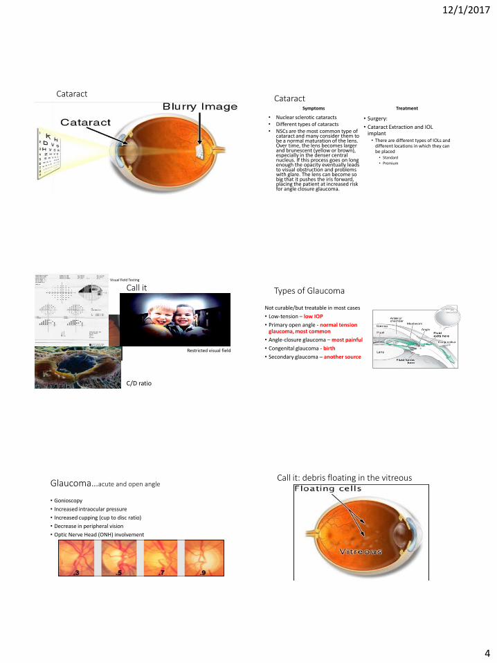
Cataract Cataract Symptoms
• Nuclear sclerotic cataracts • Different types of cataracts • NSCs are the most common type of
cataract and many consider them to be a normal maturation of the lens. Over time, the lens becomes larger and brunescent (yellow or brown), especially in the denser central nucleus. If this process goes on long enough the opacity eventually leads to visual obstruction and problems with glare. The lens can become so big that it pushes the iris forward, placing the patient at increased risk for angle closure glaucoma.
Treatment
• Surgery:
• Cataract Extraction and IOL implant
• There are different types of IOLs and different locations in which they can be placed
• Standard
• Premium
Call it Visual Field Testing
C/D ratio
Restricted visual field
Types of Glaucoma
Not curable/but treatable in most cases
• Low-tension – low IOP
• Primary open angle - normal tension glaucoma, most common
• Angle-closure glaucoma – most painful
• Congenital glaucoma - birth
• Secondary glaucoma – another source
Glaucoma…acute and open angle
• Gonioscopy
• Increased intraocular pressure
• Increased cupping (cup to disc ratio)
• Decrease in peripheral vision
• Optic Nerve Head (ONH) involvement
Call it: debris floating in the vitreous

12/1/2017
5
Floaters Symptoms
• Status of vitreous
• Age of patient
• Could be nothing/could be something
• Post Vitreous Detachment (PVD)… a really big floater
Treatment
• Dilated exam
• Surgery • Vitrectomy
Call it
Retinal Detachment
Symptoms
• Veil in vision
• Part of visual field missing
• Flashes of light
Treatment
Macular Degeneration
Dry Macular Degeneration
• The need for increasingly bright light when reading or doing close work
• Increasing difficulty adapting to low light levels, such as when entering a dimly lit restaurant
• Increasing blurriness of printed words
• A decrease in the intensity or brightness of colors
• Difficulty recognizing faces
• A gradual increase in the haziness of your overall vision
• A blurred or blind spot in the center of your field of vision
• Hallucinations of geometric shapes or people, in cases of advanced macular degeneration
Wet Macular Degeneration • Blood vessels growing in the macula
• Fluid build up
• Visual distortions, such as straight lines appearing wavy or crooked, a doorway or street sign looking lopsided
• Decreased central vision
• Decreased intensity or brightness of colors
• Well-defined blurry spot or blind spot in your field of vision
• Abrupt onset
• Rapid worsening
• Hallucinations of geometric shapes, animals or people, in cases of advanced macular degeneration
• Retinal Ophthalmologist
12/1/2017
6
Call it Diabetic Retinopathy
• Background
• Proliferative • Neovascularization
Diabetic Retinopathy • Diabetic retinopathy often has no early warning signs. Even macular
edema, which may cause vision loss more rapidly, may not have any warning signs for some time. In general, however, a person with macular edema is likely to have blurred vision, making it hard to do things like read or drive. In some cases, the vision will get better or worse during the day.
• As new blood vessels form at the back of the eye as a part of proliferative diabetic retinopathy (PDR), they can bleed (ocular hemorrhage) and blur vision. The first time this happens, it may not be very severe. In most cases, it will leave just a few specks of blood, or spots, floating in a person's visual field, though the spots often go away after a few hours.
Call both photos
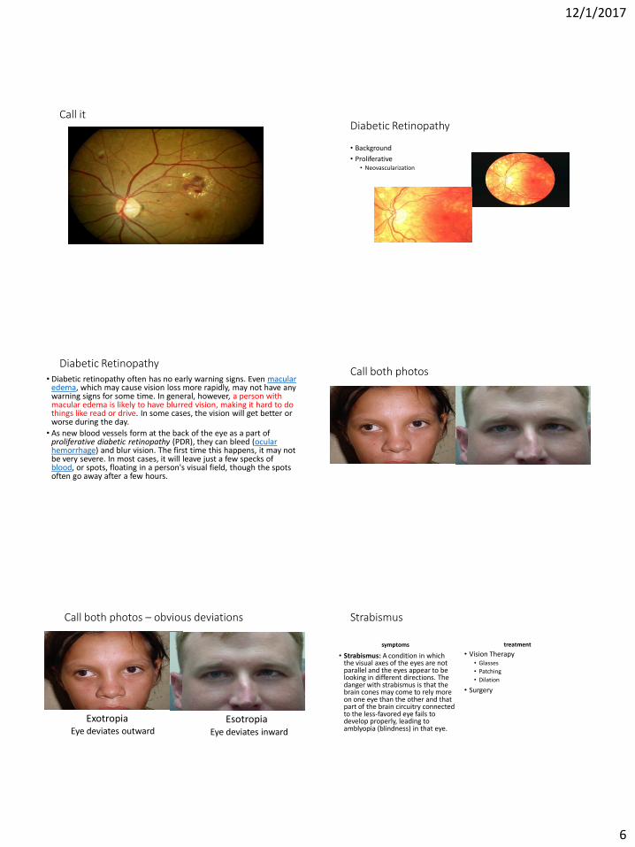
Call both photos – obvious deviations
Exotropia Esotropia Eye deviates outward Eye deviates inward
Strabismus
symptoms
• Strabismus: A condition in which the visual axes of the eyes are not parallel and the eyes appear to be looking in different directions. The danger with strabismus is that the brain cones may come to rely more on one eye than the other and that part of the brain circuitry connected to the less-favored eye fails to develop properly, leading to amblyopia (blindness) in that eye.
treatment
• Vision Therapy • Glasses
• Patching
• Dilation
• Surgery

12/1/2017
7
Call it Retinoblastoma / Leukocoria
• A white color in the center circle of the eye (pupil) when light is shone in the eye, such as when taking a flash photograph
• Eyes that appear to be looking in different directions
• Eye redness
• Eye swelling
• Retinoblastoma occurs when nerve cells in the retina develop genetic mutations that cause the cells to continue growing and multiplying when healthy cells would die. This accumulating mass of cells forms a tumor. Retinoblastoma cells can invade further into the eye and nearby structures. Retinoblastoma can also spread (metastasize) to other areas of the body, including the brain and spine.
Call it Papillidema / Optic Neuritis
• Pain. Most people who develop optic neuritis experience eye pain that's worsened by eye movement. Pain associated with optic neuritis usually peaks within several days.
• Vision loss. The extent of vision loss associated with optic neuritis varies. Most people experience at least some temporary reduction in vision. If noticeable vision loss occurs, it usually develops over the course of hours or days, and may be worsened by heat or exercise. Vision loss may be permanent in some cases.
• Loss of color vision. Optic neuritis often affects the perception of colors. You may notice that the colors of objects, particularly red ones, temporarily appear "washed out" or less vivid than normal.
• Flashing lights. Some people with optic neuritis report seeing flashing or flickering lights.
• Multiple sclerosis
• Neuromyelitis optica
Call it Central Retinal Vein Occlusion • Painless loss of monocular vision is
the usual presenting symptom of retinal artery occlusion (RAO). Ocular stroke commonly is caused by embolism of the retinal artery, although emboli may travel to distal branches of the retinal artery, causing loss of only a section of the visual field. Retinal artery occlusion represents an ophthalmologic emergency, and delay in treatment may result in permanent loss of vision.
• Immediate intervention improves chances of visual recovery, but, even then, prognosis is poor, with only 21-35% of eyes retaining useful vision. Although restoration of vision is of immediate concern, retinal artery occlusion is a harbinger for other systemic diseases that must be evaluated immediately.

12/1/2017
8
Call it Retinitis Pigmentosa
• Retinitis Pigmentosa (RP) is a group of eye diseases that affect the retina. The retina, which is located at the back of the eye, sends visual images to the brain where they are perceived. The cells in the retina that receive the visual images are called photoreceptors. There are two types of photoreceptors: rods (which are responsible for vision in low light) and cones (which are responsible for color vision and detail in high light).
• Signs of RP can usually be detected during a routine eye exam when the patient is around 10 years old. However, symptoms usually do not develop until adolescence.
Call it Other Lid Defects
Droopy or Floppy Eyelids
Can be caused by nerve or muscle defects
• Can be excessive skin upper lid (Dermatochalasis)
• Poor eyelid muscle tension (lid ptosis)
• Brow ptosis
• Floppy Eye Lid Disease – significant increase in lid laxity
Corrected by:
• Blepharoplasty
• Brow lift
• Face lift
Call it

12/1/2017
9
What Is Shingles?
After you have chickenpox, the virus that caused it, called varicella-zoster virus, remains in your body. It's always inside you, lying dormant (or asleep) in your nerve cells. At some point later in life, your immune system may weaken, allowing the virus to resurface as Shingles. You may be feeling great, but if you've had chickenpox, the Shingles virus is already inside you. And your risk for Shingles increases as you get older.
http://www.mayoclinic.org/diseases-conditions/shingles/basics/symptoms/con-20019574
At Risk
• If you've had chickenpox, the Shingles virus is inside you. And as you get older, you're at increased risk for developing the painful, blistering rash. So don't wait to talk to your doctor or pharmacist. To help you start the conversation about Shingles, here are some questions you may want to ask. You can print them and take them with you the next time you see your doctor or pharmacist. Be sure to add any other questions you may have.
Treatment
•Vaccine
•Anti-virals
•Terrasil ointment
• The antidepressants most often used to treat shingles pain are known as tricyclic antidepressants (TCAs). Examples of TCAs most commonly prescribed for people with shingles are amitriptyline, imipramine and nortriptyline
Co-Manage with PCM
• The pain and rash occur near an eye. If left untreated, this infection can lead to permanent eye damage.
• You're 70 or older, because age significantly increases your risk of complications.
• You or someone in your family has a weakened immune system (due to cancer, medications or chronic illness).
• The rash is widespread and painful
Thank you [email protected]