porcine sclera as a model of human sclera for in vitro ... · porcine sclera as a model of human...
TRANSCRIPT
Porcine sclera as a model of human sclera for in vitro transportexperiments: histology, SEM, and comparative permeability
S. Nicoli,1 G. Ferrari,2 M. Quarta,1 C. Macaluso,3 P. Govoni,4 D. Dallatana,5 P. Santi1
1Department of Pharmacy, University of Parma, Italy; 2G.B. Bietti Eye Foundation, IRCCS, Rome, Italy; 3Department ofOphthalmology, University of Parma, Italy; 4Department of Experimental Medicine, Section of Histology, University of Parma, Italy;5Department of Human Anatomy, University of Parma, Italy
Purpose: To evaluate porcine sclera as a model of human sclera for in vitro studies of transscleral drug delivery of bothlow and high molecular weight compounds.Methods: Human and porcine scleras were characterized for thickness and water content. The tissue surface was examinedby scanning electron microscopy (SEM), and the histology was studied with hematoxylin-eosin staining. Comparativepermeation experiments were performed using three model molecules, acetaminophen as the model compound for smallmolecules; a linear dextran with a molecular weight of 120 kDa as the model compound for high molecular weight drugs;and insulin, which was chosen as the model protein. Permeation parameters such as flux, lag time, and permeabilitycoefficient were determined and compared.Results: Human and porcine scleras have a similar histology and collagen bundle organization. The water content isapprox 70% for both tissues while a statistically significant difference was found for the thickness, porcine sclera beingapproximately twofold thicker than human sclera. Differences in thickness produced differences in the permeabilitycoefficient. In fact, human sclera was found to be two to threefold more permeable toward the three molecules studiedthan porcine sclera.Conclusions: The results obtained in the present paper prove that porcine sclera can be considered a good model forhuman sclera for in vitro permeation experiments of both low and high molecular weight compounds. In fact, if the differenttissue thickness is taken into account, comparable permeability was demonstrated. This suggests a possible use of thismodel in the evaluation of the transscleral permeation of new biotech compounds, which currently represent the mostinnovative and efficient therapeutic options for the treatment of ocular diseases.
Transscleral administration is considered a possible non-invasive alternative to injection to target the posterior segmentof the eye for the treatment of chorioretinal diseases. To testthe feasibility of this administration route, thepharmacokinetic and the complex ocular structure imposeseveral steps of in vitro and in vivo experimentation. The firstpreliminary step in the development of transscleral deliveryis represented by in vitro permeation studies through isolatedsclera. The reference tissue for these studies is human sclera,although its limited availability often imposes the use ofanimal models [1,2]. Different models are currently used.Rabbit is the most commonly reported model, but also cow[3] and pig are present in the literature [2]. Porcine sclera hasbeen thoroughly characterized in terms of thickness by Olsen[4] and was found to be similar to human sclera. However,few data are available on its permeability [2,5,6]. Morespecifically, to our knowledge, no literature data are presentconcerning the permeability toward high molecular weightcompounds, which currently represent the most innovative
Correspondence to: Dr. Sara Nicoli, Department of Pharmacy,University of Parma, Viale Usberti 27/A, 43100, Parma, Italy;Phone: +390521905065; FAX: +390521905006; email:[email protected]
and promising drugs for the treatment of posterior segmenteye diseases [7].
The aim of this work was to characterize and comparehuman and porcine scleras to verify adequacy, reliability, andpredictivity of porcine sclera for in vitro permeationexperiments. The characterization included histology,scanning electronic microscopy (SEM) of the outer region ofthe sclera, and measurement of water content. Moreover, thepermeability of three different model molecules through thetwo tissues was studied. The molecules chosen wereacetaminophen as the model compound for small molecules,a linear dextran with a molecular weight of 120 kDa as themodel compound for high molecular weight drugs, andinsulin, which was chosen as the model protein and also forits potential use in the treatment of diabetic retinopathy [8,9].
METHODSMaterials: Insulin from bovine pancreas, fluoresceinisothiocyanate (FITC)-dextran (FD-150; effective MW:120 kDa) acetaminophen, HEPES (4-[2-hydroxyethyl]-1-piperazineethanesulfonic acid), lysine, and EDTA werepurchased from Sigma (St. Louis, MO). For HPLC analysis,acetonitrile (HPLC grade) and distilled water were used. Allother chemicals used were of analytical grade.
Molecular Vision 2009; 15:259-266 <http://www.molvis.org/molvis/v15/a26>Received 4 November 2008 | Accepted 29 January 2009 | Published 6 February 2009
© 2009 Molecular Vision
259
Tissue preparation: Porcine globes were obtained from pigs(Large White, Landrance, Duroc; 10–11 months) that rangedin weight 145–190 kg and came from a local slaughterhouse.Porcine eyes were either used within 24 h of explantation orfrozen at −80 °C until use. After thawing, the adherent muscletissue was removed from the eye bulb, and the anteriorsegment of the eye was circumferentially cut behind thelimbus. The eye was then cut into two halves, the vitreous wasremoved, and the anterior sclera was used for permeationexperiments after the removal of the underling tissues using acotton swab. The frozen tissues were used within three monthsof explantation.
Human corneal-scleral rims, which were discardedfollowing harvesting of the corneal button (Regional CorneaBank, Bologna, Italy), were frozen in liquid nitrogen and usedwithin 15 days of explantation.
Tissue characterization: Human and porcine scleras werecharacterized in terms of thickness and water content. Thethickness was measured with a digital caliper (resolution0.001 mm; Absolute Digimatic 547–401; Mitutoyo, Milan,Italy) at the limbus, equator, and posterior pole of the porcineeye bulb, and the average value was calculated. In the case ofhuman sclera, a single measurement for each sample wasperformed since the specimens have a limited size. Thicknessmeasurements were performed before and after freezing.
For the calculation of water content of the tissue, thesclera was weighed and then dried in a dessiccator in the
presence of P2O5 to constant weight. The water content (%) isthe mass of water per unit mass of the moist specimen:
water content % =wi-wf
wix 100
where wi and wf are the initial and final weight, respectively.Optical microscopy: Pieces of human and porcine sclera(fresh or previously frozen) were fixed in 10% formaldehydeand then embedded in paraffin before sectioning (6 μm thickslices) using a microtome. All sections were then stained usingHarris hematoxylin/eosin.
Images were taken using an optical microscope NikonEclipse 80i (Nikon Instruments, Calenzano, Italy) equippedwith a camera, Nikon Digital Sight DS-2Mv, connected to thecontrol software, NIS-Elements F (Nikon Instruments).Scanning electron microscopy: Samples of scleral tissue,which were previously frozen in liquid nitrogen, were thawedand fixed in 10% formaldehyde then dehydrated in alcohol,then in absolute acetone, and treated with a critical point dryerin liquid CO2. The specimens were Au-metallized with asputtering device. The observation was performed with ascanning electron microscope (Philips, Scansion ElectronMicroscope, model 501; Philips, Hamburg, Germany).
Permeation experiments: Permeation experiments wereperformed in Franz-type diffusion cells with an area of 0.2cm2 for human sclera and 0.6 cm2 for porcine sclera.
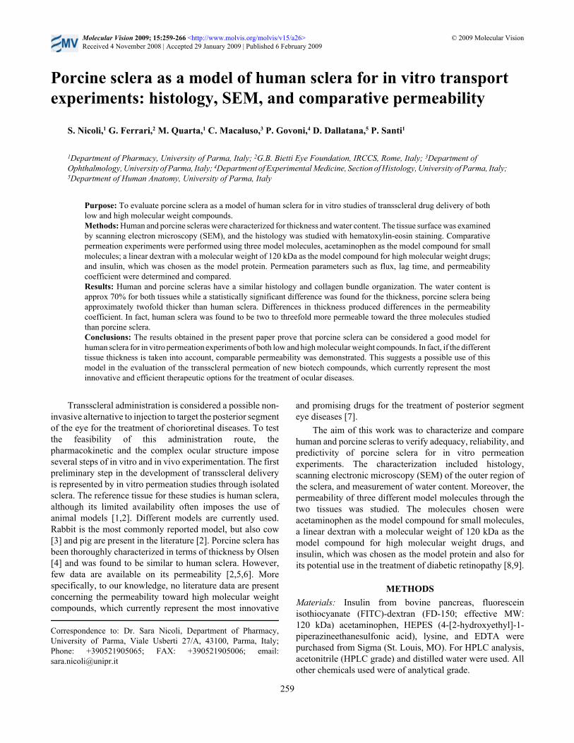
Figure 1. Histological microscopicsections of human and porcine sclerastained with hematoxylin-eosin. Emptylacunae between fibers are an artifactdue to tissue preparation. Originalmagnification was 10X in panel A and4X in panel B. In both human andporcine scleras, scattered smallfibrocyte nuclei are dispersed betweenthe bundles of interwoven collagenfibers. In porcine sclera, thicker andmore disorganized collagen bundles arevisible. From the 4X magnification (B)it is possible to appreciate differences inthickness between human and porcinesclera.
Molecular Vision 2009; 15:259-266 <http://www.molvis.org/molvis/v15/a26> © 2009 Molecular Vision
260

Preliminary control experiments demonstrated that the size ofthe cell area did not influence drug permeation.
To reduce edge damage, a thin layer of silicone lubricantwas applied to the glass surface, and the minimum forcenecessary to keep the cell sealed was applied.
The donor compartment contained alternatively 4.9 mg/ml acetaminophen, 1 mg/ml 120 kDa FITC-Dextran(FD-150), or 1 mg/ml insulin dissolved in 25 mM HEPESbuffer at pH 7.4. To improve insulin solubility in this vehicle,0.4 mM disodium EDTA and 0.3 mM lysine were added[10].
The receptor compartment contained 4 ml of 25 mMHEPES buffer (pH 7.4) added with 0.9% NaCl, kept at 37 °C,and magnetically stirred. In the case of insulin, 0.3% w/vbovine serum albumin (BSA) was added to the receptor phaseto increase the solubility of the protein. At predetermined timeintervals, the receptor solution was sampled for thedetermination of drug permeated.
The transscleral flux, i.e., the amount of drug that crosses1 cm2 of sclera in 1 h (J, µg/cm2h) was calculated as the slopeof the regression line at the steady-state while the lag time wasthe intercept of the regression line on the x-axis. Thepermeability coefficient (P, cm/s) was calculated as J/Cv
where Cv represents the concentration of the donor solution.Experiments performed with human sclera were
replicated three to four times while those performed onporcine sclera were replicated five to seven times. All theexperiments were performed using previously frozen tissues,
and acetaminophen permeability was tested also through freshpig sclera.Analytical methods: Acetaminophen and insulin wereanalyzed by HPLC with a Perkin-Elmer instrument (Norwalk,CT), which is made of an isocratic pump LC250 an UVdetector LC290, and the Perkin Elmer TurbochromWorkstation software.
Acetaminophen was analyzed using a C18 Novapak®
column (150×3.9 mm; Waters, Milford, MA) and a mobilephase composed of 92% (v/v) 10 mM sodium acetate pH 4and 8% (v/v) acetonitrile, which was pumped at 1 ml/min. Thedetector was set at 254 nm.
Insulin was analyzed using a C18 Symmetry300® column(250×4.6 mm; Waters) at 40 °C and a mobile phase composedof 73% (v/v) aqueous phase and 27% (v/v) acetonitrile, whichwas pumped at 1 ml/min. The aqueous phase containedanhydrous sodium sulfate 28.4 g/l and phosphoric acid 2.7 ml/l and was adjusted at pH 2.3 with ethanolamine. The detectorwas set at 214 nm.
FD-150 was analyzed with a fluorescence detector(Series 200a Perkin Elmer). The excitation and emission λwere 490 and 520 nm, respectively.
Affinity of insulin for the scleral tissue: Affinity of insulin forscleral tissue was estimated by measuring its partitioncoefficient between human or porcine sclera and an aqueoussolution at pH 7.4. Insulin was dissolved in the aqueousvehicle that was used as donor for the permeation experimentsat a concentration of about 100 µg/ml. This solution (0.4 ml)
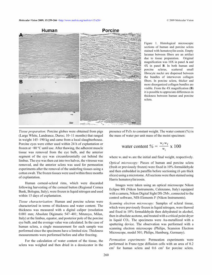
Figure 2. SEM image of the outer regionof human and porcine sclera. Originalmagnification was 5,000X in panel Aand 20,000X in panel B. Scale bar=10µm in both cases. From the pictures atlower magnification (A) it is possible toobserve branching and anastomosis ofthe collagen bundles to form denseconnective tissue. The bundles were ofvarying thickness and width and oftenintertwined with each other. Porcinebundles looked thicker at lowermagnification, but still showed a similararrangement. Moreover, highermagnification (B) did not show anydifference between human and porcinesclera in the diameter of the singlecollagen fibers
Molecular Vision 2009; 15:259-266 <http://www.molvis.org/molvis/v15/a26> © 2009 Molecular Vision
261

was added to a previously weighed amount of human orporcine sclera (50–100 mg) in a 2.0 ml vial. The insulinsolution and the scleral tissues were incubated at roomtemperature for 3 h. The solution was then filtered(regenerated cellulose, 0.45 µm pore size) and analyzed byHPLC for the determination of the final insulin concentrationin the water phase [Wf]. The insulin concentration [S] in thesclera after the incubation period was calculated as:
S =Wi x VW - Wf x VW
VS
where [Wi] is the initial concentration of insulin in the aqueousphase, [Wf] is the concentration of insulin in the aqueous phaseafter the incubation period, VW is the volume of the aqueousphase (0.4 ml), and VS is the volume of the sclera, which wasestimated by its weight and by considering the scleral densityto be equal to 1 g/ml.
The partition coefficient (Ks/w) was then calculated as:
Ks/w = SWf
xVWVS
Each experiment was replicated at least six times.Statistical analysis: The significance of the differencesbetween values was assessed using one-way ANOVAfollowed by Bonferroni test (Kaleidagraph 4.01 software[Synergy Software, Reading, PA] on a Macintosh iBook G4[Apple Computers, Cupertino, CA]). Differences wereconsidered statistically significant when p<0.05.
RESULTSTissue characterization: The mean thicknesses of porcine andhuman scleras (measured after thawing of the frozen tissue)were 1.25±0.25 mm and 0.59±0.08 mm, respectively. Somespecimens were also measured before freezing, and the valuesobtained were not statistically different from the frozen tissue.
The water content in the two species is comparable,71.6%±0.63% for human sclera and 69.5%±1.18% forporcine sclera.Optical microscopy: Histology confirmed the similaritybetween human and porcine scleras, although globally thickerand more disorganized collagen bundles were found inporcine sclera (Figure 1). Empty lacunae between fibers inboth human and porcine sclera are an artifact due to tissuepreparation. Scattered, small fibrocyte nuclei were dispersedbetween the bundles of interwoven collagen fibers. Humanspecimens show thick and interlaced bundles between theepisclera and the choroid. Porcine and human scleralspecimens were analyzed before and after freezing, and nodifferences were found in the histology of the tissues (data notshown).
Scanning electron microscopy: Scanning electron microscopy(SEM) is a very useful technique to study the threedimensional (3D) arrangement of the collagen lamellae of theouter sclera. From the pictures (Figure 2), it is possible toobserve branching and anastomosis of the collagen bundles,which form dense connective tissue. The bundles varied inthickness and width and often intertwined with each other.Porcine bundles looked thicker at lower magnification but stillshowed a similar arrangement.Permeation experiments: To compare the two tissues in termsof permeability, permeation experiments with Franz-typediffusion cells were performed across human and porcinescleras using three model permeants with differentcharacteristics: acetaminophen, insulin, and FD-150. It isworth mentioning that with these diffusion cells, it is notpossible to reproduce the natural intraocular pressure (IOP)that, to some extent, can influence the permeation of drugs[11].
Figure 3 shows the permeation profiles of the modelmolecules across human and pig scleras while flux (µg/cm2h),permeability coefficient (cm/s), and lag time (min) values arereported in Table 1. Both the data reported in Figure 3 and thevalues reported in Table 1 show that the transscleral fluxesobtained for all three molecules tested were higher for humansclera even if the differences were not large. In particular, thefluxes (and the permeability coefficients) through humansclera were two times higher than through pig sclera in thecase of acetaminophen and insulin and three times higher inthe case of FD-150. Together with fluxes and permeabilitycoefficients, a difference between the two species was alsofound in the lag time. With acetaminophen, no lag time wasdetectable for human sclera while lag time for pig sclera was35±2 min. In the case of insulin and FD-150, the lag time ofpig sclera were respectively three and two times higher thanhuman sclera.
Permeability of acetaminophen was also measuredthrough fresh porcine sclera and confirmed that the freezingprocedure did not have any effect on tissue permeability(Figure 3A).Affinity of insulin for scleral tissue: The permeation profilesof insulin were characterized by a long lag time, indicating apossible interaction between insulin and the scleral tissue. Forthis reason, the partition coefficient of insulin between thetissue and the donor solution was measured for both humanand porcine scleras and was found to be 10.3±1.2 and 7.0±1.1,respectively. The two values are not statistically differentbetween them (p=0.176), indicating a similar interaction ofthe two tissues with insulin.
DISCUSSIONThe thickness of porcine and human sclera has already beenstudied by Olsen and coworkers [4,12] that measured thethickness of big, medium, and small size eye bulbs. Eye size
Molecular Vision 2009; 15:259-266 <http://www.molvis.org/molvis/v15/a26> © 2009 Molecular Vision
262

and thickness depended upon animal weight that ranged from
Figure 3. Permeation profiles of acetaminophen, FD-150, and insulinfor porcine and human sclera. Profiles for acetaminophen (A),FD-150 (B), and insulin (C) through porcine (○) and human (●)sclera are shown. Open squares (□) in panel A indicate thepermeation profile of acetaminophen through fresh porcine sclera.Transscleral fluxes obtained for all three molecules tested werehigher for human sclera. In particular, the fluxes through humansclera were 2 times greater than through pig sclera in the case ofacetaminophen and insulin, and 3 times greater in the case of FD-150.Permeability of acetaminophen through fresh porcine scleraconfirmed that the freezing procedure did not have any effect ontissue permeability. Mean value ±SEM
2.8 to 81 kg. In the present work, the average thickness ofporcine sclera was higher than that reported by Olsen et al.[4,12] due to the consistently higher weight of the animalsused (145–190 kg).
The porcine sclera samples used in this work weretwofold thicker than the human sclera samples. Despite thisdifference, it is worth mentioning that the thickness of porcinesclera is closer to human skin than rabbit sclera, which iscurrently the most used in vitro model. In fact, rabbit sclerahas a mean thickness that is approximately one-tenth of thatof human sclera [13,14].
The value of water content found for human sclera isconsistent with literature reports [15,16]. Concerning SEMimaging and histology, no significant differences were foundbetween porcine and human scleras.
The permeability was evaluated using three modelcompounds. Acetaminophen is a model molecule for smallmolecular weight compounds (MW of 151.2 Da) with amolecular radius of approximately 0.36 nm [17] and a logP n-octanol/water of 0.2–0.89 [18]. Insulin has been chosen as amodel protein. It has a molecular weight of 5,734 Da, amolecular radius of the monomeric form of 1.13 nm [19] or2.0 nm [20], and an isoelectric point of about 5.4. FD-150 isa linear FITC-labeled dextran with high molecular weight(120 kDa) and an estimated molecular radius of 8.5 nm [21].Permeation experiments were performed using previouslyfrozen tissues since it has been demonstrated that evenconsecutive freeze–thaw treatments to -80 °C did notsignificantly affect human scleral permeability [22].
The flux of acetaminophen through human sclera wastwice that of porcine sclera (626±55 µg/cm2h for human and280±37 µg/cm2h for porcine sclera). The permeabilitycoefficient found across human sclera (3.49 [±0.3] x10-5 cm/s) is consistent with literature data. Regardless of thelipophilicity, the permeability of low molecular weight drugsthrough human sclera is typically in the range of1.0x10-5-4.0x10⁻⁵ cm/s [23].
The permeability coefficient P (cm/s) is also equal to[24]:
P = DKH
where D (cm2/s) is the diffusion coefficient of the moleculeinside the sclera, K is the partition coefficient of the moleculebetween the sclera and the donor solution, and H is thediffusional path length of the permeant. H can beapproximated to the thickness of the membrane because thesclera is a highly porous tissue. Additionally, the two tissues,human and porcine scleras, have the same architecture interms of porosity and tortuosity. By knowing the thickness ofthe barrier, the product, DK, can be calculated. As reported inTable 1, the value, DK, is similar for porcine and humanscleras. Therefore, it is reasonable that the difference in
Molecular Vision 2009; 15:259-266 <http://www.molvis.org/molvis/v15/a26> © 2009 Molecular Vision
263
permeability coefficient observed is simply due to thedifferent thickness of the two tissues.
This result indicates that porcine and human sclerasbehave in the same way toward the permeation of a smallmolecule despite a difference in tissue thickness. Thesimilarity of the two tissues in terms of structure, histology,and collagen fiber architecture corroborates this result, whichis also supported by in vitro studies on surgically-thinnedhuman sclera that demonstrated the role of the scleralthickness on transscleral transport [22].
When the permeation experiment was performed using ahigh molecular weight compound (FD-150), which wascharacterized by a molecular radius of 8.5 nm, thepermeability coefficient was 100 times lower than thepermeability coefficient of acetaminophen for both humanand porcine scleras. This is in agreement with the dependenceof the scleral permeability on the permeant molecular radius[25]. The results on the human sclera are also consistent withliterature reports. The permeability coefficient obtained here(4.8 [±0.7] x10-7 cm/s) is similar to that found by Cruysberget al. [11] (approximately 1.0x10⁻⁷ cm/s), which isincidentally identical to the value of porcine sclera. Also, inthe case of FD-150, the value, DK, which underlines theimportance of tissue thickness, was similar for both tissues.This means that porcine and human scleras behave similarlywith regards to permeability toward big molecules. This isparticularly relevant because the last generation of drugs usedor proposed for the treatment of the posterior segment eyediseases are represented by high molecular weight compoundssuch as oligonucleotides, antibodies, and other proteins.
Finally, the permeability of the sclera to insulin wastested. Insulin has been chosen as a model protein, but it hasalso been recently proposed for its potential use in the localtreatment of diabetic retinopathy [8,9]. Once again, thebehavior of human and porcine scleras toward insulin wassimilar. Despite the permeability coefficient being higher forhuman sclera, the value of DK is almost identical in the two
scleras, indicating the same behavior when the differentthickness of the two tissues is taken into account. If the resultsobtained in terms of permeability are consistent withacetaminophen and FD-150 data, the lag time found was long.The permeation experiments had to be continued for 6 h toachieve steady-state conditions (see Figure 3C). This could bedue to several reasons. One is linked to the peculiar behaviorof insulin that is able to form dimers and hexamers in solution,which results in the molecular radius of the permeant not being1.13 nm but possibly about 2.5 nm [26]. Another hypothesisis that insulin interacts with specific or non-specific bindingsites on the scleral structures. The presence of specific bindingsites for insulin has been demonstrated for chick sclera [27]while non-specific binding to the sclera has been reported fornegatively charged molecules (fluorescein andcarboxyfluorescein) [6,28] as insulin is at pH 7.4 [26].
Regardless of the nature of this interaction, we haveestimated that the affinity of insulin for the sclera was the samefor porcine and human tissues. The affinity, measured as thepartition coefficient between the sclera and the donor solution,was 10.3±1.2 (mean value±standard error of the mean [SEM])for human sclera and 7.0±1.1 for porcine sclera. These values(not statistically different between them, p=0.176) indicate asimilar behavior of the two tissues toward insulin and alsoconfirm that insulin has an effective affinity for the sclera. Infact, even if the absolute value of the partition coefficient isnot particularly high, it is much higher than the partitioncoefficient values reported in the literature, which aretypically equal or lower than 1. Examples of neutral moleculesof different lipophilicity are mannitol (0.005), budesonide(1.2), celecoxib (1.4) [2], sucrose (0.6), and FITC-labeleddextrans of various MW (0.07–0.39) [29]. There are someexceptions to these typical values such as rhodamine 6G [2],which showed high affinity for the scleral tissue because itwas electrically charged at the pH of the experiment andinteracted with the tissue.
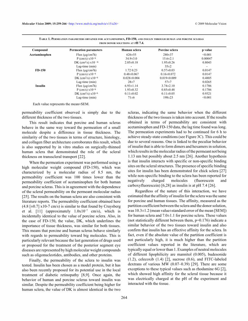
TABLE 1. PERMEATION PARAMETERS OBTAINED FOR ACETAMINOPHEN, FD-150, AND INSULIN THROUGH HUMAN AND PORCINE SCLERAS
FROM DONOR SOLUTIONS AT PH 7.4.
Compound Permeation parameters Human sclera Porcine sclera p valueAcetaminophen Flux (µg/cm2h) 626±55 280±37 <0.001
P (cm/s) x10−6 34.9±3.0 15.6±2.1 0.00047 DK (cm2/s) x10 −6 2.05±0.18 1.95±0.26 0.8043 Lag-time (min) - 35±2 -
FD-150 Flux (µg/cm2h) 1.72 0.23 0.57±0.03 0.0147 P (cm/s) x10−6 0.48±0.067 0.16±0.072 0.0147 DK (cm2/s) x10−6 0.028±0.004 0.019±0.009 0.4885 Lag-time (min) 28±7 57±7 0.0265
Insulin Flux (µg/cm2h) 6.93±1.14 3.74±2.10 0.1786 P (cm/s) x10−6 1.93±0.32 0.85±0.48 0.1786 DK (cm2/s) x10−6 0.11±0.02 0.11±0.05 0.9321 Lag-time (min) 71±6 198±23 <0.001
Each value represents the mean±SEM.
Molecular Vision 2009; 15:259-266 <http://www.molvis.org/molvis/v15/a26> © 2009 Molecular Vision
264
Detailed studies performed by Olsen et al. [4,12] haveshown important similarities between porcine and human eyesin term of intraocular anatomy and scleral thickness. Thesesimilarities are absent in other animal models such as rabbitand cow. Porcine sclera is easily obtained in large amountsfrom slaughterhouses and can efficiently substitute for rabbitsclera, which is now the most used in vitro model. The resultsobtained in the present paper further prove that porcine scleracan be considered a good model for human sclera for in vitropermeation experiments. Optical microscopy and SEMshowed a similar organization of collagen bundles of thesclera even if they looked thicker and more disorganized inthe porcine sample in comparison to the human sample.Moreover, permeation experiments demonstrated that thepermeability of porcine and human scleras is very similartoward a small molecule (151 Da), a high molecular weight(120 kDa) compound, and a model protein (5.8 kDa) exceptfor differences in tissue thickness. A very similar bindingcapacity was also demonstrated toward the model protein,suggesting a possible and reliable use of this model in theevaluation of transscleral drug permeation of new biotechcompounds, which currently represent the most innovativeand efficient therapeutic options.
Further studies are needed to confirm human and porcinesimilarities with regards to permeability toward highmolecular weight proteins.
REFERENCES1. Ambati J, Adamis AP. Transscleral drug delivery to the retina
and choroid. Prog Retin Eye Res 2002; 21:145-51. [PMID:12062532]
2. Cheruvu NP, Kompella UB. Bovine and porcine transscleralsolute transport: influence of lipophilicity and the Choroid-Bruch's layer. Invest Ophthalmol Vis Sci 2006; 47:4513-22.[PMID: 17003447]
3. Maurice DM, Polgar J. Diffusion across the sclera. Exp Eye Res1977; 25:577-82. [PMID: 590384]
4. Olsen TW, Sanderson S, Feng X, Hubbard WC. Porcine sclera:thickness and surface area. Invest Ophthalmol Vis Sci 2002;43:2529-32. [PMID: 12147580]
5. Amaral J, Fariss RN, Campos MM, Robison WG Jr, Kim H,Lutz R, Becerra SP. Transscleral-RPE permeability of PEDFand ovalbumin proteins: implications for subconjunctivalprotein delivery. Invest Ophthalmol Vis Sci 2005;46:4383-92. [PMID: 16303924]
6. Lin CW, Wang Y, Challa P, Epstein DL, Yuan F. Transscleraldiffusion of ethacrynic acid and sodium fluorescein. Mol Vis2007; 13:243-51. [PMID: 17356511]
7. Sharif NA, Klimko P. Ophthalmic Agents. In: Triggle DJ,Taylor JB, editors. Comprehensive Medicinal Chemistry II.Vol 6. Oxford (UK): Elsevier; 2006. p. 297–320.
8. McCarty MF. Nitric oxide deficiency, leukocyte activation, andresultant ischemia are crucial to the pathogenesis of diabeticretinopathy/neuropathy–preventive potential of antioxidants,essential fatty acids, chromium, ginkgolides, andpentoxifylline. Med Hypotheses 1998; 50:435-49. [PMID:9681924]
9. Koevary SB, Nussey J, Lake S. Accumulation of topicallyapplied porcine insulin in the retina and optic nerve in normaland diabetic rats. Invest Ophthalmol Vis Sci 2002;43:797-804. [PMID: 11867601]
10. Quinn R, Andrade JD. Minimizing the aggregation of neutralinsulin solutions. J Pharm Sci 1983; 72:1472-3. [PMID:6363674]
11. Cruysberg LP, Nuijts RM, Geroski DH, Gilbert JA, HendrikseF, Edelhauser HF. The influence of intraocular pressure onthe transscleral diffusion of high-molecular-weightcompounds. Invest Ophthalmol Vis Sci 2005; 46:3790-4.[PMID: 16186364]
12. Olsen TW, Aaberg SY, Geroski DH, Edelhauser HF. Humansclera: thickness and surface area. Am J Ophthalmol 1998;125:237-41. [PMID: 9467451]
13. Li SK, Zhu H, Higuchi WI. Enhanced transscleral lontophoretictransport with ion-exchange membrane. Pharm Res 2006;23:1857-67. [PMID: 16841198]
14. Li SK, Zhang Y, Zhu H, Higuchi WI, White HS. Influence ofasymmetric donor-receiver ion concentration upontransscleral iontophoretic transport. J Pharm Sci 2005;94:847-60. [PMID: 15736190]
15. Watson PG, Young RD. Scleral structure, organisation anddisease. A review. Exp Eye Res 2004; 78:609-23. [PMID:15106941]
16. Lee SB, Geroski DH, Prausnitz MR, Edelhauser HF. Drugdelivery through the sclera: effects of thickness, hydration,and sustained release systems. Exp Eye Res 2004;78:599-607. [PMID: 15106940]
17. Falk B, Garramone S, Shivkumar S. Diffusion coefficient ofparacetamol in a chitosan hydrogel. Mater Lett 2004;58:3261-5.
18. Kalantzi L, Reppas C, Dressman JB, Amidon GL, JungingerHE, Midha KK, Shah VP, Stavchansky SA, Barends DM.Biowaiver monographs for immediate release solid oraldosage forms: acetaminophen (paracetamol). J Pharm Sci2006; 95:4-14. [PMID: 16307451]
19. Laogun AA, Sheppard RJ, Grant EH. Dielectric properties ofinsulin in solution. Phys Med Biol 1984; 29:519-24. [PMID:6377337]
20. Bohidar HB. Light scattering and viscosity study of heataggregation of insulin. Biopolymers 1998; 45:1-8. [PMID:9433182]
21. Nicholson C, Tao L. Hindered diffusion of high molecularweight compounds in brain extracellular microenvironmentmeasured with integrative optical imaging. Biophys J 1993;65:2277-90. [PMID: 7508761]
22. Olsen TW, Edelhauser HF, Lim JI, Geroski DH. Human scleralpermeability. Effects of age, cryotherapy, transscleral diodelaser, and surgical thinning. Invest Ophthalmol Vis Sci 1995;36:1893-903. [PMID: 7543465]
23. Prausnitz MR, Noonan JS. Permeability of cornea, sclera, andconjunctiva: a literature analysis for drug delivery to the eye.J Pharm Sci 1998; 87:1479-88. [PMID: 10189253]
24. Martin AN. Diffusion and Dissolution. In: Martin AN,Bustamante P, editors. Physical pharmacy: physical chemicalprinciples in the pharmaceutical science. Philadelphia: Lea &Febiger; 1993. p. 324–361.
25. Ambati J, Canakis CS, Miller JW, Gragoudas ES, Edwards A,Weissgold DJ, Kim I, Delori FC, Adamis AP. Diffusion of
Molecular Vision 2009; 15:259-266 <http://www.molvis.org/molvis/v15/a26> © 2009 Molecular Vision
265
high molecular weight compounds through sclera. InvestOphthalmol Vis Sci 2000; 41:1181-5. [PMID: 10752958]
26. Langkjaer L, Brange J, Grodsky GM, Guy RH. Iontophoresisof monomeric insulin analogues in vitro: effects of insulincharge and skin pretreatment. J Control Release 1998;51:47-56. [PMID: 9685903]
27. Waldbillig RJ, Arnold DR, Fletcher RT, Chader GJ. Insulin andIGF-1 binding in chick sclera. Invest Ophthalmol Vis Sci1990; 31:1015-22. [PMID: 2162332]
28. Prausnitz MR, Edwards A, Noonan JS, Rudnick DE, EdelhauserHF, Geroski DH. Measurement and prediction of transienttransport across sclera for drug delivery to the eye. Ind EngChem Res 1998; 37:2903-7.
29. Boubriak OA, Urban JP, Bron AJ. Differential effects of agingon transport properties of anterior and posterior human sclera.Exp Eye Res 2003; 76:701-13. [PMID: 12742353]
Molecular Vision 2009; 15:259-266 <http://www.molvis.org/molvis/v15/a26> © 2009 Molecular Vision
The print version of this article was created on 30 January 2009. This reflects all typographical corrections and errata to thearticle through that date. Details of any changes may be found in the online version of the article.
266