colloids and surfaces b: biointerfaces - … · ribeiro et al. / colloids and surfaces b:...
TRANSCRIPT

Pe
Ca
b
c
Pd
a
ARR2AA
KBCMM
1
cbitCo
star
dfuds
C
h0
Colloids and Surfaces B: Biointerfaces 140 (2016) 430–436
Contents lists available at ScienceDirect
Colloids and Surfaces B: Biointerfaces
journa l homepage: www.e lsev ier .com/ locate /co lsur fb
roving the suitability of magnetoelectric stimuli for tissuengineering applications
. Ribeiro a, V. Correia a,b, P. Martins a, F.M. Gama c, S. Lanceros-Mendez a,d,∗
Centro/Departamento de Física, Universidade do Minho, Campus de Gualtar, 4710-057 Braga, PortugalAlgoritmi Research Centre, Universidade do Minho, Campus de Azurém, 4800-058 Guimarães, PortugalIBB e Institute for Biotechnology and Bioengineering, Centre of Biological Engineering, Universidade do Minho, Campus de Gualtar, 4710-057 Braga,ortugalBCMaterials, Parque Científico y Tecnológico de Bizkaia, 48160, Derio, Spain
r t i c l e i n f o
rticle history:eceived 27 October 2015eceived in revised form8 December 2015ccepted 29 December 2015
a b s t r a c t
A novel approach for tissue engineering applications based on the use of magnetoelectric materials is pre-sented. This work proves that magnetoelectric Terfenol-D/poly(vinylidene fluoride-co-trifluoroethylene)composites are able to provide mechanical and electrical stimuli to MC3T3-E1 pre-osteoblast cells andthat those stimuli can be remotely triggered by an applied magnetic field. Cell proliferation is enhancedup to ≈25% when cells are cultured under mechanical (up to 110 ppm) and electrical stimulation (up
vailable online 4 January 2016eywords:one tissue engineeringell proliferationagnetic stimulus
to 0.115 mV), showing that magnetoelectric cell stimulation is a novel and suitable approach for tissueengineering allowing magnetic, mechanical and electrical stimuli.
© 2015 Elsevier B.V. All rights reserved.
agnetoelectric
. Introduction
Tissue engineering (TE) techniques aim to mimic the physico-hemical and bioactive characteristics of natural cellular matrices,oth for therapeutic applications and fundamental biological stud-
es [1,2], the replacement and/or regeneration of lost/damagedissues or organs representing one of the main challenges [3–5].ells, scaffolds and growth factors are the three main componentsf TE strategies [6].
Biomaterials interface plays an important role on cell adhe-ion, proliferation and differentiation. Further, biomaterials can beailored not only to be passively tolerated by the organism, butlso to provide the appropriate environment to assist specific cellesponses [7].
In this way, diverse strategies are being implemented for theevelopment of novel scaffolds to support restoring native tissue
unctionalities, including bone, muscle and nerve [1,8]. In partic-
lar, an increasing number of patients suffer from bone relatediseases [9], bone TE becoming increasingly relevant and withtrong socio-economic impact [10,11].∗ Corresponding author at: Center/Department of Physics, University of Minho,ampus de Gualtar, Braga 4710-057, Portugal. Fax: +351 253 604061.
E-mail address: [email protected] (S. Lanceros-Mendez).
ttp://dx.doi.org/10.1016/j.colsurfb.2015.12.055927-7765/© 2015 Elsevier B.V. All rights reserved.
Therefore, the development of new materials for bone TE isunder intense investigation [9,11]. Bone TE strategies are typicallybased on scaffolds as passive cell supports, including porous mate-rials [12], 3D architectures [13] and fibers [14] as well as materialsreinforced with hydroxyapatite [15], but a suitable approach hasnot been achieved so far.
Most of those strategies do not take into account one of therelevant properties of natural bone tissue, which is its piezo-electricity [16]. This means that when a mechanical solicitation(stress) is applied to the bone, it produces a voltage variationthat strongly influences characteristic and performance of bonetissue all along its developing and functional time. In this way,the use of electrically active biomaterials is an attractive andprobably necessary approach for bone TE. The potential of suchelectroactive smart materials in this field exploited so far mainlypiezoelectric poly(vinylidene fluoride) (PVDF) or copolymers suchas poly(vinylidene fluoride-co-trifluoroethylene) (PVDF-TrFE), thepiezoelectric polymers with the largest response [17–19]. Indeed,the influence of surface polarity of PVDF films on bone cell responseshows that polymer surface charges promote cell adhesion, pro-liferation and differentiation [18,20]. Further, under dynamic
mechanical conditions, piezoelectric substrates improve osteoblastgrowth and differentiation [17,18]. This biomimetic approach isparticularly relevant as bone, like other tissues, is physiologicallysubjected to mechano-electrical solicitations related to walking,
ces B: Biointerfaces 140 (2016) 430–436 431
jiuefcb
itpotitteaswp
btM((ittadianmetc
MpmipMianpp
2
2
tEFT
Table 1Scaffolds used for cell culture.
Denomination Samples Polarization Stimulus Film response
A PVDF-TrFE non poled × Magnetic NoneB PVDF-TrFE/TD non poled × MechanicalC PVDF-TrFE/TD “poled +”
√Electrical
C. Ribeiro et al. / Colloids and Surfa
umping and running [16]. However, in some cases, the patient ismmobilized due to serious health condition, and as a result the nat-ral mechanical stimulus does not occur [21], thus decreasing theffectiveness of piezoelectric biomaterials. Such limitation claimsor the development of new materials able to remotely mechani-al and/or electrically stimulate tissues from outside of the humanody [22,23] and/or for specific cell cultures in bioreactors.
Magnetoelectric (ME) composite materials provide such annnovative tool, allowing the use of an external magnetic fieldo remotely control tissue stimulation [23], without the need ofatient movement. Those composites consist on the combinationf magnetostrictive and piezoelectric materials, the ME effect beinghe result of a product property [24]. The mechanical deformationnduced by a magnetic field due to the magnetostriction of one ofhe components, results in a electrical polarization variation dueo the piezoelectric effect of the other phase, allowing large MEffects at room temperature [25,26]. Thus, the magnetic actuationbility of the ME composite allows the mechanical and electricaltimulus of neighboring cells [27]. As bone TE requires biomaterialsith flexibility, lightweight, versatility and biocompatibility [8,28],
olymer-based ME materials can be taken to advantage [27].Two main types of polymer-based ME composites can
e found in the literature: laminated composites and par-iculated micro and nanocomposites [27]. Despite the lower
E response of P(VDF-TrFE)/CoFe2O4 nanocomposites [29]42 mV cm−1 Oe−1)—four orders of magnitude lower than the one383 V cm−1 Oe−1) reported for P(VDF-TrFE)/Metglas 2605SA1 lam-nates [30] - its higher flexibility, simple fabrication, easy shaping,he possibilities of miniaturization and the absence of degrada-ion at the piezoelectric/magnetostrictive interface are obviousdvantages [27,30,31]. Further, particulate composites allow theevelopment of geometries suitable for tissue engineering, includ-
ng spheres [25] and fiber mats [32], allowing also cell culturend specific TE approaches. Finally, the incorporation of mag-etic nanoparticles into scaffolds acting synergistically with theagnetic field in vivo will improve cellular proliferation and differ-
ntiation, and promote an enhancement of tissue integration intohe scaffold, a crucial step towards the clinical applications of theomposites [33,34].
Thus, this work demonstrates the suitability of polymer-basedE composites for tissue engineering, providing thus a novel
latform for innovative tissue engineering strategies based onagnetic, electrical and mechanical stimuli. In particular, the
nfluence of the external magnetic field stimulation in MC3T3-E1re-osteoblast cells proliferation [35] on a Terfenol-D/P(VDF-TrFE)E composite scaffold is reported. Terfenol-D was chosen due to
ts biocompatibility and high magnetostrictive coefficient [36,37]nd P(VDF-TrFE) due to its proven potential for bone, muscle, andeuronal tissue engineering applications, biocompatibility, highiezoelectric coefficient and crystallization on the piezoelectrichase from solvent casted samples [38,39].
. Experimental
.1. Materials
All the chemicals and particles were used as received from
he suppliers: Terfenol-D (TD) ≈1 �m of random particles fromtrema Products, N,N-dimethylformamide (DMF, pure grade) fromluka and poly(vinylidene fluoride-co-trifluoroethylene), P(VDF-rFE), from Solvay Solexis.2.2. Films preparation
Films were prepared based on the method presented in [40,41]for similar polymer-based composite compositions. In short,Terfenol-D particles were added to DMF and placed in an ultra-sound bath for 8 h in order to ensure proper dispersion of theparticles. P(VDF-TrFE) polymer was then added and mixed during2 h with the help of a mechanical Teflon stirrer, in an ultrasoundbath to avoid magnetic particle agglomeration during the mixingprocess. After that, the solution was spread in a clean glass sub-strate and solvent evaporation took place inside an oven at 210 ◦Cfor 10 min. Polymer crystallization was achieved by cooling downfilms to room temperature. At the end of this process, TD parti-cles were inside the polymer matrix and ≈50 �m thick films werepeeled from the glass substrate.
TD/P(VDF-TrFE) composites with 40 weight percentage(40 wt.%) of TD particles content were produced, since preliminarytests demonstrated that TD such composition provides a good MEresponse with suitable mechanical properties (e.g., flexibility) [29].
Film poling was achieved using an optimized procedure, con-sisting on a corona treatment at 10 kV during 120 min at 120 ◦C,in a home-made chamber, and cooling down to room temperatureunder applied electric field. The piezoelectric response (d33) of thesamples was analyzed with a wide range d33-meter (model 8000,APC Int., Ltd.).
For in vitro assays, circular films were cut with 13 mm of diam-eter. Non-poled films (with and without magnetic particles) wereused to study the effect of the magnetic field and magnetostrictionin the cell culture and poled films to study the influence of the MEstimulus in cell behavior (Table 1).
For sterilization proposes, the films were exposed to ultravioletlight (UV) for 1 h on each side. After that, samples were placed instandard 24-well cell culture plates and washed 5 times (5 min eachtime) with a phosphate buffer saline (PBS) 1 × solution.
2.3. ME characterization
In order to obtain the out-of-plane ME coefficient �33, a DC andAC magnetic field were applied along the direction of the electricpolarization of the composites, i.e., perpendicular to the surface.
The AC driving magnetic field of 1 Oe amplitude at ≈10 kHz(resonance frequency of the composite) was provided by a pair ofHelmholtz coils and the DC field with a maximum value of 0.5 Twas applied by an electromagnet.
The resonance frequency (fr) of the composites was determinedby Eq. (1):
fr = n
2l
√EY
�(1)
where n, EY and � are the harmonic mode order, in-plane Young’smodulus and density of the composites, respectively.
The induced ME voltage was measured with a StandfordResearch Lock-in amplifier (SR530).
432 C. Ribeiro et al. / Colloids and Surfaces B: Biointerfaces 140 (2016) 430–436
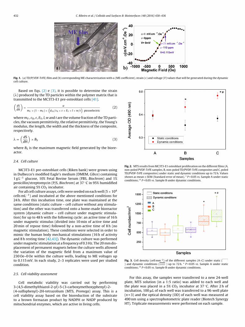
F E coefficient), strain (ε) and voltage (V) values that will be generated during the dynamicc
(t(
wcmr
�
wa
2
i1pa
c2ststu2mmaupt2tc
2
3(ctm
Fig. 2. MTS results from MC3T3-E1 osteoblast proliferation on the different films (A,non-poled PVDF-TrFE samples, B, non-poled TD/PVDF-TrFE composites and C, poledTD/PVDF-TrFE composites) under static and dynamic conditions up to 72 h. Valuesshown as mean ± SEM (Standard error of mean). * P < 0.05 vs. Sample A under staticconditions; # P < 0.05 vs. Sample B under dynamic conditions.
ig. 1. (a) TD/P(VDF-TrFE) film and (b) corresponding ME characterization with � (Mell culture.
Based on Eqs. (2) e (3), it is possible to determine the strain�) produced by the TD particles within the polymer matrix that isransmitted to the MC3T3-E1 pre-osteoblast cells [41].
dSdH
)= ˛
mV × (1 − mV ) ×(d33/ε0 × ε × EY × l × w/t
)piezoelectric
(2)
here mV , ε0, ε, EY, l, w and t are the volume fraction of the TD parti-les, the vacuum permittivity, the relative permittivity, the Young’sodulus, the length, the width and the thickness of the composite,
espectively.
=(dSdH
)× BS (3)
here BS is the maximum magnetic field generated by the biore-ctor.
.4. Cell culture
MC3T3-E1 pre-osteoblast cells (Riken bank) were grown usingn Dulbecco’s modified Eagle’s medium (DMEM, Gibco) containing
g L−1 glucose, 10% Fetal Bovine Serum (FBS, Biochrom) and 1%enicillin/streptomycin (P/S, Biochrom) at 37 ◦C in 95% humidifiedir containing 5% CO2 incubator.
For all cell culture assays, cells were seeded on each well (5 × 10⁴
ells mL−1) and incubated at the above mentioned conditions for4 h. After this incubation time, one plate was maintained at theame conditions (static culture – cell culture without any stimula-ion) and the other was transferred onto a home-made bioreactorystem (dynamic culture – cell culture under magnetic stimula-ion) for up to 48 h with the following cycle: an active time of 16 hnder magnetic stimulus (divided into 10 min of active time and0 min of repose time) followed by a non-active time of 8 h (noagnetic stimulation). These conditions were selected in order toimic the human body mechanical stimulations (16 h of activity
nd 8 h resting time [42,43]). The dynamic culture was performednder magnetic stimulation at a frequency of 0.3 Hz. The 20 mm dis-lacement of permanent magnets below the culture wells allowedhe variation of the magnetic field from a maximum value of30 Oe–0 Oe within the culture wells, leading to ME voltages upo 0.115 mV. In each study, 2–3 replicates were used per studiedondition.
.5. Cell viability assessment
Cell metabolic viability was carried out by performing-(4,5-dimethylthiazol-2-yl)-5-(3-carboxymethoxyphenyl)-2-
4-sulfophenyl)-2H-tetrazolium (MTS, Promega) assay. This is aell viability assay based on the bioreduction of the substrateo a brown formazan product by NADPH or NADP produced byitochondrial enzymes, which are active in living cells.
Fig. 3. Cell density (cell mm−2) of the different samples (A–C) under static () and dynamic conditions ( ) up to 72 h. * P < 0.05 vs. Sample A under staticconditions; # P < 0.05 vs. Sample B under dynamic conditions.
For this assay, the samples were transferred to a new 24-wellplate, MTS solution (in a 1:5 ratio) was added to each well andthe plate was placed in a 5% CO2 incubator at 37 ◦C. After 2 h of
incubation, 100 �L of each well was transferred to a 96-well plate(n = 3) and the optical density (OD) of each well was measured at490 nm using a spectrophotometric plate reader (Biotech SynergyHT). Triplicate measurements were performed on each sample.
C. Ribeiro et al. / Colloids and Surfaces B: Biointerfaces 140 (2016) 430–436 433
F lds ws colou
2
nf
wdsmfips
P(pDt
ig. 4. Representative images of pre-osteoblast culture after 72 h on A, B and C scaffotained with TRITC-red). Scale bar = 200 �m. (For interpretation of the references to
.6. Cell morphology and cell number
The samples collected from cell culture were transferred for aew 24-well plate, washed with PBS 1x and the cells fixed with 4%
ormaldehyde (Panreac) for 10 min at 37 ◦C in a 5% CO2 incubator.For cell number quantification, after fixation, all samples were
ashed with PBS 1x and stained for 10 min with 1 �g mL−1 of a 4,6-iamidino-2-phenylindole (DAPI, Sigma) solution. After that, theamples were washed with PBS 1x and observed in a fluorescenceicroscopy (Olympus BX51 Microscope). Twenty representative
elds of each well were selected in a blinded and systematicrocedure using 10× magnification. For cell counting, the ImageJoftware was used. Finally, cell number per mm2 were calculated.
For cell morphology, after fixation, samples were washed withBS 1x three times and incubated in 0.1 �g mL−1 of red phalloidinSigma) solution for 45 min at room temperature. Finally, the sam-
les were again washed with PBS 1x and incubated in 1 �g mL−1API solution for 10 min. Cells were washed again with PBS 1x andhen visualized in a fluorescence microscopy.
Fig. 5. Representative images of cell pro
ith static and dynamic conditions (nucleus stained with DAPI-blue and cytoskeletonr in this figure legend, the reader is referred to the web version of this article).
2.7. Statistical analysis
All quantitative data were analyzed using GraphPad Prism(v6.00). For each data set, each condition was compared using one-way analysis of variance (ANOVA) statistical evaluation followed byTukey’s test. Differences were considered to be significant when P <0.05. The data of cell density are displayed in boxplots, the boxplotscontaining the following descriptive statistics: median (horizontalline), mean (square), interquartile range (box) and whiskers (out-lier).
3. Results
It has been established that mechanical forces influenceosteoblast activity [44,45]. Beyond the mechanical stimulus, it has
been also proven that charged surfaces and piezoelectric materialsenhance bone tissue regeneration [17,46]. In view of these facts,three different samples were used to prove the suitability of the MEeffect for tissue engineering applications: A – non-poled polymerliferation to each distinct scaffold.

4 ces B:
spmeopsdwit
pvltumedarwrtPbct
a
Tscmfirfit
ft2ttacc
e
irppfidiT
T˛
34 C. Ribeiro et al. / Colloids and Surfa
amples without magnetostrictive particles, acting as passive sup-orts for cell culture (it is to notice here that poled samples withoutagnetic fillers will not have any reaction to the magnetic field
ither, showing therefore the same response that the non-polednes under dynamic conditions); B – non-poled polymeric sam-les with magnetostrictive particles, with can apply mechanicaltimulation to the cells when subjected to a varying magnetic field,ue to the magnetostrictive effect; and C – poled polymer samplesith magnetostrictive particles, which together with the mechan-
cal stimulation, will induce a varying electrical surface charge dueo the piezoelectric effect, as defined by the ME coupling.
It is important to notice at this point that the effect of sampleoling on protein adsorption and cell behavior has been pre-iously addressed in [46] and [20], respectively. Sample polingeads to an overall net surface charge influencing protein adsorp-ion and cell response, as well as improved piezoelectric responsender mechanical stimuli [17,18]. In the present investigation, as aagnetic stimulus is used to trigger the mechanical and the piezo-
lectric response of the samples and once PVDF-TrFE poled sampleso not react to the magnetic stimulus, non-poled PVDF-TrFE is useds negative control. As both poled and non-poled samples do noteact to the applied magnetic field, the differences among themould be the same as reported in [20,46]. There variations are not
elevant to the present investigation in which the effect of magne-ostrictive and magnetoelectric effects are considered. With poledVDF-TrFE samples as negative control, no differences will existetween static and dynamic magnetic conditions either, but weould have a higher number of cells in the poled samples than inhe non-poled ones, as previously reported [46,20].
Fig. 1 shows an image of the TD/P(VDF-TrFE) ME composite filmnd its ME characterization.
Fig. 1a shows the flexibility and easy shaping of the TD/P(VDF-rFE) film. The ME response of the TD/P(VDF-TrFE) compositetudied between −1000 and 1000 Oe is shown in Fig. 1b. The MEoefficient (�), that correlates the voltage generated across theaterial (�V) with the thickness (t) and the applied AC magnetic
eld (BAC), = (�V)/(t × BAC), increases until the ME saturation iseached (≈35 mV cm−1 Oe−1) at ≈500 Oe. For higher DC magneticelds, a decrease in the induced voltage is observed resulting from
he saturation of the magnetostriction [47–49].Since the DC magnetic field at which the cell culture is per-
ormed will vary between 0 and ≈230 Oe (arrows of Fig. 1b),he maximum �, strain and voltage within the films will be3 mV cm−1 Oe−1, 110 ppm and 0.115 mV, respectively. Withouthe application of the DC field, �, strain and voltage values withinhe films will be 0. In this way all these parameters will be continu-lly changed from the maximum value to zero during the dynamicell culture and all the parameters will be kept zero during the staticondition.
The strain value was determined from Eqs. (2) and (3) (seexperimental methods) using the parameters shown in Table 2.
The influence of the magnetic stimulus on cell proliferationn the different PVDF-TrFE and TD/PVDF-TrFE composite films isepresented in Fig. 2. As previously mentioned, cell culture waserformed in three modes: Samples A, non-poled PVDF-TrFE sam-les, with no mechanical or electrical reaction to the magnetic
eld; samples B, non-poled TD/PVDF-TrFE composites, which pro-uce mechanical variations of the magnetostrictive nanoparticlesn response to the magnetic field variation and samples C, poledD/PVDF-TrFE composites, which also produce an electric voltage
able 2, mv, d33, ε, EY, l, w, and t values used to determine � and dS/dh.
mV cm−1 Oe−1 mv |d33| pC N−1 ε EY GPa
23 0.2 6 25 1.4
Biointerfaces 140 (2016) 430–436
variation. Further, in order to mimic the human body mechanicalstimulation along the day, the dynamic culture was performed withan active time of 16 h and a non-active time of 8 h.
It was observed that dynamic culture (cell culture under a vary-ing magnetic field) improves cell proliferation on TD/PVDF-TrFEfilms. No significant differences between the static and dynamicconditions were observed on the negative control (PVDF-TrFEfilms—A samples), revealing that MC3T3-E1 osteoblast prolifera-tion is not affected by the application of DC magnetic fields onsamples without magnetostrictive nanoparticles.
On B samples, an increase of ≈20% in cell number from staticto dynamic conditions was detected as a result of the mechani-cal stimulation of cells, confirming the mechanical sensitivity ofosteoblasts reported on previous studies [44,45].
On the other hand, on C samples a higher increase of ≈25% inthe number of cells from static to dynamic conditions was observedas a result of the mechanical and electrical stimulation of cells. Inthis way, together with the mechanical stimulus (up to 110 ppmstrain variation), the varying charged surfaces and electrical stim-ulation (up to 0.115 mV voltage variation) enhance the bone tissueproliferation [17,18].
The differences between the cell numbers detected on the Band C scaffolds is related with the piezoelectric coefficient of thelater (d33 ≈ −6 pC N−1–similar to piezoelectric coefficient found inhuman bones [50]) resultant from the PVDF-TrFE poling. Such alow value of d33, as compared with pristine PVDF-TrFE films [39],is due to the large amount (40%) of TD particles (1 �m) that pre-vent a homogeneous poling of the samples as a result of the lowresistance Terfenol-D particle percolation path [51].
Cell density on the different substrates was quantified and dis-played in Fig. 3.
Fig. 3 shows that higher cell density values are obtained underdynamic conditions on B and C samples, which is consistent withthe MTS results. The highest cell density value was detected on Cscaffolds under dynamic conditions with an average value of 962cells mm2, with minimal and maximal densities of 659 and 1455cells mm2 respectively, whereas in static conditions the averagevalue was 591 cells mm2. Regarding B scaffolds, it was observedan average value of 504 cells mm2 in static conditions and 817cells mm2 in dynamic conditions. Finally, concerning A scaffolds,similar average values (≈550 cells mm2) were observed for bothconditions (static and dynamic). The lower the difference betweenthe highest and the smallest cell numbers for the same scaffold,the more uniform will be the cell distribution in the biomaterial.In this way, B and C scaffolds colonized under dynamic conditionsshow preferential areas of cell growth as a result of the local inter-actions between cells and mechanical/electrical stimulus (locatedpreferentially close to the magnetic particles).
Finally, cell morphology, number and density were analyzed byfluorescence microscopy (Fig. 4).
No significant differences on cell morphology were foundbetween the different films. Nevertheless, results from Figs. 1 and 2are confirmed: no differences are detected on cell behaviorbetween static and dynamic conditions on the A scaffolds; a highnumber of cells was detected under dynamic conditions for B and Cscaffolds, the highest number of cells observed on the later. It was
also observed the existence of preferential areas of cell growth onscaffolds with TD (B and C).w×l×t (mm × mm × �m) ds/dH × 10−9 � ppm
10 × 4 × 50 6 110

ces B:
4
temmocsee
[pglcaucic[t
5
tTpabficsfi
A
gbeVStGSt
R
[
[
[
[
[
[
[
[
[
[
[
[
[
[
[
[
[
[
[
[
[
[
[
[
C. Ribeiro et al. / Colloids and Surfa
. Discussion
This reports shows the suitability of the ME effect for novelissue engineering strategies and, in particular, for bone tissuengineering. The prepared films allow cell culture under magnetic,echanical and electrical stimuli (Fig. 5), triggered by an externalagnetic field, being the highest increase of ≈25% in cells number
bserved as a result of the mechanical and electrical stimulation ofells. Simultaneously with the mechanical stimulus (up to 110 ppmtrain) – Fig. 5, mechanical -, the varying charged surfaces andlectrical stimulations (up to 0.115 mV) – Fig. 5, mechano-electric,nhanced the MC3T3-E1 pre-osteoblast cells proliferation.
Whereas, magnetic field does not seem to influence cell growth52], mechanical cues act in diverse cellular processes ranging fromroliferation to transcription and organogenesis due to changes inene expression, second messenger signaling (such as intracellu-ar Ca2+), focal adhesion complexes and internal remodeling of theytoarchitecture [53,42]. Further, mechano-electrical stimulationffects cell adhesion, orientation and migration and influences reg-lation of morphological and phenotypic processes involved in theell proliferation and differentiation [54,55], resulting in a changen the cell membrane voltage, GAG synthesis, expression of extra-ellular genes, reduction of inflammatory mediators, among others42]. Such effects that can be further investigated and tailored withhe help of this innovative platform for tissue engineering.
. Conclusions
This work demonstrates the suitability of the magnetoelec-ric effect for tissue engineering applications. Magnetoelectricerfenol-D/poly(vinylidene fluoride-co-trifluoroethylene) com-osites, acting as cell supports, were used to provide mechanicalnd electrical stimuli to MC3T3-E1 pre-osteoblast cells, the stimulieing remotely triggered by the application of a varying magneticeld. Cell proliferation was enhanced up to ≈25% when cells areultured under mechanical and electrical stimulation. Thus, it ishown that magnetoelectric cell stimulation is a suitable approachor novel tissue engineering strategies, allowing magnetic, mechan-cal and electrical stimuli of cells both in vivo and in vitro.
cknowledgements
This work is funded by FEDER funds through the “Pro-rama Operacional Fatores de Competitividade—COMPETE” andy national funds arranged by FCT—Fundac ão para a Ciência
a Tecnologia, project reference PEST-C/FIS/UI607/2014. C.R.,.C. and P.M. thank the FCT for the SFRH/BPD/90870/2012,FRH/BPD/96227/2013 and SFRH/BPD/97739/2013 grants, respec-ively. The authors thank financial support from the Basqueovernment Industry Department under the ELKARTEK Program..L.M. thanks the Diputación de Bizkaia for financial support underhe Bizkaia Talent program.
eferences
[1] S. Chung, M.W. King, Design concepts and strategies for tissue engineeringscaffolds, Biotechnol. Appl. Biochem. 58 (2011) 423–438.
[2] M.P. Lutolf, J.A. Hubbell, Synthetic biomaterials as instructive extracellularmicroenvironments for morphogenesis in tissue engineering, Nat. Biotechnol.23 (2005) 47–55.
[3] T.A. Einhorn, L.C. Gerstenfeld, Fracture healing: mechanisms andinterventions, Nat. Rev. Rheumatol. 11 (2015) 45–54.
[4] E. Quinlan, A. López-Noriega, E. Thompson, H.M. Kelly, S.A. Cryan, F.J. O’Brien,Development of collagen-hydroxyapatite scaffolds incorporating PLGA and
alginate microparticles for the controlled delivery of rhBMP-2 for bone tissueengineering, J. Control. Release 198 (2015) 71–79.[5] P.S. Malchesky, Artificial organs 2014: a year in review, Artif. Organs 39(2015) 260–287.
[6] Y. Ikada, Challenges in tissue engineering, J. R. Soc. Interface 3 (2006) 589–601.
[
[
Biointerfaces 140 (2016) 430–436 435
[7] L. Bacakova, E. Filova, M. Parizek, T. Ruml, V. Svorcik, Modulation of celladhesion, proliferation and differentiation on materials designed for bodyimplants, Biotechnol. Adv. 29 (2011) 739–767.
[8] F.J. O’Brien, Biomaterials & scaffolds for tissue engineering, Mater. Today 14(2011) 88–95.
[9] A.R. Costa-Pinto, R.L. Reis, N.M. Neves, Scaffolds based bone tissueengineering: the role of chitosan, Tissue Eng. B 17 (2011) 331–347.
10] S. Pina, J.M. Oliveira, R.L. Reis, Natural-based nanocomposites for bone tissueengineering and regenerative medicine: a review, Adv. Mater. 27 (2015)1143–1169.
11] J. Venkatesan, I. Bhatnagar, P. Manivasagan, K.-H. Kang, S.-K. Kim, Alginatecomposites for bone tissue engineering: a review, Int. J. Biol. Macromol. 72(2015) 269–281.
12] S.J. Simske, R.A. Ayers, T.A. Bateman, Porous materials for bone engineering,Mater. Sci. Forum 250 (1997) 151–182.
13] M.E. Gomes, A.S. Ribeiro, P.B. Malafaya, R.L. Reis, A.M. Cunha, A new approachbased on injection moulding to produce biodegradable starch-basedpolymeric scaffolds: morphology, mechanical and degradation behaviour,Biomaterials 22 (2001) 883–889.
14] Q.P. Pham, U. Sharma, A.G. Mikos, Electrospinning of polymeric nanofibers fortissue engineering applications: a review, Tissue Eng. 12 (2006) 1197–1211.
15] J.F. Mano, C.M. Vaz, S.C. Mendes, R.L. Reis, A.M. Cunha, Dynamic mechanicalproperties of hydroxyapatite-reinforced and porous starch-based degradablebiomaterials, J. Mater. Sci. Mater. Med. 10 (1999) 857–862.
16] E. Fukada, I. Yasuda, On the piezoelectric effect of bone, J. Phys. Soc. Jpn. 12(1957) 1158–1162.
17] C. Ribeiro, J. Pärssinen, V. Sencadas, V. Correia, S. Miettinen, V.P. Hytönen, S.Lanceros-Méndez, Dynamic piezoelectric stimulation enhances osteogenicdifferentiation of human adipose stem cells, J. Biomed. Mater. Res. A 103(2015) 2172–2175.
18] C. Ribeiro, S. Moreira, V. Correia, V. Sencadas, J.G. Rocha, F.M. Gama, J.L. GómezRibelles, S. Lanceros-Méndez, Enhanced proliferation of pre-osteoblastic cellsby dynamic piezoelectric stimulation, RSC Adv. 2 (2012) 11504–11509.
19] M.T. Rodrigues, M.E. Gomes, J.F. Mano, R.L. Reis, �-PVDF membranes inducecellular proliferation and differentiation in static and dynamic conditions,Mater. Sci. Forum 587–588 (2008) 72–76.
20] J. Parssinen, H. Hammarén, R. Rahikainen, V. Sencadas, C. Ribeiro, S.Vanhatupa, S. Miettinen, S. Lanceros-Méndez, V.P. Hytönen, Enhancement ofadhesion and promotion of osteogenic differentiation of human adipose stemcells by poled electroactive poly(vinylidene fluoride), J. Biomed. Mater. Res. A103 (2015) 919–928.
21] M. Mehta, K. Schmidt-Bleek, G.N. Duda, D.J. Mooney, Biomaterial delivery ofmorphogens to mimic the natural healing cascade in bone, Adv. Drug Deliv.Rev. 64 (2012) 1257–1276.
22] J. Dobson, Remote control of cellular behaviour with magnetic nanoparticles,Nat. Nanotechnol. 3 (2008) 139–143.
23] R. Guduru, S. Khizroev, Magnetic field-controlled release of Paclitaxel drugfrom functionalized magnetoelectric nanoparticles, Part. Part. Syst. Char. 31(2014) 605–611.
24] D.M. Evans, A. Schilling, A. Kumar, D. Sanchez, N. Ortega, M. Arredondo, R.S.Katiyar, J.M. Gregg, J.F. Scott, Magnetic switching of ferroelectric domains atroom temperature in multiferroic PZTFT, Nat. Commun. 4 (2013) 1534.
25] R. Goncalves, P. Martins, D.M. Correia, V. Sencadas, J.L. Vilas, L.M. Leon, G.Botelho, S. Lanceros-Mendez, Development of magnetoelectricCoFe2O4/poly(vinylidene fluoride) microspheres, RSC Adv. 5 (2015)35852–35857.
26] Y. Li, Z. Wang, J. Yao, T. Yang, Z. Wang, J.M. Hu, C. Chen, R. Sun, Z. Tian, J. Li, L.Q.Chen, D. Viehland, Magnetoelectric quasi-(0–3) nanocompositeheterostructuresetoelectric quasi-(0–3) nanocomposite heterostructures, Nat.Commun. 6 (2015) 6680.
27] P. Martins, S. Lanceros-Méndez, Polymer-based magnetoelectric materials,Adv. Funct. Mater. 23 (2013) 3371–3385.
28] A.S. Mistry, A.G. Mikos, Tissue engineering strategies for bone regeneration,Adv. Biochem. Eng. Biotechnol. 95 (2005) 1–22.
29] P. Martins, R. Gonc alves, S. Lanceros-Mendez, A. Lasheras, J. Gutiérrez, J.M.Barandiarán, Effect of filler dispersion and dispersion method on thepiezoelectric and magnetoelectric response of CoFe2O4/P(VDF-TrFE)nanocomposites, Appl. Surf. Sci. 313 (2014) 215–219.
30] J. Jin, S.G. Lu, C. Chanthad, Q. Zhang, M.A. Haque, Q. Wang, Multiferroicpolymer composites with greatly enhanced magnetoelectric effect under alow magnetic bias, Adv. Mater. 23 (2011) 3853–3858.
31] M. Silva, S. Reis, C.S. Lehmann, P. Martins, S. Lanceros-Mendez, A. Lasheras, J.Gutiérrez, J.M. Barandiarán, Optimization of the magnetoelectric response ofpoly(vinylidene fluoride)/epoxy/vitrovac laminatesmization of themagnetoelectric response of poly(vinylidene fluoride)/epoxy/vitrovaclaminates, ACS Appl. Mater. Interfaces 5 (2013) 10912–10919.
32] R. Gonc alves, P. Martins, X. Moya, M. Ghidini, V. Sencadas, G. Botelho, N.D.Mathur, S. Lanceros-Mendez, Magnetoelectric CoFe2O4/polyvinylidenefluoride electrospun nanofibres, Nanoscale 7 (2015) 8058–8061.
33] H.Y. Xu, N. Gu, Magnetic responsive scaffolds and magnetic fields in bonerepair and regeneration, Front. Mater. Sci. 8 (2014) 20–31.
34] S. Gil, J.F. Mano, Magnetic composite biomaterials for tissue engineering,Biomater. Sci. 2 (2014) 812–818.
35] G.S. Baht, D. Silkstone, L. Vi, P. Nadesan, Y. Amani, H. Whetstone, Q. Wei, B.A.Alman, Exposure to a youthful circulaton rejuvenates bone repair throughmodulation of �-catenin, Nat. Commun. 6 (2015) 7131.

4 ces B:
[
[
[
[
[
[
[
[
[
[
[
[
[
[
[
[
[
[
[54] H.S. Dhowre, S. Rajput, N.A. Russell, M. Zelzer, Responsive cell-materialinterfaces, Nanomedicine 10 (2015) 849–871.
[55] M.L. Hernández-Bule, C.L. Paíno, M.A. Trillo, A. Úbeda, Electric stimulation at
36 C. Ribeiro et al. / Colloids and Surfa
36] S. Dong, J. Cheng, J.F. Li, D. Viehland, Enhanced magnetoelectric effects inlaminate composites of Terfenol-D/Pb(ZrTi)O3 under resonant drive, Appl.Phys. Lett. 83 (2003) 4812–4814.
37] P. Pouponneau, L. Yahia, Y. Merhi, L.M. Epure, S. Martel, Biocompatibility ofcandidate materials for the realization of medical microdevices, Annu. Int.Conf. Proc. IEEE Eng. Med. Biol. Soc. 1 (2006) 2362–2365.
38] C. Ribeiro, D.M. Correia, S. Ribeiro, V. Sencadas, G. Botelho, S.Lanceros-Méndez, Piezoelectric poly(vinylidene fluoride) microstructure andpoling state in active tissue engineering, Eng. Life Sci. 15 (2015) 351–356.
39] P. Martins, A.C. Lopes, S. Lanceros-Mendez, Electroactive phases ofpoly(vinylidene fluoride): determination, processing and applications, Prog.Polym. Sci. 39 (2014) 683–706.
40] P. Martins, A. Lasheras, J. Gutierrez, J.M. Barandiaran, I. Orue, S.Lanceros-Mendez, Optimizing piezoelectric and magnetoelectric responseson CoFe2O4/P(VDF-TrFE) nanocomposites, J. Phys. D Appl. Phys. 44 (2011)495303.
41] P. Martins, M. Silva, S. lanceros-mendez, Determination of themagnetostrictive response of nanoparticles via magnetoelectricmeasurements, Nanoscale 21 (2015) 9457–9461.
42] X. Yuan, D.E. Arkonac, P.H.G. Chao, G. Vunjak-Novakovic, Electricalstimulation enhances cell migration and integrative repair in the meniscus,Sci. Rep. 4 (2015) 3674.
43] K. Spiegel, R. Leproult, E. Van Cauter, Impact of sleep debt on metabolic andendocrine function, Lancet (1999) 1435–1439.
44] S.H. Cartmell, A. Keramane, G.R. Kirkham, S.B. Verschueren, J.L. Magnay, A.J. ElHaj, J. Dobson, Use of magnetic particles to apply mechanical forces for bone
tissue engineering purposes, J. Phys. Conf. Ser. 17 (2005) 77–80.45] J. Meng, B. Xiao, Y. Zhang, J. Liu, H. Xue, J. Lei, H. Kong, Y. Huang, Z. Jin, N. Gu,H. Xu, Super-paramagnetic responsive nanofibrous scaffolds under staticmagnetic field enhance osteogenesis for bone repair in vivo, Sci Reports 3(2013) 2655.
Biointerfaces 140 (2016) 430–436
46] C. Ribeiro, J.A. Panadero, V. Sencadas, S. Lanceros-Méndez, M.N. Tamano, D.Moratal, M. Salmerón-Sánchez, J.L. Gómez Ribelles, Fibronectin adsorptionand cell response on electroactive poly(vinylidene fluoride) films, Biomed.Mater. 7 (2012) 035004.
47] X.W. Dong, B. Wang, K.F. Wang, J.G. Wan, J.M. Liu, Ultra-sensitive detection ofmagnetic field and its direction using bilayer PVDF/Metglas laminate, Sens.Actuator A-Phys. 153 (2009) 64–68.
48] Y.S. Koo, K.M. Song, N. Hur, J.H. Jung, T.H. Jang, H.J. Lee, T.Y. Koo, Y.H. Jeong,J.H. Cho, Y.H. Jo, Strain-induced magnetoelectric coupling in BaTiO3/Fe3O4
core/shell nanoparticles, Appl. Phys. Lett. 94 (2009) 032903.49] Y.X. Zheng, Q.Q. Cao, C.L. Zhang, H.C. Xuan, L.Y. Wang, D.H. Wang, Y.W. Du,
Study of uniaxial magnetism and enhanced magnetostriction inmagnetic-annealed polycrystalline CoFe2O4, J. Appl. Phys. 110 (2011) 043908.
50] C. Halperin, S. Mutchnik, A. Agronin, M. Molotskii, P. Urenski, M. Salai, G.Rosenman, Piezoelectric effect in human bones studied in nanometer scale,Nano Lett. 4 (2004) 1253–1256.
51] Y. Zeng, G. Bao, J. Yi, G. Zhang, S. Jiang, The influence of inducing magneticfield on the magnetoelectric effect of particulate magnetoelectric composites,J. Alloys Compd. 630 (2015) 183–188.
52] J. Miyakoshi, The review of cellular effects of a static magnetic field, Sci.Technol. Adv. Mater. 7 (2006) 305–307.
53] N.V. Bukoreshtliev, K. Haase, A.E. Pelling, Mechanical cues in cellularsignalling and communication, Cell Tissue Res. 352 (2013) 77–94.
448 kHz promotes proliferation of human mesenchymal stem cells, Cell Phys.Biochem. 34 (2014) 1741–1755.