cloning of cvipii nicking and modification system from chlorella
TRANSCRIPT

Cloning of CviPII nicking and modification systemfrom chlorella virus NYs-1 and application of Nt.CviPIIin random DNA amplificationSiu-hong Chan, Zhenyu Zhu, James L. Van Etten1 and Shuang-yong Xu*
New England Biolabs, Inc., 32 Tozer Road, Beverly, MA 01915, USA and 1Department of Plant Pathology andNebraska Center for Virology, University of Nebraska, Lincoln, NE 68583, USA
Received October 15, 2004; Revised and Accepted November 5, 2004
ABSTRACT
The cloning and expression of the CviPII DNA nickingand modification system encoded by chlorella virusNYs-1 is described. The system consists of a co-linearMTase encoding gene (cviPIIM ) and a nicking endo-nuclease encoding gene (cviPIINt) separated by 12 nt.M.CviPII possesses eight conserved amino acidmotifs (I to VIII) typical of C5 MTases, but, like anotherchlorella virus MTase M.CviJI, lacks conserved motifsIX and X. In addition to modification of the first cyto-sine in CCD (D = A, G or T) sequences, M.CviPII modi-fies both the first two cytosines in CCAA and CCCGsites as well. Nt.CviPII has significant amino acidsequence similarity to Type II restriction endonu-clease CviJI that recognizes an overlapping sequence(RG^CY). Nt.CviPII was expressed in Escherichia coliwith or without a His-tag in a host pre-modified byM.CviPII. Recombinant Nt.CviPII recognizes theDNA sequence CCD and cleaves the phosphodiesterbond 50 of the first cytosine while the other strand ofDNA at this site is not affected. Nt.CviPII displays sitepreferences with CCR (R =A or G) sites preferred overCCT sites. Nt.CviPII is active from 16 to 65�C with atemperature optimum of 30–45�C. Nt.CviPII can beused to generate single-stranded DNAs (ssDNAs)for isothermal strand-displacement amplification.Nt.CviPII was used in combination with Bst DNA poly-merase I large fragment to rapidly amplify anonymousDNA from genomic DNA or from a single bacterialcolony.
INTRODUCTION
DNA nicking endonucleases (NEases) cleave one strand ofDNA in a sequence-specific and strand-specific manner.Although there are over 240 Type II restriction endonucleases(REases) with unique specificities isolated from bacterial andviral sources, only a few site-specific NEases are currentlycommercially available (1). Efforts to develop more NEasesconsist of both genetic engineering of existing REases andscreening of bacterial and viral sources for new enzymes.
The NEases N.BstNBI and N.BstSEI (50-GAGTCN4^-30)were found in Bacillus stearothermophilus (2,3) andthe bottom-strand NEase Nb.BsrDI (50-^CATTGC-30),a large subunit of BsrDI, was found in another isolate ofB.stearothermophilus D70 (unpublished results). Two naturalNEases Nt.CviPII (^CCD) (4) and Nt.CviQXI (R^AG) (5) arecoded by chlorella viruses.
Plaque-forming chlorella viruses are ubiquitous in fresh-water environments throughout the world and titers as highas 100 000 infectious particles per ml of native water havebeen reported (6). The viruses have many unusual propertiesincluding double-stranded DNA (dsDNA) genomes as large as380 kb. Chlorella viruses can be distinguished from oneanother by the amount and site-specificity of genomic DNAmethylation (7). DNA from each chlorella virus contains C5-methylcytosine (m5C) in amounts ranging from 0.1 to 47% ofthe total cytosines. Many viruses also contain N6-methylade-nine (m6A) in amounts ranging from 1.5 to 37% of the totaladenines. These properties led to the discovery that the chlor-ella viruses encode multiple DNA MTases and site-specificNEases/REases that cleave either one or both strands ofdsDNA (8,9).
The virus NYs-1 genome contains a high level of m5C(47%) as well as 11% m6A (10). About half of the m5Cmethylation can be attributed to the NYs-1 encoded dinucleo-tide DNA MTase M.CviPI, which modifies GC sequences (11).NYs-1 encodes a site-specific NEase (now called Nt.CviPII)reported to recognize the sequence CCD and cleave 50 to thefirst cytosine (4). The enzyme cleaves CmCD sequences butnot mCCD sequences. Nt.CviPII creates breaks in dsDNAwhenever two CCD sequences on opposite strands are closeenough for the strands to separate; when the CCD sequenceson opposite strands are further apart only a portion of thestrands separate.
The low quantities of Nt.CviPII produced in a NYs-1infected lysate limits its potential use in manipulatingDNA. To overcome this limitation, we cloned and expressedthis nicking–modification (N–M) system in Escherichia coli.Here, we describe the characterization of the CviPII N–Msystem, and Nt.CviPII’s use in generating DNA oligonucleo-tides for random DNA amplification. Cloning Nt.CviPIIrequired overcoming the toxicity problem caused by the fre-quent DNA nicking activity of the enzyme so as to produce asufficient amount of recombinant Nt.CviPII. We also describe
*To whom correspondence should be addressed. Tel: +1 978 927 7287; Fax: +1 978 921 1350; Email: [email protected]
Nucleic Acids Research, Vol. 32 No. 21 ª Oxford University Press 2004; all rights reserved
Nucleic Acids Research, 2004, Vol. 32, No. 21 6187–6199doi:10.1093/nar/gkh958
Downloaded from https://academic.oup.com/nar/article-abstract/32/21/6187/1101502by gueston 29 March 2018

a novel method for isothermal DNA amplification withoutaddition of exogenous primers.
MATERIALS AND METHODS
Molecular cloning of CviPII MTase and NEase genes
Procedures used to produce and purify chlorella virus NYs-1and isolate NYs-1 DNA were described previously (12).NYs-1 DNA was partially digested by 2-fold serial dilutionsof Sau3AI and ligated into BamHI-digested and CIP-treatedpUCAC (a derivative of pUC19 with a chloramphenicol-resistant gene inserted into the AflIII site). The ligatedproducts were used to transform E.coli ER1992 (13). Clonesthat expressed M.CviPII were selected by digesting pooledampicillin (Amp) and chloramphenicol (Cam) resistantplasmids with MspI (cleaves CCGG and CmCGG sequencesbut not mCCGG sequence) prior to a second round of E.coliER1992 transformation. Clones partially resistant to MspIwere selected, which could contain the cviPIIM gene. Insertsfrom the plasmids were sequenced using pUC universal for-ward and reverse primers as well as custom-designed primers.The sequences were subjected to a BLAST search againstREBASE (1) and GenBank (14).
DNA sequence adjacent to the cviPIIM gene was obtainedby directly sequencing the NYs-1 genomic DNA using customprimers. To clone the cviPIIM and cviPIINt genes for expres-sion, PCR primers were designed to amplify the genes fromNYs-1 genomic DNA (Table 1). The MTase gene was clonedinto the SphI and SalI sites of pUC19 where it is under thecontrol of the lac promoter and the plasmid used to transformE.coli ER2502. The resultant pUC-cviPIIM was transferredinto E.coli expression host ER2683. An N-terminal [His]6-tagcoding sequence was added to the cviPIINt gene during PCR.The amplified product was ligated downstream of the tacpromoter (15) sequence of plasmid pR976 (obtainedfrom I. Hall and P. Riggs, NEB) such that the start codon
was situated 18 nt downstream from the ribosome-bindingsite. The ligated DNA was used to transform the pre-modifiedER2683 containing pUC-cviPIIM. All bacterial strains wereobtained from M. Sibley and E. Raleigh of NEB.
MTase protection assay
Modified plasmid pUC-cviPIIM was isolated from ER2502[pUC-cviPIIM] cells after growing in medium containing0.25 mM IPTG. The plasmid DNA was incubated withMspI (C^CGG) or ScrFI (CC^NGG) at 37�C for 1 h inNEBuffer 2 (10 mM Tris–HCl, 50 mM NaCl, 10 mM MgCl2and 1 mM DTT, pH 7.9). The digested DNA was analyzed byagarose gel electrophoresis.
Determination of methylation site
Plasmid pUC-cviPIIM used in the MTase protection assay wastreated with sodium bisulfite (EZ DNA Methylation Kit, ZymoResearch) and the treated DNA was purified on a spin-column.The cviPIIM gene was amplified by PCR using primers MP-SphI-F and MP-SalI-F (Table 1). Untreated plasmid, amplifiedby the same pair of primers, served as a control. The PCRproducts were gel-purified and sequenced using the same PCRprimers. Methylated cytosines are protected from sodiumbisulfite, which converts un-modified cytosines to uracils;the uracils are amplified as thymidines in PCR. Thus, compar-ison of sodium bisulfite treated DNA sequences with controlsequences identifies the sites containing m5C: cytosines-turned-thymidines were un-modified cytosines while thosethat remained cytosines were methylated.
Expression and purification of recombinant Nt.CviPII
A single colony of ER2683, transformed with both pUC-cvi-PIIM and pR976-cviPIINt, was grown to mid-log phase in 2 lof rich media (10 g/l of Tryptone, 5 g/l of yeast extract and 5 g/lNaCl, pH adjusted to 7.2 with NaOH) containing Ampicillin,(Amp, 0.1 mg/ml), kanamycin (Km, 0.05 mg/ml) and
Table 1. Oligos for expression and construction of nicking substrates
Name Sequence (50 to 30) Remarks
Cloning of cviPIIMMP-SphI-F 50-GGTGGTGCATGCAAATAAATGCGTACAAAGTGATCGTATTTTTATC-30 SphI site added for cloningMP-SalI-R 50-GGTGGTGTCGACTTAATAATGCATAAGATCACCCAAATATTC-30 SalI site added for cloningCloning of cviPIINtNP-Nhis-PstI-F 50-GTGCTGCAGATGCATCATCATCATCATCATTATATATATATGTCTA
CTCCGCAGGCAAAGACCAAATAT-30N-terminal His-tag and PstI siteadded for cloning
NYS1-2490-KpnI-R 50-CGGGGTACCTTAGTCGATCTCCAATCGGCCATTACGATATCGATG-30 KpnI site added for cloningConstruction of Nt.CviPII substrateNP-sub-F2 50-GTGAATTCACAAGTTAATACGACTCACTATAGCTGAAGTCGCGACT
GCGTCAGCGAACAGCAG-30EcoRI site added for cloning
NP-sub-F1 50CGCGACTGCGTCAGCGAACAGCAGCTGTGCAGACGTTACGAGCACGACTAGCACAAGTGCTGA 30
NP-sub-R1 50-GTATCGCTCGAGCAGCTGCTGCTCGCTGATCACGTTACGACTTCAGCACTTGTGCTAGTCGTGCTC-30
NP-sub-R2-CCA 50-AGTAAGCAAGCTTATCGCTTGCATTCGCCACAAGTATCTTACGACGTATCGCTCGAGCAGCTGCTGCTC-30
HindIII site added for cloning
NP-sub-R2-CCG 50-AGTAAGCAAGCTTATCGCTTGCATTCGCCGCAAGTATCTTACGACGTATCGCTCGAGCAGCTGCTGCTC-30
NP-sub-R2-CCC 50-AGTAAGCAAGCTTATCGCTTGCATTCGCCCCAAGTATCTTACGACGTATCGCTCGAGCAGCTGCTGCTC-30
NP-sub-R2-CCT 50-AGTAAGCAAGCTTATCGCTTGCATTCGCCTCAAGTATCTTACGACGTATCGCTCGAGCAGCTGCTGCTC-30
Bold sequences CCN sites.
6188 Nucleic Acids Research, 2004, Vol. 32, No. 21
Downloaded from https://academic.oup.com/nar/article-abstract/32/21/6187/1101502by gueston 29 March 2018

tetracycline (Tc, 0.01 mg/ml) at 30�C at 280 r.p.m. One liter ofculture was induced with 0.25 mM IPTG and the other was not.Both cultures were incubated at 16�C for 18 h and cells wereharvested by centrifugation (3550 g). The cell pellets (wetweight of 3.9 g for the induced culture and 5.0 g for theuninduced culture) were sonicated in 100 ml of 20 mMNaHPO4 and 0.1 M NaCl, pH 7.4. After cleared lysateswere obtained by centrifugation (26 700 g), the soluble frac-tions were loaded on an SP FF column (25 ml bed volume,Amersham Biosciences) and eluted with a linear gradient of0.1–1 M NaCl. Two-microliter fractions were incubated with0.5 mg of pUC19 at 37�C for 1 h. Reactions were stopped byadding 25 mM EDTA and analyzed by agarose gel electro-phoresis.
To purify recombinant His-tagged Nt.CviPII, the cellextracts derived from 6 l of cell cultures were loaded ona Nickel-charged HisTrap column (HisTrap HP, 5 ml bedvolume, Amersham Biosciences) and protein was eluted witha step gradient of 50, 100, 200, 300, 400 and 500 mM imidazolein 20 mM NaHPO4 and 0.5 M NaCl, pH 7.4. DNA nickingactivity was detected in fractions eluting in 50 and 300 mMimidazole. The 50 mM imidazole fraction was further purifiedon a heparin FF column (25 ml bed volume, Amersham Bio-sciences) and an SP FF column (25 ml bed volume, AmershamBiosciences) (this preparation is referred to as His�-Nt.CviPII).The 300 mM imidazole fraction was concentrated and assayed(referred to as His+-Nt.CviPII). All purified protein prepara-tions were concentrated by VIVASPIN 20 (10 000 MWCO,VIVASCIENCE) and stored in 10 mM NaHPO4 and 250 mMNaCl, pH 7.0 with 50% glycerol at �20�C.
DNA nicking activity assay
Nicking activity was assayed with 0.5 mg of pUC19 inNEBuffer 4 (20 mM Tris–acetate, 10 mM magnesium acetate,50 mM potassium acetate and 1 mM DTT, pH 7.9) with0.1 mg/ml BSA for 1 h at 37�C. Nt.CviPII nicking activitywas distinguished from non-specific host nucleases by exam-ining the banding pattern of the digested DNA. To check fornon-specific exonuclease activity, reactions were incubated at37�C for 16 h. For Nt.CviPII unit calculations, 1 mg of pUC19was exposed to the same conditions. Protein concentrationswere determined by the Bradford assay (BioRad ProteinAssay) using BSA as a standard. To determine the temperature-dependence of the activity, 1 U of Nt.CviPII was incubatedwith 0.5 mg of pUC19 at various temperatures for 1 h.For single-stranded DNAs (ssDNA) cleavage activity deter-mination, 250 ng of ss-M13 phage DNA was incubated with0.5 or 1 U of Nt.CviPII at 37�C for 1 h. Nt.CviPII-cleaved DNAwas subjected to electrophoresis in either 1.5% agarose gels,or 6% polyacrylamide gel (PAG) containing 7 M urea in1· TBE buffer.
Determination of Nt.CviPII cleavage site
Double-stranded DNA substrates of 189 bp containing singleCCA, CCT, CCC or CCG sites at 160–162 nt (Table 1) wereconstructed by PCR using the ‘inside-out’ method (16).The substrates contain an internal XhoI site (C^TCGAG) asa control to monitor cleavage. They also have EcoRI andHindIII sites at the 50 and 30 ends, respectively, for ligationinto pUC19. The insert DNA sequence was confirmed
by sequencing. Two micrograms of pUC19-substrate wasincubated with Nt.CviPII at 37�C for 1 h. Reactions wereterminated with 25 mM EDTA and DNA samples werepurified by QIAprep spin columns (Qiagen). One-eighthdilutions of the purified cleaved products were sequencedwith custom primers that anneal at the 50 end of the substrateDNA (run-off sequencing).
Amplification of genomic DNA
Two hundred nanograms of Thermus thermophilus genomicDNA were incubated with 1 U of Nt.CviPII and either 16 U ofBst DNA polymerase large fragment, or 8 U of Taq DNApolymerase or 4 U of Vent DNA polymerase in ThermoPolreaction buffer at 55�C (20 mM Tris–acetate, 10 mM KCl,10 mM (NH4)2SO4, 2 mM MgSO4 and 0.1% Triton X-100,pH 8.5), or 10 U of Klenow fragment of E.coli DNA poly-merase I in EcoPol buffer (10 mM Tris–HCl, 5 mM MgCl2 and7.5 mM DTT, pH 7.5) at 37�C for 30 min supplemented with0.1 mM dNTPs. For DNA amplification from E.coli, singlecolonies were suspended in 50 ml of water, heated at 94�C for8 min, centrifuged, and 20 ml of the supernatant was usedfor amplification. Amplified DNAs were subjected to electro-phoresis on either 1.5–2% agarose gels or 6% PAG containing7 M urea in 1· TBE buffer.
RESULTS
Cloning and identification of cviPIIM
Plasmid DNA from E.coli cells that express M.CviPII (modi-fies the underlined C in CCD sequence) should be resistant toMspI digestion (modified mCCGG sequence is resistant toMspI cleavage). Eighteen plasmids were screened for resis-tance to MspI digestion after MspI challenge of the Sau3AIpartial genomic DNA library. All 18 plasmids were partiallyresistant and insert DNAs from six isolates were sequenced.All six sequences had an identical open reading frame (ORF)(364 codons) that had 45.2% amino acid identity to NYs-1encoded M.CviPI (recognition sequence GC) (11) and 41%amino acid identity to chlorella virus IL-3A encoded M.CviJI(recognition sequence RGCY) (17) (Figure 1A).
Amino acid sequence alignment of M.CviPII with M.CviPI,M.CviJI and M.HhaI supports its identification as an m5CMTase (Figure 1A). M.HhaI is a prototypic m5C MTase thatpossesses all 10 motifs characteristic of m5C MTase motifs andfor which structures at atomic resolution were reported (18,19).Alignment with M.HhaI showed that of the six most highlyconserved motifs in other m5C MTases (motifs I, IV, VI,VIII, XI and X) (20,21), high conservation of motifs I to IVis found in all three chlorella virus enzymes. Motifs VI and VIIIare identified in both M.CviPII and M.CviPI, albeit with lesseramino acid conservation. In M.HhaI and most m5C MTases,motifs I through VIII and the C-terminal part of motif X con-stitutes the large domain of the structure, whereas motifs IX andthe N-terminal part of motif X make up the small domain.A variable target recognition domain (TRD), which is respon-sible for sequence recognition, links motif VIII to motif IX (22).Alignment of M.CviPII and M.CviPI against M.HhaI also indi-cated that the invariable active site residues Cys81, Glu119and Arg165 of M.HhaI are found in all three chlorella virus
Nucleic Acids Research, 2004, Vol. 32, No. 21 6189
Downloaded from https://academic.oup.com/nar/article-abstract/32/21/6187/1101502by gueston 29 March 2018

enzymes. Residues that interact with S-adenosyl-L-methionine(AdoMet), namely Phe18, Gly20, Gly22 of motif I (FXGXGX),Glu40 of motif II, Asp60 of motif III, Pro80 of motif IV andLeu100 of motif V are also conserved. Motifs IX and X,however, do not appear to be present in these virus-encodedMTases. The absence of motifs IX and X in M.CviJI wasreported previously (17,20). Nevertheless, the putative variableregions (TRD) following domain VIII of M.CviPII, M.CviPI,and M.CviJI share significant similarity. Sequence identitiesin this region are 32% between M.CviPII and M.CviPI,26% between M.CviPII and M.CviJI, but only 8.3% betweenM.CviPII and M.HhaI. The high sequence identity in the
predicted TRDs of M.CviPII and M.CviPI is unexpectedbecause M.CviPII and M.CviPI modify CCD and GC sites,respectively, although both recognize short target sequences.
The putative cviPIIM gene was amplified by PCR, ligatedinto pUC19 and transferred into E.coli ER2502. The resultantplasmid, pUC-cviPIIM, was partially resistant to MspI but notto ScrF1 (Figure 2A). MspI cleaves CmCGG (23,24) but notmCCGG (25), whereas ScrFI cleaves mCCNGG (Nelson M.,unpublished data) but not CmCNGG (25). This indirect evi-dence (resistant to MspI digestion) indicates that M.CviPIImodifies the first cytosine of the CCGG sequence, whichagrees with results obtained with the native enzyme (4).
6190 Nucleic Acids Research, 2004, Vol. 32, No. 21
Downloaded from https://academic.oup.com/nar/article-abstract/32/21/6187/1101502by gueston 29 March 2018

In addition, pre-modification of the E.coli expression host bythe MTase allowed expression of Nt.CviPII (see below).Sequencing the PCR derived product from the sodiumbisulfite-treated pUC-cviPIIM indicated that most of thefirst but not the second cytosines in CCG, CCA and CCTsequences were modified (Figure 2B). Surprisingly, thefirst two cytosines in the sequences CCCG and CCAA werealso modified (Figure 2B, Table 2). Within CCCG sequence,there are overlapping CCC and CCG sites. Modification ofthe first cytosine in the CCC sites showed that M.CviPIIalso modifies CCC site (CCC is not cleaved by its cognateNt.CviPII NEase). Methylation of the second cytosinein CCCG corresponds to the first cytosine of the CCG site.However, modification of both the first and second cytosinesin CCAA sequences suggests that M.CviPII displays a relaxedrecognition mode under over-expression conditions (thecviPIIM gene was cloned in a high-copy-number plasmidand IPTG-induced). We do not know if this is also a propertyof the native enzyme when expressed by the virus. Wealso found that M.CviQXI from chlorella virus NY-2Adisplayed a more relaxed specificity than the cognatenicking endonuclease when it is cloned in pUC19: the speci-ficities of Nt.CviQXI and M.CviQXI are R^AG and AG,respectively (unpublished results). It is not clear whetherthis is a general property of viral DNA modification and pro-tection systems.
Cloning and Identification of cviPIINt
In chlorella viruses, like bacteria, REase genes arealways located near their cognate DNA MTase gene, althoughthe exact spacing and relative orientation of the two genescan vary (26). Therefore, we sequenced NYs-1 DNAadjacent to cviPIIM and identified an ORF of 349 codons(in the same orientation) that began 12 nucleotides down-stream of the cviPIIM stop codon (Figure 3A). The ORFhad low but significant amino acid sequence identity to chlor-ella virus IL-3A encoded REase CviJI (RG^CY, 23.5% iden-tity) (Figure 1B) (17,27). The half site of CviJI (CY, Y = C orT) is similar to the Nt.CviPII recognition sequence CCD.Nt.CviPII does not resemble any other REases or NEases inGenBank.
Due to the frequent number of Nt.CviPII nicking sites, weanticipated that it would be difficult to clone the cviPIINt gene
Figure 1. (A) Alignment of M.CviPII, M.CviPI, M.CviJI and M.HhaI. Motifs I through X are indicated at the bottom of the alignment and conserved residues areindicated by dots. Sequences that are identical or similar are shown in black or grey boxes, respectively. Asterisks indicate catalytic residues and hashes indicateS-adenosyl-L-methionine binding residues. (B) Alignment of Nt.CviPII and CviJI amino acid sequences. Identical residues are shown in black boxes and similarresidues are shaded in grey. Two Type II restriction REase active site motifs (P-D/E-Xn-D/E/S-Z-K/E; Z = hydrophobic residue) are found in Nt.CviPII sequences:Ser126-Asp127-X12-Glu139-Ile140-Lys141 and V189-Glu190-X21-Glu211-Val212-Lys213. The latter motif can be partially aligned to the proposed active site ofCviJI. Asterisks indicate conserved residues of the active site motif.
Table 2. Sites modified by M.CviPII in the ORF of M.CviPII in pUC-cviPIIM
Modified sites (mC) Tally
CCG 7CCA 2CCT 4CCCG 1CCAA 4
Nucleic Acids Research, 2004, Vol. 32, No. 21 6191
Downloaded from https://academic.oup.com/nar/article-abstract/32/21/6187/1101502by gueston 29 March 2018

in E.coli. Therefore, we initially tried to express Nt.CviPII inan in vitro transcription and translation system. A low level ofnicking activity was detected; however, it was difficult toachieve a clear digestion pattern (data not shown). To achieve
sufficient enzyme for purification, we tried to express therecombinant Nt.CviPII in E.coli. ER2683 whose genomewas pre-modified by expression of M.CviPII from the plasmidpUC-cviPIIM. Additional measures were taken to construct a
Figure 2. (A) Partial resistance to MspI challenge. pUC-cviPIIM was incubated with MspI or ScrFI. MspI does not cleave mCCGG whereas ScrFI cleaves mCCNGG.(B) Identification of methylation site. DNA sequences of PCR products derived from sodium bisulfite-treated pUC-cviPIIM were compared with those fromuntreated pUC-cviPIIM. The change of cytosine to thymidine corresponds to unmodified cytosine, whereas cytosine in both sodium bisulfite-treated and un-treatedplasmids indicates C5-methylcytosine. The recognition sites stated on top of each panel are underlined in red and modified nucleotides are indicated by down arrows.
6192 Nucleic Acids Research, 2004, Vol. 32, No. 21
Downloaded from https://academic.oup.com/nar/article-abstract/32/21/6187/1101502by gueston 29 March 2018

stable expression clone: (i) a low copy number plasmid pR976with Ptac and p15A replication origin was used as the cloningvector for the cviPIINt gene; (ii) the cviPIINt gene wasinserted 18 nt downstream of the ribosome-binding site toreduce Nt.CviPII expression. Efforts to express M.CviPII inpACYC184 (pACYC-cviPIIM gave only marginal modifica-tion of the host genomic DNA) and Nt.CviPII in pET21avectors failed to generate a stable expression clone (datanot shown).
Expression and purification of Nt.CviPII
Expression strain ER2683 (pUC-cviPIIM, pR976-cviPIINt)was successfully constructed. The Nt.CviPII expressionplasmid alone could not transform E.coli cells, indicatingthat residual expression of Nt.CviPII in the absence of thecognate methylase is lethal to the host. To find out the basallevel of expression, Nt.CviPII activity was compared with,and without IPTG induction. In the presence of IPTG, mostfractions eluted from an SP FF column exhibited significantlyhigher DNA nicking activity (Figure 4A, top panel) thanfractions from an uninduced culture (Figure 4A, bottompanel). IPTG induction of cells grown at 16�C producedmore Nt.CviPII activity than cells grown at higher tempera-tures (data not shown). The pI value of Nt.CviPII was cal-culated to be 9.82 using ProtParam (28). For purification of
the His-tagged Nt.CviPII, lysate was applied to a HisTrapcolumn and protein eluted with an imidazole step gradient.Unexpectedly, nicking activity eluted at both low (50 mM)and high (300 mM) imidazole concentrations. The low imi-dazole-eluted activity was further purified through heparin FFand SP columns. N-terminal sequencing of the purifiedprotein revealed the sequence MSTPQAKTKYY, which cor-responds to amino acids 5–15 of Nt.CviPII. Thus this fractioncontains a protein initiated at the cviPIINt fifth codon, whichis an ATG. The mass of this protein is 34 110 Da, comparedwith a predicted value of 40 069 Da. This preparation wasdesignated His�-Nt.CviPII.
Protein that eluted at 300 mM imidazole was concentratedand two bands of �34–36 kDa were observed by SDS–PAGE(data not shown). N-terminal sequencing established that theupper band was Nt.CviPII with a His6 tag while the lower bandwas an E.coli 2-dehydro-3-deoxyphosphoheptonate aldolasecontaminant. The two proteins separated on an SP column,resulting in pure Nt.CviPII, as judged by SDS–PAGE(Figure 3B). This protein was designated His+-Nt.CviPII.Because this latter preparation was cleaner than His�-Nt.CviPII, it was used in all subsequent experiments.
Nt.CviPII nicking activity
Nt.CviPII activity was measured at a range of temperatureswith a pUC19 substrate (Figure 4B). DNA nicking activity ishighest at 30–45�C; nicking activity at 20–25�C is higher thanthat at 55–60�C. Nt.CviPII showed lowest activity at 65�Cprobably due to partial thermal denaturation. At 30–45�C,the cleavage products of pUC19 appear to be �200 bp in1.5% agarose gel electrophoresis (Figure 4B). A time courseexperiment at 37�C established that a stable cleavage patternwas reached after 1 h incubation (results not shown). Longerincubation times or addition of more enzyme did not alter thepattern. Moreover, the pattern is not the result of Nt.CviPIIinactivation at 37�C because pre-incubation of Nt.CviPII at37�C for 90 min did not alter subsequent enzyme activity (datanot shown). Extended incubation of pUC19 with two batchesof Nt.CviPII (His+ and His�) for 16 h indicated no significantcontamination with non-specific nucleases (data not shown).One unit of Nt.CviPII activity is defined as the amount ofenzyme needed to cleave pUC19 into �200 bp products in1 h at 37�C, as judged by electrophoresis in 1.5% agarose gels.Using this criterion, the specific activity of His+-Nt.CviPII is9410 U/mg of protein.
Analyzing the pUC19 cleavage products by electrophoresison a urea PAG indicated that the ssDNA fragments weresmaller than 150 nt (Figure 4C). NEBcutter (Roberts R.and Vincze T., www.neb.com) identified 252 CCD sitesdistributed on both strands of pUC19. Upon complete diges-tion, the longest and the shortest fragments are expected to be�50 and �20 bp, respectively. However, the end products ofpUC19 are ssDNAs that ranged from 25 to 150 nt (Figure 4C),suggesting that some sites are not nicked efficiently. There-fore, some Nt.CviPII cleavage sites in pUC19 are apparen-tly less susceptible to cleavage than others (see below).To determine if Nt.CviPII is active toward ssDNA, ss-M13DNA was digested by Nt.CviPII. However, only partial diges-tion was achieved when compared with digestion of pUC19
Figure 3. (A) The CviPII N-M system contains 2332 nt and two completeORFs. The cviPIIM gene contains m5C DNA MTase motifs I through VIII asindicated (see also Figure 2). The cviPIINt gene contains two Type II REaseactive site motifs (see also Figure 2B). (B) Purified recombinant Nt.CviPII wasanalyzed on a 4–10% SDS–PAGE.
Nucleic Acids Research, 2004, Vol. 32, No. 21 6193
Downloaded from https://academic.oup.com/nar/article-abstract/32/21/6187/1101502by gueston 29 March 2018

C
B
A
Figure 4. (A) Control of Nt.CviPII expression. Soluble extracts from equivalent induced (upper panel) and un-induced cultures (lower panel) were loaded on an SP FFcolumn, eluted with a linear gradient of 0.1–1 M NaCl, and fractions were assayed for activity. (B) About 0.5 mg of pUC19 was incubated with 1 U of Nt.CviPII at thedesignated temperature for 1 h. Reactions were stopped and analyzed by electrophoresis on a 1.5% agarose gel. (C) Nt.CviPII-cleaved pUC19 and ss-M13 phageDNAs were analyzed by electrophoresis on a 6% PAG with 7 M urea. I, input; FT, flow-through; N, nicked pUC19; L, linearized pUC19; S, supercoiled pUC19;100 bp, 100 bp DNA size marker; and LMW, low molecular weight DNA size marker.
6194 Nucleic Acids Research, 2004, Vol. 32, No. 21
Downloaded from https://academic.oup.com/nar/article-abstract/32/21/6187/1101502by gueston 29 March 2018

(Figure 4C). It is most likely that Nt.CviPII cleaved transientduplex DNA formed by the ss-M13 DNA.
Nt.CviPII cleavage specificity
To construct CCD sites with the same flanking sequence,synthetic dsDNA oligonucleotides (180 nt) that contain singleCCA, CCG, CCT and CCC sites were ligated into pUC19.The plasmids were incubated with Nt.CviPII, analyzed onagarose gels, and the cleavage sites determined by ‘run-off’
sequencing. When a nick occurs in a dsDNA, the sequencingreaction stops after the nicked base and the peaks in thesequencing chromatogram diminish sharply. An extra adenineis added to the 30 end of the newly synthesized ssDNA due tothe template-independent polymerase activity of Taq DNApolymerase used in the sequencing reaction. The adeninepeak also helps identify the cleavage site. Consistent with aprevious report (4), run-off sequencing showed that the DNAwas cleaved 50 of the first cytosine in CCA, CCG and CCTsequences, but not at the CCC site (Figure 5). However,
Figure 5. Run-off sequencing of Nt.CviPII-cleaved DNA. The sequences were read as reverse-complement by the sequencing primer. Therefore, TGG correspondsto CCA, CGG to CCG, GGG to CCC and AGG to CCT. Triplet sequences in red boxes are CCN sites that are added to the substrate DNA. Those in green boxes arenative CCA sequences of pUC19. Arrows under the chromatographs indicate the nt adenine added by the template-independent activity of Taq DNA polymerase usedin the sequencing reactions. Arrow heads also indicate the nicking site (complement).
Nucleic Acids Research, 2004, Vol. 32, No. 21 6195
Downloaded from https://academic.oup.com/nar/article-abstract/32/21/6187/1101502by gueston 29 March 2018

cleavage is less favorable at the CCT site than the CCA andCCG sites as only a small amount of the CCT substrate wascleaved (Figure 5, bottom panel). A small peak of adeninefollowing the AGG triplet also indicates that some cleavageoccurred at the CCT site. For the CCC site, there are alwaysoverlapping CCC and CCN sites because the base followingCCC might not be a cytosine. In the synthetic substrate, theCCC site is followed by an adenine. Cleavage took place 50 tothe second cytosine in the CCCA sequence, indicating thatNt.CviPII recognizes the overlapping CCA site (boxed in blue,panel CCC, Figure 5) but not the CCC site. Therefore, CCA,CCG and CCT but not CCC sites are recognized and cleavedby Nt.CviPII.
Isothermal amplification of DNA
Because of the high frequency of Nt.CviPII cleavage sites andits single-stranded cleavage product, Nt.CviPII was used inconjunction with several DNA polymerases in isothermalrandom DNA amplification. E.coli DNA was incubatedwith Nt.CviPII and various DNA polymerases at varioustemperatures for 30 min. Bst DNA polymerase I largefragment generated the highest yield of DNA. The large frag-ment of DNA polymerase I from Thermomicrobium roseum(unpublished data) also synthesized significant amounts of
DNA while Vent DNA polymerase and Taq DNA polymerasedid not synthesize any DNA (Figure 6A). E.coli DNApolymerase Klenow fragment generated a small amount ofamplified DNA. Random DNA amplification can be achievedfrom T.thermophilus HB27 and l DNA in the presence ofNt.CviPII/Bst DNA polymerase I large fragment but notwith Bst DNA polymerase large fragment alone (Figure 6B).As shown in urea PAGE, the amplified DNA ranged from 25 to200 nt from T.thermophilus genomic DNA and some largerbackground DNA were also detected from l DNA (Figure 6C).The DNA mass can be amplified approximately 50-fold with10 ng input DNA generating 500 ng of amplified small DNAfragments. The Nt.CviPII/Bst DNA pol amplified products canbe diluted and further amplified by addition of [N]6 or [N]9
random primers and Bst DNA pol large fragment by incuba-tion at 50�C for 60 min; the second rounds of random ampli-fication generated major products in the range of 1 to 2 kb forE.coli DNA (data not shown).
Efforts were also made to amplify DNA from a singleE.coli colony using Nt.CviPII and Bst DNA polymeraseI large fragment (Figure 6D, lane 2). Heating the colony at94�C for 8 min was necessary to release the DNA for optimalamplification (Figure 6D, lane 6). Inclusion of the 4-basecutting REase CviTI (RG^CY) or MspI (C^CGG) didnot significantly alter the size of the amplified products(Figure 6D, lanes 1 and 4).
DISCUSSION
One unusual property of the chlorella viruses is that theyencode multiple Type II DNA restriction and modificationsystems (8,9). This manuscript describes the identificationand characterization of two genes from chlorella virusNYs-1 that encode a DNA NEase (Nt.CviPII) and its com-panion m5C MTase (M.CviPII). This is the first report char-acterizing recombinant M.CviPII and Nt.CviPII. Anothernicking enzyme, Nt.CviQXI from chlorella virus NY-2A,has been synthesized in vitro (unpublished results). Nt.CviQXInicks at R^AG sites (5).
NYs-1 has one of the most heavily methylated chlorellavirus genomes known: 48% of the cytosines are m5C and11% of the adenines are m6A (10). Previously, an NYs-1-encoded orphan m5C MTase M.CviPI was cloned and char-acterized (11); M.CviPI methylates GC sequences. In thecurrent paper, we show that M.CviPII methylates not onlyCCD sequences but also the first and second cytosines inCCCG sequences (overlapping CCC and CCG sites) andboth cytosines at CCAA sites. Because both NYs-1 MTasesmodify such short sequences, M.CviPI and M.CviPII couldaccount for most, if not all, of the 48% m5C in NYs-1 DNA.None of the NYs-1-encoded m6A DNA MTases has beencharacterized.
M.CviPII is one of the few cloned DNA MTases thatcatalyzes m5C methylation of a trinucleotide sequencede novo. The other two frequent m5C MTases are orphanMTases: M.CviPI and M.SssI from Spiroplasma sp. strainMQ1, which methylate GC and CG sites (29), respectively.DNA MTases that methylate DNA frequently have been usedto study yeast chromatin structure in vivo (11,30,31) andM.CviPII may be useful in similar studies.
Figure 6. Isothermal random DNA amplification. (A) E.coli DNA wasamplified using Nt.CviPII in combination with Bst DNA polymerase I largefragment (Bst), T.roseum (Tro) DNA polymerase large fragment, Vent DNApolymerase (Vent), Taq DNA polymerase (Taq) or Klenow fragment of E.coliDNA polymerase I (Klenow). (B) T.thermophilus and l DNAs were amplifiedwith Nt.CviPII and Bst DNA polymerase I large fragment. The amplifiedproducts were analyzed by 1.5% agarose gel electrophoresis. (C) The samecleavage reaction products as in (B) were analyzed on a 6% PAG with 7 M urea.(D) DNA was amplified from single colonies of E.coli with or without 4-basecutters CviTI (RG^CY) or MspI (C^CGG).
6196 Nucleic Acids Research, 2004, Vol. 32, No. 21
Downloaded from https://academic.oup.com/nar/article-abstract/32/21/6187/1101502by gueston 29 March 2018
The companion to M.CviPII is a nicking endonucleaseNt.CviPII, which recognizes the short sequence CCD (D = A,G, or T) and cleaves the phosphodiester bond 50 to the firstcytosine. However, not all CCD sequences are cleaved withequal efficiency: CCA and CCG sites are preferred over CCTsites. From sequence analysis, Nt.CviPII contains two putativeType II REase active site motifs (P-D/E-Xn-D/E/S-Z-K/E;Z = hydrophobic residue; Figures 1B and 3A). As a NEase,it is most likely that one of the active sites is functional.Mutagenesis studies should be carried out to determine thecatalytic residues in the enzyme. In addition, biochemicalstudies are needed to determine the mechanism of nicking byNt.CviPII, e.g. does the enzyme function as a monomer or adimer? A previous study indicates that loss of the dimerizationsite of MlyI could generate a NEase (32). Alternatively, loss ofone catalytic site could produce either a top strand or a bottomstrand NEase (e.g. Nt.BsaI and Nb.BsaI) (33).
Restriction and modification genes are widespread in thechlorella viruses and their presence leads to the question: whatis the origin of the genes? Several observations suggest that
bacterial and chlorella virus restriction and modificationenzymes have evolved from a common progenitor: (i)amino acid sequence motifs characteristic of bacterial m5Cand m6A DNA MTases also exist in the chlorella virusDNA MTases. However, the three characterized chlorellavirus m5C MTases lack motifs IX and X that are present inbacterial m5C MTases. (ii) some chlorella virus MTases haveup to 37% amino acid identity with bacterial MTases. (iii)Chlorella virus REase genes (5,27,34,35), like bacterialREase genes (26), are adjacent to their companion DNAMTase gene. (iv) Chlorella virus MTase genes have upstreamregions that function as promoters in E.coli (36,37).
Although evidence suggests that bacterial and chlorellavirus DNA MTases probably have common evolutionaryorigins, amino acid and promoter sequence data suggestthat the MTase genes have been associated with chlorellaviruses for a very long time. The G+C content of thecviPIIM gene is 43%, which is close to the �40% G+Ccontent of the NYs-1 genome (10). However, the G+Ccontent of the cviPIINt gene is lower (32%), suggesting
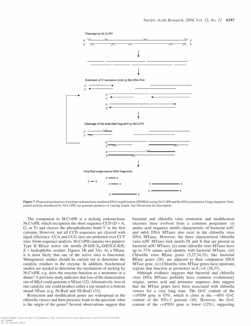
Figure 7. Proposed mechanism of nicking-endonuclease-mediated DNA amplification (NEMDA) using Nt.CviPII and Bst DNA polymerase I large fragment. Note:partial nicking introduced by Nt.CviPII can generate products of varying length. See Discussion for description.
Nucleic Acids Research, 2004, Vol. 32, No. 21 6197
Downloaded from https://academic.oup.com/nar/article-abstract/32/21/6187/1101502by gueston 29 March 2018

that this gene might have been acquired separately from thecviPIIM gene.
Several methods used for isothermal genome-wide ampli-fication of DNA have been described recently. An earlyversion of strand displacement amplification (SDA) dependson synthesis of hemi-phosphorothioated HincII or AvaI sites.where the appropriate REase nicks the template DNA on theun-modified strand to allow separation of the amplified strand(38–40). Rolling circle amplification requires a circular tem-plate (e.g. certain virus genomes, cloned plasmids or padlock-mediated circularized DNA) and random primers (41,42).More recently, specially designed DNA oligonucleotideshave been used with Nt.BstNBI to conduct linear and expo-nential amplification of short oligonucleotides (43). Being athermostable enzyme, Nt.BstNBI was incubated with templateDNA, dNTPs and Vent exo- DNA polymerase. At 60�C, thenicked fragments (designed to be 12 nt) separated from thedouble strand template such that a free 30 end was available fortemplate-dependent amplification. To achieve exponentialamplification, an oligonucleotide with sequence complemen-tary to the target sequence is added as the ‘trigger’ oligonu-cleotide. On the other hand, using thermocycling, REases thatcleave DNA frequently have been used to generate oligonu-cleotides for 2-step DNA amplification. CviJI*, which cleavesRG^CN and YG^CY sites in the presence of ATP, generatesdsDNA fragments of 20–60 bp from Bacillus sp. (44).After heat inactivation, the cleavage products are mixed withBacillus genomic DNA, a thermostable DNA polymerase, anddNTPs for amplification by thermocycling. The amplifiedDNA ranged from 0.1–10 kb in size, was independent ofthe size of the template DNA, and gave high sensitivity inSouthern blots when biotinylated-dUTP was included in theamplification reaction.
As demonstrated in this paper, Nt.CviPII and Bst DNApolymerase I large fragment can be used to amplifyDNA in a single-step isothermal reaction. This process istermed nicking-endonuclease-mediated DNA amplification(NEMDA). Because Nt.CviPII is partially active at 55�C,the amplification reaction can be carried out at 55�C wheredenaturation and separation of nicked DNA (oligos) arefavored. Nt.CviPII cleaves DNA frequently and produces sin-gle-stranded products or partial duplex DNAs with 50 over-hangs (Figure 7). Bst DNA polymerase I large fragment thenfills in at the 30 end until it reaches the end of the template. Theextended DNA acts as a substrate for Nt.CviPII, which, in turn,provides new substrate for the polymerase, allowing linearamplification. The hallmark of NEMDA is that exogenousprimers are not needed for DNA amplification, since partialduplex DNA is generated from the substrate DNA. From ourcollection of DNA polymerases, only Bst DNA polymerase Ilarge fragment and Thermomicrobium roseum (Tro) DNApolymerase I large fragment produce significant amounts ofamplified products. These polymerases possess relatively highstrand displacement activity among the polymerases we tested.Strand displacement activity may be involved in removing thenicked fragment and revealing a recessive 30 end for template-dependent amplification.
Unlike rolling circle amplification that can be used in wholegenome amplification, NEMDA does not cover the entiregenome. Nevertheless, the amplified DNA can be used as aprobe to detect target DNA by Southern blotting (unpublished
data). We have also demonstrated that incubation of randomDNA oligonucleotides and fresh Bst DNA polymerase largefragment with the amplified product can further amplify theDNA. Alternatively, the amplified DNA can be purified andused as primers for direct amplification of genomic DNAthrough isothermal or thermocycling procedures.
Randomly amplified DNA has been used as a highlysensitive probe for arrays of DNA oligonucleotides carrying‘signature sequences’ for biological agents of high concernsuch as E.coli O157:H7 (45,46). The DNA amplificationmethod presented here does not require synthesis of primersand can generate large quantities of ssDNA from a singlebacterial colony in a short time frame (e.g. 10 to 30 min).The procedure can be adapted for environmental or clinicalsamples and labels, such as biotin or fluorescein, can beincorporated into the amplified products by including modifieddNTP. Development of timely, sensitive and specific detectionmethods to identify important pathogens is of tremendousinterest to bio-defense and public health.
ACKNOWLEDGEMENTS
We thank Richard Roberts for critical reading of themanuscript, Jim Gurnon for isolating NYs-1 DNA, ShelleyCushing and Jack Benner II for mass spectrometry and proteinsequencing, Elisabeth Raleigh, Jim Samuelson, and RebeccaKucera for discussion, Jim Samuelson for independent con-firmation of NEMDA, Don Comb for support and encourage-ment. This work is partly supported by NIH STTR grant1R41GM070212-01. The sequence has been deposited inGenBank with accession number of AY781802.
REFERENCES
1. Roberts,R.J., Vincze,T., Posfai,J. and Macelis,D. (2003) REBASE:restriction enzymes and methyltransferases. Nucleic Acids Res.,31, 418–420.
2. Morgan,R.D., Calvet,C., Demeter,M., Agra,R. and Kong,H. (2000)Characterization of the specific DNA nicking activity of restrictionendonuclease. N. BstNBI. Biol. Chem., 381, 1123–1125.
3. Abdurashitov,M.A., Belichenko,O.A., Shevchenko,A.V. andDegtyarev,S.K. (1996) N.BstSE—site-specific nuclease from Bacillusstearothermophilus SE-589—restriction endonuclease production.Molek. Biol., 30, 1261–1267.
4. Xia,Y.N., Morgan,R., Schildkraut,I. and Van Etten,J.L. (1988)A site-specific single strand endonuclease activity induced by NYs-1virus infection of a Chlorella-like green alga. Nucleic Acids Res., 16,9477–9487.
5. Zhang,Y., Nelson,M., Nietfeldt,J., Xia,Y., Burbank,D., Ropp,S. andVan Etten,J.L. (1998) Chlorella virus NY-2A encodes at least 12 DNAendonuclease/methyltransferase genes. Virology, 240, 366–375.
6. Van Etten,J.L. (2003) Unusual life style of giant chlorella viruses.Annu. Rev. Genet., 37, 153–195.
7. Van Etten,J.L., Lane,L.C. and Meints,R.H. (1991) Viruses and virus-likeparticles of eukaryotic algae. Microbiol. Rev., 55, 586–620.
8. Nelson,M., Burbank,D.E. and Van Etten,J.L. (1998) Chlorella virusesencode multiple DNA methyltransferases. Biol. Chem., 379, 423–428.
9. Nelson,M., Zhang,Y. and Van Etten,J.L. (1993) DNA methyltransferasesand DNA site-specific endonucleases encoded by chlorella viruses.In Jost,P.J. Saluz,H.P., eds, DNA Methylation: Molecular Biology andBiological Significance, Birkhauser, Basel, pp. 186–211.
10. Schuster,A.M., Burbank,D.E., Meister,B., Skrdla,M.P., Meints,R.H.,Hattman,S., Swinton,D. and Van Etten,J.L. (1986) Characterization ofviruses infecting a eukaryotic Chlorella-like green alga. Virology,150, 170–177.
6198 Nucleic Acids Research, 2004, Vol. 32, No. 21
Downloaded from https://academic.oup.com/nar/article-abstract/32/21/6187/1101502by gueston 29 March 2018
11. Xu,M., Kladde,M.P., Van Etten,J.L. and Simpson,R.T. (1998) Cloning,characterization and expression of the gene coding for a cytosine-5-DNA methyltransferase recognizing GpC. Nucleic Acids Res., 26,3961–3966.
12. Van Etten,J.L., Schuster,A.M., Girton,L., Burbank,D.E., Swinton,D. andHattman,S. (1985) DNA methylation of viruses infecting a eukaryoticChlorella-like green alga. Nucleic Acids Res., 13, 3471–3478.
13. Fomenkov,A., Xiao,J.-P., Dila,D., Raleigh,E. and Xu,S.-Y. (1994)The ‘endo-blue method’ for direct cloning of restriction endonucleasegenes in E.coli. Nucleic Acids Res., 22, 2399–2403.
14. Altschul,S.F., Madden,T.L., Schaffer,A.A., Zhang,J., Zhang,Z.,Miller,W. and Lipman,D.J. (1997) Gapped BLAST and PSI-BLAST:a new generation of protein database search programs. Nucleic Acids Res.,25, 3389–3402.
15. de Boer,H.A., Comstock,L.J. and Vasser,M. (1983) The tac promoter:a functional hybrid derived from the trp and lac promoters. Proc. NatlAcad. Sci. USA, 80, 21–25.
16. Gao,X., Yo,P., Keith,A., Ragan,T.J. and Harris,T.K. (2003)Thermodynamically balanced inside-out (TBIO) PCR-based genesynthesis: a novel method of primer design for high-fidelity assembly oflonger gene sequences. Nucleic Acids Res., 31, e143.
17. Shields,S.L., Burbank,D.E., Grabherr,R. and Van Etten,J.L. (1990)Cloning and sequencing the cytosine methyltransferase gene M. CviJIfrom Chlorella virus IL-3A. Virology, 176, 16–24.
18. Cheng,X., Kumar,S., Posfai,J., Pflugrath,J.W. and Roberts,R.J. (1993)Crystal structure of the HhaI DNA methyltransferase complexed withS-adenosyl-L-methionine. Cell, 74, 299–307.
19. Klimasauskas,S., Kumar,S., Roberts,R.J. and Cheng,X. (1994) HhaImethyltransferase flips its target base out of the DNA helix. Cell, 76,357–369.
20. Kumar,S., Cheng,X., Klimasauskas,S., Mi,S., Posfai,J., Roberts,R.J. andWilson,G.G. (1994) The DNA (cytosine-5) methyltransferases. NucleicAcids Res., 22, 1–10.
21. Posfai,J., Bhagwat,A.S., Posfai,G. and Roberts,R.J. (1989) Predictivemotifs derived from cytosine methyltransferases. Nucleic Acids Res., 17,2421–2435.
22. Lauster,R., Trautner,T.A. and Noyer-Weidner,M. (1989)Cytosine-specific type II DNA methyltransferases. A conserved enzymecore with variable target-recognizing domains. J. Mol. Biol., 206,305–312.
23. Hou,P., Ji,M., He,N. and Lu,Z. (2004) Microarray-based method toevaluate the accuracy of restriction endonucleases HpaII and MspI.Biochem. Biophys. Res. Commun., 314, 110–117.
24. Ben-Hattar,J. and Jiricny,J. (1988) Effect of cytosine methylation on thecleavage of oligonucleotide duplexes with restriction endonucleasesHpaII and MspI. Nucleic Acids Res., 16, 4160.
25. Nelson,M., Christ,C. and Schildkraut,I. (1984) Alteration of apparentrestriction endonuclease recognition specificities by DNA methylases.Nucleic Acids Res., 12, 5165–5173.
26. Wilson,G.G. and Murray,N.E. (1991) Restriction and modificationsystems. Annu. Rev. Genet., 25, 585–627.
27. Swaminathan,N., Mead,D.A., McMaster,K., George,D., Van Etten,J.L.and Skowron,P.M. (1996) Molecular cloning of the three base restrictionendonuclease R.CviJI from eukaryotic Chlorella virus IL-3A. NucleicAcids Res., 24, 2463–2469.
28. Gasteiger,E., Gattiker,A., Hoogland,C., Ivanyi,I., Appel,R.D. andBairoch,A. (2003) ExPASy: the proteomics server for in-depth proteinknowledge and analysis. Nucleic Acids Res., 31, 3784–3788.
29. Renbaum,P., Abrahamove,D., Fainsod,A., Wilson,G.G., Rottem,S. andRazin,A. (1990) Cloning, characterization, and expression in Escherichia
coli of the gene coding for the CpG DNA methylase from Spiroplasma sp.strain MQ1(M.SssI). Nucleic Acids Res., 18, 1145–1152.
30. Kladde,M.P. and Simpson,R.T. (1994) Positioned nucleosomes inhibitDam methylation in vivo. Proc. Natl Acad. Sci. USA, 91, 1361–1365.
31. Kladde,M.P., Xu,M. and Simpson,R.T. (1996) Direct study ofDNA–protein interactions in repressed and active chromatin in livingcells. EMBO J, 15, 6290–6300.
32. Xu,Y., Lunnen,K.D. and Kong,H. (2001) Engineering a nickingendonuclease N.AlwI by domain swapping. Proc. Natl Acad. Sci. USA,98, 12990–12995.
33. Zhu,Z., Samuelson,J.C., Zhou,J., Dore,A. and Xu,S.Y. (2004)Engineering strand-specific DNA nicking enzymes from the type IISrestriction endonucleases BsaI, BsmBI, and BsmAI. J. Mol. Biol., 337,573–583.
34. Skowron,P.M., Swaminathan,N., McMaster,K., George,D., VanEtten,J.L. and Mead,D.A. (1995) Cloning and applications of the two/three-base restriction endonuclease R.CviJI from IL-3A virus-infectedChlorella. Gene, 157, 37–41.
35. Li,Y., Lu,Z., Sun,L., Ropp,S., Kutish,G.F., Rock,D.L. and Van Etten,J.L.(1997) Analysis of 74 kb of DNA located at the right end of the 330-kbchlorella virus PBCV-1 genome. Virology, 237, 360–377.
36. Narva,K.E., Wendell,D.L., Skrdla,M.P. and Van Etten,J.L. (1987)Molecular cloning and characterization of the gene encoding the DNAmethyltransferase,M.CviBIII, from Chlorella virus NC-1A. NucleicAcids Res., 15, 9807–9823.
37. Stefan,C., Xia,Y.N. and Van Etten,J.L. (1991) Molecular cloning andcharacterization of the gene encoding the adenine methyltransferaseM.CviRI from Chlorella virus XZ-6E. Nucleic Acids Res., 19, 307–311.
38. Milla,M.A., Spears,P.A., Pearson,R.E. and Walker,G.T. (1998) Use ofthe restriction enzyme AvaI and exo-Bst polymerase in stranddisplacement amplification. Biotechniques, 24, 392–396.
39. Walker,G.T., Little,M.C., Nadeau,J.G. and Shank,D.D. (1992)Isothermal in vitro amplification of DNA by a restriction enzyme/DNApolymerase system. Proc. Natl Acad. Sci. USA, 89, 392–396.
40. Walker,G.T., Nadeau,J.G., Spears,P.A., Schram,J.L., Nycz,C.M. andShank,D.D. (1994) Multiplex strand displacement amplification (SDA)and detection of DNA sequences from Mycobacterium tuberculosis andother mycobacteria. Nucleic Acids Res., 22, 2670–2677.
41. Rector,A., Tachezy,R. and Van Ranst,M. (2004) A sequence-independent strategy for detection and cloning of circular DNA virusgenomes by using multiply primed rolling-circle amplification.J. Virol., 78, 4993–4998.
42. Smirnov,D.A., Burdick,J.T., Morley,M. and Cheung,V.G. (2004)Method for manufacturing whole-genome microarrays by rolling circleamplification. Genes Chromosomes Cancer, 40, 72–77.
43. Van Ness,J., Van Ness,L.K. and Galas,D.J. (2003) Isothermal reactionsfor the amplification of oligonucleotides. Proc. Natl Acad. Sci. USA,100, 4504–4509.
44. Swaminathan,N., McMaster,K., Skowron,P.M. and Mead,D.A. (1998)Thermal cycle labeling: zeptomole detection sensitivity and microgramprobe amplification using CviJl* restriction-generated oligonucleotides.Anal. Biochem., 255, 133–141.
45. Vora G.J.,M.,C.E., Stenger,D.A. and Andreadis,J.D. (2004) Nucleic acidamplification stratagies for DNA microarray-based pathogen detection.Appl. Environ. Microbiol., 70, 3047–3054.
46. Yang,J.M., Bell,J., Huang,Y., Tirado,M., Thomas,D., Forster,A.H.,Haigis,R.W., Swanson,P.D., Wallace,R.B., Martinsons,B. et al. (2002)An integrated, stacked microlaboratory for biological agent detectionwith DNA and immunoassays. Biosens. Bioelectron., 17,605–618.
Nucleic Acids Research, 2004, Vol. 32, No. 21 6199
Downloaded from https://academic.oup.com/nar/article-abstract/32/21/6187/1101502by gueston 29 March 2018