clinical update management of pericardial effusion...review clinical update management of...
TRANSCRIPT
REVIEW
Clinical update
Management of pericardial effusionMassimo Imazio1* and Yehuda Adler2
1Department Cardiology, Maria Vittoria Hospital, Via Luigi Cibrario 72, Torino 10141, Italy; and 2Chaim Sheba Medical Center, Tel Hashomer, and Sackler University, Tel Aviv, Israel
Received 26 June 2012; revised 10 August 2012; accepted 9 October 2012; online publish-ahead-of-print 2 November 2012
Pericardial effusion is a common finding in clinical practice either as incidental finding or manifestation of a systemic or cardiac disease. Thespectrum of pericardial effusions ranges from mild asymptomatic effusions to cardiac tamponade. The aetiology is varied (infectious, neo-plastic, autoimmune, metabolic, and drug-related), being tuberculosis the leading cause of pericardial effusions in developing countriesand all over the world, while concurrent HIV infection may have an important promoting role in this setting. Management is guided bythe haemodynamic impact, size, presence of inflammation (i.e. pericarditis), associated medical conditions, and the aetiology whenever pos-sible. Pericardiocentesis is mandatory for cardiac tamponade and when a bacterial or neoplastic aetiology is suspected. Pericardial biopsy isgenerally reserved for cases with recurrent cardiac tamponade or persistence without a defined aetiology, especially when a bacterial orneoplastic aetiology is suspected and cannot be assessed by other conventional and less invasive means. A true isolated effusion may notrequire a specific treatment if the patient is asymptomatic, but large ones are at risk of progression to cardiac tamponade (up to onethird). Pericardiocentesis alone may be curative for large effusions, but recurrences are also common and pericardiectomy or less invasiveoptions (i.e. pericardial window) should be considered with recurrent cardiac tamponade or symptomatic pericardial effusion (either circum-ferential or loculated). The aim of this paper was to summarize and critically evaluate current knowledge on the management of pericardialeffusion.- - - - - - - - - - - - - - - - - - - - - - - - - - - - - - - - - - - - - - - - - - - - - - - - - - - - - - - - - - - - - - - - - - - - - - - - - - - - - - - - - - - - - - - - - - - - - - - - - - - - - - - - - - - - - - - - - - - - - - - - - - - - - - - - - - - - - - - - - - - - - - - - - - - - - - - - - - -Keywords Pericardial effusion † Aetiology † Diagnosis † Management † Pericarditis
Introduction
Probably no serious disease is so frequently overlooked by the prac-titioner. Postmortem experience shows how often pericarditis is notrecognized or goes to resolution and adhesion without attractingnotice (Osler, The Principles and Practice of Modern Medicine, 1892).
Pericardial effusion is a common finding in clinical practice either asincidental finding or manifestation of a systemic or cardiac disease.The spectrum of pericardial effusions ranges from mild asymptom-atic effusions to cardiac tamponade. Moreover, pericardial effusionmay accumulate slowly or suddenly.1– 3 Unfortunately, there arefew epidemiological data on the incidence and prevalence ofsuch effusions in the clinical setting. In Maria Vittoria hospital, anurban general hospital in Torino and an Italian referral centre forpericardial diseases, the mean annual incidence and prevalence ofpericardial effusion have been, respectively, 3 and 9% in a 6years experience of the echo laboratory (2000–05).4 Such datamainly depend on the epidemiological background (especiallydeveloped vs. developing country, where tuberculosis is a leading
cause of pericardial disease and concurrent HIV infection mayhave an important promoting role),5 the institutional setting(tertiary referral centre vs. secondary and general hospitals), andthe availability of specific subspecialties (especially nephrology,rheumatology, and oncology).
No specific guidelines and management recommendations havebeen issued by medical societies beyond the 2000 national Spanishguidelines6 and the 2004 European Society of Cardiology guide-lines on the management of pericardial diseases,7 and some narra-tive reviews on the topic,4,8,9 whereas no specific guidelines havebeen issued from the American Heart Association (AHA) andAmerican College of Cardiology (ACC).
It is the aim of this review to critically evaluate current knowl-edge on the management of pericardial effusion. A thorough litera-ture review has been performed without language restriction withthe MeSH term ‘pericardial effusion’ or ‘pericardial’[All Fields] and‘effusion’[All Fields]) or ‘pericardial effusion’[All Fields]. After theinitial identification of 1520 papers, 139 papers were selected fordetailed review based on novelty or important data leading tothe final inclusion of 50 papers in the final reference list.
* Corresponding author. Tel: +39 011 4393391, Fax: +39 011 4393334, Email: [email protected]
Published on behalf of the European Society of Cardiology. All rights reserved. & The Author 2012. For permissions please email: [email protected]
European Heart Journal (2013) 34, 1186–1197doi:10.1093/eurheartj/ehs372
Initial evaluation andpathophysiological issuesWhen a pericardial effusion is detected, the first step is to evaluateits size and haemodynamic importance, as well as the possible as-sociation with concomitant diseases. Echocardiography is the firstdiagnostic tool for this assessment as also acknowledged by theAHA/ACC guidelines on the use of echocardiography, that gavea class I indication for the use of echocardiography in any caseof suspected pericardial disease.10
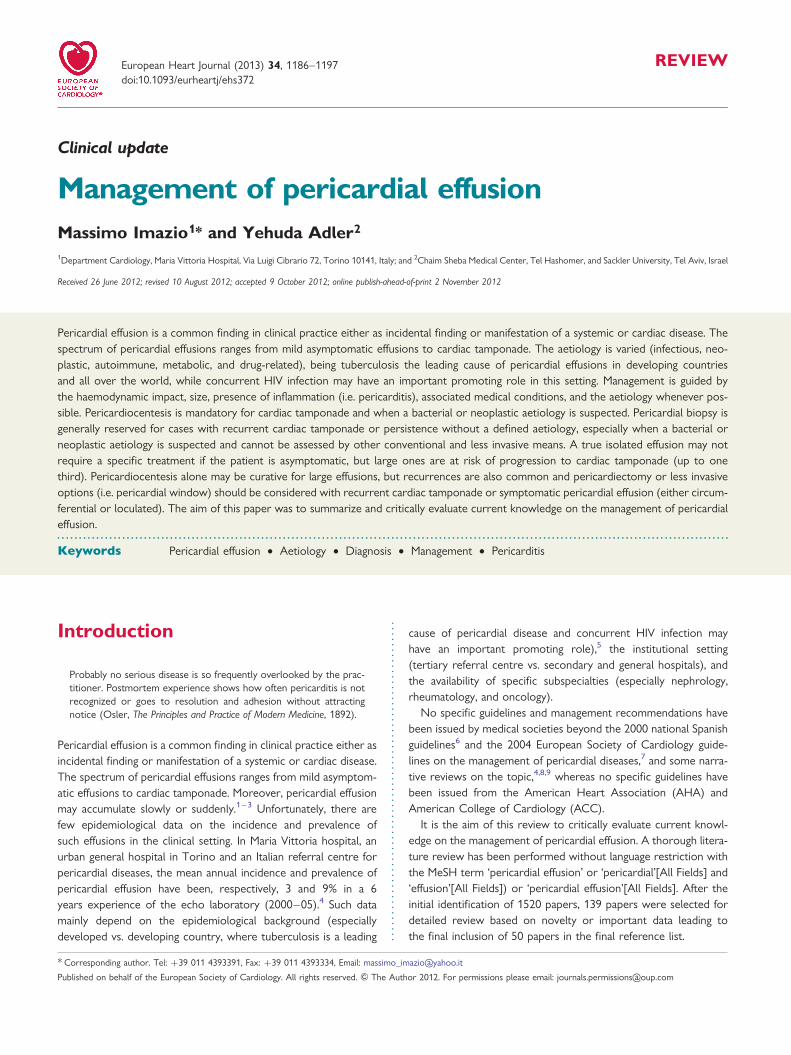
Pericardial effusion may be classified based on its onset (acute,subacute vs. chronic when dating .3 months), distribution (cir-cumferential or loculated), haemodynamic impact (none, cardiactamponade, effusive-constrictive), composition (exudates, transu-date, blood, rarely air, or gas from bacterial infections), and espe-cially by its size as mild, moderate, and large (Table 1) based on asimple semiquantitative echocardiographic assessment (Figure 1)that has been demonstrated useful also to estimate the risk ofspecific aetiology and complications during follow-up.4,7,9,11,12
The normal pericardial sac contains 10–50 mL of pericardialfluid acting as a lubrificant between the pericardial layers. Surpris-ingly, little is known about the formation and removal of pericardialfluid, because of the paucity of comprehensive studies, especiallyin human subjects, and methodological difficulties to distinguishbetween the dynamics of normal pericardial fluid and those of apathological effusion (Supplementary material online, ReferenceA). Nevertheless, normal pericardial fluid is generally consideredan ultrafiltrate of plasma (Supplementary material online, Refer-ence B). The arrangement of lymphatic vessels is complex andhas been described in human cadavers (Supplementary materialonline, References C and D). The lymphatic vessels include differ-ent pathways according to ventral, lateral, and posterior surfaces,
but, in any case, terminate to mediastinal, tracheobronchial, or iux-taesophageal lymph nodes. On the ventral surface, the lymphaticsof the parietal pericardium connect to lymphatics in the pericardialfat and areolar tissue. On the lateral and posterior surfaces, thelymphatics of the parietal pericardium anastomose with lymphaticsof the reflected mediastinal pleura (Supplementary material online,Reference C). Lymphatic drainage of the pericardium to the medi-astinal and tracheobronchial lymph nodes and interactions withpleural lymphatics (Supplementary material online, Reference D)provide the anatomical basis for pathological involvement of thepericardium in specific diseases (i.e. pleuro-pulmonary diseasessuch as pulmonary tuberculosis and lung cancer).
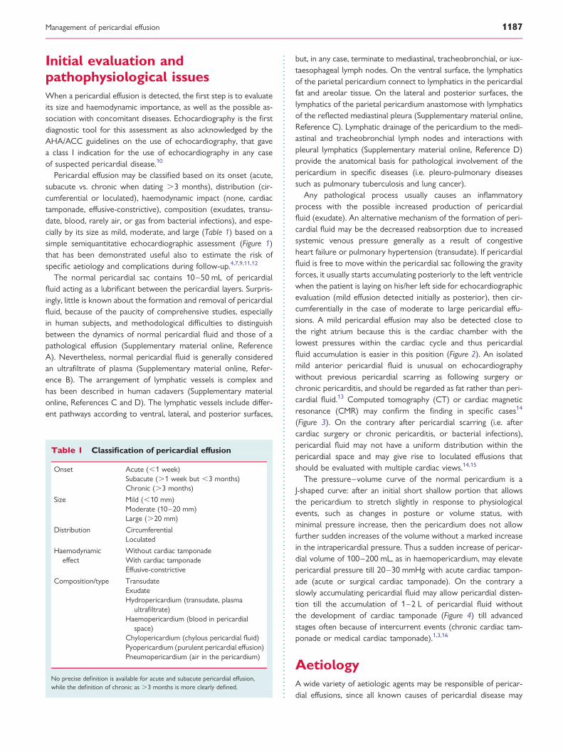
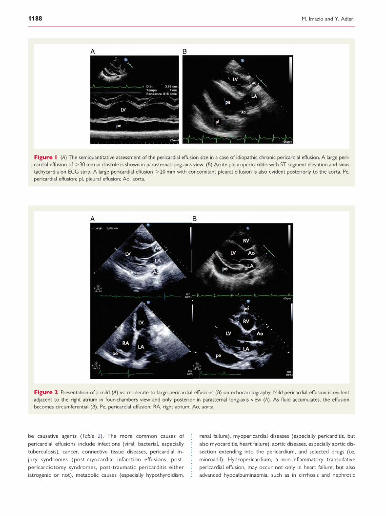
Any pathological process usually causes an inflammatoryprocess with the possible increased production of pericardialfluid (exudate). An alternative mechanism of the formation of peri-cardial fluid may be the decreased reabsorption due to increasedsystemic venous pressure generally as a result of congestiveheart failure or pulmonary hypertension (transudate). If pericardialfluid is free to move within the pericardial sac following the gravityforces, it usually starts accumulating posteriorly to the left ventriclewhen the patient is laying on his/her left side for echocardiographicevaluation (mild effusion detected initially as posterior), then cir-cumferentially in the case of moderate to large pericardial effu-sions. A mild pericardial effusion may also be detected close tothe right atrium because this is the cardiac chamber with thelowest pressures within the cardiac cycle and thus pericardialfluid accumulation is easier in this position (Figure 2). An isolatedmild anterior pericardial fluid is unusual on echocardiographywithout previous pericardial scarring as following surgery orchronic pericarditis, and should be regarded as fat rather than peri-cardial fluid.13 Computed tomography (CT) or cardiac magneticresonance (CMR) may confirm the finding in specific cases14
(Figure 3). On the contrary after pericardial scarring (i.e. aftercardiac surgery or chronic pericarditis, or bacterial infections),pericardial fluid may not have a uniform distribution within thepericardial space and may give rise to loculated effusions thatshould be evaluated with multiple cardiac views.14,15
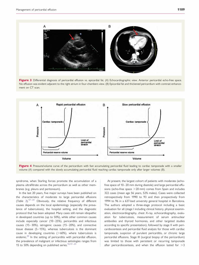
The pressure–volume curve of the normal pericardium is aJ-shaped curve: after an initial short shallow portion that allowsthe pericardium to stretch slightly in response to physiologicalevents, such as changes in posture or volume status, withminimal pressure increase, then the pericardium does not allowfurther sudden increases of the volume without a marked increasein the intrapericardial pressure. Thus a sudden increase of pericar-dial volume of 100–200 mL, as in haemopericardium, may elevatepericardial pressure till 20–30 mmHg with acute cardiac tampon-ade (acute or surgical cardiac tamponade). On the contrary aslowly accumulating pericardial fluid may allow pericardial disten-tion till the accumulation of 1–2 L of pericardial fluid withoutthe development of cardiac tamponade (Figure 4) till advancedstages often because of intercurrent events (chronic cardiac tam-ponade or medical cardiac tamponade).1,3,16
AetiologyA wide variety of aetiologic agents may be responsible of pericar-dial effusions, since all known causes of pericardial disease may
Table 1 Classification of pericardial effusion
Onset Acute (,1 week)Subacute (.1 week but ,3 months)Chronic (.3 months)
Size Mild (,10 mm)Moderate (10–20 mm)Large (.20 mm)
Distribution CircumferentialLoculated
Haemodynamiceffect
Without cardiac tamponadeWith cardiac tamponadeEffusive-constrictive
Composition/type TransudateExudateHydropericardium (transudate, plasma
ultrafiltrate)Haemopericardium (blood in pericardial
space)Chylopericardium (chylous pericardial fluid)Pyopericardium (purulent pericardial effusion)Pneumopericardium (air in the pericardium)
No precise definition is available for acute and subacute pericardial effusion,while the definition of chronic as .3 months is more clearly defined.
Management of pericardial effusion 1187
be causative agents (Table 2). The more common causes ofpericardial effusions include infections (viral, bacterial, especiallytuberculosis), cancer, connective tissue diseases, pericardial in-jury syndromes (post-myocardial infarction effusions, post-pericardiotomy syndromes, post-traumatic pericarditis eitheriatrogenic or not), metabolic causes (especially hypothyroidism,
renal failure), myopericardial diseases (especially pericarditis, butalso myocarditis, heart failure), aortic diseases, especially aortic dis-section extending into the pericardium, and selected drugs (i.e.minoxidil). Hydropericardium, a non-inflammatory transudativepericardial effusion, may occur not only in heart failure, but alsoadvanced hypoalbuminaemia, such as in cirrhosis and nephrotic
Figure 1 (A) The semiquantitative assessment of the pericardial effusion size in a case of idiopathic chronic pericardial effusion. A large peri-cardial effusion of .30 mm in diastole is shown in parasternal long-axis view. (B) Acute pleuropericarditis with ST segment elevation and sinustachycardia on ECG strip. A large pericardial effusion .20 mm with concomitant pleural effusion is also evident posteriorly to the aorta. Pe,pericardial effusion; pl, pleural effusion; Ao, aorta.
Figure 2 Presentation of a mild (A) vs. moderate to large pericardial effusions (B) on echocardiography. Mild pericardial effusion is evidentadjacent to the right atrium in four-chambers view and only posterior in parasternal long-axis view (A). As fluid accumulates, the effusionbecomes circumferential (B). Pe, pericardial effusion; RA, right atrium; Ao, aorta.
M. Imazio and Y. Adler1188
syndrome, when Starling forces promote the accumulation of aplasma ultrafiltrate across the pericardium as well as other mem-branes (e.g. pleura and peritoneum).
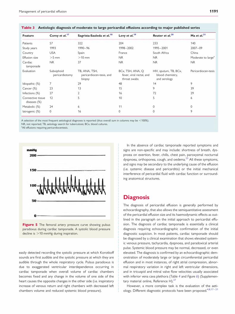
In the last 20 years, five major surveys have been published onthe characteristics of moderate to large pericardial effusions(Table 3).17– 21 Obviously, the relative frequency of differentcauses depends on the local epidemiology (especially the preva-lence of tuberculosis), the hospital setting, and the diagnosticprotocol that has been adopted. Many cases still remain idiopathicin developed countries (up to 50%), while other common causesinclude especially cancer (10–25%), pericarditis and infectiouscauses (15–30%), iatrogenic causes (15–20%), and connectivetissue disease (5–15%), whereas tuberculosis is the dominantcause in developing countries (.60%), where tuberculosis isendemic.22 In the setting of pericarditis with pericardial effusion,the prevalence of malignant or infectious aetiologies ranges from15 to 50% depending on published series.12,17 –21
At present, the largest cohort of patients with moderate (echo-free space of 10–20 mm during diastole) and large pericardial effu-sions (echo-free space .20 mm) comes from Spain and includes322 cases (mean age 56 years, 52% males). Cases were collectedretrospectively from 1990 to 93 and then prospectively from1994 to 96 in a 637-bed university general hospital in Barcelona.The authors adopted a three-stage protocol including a basicevaluation for all (stage I including clinical history, physical examin-ation, electrocardiography, chest X-ray, echocardiography, evalu-ation for tuberculosis, measurement of serum antinuclearantibodies and thyroid hormones, and other targeted studiesaccording to specific presentation), followed by stage II with peri-cardiocentesis and pericardial fluid analysis for those with cardiactamponade, suspicion of purulent pericarditis, or chronic largepericardial effusions. Stage III (surgical biopsy of the pericardium)was limited to those with persistent or recurring tamponadeafter pericardiocentesis, and when the effusion lasted for .3
Figure 3 Differential diagnosis of pericardial effusion vs. epicardial fat. (A) Echocardiographic view. Anterior pericardial echo-free space.No effusion was evident adjacent to the right atrium in four-chambers view. (B) Epicardial fat and thickened pericardium with contrast enhance-ment on CT scan.
Figure 4 Pressure/volume curve of the pericardium with fast accumulating pericardial fluid leading to cardiac tamponade with a smallervolume (A) compared with the slowly accumulating pericardial fluid reaching cardiac tamponade only after larger volumes (B).
Management of pericardial effusion 1189
weeks after admission and without an aetiologic diagnosis. Themost common diagnoses included: acute idiopathic pericarditis(20%), iatrogenic effusions (16%), cancer (13%), and chronicidiopathic pericardial effusion (9%). In 60% of cases, the cause ofpericardial effusion was a known medical condition.18
Clinical presentationThe clinical presentation of pericardial effusion is varied accordingto the speed of pericardial fluid accumulation as mentioned in theintroduction, and the aetiology of the effusion with possible symp-toms that may be related to the causative disease. The rate of peri-cardial fluid accumulation is critical for the clinical presentation. Ifpericardial fluid is quickly accumulating such as for wounds or
iatrogenic perforations, the evolution is dramatic and only smallamounts of blood are responsible of a quick rise of intrapericardialpressure and overt cardiac tamponade in minutes. On the contrarya slowly accumulating pericardial fluid allows the collection of alarge effusion in days to weeks before a significant increase in peri-cardial pressure becomes responsible of symptoms and signs.3
Classical symptoms include dyspnoea on exertion progressing toorthopnoea, chest pain, and/or fullness. Additional occasionalsymptoms due to local compression may include nausea (dia-phragm), dysphagia (oesophagus), hoarseness (recurrent laryngealnerve), and hiccups (phrenic nerve). Non-specific symptomsinclude also cough, weakness, fatigue, anorexia and palpitationsand reflect the compressive effect of the pericardial fluid on con-tiguous anatomic structures or reduced blood pressure and sec-ondary sinus tachycardia.23
The classical findings of cardiac tamponade have been describedby the thoracic surgeon Beck in 1935.24 Beck identified a triad in-cluding hypotension, increased jugular venous pressure, and a smalland quiet heart. This triad was classically identified in ‘surgical tam-ponade’ with acute cardiac tamponade due to intrapericardialhaemorrhage because of trauma, myocardial or aortic rupture.The Beck triad may be lacking in patients with ‘medical tamponade’with slowly accumulating pericardial fluid. Hypotension is absoluteor relative. Acute cardiac tamponade is usually associated with lowblood pressure (,90 mmHg) but may be only slightly reduced insubacute, chronic tamponade. Hypertensive patients may havenormal to mildly elevated blood pressure concomitant to cardiactamponade.25 Fever is a non-specific sign that may be associatedwith pericarditis either infectious or immune-mediated (i.e. system-ic inflammatory diseases).
On physical examination classical signs include neck vein disten-tion with elevated jugular venous pressure at bedside examination,pulsus paradoxus, and diminished heart sounds on cardiac auscul-tation. Pericardial friction rubs are rarely reported, they can beusually detected in patients with concomitant pericarditis. Rubswhich occur during the maximal movement of the heart withinits pericardial sac, are said to be generated by friction betweenthe two inflamed layers of the pericardium. However, this com-monly offered explanation for its mechanism may be an oversimpli-fication as patients with a pericardial effusion may also have anaudible friction rub, and there is no precise correlation betweenpericardial rubs and size of the effusion, although pericardial rubsmay be easier to hear in patients without a pericardial effusion,but this finding is not universal and is not well-documented(Supplementary material online, Reference E). In a report of 100patients with acute pericarditis, a pericardial rub was present in85% of cases without an effusion (Supplementary material online,Reference F). This prevalence is considerably higher than the35% incidence of friction rubs reported in another series (Supple-mentary material online, Reference G).
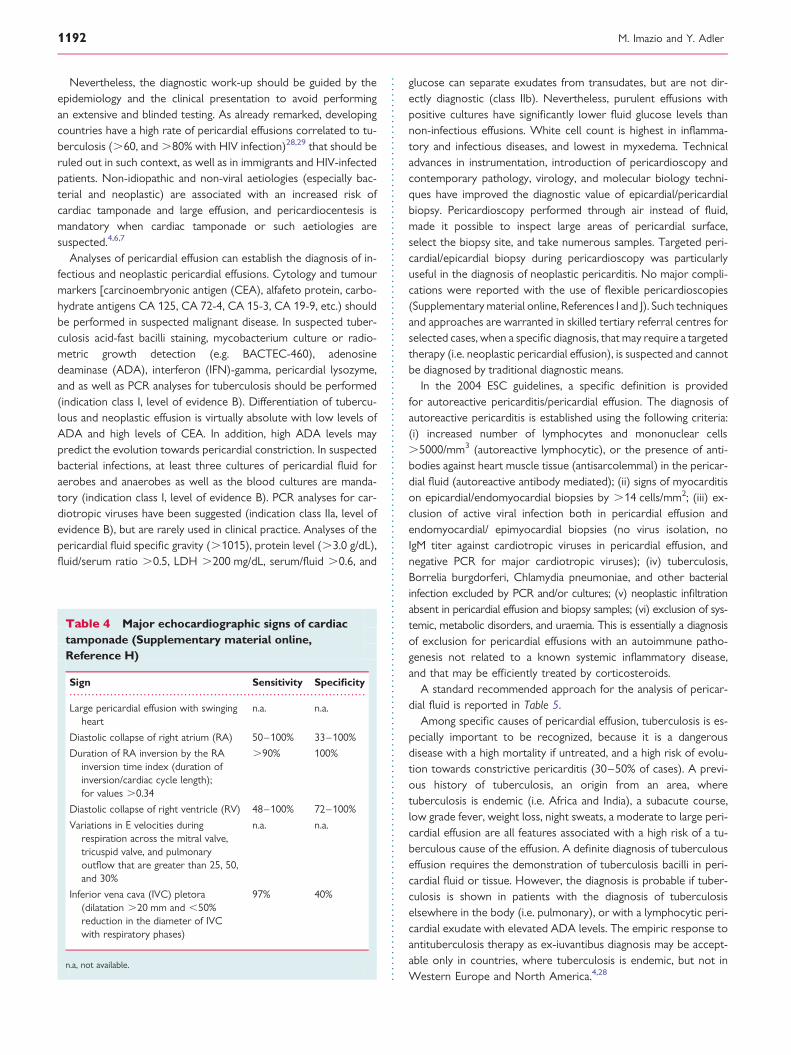
Pulsus paradoxus has been described for the first time by Kuss-maul in 1873 as a palpable reduction of radial pulse on inspirationin patients with cardiac tamponade.26 The so-called paradox wasthe ‘waxing and waning’ of the peripheral pulse, in contrast tothe unvarying strength of the apical cardiac impulse. Pulsus para-doxus is classically defined as an inspiratory reduction of at least10 mmHg of the systolic blood pressure (Figure 5). It can be
. . . . . . . . . . . . . . . . . . . . . . . . . . . . . . . . . . . . . . . . . . . . . . . . . . . . . . . . . . . . . . . . . . . . . . . . . . . . . . . .
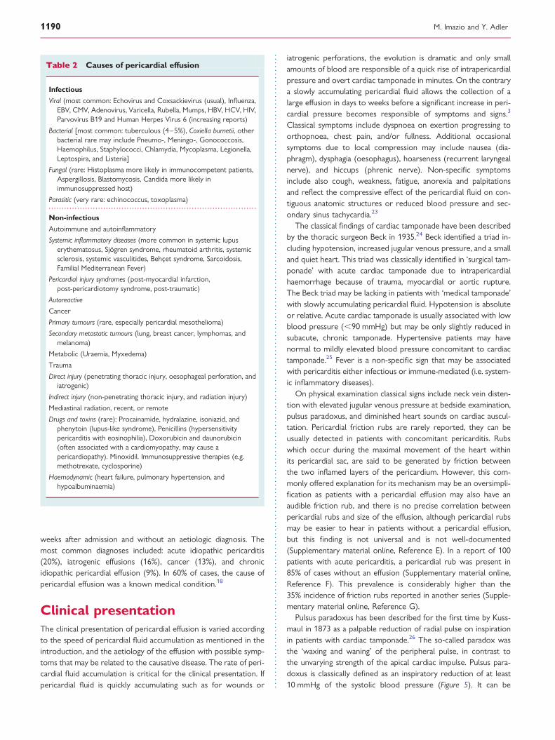
Table 2 Causes of pericardial effusion
Infectious
Viral (most common: Echovirus and Coxsackievirus (usual), Influenza,EBV, CMV, Adenovirus, Varicella, Rubella, Mumps, HBV, HCV, HIV,Parvovirus B19 and Human Herpes Virus 6 (increasing reports)
Bacterial [most common: tuberculous (4–5%), Coxiella burnetii, otherbacterial rare may include Pneumo-, Meningo-, Gonococcosis,Haemophilus, Staphylococci, Chlamydia, Mycoplasma, Legionella,Leptospira, and Listeria]
Fungal (rare: Histoplasma more likely in immunocompetent patients,Aspergillosis, Blastomycosis, Candida more likely inimmunosuppressed host)
Parasitic (very rare: echinococcus, toxoplasma)
Non-infectious
Autoimmune and autoinflammatory
Systemic inflammatory diseases (more common in systemic lupuserythematosus, Sjogren syndrome, rheumatoid arthritis, systemicsclerosis, systemic vasculitides, Behcet syndrome, Sarcoidosis,Familial Mediterranean Fever)
Pericardial injury syndromes (post-myocardial infarction,post-pericardiotomy syndrome, post-traumatic)
Autoreactive
Cancer
Primary tumours (rare, especially pericardial mesothelioma)
Secondary metastatic tumours (lung, breast cancer, lymphomas, andmelanoma)
Metabolic (Uraemia, Myxedema)
Trauma
Direct injury (penetrating thoracic injury, oesophageal perforation, andiatrogenic)
Indirect injury (non-penetrating thoracic injury, and radiation injury)
Mediastinal radiation, recent, or remote
Drugs and toxins (rare): Procainamide, hydralazine, isoniazid, andphenytoin (lupus-like syndrome), Penicillins (hypersensitivitypericarditis with eosinophilia), Doxorubicin and daunorubicin(often associated with a cardiomyopathy, may cause apericardiopathy). Minoxidil. Immunosuppressive therapies (e.g.methotrexate, cyclosporine)
Haemodynamic (heart failure, pulmonary hypertension, andhypoalbuminaemia)
M. Imazio and Y. Adler1190
easily detected recording the systolic pressure at which Korotkoffsounds are first audible and the systolic pressure at which they areaudible through the whole respiratory cycle. Pulsus paradoxus isdue to exaggerated ventricular interdependence occurring incardiac tamponade when overall volume of cardiac chambersbecomes fixed and any change in the volume of one side of theheart causes the opposite changes in the other side (i.e. inspiratoryincrease of venous return and right chambers with decreased leftchambers volume and reduced systemic blood pressure).
In the absence of cardiac tamponade reported symptoms andsigns are non-specific and may include: shortness of breath, dys-pnoea on exertion, fever, chills, chest pain, paroxysmal nocturnaldyspnoea, orthoponea, cough, and oedema.23 All these symptoms,and signs may be secondary to the underlying cause of the effusion(i.e. systemic disease and pericarditis) or the initial mechanicalinterference of pericardial fluid with cardiac function or surround-ing anatomical structures.
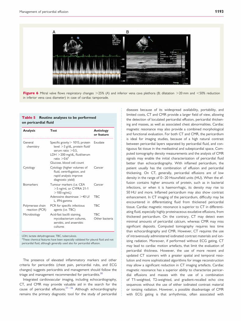
DiagnosisThe diagnosis of pericardial effusion is generally performed byechocardiography, that also allows the semiquantitative assessmentof the pericardial effusion size and its haemodynamic effects as out-lined in the paragraph on the initial approach to pericardial effu-sion. The diagnosis of cardiac tamponade is essentially a clinicaldiagnosis requiring echocardiographic confirmation of the initialdiagnostic suspicion. In most patients, cardiac tamponade shouldbe diagnosed by a clinical examination that shows elevated system-ic venous pressure, tachycardia, dyspnoea, and paradoxical arterialpulse. Systemic blood pressure may be normal, decreased, or evenelevated. The diagnosis is confirmed by an echocardiographic dem-onstration of moderately large or large circumferential pericardialeffusion and in most instances, of right atrial compression, abnor-mal respiratory variation in right and left ventricular dimensions,and in tricuspid and mitral valve flow velocities usually associatedwith inferior vena cava plethora (Table 4 and Figure 6) (Supplemen-tary material online, Reference H).27
However, a more complex task is the evaluation of the aeti-ology. Different diagnostic protocols have been proposed.4,8,17–21
. . . . . . . . . . . . . . . . . . . . . . . . . . . . . . . . . . . . . . . . . . . . . . . . . . . . . . . . . . . . . . . . . . . . . . . . . . . . . . . . . . . . . . . . . . . . . . . . . . . . . . . . . . . . . . . . . . . . . . . . . . . . . . . . . . . . . . . . . . . . . . . . . . . . . . . . . . . . . . . . . . . . . . . . . . . . . . .
Table 3 Aetiologic diagnosis of moderate to large pericardial effusions according to major published series
Feature Corey et al.17 Sagrista-Sauleda et al.18 Levy et al.19 Reuter et al.20 Ma et al.21
Patients 57 322 204 233 140
Study years 1993 1990–96 1998–2002 1995–2001 2007–09
Country USA Spain France South Africa China
Effusion size .5 mm .10 mm NR NR Moderate to largea
Cardiactamponade
NR 37 NR NR NR
Evaluation Subxiphoidpericardiotomy
TB, ANA, TSH,pericardiocen-tesis, andbiopsy
BCx, TSH, ANA, Qfever, viral rectal, andthroat swabs
HIV, sputum, TB, BCx,blood chemistry,and serology
Pericardiocen-tesis
Idiopathic (%) 7 29 48 14 9
Cancer (%) 23 13 15 9 39
Infections (%) 27 2 16 72 29
Connective tissuediseases (%)
12 5 10 5 6
Metabolic (%) 24 6 11 0 0
Iatrogenic (%) 0 16 0 0 9
A selection of the most frequent aetiological diagnoses is reported (thus overall sum in columns may be ,100%).NR, not reported; TB, aetiology search for tuberculosis; BCx, blood cultures.aAll effusions requiring pericardiocentesis.
Figure 5 The femoral artery pressure curve showing pulsusparadoxus during cardiac tamponade. A systolic blood pressuredecline is .10 mmHg during inspiration.
Management of pericardial effusion 1191
Nevertheless, the diagnostic work-up should be guided by theepidemiology and the clinical presentation to avoid performingan extensive and blinded testing. As already remarked, developingcountries have a high rate of pericardial effusions correlated to tu-berculosis (.60, and .80% with HIV infection)28,29 that should beruled out in such context, as well as in immigrants and HIV-infectedpatients. Non-idiopathic and non-viral aetiologies (especially bac-terial and neoplastic) are associated with an increased risk ofcardiac tamponade and large effusion, and pericardiocentesis ismandatory when cardiac tamponade or such aetiologies aresuspected.4,6,7
Analyses of pericardial effusion can establish the diagnosis of in-fectious and neoplastic pericardial effusions. Cytology and tumourmarkers [carcinoembryonic antigen (CEA), alfafeto protein, carbo-hydrate antigens CA 125, CA 72-4, CA 15-3, CA 19-9, etc.) shouldbe performed in suspected malignant disease. In suspected tuber-culosis acid-fast bacilli staining, mycobacterium culture or radio-metric growth detection (e.g. BACTEC-460), adenosinedeaminase (ADA), interferon (IFN)-gamma, pericardial lysozyme,and as well as PCR analyses for tuberculosis should be performed(indication class I, level of evidence B). Differentiation of tubercu-lous and neoplastic effusion is virtually absolute with low levels ofADA and high levels of CEA. In addition, high ADA levels maypredict the evolution towards pericardial constriction. In suspectedbacterial infections, at least three cultures of pericardial fluid foraerobes and anaerobes as well as the blood cultures are manda-tory (indication class I, level of evidence B). PCR analyses for car-diotropic viruses have been suggested (indication class IIa, level ofevidence B), but are rarely used in clinical practice. Analyses of thepericardial fluid specific gravity (.1015), protein level (.3.0 g/dL),fluid/serum ratio .0.5, LDH .200 mg/dL, serum/fluid .0.6, and
glucose can separate exudates from transudates, but are not dir-ectly diagnostic (class IIb). Nevertheless, purulent effusions withpositive cultures have significantly lower fluid glucose levels thannon-infectious effusions. White cell count is highest in inflamma-tory and infectious diseases, and lowest in myxedema. Technicaladvances in instrumentation, introduction of pericardioscopy andcontemporary pathology, virology, and molecular biology techni-ques have improved the diagnostic value of epicardial/pericardialbiopsy. Pericardioscopy performed through air instead of fluid,made it possible to inspect large areas of pericardial surface,select the biopsy site, and take numerous samples. Targeted peri-cardial/epicardial biopsy during pericardioscopy was particularlyuseful in the diagnosis of neoplastic pericarditis. No major compli-cations were reported with the use of flexible pericardioscopies(Supplementary material online, References I and J). Such techniquesand approaches are warranted in skilled tertiary referral centres forselected cases, when a specific diagnosis, that may require a targetedtherapy (i.e. neoplastic pericardial effusion), is suspected and cannotbe diagnosed by traditional diagnostic means.
In the 2004 ESC guidelines, a specific definition is providedfor autoreactive pericarditis/pericardial effusion. The diagnosis ofautoreactive pericarditis is established using the following criteria:(i) increased number of lymphocytes and mononuclear cells.5000/mm3 (autoreactive lymphocytic), or the presence of anti-bodies against heart muscle tissue (antisarcolemmal) in the pericar-dial fluid (autoreactive antibody mediated); (ii) signs of myocarditison epicardial/endomyocardial biopsies by .14 cells/mm2; (iii) ex-clusion of active viral infection both in pericardial effusion andendomyocardial/ epimyocardial biopsies (no virus isolation, noIgM titer against cardiotropic viruses in pericardial effusion, andnegative PCR for major cardiotropic viruses); (iv) tuberculosis,Borrelia burgdorferi, Chlamydia pneumoniae, and other bacterialinfection excluded by PCR and/or cultures; (v) neoplastic infiltrationabsent in pericardial effusion and biopsy samples; (vi) exclusion of sys-temic, metabolic disorders, and uraemia. This is essentially a diagnosisof exclusion for pericardial effusions with an autoimmune patho-genesis not related to a known systemic inflammatory disease,and that may be efficiently treated by corticosteroids.
A standard recommended approach for the analysis of pericar-dial fluid is reported in Table 5.
Among specific causes of pericardial effusion, tuberculosis is es-pecially important to be recognized, because it is a dangerousdisease with a high mortality if untreated, and a high risk of evolu-tion towards constrictive pericarditis (30–50% of cases). A previ-ous history of tuberculosis, an origin from an area, wheretuberculosis is endemic (i.e. Africa and India), a subacute course,low grade fever, weight loss, night sweats, a moderate to large peri-cardial effusion are all features associated with a high risk of a tu-berculous cause of the effusion. A definite diagnosis of tuberculouseffusion requires the demonstration of tuberculosis bacilli in peri-cardial fluid or tissue. However, the diagnosis is probable if tuber-culosis is shown in patients with the diagnosis of tuberculosiselsewhere in the body (i.e. pulmonary), or with a lymphocytic peri-cardial exudate with elevated ADA levels. The empiric response toantituberculosis therapy as ex-iuvantibus diagnosis may be accept-able only in countries, where tuberculosis is endemic, but not inWestern Europe and North America.4,28
. . . . . . . . . . . . . . . . . . . . . . . . . . . . . . . . . . . . . . . . . . . . . . . . . . . . . . . . . . . . . . . . . . . . . . . . . . . . . . . .
Table 4 Major echocardiographic signs of cardiactamponade (Supplementary material online,Reference H)
Sign Sensitivity Specificity
Large pericardial effusion with swingingheart
n.a. n.a.
Diastolic collapse of right atrium (RA) 50–100% 33–100%
Duration of RA inversion by the RAinversion time index (duration ofinversion/cardiac cycle length);for values .0.34
.90% 100%
Diastolic collapse of right ventricle (RV) 48–100% 72–100%
Variations in E velocities duringrespiration across the mitral valve,tricuspid valve, and pulmonaryoutflow that are greater than 25, 50,and 30%
n.a. n.a.
Inferior vena cava (IVC) pletora(dilatation .20 mm and ,50%reduction in the diameter of IVCwith respiratory phases)
97% 40%
n.a, not available.
M. Imazio and Y. Adler1192
The presence of elevated inflammatory markers and othercriteria for pericarditis (chest pain, pericardial rubs, and ECGchanges) suggests pericarditis and management should follow thetriage and management recommended for pericarditis.30
Integrated cardiovascular imaging, including echocardiography,CT, and CMR may provide valuable aid in the search for thecause of pericardial effusions.31– 36 Although echocardiographyremains the primary diagnostic tool for the study of pericardial
diseases because of its widespread availability, portability, andlimited costs, CT and CMR provide a larger field of view, allowingthe detection of loculated pericardial effusion, pericardial thicken-ing and masses, as well as associated chest abnormalities. Cardiacmagnetic resonance may also provide a combined morphologicaland functional evaluation. For both CT and CMR, the pericardiumis ideal for imaging studies, because of a high natural contrastbetween pericardial layers separated by pericardial fluid, and con-tiguous fat tissue in the mediastinal and subepicardial space. Com-puted tomography density measurements and the analysis of CMRsignals may enable the initial characterization of pericardial fluidbetter than echocardiography. With inflamed pericardium, thepatient usually has the combination of effusion and pericardialthickening. On CT, generally, pericardial effusions are of lowdensity in the range of 0–20 Hounsfield units (HU). When the ef-fusion contains higher amounts of protein, such as in bacterialinfections, or when it is haemorrhagic, its density may rise to50 HU and more. Inflamed pericardium may also show contrastenhancement. In CT imaging of the pericardium, difficulty may beencountered in differentiating fluid from thickened pericardialtissue. Cardiac magnetic resonance is superior to CT in differenti-ating fluid, especially highly proteinaceous exudative effusions, fromthickened pericardium. On the contrary, CT may detect evenminimal amounts of pericardial calcium, whereas CMR may misssignificant deposits. Computed tomography requires less timethan echocardiography and CMR. However, CT requires the useof intravenously administered iodinated contrast materials and ion-izing radiation. Moreover, if performed without ECG gating, CTmay lead to cardiac motion artefacts, that limit the evaluation ofpericardial thickness. However, the use of more recent andupdated CT scanners with a greater spatial and temporal reso-lution and more sophisticated algorithms for image reconstructionmay allow a significant reduction in CT imaging artefacts. Cardiacmagnetic resonance has a superior ability to characterize pericar-dial effusions and masses with the use of a combinationof T1-weighted, T2-weighted, and gradient-recalled echo cinesequences without the use of either iodinated contrast materialor ionizing radiation. However, a possible disadvantage of CMRwith ECG gating is that arrhythmias, often associated with
Figure 6 Mitral valve flows respiratory changes .25% (A) and inferior vena cava plethora (B; dilatation .20 mm and ,50% reductionin inferior vena cava diameter) in case of cardiac tamponade.
. . . . . . . . . . . . . . . . . . . . . . . . . . . . . . . . . . . . . . . . . . . . . . . . . . . . . . . . . . . . . . . . . . . . . . . . . . . . . . . .
Table 5 Routine analyses to be performedon pericardial fluid
Analysis Test Aetiologyor feature
Generalchemistry
Specific gravity. 1015, proteinlevel .3 g/dL, protein fluid/serum ratio .0.5,
Exudate
LDH .200 mg/dL, fluid/serumratio .0.6a
Glucose, blood cell count
Cytology Cytology (higher volumes offluid, centrifugation, andrapid analysis improvediagnostic yield)
Cancer
Biomarkers Tumour markers (i.e. CEA.5 ng/mL or CYFRA 21-1.100 ng/mL)
Cancer
Adenosine deaminase .40 U/L, IFN-gamma
TBC
Polymerase chainreaction (PCR)
PCR for specific infectiousagents (i.e. TBC)
TBC
Microbiology Acid-fast bacilli staining,mycobacterium cultures,aerobic, and anaerobiccultures
TBCOther bacteria
LDH, lactate dehydrogenase; TBC, tuberculosis.aThese chemical features have been especially validated for pleural fluid and notpericardial fluid, although generally used also for pericardial effusion.
Management of pericardial effusion 1193
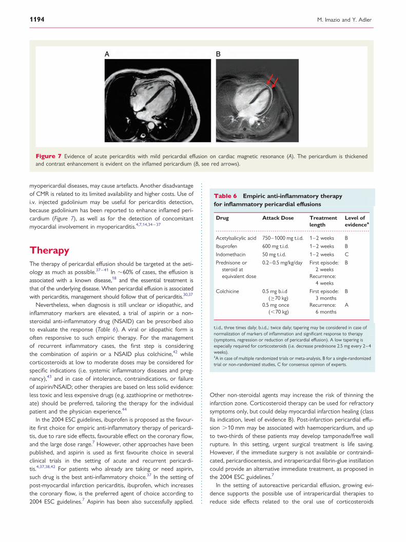
myopericardial diseases, may cause artefacts. Another disadvantageof CMR is related to its limited availability and higher costs. Use ofi.v. injected gadolinium may be useful for pericarditis detection,because gadolinium has been reported to enhance inflamed peri-cardium (Figure 7), as well as for the detection of concomitantmyocardial involvement in myopericarditis.4,7,14,34– 37
TherapyThe therapy of pericardial effusion should be targeted at the aeti-ology as much as possible.37–41 In �60% of cases, the effusion isassociated with a known disease,18 and the essential treatment isthat of the underlying disease. When pericardial effusion is associatedwith pericarditis, management should follow that of pericarditis.30,37
Nevertheless, when diagnosis is still unclear or idiopathic, andinflammatory markers are elevated, a trial of aspirin or a non-steroidal anti-inflammatory drug (NSAID) can be prescribed alsoto evaluate the response (Table 6). A viral or idiopathic form isoften responsive to such empiric therapy. For the managementof recurrent inflammatory cases, the first step is consideringthe combination of aspirin or a NSAID plus colchicine,42 whilecorticosteroids at low to moderate doses may be considered forspecific indications (i.e. systemic inflammatory diseases and preg-nancy),43 and in case of intolerance, contraindications, or failureof aspirin/NSAID; other therapies are based on less solid evidence:less toxic and less expensive drugs (e.g. azathioprine or methotrex-ate) should be preferred, tailoring the therapy for the individualpatient and the physician experience.44
In the 2004 ESC guidelines, ibuprofen is proposed as the favour-ite first choice for empiric anti-inflammatory therapy of pericardi-tis, due to rare side effects, favourable effect on the coronary flow,and the large dose range.7 However, other approaches have beenpublished, and aspirin is used as first favourite choice in severalclinical trials in the setting of acute and recurrent pericardi-tis.4,37,38,42 For patients who already are taking or need aspirin,such drug is the best anti-inflammatory choice.37 In the setting ofpost-myocardial infarction pericarditis, ibuprofen, which increasesthe coronary flow, is the preferred agent of choice according to2004 ESC guidelines.7 Aspirin has been also successfully applied.
Other non-steroidal agents may increase the risk of thinning theinfarction zone. Corticosteroid therapy can be used for refractorysymptoms only, but could delay myocardial infarction healing (classIIa indication, level of evidence B). Post-infarction pericardial effu-sion .10 mm may be associated with haemopericardium, and upto two-thirds of these patients may develop tamponade/free wallrupture. In this setting, urgent surgical treatment is life saving.However, if the immediate surgery is not available or contraindi-cated, pericardiocentesis, and intrapericardial fibrin-glue instillationcould provide an alternative immediate treatment, as proposed inthe 2004 ESC guidelines.7
In the setting of autoreactive pericardial effusion, growing evi-dence supports the possible use of intrapericardial therapies toreduce side effects related to the oral use of corticosteroids
Figure 7 Evidence of acute pericarditis with mild pericardial effusion on cardiac magnetic resonance (A). The pericardium is thickenedand contrast enhancement is evident on the inflamed pericardium (B, see red arrows).
. . . . . . . . . . . . . . . . . . . . . . . . . . . . . . . . . . . . . . . . . . . . . . . . . . . . . . . . . . . . . . . . . . . . . . . . . . . . . . . . .
Table 6 Empiric anti-inflammatory therapyfor inflammatory pericardial effusions
Drug Attack Dose Treatmentlength
Level ofevidencea
Acetylsalicylic acid 750–1000 mg t.i.d. 1–2 weeks B
Ibuprofen 600 mg t.i.d. 1–2 weeks B
Indomethacin 50 mg t.i.d. 1–2 weeks C
Prednisone orsteroid atequivalent dose
0.2–0.5 mg/kg/day First episode:2 weeks
B
Recurrence:4 weeks
Colchicine 0.5 mg b.i.d(≥70 kg)
First episode:3 months
B
0.5 mg once(,70 kg)
Recurrence:6 months
A
t.i.d., three times daily; b.i.d.,: twice daily; tapering may be considered in case ofnormalization of markers of inflammation and significant response to therapy(symptoms, regression or reduction of pericardial effusion). A low tapering isespecially required for corticosteroids (i.e. decrease prednisone 2.5 mg every 2–4weeks).aA in case of multiple randomized trials or meta-analysis, B for a single-randomizedtrial or non-randomized studies, C for consensus opinion of experts.
M. Imazio and Y. Adler1194
(Supplementary material online, Reference K). Systemic corticos-teroids offer an effective treatment option for autoreactive peri-carditis; however, their use is limited by adverse effects and theyare an independent risk factor for pericarditis recurrence. In a re-cently published systematic review (Supplementary material online,Reference L), one case series and three open-label trials evaluatingintrapericardial triamcinolone for the management of autoreactivepericarditis were reviewed. Included studies were limited by smallsample sizes (n ¼ 2–84), lack of control groups, short durations offollow-up (24 h–12 months), use of adjuvant agents, lack of patientdemographic data, subjective report of symptom relief, and lackof consistent dose of intrapericardial triamcinolone. Despitethese limitations, available data suggest symptom resolution andreduced pericarditis recurrence with the administration of intra-pericardial triamcinolone to patients with autoreactive pericarditis(Supplementary material online, Reference L).
When a pericardial effusion becomes symptomatic withoutevidence of inflammation or when empiric anti-inflammatorydrugs are not successful, drainage of the effusion should be consid-ered. Pericardiocentesis with prolonged pericardial drainage till,30 mL/24 h is recommended to promote adherence of pericar-dial layers and prevent further accumulation of fluid, althoughevidence to support this indication is based on case reports, retro-spective studies, and expert opinion. If pericardiocentesis is notfeasible or fails, the creation of a so-called pericardial windowshould be considered either by conventional heart surgery orvideo-assisted thoracoscopy. Balloon pericardiotomy is an alterna-tive to surgical creation of a pericardial window, which has beenshown successful especially in the setting of neoplastic pericardialdisease. The technique involves inserting a deflated single catheteror double balloon catheters into the pericardial space using a sub-xiphoid approach under fluoroscopic or echocardiographic guid-ance. Although successful in preventing recurrence in .80% ofcases, stretching of the pericardium is often painful so appropriateanalgesia is recommended.37,38
The 2004 ESC guidelines gave a class IIa recommendation topericardiectomy for frequent and highly symptomatic recurrencesresistant to medical treatment. Other reported indications includerepeated recurrences with cardiac tamponade, and evidence ofserious steroid toxicity.7 Although surgical experiences are notalways concordant, pericardiectomy is generally considered as atherapeutic option of doubtful efficacy in recurrent idiopathic peri-carditis or pericardial effusion and should be considered only in ex-ceptional cases. Chronic permanent constriction remains themajor indication for such intervention.37 However, incessant peri-carditis, as distinguished from recurrent intermittent pericarditis,may respond favourably to surgical removal, especially in the pres-ence of recurrent pericardial effusion.45 An idiopathic chronic peri-cardial effusion is defined as a collection of pericardial fluid thatpersists for .3 months and has no apparent cause; large effusionshave a risk of progression to cardiac tamponade (up to one-third,according to a Spanish study). On this basis some authors haveadvocated the need for pericardiectomy for such cases, whenevera large effusion recurs after pericardiocentesis.46 Since drainage isrelatively safe and easy in some cases with guided pericardiocen-tesis, drainage has been recommended for large subacute effusions,that do not respond to empiric therapy, and are stable after several
weeks (e.g. 6–8 weeks), especially when there are signs of right-sided collapse, in order to prevent the possible progression ofthe effusion towards tamponade following additional events (e.g.pericarditis, bleeding following chest trauma).47
Unfortunately, there are no proven effective medical therapiesto reduce an isolated effusion; in the absence of inflammation,NSAID as well as colchicine and corticosteroids are generallynot efficacious.42 Pericardiocentesis alone may be necessary forthe resolution of large effusions but recurrences are alsocommon and pericardiectomy or less invasive options (i.e. pericar-dial window) should be considered whenever fluid reaccumulates,becomes loculated, or biopsy material is required.37 The feasibilityof pericardiocentesis is high (.90%) in patients with anterior effu-sion, while the rate of success is ,60% with small, posteriorlylocated effusions. Pericardiocentesis with echocardiography guid-ance is feasible in .95% of cases. The most serious complicationsof pericardiocentesis are laceration and perforation of the myocar-dium and the coronary vessels. In addition, patients can experienceair embolism, pneumothorax, arrhythmias (usually vasovagal brady-cardia), and puncture of the peritoneal cavity or abdominal viscera.Internal mammary artery fistulas, acute pulmonary oedema, andpurulent pericarditis were rarely reported. The safety was improvedwith echocardiographic or fluoroscopic guidance. Recent largeechocardiographic series reported an incidence of major complica-tions of 1–1.6%. In a large series of fluoroscopy-guided percutan-eous pericardiocenteses, cardiac perforations occurred in ,1%,serious arrhythmias in 0.6%, arterial bleeding in 1.1%, pneumothoraxin 0.6%, infection in 0.3%, and a major vagal reaction in 0.3%.6,7 Theneed for intervention in all cases remains controversial and requiresthe understanding of the possible benefit/risk ratio (i.e. possibletrigger of recurrences).48
Special considerations should be done for the management ofneoplastic pericardial effusions (Supplementary material online,References L and M–Q). Malignant pericardial effusion andcardiac tamponade are known complications of many advancedmalignancies, such as breast cancer, lung cancer, lymphomas, andleukaemias. There are many treatment options available, rangingfrom simple drainage to thoracic surgery. It is essential that treatingphysicians choose a treatment plan in the context of the cancerstage, the patient’s prognosis, the success rates and risks of thevarious modalities, and local availability and expertise. Given thepoor prognosis of most patients presenting with malignant pericar-dial effusions, reducing symptoms and improving the quality of lifeare the primary goals of treatment. Management of these patientsrequires multidisciplinary approaches with cooperation betweencardiologists, cardiac surgeons, oncologists, radiotherapists, andpalliative care physicians.38 Immediate relief of symptoms may beobtained with percutaneous drainage or with a surgical approach.For long-term prevention of recurrences, various approaches havebeen proposed: extended drainage, pericardial window (either sur-gical or percutaneous balloon pericardiostomy), sclerosing localtherapy, local and/or systemic chemotherapy, or radiationtherapy. The outcomes of various therapeutic approaches varyfor different tumour types. Lymphoma and leukaemias can be suc-cessfully treated with systemic chemotherapy; for solid tumours,percutaneous drainage and the use of systemic and/or local scler-osing and antineoplastic therapy seems to offer the best chance of
Management of pericardial effusion 1195
success. The use of ‘pure’ sclerosing agents has been replaced byagents with both sclerosing and antineoplastic activity (bleomycinor thiotepa), which seems to be quite effective in breast cancer,at least when associated with systemic chemotherapy. Local chemo-therapy with platinum, mitoxantrone, and other agents may lead togood local control of the disease, but the addition of systemicchemotherapy is probably relevant to improve survival. The ration-ale of local instillation of antineoplastic agents is to cure the metas-tases, rather than simply prevent effusion by mechanical means.Intrapericardial treatment tailored to the type of cancer indicatesthat cisplatin is more effective in secondary lung cancer, whereasthiotepa seems to be effective in breast cancer.7 Surgical or percu-taneous pericardiostomy and radiation therapy may be useful in re-curring effusions or cases not responsing to other treatments(Supplementary material online, References P and Q).40
Prognosis and follow-upThe prognosis of pericardial effusion is essentially related to theaetiology, and, thus, it is important to identify specific aetiologythat requires targeted therapies. The size of the effusion is corre-lated to the prognosis, because moderate to large effusions aremore common for specific aetiologies, such as bacterial, neoplasticor related to a systemic inflammatory disease.4,12,30 Bacterial aeti-ologies as well as post-radiation pericardial diseases, pericardialinjury syndromes have an increased risk of developing
complications either early or intermediate (cardiac tamponade,recurrences) or late (constrictive pericarditis).49 Idiopathic pericar-dial effusion and pericarditis have an overall good prognosis with avery low risk of complications especially if the effusion is mild tomoderate. In contrast with these observations, a recently publishedprospective study has shown that even with mild pericardial effu-sion the overall prognosis may be worse than in age- and sex-matched controls.50
Large idiopathic chronic effusion (.3 months) have a 30–35%risk of progression to cardiac tamponade.46 Also subacute (4–6weeks) large effusions not responsive to conventional therapyand with echocardiographic signs of right chambers collapse mayhave an increased risk of progression according to some authorswho recommend preventive drainage in such cases.47
Idiopathic pericarditis has a very low documented risk of con-strictive pericarditis despite even several recurrences: here the riskis related to the aetiology and not the number of recurrences.49
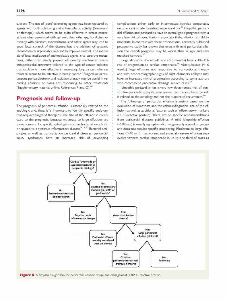
The follow-up of pericardial effusion is mainly based on theevaluation of symptoms and the echocardiographic size of the ef-fusion, as well as additional features such as inflammatory markers(i.e. C-reactive protein). There are no specific recommendationsfrom pericardial diseases guidelines. A mild idiopathic effusion(,10 mm) is usually asymptomatic, has generally a good prognosisand does not require specific monitoring. Moderate to large effu-sions (.10 mm) may worsen and especially severe effusions mayevolve towards cardiac tamponade in up to one-third of cases as
Figure 8 A simplified algorithm for pericardial effusion triage and management. CRP, C-reactive protein.
M. Imazio and Y. Adler1196
already mentioned. We usually recommend to tailor the follow-upto the aetiology, adopted therapies (usually a control after 1–2weeks and then after 1 month and 6 months), and monitoringthe possible evolution towards worsening pericardial effusion,cardiac tamponade, and constrictive pericarditis. For idiopathicmoderate effusions, an appropriate timing for echocardiographicfollow-up may be an echocardiogram every 6 months. For asevere effusion, an echocardiographic follow-up may be every3–6 months. A tailored follow-up is warranted also consideringthe relative stability or evolution of the size (i.e. a worsening effu-sion may require a closer timing also guided by symptoms).4
ConclusionsRelatively, few data have been published on the management ofpericardial effusion and there is a lack of contemporary prospect-ive series with unselected patients with pericardial effusions of dif-ferent entity (small vs. moderate vs. large). Ongoing prospectivestudies, registries, and new updated guidelines from medical soci-eties will provide more evidence based data to guide the manage-ment of pericardial effusion. A simplified algorithm for the triageand management of pericardial effusion is illustrated in Figure 8.
Supplementary materialSupplementary material is available at European Heart Journalonline.
Conflict of interest: none declared.
References1. Shabetai R. Pericardial effusion: haemodynamic spectrum. Heart 2004;90:255–256.2. Shabetai R. Function of the normal pericardium. Clin Cardiol 1999;22(1 Suppl 1):
I4–I5.3. Spodick DH. Acute cardiac tamponade. N Engl J Med 2003;349:684–690.4. Imazio M, Mayosi BM, Brucato A, Markel G, Trinchero R, Spodick DH, Adler Y.
Triage and management of pericardial effusion. J Cardiovasc Med (Hagerstown)2010;11:928–935.
5. Ntsekhe M, Mayosi BM. Tuberculous pericarditis with and without HIV. Heart FailRev. Advance Access published March 18, 2012, doi:10.1007/s10741-012-9310-6.
6. Sagrista Sauleda J, Almenar Bonet L, Angel Ferrer J, Bardajı Ruiz A, BoschGenover X, Guindo Soldevila J, Merce Klein J, Permanyer Miralda C, Tello deMeneses Becerra R. The clinical practice guidelines of the Sociedad Espanolade Cardiologıa on pericardial pathology. Rev Esp Cardiol 2000;53:394–412.
7. Maisch B, Seferovic PM, Ristic AD, Erbel R, Rienmuller R, Adler Y,Tomkowski WZ, Thiene G, Yacoub MH; Task Force on the Diagnosis and Man-agement of Pericardial Diseases of the European Society of Cardiology. Guide-lines on the diagnosis and management of pericardial diseases executivesummary: the Task force on the diagnosis and management of pericardial diseasesof the European Society of Cardiology. Eur Heart J 2004;25:587–610.
8. Sagrista-Sauleda J, Merce AS, Soler-Soler J. Diagnosis and management of pericar-dial effusion. World J Cardiol 2011;3:135–143.
9. Soler-Soler J, Sagrista-Sauleda J, Permanyer-Miralda G. Management of pericardialeffusion. Heart 2001;86:235–240.
10. Cheitlin MD, Armstrong WF, Aurigemma GP, Beller GA, Bierman FZ, Davis JL,Douglas PS, Faxon DP, Gillam LD, Kimball TR, Kussmaul WG, Pearlman AS,Philbrick JT, Rakowski H, Thys DM, Antman EM, Smith SC Jr, Alpert JS,Gregoratos G, Anderson JL, Hiratzka LF, Hunt SA, Fuster V, Jacobs AK,Gibbons RJ, Russell RO; American College of Cardiology; American Heart Asso-ciation; American Society of Echocardiography. ACC/AHA/ASE 2003 guidelineupdate for the clinical application of echocardiography: summary article: areport of the American College of Cardiology/American Heart AssociationTask Force on Practice Guidelines (ACC/AHA/ASE Committee to Update the
1997 Guidelines for the Clinical Application of Echocardiography). Circulation2003;108:1146–1162.
11. Weitzman LB, Tinker WP, Kronzon I, Cohen ML, Glassman E, Spencer FC. Theincidence and natural history of pericardial effusion after cardiac surgery—anechocardiographic study. Circulation 1984;69:506–511.
12. Imazio M, Cecchi E, Demichelis B, Ierna S, Demarie D, Ghisio A, Pomari F,Coda L, Belli R, Trinchero R. Indicators of poor prognosis of acute pericarditis.Circulation 2007;115:2739–2744.
13. Najib MQ, Ganji JL, Raizada A, Panse PM, Chaliki HP. Epicardial fat can mimic peri-cardial effusion on transoesophageal echocardiogram. Eur J Echocardiogr 2011;12:804.
14. Alter P, Figiel JH, Rupp TP, Bachmann GF, Maisch B, Rominger MB. MR, CT, andPET imaging in pericardial disease. Heart Fail Rev. Advance Access publishedMarch 25, 2012, doi:10.1007/s10741-012-9309-z.
15. Elavunkal J, Bright L, Stone MB. Emergency ultrasound identification of loculatedpericardial effusion: the importance of multiple cardiac views. Acad Emerg Med2011;18:e29.
16. Saito Y, Donohue A, Attai S, Vahdat A, Brar R, Handapangoda I, Chandraratna PA.The syndrome of cardiac tamponade with ‘small’ pericardial effusion. Echocardiog-raphy 2008;25:321–327.
17. Corey GR, Campbell PT, Van Trigt P, Kenney RT, O’Connor CM, Sheikh KH,Kisslo JA, Wall TC. Etiology of large pericardial effusions. Am J Med 1993;95:209–213.
18. Sagrista-Sauleda J, Merce J, Permanyer-Miralda G, Soler-Soler J. Clinical clues tothe causes of large pericardial effusions. Am J Med 2000;109:95–101.
19. Levy PY, Corey R, Berger P, Habib G, Bonnet JL, Levy S, Messana T, Djiane P,Frances Y, Botta C, DeMicco P, Dumon H, Mundler O, Chomel JJ, Raoult D. Etio-logic diagnosis of 204 pericardial effusions. Medicine (Baltimore) 2003;82:385–391.
20. Reuter H, Burgess LJ, Doubell AF. Epidemiology of pericardial effusions at a largeacademic hospital in South Africa. Epidemiol Infect 2005;133:393–399.
21. Ma W, Liu J, Zeng Y, Chen S, Zheng Y, Ye S, Lan L, Liu Q, Weig HJ, Liu Q. Causesof moderate to large pericardial effusion requiring pericardiocentesis in 140 HanChinese patients. Herz 2012;37:183–187.
22. Syed FF, Ntsekhe M, Mayosi BM. Tailoring diagnosis and management of pericar-dial disease to the epidemiological setting. Mayo Clin Proc 2010;85:866; authorreply 866.
23. Roy CL, Minor MA, Brookhart MA, Choudhry NK. Does this patient with a peri-cardial effusion have cardiac tamponade? JAMA 2007;297:1810–1818.
24. Beck C. Two cardiac compression triads. J Am Med Assoc 1935;104:714–716.25. Brown J, MacKinnon D, King A, Vanderbush E. Elevated arterial blood pressure in
cardiac tamponade. N Engl J Med 1992;327:463–466.26. Kussmaul A. Uber schwielige Mediastino- Perikarditis und den paradoxen Puls.
Berl Klin Wochenschr 1873;10:433–435.27. Fowler NO. Cardiac tamponade. A clinical or an echocardiographic diagnosis?
Circulation 1993;87:1738–1741.28. Mayosi BM, Burgess LJ, Doubell AF. Tuberculous pericarditis. Circulation 2005;
112:3608–3616.29. Mayosi BM. Contemporary trends in the epidemiology and management of car-
diomyopathy and pericarditis in sub-Saharan Africa. Heart 2007;93:1176–1183.30. Imazio M, Trinchero R. Triage and management of acute pericarditis. Int J Cardiol
2007;118:286–294.31. Yared K, Baggish AL, Picard MH. Multimodality imaging of pericardial diseases.
JACC Cardiovasc Imaging 2010;3:650–660.32. Verhaert D, Gabriel RS, Johnston D. The role of multimodality imaging in the
management of pericardial disease. Circ Cardiovasc Imaging 2010;3:333–343.33. Rajiah P, Kanne JP. Computed tomography of the pericardium and pericardial
disease. J Cardiovasc Comput Tomogr 2010;4:3–18.34. Imazio M, Brucato A, Derosa FG, Lestuzzi C, Bombana E, Scipione F, Leuzzi S,
Cecchi E, Trinchero R, Adler Y. Aetiological diagnosis in acute and recurrent peri-carditis: when and how. J Cardiovasc Med (Hagerstown) 2009;10:217–230.
35. Bogaert J, Francone M. Cardiovascular magnetic resonance in pericardial diseases.J Cardiovasc Magn Reson 2009;11:11–14.
36. Imazio M, Trinchero R. Myopericarditis: etiology, management, and prognosis.Int J Cardiol 2008;127:17–26.
37. Imazio M, Spodick DH, Brucato A, Adler Y. Controversial issues in the manage-ment of pericardial diseases. Circulation 2010;121:916–928.
38. Imazio M, Brucato A, Mayosi BM, Derosa FG, Lestuzzi C, Macor A, Trinchero R,Spodick DH, Adler Y. Medical therapy of pericardial diseases: part II: noninfectiouspericarditis, pericardial effusion and constrictive pericarditis. J Cardiovasc Med(Hagerstown) 2010;11:785–794.
39. Syed FF, Mayosi BM. A modern approach to tuberculous pericarditis. Prog Cardi-ovasc Dis 2007;50:218–236.
40. Lestuzzi C. Neoplastic pericardial disease: old and current strategies for diagnosisand management. World J Cardiol 2010;2:270–279.
Management of pericardial effusion 1197

41. Imazio M. Pericardial involvement in systemic inflammatory diseases. Heart 2011;97:1882–1892.
42. Imazio M, Brucato A, Trinchero R, Spodick DH, Adler Y. Colchicine for pericar-ditis: hype or hope? Eur Heart J 2009;30:532–539.
43. Imazio M, Brucato A, Cumetti D, Trinchero R. Corticosteroids for recurrent peri-carditis: high versus low doses: a nonrandomized observation. Circulation 2008;118:667–671.
44. Vianello F, Cinetto F, Cavraro M, Battisti A, Castelli M, Imbergamo S,Marcolongo R. Azathioprine in isolated recurrent pericarditis: a single centreexperience. Int J Cardiol 2011;147:477–478.
45. Hota SS, Chow CM, Bonneau D. Surgical treatment for incessant pericarditis. CanJ Cardiol 2009;25:161–162.
46. Sagrista-Sauleda J, Angel J, Permanyer-Miralda G, Soler-Soler J. Long-term follow-up of idiopathic chronic pericardial effusion. N Engl J Med 1999;341:2054–2059.
47. Little WC, Freeman GL. Pericardial disease. Circulation 2006;113:1622–1632.48. Lange RA, Hillis DH. Acute pericarditis. N Engl J Med 2004;351:2195–2202.49. Imazio M, Brucato A, Maestroni S, Trinchero R. Risk of constrictive pericarditis
after acute pericarditis. Circulation 2011;124:1270–1275.50. Mitiku TY, Heidenreich PA. A small pericardial effusion is a marker of increased
mortality. Am Heart J 2011;161:152–157.
M. Imazio and Y. Adler1197a